The identification of mobile species from silicone gels used in burns scar remediation
10
Silicon Chemistry 2: 1–10, 2003. © 2004 Kluwer Academic Publishers. Printed in the Netherlands. 1 The identification of mobile species from silicone gels used in burns scar remediation Washington Sanchez 1 , Nikole Hynard 1 , John Evans 2 & Graeme George 1, ∗ 1 Centre for Instrumental and Developmental Chemistry, and 2 Centre for Rehabilitation Science and Engineering, Queensland University of Technology, Brisbane 4000, Australia ∗ Author for correspondence (e-mail: [email protected]) (Received 9 July 2002; accepted 21 February 2003) Key words: burn scars, diffusion, Energy Dispersive X-ray mapping, MALDI-MS, polydimethyl siloxane gels, silicone oligomers Abstract The mobile species that may migrate from polydimethyl siloxane medical gel sheeting into skin have been iden- tified by MALDI-MS. The chloroform-extractable species from the bulk gel comprised predominantly cyclic oligomers with a mass distribution peaking at n = 17, but in an aqueous environment the species at the sur- face were predominantly linear siloxanes with one hydrophilic end group (methyl/hydroxyl, methyl/methylol- or methyl/methoxy-). By using a gelatine matrix as a model substrate, the distribution of silicon after application of the silicone gel for 16 weeks was determined by Energy-dispersive X-Ray mapping of the sectioned gelatine. Extension of this method to tissue is complicated by the high background of silicon in the dermal layer, but it is demonstrated that MALDI-MS of silicone oligomers may be achieved directly from the surface of human skin sections. The association of the linear and cyclic oligomers with proteins relevant in hypertrophic scarring is considered. Introduction It has been reported that polydimethyl siloxane silic- one gel sheets may offer a simple treatment for the severe scarring which often accompanies burns [1, 2] and this has been adopted as a routine clinical treatment. All wounds heal by scar formation, but in certain instances hypertrophic scars may form which are red, raised and thickened because of overproduc- tion of all components of the extracellular matrix [3]. These frequently occur following a thermal injury, or other type of deep dermal wound, and are of concern in recovery from major burns injuries. Hypertrophic scarring is a delayed onset phenomenon, usually ap- pearing 4–8 weeks after re-epithelialisation. The most striking feature of hypertrophic scars is the continu- ing build-up of collagen over long periods of time, compared to the remodelling and collagen loss in nor- mal scars. The synthesis and degradation of collagen occur concurrently in the wound healing process and hence this balance controls the final amount of fibrous protein deposited. In problem scars, this balance of collagen turnover is disrupted [3, 4]. Silicone gel sheets have been clinically proven to rehabilitate hypertrophic scars; as an alternative to pressure therapy and corticosteroid injections, this treatment is virtually free of side effects. Most recently the gel sheets have been marketed for self-therapy and are available across-the-counter to allow treat- ment of problem scars from a range of injuries in addition to burns [5]. These polymeric sheets are transparent, flexible gels that are worn over the scar. Silicone gel sheets can be applied as soon as there is re-epithelialisation over the wounded site, with the benefit that they adhere and mould to any body con- tour. There is an immediate decrease in redness and sensitivity, but the minimum time of use required for a noticeable effect on the scar morphology is twelve hours a day for about one to two months. Silicone gel therapy often works when other conventional treat-
-
Upload
washington-sanchez -
Category
Documents
-
view
213 -
download
0
Transcript of The identification of mobile species from silicone gels used in burns scar remediation

Silicon Chemistry 2: 1–10, 2003.© 2004 Kluwer Academic Publishers. Printed in the Netherlands.
1
The identification of mobile species from silicone gels used in burns scarremediation
Washington Sanchez1, Nikole Hynard1, John Evans2 & Graeme George1, ∗1Centre for Instrumental and Developmental Chemistry, and 2Centre for Rehabilitation Science and Engineering,Queensland University of Technology, Brisbane 4000, Australia∗Author for correspondence (e-mail: [email protected])
(Received 9 July 2002; accepted 21 February 2003)
Key words: burn scars, diffusion, Energy Dispersive X-ray mapping, MALDI-MS, polydimethyl siloxane gels,silicone oligomers
Abstract
The mobile species that may migrate from polydimethyl siloxane medical gel sheeting into skin have been iden-tified by MALDI-MS. The chloroform-extractable species from the bulk gel comprised predominantly cyclicoligomers with a mass distribution peaking at n = 17, but in an aqueous environment the species at the sur-face were predominantly linear siloxanes with one hydrophilic end group (methyl/hydroxyl, methyl/methylol-or methyl/methoxy-). By using a gelatine matrix as a model substrate, the distribution of silicon after applicationof the silicone gel for 16 weeks was determined by Energy-dispersive X-Ray mapping of the sectioned gelatine.Extension of this method to tissue is complicated by the high background of silicon in the dermal layer, but it isdemonstrated that MALDI-MS of silicone oligomers may be achieved directly from the surface of human skinsections. The association of the linear and cyclic oligomers with proteins relevant in hypertrophic scarring isconsidered.
Introduction
It has been reported that polydimethyl siloxane silic-one gel sheets may offer a simple treatment for thesevere scarring which often accompanies burns [1,2] and this has been adopted as a routine clinicaltreatment. All wounds heal by scar formation, but incertain instances hypertrophic scars may form whichare red, raised and thickened because of overproduc-tion of all components of the extracellular matrix [3].These frequently occur following a thermal injury, orother type of deep dermal wound, and are of concernin recovery from major burns injuries. Hypertrophicscarring is a delayed onset phenomenon, usually ap-pearing 4–8 weeks after re-epithelialisation. The moststriking feature of hypertrophic scars is the continu-ing build-up of collagen over long periods of time,compared to the remodelling and collagen loss in nor-mal scars. The synthesis and degradation of collagenoccur concurrently in the wound healing process and
hence this balance controls the final amount of fibrousprotein deposited. In problem scars, this balance ofcollagen turnover is disrupted [3, 4].
Silicone gel sheets have been clinically provento rehabilitate hypertrophic scars; as an alternativeto pressure therapy and corticosteroid injections, thistreatment is virtually free of side effects. Most recentlythe gel sheets have been marketed for self-therapyand are available across-the-counter to allow treat-ment of problem scars from a range of injuries inaddition to burns [5]. These polymeric sheets aretransparent, flexible gels that are worn over the scar.Silicone gel sheets can be applied as soon as thereis re-epithelialisation over the wounded site, with thebenefit that they adhere and mould to any body con-tour. There is an immediate decrease in redness andsensitivity, but the minimum time of use required fora noticeable effect on the scar morphology is twelvehours a day for about one to two months. Siliconegel therapy often works when other conventional treat-

2
ments have failed, and while it appears to reduce theamount of scar tissue, its specific mode of action isuncertain. Mechanisms proposed include: increasedskin temperature to increase the rate of collagenasereactions; control of skin hydration; a direct chemicalreaction of components migrating from the gel or theirmechanical effect on the scar [3, 6].
In this study we are investigating the nature andmigration of the low molecular weight species fromthese silicone gels and in particular the detailed identi-fication of the species which are present on the surfaceof the gel under different conditions and those whichare able to migrate into a collagen-like matrix.
Experimental
The silicone gel used was Cica-Care® [5] obtainedfrom the Burns Unit, Royal Brisbane Hospital.MALDI-TOF-MS has been used to determine thechemical composition, molar mass and oligomeric dis-tribution of both the species present in the bulk andthose that may migrate from the surface of the silic-one gel. In the studies to determine the composition oflow molecular weight species present in the bulk, thegel was exhaustively extracted with chloroform. Thisresulted in a loss in weight of 36% from the cross-linked gel. In another experiment the gel was extractedwith water to determine if any water-soluble or hy-drophilic species were present in the gel. The weightloss was much less (4%). For the analysis of surfacespecies, transfer to the MALDI target was achieved bytouching the surface of the gel to the stainless steelplate, which formed the MALDI target. To prepare thesample for MALDI analysis, typically 1 pg of samplewas added to the matrix (20:1 of 4-hydroxybenzilidenemalononitrile:sodium iodide) on the target plate andallowed to dry. The ions from 450 N2 laser shots at337 nm were analysed in a Micromass Tof-Spec 2Etime of flight mass spectrometer [7].
GC/MS was used to detect any lower molecularmass species below 960 Da that may be present inthe extracts. Samples were prepared by separate ex-tractions of the silicone gel with methanol. Thesewere later analysed with a Fisons 8000 gas chroma-tograph with a HT5 capillary column combined witha quadrupole MD800 mass spectrometer for structuralanalysis. The 12 m column has a 5% phenyl (equiv.)polycarborane-siloxane with an internal diameter of0.22 mm. All injections were made in split-less modeand with a purge activation time of 60 s. During the
purge activation, the column temperature was heldat 40 ◦C after which the temperature was ramped to350 ◦C at a rate of 15 ◦C/min.
Gelatine, which is hydrolysed bovine collagen,has been used as a model system to determine themigration of silicone species from the medical gel.Gelatine was cast from solution containing an anti-biotic to prevent microbial attack during the trial. Thesilicone gel was placed in contact with the gelatinesurface for 16 weeks and then sectioned after freeze-drying to obtain cross-sections for elemental mappingof silicon by Energy Dispersive X-Ray (EDX) ana-lysis in the Scanning Electron Microscope. ScanningTransmission Electron Microscopy (STEM) has alsobeen performed on sectioned scar tissue. Samplespreviously stored under liquid nitrogen were cryo-sectioned to 1–2 µm by 1 mm2 with an RMCTVIIultramicrotome at liquid nitrogen temperatures using aglass knife. The cryo-sections were then collected ondouble grids and immediately freeze-dried under va-cuum (10−4 mmHg) overnight. X-ray microanalyseswere performed with a Philips CM200 scanning trans-mission electron microscope at 120 kV and a siliconmap was constructed.
Results and discussion
Figure 1 shows the MALDI mass spectrum of thelow molecular weight silicone species obtained by ex-tracting the gel with chloroform. The spectrum showsthe species present from mass 1080 Da to 2640 Da.The cut off of 1080 Da is set to ensure that interfer-ence from adduct ions of the matrix (n × 192 Da) iseliminated in the analyte spectrum. In Figure 1a theprogression of peaks with a separation of 74 Da cor-responding to [(CH3)2-Si-O]n is the dominant feature.The peaks correspond to the sodiated species for in-creasing values of n, with a peak at around n = 17.Such species are normally not detected by analyticalmethods such as GC/MS due to mass limitations andthis reflects the sensitivity of MALDI-MS. It is re-cognised, however, that poly-disperse samples showselective ionisation and detection so that results fromun-fractionated samples may be biased to lower molarmasses [8]. The sodiated species in the MALDI spec-tra (Figure 1a) are observed due to the presence ofsodium iodide in the matrix to aid ionization. The twomost likely species present in a polydimethylsiloxanesample are the linear (a) and cyclic (b) oligomers, asshown below.

3
Figure 1a. MALDI-MS analysis of low molecular weight silicone oligomers from silicone medical gel after chloroform extraction. Theprogressions due to cyclic ( ) methyl/methylol, methyl/methoxy-terminated ( ) and methyl/hydroxy-terminated oligomers ( ) are shown.
Figure 1b. Isotopic prediction of MALDI-MS analysis of low molecular weight silicone oligomers for: (A) NaSi17C34H102O17, (B)NaSi17C34H102O17SiC2H6CH3OCH3 and (C) NaSi17C34H102O17SiC2H6CH3OH, maxima at n = 17, from silicone medical gel afterchloroform extraction.
4
Since PDMS is synthesised by an equilibrium ringopening polymerisation of the cyclic starting ma-terial with n = 4 (octamethyl cyclotetrasiloxane orD4), there will always be cyclic oligomers at equi-librium. Thus ∼ 10% of the silicone is present aslow molecular weight cyclic oligomers in the PDMSin addition to the linear oligomer species. Using theinstrumental software it is possible to simulate theisotopic species to be expected for the linear andcyclic oligomers for n = 17 [7]. When this is per-formed (as shown in Figure 1b), the spectrum may beassigned to the cyclic species, with a smaller contri-bution from linear species which are not those withmethyl functionalities at each end (as shown in (a)above) but rather those having one end group, eithera methoxy or a methylol group. Simulations havebeen performed for all possible terminal groups inaddition to the methyl/hydroxyl-, methyl/methylol-and methyl/methoxy combinations shown in Fig-ure 1, but no further improvement to fit of thedata was obtained. The combinations of end groupstried were hydrogen/hydrogen, methyl/hydrogen,methyl/methyl, methyl/hydroxyl, hydroxyl/hydroxyl,methoxy/methoxy, methylol/methylol, methyl/vinyl,silanol/vinyl and vinyl/vinyl. It is therefore concludedthat extraction of the silicone gels with chloroformremoves all species, both cyclic and linear, but thedetection by MALDI of the cyclic species may be fa-voured due to the ionization process [9]. The speciesbelow 960 Da have been determined by GC/MS. Thisresulted in the positive identification of peaks corres-ponding to D5 and D6. The absence of D4 is consistentwith the stripping of this volatile species from the gel.
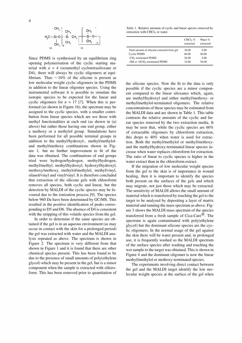
In order to determine if the same species are ob-tained if the gel is in an aqueous environment (as mayoccur in contact with the skin for a prolonged period)the gel was extracted with water and the MALDI ana-lysis repeated as above. The spectrum is shown inFigure 2. The spectrum is very different from thatshown in Figure 1 and it is found that there are otherchemical species present. This has been found to bedue to the presence of small amounts of poly(ethyleneglycol) which may be present in the gel, but is a minorcomponent when the sample is extracted with chloro-form. This has been removed prior to quantitation of
Table 1. Relative amounts of cyclic and linear species removed byextraction with CHCl3 or water.
CHCl3 % Water %extracted extracted
Total amount of silicone extracted from gel 36.00 4.00Cyclic PDMS 60.00 40.00-CH3 terminated PDMS 26.00 4.00-OH or -OCH3 terminated PDMS 14.00 56.00
the silicone species. Now the fit to the data is onlypossible if the cyclic species are a minor compon-ent compared to the linear siloxanes which, again,are methyl/hydroxyl and either methyl/methoxy- ormethyl/methylol-terminated oligomers. The relativeconcentrations of these species may be estimated fromthe MALDI data and are shown in Table 1. This tablecontrasts the relative amounts of the cyclic and lin-ear species removed by the two extraction media. Itmay be seen that, while the cyclic species are 60%of extractable oligomers by chloroform extraction,this drops to 40% when water is used for extrac-tion. Both the methyl/methylol or methyl/methoxy-and the methyl/hydroxy-terminated linear species in-crease when water replaces chloroform for extraction.The ratio of linear to cyclic species is higher in thewater extract than in the chloroform extract.
If the migration of low molecular weight speciesfrom the gel to the skin is of importance in woundhealing, then it is important to identify the speciesboth present on the surfaces of the gels and whichmay migrate, not just those which may be extracted.The sensitivity of MALDI allows the small amount ofmaterial which is transferred by touching the gel to thetarget to be analysed by depositing a layer of matrixmaterial and running the mass spectrum as above. Fig-ure 3 shows the MALDI mass spectrum of the speciestransferred from a fresh sample of Cica-Care®. Thespectrum is again contaminated with poly(ethyleneglycol) but the dominant silicone species are the cyc-lic oligomers. In the normal usage of the gel againstthe skin there will be water present and, in prolongeduse, it is frequently washed so the MALDI spectrumof the surface species after washing and touching thewet sample to the target was obtained. This is shown inFigure 4 and the dominant oligomer is now the linearmethyl/methylol or methoxy-terminated species.
The experiments involving direct contact betweenthe gel and the MALDI target identify the low mo-lecular weight species at the surface of the gel when
5
Figure 2. MALDI-MS analysis of low molecular weight silicone oligomers from silicone medical gel after water extraction. The spec-trum shows contamination by polyethylene glycol at low molecular weight. The progressions due to cyclic ( ) methyl/methylol,methyl/methoxy-terminated ( ) and methyl/hydroxy-terminated oligomers ( ) are shown.
Figure 3. MALDI-MS of components transferred from fresh silicone medical gel by pressing against metal MALDI target. The spectrum showscontamination by polyethylene glycol at low molecular weight. The progression due to cyclic oligomers ( ) is shown.

6
Figure 4. MALDI-MS of components transferred from water-washed silicone medical gel by pressing against metal MALDI target. Theprogression due to methyl/methylol- (methyl/methoxy)-terminated oligomers ( ) is shown.
either wet or dry, but do not show that they are ableto migrate into skin or scar tissue. For this we haveused the model system of a gelatine matrix, sincehypertrophic scarring results from over-production ofcollagen in the dermis. The gelatine provides a mat-rix of hydrolysed Type 1 bovine collagen which maybe readily freeze-dried and sectioned in order to de-termine the distribution of silicon. While, in principle,MALDI may be used for mapping experiments sincethe instrumental software allows a line map to be con-structed along a line of laser shots, the low resolutionof 100 µm limits the usefulness in this application.EDX in the Scanning Electron Microscope providesan alternative semi-quantitative technique [10], but thewater in the gel must be removed by freeze-drying.
Figure 5 shows representative EDX spectra fromdifferent positions on proceeding from the front sur-face (a), through the bulk (b) to the back surface (c)of a sectioned sample of gelatine after contact with apatch of silicone gel sheeting for 16 weeks. It is seenthat the silicon signal is well resolved from the otherbands. The sulphur and chlorine bands arise from theantibiotic added to the gelatine (as seen in the controlsample (d)). A map may be constructed of silicon dis-tribution over the entire thickness of the gel and thisis shown in Figure 6 at two levels of threshold sens-itivity. (Note that silicon appears as light regions inthese maps.) The images show that there is migration
of silicon into the collagen layer and there are someareas in the bulk where a high local concentration isachieved. It is noted from the map that the highestconcentration occurs at the side in contact with gel, buta significant concentration is detected on the surfaceaway from the gel as well as in the bulk of the gelatine.The front is the face to which the patch is applied, butthe high concentration on the back surface suggeststhat there has been migration through the gelatine andaggregation on the back surface.
The siloxanes, being highly surface active, willaggregate at the air–gelatine interface, but there mustbe diffusion through either the water or the hydro-phobic collagen in order to penetrate the gelatine. Thecyclic and methyl-terminated linear species are highlyhydrophobic and association with the collagen wouldbe expected. For example, it has been reported thathydrophobic interaction between heme proteins andPDMS leads to denaturation [11] and that small cyc-lics with n = 4 (octamethyl cyclotetrasiloxane or D4)induce conformational changes in fibrinogenin andfibronectin, which are wound healing proteins [12].Based on these results, the presence of zones in thebulk with high concentration would suggest strong hy-drophobic interaction between the siloxane oligomersand the collagen in certain areas. However, an inter-esting result, noted earlier, is that while the surface ofthe silicone gel is rich in the cyclic species, these are

7
Figure 5. EDX elemental analysis of gelatine cross-section after16 weeks in contact with Cica-Care® silicone gel: (a) Surface incontact with gel; (b) Bulk (centre); (c) Back surface; (d) Controlsample.
Figure 6. EDX elemental map for silicon of gelatine cross-sectionafter 16 weeks in contact with Cica-Care® silicone gel. Siliconappears as white zones against a dark background for differentthreshold sensitivities in (a) and (b).
a minor component compared to the linear oligomersthat are detected at the surface by MALDI when thegel is wet.
It has been recently reported that linear siliconesmodified with hydrophilic groups will associate withproteins at interfaces and stabilize them against de-naturation [13]. Whether simple hydroxy group ter-mination is sufficient to achieve this stabilization orwhether the silicone facilitates denaturation throughinversion of the structure on hydrophobic interactionis of fundamental interest in a likely mechanism ofremediation of hypertrophic scarring by these mobileand associative species. As there is only a limitedamount of extrapolation which may be made from thehydrolysed collagen matrix to the dermal layer, studieshave commenced on the species which may actually

8
migrate into skin and scar tissue from a silicone gelsheet.
Figure 7a shows a STEM photograph of a cross-section of scar tissue; the extent of the epidermis andthe lower dermal layer are indicated. Figure 7b showsthe X-ray microanalysis map for silicon from the samecross-section. It is seen that silicon is widely distrib-uted in the sample and that the highest concentrationappears in the interfacial region between the dermisand epidermis. The concentration in the epidermiswhich has developed by re-epithelialisation followingthe burn is very low compared to that in the dermisand the distribution declines from the maximum atthe interface towards the inner dermal layer. It shouldbe noted that silicon is present in healthy skin andis linked to collagen development, so the extensionfrom the model system to skin is complicated by thisand also the ubiquitous use of silicones in skin-careproducts, both of which will result in a high back-ground against which measurements are to be made.The form of the silicon is not known and MALDI-MSanalysis of the silicone species on skin is required.
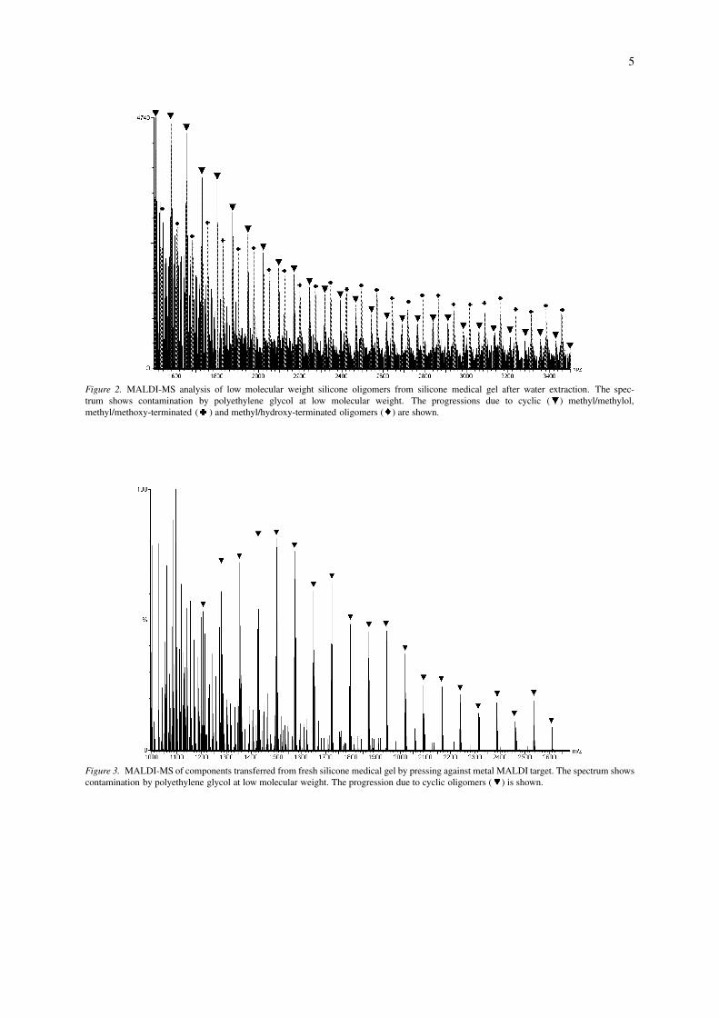
In preliminary studies on scar tissue to which achloroform extract has been applied (which, as notedabove in Figure 3, consists of predominantly cyclicspecies), it has been shown that ionisation and ana-lysis of the low molecular weight silicones may beachieved (but at poorer S/N). This is shown in Fig-ure 8, which is the MALDI mass spectrum from thesurface of burn scar to which the silicone had beenapplied. A fine spray of the matrix was applied to thepartially dehydrated skin prior to analysis. In anotherreport of molecular imaging of biological samples us-ing MALDI-TOF mass spectrometry [14], the directanalysis of tissue produced interference from signalsfrom abundant molecules such as lipid and proteinfragments around the lower molecular weight region(MW < 1500). In this investigation, skin both withand without a matrix solution deposited onto it wasanalysed by MALDI-TOF mass spectrometry and nointerfering biological species were detected, possiblybecause the analysis conditions had been optimisedspecifically for the PDMS oligomers. Thus, in prin-ciple the studies of the model gelatine system maybe directly extended to skin and scar tissue to de-termine if the cyclic or linear siloxane species arethose which migrate under the conditions prevailingon the surface of a scar when a silicone gel patch isapplied. Examination of Figure 8 suggests that thismay be determined by a careful comparison of theoligomers that are desorbed and analysed. Compar-
Figure 7. (a) STEM image of a cross-section of scar tissue show-ing extent of epidermis (70 µm); (b) EDX map of silicon showingmaximum intensity at epidermis/dermis interface.
ison of the MALDI spectra in Figure 1 (the extract asapplied) and Figure 8 shows that the methyl/methylol-(or methoxy-) terminated oligomer is not desorbedfrom the scar tissue. There may be several explan-ations for this behaviour, but one possibility is thatthese oligomers have migrated preferentially into theepidermis and are strongly associated with the proteinsor other extracellular matrix components.
Studies are underway of skin and scar tissue todetermine more decisively the species which may mi-grate through the stratum corneum to the dermis andthe effect this may have on the properties of the col-lagen. The challenge in direct MALDI analysis fromsections of skin such as that shown in Figure 7 isthe limited spatial resolution of the Nitrogen laserpulse used for ionisation. This is typically 100 µm,while the entire thickness of the epidermis after re-epithelialisation is much thinner than in healthy tissue[3] and in Figure 7 is ∼ 70 µm.
9
Figure 8. MALDI mass spectrum from surface of scar tissue after contact with (chloroform) extract of Cica-Care® silicone gel showing thepresence of cyclic ( ) and methyl/hydroxyl-terminated ( ) oligomers.
Conclusions
Low molecular weight species in silicone medicalsheeting may be characterised by MALDI-MS andGC/MS analysis on solvent extracts. Chloroform ex-traction of the gel resulted in a loss of 36% byweight from the cross-linked network and MALDIanalysis together with isotopic pattern fitting showedpredominantly (∼ 60%) cyclic PDMS oligomers (witha maximum at n = 17) with a smaller amount ofmethyl/methylol or methoxy-terminated linear spe-cies. The GC/MS showed the presence of D5 and D6but no D4 in the silicone oil extracted from the medicalgel. In contrast, water extracts only 4% of siloxaneoligomers. These had a lower level of cyclics (∼ 40%)and the dominant species were linear siloxanes. Theseoligomers are also identified after the silicone gel isplaced in contact with the MALDI metal target plateand the species transferred to the surface are analysedby MALDI. The fresh gel transferred cyclic species,while after washing in water the methyl/methylol- ormethyl/methoxy-terminated linear species were trans-ferred. The migration of silicones into a gelatine gelafter 16 weeks exposure from a fresh silicone gel hasbeen demonstrated by mapping of elemental silicon byEDX. This shows that the oligomers migrate rapidlyto the back surface, but regions of high concentra-tion also occur in the bulk. Cyclic species and inparticular all species below 960 Da (i.e. D4, D5 andD6) were absent from the siloxane oligomers extrac-
ted from the gelatine after contact with the medicalgel. The nature of the collagen-siloxane interaction issignificant, since depending on whether hydrophilic orhydrophobic interactions dominate, the protein may beeither denatured or stabilized. Extension of the modelstudies to human skin and scar tissue is complicatedby the high background silicon levels determined byX-ray elemental mapping, but it has been demon-strated that desorption of oligomers may be achievedin MALDI so that the siloxane oligomers may, underfavourable circumstances, be identified. However, itis recognised that there are limitations to the MALDItechnique, primarily the inability to obtain quantitativeanalysis of samples that are highly polydisperse, butthis may be corrected by coupling size exclusion chro-matography to MALDI, as demonstrated by Montaudoet al. [8].
Acknowledgements
This study has been done in conjunction with theBurns Unit of the Royal Brisbane Hospital (Dr M.Muller, Dr E. Cheng and Prof S. Pegg) through anARC Linkage, APA-I award (to WS).

10
References
1. Perkins, K., Davey, R.B. & Wallis, K.A. 1982 Silicone gel:A new treatment for burn scars and contractures. Burns 9,201–204.
2. Ohmori, S. 1998 Effectiveness of silastic sheet coverage in thetreatment of scar keloid (hypertrophic scar). Aesth. Plast. Surg.12, 95–99.
3. Su, C.W., Alizadeh, K., Broddie, A. & Lee, R.C. 1998 Theproblem scar. Clinic. Plast. Surg. 25, 451–465.
4. Stadelmann, W.K., Digenis, A.G. & Tobin, G.R. 1998Physiology and healing dynamics of chronic cutaneouswounds. Am. J. Surg. 176, 26S–38S.
5. Cica-Care® (Smith and Nephew, Largo, FL).6. Sawada, Y. & Sone, K. 1990 Treatment of scars and keloids
with a cream containing silicone oil. Br. J. Plast. Surg. 43,683–688.
7. Hunt, S.M. & George, G.A. 2000 Characterization of siloxaneresidues from polydimethylsiloxane elastomers by MALDI-TOF-MS. Polym. Int. 49 (7), 633–645.
8. Montaudo, G., Montaudo, M.S., Puglisi, C. & Samperi, F.1995 Molecular weight distribution of poly(dimethylsiloxane)by combining matrix-assisted laser desorption/ionisation time-of-flight mass spectrometry with gel-permeation chromato-
graphy fractionation. Rapid Commun. Mass Spectrom. 9,1158–1163.
9. Axelsson, J., Scrivener, E., Haddleton, D.M. & Derrick, P.J.1996 Mass discrimination effects in an ion detector and othercauses of shifts in polymer mass distributions measured bymatrix-assisted laser desorption/ionisation time-of-flight massspectrometry. Macromolecules 29, 8875–8882.
10. Morgan, A.J. 1985 X-ray Microanalysis in Electron Micro-scopy for Biologists. Oxford, UK: Oxford University Press.
11. Anderson, A.B. & Robertson, C.R. 1995 Absorption spectraindicate conformational alteration of myoglobin adsorbed onpolydimethylsiloxane. Biophys. J. 68 (5), 2091–2097.
12. Sun, L., Alexander, H., Lattarulo, N., Blumenthal, N.C., Ricci,J.L. & Chen, G. 1998 Protein denaturation induced by cyclicsilicone. Biomaterials 18 (24), 1593–1597.
13. Zelisko, P.M., Brook, M.A. & Bartzoka, V. 2001 Deliv-ery of proteinacious materials from silicone protected micro-particles and water-in-silicone oil emulsions. In Proceedings –28th International Symposium on Controlled Release of Bio-active Materials and 4th Consumer & Diversified ProductsConference. San Diego, CA, USA.
14. Caprioli, R.M., Farmer, T.B. & Gile, J. 1997 Molecularimaging of biological samples: Localisation of peptides andproteins using MALDI-TOF MS. Anal. Chem. 69, 4751–4760.