The Hox gene Ultrabithorax modulates the shape and … Lab/2003... · leg of Drosophila by...
12
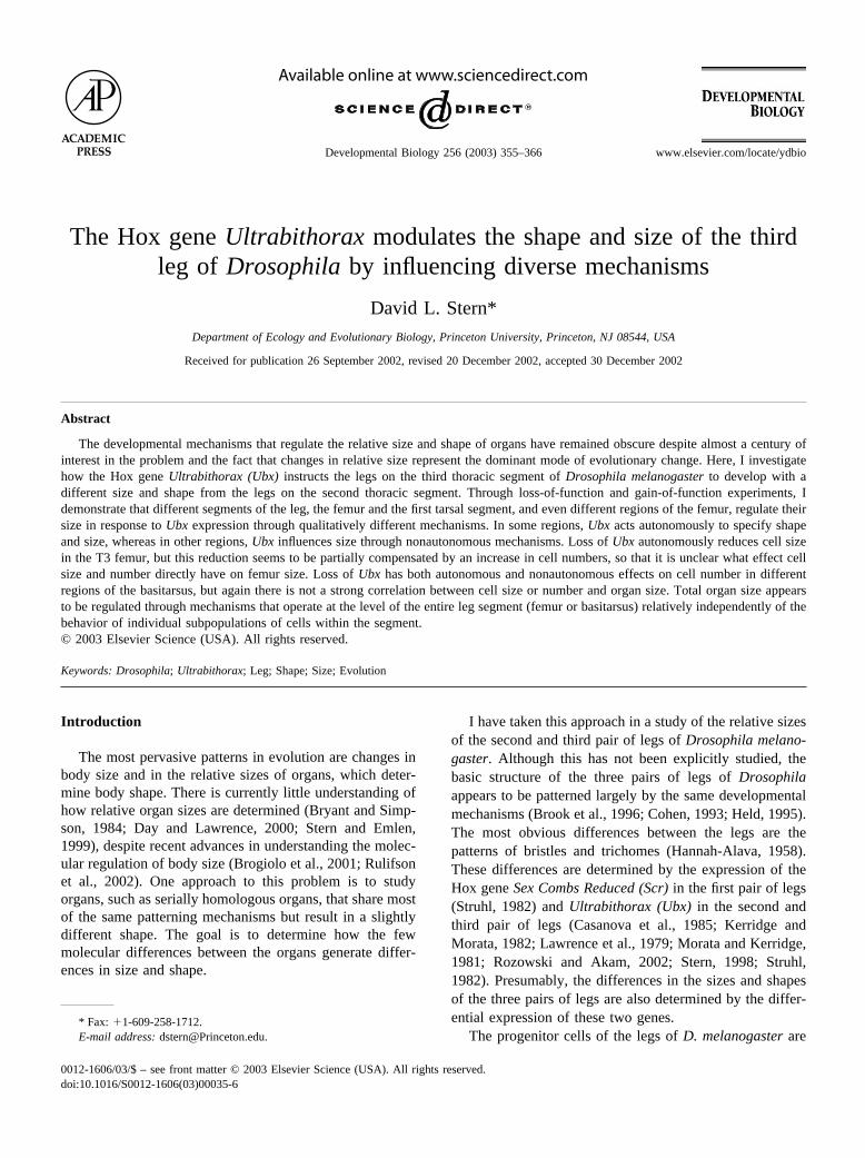
The Hox gene Ultrabithorax modulates the shape and size of the third leg of Drosophila by influencing diverse mechanisms David L. Stern* Department of Ecology and Evolutionary Biology, Princeton University, Princeton, NJ 08544, USA Received for publication 26 September 2002, revised 20 December 2002, accepted 30 December 2002 Abstract The developmental mechanisms that regulate the relative size and shape of organs have remained obscure despite almost a century of interest in the problem and the fact that changes in relative size represent the dominant mode of evolutionary change. Here, I investigate how the Hox gene Ultrabithorax (Ubx) instructs the legs on the third thoracic segment of Drosophila melanogaster to develop with a different size and shape from the legs on the second thoracic segment. Through loss-of-function and gain-of-function experiments, I demonstrate that different segments of the leg, the femur and the first tarsal segment, and even different regions of the femur, regulate their size in response to Ubx expression through qualitatively different mechanisms. In some regions, Ubx acts autonomously to specify shape and size, whereas in other regions, Ubx influences size through nonautonomous mechanisms. Loss of Ubx autonomously reduces cell size in the T3 femur, but this reduction seems to be partially compensated by an increase in cell numbers, so that it is unclear what effect cell size and number directly have on femur size. Loss of Ubx has both autonomous and nonautonomous effects on cell number in different regions of the basitarsus, but again there is not a strong correlation between cell size or number and organ size. Total organ size appears to be regulated through mechanisms that operate at the level of the entire leg segment (femur or basitarsus) relatively independently of the behavior of individual subpopulations of cells within the segment. © 2003 Elsevier Science (USA). All rights reserved. Keywords: Drosophila; Ultrabithorax; Leg; Shape; Size; Evolution Introduction The most pervasive patterns in evolution are changes in body size and in the relative sizes of organs, which deter- mine body shape. There is currently little understanding of how relative organ sizes are determined (Bryant and Simp- son, 1984; Day and Lawrence, 2000; Stern and Emlen, 1999), despite recent advances in understanding the molec- ular regulation of body size (Brogiolo et al., 2001; Rulifson et al., 2002). One approach to this problem is to study organs, such as serially homologous organs, that share most of the same patterning mechanisms but result in a slightly different shape. The goal is to determine how the few molecular differences between the organs generate differ- ences in size and shape. I have taken this approach in a study of the relative sizes of the second and third pair of legs of Drosophila melano- gaster. Although this has not been explicitly studied, the basic structure of the three pairs of legs of Drosophila appears to be patterned largely by the same developmental mechanisms (Brook et al., 1996; Cohen, 1993; Held, 1995). The most obvious differences between the legs are the patterns of bristles and trichomes (Hannah-Alava, 1958). These differences are determined by the expression of the Hox gene Sex Combs Reduced (Scr) in the first pair of legs (Struhl, 1982) and Ultrabithorax (Ubx) in the second and third pair of legs (Casanova et al., 1985; Kerridge and Morata, 1982; Lawrence et al., 1979; Morata and Kerridge, 1981; Rozowski and Akam, 2002; Stern, 1998; Struhl, 1982). Presumably, the differences in the sizes and shapes of the three pairs of legs are also determined by the differ- ential expression of these two genes. The progenitor cells of the legs of D. melanogaster are * Fax: 1-609-258-1712. E-mail address: [email protected]. R Available online at www.sciencedirect.com Developmental Biology 256 (2003) 355–366 www.elsevier.com/locate/ydbio 0012-1606/03/$ – see front matter © 2003 Elsevier Science (USA). All rights reserved. doi:10.1016/S0012-1606(03)00035-6
Transcript of The Hox gene Ultrabithorax modulates the shape and … Lab/2003... · leg of Drosophila by...
The Hox gene Ultrabithorax modulates the shape and size of the thirdleg of Drosophila by influencing diverse mechanisms
David L. Stern*Department of Ecology and Evolutionary Biology, Princeton University, Princeton, NJ 08544, USA
Received for publication 26 September 2002, revised 20 December 2002, accepted 30 December 2002
Abstract
The developmental mechanisms that regulate the relative size and shape of organs have remained obscure despite almost a century ofinterest in the problem and the fact that changes in relative size represent the dominant mode of evolutionary change. Here, I investigatehow the Hox gene Ultrabithorax (Ubx) instructs the legs on the third thoracic segment of Drosophila melanogaster to develop with adifferent size and shape from the legs on the second thoracic segment. Through loss-of-function and gain-of-function experiments, Idemonstrate that different segments of the leg, the femur and the first tarsal segment, and even different regions of the femur, regulate theirsize in response to Ubx expression through qualitatively different mechanisms. In some regions, Ubx acts autonomously to specify shapeand size, whereas in other regions, Ubx influences size through nonautonomous mechanisms. Loss of Ubx autonomously reduces cell sizein the T3 femur, but this reduction seems to be partially compensated by an increase in cell numbers, so that it is unclear what effect cellsize and number directly have on femur size. Loss of Ubx has both autonomous and nonautonomous effects on cell number in differentregions of the basitarsus, but again there is not a strong correlation between cell size or number and organ size. Total organ size appearsto be regulated through mechanisms that operate at the level of the entire leg segment (femur or basitarsus) relatively independently of thebehavior of individual subpopulations of cells within the segment.© 2003 Elsevier Science (USA). All rights reserved.
Keywords: Drosophila; Ultrabithorax; Leg; Shape; Size; Evolution
Introduction
The most pervasive patterns in evolution are changes inbody size and in the relative sizes of organs, which deter-mine body shape. There is currently little understanding ofhow relative organ sizes are determined (Bryant and Simp-son, 1984; Day and Lawrence, 2000; Stern and Emlen,1999), despite recent advances in understanding the molec-ular regulation of body size (Brogiolo et al., 2001; Rulifsonet al., 2002). One approach to this problem is to studyorgans, such as serially homologous organs, that share mostof the same patterning mechanisms but result in a slightlydifferent shape. The goal is to determine how the fewmolecular differences between the organs generate differ-ences in size and shape.
I have taken this approach in a study of the relative sizesof the second and third pair of legs of Drosophila melano-gaster. Although this has not been explicitly studied, thebasic structure of the three pairs of legs of Drosophilaappears to be patterned largely by the same developmentalmechanisms (Brook et al., 1996; Cohen, 1993; Held, 1995).The most obvious differences between the legs are thepatterns of bristles and trichomes (Hannah-Alava, 1958).These differences are determined by the expression of theHox gene Sex Combs Reduced (Scr) in the first pair of legs(Struhl, 1982) and Ultrabithorax (Ubx) in the second andthird pair of legs (Casanova et al., 1985; Kerridge andMorata, 1982; Lawrence et al., 1979; Morata and Kerridge,1981; Rozowski and Akam, 2002; Stern, 1998; Struhl,1982). Presumably, the differences in the sizes and shapesof the three pairs of legs are also determined by the differ-ential expression of these two genes.
The progenitor cells of the legs of D. melanogaster are* Fax: �1-609-258-1712.E-mail address: [email protected].
R
Available online at www.sciencedirect.com
Developmental Biology 256 (2003) 355–366 www.elsevier.com/locate/ydbio
0012-1606/03/$ – see front matter © 2003 Elsevier Science (USA). All rights reserved.doi:10.1016/S0012-1606(03)00035-6

first determined in the embryo as a population of 15–20cells in the 3 thoracic segments (Cohen, 1993). These cellsinvaginate into the embryo as an epithelium to form imag-inal discs. The imaginal discs proliferate during the 3 larvalinstars, becoming patterned into progressively more subdi-vided domains in all 3 axes (anterior–posterior, dorsal–ventral, and proximal–distal). After pupariation, the legdisks evaginate, mainly by rearrangement of cell positions.After pupation, at 12 h after puparium formation (APF),they elongate to their maximal length as large sacs (Fristromand Fristrom, 1993), apparently largely by changes in cellshape, although the legs do undergo some cell proliferationwith most cells dividing at least once (Graves and Schu-biger, 1982). The pupal cuticle apolyses at approximately18 h APF and the legs begin to constrict, only slightly in theproximal–distal direction and mainly circumferentially. Thecellular mechanisms of this constriction are not known, butmay involve cell rearrangement and shape change. It is notknown whether cell death plays a role in this stage of leg de-velopment. The legs stop constricting when they have reachedtheir final circumferential width, at approximately 36 h APF,and the legs then proceed to final differentiation of the bristles,trichomes, and adult cuticle (Fristrom and Fristrom, 1993).
It is possible that the Hox genes act at any of a numberof times during growth and patterning of the legs to controlorgan size (Roch and Akam, 2000; Rozowski and Akam,2002; Weatherbee et al., 1998). For example, they couldcontrol cell proliferation during larval growth or cell sizeand shape during the pupal period through direct transcrip-tional regulation of genes controlling cell proliferation andshape. That is, Ubx may modulate the response to nonau-tonomous growth regulators, such as wg and dpp, throughcell-autonomous mechanisms to cause subtle changes in
size and shape. Alternatively, Ubx could modify the pat-terning system itself; for example, by modulating the ex-pression of dpp or wg (Keisman et al., 2001), indirectlyinfluencing cell proliferation and size. In the case of thecontrol of wing versus haltere morphology, Ubx acts earlyto alter the patterning system (Weatherbee et al., 1998) andto specify some haltere-specific features of cells (Roch andAkam, 2000), but it also acts directly later to specify finalcell morphology (Roch and Akam, 2000). The differencesbetween the legs are more subtle than the differences be-tween the wing and haltere, and it is less obvious that Ubxmight act upon central growth regulators such as dpp andwg to modify leg size.
The first question to address is, therefore, whether Ubxregulates leg size by modifying organ size via cell-autono-mous or nonautonomous mechanisms. I have performedloss- and gain-of-function experiments to dissect the role ofUbx in controlling size and shape in different regions of thethird pair of legs. Despite the fact that the differencesbetween the second and third pair of legs are slight, I havefound that Ubx influences leg shape and size via bothnonautonomous and cell-autonomous mechanisms and isrequired at different times to instruct at least some differ-ences. In addition, although Ubx autonomously and nonau-tonomously affects cell number and cell size, there does notappear to be a simple relationship between these cellularchanges and the changes in organ shape.
Materials and methods
The three femurs and basitarsi of D. melanogaster andthe landmarks used for length and width measurements are
Fig. 1. The three pairs of legs of D. melanogaster are different shapes and sizes. (A) From top to bottom, the T1, T2, and T3 femurs increase in length. Whenmeasured from proximal to distal tip, as indicated by the vertices of the red lines, the T3 leg is considerably longer than the T2 leg. The red brackets below the legsindicate the horizontal position of the proximal campaniform sensillae used as a landmark for all femur length measurements. The vertical dotted blue lines indicatethe proximal boundary of the campaniform sensillae. The horizontal blue lines and the vertical green lines indicate representative leg length and width measurements,respectively. (B) From top to bottom, the T1, T2, and T3 basitarsal segments increase in length. The red lines connect the bristle landmarks used on each leg forbasitarsal length measurements. The horizontal blue and vertical green lines indicate the length and width measurements, respectively, for these legs.
356 D.L. Stern / Developmental Biology 256 (2003) 355–366

shown in Fig. 1. (The pro-, meso-, and meta-thoracic seg-ments are abbreviated as T1, T2, and T3.) Variation in thesize of the T2 and T3 legs between individuals is much
greater than variation between the two legs within an indi-vidual (unpublished observations). However, the variationbetween individuals is caused primarily by variation in total
Fig. 2. Loss of Ubx autonomously alters the shape of the proximal T3 femur. Outlines of the proximal femur are shown to the right of pictures in black, andthe dorsal and ventral boundaries of the clones are shown in pink. Although the posterior of T2 and T3 is normally devoid of bristles, loss of Ubx leads toderepression of bristles allowing scoring for forked� bristles. (A) A wild-type T2 femur has a short distance (illustrated with blue line) between the ventralcampaniform sensillae and the most proximal edge of the anterior femur. (B) A Ubx clone that fills the anterior compartment of T2 has no effect on the shapeof the proximal femur. (C) A wild-type T3 femur has a distance approximately three times larger than T2 between the ventral campaniform sensillae andthe most proximal edge of the anterior femur. (D) A Ubx clone that fills the anterior compartment of T3 transforms the proximal femur into the shape of theT2 femur. [Clones that included a smaller region of the anterior femur caused only local transformation of shape (not shown), as seen in the posterior femur(I).] (E) A control clone has no effect on the shape of the proximal femur. (F, H) The posterior proximal femur of a wild-type T2 (F) and T3 (H) leg. (G)A Ubx clone on part of the T2 posterior femur has no effect on the proximal femur, whereas (I) a Ubx clone on the T3 posterior femur causes an autonomousreduction in the distance between the proximal edge of the femur and the campaniform sensillae.Fig. 3. The length of the T3 femur, measured between the ventral campaniform sensillae and the distal edge of the femur on the anterior surface, is reducedby Ubx clones in either the anterior or posterior compartment. (A) Anterior surface of wild-type T3 femur. (B) Anterior surface of T3 leg carrying a Ubxclone in the anterior compartment. The dorsal and ventral borders of the clone are outlined in pink, and the landmarks used for measurement are marked withblue lines. (C) Posterior surface of wild-type T3 femur. (D) Posterior surface of T3 leg carrying a Ubx clone (marked as in B) in the posterior compartment.Note that, although the dorsal boundary of this clone appears lateral, this clone in fact fills the posterior compartment. The respective lengths of the legs in(A–D) are illustrated below the pictures in (A�–D�).
357D.L. Stern / Developmental Biology 256 (2003) 355–366

body size. To eliminate the effect of body size in myanalysis, I compared all treatments within individuals, andmost comparisons are expressed as a percentage change inlength relative to the control leg. Measured within individ-uals and using the landmarks shown, the T3 femur is 5.8 �0.59% (95% confidence interval on mean) longer (N � 39)and 22.1 � 1.83% wider (N � 39) than the T2 femur, andthe T3 basitarsus is 7.2 � 1.79% longer (N � 41) and 17.3� 3.52% wider (N � 27) than the T2 basitarsus.
Clonal analysis
I generated chimaeric individuals that carried a domainof cells in one leg that was homozygous for a protein-nullmutation in the Ubx gene. All other cells in the legs carriedthis mutation in the heterozygous state and, as far as I couldtell, were phenotypically wild-type. I therefore comparedthe size of the leg carrying the mutant tissue with thecorresponding leg on the other side of the body that ap-peared wild-type. I used the technique of making Minuteclones (Morata and Ripoll, 1975), which generates rare,large clones. Virgin females carrying the alleles f 36a;M(3)wf �87/TM3 were crossed to males carrying Ubx1e11/TM1.Control clones were generated by crossing f 36a;M(3)w f �87/TM3 females to males homozygous for a third chromosomecarrying the alleles st1ppe11. Larvae were exposed to X-rays(1000 rad) between 24 and 72 h after egglaying. As anadditional control, I generated Minute clones with a secondprotein-null mutation, UbxBelt8, to determine whether ob-served effects were specific to the Ubx1e11 chromosomeused for the majority of the analysis. The UbxBelt8 allele wasgenerated previously in an X-ray mutagenesis screen fornew Ubx mutations (Stern, 1998) on the wild-type geneticbackground Beltsville (stock number �18 from the collec-tion of Michael Ashburner, Cambridge), which was origi-nally collected in Beltsville, Missouri, USA in 1985.
Leg measurements
Flies preserved in 80% ethanol were inspected under abinocular microscope for any legs containing forked� bris-tles. If a single leg with forked� bristles was found, all sixlegs were mounted to allow comparison between legs on thesame fly. Legs were dissected, briefly rinsed in distilledwater, and mounted in Hoyer’s medium (Stern and Sucena,2000). All mounted legs were inspected for clones, and theposition of clones on each leg was mapped. The size ofclones was calculated as the number of bristle rows con-taining forked� bristles. Images of femurs and tarsi werethen captured on a compound microscope fitted with adigital camera, and legs were analyzed on a Macintoshcomputer by using the public domain NIH Image program(developed at the U.S. National Institutes of Health andavailable on the Internet at http://rsb.info.nih.gov/nih-image/).
To allow quantitative analysis of the effects of Ubx�
clones, I examined T3 legs for morphological landmarks
that are unaffected by Ubx� clones. All 3 pairs of legs carryapproximately 11 campaniform sensillae in approximatelythe same ventral–proximal location (marked with red brack-ets in Figs. 1, 2, and 7). In addition, I found that the numberof campaniform sensillae was unchanged in Ubx� clones onany of the 3 legs and that the approximate location is thesame. I therefore treated the campaniform sensillae as alandmark that is not influenced by Ubx expression.
I measured femur length as the distance between theanterior ventral campaniform sensilla and the distal tip ofthe femur, measured approximately parallel to the anteriorsecond bristle row from the ventral anterior–posteriorboundary (blue lines in Fig. 1A). The width of the femurwas measured perpendicular to this line at the halfway pointbetween the landmarks (green lines in Fig. 1A). The lengthof the basitarsal segment was measured as the distancebetween the point of insertion of the most proximal andmost distal large bristles on the ventral side (blue lines inFig. 1B) and basitarsal width at the midpoint of this line(green lines in Fig. 1B)
Cell size and number estimation
Over most of the surface of the wing blade, cells differ-entiate a single trichome (Dobzhansky, 1929). Similarly,over most of the femur, cells differentiate individualtrichomes (unpublished observations). However, in someregions, particularly the proximal femur, some cells differ-entiate multiple trichomes (unpublished observations) andin other regions–for example, the entire posterior third fe-mur and part of the posterior second femur–cells do notdifferentiate trichomes (Stern, 1998). Therefore, I estimatedcell surface area (hereafter referred to as “cell size”) in aregion near the middle of the anterior third femur by deter-mining the density of trichomes in the area between bristlerows. To determine the effect of loss of Ubx on cell size,trichome density was estimated in the same relative area oflegs with and without clones of the same individual. Foreach individual, four regions were normally measured andaveraged for each leg.
The basitarsi of Drosophila do not differentiate enoughtrichomes to allow estimation of cell size via trichomedensity. Instead, I adopted an indirect method of estimatingcell size that was shown by Held (1979) to be accurate forthe second leg basitarsus. Held showed that there is a con-stant number of cells between bristles in a bristle row. Cellnumber can therefore be estimated as directly proportionalto the number of bristles in a bristle row, although I presentand analyze the bristle counts at face value.
Statistical analysis
I tested for an effect of clones on leg length by compar-ing the percent reduction {[(length of clonal leg � length ofnonclonal leg)/length of nonclonal leg] � 100} in Ubx�
clonal legs with the percent reduction in Ubx� control
358 D.L. Stern / Developmental Biology 256 (2003) 355–366

clones (clones made using the st1ppe11 chromosome) usingan unpaired t test. I tested for the effect of clone size andclone position (anterior vs posterior compartment) by Anal-ysis of Covariance (ANCOVA) where clone size was thecovariate. Cell sizes and bristle numbers were compared byusing unpaired t tests. All analyses were performed withJMP4 (SAS Institute).
Ectopic Ubx expression
To examine the effects of ectopic expression of Ubx onleg size and shape, I expressed Ubx from a heat shock-inducible transgene, HSUbx-1a (Mann, 1990) and comparedthe morphology of second and third legs. If expression of Ubxis sufficient to generate specific features of the third leg,then expression from the Ubx transgene should transformthe morphology of the second leg into that of the third leg.
White prepupae (0–30 min after puparium formation;APF) from the cross st1ppe11 � HSUbx-1a were collectedand maintained on moistened Kimwipes in petri dishes at25°C and aged until heat-shock treatment. Petri dishes weresealed with Parafilm and immersed in a 37°C water bath for1 h. Separate samples were heat-shocked at 2-h intervalsfrom 0 to 48 h APF. Samples heat-shocked at 0–6 h APFand some heat-shocked at 8 h APF failed to differentiatecuticle, whereas all others developed to pharate adults butfailed to eclose. Legs were mounted in Hoyer’s medium,and leg sizes were quantified as above. The extent of trans-formation of the proximal femur was quantified as thedistance between the most proximal campaniform sensillaand the most proximal tip of the joint, measured approxi-mately along the major axis of the femur.
Results
Ubx is required to generate the precise shape of theproximal femur of the third leg
The distance between the campaniform sensillae and themost proximal tip of the femur is approximately three timeslarger on the T3 femur than the T2 femur (anterior: Fig. 2Avs. C; posterior: Fig. 2F vs. H). In addition, the anteriorproximal T3 femur resembles a rhinoceros head in profile(Fig. 2C). Ubx� clones autonomously reduced the distancebetween the campaniform sensillae and the proximal T3femur on both the anterior (Fig. 2D) and posterior (Fig. 2I)side. The difference in length of this proximal femur regionaccounts for most of the difference in total femur lengthbetween T2 and T3 (Fig. 1). Therefore, this region wasexcluded from all subsequent measurements of femur lengthby measuring length from the campaniform sensillae to thedistal end of the femur (see Fig. 1A). Control clones madewith a Ubx� allele had no effect on this part of the leg (Fig.2E), and clones made with the UbxBelt8 allele had the sameeffect as the Ubxl allele (unpublished observations). Ubx
clones had no effect on the shape of the anterior T2 prox-imal femur (Fig. 2B) and caused no or perhaps a smallreduction in size of the posterior T2 proximal femur (Fig.2G), a region where Ubx is also expressed (Stern, 1998).
Ubx is required to determine the length of the third legfemur and basitarsus
Loss-of-function Ubx� clones caused similar effects onthe lengths of femurs and basitarsi. Ubx� clones caused asignificant reduction in T3 femur (Figs. 3 and 4C) andbasitarsus (Fig. 4F, see Fig. 6) length. The magnitude of thereduction in femur length was independent of the location ofthe clone in the anterior or posterior compartment (Figs. 3and 4C) and also independent of the size of the clone (Fig.4C). [For example, for the T3 femur, ANCOVA with clonesize as a covariate yields no significant effects for clone size(F1,32 � 0.0758, P � 0.78) and for anterior vs. posteriorcompartment (F1,32 � 0.0209, P � 0.89)]. With a fewexceptions, to be discussed in detail below, clones on thebasitarsus caused a similar reduction in basitarsus length,independent of their size or location (Fig. 4F). The inde-pendence of leg length reduction on clone size and locationwas unexpected and suggests that loss of Ubx from oneregion causes nonautonomous reduction in length across theentire leg segment. [Recall that all leg lengths were mea-sured by using the same landmarks located in the anteriorcompartment (Fig. 1), yet clones located in both the poste-rior and anterior compartments had the same effect on leglength.] This nonautonomous effect did not propagateacross leg segments, for example from femur to basitarsus,since some legs carried clones on only one segment andshowed length reduction in only that segment. Ubx� cloneshad no significant effect on femur or basitarsus length on thefirst leg (Fig. 4A and D), and clones on the second legcaused a small reduction in femur length (Fig. 4B) and anonsignificant reduction in basitarsus length (Fig. 4E).Clones made with the UbxBelt8 chromosome had effects ofsimilar magnitude on the femur and tarsus length to thosemade with the Ubx1 chromosome (unpublished observa-tions). Control clones made with an Ubx� allele had noeffect on leg length, but the amount of variation among legswas similar to that seen in Fig. 4 (unpublished observations).This suggests that most of the variation was caused by normalasymmetry in leg length and, more likely, measurement errorand not caused by the specific experimental treatment.
The observed reductions in femur length are consistentwith the hypothesis that loss of Ubx in T3 causes a reductionin leg length to a normal leg without Ubx function, ratherthan that the reductions were caused by nonspecific effectsof loss of Ubx. This is because the T3 femurs were reducedin length, on average, to approximately the length of T2 legson the same fly (unpublished observations). In contrast, theT3 basitarsi were reduced to a length approximately 7.8%shorter than the T2 legs (unpublished observations). Thisdeviation from the simple expectation appears to have been
359D.L. Stern / Developmental Biology 256 (2003) 355–366

caused by an excessive reduction in length of a subset ofclones that also showed an increase in basitarsus width,which is discussed in detail in a subsequent section.
Loss of Ubx in the femur causes autonomous reduction ofcell size, but the relevance to the change in organ size isunclear
To assess the role of changes in cell size and number onthe observed reduction in femur length, I estimated cell size
in the same regions of legs with and without clones. Cellarea in Ubx� clones on the anterior T3 femur was autono-mously reduced by 16.6% (�3.55%, standard error of themean; N � 12; t test against hypothesized mean of 0 ��4.68, P � 0.0003), which implies that cell diameter wasautonomously reduced by approximately 8.7%. This reduc-tion in cell diameter is approximately twice as large as theobserved reduction in leg length of 4.2%. To determinewhether Ubx� clones caused a nonautonomous reduction in
Fig. 4. Ubx clones reduce the lengths of the T3, and to a smaller extent the T2, femur, and basitarsus. The size of clones, as the number of bristle rows, is plottedon the horizontal axis, and the percent reduction in the leg carrying the clone compared with the respective wild-type leg from the other side of the animal is plottedon the vertical axis. Legs with clones on the anterior and posterior surfaces are plotted as blue diamonds and red triangles, respectively. (A, D) There is no effectof Ubx clones on the T1 femur (t � 1.3, df � 38, P � 0.20) or basitarsus (t � �1.93, df � 37, P � 0.06). (B, E) Ubx clones cause a small reduction in the lengthof the T2 femur (t � 2.6, df � 93, P � 0.01) and a nonsignificant effect on the length of the T2 basitarsus (t � 1.43, df � 60, P � 0.16). (C, F) Ubx clones causea significant reduction in the T3 femur (t � 4.08, df � 60, P � 0.0002) and basitarsus (t � 6.97, df � 49, P � 0.0001) length.
360 D.L. Stern / Developmental Biology 256 (2003) 355–366

cell size, I estimated the change in cell size on the anteriorT3 femur for legs with clones in the posterior compartment.These legs did not show a significant reduction in cell size(mean � �0.8 � 2.11%, N � 14; t � �0.4017, P � 0.35),suggesting that loss of Ubx causes only autonomous reduc-tion in cell size in the T3 femur.
These results are difficult to reconcile with the observednonautonomous reduction in femur length (Fig. 4C). Iffemur length was strictly determined by changes in cell size,I should have observed both autonomous and nonautono-mous reduction in cell size. In addition, if femur length wasdetermined strictly by changes in cell size, I should not haveobserved a nonautonomous effect of clones on femur size.Instead, I would have observed a reduction in leg length foranterior clones (where the landmarks are located) and asmall or negligible reduction for posterior clones. I mightalso have expected to observe a distortion of femur shapedue to this unequal length reduction in the anterior andposterior compartments, but this was not observed. There-fore, the autonomous cell size reduction is presumably com-pensated by a small increase in cell number in the areacarrying the clone and a nonautonomous reduction in cellnumber in the nonclonal tissue.
Loss of Ubx in clones does not cause a strong reductionof femur or basitarsus width
In contrast to the effects on leg length, loss of Ubx inclones in the anterior compartment had no discernible effecton femur width, whereas posterior clones caused a smallreduction in femur width (Fig. 5). By analysis of covariance(ANCOVA), the slopes for anterior and posterior clones(Fig. 5) are not significantly different (F1,38 � 0.12, P �0.73), and there is no significant effect of clone size onreduction in femur width (F1,38 � 2.43, P � 0.13). How-
ever, the intercepts of the two lines are significantly differ-ent (F1,38 � 7.16, P � 0.01), indicating that posterior cloneshad a stronger effect on leg width than anterior clones. Thereduction caused by posterior clones is of small magnitudeand does not cause the T3 legs to be reduced to the width ofT2 legs (unpublished observations). Loss of Ubx in clonesin the posterior compartment and most of the anterior com-partment of the T3 basitarsus had no significant effect on thewidth of the T3 basitarsus (Fig. 6A, B, E, and F; andunpublished observations).
Loss of Ubx in the most ventral anterior cells of the T3basitarsus causes the production of ectopic bristles andswollen tarsi
Ubx� clones found in the most ventral cells in the ante-rior compartment of the basitarsus caused a large increase inthe width of the basitarsus (Fig. 6C and D) and the produc-tion of an ectopic row of bristles between bristle rows 1 and8 (Fig. 6G). In every leg that displayed ectopic bristles, thebasitarsus was swollen to 16–74% greater than normalwidth (N � 8), whereas all other clones combined showeda range in basitarsal width of �10 � 14%. This basitarsalwidening was associated with a more extreme reduction in
Fig. 5. Ubx clones in the posterior compartment cause a weak reduction ofthe T3 femur width. The axes are the same as in Fig. 4. Legs with cloneson the anterior and posterior surface are plotted as blue diamonds and redsquares, respectively. Regression lines are shown in blue and pink for theanterior and posterior clones, respectively. (Anterior clones: y � �0.0103x� 0.0043, r2 � 0.0602; posterior clones: y � �0.0168x � 0.0235, r2 �0.0626.)
Fig. 6. Ubx clones reduce the length, and in some cases increase the width,of the T3 basitarsus. (A, C, E) The wild-type basitarsi from the same flieswith clones in the (B) dorsal–anterior, (D) ventral–anterior, and (F) entireposterior compartments. The lengths of the legs are illustrated below thephotographs as gray bars (A�–F�) and widths as black bars (A�–F�). Thelocation of the clonal tissue is illustrated as blue shading in the line drawingbelow each pair of legs. The dorsal (d), anterior (a), ventral (v), andposterior (p) regions of the legs are indicated to the left of each illustrationand the row numbers to the right. The ectopic bristles are indicated in redin the illustration below (C) and (D). (G) Part of the anterior side of abasitarus carrying an anterior ventral clone showing rows 6–8 (yellow,green, and blue arrowheads) and the ectopic bristles (red arrows).
361D.L. Stern / Developmental Biology 256 (2003) 355–366

length than found in other legs carrying clones. For exam-ple, a strong correlation between change in length andchange in width was found for anterior clones (r � 0.6164)but not for posterior clones (r � 0.0686), which showed nowidening. The slope of the regression of change in width onchange in length for anterior clones was approximately�1.5, which is approximately what would be expected if thetotal area of the basitarsus is conserved, suggesting that thisswelling does not involve extra growth. (This conclusion isbased on the assumption that the surface of the basitarsusapproximates a cylinder, in which case a change in length isdirectly proportional to a change in width (area of a cylinder� � � diameter � length.) However, the swelling causesthe middle of the basitarsus to swell more than the ends(Fig. 6D), so the change in length decreases at a rate some-what smaller than the change in width.
I performed three experiments to control for the possi-bility that the ectopic bristles and basitarsal swelling werean artifact. First, I examined Ubx clones in the same posi-tion of the first and second pair of legs and found that theydid not display ectopic bristles or swelling, suggesting thatthe effect was specific to legs normally expressing highlevels of Ubx (i.e., T3). Second, I controlled for the possi-bility that this effect was caused by a mutation in an inde-pendent gene on the right arm of the third chromosome bymaking Minute clones with an independently derived pro-tein-null mutation, UbxBelt8, and this allele also causedectopic bristles and basitarsal swelling when the clone ranthrough the most ventral anterior cells. Third, I controlledfor the possibility that this effect was caused by the Minute-bearing chromosome by making clones with a Ubx� chro-mosome carrying the alleles st1ppe11, and these legs did notdisplay ectopic bristles or basitarsal swelling when clonesran through the most ventral anterior cells.
One possible explanation is that these effects result froman interaction of anterior and posterior cells that is depen-dent on both sets of cells expressing Ubx. For example, inmy clonal analysis, cells in the most ventral–anterior rowlose Ubx and are juxtaposed with cells in the posteriorcompartment that express Ubx. However, we can reject thishypothesis, because flies that carry mutations that eliminateUbx expression from the entire anterior compartment butnot the posterior compartment (for example, bx8/Ubx1) donot show this basitarsal swelling (unpublished observa-tions). This suggests that the observed basitarsal swelling iscaused by autonomous alteration of processes within theanterior ventral-most cells.
Loss of Ubx in the basitarsus causes autonomous,nonautonomous, or no reduction of cell number,depending on location of the clone, but therelevance to organ size is unclear
Overall, there is a weak correlation between the reduc-tion in the number of bristles in rows, which is proportionalto cell numbers (Held, 1979), and the magnitude of the
reduction in leg length (r � 0.326, N � 38) for legs withUbx� clones. In other words, approximately 10% (r2) of thevariation in leg length is explained by variation in bristlenumber. However, different bristle rows displayed differentresponses to Ubx� clones (Table 1). For example, in rows 2,4, and 8, Ubx� clones caused an autonomous reduction inbristle number of approximately 14%, which is similar tothe average length reduction of about 15% for basitarsicarrying clones (Fig. 4F). However, the other rows do notdisplay a significant reduction of bristle number, suggestingthat length is reduced in these rows primarily by reductionin cell size. In addition, I detected nonautonomous effects ofclones in some positions on bristle number in adjoiningrows. The most striking effect was seen in row 1, the mostventral row in the posterior compartment, when a clone wasfound in row 8, the most ventral row of the anterior com-partment. In this case, bristle number was reduced in row 1by 16%. In contrast, when the clone was found anywhereelse in the basitarsus except row 1 or 8, row 1 showed nosignificant reduction in bristle number (Table 1). It is strik-ing that this effect was seen with clones in row 8, which isthe location where clones also generated ectopic bristles andbasitarsal widening. Therefore, different regions of the ba-sitarsus respond in a heterogeneous manner to loss of Ubx,and it is not clear what role these cellular changes play inthe generation of total organ size.
Ectopic expression of Ubx during pupal development cangenerate some, but not all, features of the T3 leg size andshape in the T2 legs
To begin to explore the temporal requirements for Ubxfunction in the regulation of leg shape and size, I examined
Table 1The effect of Ubx� clones on the number of bristles in each of the eightbristle rows of the basitarsus
Rowa Percent change � S.E.b tc Pd N
1 5.7 � 4.59 1.24 0.8641 62 �13.8 � 2.15 �6.44 0.0003 73 �10.3 � 9.13 �1.13 0.1435 104 �15.1 � 3.02 �5.00 0.0008 85 �5.3 � 3.46 �1.54 0.0764 126 �5.9 � 2.96 �1.98 0.0313 197 6.9 � 4.98 1.40 0.9085 168 �12.7 � 3.96 �3.20 0.0048 111 with row 8 clonee �15.6 � 3.05 �5.09 0.0002 111 without row 8 clonee �2.4 � 2.36 �1.02 0.1601 21
a See Fig. 6 for the position of rows.b The numbers represent the average percentage change in the number of
bristles between the leg with a clone and the other leg of the sameindividual without a clone � the standard error of the mean.
c t test value for test that mean � 0.d P values uncorrected for multiple tests are shown. To correct for
multiple tests 10 tests), a t value is conservatively considered significant atthe P � 0.005 level (0.05/10).
e Values are for legs that do not themselves carry a clone in row 1, butfor which a clone is found elsewhere on the leg.
362 D.L. Stern / Developmental Biology 256 (2003) 355–366

the effect of expressing Ubx ubiquitously at different timesduring pupal development. (Unfortunately, ubiquitous Ubxexpression before pupation—approximately 8 h APF—causes developmental arrest prior to cuticle deposition.) Iexamined these animals with the expectation that expressionof Ubx should make the T2 legs more similar to T3 legs.
Ubiquitous expression of Ubx during pupal developmentcaused a specific transformation of the shape of the proxi-mal femur of T2, and to a lesser extent T1, toward the shapeof T3 (Fig. 7). This effect of ectopic Ubx was strongest fromapproximately 8 to 20 h after pupariation, after which timethe effect became progressively less strong (Fig. 7F). Therewas no significant effect of Ubx on proximal femur shapeafter 28 h APF (Fig. 7F). Similar heat-shock treatments hadno discernible effects on the legs of wild-type flies (unpub-lished data).
Ubiquitous expression of Ubx after pupation did notincrease the length or width of the T2 femur or basitarsus sothat they were more similar to that of T3 (unpublishedobservations).
Discussion
Ubx appears to regulate the final size and shape of thethird pair of legs via different mechanisms in differentregions of the leg. The most obvious difference is betweenregulation of femur and basitarsal length, which appearsmainly to involve nonautonomous regulation between allthe cells of the segment, and the growth of the most prox-imal femur, which appears to involve autonomous influenceof Ubx. In addition, the timing of these controls appears tobe different. Ubx influences proximal femur shape afterpupation, since the proximal femur shape could be mim-icked by overexpressing Ubx in the second leg during pupaldevelopment (Fig. 7). In contrast, Ubx is required between24 h AEL and pupation to influence leg length, since cloneswere induced between 24 and 72 h AEL and overexpressionof Ubx in the pupal period did not influence the length of thesecond leg.
The role of communication across compartmentboundaries
The nonautonomous effect of loss of Ubx function on leglength appears to reflect a mechanism of size controlwhereby cells in the adjacent anterior and posterior com-partments are able to communicate information about thelength of the leg segment. This communication may involvesignaling molecules or it may be entirely mechanical, forexample, involving the detection of tension across an epi-thelium. Whatever this mechanism is, the truly confoundingaspect of this nonautonomy is that all of the cells in the legsegment, including all of those that still express Ubx, re-spond to the minority of cells that have lost Ubx expression,and together they reduce leg length to the size of a leg in
which none of the cells express Ubx. In other words, theabsence of Ubx from some cells of the leg segment appearsdominant to the expression of Ubx in the remaining cellsfor leg length. This does not appear to be the case for legwidth.
Loss of Ubx in clones had only a small effect on femurwidth when the clones were found in the posterior compart-ment (Fig. 5). I interpret this result to mean either that cellsrespond autonomously to the expression of Ubx in deter-mining leg width, and therefore I could only detect a smallreduction in width caused by a minority of cells that had lostUbx expression, or that the expression of Ubx in the major-ity of cells is largely dominant to its absence from a minor-ity of cells, particularly if the minority is in the anteriorcompartment.
The loss of Ubx in cells of the ventral basitarsus causedthe most surprising effect, the production of ectopic bristles,a dramatic increase in the width of the basitarsus (Fig. 6),and a nonautonomous decrease in bristle number in theadjoining bristle row of the posterior compartment (Table1). This is the only case in which the resulting phenotypescannot be construed as a homeotic transformation from a T3to a T2 leg. The location of these clones, in the most ventralcells of the anterior compartment, suggests that Ubx mayinfluence wingless (wg) signaling during the development ofthe T3 basitarsus, because wg is expressed in this domainand is required for driving leg growth and patterning (Gal-indo et al., 2002). This view is supported, but not proven, bythe observation that loss of wg function during larval de-velopment causes a similar widening of the basitarsal seg-ments of all three pairs of legs. I have observed tarsalwidening in flies carrying a temperature-sensitive allele ofwg that were shifted to the restrictive temperature between88 and 110 h after egg laying (unpublished data). [Thisphenomenon can be seen in a first leg basitarsus in Fig. 1cof a recent paper by Galindo et al. (2002), who performedthe same experiment.] However, I did not detect any differ-ences in expression of wg protein between the T2 and T3legs and I could not detect an obvious change in wg expres-sion in Ubx null clones in developing T3 leg discs (unpub-lished observations).
One model consistent with the current observations isthat Ubx is required early to upregulate a signaling pathwaylocated in the ventral row of tarsal cells, but that it isrequired later to repress the same pathway. This two-stepmodel is favored because uniform removal of Ubx from theentire basitarsus from the earliest stages of development(for example, in flies carrying the allelic combinationabx1bx3pbx1/Df Ubx) does not cause basitarsal widening orthe production of ectopic bristles, but instead transforms theT3 basitarsus to a T2 basitarsus.
An alternative interpretation is that Ubx is required toupregulate wg throughout T3 tarsal development. Late re-moval of Ubx may then cause a drop in wg leading to tarsalwidening and the development of ectopic bristles. wg func-tion in the basitarsus is known to be concentration depen-
363D.L. Stern / Developmental Biology 256 (2003) 355–366

dent, with more ventral bristle fates requiring higher levelsof wg activity (Johnston and Schubiger, 1996; Struhl andBasler, 1993). It is therefore worth noting that the ectopicbristles have a thick shape similar to row 8 bristles (Fig.
6G). This ectopic bristle row may therefore be interpreted asa lateral transformation caused by a slight reduction in wglevel in the most ventral cells.
Whatever the true model of this function of wg may be,the nonautonomous effects I have observed are similar tothose recently reported for Ubx control of wg expression inthe haltere (Weatherbee et al., 1998) and doublesex controlof wg and decapentaplegic (dpp) functions in the genitalimaginal disc (Keisman et al., 2001). However, whereascontrol of wg and dpp in the haltere and genital discs causesa dramatic alteration in growth patterns, the effect on thebasitarsus is of much smaller magnitude. Thus, Ubx mayhave only a small effect on this signaling process in thebasitarsus. This suggests the intriguing possibility that themajor determinants of organ growth, wg and dpp, are reg-ulated by a panoply of patterning genes that subtly controlthe function of these signaling molecules leading to slightalterations in organ shape in Drosophila. Galindo et al.(2002) have demonstrated that wg is required to establishdistal elements of the leg before 84 h AEL, but that after thistime, wg is apparently required only for patterning ventralelements and also, though not noted by the authors, todetermine the correct shape of the leg segments (see theirFig. 1c). It is therefore possible that other genes, such asUbx, influence wg action in different ways if they act atdifferent times during leg development. The wg and dppsignaling pathways might therefore commonly play a dualrole of controlling proliferation and patterning of the majorproximal–distal elements early during development andthen contribute to more subtle effects on organ size andshape later during development.
Effects on length versus width
One important caveat about the effects of clones on legshape is that clones in the legs always run in a proximal–distal direction along the length of the leg (Garcia-Bellido etal., 1973; Kerridge and Morata, 1982; Lawrence et al.,1979). Therefore, while clones tend to run along the longaxis of the leg and cross leg-segment boundaries, they neverstraddle the circumference of the leg. It may be worthconsidering the effects of Ubx clones on leg shape in thislight. First, Ubx clones had a strong and nonautonomouseffect on leg length and a weak and apparently autonomouseffect on leg width. One possibility is that leg length isregulated either by cells at the boundaries of leg segments orby a continuous length of epithelium within a segment andthat width is regulated by a contiguous circle of cells aroundthe leg. The observed effects on leg length still require somekind of nonautonomous communication between cellsthroughout the leg. However, a similar mechanism may acton leg width but may have gone undetected because of theinability to eliminate Ubx from a contiguous ring of cellsaround the leg.
Fig. 7. Ectopic expression of Ubx from a heat-shock inducible promoterduring pupal development can transform the T2 and T1 proximal femur toadopt a shape similar to the T3 proximal femur. The position of theproximal ventral campaniform sensillae is marked with a red bracket. (A)A wild-type T3 proximal femur. (B) A wild-type T2 proximal femur. (C)A T2 proximal femur from a fly heat-shocked at 12 h APF. (D) A wild-typeT1 proximal femur. (E) A T1 proximal femur from a fly heat-shocked at12 h APF. (F) A time-course of the effect of overexpression of Ubx on thedistance between the T2 proximal campaniform sensillae and the mostproximal tip of the femur. The strongest effect is observed at approximately16 h APF, and overexpression causes no significant transformation after28 h APF. The error bars are 95% confidence intervals on the mean. Thevalues for the control legs (c), from flies carrying the heat-shock induciblepromoter but not heat shocked, are shown as red (mean) and orange (95%confidence intervals) lines.
364 D.L. Stern / Developmental Biology 256 (2003) 355–366

The role of cell size and number in controlling organ size
Cell number and cell size do not appear to be related inany obvious way to total leg size. These observations areconsistent with a growing body of evidence that organ sizesare determined without regard to the precise regulation ofcell number or cell size (Garcia-Bellido et al., 1973; Jameset al., 1995; McCabe, 1997; Morata and Ripoll, 1975;Neufeld et al., 1998; Partridge et al., 1994; Stevenson et al.,1995; Weigmann et al., 1997). These observations suggestthat organ size is not directly determined by mechanismsthat control cell number or size, although Ubx clearly doesaffect cell size and number both in the legs, as shown, andin the haltere (Roch and Akam, 2000). It is more likely thatcell size and number are sufficiently plastic to allow regu-lation of sizes and numbers to satisfy the true, and unknown,regulator of size.
Evolutionary considerations
Ubx appears to modify leg shape through a variety ofmechanisms and influences different parts of a single legthrough different growth mechanisms. From an evolution-ary perspective, this observation suggests that the control ofleg shape and size by Ubx has evolved by the independentco-option of Ubx transcriptional regulation by differentmechanisms of growth control in different parts of a singleleg. This observation is also consistent with recent studies ofthe genetic architecture of variation in organ shape, whichhave invariably found that variation in organ shape is in-fluenced by a large number of loci each of small effect (Liuet al., 1996; True et al., 1997; Weber et al., 1999; Weber,1990, 1992; Zeng et al., 2000). Patterns of allometry withinand between species provide correlative evidence and haveled to speculations that developmental mechanisms con-strain or limit the patterns of natural variation. However,this study combined with the results of selection experi-ments (Beldade et al., 2002; Weber, 1990, 1992) suggeststhat the subcomponents of individual organs are regulatedby developmental mechanisms that possess at least someindependence and are therefore amenable to change bynatural selection. The striking patterns of allometry ob-served in the natural world are therefore more likely torepresent the consequence of natural selection for theseparticular shapes rather than mechanistic limitations onwhat is possible.
Acknowledgments
I am grateful to Michael Akam for his unstinting support.Most of this work was performed when I worked in MichaelAkam’s laboratory as a BBSRC David Phillips ResearchFellow at the Dept. of Zoology, University of Cambridgeand a Senior Research Fellow at Churchill College, Cam-bridge. I am grateful to the BBSRC, Cambridge University,
and Churchill College for supporting my work over the longhaul. In the final stages, this work was supported by Prince-ton University and an NIH grant. Michael Akam andAlistair McGregor provided helpful comments on the manu-script. Fly stocks were kindly provided by the Bloomingtonand Garcia-Bellido stock centers, Ed Lewis, and MichaelAkam.
References
Beldade, P., Koops, K., Brakefield, P.M., 2002. Developmental constraintsversus flexibility in morphological evolution. Nature 416, 844–847.
Brogiolo, W., Stocker, H., Ikeya, T., Rintelen, F., Fernandez, R., Hafen, E.,2001. An evolutionarily conserved function of the Drosophila insulinreceptor and insulin-like peptides in growth control. Curr. Biol. 11,213–221.
Brook, W.J., Diaz-Benjumea, F.J., Cohen, S.M., 1996. Organizing spatialpattern in limb development. Annu. Rev. Cell Dev. Biol. 12, 161–180.
Bryant, P.J., Simpson, P., 1984. Intrinsic and extrinsic control of growth indeveloping organs. Q. Rev. Biol. 59, 387–415.
Casanova, J., Sanchez-Herrero, E., Morata, G., 1985. Prothoracic transfor-mation and functional structure of the Ultrabithorax gene of Drosoph-ila. Cell 42, 663–669.
Cohen, S.M., 1993. Imaginal disc development, in: Bate, M., Arias, A.M.(Eds.), The Development of Drosophila melanogaster, Vol. 2, ColdSpring Harbor Laboratory Press, Plainview, NY, pp. 747–841.
Day, S.J., Lawrence, P.A., 2000. Measuring dimensions: the regulation ofsize and shape. Development 127, 2977–2987.
Dobzhansky, T., 1929. The influence of the quantity and quality of chro-mosomal material on the size of the cells in Drosophila melanogaster.Wilhelms Roux’s Arch. Dev. Biol. 115, 363–379.
Fristrom, D., Fristrom, J.W., 1993. The metamorphic development of theadult epidermis, in: Bate, M., Arias, A.M. (Eds.), The Developmentof Drosophila melanogaster, Cold Spring Harbor Laboratory Press,Plainview, NY, pp. 843–897.
Galindo, M.I., Bishop, S.A., Greig, S., Couso, J.P., 2002. Leg patterningdriven by proximal–distal interactions and EGFR signaling. Science297, 256–259.
Garcia-Bellido, A., Ripoll, P., Morata, G., 1973. Developmental compart-mentalisation of the wing disk of Drosophila. Nature 245, 251–253.
Graves, B.J., Schubiger, G., 1982. Cell cycle changes during growth anddifferentiation of imaginal leg discs in Drosophila melanogaster. Dev.Biol. 93, 104–110.
Hannah-Alava, A., 1958. Morphology and chaetotaxy of the legs of Dro-sophila melanogaster. J. Morphol. 103, 281–310.
Held Jr., L.I., 1979. Pattern as a function of cell number and cell size on thesecond-leg basitarsus of Drosophila. Wilhelms Roux’s Arch. Dev. Biol.187, 105–127.
Held Jr., L.I., 1995. Axes, boundaries and coordinates: the ABCs of fly legdevelopment. BioEssays 17, 721–732.
James, A.C., Azevedo, R.B.R., Partridge, L., 1995. Cellular basis anddevelopmental timing in a size cline of Drosophila melanogaster.Genetics 140, 659–666.
Johnston, L., Schubiger, G., 1996. Ectopic expression of wingless inimaginal discs interferes with decapentaplegic expression and alterscell determination. Development 122, 3519–3529.
Keisman, E.L., Christiansen, A.E., Baker, B.S., 2001. The sex determina-tion gene doublesex regulates the A/P organizer to direct sex-specificpatterns of growth in the Drosophila genital imaginal disc. Deve. Cell1, 215–225.
Kerridge, S., Morata, G., 1982. Developmental effects of some newlyinduced Ultrabithorax alleles of Drosophila. Embyrol. Exp. Morphol.68, 211–234.
365D.L. Stern / Developmental Biology 256 (2003) 355–366

Lawrence, P.A., Struhl, G., Morata, G., 1979. Bristle patterns and com-partment boundaries in the tarsi of Drosophila. J. Embryol. Exp. Mor-phol. 51, 195–208.
Liu, J., Nercer, J.M., Stam, L.F., Gibson, G.C., Zeng, Z.-B., Laurie, C.C.,1996. Genetic analysis of a morphological shape difference in the malegenitalia of Drosophila simulans and D. mauritiana. Genetics 142,1129–1145.
Mann, R.S., Hogness, D.S., 1990. Functional dissection of Ultrabithoraxproteins in D. melanogaster. Cell 60, 597–610.
McCabe, J., French, V., Partridge, L., 1997. Joint regulation of cell size andcell number in the wing blade of Drosophila melanogaster. Gene. Res.69, 61–68.
Morata, G., Kerridge, S., 1981. Sequential functions of the bithorax com-plex of Drosophila. Nature 290, 778–781.
Morata, G., Ripoll, P., 1975. Minutes: mutants of Drosophila autono-mously affecting cell division rate. Dev. Biol. 42, 211–221.
Neufeld, T.P., de la Cruz, A.F.A., Johnston, L.A., Edgar, B.A., 1998.Coordination of growth and cell division in the Drosophila wing. Cell93, 1183–1193.
Partridge, L., Barrie, B., Fowler, K., French, V., 1994. Evolution anddevelopment of body size and cell size in Drosophila melanogaster inresponse to temperature. Evolution 48, 1269–1276.
Roch, F., Akam, M., 2000. Ultrabithorax and the control of cell morphol-ogy in Drosophila halteres. Development 127, 97–107.
Rozowski, M., Akam, M., 2002. Hox gene control of segment-specificbristle patterns in Drosophila. Genes Dev. 16, 1150–1162.
Rulifson, E.J., Kim, S.K., Nusse, R., 2002. Ablation of insulin-producingneurons in flies: growth and diabetic phenotypes. Science 296, 1118–1120.
Serrano, N., O’Farrell, P.H., 1997. Limb morphogenesis: connections be-tween patterning and growth. Curr. Biol. 7, R186–R195.
Stern, D.L., 1998. A role of Ultrabithorax in morphological differencesbetween Drosophila species. Nature 396, 463–466.
Stern, D.L., Emlen, D.J., 1999. The developmental basis for allometry ininsects. Development 126, 1091–1101.
Stern, D.L., Sucena, E., 2000. Preparing larval and adult cuticles for lightmicroscopy, in: Ashburner, M., Hawley, S., Sullivan, B. (Eds.), Dro-sophila: A Laboratory Manual, Cold Spring Harbor Laboratory Press,Cold Spring Harbor, NY, pp. 601–615.
Stevenson, R.D., Hill, M.F., Bryant, P.J., 1995. Organ and cell allometry inHawaiian Drosophila: how to make a big fly. Proc. R. Soc. Lond. Biol.Sci. 259, 105–110.
Struhl, G., 1982. Genes controlling segmental specification in the Dro-sophila thorax. Proc. Nat. Acad. Sci. USA 79, 7380–7384.
Struhl, G., Basler, K., 1993. Organizing activity of wingless protein inDrosophila. Cell 72, 527–540.
True, J.R., Liu, J., Stam, L.F., Zeng, Z.-B., Laurie, C.C., 1997. Quantitativegenetic analysis of divergence in male secondary sexual traits betweenDrosophila simulans and Drosophila mauritiana. Evolution 51, 816–832.
Weatherbee, S.D., Halder, G., Kim, J., Hudson, A., Carroll, S., 1998.Ultrabithorax regulates genes at several levels of the wing-patterninghierarchy to shape the development of the Drosophila haltere. GenesDev. 12, 1474–1482.
Weber, K., Eisman, R., Morey, L., Patty, A., Sparks, J., Tausek, M., Zeng,Z.-B., 1999. An analysis of polygenes affecting wing shape on chro-mosome 3 in Drosophila melanogaster. Genetics 153, 773–786.
Weber, K.E., 1990. Selection on wing allometry in Drosophila melano-gaster. Genetics 126, 975–989.
Weber, K.E., 1992. How small are the smallest selectable domains ofform? Genetics 130, 345–353.
Weigmann, K., Cohen, S.M., Lehner, C.F., 1997. Cell cycle progression,growth and patterning in imaginal discs despite inhibition of celldivision after inactivation of Drosophila Cdc2 kinase. Development124, 3555–3563.
Zeng, Z.-B., Liu, J., Stam, L.F., Kao, C.-H., Mercer, J.M., Laurie, C.C.,2000. Genetic architecture of a morphological shape difference be-tween two Drosophila species. Genetics 154, 299–310.
366 D.L. Stern / Developmental Biology 256 (2003) 355–366