Iberian Odonata distribution: BOS Arthropod Collection digitisation

RESEARCH Open Access
The head morphology of Pyrrhosomanymphula larvae (Odonata: Zygoptera)focusing on functional aspects of themouthpartsSebastian Büsse1* , Thomas Hörnschemeyer2 and Stanislav N. Gorb1
Abstract
Background: The understanding of concerted movements and its underlying biomechanics is often complex andelusive. Functional principles and hypothetical functions of these complex movements can provide a solid basis forbiomechanical experiments and modelling. Here a description of the cephalic anatomy of Pyrrhosoma nymphula(Zygoptera, Coenagrionidae) focusing on functional aspects of the mouthparts using micro computed tomography(μCT) is presented.Results: We compared six different instars of the damselfly P. nymphula as well as one instar of the dragonflyAeshna cyanea and Epiophlebia superstes each. In total 42 head muscles were described with only minor differencesof the attachment points between the examined species and the absence of antennal muscle M. scapopedicellarismedialis (0an7) in Epiophlebia as a probable apomorphy of this group. Furthermore, the ontogenetic differencesbetween the six larval instars are minor; the only considerable finding is the change of M. submentopraementalis(0la8), which is dichotomous in the early instars (I1,I2 and I3) with a second point of origin at the postero-lateralbase of the submentum. This dichotomy is not present in any of the older instars studied (I6, middle-late andpen-ultimate).
Conclusion: However, the main focus of the study herein, is to use these detailed morphological descriptionsas basis for hypothetic functional models of the odonatan mouthparts. We present blueprint like descriptionof the mouthparts and their musculature, highlighting the caused direction of motion for every single muscle.This data will help to elucidate the complex concerted movements of the mouthparts and will contribute tothe understanding of its biomechanics not in Odonata only.
Keywords: Dragonfly (Anisoptera), Damselfly (Zygoptera), Functional morphology, Ontogenesis, Muscleequipment, Prehensile mask, Feeding apparatus, Micro computed tomography (μCT), Synchrotron radiationmicro computed tomography (SRμCT)
BackgroundInsects evolved a staggering diversity of mouthparts andfeeding modes [1, 2]. The feeding process usually re-quires a complex interaction of several specially shapedmouthpart elements – labrum, mandibles, maxillae andlabium - moved in a concerted action by muscles
through specialised joints supplemented by membranousregions. Such coordinated movements of the mouthpartswere studied in exemplary insects using for exampletomographic filming techniques [3], but remain poorlyunderstood concerning muscle activation [4] and neur-onal control [5].Odonata larvae shows striking differences in their
mouthpart organisation compared to adults and conse-quently differ in details of their feeding mode. Odonatalarvae are aquatic predators, while the adults hunt their
* Correspondence: [email protected] of Functional Morphology and Biomechanics, Institute ofZoology, Christian-Albrechts-Universität zu Kiel, Am Botanischen Garten 9,24118 Kiel, GermanyFull list of author information is available at the end of the article
© The Author(s). 2017 Open Access This article is distributed under the terms of the Creative Commons Attribution 4.0International License (http://creativecommons.org/licenses/by/4.0/), which permits unrestricted use, distribution, andreproduction in any medium, provided you give appropriate credit to the original author(s) and the source, provide a link tothe Creative Commons license, and indicate if changes were made. The Creative Commons Public Domain Dedication waiver(http://creativecommons.org/publicdomain/zero/1.0/) applies to the data made available in this article, unless otherwise stated.
Büsse et al. Frontiers in Zoology (2017) 14:25 DOI 10.1186/s12983-017-0209-x

prey on the fly [6]. The labium of Odonata larvaeshows the most drastic differences compared to thatof adults. The larval labium is modified into a pre-hensile labial mask that is used for capturing poten-tially fast moving organisms up to predator’s ownsize. The mandibles and maxillae, however, show onlyminor differences compared to the adults. Some ofthese differences in the outer anatomy are reflected inthe muscle configuration [7–9].This transmutation of the labium into a prehensile
mask is the most distinctive character of odonatanlarvae and rather unique within the insects. Struc-tural aspects of the larval mouthparts of Odonataand the functional mechanism of this catching appar-atus have been investigated in various degrees of de-tail [7, 10–14]. Snodgrass [8], Pritchard [10], and Blankeet al. [9] provided detailed description of the head anat-omy, focusing on the cuticular features and themuscle arrangement. Furthermore, Olesen [11, 12],Tanaka & Hisada [13] and Parry [14] focussed on thefunctionality of the labium, studying the mechanismthat is responsible for extending the prehensile mask.Their results indicated that the main driving forcecontributing to the labium extension is an increase inhaemolymph pressure generated by the respiratorysystem of Anisoptera using an internal rectal organ –the branchial chamber. Investigations on this topic inZygoptera larvae are scarce and their overall morph-ology differs significantly from that of Anisoptera; interms of respiration they use external gills – caudallamellae – for respiration [15] instead of the internalorgan mentioned for Anisoptera [6]. The internal anat-omy of Zygoptera is controversial and differs from that ofAnisoptera: i) extensively like described for example inWhedon [16] or ii) in some functional aspects as de-scribed in Miller [17]. However, more recent studies showthat Zygoptera larvae are able to intake water into theirhindgut and use this mechanism for respiration or supple-menting respiration [15, 17–19].This study was undertaken to better understand the
functional morphology of the larval mouthpart systemof Odonata. We describe the cephalic anatomy ofPyrrhosoma nymphula (Zygoptera, Coenagrionidae)with a focus on detailed 3D description of the mouth-part musculature and its potential function in thefeeding process. The idea is to present hypotheticalfunctions of different muscles in the complex move-ments of the odonatan larval feeding apparatus. Thispaper provides a solid basis for future biomechanicalexperiments and modelling. The results, obtained onthe representative of Zygoptera, are compared withanisopteran mouthpart musculature and that ofEpiophlebia, in order to allow more general conclu-sions for Odonata.
ResultsHead capsule (Fig. 1a-d)The strongly sclerotized and dorso-ventrally flattenedhead is almost three times as broad as long. It isprognathous and only sparsely covered with setae –some on the labrum and laterally at the posterior partof the occiput. The strongly convex and globular eyesprotruding laterally are distinctly separated dorsally byalmost three times their own width. The occipitalridge marks the anterior border of the occiput and isdiscernable but weakly developed. The postocciput istriangular and strongly developed. The frons isslightly rounded and declines towards the clypeus.The ocelli are barely discernable in SEM images, butthey are present as the tomography data show. Theclypeus is divided into a postclypeus and a smallanteclypeus. The most important internal structurefor muscle attachment is the tentorium. It consists ofa rod-like corpotentorium, anterior, dorsal and poster-ior tentorial arms. The anterior tentorial arms arestraight and connected via musculature (0te3) to thehead capsule; tentorial pits are not discernable. Theweakly developed posterior tentorial arms serve as at-tachment points of the maxillar muscles 0mx4 and0mx4.
MusculatureM. tentoriofrontalis dorsalis (0te3) – O: apex of dorsaltentorial arms I: frons, posterior at antennal base.
Antennae (Fig. 1a,c)The antennae reach out beyond the mouthparts andare composed of scapus, pedicellus and five flagello-meres. The pedicellus is twice as long as the scapus,and the flagellum is longer than scapus and pedicellustogether. The tentorium is the attachment point ofthe antennal muscles. Antennal heart muscles are ab-sent. Instead, the pharynx and a sac-like structure, sit-uated in front of the brain, hardly discernable in theμCT-data, are connected with the antennal vessels,supporting haemolymph flow [20, 21].
Musculature (Figures cf. [8])M. tentorioscapalis anterior (0an1) – O: mesal at thedorsal tentorial arm I: anterior at the base of the sca-pus. M. tentorioscapalis posterior (0an2) – O: mesalat the dorsal tentorial arm, dorsal to 0an1 I: posteriorat the base of the scapus. M. frontopedicellarius(0an5) – O: dorsal tentorial arm, close to 0an2 I: ven-tral at the base of the pedicellus. M. scapopedicellarislateralis (0an6) – O: antero-lateral at the base of thescapus I: antero-lateral at the base of the pedicellus.M. scapopedicellaris medialis (0an7) – O: meso-lateral
Büsse et al. Frontiers in Zoology (2017) 14:25 Page 2 of 13

Fig. 1 SEM micrographs of Pyrrhosoma nymphula larva. a-d Head capsule e-j. Mouthparts a. Frontal view b. Ventral view c. Lateral view d. Dorsalview e. Maxilla, dorsal view f. Mandible, ventral view g. Mandible, dorso-lateral view h. Maxilla, median view i. Maxilla, lateral view j. Labium, dorsalview. Abbreviations: acl – anteclypeus, antb – antennal base, ce – compound eye, ca – cardo, cl – cleavage line, dse – dentisetae, eh – end hook,fl – flagellum, fr – frons, inc – incisivi, lac – lacinia, lb. – labium, lbr – labrum, lp – labial palp, ls – labial sutur, loc – lateral occellus, ma – membranousarea, mh – movable hook, ml – median lobe, mo – mola, moc – median occellus, mp – maxillar palpus, oc – occiput, ocr – occipital ridge, pcl –postclypeus, pe – pedicellus, pm – postmentum, poc – post occiput, por – postoccipital region, prm – praementum, sc – scapus, set –setae, st – stipes
Büsse et al. Frontiers in Zoology (2017) 14:25 Page 3 of 13
at the base of the scapus I: postero-lateral at the baseof the pedicellus.
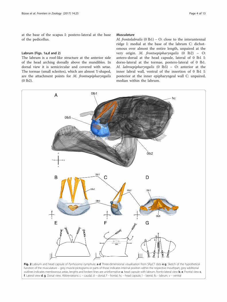
Labrum (Figs. 1a,d and 2)The labrum is a roof-like structure at the anterior sideof the head arching dorsally above the mandibles. Indorsal view it is semicircular and covered with setae.The tormae (small sclerites), which are almost Y-shaped,are the attachment points for M. frontoepipharyngalis(0 lb2).
MusculatureM. frontolabralis (0 lb1) – O: close to the interantennalridge I: medial at the base of the labrum C: dichot-omous over almost the entire length, unpaired at thevery origin. M. frontoepipharyngalis (0 lb2) – O:antero-dorsal at the head capsule, lateral of 0 lb1 I:dorso-lateral at the tormae, postero-lateral of 0 lb1.M. labroepipharyngalis (0 lb5) – O: anterior at theinner labral wall, ventral of the insertion of 0 lb1 I:posterior at the inner epipharyngeal wall C: unpaired,median within the labrum.
Fig. 2 Labrum and head capsule of Pyrrhosoma nymphula. a-d Three-dimensional visualisation from SRμCT data e-g. Sketch of the hypotheticalfunction of the musculature – grey muscle-pictograms or parts of those indicates internal position within the respective mouthpart, grey additionaloutlines indicates membranous areas, lengths and broken lines are uninformative a. head capsule with labrum, fronto-lateral view b, e. Frontal view c,f. Lateral view d, g. Dorsal view. Abbreviations: c – caudal, d – dorsal, f – frontal, hc – head capsule, l – lateral, lb. - labrum, v – ventral
Büsse et al. Frontiers in Zoology (2017) 14:25 Page 4 of 13

Mandibles (Figs. 1f,g and 3)The mandibles are strongly sclerotized and show thetypical dicondylic (two articulations points) ball-and-socket type. They are somewhat triangular – slightlyelongated – from a dorsal view. The mandibles have fourincisivi at the tip, which are broadly connected with eachother at their bases, but divided into two groups by anincision. One lateral incisivus is located closer towardsthe mouth opening. The mola is separated into twoareas and composed of one incisivus each. Laterally of
the molar lobe, two reinforcing ridges run in cranialdirection.
MusculatureM. craniomandibularis internus (0md1) – O: broadareas of the head capsule, postero-dorsal and postero-lateral I: postero-lateral at the mandible, abbuctor ten-don C: the largest muscle within the head capsule. M.craniomandibularis externus posterior (0md3) – O:postero-lateral at the head capsule I: latero-ventral at the
Fig. 3 Mandible and head capsule of Pyrrhosoma nymphula. a-d Three-dimensional visualisation from SRμCT data e-g. Sketch of the hypotheticalfunction of the musculature – grey muscle-pictograms or parts of those indicates internal position within the respective mouthpart, grey additionaloutlines indicates membranous areas, lengths and broken lines are uninformative a. head capsule with labrum, fronto-lateral view b, e. Frontal view c,f. Lateral view d, g. Dorsal view. Abbreviations: c – caudal, d – dorsal, f – frontal, hc – head capsule, l – lateral, md – mandible, v – ventral
Büsse et al. Frontiers in Zoology (2017) 14:25 Page 5 of 13
mandible, abductor tendon. M. hypopharyngomandibu-laris (0md4) – O: dorso-lateral at the suspensorial bar ofthe hypopharynx I: lateral within the mandible. M. ten-toriomandibularis lateralis inferior (0md6) – O: anteriortentorial arm, via tendon I: lateral within the mandibleC: same tendon as 0md8. M. tentoriomandibularis med-ialis superior (0md7) – O: dorsal tentorial arm I:postero-dorsal at the mandible C: weakly developed. M.tentoriomandibularis medialis inferior (0md8) – O: an-terior tentorial arm, via tendon I: posterio-lateral withinthe mandible C: same tendon as 0md6.
Maxillae (Figs. 1e,h & i and 4)The maxillae are located between the mandibles and thelabium, dorso-lateral of the hypopharynx. They are de-veloped to similar extent as in the adult [22]. The maxil-lae are composed of four parts: cardo, stipes, maxillarpalp, and lacinia, a galea is absent. The undivided andtriangular cardo is connected via the cardo-stipital mem-brane to the stipes (no joint is developed) – enabling itsmovement with respect to the stipes. The latter is almostrectangular in ventral view and divided into basistipesand mediostipes by a longitudinal stipital ridge. At thedistal end of the stipes, the maxillary palp originates lat-erally, and the lacinia originates distally. The maxillarypalp is connected via a socket, whereas the lacinia isfused with the stipes.
MusculatureM. craniocardinalis (0mx1) – O: ventro-lateral at thehead capsule between 0md1 and 0md3 I: basal via a ten-don at the cardo C: fan-shaped muscle (origin). M. cranio-lacinialis (0mx2) – O: at the head capsule, postero-dorsalto 0mx1 I: basal at the lacinia. M. tentoriocardinalis(0mx3) – O: lateral at the anterior tentorial arm I: withinthe cardo. M. tentoriostipitalis anterior (0mx4) – O:ventro-lateral at the corpotentorium I: ventral at the sti-pes, slightly fan-shaped. M. tentoriostipitalis posterior(0mx5) – O: ventro-lateral base of anterior tentorial arm,close to 0mx3 I: at the stipes. M. stipitolacinialis (0mx6) –O: ventro-lateral at the base of the stipes I: base of lacinia.M. stipitopalpalis externus (0mx8) – O: lateral within thestipes close to 0mx10 I: posterior on the base of the pal-pus. M. stipitopalpalis internus (0mx10) – O: lateralwithin the stipes close to 0mx8 I: anterior on the base ofthe palpus.
Labium (Figs. 1b–j and 5)The labium is developed as prehensile mask responsiblefor prey capturing. It consists of post- and prementum,ligula, and palps with movable hooks. Glossa and para-glossa are not separated from the prementum by a par-ticular ridge. The post- and prementum are connectedvia a cubital-like hinge joint, the so-called prementum-
postmentum joint (p-p joint), enabling movement ofboth parts relative to each other. The prehensile mask isconnected to the head capsule ventrally via the postmen-tum. The postmentum shows the largest width at the ap-ical end at the connection with the palps. It narrowstowards the joint to almost half of its width. The labialpalps are located at the apical tip of the prehensile mask.They are flexibly connected to the mask and show ablunt end hook and a pointed movable hook. Muscula-ture inserting at the end hook is absent.
MusculatureM. tentoriopraementalis (0la5) – O: posterior at the cor-potentorium I: lateral at the premental edge. M. submen-topraementalis (0la8) – O: at the posterior part of thesubmentum I: dorsal at the prementum C: dichotomousin the early instars (I1,I2,I3) O2: at the postero-lateralbase of the submentum. M. praementopalpalis internus(0la13) – O: median at the prementum, ventral of 0la14I: antero-median at the base of the palpus. M. praemen-topalpalis externus (0la14) – O: median at the premen-tum, dorsal of 0la13 I: lateral at the base of the palpus.M. praementomembranus (0la15) – O: anterio-lateral atthe postmentum I: postero-lateral at the prementum.
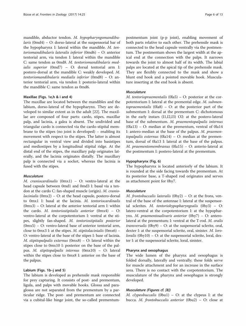
Hypopharynx (Fig. 6)The hypopharynx is located anteriorly of the labium. Itis rounded at the side facing towards the prementum. Atits posterior base, a T-shaped rod originates and servesas attachment point for 0hy7.
MusculatureM. frontobuccalis lateralis (0hy2) – O: at the frons, ven-tral of the base of the antennae I: lateral at the suspensor-ial sclerites. M. tentoriohypopharyngealis (0hy3) – O:latero-ventral at the corpotentorium I: at the hypophar-ynx. M. praementosalivaris anterior (0hy7) – O: antero-lateral at the prementum; I: ventral at the T-rod. M. oralistransversalis (0hy9) – O: at the suspensorial sclerite, oral,dexter I: at the suspensorial sclerite, oral, sinister. M. loro-loralis (0hy10) – O: at the suspensorial sclerite, loral, dex-ter I: at the suspensorial sclerite, loral, sinister.
Pharynx and oesophagusThe wide lumen of the pharynx and oesophagus isfolded dorsally, laterally and ventrally; these folds servefor muscle attachment and for an increase in the surfacearea. There is no contact with the corpotentorium. Themusculature of the pharynx and oesophagus is stronglydeveloped.
Musculature (Figures cf. [8])M. clypeobuccalis (0bu1) – O: at the clypeus I: at thebucca. M. frontobuccalis anterior (0bu2) – O: close at
Büsse et al. Frontiers in Zoology (2017) 14:25 Page 6 of 13

the interantenal ridge, posterior to 0 lb1 I: dorsal atthe bucca. M. frontobuccalis posterior (0bu3) – O:posterior at the frons I: dorsal at the bucca. M. ten-toriobuccalis lateralis (0bu4): O: base of dorsal tentor-ial arms; I: lateral at the bucca. M. tentoriobuccalisanterior (0bu5) – O: at the corpotentorium I: ventralat the bucca. M. tentoriobuccalis posterior (0bu6) –O: dorsal at the anterior tentorial arm I: ventral atthe bucca. M. verticopharyngalis (0 ph 1) – O: at theocciput, medial of 0md1 I: dorsal at the pharynx. M.tentoriopharyngalis (0 ph 2) – O: at the corpotentor-ium I: ventral at the pharynx.
DiscussionComparative morphologyNinety-one head muscles are known so far for insects[23–25]. The most recent account for larval Odonata de-scribed 41 muscles for Epiophlebia [9]. Here we describe42 head muscles for larval Pyrrhosoma nymphula.The difference is due to the absence of antennal
muscle 0an7 in Epiophlebia (E. superstes), which seemsto be a peculiarity of Epiophlebia, since it is present inAnisoptera (Aeshna cyanea) as well as Zygoptera (Pyr-rhosoma nymphula) – see also Blanke et al. [9]. Further-more, there is a shift of the origin point of the muscle
Fig. 4 Maxilla and head capsule of Pyrrhosoma nymphula. a-d Three-dimensional visualisation from SRμCT data e-g. Sketch of the hypotheticalfunction of the musculature – grey muscle-pictograms or parts of those indicates internal position within the respective mouthpart, grey additionaloutlines indicates membranous areas, lengths and broken lines of are uninformative a. head capsule with labrum, fronto-lateral view b, e. Frontal viewc, f. Lateral view d, g. Dorsal view. Abbreviations: c – caudal, d – dorsal, f – frontal, hc – head capsule, l – lateral, mx – maxilla, v – ventral
Büsse et al. Frontiers in Zoology (2017) 14:25 Page 7 of 13

0an5 from the frons to the tentorium – 0an5 might takeover the function of 0an7 – we follow the suggestedhomologization from Blanke et al. [9].The peculiarities of the head musculature in Odonata
larvae in comparison to adults mentioned by Blanke etal. [9] can be confirmed. More precisely, the absence ofM. tentoriomandibularis lateralis superior (0md5), M.
postoccipitopharyngealis (0 ph 3) and M. postoccipitalo-hypopharyngealis (0hy4) as well as the presence of thelabial muscle M. tentoriopraementalis (0la5), antennalmuscle M. tentoriofrontalis dorsalis (0te3), and the pres-ence of the hypopharyngeal muscles (0hy2, 0hy3 0hy9,0hy10), buccal muscles (0bu3, 0bu4, 0bu5), andpharyngeal muscule (0 ph 2) can be confirmed.
Fig. 5 Labium and head capsule of Pyrrhosoma nymphula. a-d Three-dimensional visualisation from SRμCT data E-G. Sketch of the hypotheticalfunction of the musculature – grey muscle-pictograms or parts of those indicates internal position within the respective mouthpart, grey additionaloutlines indicates membranous areas, lengths and broken lines are uninformative a. head capsule with labrum, fronto-lateral view b, e. Frontal view c,f. Lateral view d, g. Dorsal view. Abbreviations: c – caudal, d – dorsal, f – frontal, hc – head capsule, hy – hypopharynx, l – lateral, la –labium, v – ventral
Büsse et al. Frontiers in Zoology (2017) 14:25 Page 8 of 13
Muscle 0 lb1 of the labrum is a dichotomous musclein P. nymphula while it is an unpaired muscle in Epioph-lebia. The mandible muscle 0md7 is weakly developedcompared to that in representatives of Epiprocta.
Muscle ontogenesisDifferent instars of P. nymphula only show minor ana-tomical differences. Accompanied by the continuous in-crease in size, a slight shift of head proportions isnoticeable during ontogenesis. Additionally, the premen-tum becomes elongated, if compared to the submentum,and the labium becomes elongated rather than widened.Small teeth-like sclerotized structures appear anteriorlyat the lateral edge of the prementum [26] of instar 6.Generally, characters used in taxonomy [26], like therows of setae on the head capsule especially at thedorso-caudal postocciput or on the labial palpus andventro-medial of the prementum, are fully developed inlater instars. Furthermore, the mode of crypsis changestowards instars 5 and 6 from a hyaline almost transpar-ent body, which supposedly leads to near-invisibility inthe body of water, to a piebald cuticle comprising differ-ent shades of beige and brown with spots of whitish andblackish to camouflage in the benthos [6].The only muscular change within the larval stages is
revealed concerning M. submentopraementalis (0la8),which is dichotomous in the early instars (I1,I2 and I3)with a second point of origin at the postero-lateral baseof the submentum. This dichotomy is not present in anyof the older instars studied (I6, middle-late and pen-ultimate).Compared to the adult, drastic differences in the
overall morphology and muscle arrangement (cf.‘comparative morphology’ section) occur not only be-cause of the transmutation of the labium (from its
insect ground pattern) into a prehensile mask. There-orientation of the mouthparts from prognathous inthe larva to orthognathous in the adult takes place.This modification leads to the adaptation to the dif-ferent prey-capturing mode in the adult. The adultsuse their specialised legs as a basket for catching preyin flight [27] – the prey is brought to the mouthfrom ventral. Whereas, the larva uses its prehensilemask for prey-capturing under water – the prey isbrought to the mouth from frontal. Furthermore, thepresence of the hypopharyngeal muscles (0hy2, 0hy30hy9, 0hy10), buccal muscles (0bu3, 0bu4, 0bu5), andpharyngeal muscle (0 ph 2) and the strong develop-ment, within the larvae, of the latter two musclegroups might indicate further adaptations for inges-tion within an aquatic habitat – ingestion via suckingthe food “solved” in water. However, the most exten-sive change is the transmutation of the labium. Here,an elongation of the pre- and postmentum as well asthe transformation of the labial palps into prey grasp-ing organs occur. The abductor – M. praementopal-palis externus (0la14) – and adductor – M.praementopalpalis internus (0la13) – of the formerpalpi are greatly enlarged and the points of origin istranslocated to the base of the postmentum to in-crease the applicable force. Two well-developed joints,prementum-head joint (P-H joint) ventral of thehypopharynx and the prementum-postmentum joint(P-P joint), lead to an increase of movability of theprehensile mask. Furthermore, the T-shaped rod or T-rod is characteristic for larval Odonata [8] (more pre-cisely the hypopharyngeal apodem) serves as an im-portant attachment structure for the redirectedmuscles (see also subsection ‘labium’ within the nextsection).
Fig. 6 Hypopharynx and head capsule of Pyrrhosoma nymphula. Three-dimensional visualisation from SRμCT data in fronto-lateral view. Abrevia-tion: c – caudal, d – dorsal, f – frontal, l – lateral, v – ventral, Tr – T-rod
Büsse et al. Frontiers in Zoology (2017) 14:25 Page 9 of 13

Functional significance of different groups of mouthpartsmusclesThe labrum (Fig. 2) limits the preoral cavity anteriorly. Itis connected to the clypeal area by the membranous cly-peolabral suture, which enables movement of the la-brum. This movement is realised by the antagonisticaction of muscles 0 lb1, 0 lb2. The muscle 0 lb5 com-presses the labrum due to its attachment at its anteriorand posterior (epipharyngeal) wall, the cuticle elasticitymight function as muscle antagonist for returning of thelabrum shape to its original condition. The muscle 0 lb1is attached anterio-medially of the labrum and enablesits dorso-lateral movement. The muscle 0 lb2 is the an-tagonist that is attached postero-laterally of the labrumand enables its dorso-medial movement.The mandibles (Fig. 3) are strongly sclerotized jaws for
crushing harder parts of the prey [6]. The movability ofthe mandibles is restricted to one axis due to the anter-ior and posterior ball-and-socket joints. This holds truefor Odonata similar to other groups of insects [28].These joints span a virtual axis of rotation and this rota-tion is produced by the antagonistic action of 0md1, theadductor muscle, and 0md3, the abductor muscle. Mus-cles 0md4, 6, 7 and 8 are often reduced in other wingedinsects [1]. Muscle 0md4 is attached antero-lateralwithin the mandible and at the hypopharynx, where itmight support the movement of the hypopharyngealsclerites. Muscles 0md6 and 0md8 are attached lateraland postero-lateral within the mandible, respectively.They enable a dorso-lateral and postero-dorsal move-ment of the mandible and support in this function themain abductor 0md3 [4].The maxillae (Fig. 4) are specialised for manipulating,
handling and sensing the food. The feeding movementsof the maxillae are mainly protraction and retraction [8].The muscles are able to move the maxillae in almostevery direction, because flexible membranous regionsand the cardo-cranial joint implement the suspension.The muscles 0mx1, 2, 3, 4 and 5 are used to move themaxilla very precisely: 0mx1 – is attached postero-lateralon the maxilla at the cardo (lateral to the head) and en-ables a dorso-median movement of the cranial part ofthe maxilla; 0mx2 – is attached antero-median on themaxilla at the lacinia and enables a dorsal (slightlydorso-lateral) movement of the apical part of the max-illa; 0mx3 – is attached postero-median on the maxillawithin the cardo and enables a dorsal (slightly dorso-lateral) movement of the cranial part of the maxilla;0mx4 – is attached median on the maxilla at the stipesand enables a dorsal movement of the maxilla; 0mx5 –is attached antero-lateral (medial to the head) and en-ables a dorso-lateral movement of the maxilla. Duringthe protracting process the laciniae are used to grasp theprey, provided by the prehensile mask, by thrusting
forward beyond the mandibles [8]. After retracting themaxillae, the food grasped by the laciniae, is delivered tothe mandibles, where the digestive process starts. Themuscle 0mx6 runs completely within the maxillae and isattached at the median base of the lacinia and might en-able abduction. During or before this process the palpsare used to sense the food. The maxillary palp is, due toits connection with the stipes via a ball and socket joint,very movable. The abductor muscle 0mx8 enables aventro-lateral movement of the palp, whereas the oppos-ing adductor muscle 0mx10 enables a dorso-medialmovement of the palp. Combined with the dorso-ventralmovement of both 0mx1 and 0mx2 the sensing in everydirection is enabled.The locking mechanism of the prehensile mask, as de-
scribed by Olesen [12], where the maxillae are supposedto be used to prevent propelling of the prehensile maskcould not be confirmed.The labium (Fig. 5) performs the most interesting bio-
mechanical movement in Odonata larvae (cf. Additionalfile 1: High-Speed Video). It represents a strong modifi-cation of mouthparts within the insects: the modificationof the labium into a prehensile mask used for prey cap-turing. The process of prey capturing can be divided into two different events: i) propelling of the prehensilemask towards the prey, and ii) grasping the prey usingthe movable pointed hooks.The muscles 0la5, 0la8, 0la15, and 0hy7 are used for
retracting the prehensile mask after the strike and tohold it in its resting position. They could presumablywork as an antagonist against the pressure, while the jetpropulsion in Anisoptera takes place, as suggested byPritchard [10]. While muscles 0la5, 0la8 and 0hy7 mighthelp to prevent propelling the prementum, muscle 0la15might prevent the shearing of the postmentum againstthe head capsule. The movability of the prehensile maskis restricted by two joints, the prementum-head joint (P-H joint) and the prementum-postmentum joint (P-Pjoint). More precisely: 0la5 – is attached medially at thebase of the postmentum and enables a dorsal movementtowards the head, slightly restricted by the P-H joint forretraction/holding; 0la8 – is running from the ventralbase of the prementum to the apical side of the post-mentum; this muscle locks the movability within the p-pjoint; 0la15 – is attached at the very base of the post-mentum and might prevent the shearing of the post-mentum against the head capsule and/or helps to lockthe post- and prementum to each other; 0hy7 – is at-tached at the ventral base of the prementum directly atthe labial articulation and enables a dorsal movement to-wards the head, strongly restricted by the P-H joint forretraction/holding. All these muscles might be includedin the propelling movement of the prehensile mask, tosteer during and/or positioning the mask. However, it
Büsse et al. Frontiers in Zoology (2017) 14:25 Page 10 of 13
becomes clear that the propelling of the prehensile labialmask cannot be powered by normal muscle contraction,since muscle contraction enables only the oppositemovement.However, both muscles 0la5 and 0hy7 become
deflected in the completely drawn-in position of the pre-hensile labial mask. The muscle 0la5 is bent by the T-rod apodem (see above paragraph ‘muscle ontogenesis),as described by Blanke and colleagues [9]: the bent endsof this apodem deflect the muscle 0la5 towards thethorax. The same is true for the muscle 0hy7: here thedorsal base of the prementum functions as ‘deflectionsheave’ and redirects the muscle towards the tip ofprementum.So far the results of previous investigations indicate
that the main force for the labium extension is producedby abdominal dorso-ventral muscles and transmitted tothe labium as haemolymph pressure increases [11–14].The same abdominal muscles are also involved in themechanism of water intake into the digestive tract, to in-crease the body pressure, for respiration and swimmingby jet propulsion [12, 29–31]; the latter is an escapemechanism in anisopteran larvae [6]. Epiprocta [Anisop-tera + Epiophebia] use a dedicated internal respirationorgan and are able to escape via jet propulsion (neverobserved in Epiophlebia) [32]. The ability to increasehaemolymph pressure by closing their anal valve, inorder to propel the prehensile mask, is therefore studiedin anisopteran taxa only. The mechanism of propellingthe prehensile mask in Zygoptera is not properly studiedso far, due to missing experimental investigations and adiffering larval anatomy as mentioned above. Caillère[33, 34] investigated the prey capturing strike of Zygop-tera mentioning a convergent movement of the gills andthe beginning of a forward movement of the digestivetract (abdomen) just before the prey capturing process.The extension of the prehensile mask and the forwardmovement of the abdomen stop simultaneously, and atalmost the same time, the convergent movement of thegills ceases [33, 34]. This observation as well as the in-vestigations by Eriksen [15], Miller [17, 18] andSesterhenn and colleagues [19] confirm that also Zygop-tera are able of an water intake into their digestive tracteven though their external gills are responsible for a sig-nificant part of the gas exchange [15], as already Tillyard[7] suggested. However, the internal anatomy differs atleast in the “lack of the diaphragm and the sub-intestinalmuscle, which allows anisoptera larvae to suck water dir-ectly into the branchial chamber” ([17],p.386). Zygopteraare therefore restricted to a more gulping like ventila-tion, most likely due to the mentioned differences and asmaller volume of the related respiratory organs [17].Since it is implausible – parsimony principle – that
Zygoptera and Anisoptera use different mechanisms for
such a highly complex biomechanical process, we safelycan assume that the extension of the labium in Anisop-tera as well as in Zygoptera is based on the same princi-ples. Nevertheless, the mechanism of the maskpropelling within the Odonata – considering Zygopteraand Anisoptera – should be reinvestigated because ofthe significant differences mentioned.The second important process in prey capturing, is the
prey grasping, which is realised by the strongest muscu-lature found in the prehensile mask (Fig. 5). The ad-ductor muscle 0la13 realizes the grasping and clutchingof the prey at the tip of the labium by closing the labialpalps with the movable hooks. The abductor muscle0la14 realizes the opening of the labial palps, to releasethe prey remains and bring the palps in the initial pos-ition for new prey capturing process.The hypopharynx (Fig. 5) is situated in the preoral cav-
ity in front of the functional mouth [1]. The strongestmuscle 0hy3 is the retractor of the hypopharynx, origin-ating on the tentorium to allow a caudal movement andtherefore a widening of the oral cavity. Muscle 0hy2 ori-ginating at the frons, most likely is able to protract thehypopharynx into the oral cavity, serving as antagonistto 0hy3. In Odonata, the unique T-rod or T-shaped rodis originating from the hypopharynx and 0hy7, whichoriginates on the T-rod and inserts within the prehensilelabial mask plays an important role in its movement (cf.paragraph on the labium). The small muscles 0hy9 and0hy10 attaching at the left and the right side of the sus-pensorial sclerites might deform the hypopharynxlaterally.
MethodsWe studied six (L1, L2, L3, L6, middle-late, pen-ultimate) instars of Pyrrhosoma nymphula (Sulzer, 1776)(Zygoptera; Coenagrionidae), for comparison we also in-vestigated larval specimens (late instars) of Aeshna cya-nea (Müller, 1764) (Anisoptera; Aeshnidae), andEpiophlebia superstes (Sélys, 1889) (Epiprocta; Epiophle-biidae). All figures show the pen-ultimate instar. Thespecimens were fixed in alcoholic Bouin solution (=Duboscq-Brasil) and stored in 70–80% ethanol [35]. Allapplicable regulations concerning the protection of free-living species were followed. All necessary permits wereobtained for collecting Odonata at the BillingshäuserSchlucht, Göttingen, Germany (permission granted by“Untere Naturschutzbehörde” file reference AZ.67.2.5Wei). Prior to scanning (both CT and SEM), the sampleswere dehydrated in an ascending ethanol series anddried at the critical point (Balzers CPD030) or usingHexamethyldisilazan (HMDS) [35].High resolution X-ray tomography (μCT) was carried
out using a SkyScan 1172 desktop micro-CTscanner(Bruker micro-CT, Kontich, Belgium) at 40 kV and
Büsse et al. Frontiers in Zoology (2017) 14:25 Page 11 of 13
250 μA with images taken every 0.25°. Additionally, weused the synchrotron radiation micro-computed tomog-raphy (SRμCT) setup at the Tomcat beamline of theSwiss Light Source, Villigen, Switzerland [36].Segmentation and visualization of the data were done
with Amira 5.4.3 (FEI SAS, France, www.vsg3d.com) andPhotoshop CS3 (Adobe SystemInc.). Please refer to Betzet al. [37] for further information on the general setupfor SRμCT and to Büsse et al. [38] for information onsegmentation, labelling and visualization with Amira.For SEM, the samples were dehydrated in an ascend-
ing ethanol series, critical point dried (Quorum E3000)and sputter-coated with gold-palladium (10 nm thick-ness; Leica Bal-TEC SCD500). Afterwards the sampleswere mounted on a rotatable sample holder [39] and ex-amined in a Hitachi TM3000 scanning electron micro-scope at an accelerating voltage of 15 kV.For the high-speed video recordings a Photron Fas-
tcam SA1.1 (model 675 K–M1, www.vkt.de) equipedwith a 105 mm/1:2.8 macro lens (Sigma, Japan,www.sigma-photo.co.jp) mounted on a Manfrotto055tripod with Manfrotto410 geared head (Manfrotto, Italy,www.manfrotto.com) and two Dedocol COOLT3 lightsources (Dedotech, Switzerland, https://dedotec.ch) wasused. The labial strike was shot with 5400 frames persecond (fps) (1/frame, Trigger Mode: End, Resolution1024 × 1024). The video was saved as 16-bit TIFFimage-stack and later reconstructed into a video format(AVI) using ImageJ 1.51e (National Institutes of Health,USA, https://imagej.nih.gov/ij/).The juvenile stage in Odonata, Ephermeroptera and
Plecoptera is suggested to be called “naiad “followingthe terminology proposed in Bybee et al. [40]. How-ever, since for Odonata the more commonly usedname is “larva”, we decided to be consistent withinthe terminology, mainly used in the community, andused here the more general term [41, 42]. Anatomicalstructures are described using the nomenclature ofBeutel et al. [43], muscle designations are made usingthe nomenclature of Wipfler et al. [25]. Muscles aredescribed stating their origin (O) and their insertion(I) followed by (C) some special characteristics, ifpresent.
Additional file
Additional file 1: High-speed video of the prey capturing process of aZygoptera larva. (MOV 12373 kb)
AcknowledgementsWe are grateful for the support by the members of the FunctionalMorphology and Biomechanics Group at Kiel University, especially to EstherAppel and Dr. Lars Heepe. We also want to thank Sebastian Boge forpreparatory work. Furthermore, our special thanks goes to Dr. AlexanderBlanke for proofreading and many elucidating discussions.
The support of the Swiss-Light-Source at the Paul-Scherrer Institut for grantingbeamtime at the TOMCAT beamline (Proposal No.: 20120025, granted to Dr.Sonja Wedmann, Senckenberg Gesellschaft für Naturforschung, Frankfurt a. M.,Germany, as head of a combined fossil and extent entomology research group)is gratefully acknowledged.Last but not least, we want to thank the unknown Reviewers for theirconstructive and helpful comments.
FundingTH was supported through DFG grant HO2306/6–1, 2. The project wasfinanced and SB directly supported through the DFG grant BU3169/1–1.
Availability of data and materialsThe necessary data will be available in the supplement. The analysed raw-datasets are available from the corresponding author on reasonable request.
Authors’ contributionsSB, TH and SNG designed the study. SB and SNG conducted the μCT andSEM analysis. SB and TH carried out the analysis of the raw data. SB wrotethe manuscript. All authors read, revised and approved the manuscript.
Competing interestsThe authors declare that they have no competing interests.
Consent for publicationNot applicable.
Ethics approvalNo permission of research ethics or animal ethics committees was necessary.All applicable regulations concerning the protection of free-living specieswere followed.
Publisher’s NoteSpringer Nature remains neutral with regard to jurisdictional claims inpublished maps and institutional affiliations.
Author details1Department of Functional Morphology and Biomechanics, Institute ofZoology, Christian-Albrechts-Universität zu Kiel, Am Botanischen Garten 9,24118 Kiel, Germany. 2Senckenberg Gesellschaft für Naturforschung,Senckenberganlage 25, 60325 Frankfurt, Germany.
Received: 17 January 2017 Accepted: 26 April 2017
References1. Snodgrass RE. Principles of insect morphology. New York: Mc Graw-Hill
Book Company; 1935.2. Grimaldi D, Engel MS. Evolution of the insects. Cambridge: University Press;
2005.3. Schmitt C, Rack A, Betz O. Analyses of the mouthpart kinematics in
Periplaneta americana (Blattodea, Blattidae) by using synchrotron-based X-ray cineradiography. J Exp Biol. 2014;217:3095–107.
4. David S, Funken J, Potthast W, Blanke A. Musculoskeletal modelling of thedragonfly mandible system as an aid to understanding the role of singlemuscles in an evolutionary context. J Exp Biol. 2016;219:1041–9.
5. Gronenberg SJW. The control of mandible movements in the antOdontomachus. J Insect Physiol. 1999;45:231–40.
6. Corbet PS. Dragonflies: behavior and ecology of Odonata. New York: CornellUniv Press; 1999.
7. Tillyard RJ. The biology of dragonflies (Odonata or Paraneuroptera).Cambridge: University Press; 1917.
8. Snodgrass RE. The dragonfly larva. Smith Misc Coll. 1954;12:38.9. Blanke A, Büsse S, Machida R. Coding characters from different life
stages for phylogenetic reconstruction: a case study on dragonfly adultsand larvae, including a description of the larval head anatomy ofEpiophlebia superstes (Odonata: Epiophlebiidae). Zool J Linnean Soc.2015;174:718–32.
10. Pritchard G. Prey capture by dragonfly larvae (Odonata; Anispotera). Canad JZool. 1965;43:271–89.
Büsse et al. Frontiers in Zoology (2017) 14:25 Page 12 of 13
11. Olesen J. The hydraulic mechanism of labial extension and jet propulsion indragonfly nymphs. J Comp Physiol. 1972;81:53–5.
12. Olesen J. Prey-capture in dragonfly nymphs (Odonata; Insecta): labialprotraction by means of a multi purpose ab- dominal pump. Vid Medd DanNaturalist Foren. 1979;141:81–96.
13. Tanaka Y, Hisada M. The hydraulic mechanism of the preda- tory strike indragonfly larvae. J Exp Biol. 1980;88:1–19.
14. Parry DA. Labial extension in the dragonfly larva Anax Imperator. J Exp Biol.1983;107:495–9.
15. Eriksen CH. Respiratory roles of caudal lamellae (gills) in a Lestid damselfly(Odonata:Zygoptera). J North Am Benthol Soc. 1986;5:16–27.
16. Whedon AD. The comparative morphology and possible Adaptions of theabdomen in the Odonata. Trans Am Entomol Soc. 1918;44:373–437.
17. Miller PL. Responses of rectal pumping to oxygen lack by larval Calopteryxsplendens (Zygoptera: Odonata). Physiol Entomol. 1993;18:379–88.
18. Miller PL. The responses of rectal pumping in some zygopteran larvae(Odonata) to oxygen and ion availability. J Insect Physiol. 1994;40:333–9.
19. Sesterhenn TM, Reardon EE, Chapman LJ. Hypoxia and lost gills: respiratoryecology of temperate larval damselfly. J Insect Physiol. 2013;59:19–25.
20. Pass G. Accessory pulsatile organs: evolutionary innovations in insects. AnnRev Entomol. 2000;45:495–518.
21. Pass G. Phylogenetic relationships of the orders of hexapoda: contributionsfrom the circulatory organs for a morphological data matrix. Arthropod SystPhylo. 2006;64:165–203.
22. Blanke A, Beckmann F, Misof B. The head anatomy of Epiophlebia superstes(Odonata: Epiophlebiidae). Org Div Evol. 2013;13:55–66.
23. Kéler S v. Entomologisches Wörterbuch. Berlin: Akademieverlag Berlin; 1963.24. Matsuda R. Morphology and evolution of the insect head. Mem Am Inst
Ent. 1965;4:1–334.25. Wipfler B, Machida AR, Müller RB, Beutel RG. On the head morphology of
Grylloblattodea (Insecta) and the systematic position of the order, with anew nomenclature for the head muscles of Dicondylia. Syst Entomol. 2011;36:241–66.
26. Heidemann H, Seidenbusch R. Die Libellenlarven Deutschlands – Handbuchfür Exuviensammler. Keltern: Goecke & Evers; 2002.
27. Leipelt KG, Suhling F, Gorb SN. Ontogenetic shifts in functional morphologyof dragonfly legs (Odonata: Anisoptera). Zoology. 2010;113:317–25.
28. Gorb SN, Beutel RG. Head-capsule design and mandible control in beetlelarvae: a three-dimensional approach. J Morph. 2000;244:1–14.
29. Pickard RS, Mill PJ. Ventilatory muscle activity in intact preparations ofAeschnid dragonfly larvae. J Exp Biol. 1972;6:527–36.
30. Mill PJ, Pickard RS. Anal valve movement and normal ventilation in Aeshniddragonfly larvae. J Exp Biol. 1972;56:537–43.
31. Mill PJ, Pickard RS. Jet-propulsion in Anisopteran dragonfly larvae. J CompPhysiol. 1975;97:320–38.
32. Tabaru N. Larval development of Epiophlebia superstes in Kyushu. Tombo.1984;27:27–31.
33. Caillère L. Long Term Learning in Agrion (Syn. Calopteryx) splendens Harris1782 Larvae (Insecta, Odonata). Z Vergl Physiol. 1970;69:284–95.
34. Caillère L. Dynamics of the strike in a Agrion (syn. Calopteryx) splendensHarris. larvae (Odonata: Calopterygidae). Odonatologica. 1972;1:11-19.
35. Romeis B. Mikroskopische Technik. Urban und Schwarzenberg: München; 1987.36. Stampanoni M, Mokso R, Marone F, Vila-Comamala J, Gorelick S, Trtik P,
Jefimovs K, David C. Phase-contrast tomography at the nanoscale usinghard X-rays. Physic Rev B. 2010;81:140105.
37. Betz O, Wegst U, Weide D, Heethoff M, Helfen L, Lee W-K, Cloetens P.Imaging applications of synchrotron X-ray phase-contrast microtomographyin biological morphology and biomaterials science. I. General aspects of thetechnique and its advantages in the analysis of millimetre-sized arthropodstructure. J Microsc. 2007;227:51–71.
38. Büsse S, Helmker B, Hörnschemeyer T. The thorax morphology ofEpiophlebia (Insecta: Odonata) nymphs - including remarks on ontogenesisand evolution. Sci Rep. 2015;5:12835.
39. Pohl HA. Scanning electron microscopy specimen holder for viewingdifferent angles of a single specimen. Microsc Res Tech. 2010;73:1073–6.
40. Bybee SM, Hansen Q, Büsse S, Wightman H, Branham MA. For consistency’ssake: the precise use of nymph, larva and naiad within insecta. SystEntomol. 2015;40:667–70.
41. Sahlén G, Suhling F, Martens A, Gorb SN, Fincke OM. For consistency's sake?A reply to Bybee et al. Syst Entomol. 2016;41:307–8.
42. Büsse S, Bybee SM. Larva, nymph and naiad – a response to the replies toBybee et al. 2015 and the results of a survey within the entomologicalcommunity. Syst Entomol. 2017;42:11–4.
43. Beutel RG, Friedrich F, Yang X-K, Ge S-Q. Insect morphology and phylogeny:a textbook for students of entomology. Berlin: De Gruyter; 2013.
• We accept pre-submission inquiries
• Our selector tool helps you to find the most relevant journal
• We provide round the clock customer support
• Convenient online submission
• Thorough peer review
• Inclusion in PubMed and all major indexing services
• Maximum visibility for your research
Submit your manuscript atwww.biomedcentral.com/submit
Submit your next manuscript to BioMed Central and we will help you at every step:
Büsse et al. Frontiers in Zoology (2017) 14:25 Page 13 of 13