The fundamental plan of the retina - Harvard...
10
A simple concept of the retina’s function—lateral inhibition by horizontal and amacrine cells, a direct pathway mediated by bipolar cells—is part of the everyday canon of neurobiolo- gy. In reality, the retina is a more complex and more subtle structure than the textbooks imply. This is of course true also for other structures of the central nervous system—such as the hippocampus or cortex—where a similar mismatch exists between a simple iconic physiology and the facts of the bio- logical structure. Here I make an initial attempt to come to grips with the real retina, to encompass the system’s actual cel- lular complexity. Neuroanatomical studies have reached a milestone. The iden- tification and classification of retinal neurons (Fig. 1), begun more than 100 years ago by Santiago Ramon y Cajal, is nearing completion—the first time that this has been accomplished for any significantly complex structure of the mammalian CNS. This statement is possible because much of the recent work on reti- nal cell populations has been quantitative. Staining cells as whole populations permits comparison of their numerical frequency. More importantly, when the number of cells of a general class (such as amacrine cells) is known, one can then determine when the identified types add up to the class total 1–4 . Much detail remains to be learned, and a few additional cell types are sure to be discovered. However, we now know at least that no large cell populations remain unidentified, that there are no major pieces ‘missing’ within the retina’s machinery 5 . Unexpectedly, for most mammals, the numbers of bipolar and amacrine cells are distributed fairly evenly among the dif- ferent types. This differs from initial impressions, which were much influenced by early studies in primates. The primate fovea is anomalous in being dominated numerically by a single type of retinal ganglion cell, with an associated, specialized type of bipo- lar cell (see below). In other mammalian retinas, and away from the fovea in primates, individual bipolar, amacrine and ganglion cell types are numerically distributed in a more level way. Although variations certainly exist (generally, there are fewer wide-field than narrow-field neurons), there are no dominant types. In other words, the retina is not composed of a few major players surrounded by a diverse cast of minor ones. Instead, it consists of many parallel, anatomically equipotent microcircuits. How can this awesome list of cell types be sorted? What uni- fying principles might allow us to conceive of the retina more The fundamental plan of the retina Richard H. Masland Howard Hughes Medical Institute, Wellman 429, Massachusetts General Hospital, Harvard Medical School, Boston, Massachusetts 02114, USA Correspondence should be addressed to R.M. ([email protected]) The retina, like many other central nervous system structures, contains a huge diversity of neuronal types. Mammalian retinas contain approximately 55 distinct cell types, each with a different function. The census of cell types is nearing completion, as the development of quantitative methods makes it possible to be reasonably confident that few additional types exist. Although much remains to be learned, the fundamental structural principles are now becoming clear. They give a bottom-up view of the strategies used in the retina’s processing of visual information and sug- gest new questions for physiological experiments and modeling. simply? From the work of many laboratories 6–11 , the fundamen- tal backbone of the retina’s structural organization has come into view. It reinforces certain principles learned from physiological experiments, and suggests new questions for further ones. Here I review the retina’s structure and point out some unresolved functional issues that it suggests. Parallel pathways from cones to ganglion cells A typical mammalian retina contains 9–11 different types of cone-driven bipolar cells. These represent an assortment of path- ways from cones to the inner retina, each carrying a different type of information. This diversity was initially shown in the cells’ structures and the distinct proteins that each expresses. Electro- physiological experiments are now beginning to reveal its func- tional consequences. In most mammalian species, rods outnumber cones by approximately 20-fold, and rods were once considered the pri- mordial photoreceptors. However, molecular cloning of the visual pigments (opsins) that render these cells light-sensitive led to the conclusion that cone pigments evolved long before rhodopsin, the rod pigment 12–14 . The early photoreceptor thus seems to have been some type of cone (Fig. 2a). In retrospect, this makes sense; in building a cell to detect light, one would surely design it for times when copious light is available. (In starlight, a human rod photoreceptor has been calculated to receive only one photon every 10 minutes 8,15 .) Cones are asso- ciated with a complex network of postsynaptic cells, whereas the circuitry strictly associated with rods is minimal; even though rods outnumber cones, most mammalian retinas have 8 to 10 cone-driven neurons for every cell associated primari- ly with the rod pathway. The existence of multiple subclasses of cone-driven bipo- lar cells (‘cone bipolars’) was initially predicted on structural and molecular grounds 11,16,17 . First, bipolar cells branch at dif- ferent levels of the inner plexiform layer 18 , which contain processes of different types of amacrine and ganglion cells. Some ganglion cell types have dendrites confined mainly to level 1 of the inner plexiform layer, others to level 2, and so on. The inner plexiform layer, named as though it formed a sin- gle, tangled ‘plexus,’ is in fact an ordered stack of synaptic planes, more like a club sandwich than a plate of spaghetti. Specific bipolar cells make their synapses within specific planes, review nature neuroscience • volume 4 no 9 • september 2001 877 © 2001 Nature Publishing Group http://neurosci.nature.com © 2001 Nature Publishing Group http://neurosci.nature.com
Transcript of The fundamental plan of the retina - Harvard...

A simple concept of the retina’s function—lateral inhibitionby horizontal and amacrine cells, a direct pathway mediatedby bipolar cells—is part of the everyday canon of neurobiolo-gy. In reality, the retina is a more complex and more subtlestructure than the textbooks imply. This is of course true alsofor other structures of the central nervous system—such as thehippocampus or cortex—where a similar mismatch existsbetween a simple iconic physiology and the facts of the bio-logical structure. Here I make an initial attempt to come togrips with the real retina, to encompass the system’s actual cel-lular complexity.
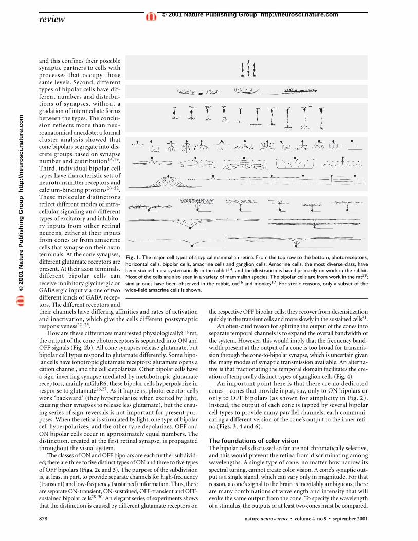
Neuroanatomical studies have reached a milestone. The iden-tification and classification of retinal neurons (Fig. 1), begunmore than 100 years ago by Santiago Ramon y Cajal, is nearingcompletion—the first time that this has been accomplished forany significantly complex structure of the mammalian CNS. Thisstatement is possible because much of the recent work on reti-nal cell populations has been quantitative. Staining cells as wholepopulations permits comparison of their numerical frequency.More importantly, when the number of cells of a general class(such as amacrine cells) is known, one can then determine whenthe identified types add up to the class total1–4. Much detailremains to be learned, and a few additional cell types are sure tobe discovered. However, we now know at least that no large cellpopulations remain unidentified, that there are no major pieces‘missing’ within the retina’s machinery5.
Unexpectedly, for most mammals, the numbers of bipolarand amacrine cells are distributed fairly evenly among the dif-ferent types. This differs from initial impressions, which weremuch influenced by early studies in primates. The primate foveais anomalous in being dominated numerically by a single type ofretinal ganglion cell, with an associated, specialized type of bipo-lar cell (see below). In other mammalian retinas, and away fromthe fovea in primates, individual bipolar, amacrine and ganglioncell types are numerically distributed in a more level way.Although variations certainly exist (generally, there are fewerwide-field than narrow-field neurons), there are no dominanttypes. In other words, the retina is not composed of a few majorplayers surrounded by a diverse cast of minor ones. Instead, itconsists of many parallel, anatomically equipotent microcircuits.
How can this awesome list of cell types be sorted? What uni-fying principles might allow us to conceive of the retina more
The fundamental plan of the retina
Richard H. Masland
Howard Hughes Medical Institute, Wellman 429, Massachusetts General Hospital, Harvard Medical School, Boston, Massachusetts 02114, USA
Correspondence should be addressed to R.M. ([email protected])
The retina, like many other central nervous system structures, contains a huge diversity of neuronaltypes. Mammalian retinas contain approximately 55 distinct cell types, each with a differentfunction. The census of cell types is nearing completion, as the development of quantitativemethods makes it possible to be reasonably confident that few additional types exist. Althoughmuch remains to be learned, the fundamental structural principles are now becoming clear. Theygive a bottom-up view of the strategies used in the retina’s processing of visual information and sug-gest new questions for physiological experiments and modeling.
simply? From the work of many laboratories6–11, the fundamen-tal backbone of the retina’s structural organization has come intoview. It reinforces certain principles learned from physiologicalexperiments, and suggests new questions for further ones. HereI review the retina’s structure and point out some unresolvedfunctional issues that it suggests.
Parallel pathways from cones to ganglion cellsA typical mammalian retina contains 9–11 different types ofcone-driven bipolar cells. These represent an assortment of path-ways from cones to the inner retina, each carrying a different typeof information. This diversity was initially shown in the cells’structures and the distinct proteins that each expresses. Electro-physiological experiments are now beginning to reveal its func-tional consequences.
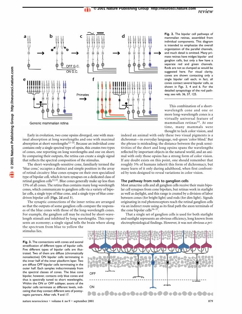
In most mammalian species, rods outnumber cones byapproximately 20-fold, and rods were once considered the pri-mordial photoreceptors. However, molecular cloning of thevisual pigments (opsins) that render these cells light-sensitiveled to the conclusion that cone pigments evolved long beforerhodopsin, the rod pigment12–14. The early photoreceptor thusseems to have been some type of cone (Fig. 2a). In retrospect,this makes sense; in building a cell to detect light, one wouldsurely design it for times when copious light is available. (Instarlight, a human rod photoreceptor has been calculated toreceive only one photon every 10 minutes8,15.) Cones are asso-ciated with a complex network of postsynaptic cells, whereasthe circuitry strictly associated with rods is minimal; eventhough rods outnumber cones, most mammalian retinas have8 to 10 cone-driven neurons for every cell associated primari-ly with the rod pathway.
The existence of multiple subclasses of cone-driven bipo-lar cells (‘cone bipolars’) was initially predicted on structuraland molecular grounds11,16,17. First, bipolar cells branch at dif-ferent levels of the inner plexiform layer18, which containprocesses of different types of amacrine and ganglion cells.Some ganglion cell types have dendrites confined mainly tolevel 1 of the inner plexiform layer, others to level 2, and so on.The inner plexiform layer, named as though it formed a sin-gle, tangled ‘plexus,’ is in fact an ordered stack of synapticplanes, more like a club sandwich than a plate of spaghetti.Specific bipolar cells make their synapses within specific planes,
review
nature neuroscience • volume 4 no 9 • september 2001 877
©20
01 N
atu
re P
ub
lish
ing
Gro
up
h
ttp
://n
euro
sci.n
atu
re.c
om
© 2001 Nature Publishing Group http://neurosci.nature.com
878 nature neuroscience • volume 4 no 9 • september 2001
and this confines their possiblesynaptic partners to cells withprocesses that occupy thosesame levels. Second, differenttypes of bipolar cells have dif-ferent numbers and distribu-tions of synapses, without agradation of intermediate formsbetween the types. The conclu-sion reflects more than neu-roanatomical anecdote; a formalcluster analysis showed thatcone bipolars segregate into dis-crete groups based on synapsenumber and distribution16,19.Third, individual bipolar celltypes have characteristic sets ofneurotransmitter receptors andcalcium-binding proteins20–22.These molecular distinctionsreflect different modes of intra-cellular signaling and differenttypes of excitatory and inhibito-ry inputs from other retinalneurons, either at their inputsfrom cones or from amacrinecells that synapse on their axonterminals. At the cone synapses,different glutamate receptors arepresent. At their axon terminals,different bipolar cells canreceive inhibitory glycinergic orGABAergic input via one of twodifferent kinds of GABA recep-tors. The different receptors andtheir channels have differing affinities and rates of activationand inactivation, which give the cells different postsynapticresponsiveness22–25.
How are these differences manifested physiologically? First,the output of the cone photoreceptors is separated into ON andOFF signals (Fig. 2b). All cone synapses release glutamate, butbipolar cell types respond to glutamate differently. Some bipo-lar cells have ionotropic glutamate receptors: glutamate opens acation channel, and the cell depolarizes. Other bipolar cells havea sign-inverting synapse mediated by metabotropic glutamatereceptors, mainly mGluR6; these bipolar cells hyperpolarize inresponse to glutamate26,27. As it happens, photoreceptor cellswork ‘backward’ (they hyperpolarize when excited by light,causing their synapses to release less glutamate), but the ensu-ing series of sign-reversals is not important for present pur-poses. When the retina is stimulated by light, one type of bipolarcell hyperpolarizes, and the other type depolarizes. OFF andON bipolar cells occur in approximately equal numbers. Thedistinction, created at the first retinal synapse, is propagatedthroughout the visual system.
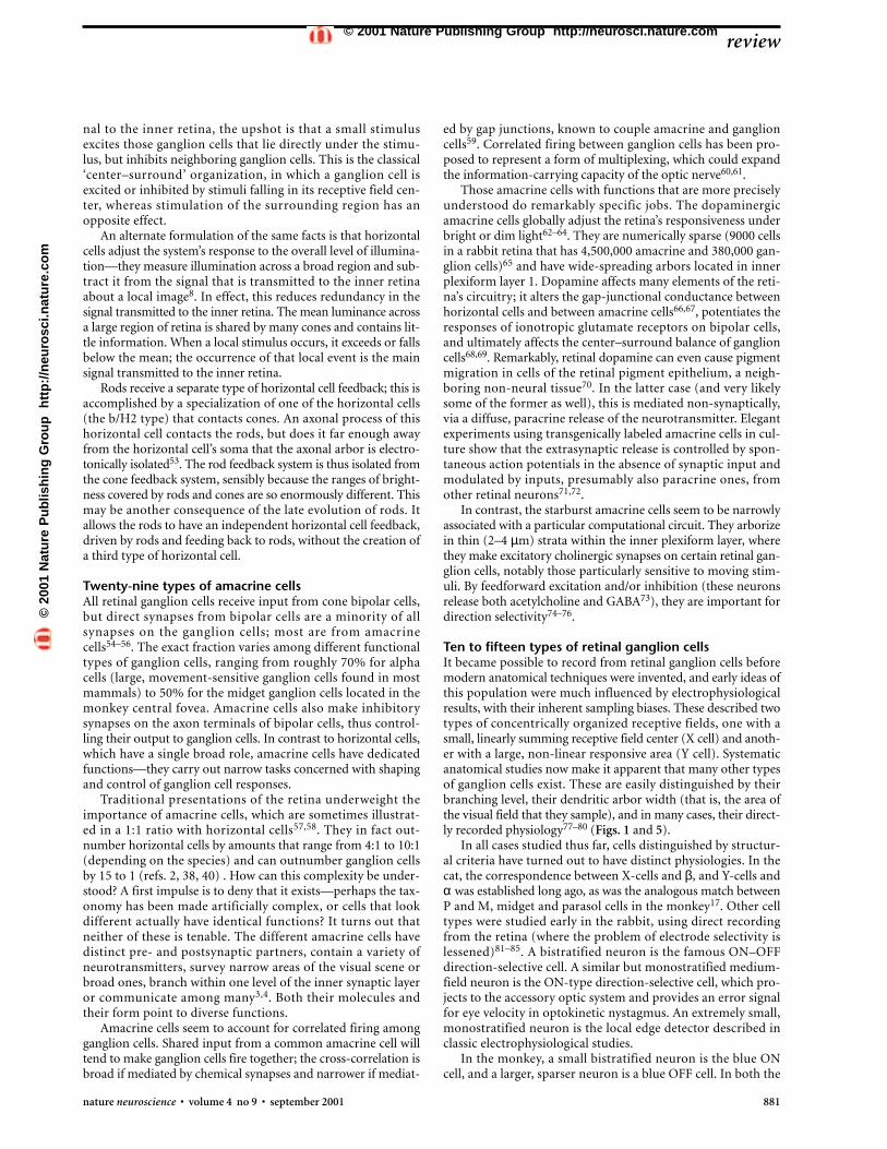
The classes of ON and OFF bipolars are each further subdivid-ed; there are three to five distinct types of ON and three to five typesof OFF bipolars (Figs. 2c and 3). The purpose of the subdivisionis, at least in part, to provide separate channels for high-frequency(transient) and low-frequency (sustained) information. Thus, thereare separate ON-transient, ON-sustained, OFF-transient and OFF-sustained bipolar cells28–30. An elegant series of experiments showsthat the distinction is caused by different glutamate receptors on
the respective OFF bipolar cells; they recover from desensitizationquickly in the transient cells and more slowly in the sustained cells31.
An often-cited reason for splitting the output of the cones intoseparate temporal channels is to expand the overall bandwidth ofthe system. However, this would imply that the frequency band-width present at the output of a cone is too broad for transmis-sion through the cone-to-bipolar synapse, which is uncertain giventhe many modes of synaptic transmission available. An alterna-tive is that fractionating the temporal domain facilitates the cre-ation of temporally distinct types of ganglion cells (Fig. 4).
An important point here is that there are no dedicatedcones—cones that provide input, say, only to ON bipolars oronly to OFF bipolars (as shown for simplicity in Fig. 2).Instead, the output of each cone is tapped by several bipolarcell types to provide many parallel channels, each communi-cating a different version of the cone’s output to the inner reti-na (Figs. 3, 4 and 6).
The foundations of color visionThe bipolar cells discussed so far are not chromatically selective,and this would prevent the retina from discriminating amongwavelengths. A single type of cone, no matter how narrow itsspectral tuning, cannot create color vision. A cone’s synaptic out-put is a single signal, which can vary only in magnitude. For thatreason, a cone’s signal to the brain is inevitably ambiguous; thereare many combinations of wavelength and intensity that willevoke the same output from the cone. To specify the wavelengthof a stimulus, the outputs of at least two cones must be compared.
review
Fig. 1. The major cell types of a typical mammalian retina. From the top row to the bottom, photoreceptors,horizontal cells, bipolar cells, amacrine cells and ganglion cells. Amacrine cells, the most diverse class, havebeen studied most systematically in the rabbit3,4, and the illustration is based primarily on work in the rabbit.Most of the cells are also seen in a variety of mammalian species. The bipolar cells are from work in the rat39;similar ones have been observed in the rabbit, cat16 and monkey17. For steric reasons, only a subset of thewide-field amacrine cells is shown.©
2001
Nat
ure
Pu
blis
hin
g G
rou
p
htt
p:/
/neu
rosc
i.nat
ure
.co
m© 2001 Nature Publishing Group http://neurosci.nature.com
This combination of a short-wavelength cone and one ormore long-wavelength cones is avirtually universal feature ofmammalian retinas14. At onetime, many mammals werethought to lack color vision, and
indeed an animal with only these two visual pigments is adichromat—in everyday language, red–green ‘color blind.’ Butthe phrase is misleading; the distance between the peak sensi-tivities of the short and long opsins spans the wavelengthsreflected by important objects in the natural world, and an ani-mal with only those opsins has a strong form of color vision.If any doubt exists on this point, one should remember thatroughly 5% of humans inherit this form of dichromacy, butmany learn of it only during adulthood, when first confront-ed by tests designed to reveal variations in color vision.
The pathway from rods to ganglion cellsMost amacrine cells and all ganglion cells receive their main bipo-lar cell synapses from cone bipolars, but retinas work in starlightas well as daylight, and this range is created by a division of laborbetween cones (for bright light) and rods (for dim light). Signalsoriginating in rod photoreceptors reach the retinal ganglion cellsvia an indirect route using as its final path the axon terminals ofthe cone bipolar cells34–37.
That a single set of ganglion cells is used for both starlightand sunlight represents an obvious efficiency, long known fromelectrophysiological findings. However, it was not obvious a pri-
Fig. 3. The connections with cones and axonalstratification of different types of bipolar cells.Five different types of bipolar cells are illus-trated. Two of them are diffuse (chromaticallynonselective) ON bipolar cells terminating inthe inner half of the inner plexiform layer. Twoare diffuse OFF bipolar cells terminating in theouter half. Each samples indiscriminately fromthe spectral classes of cones. The blue conebipolar, however, contacts only blue cones andthus is spectrally tuned to short wavelengths.Within the ON or OFF sublayer, axons of thebipolar cells terminate at different levels, indi-cating that they contact different sets of postsy-naptic partners. After refs. 9 and 17.
Fig. 2. The bipolar cell pathways ofmammalian retinas, assembled fromindividual components. This diagramis intended to emphasize the overallorganization of the parallel channels,and much detail is omitted. Many pri-mate retinas have midget bipolar andganglion cells, but only a few have aseparate red and green channels.Rods are not as clumped as would besuggested here. For visual clarity,cones are shown contacting only asingle bipolar cell each; in fact, allcones contact several bipolar cells, asshown in Figs. 3, 4 and 6. For thedetailed synaptology of the rod path-way, see refs. 36, 37, 125.
Early in evolution, two cone opsins diverged, one with max-imal absorption at long wavelengths and one with maximalabsorption at short wavelengths12–14. Because an individual conecontains only a single spectral type of opsin, this creates two typesof cones, one reporting on long wavelengths and one on short;by comparing their outputs, the retina can create a single signalthat reflects the spectral composition of the stimulus.
The short-wavelength-sensitive cone, familiarly termed the‘blue cone,’ occupies a distinct and simple position in the arrayof retinal circuitry: blue cones synapse on their own specializedtype of bipolar cell, which in turn synapses on a dedicated class ofretinal ganglion cells32,33. Blue cones generally make up less than15% of all cones. The retina thus contains many long-wavelengthcones, which communicate to ganglion cells via a variety of bipo-lar cells, a single type of blue cone, and a single type of blue cone-driven bipolar cell (Figs. 2d and 3).
The synaptic connections of the inner retina are arrangedso that the outputs of some ganglion cells compare the respons-es of the blue cones with those of the long-wavelength cones.For example, the ganglion cell may be excited by short-wave-length stimuli and inhibited by long wavelengths. This repre-sents an economy; a single signal tells the brain where alongthe spectrum from blue to yellow thestimulus lies.
review
nature neuroscience • volume 4 no 9 • september 2001 879
a b c d
e f
©20
01 N
atu
re P
ub
lish
ing
Gro
up
h
ttp
://n
euro
sci.n
atu
re.c
om
© 2001 Nature Publishing Group http://neurosci.nature.com

The retina of a macaque monkey con-tains approximately 1,500,000 retinal gan-glion cells; a cat, 160,000; a rabbit, 380,000(refs. 1, 9, 42) . Around 70% of the gan-glion cells of the monkey’s retina are midgetcells. They have a simple center–surroundorganization with linear spatial summationin the receptive field center. Associated with
midget ganglion cells is a special midget bipolar cell. In thefovea, an individual ganglion cell receives direct input fromonly a single cone. The fundamental advantage offered by amidget system is a high sampling density, which enables greatspatial resolution. In the central fovea, the spatial resolutionof the entire system—photoreceptors, bipolars and ganglioncells—is limited only by the cone packing density43.
In humans and some species of monkey, gene duplicationfollowed by mutations affecting a few amino acids caused thelong-wavelength opsin present in all mammals to evolve intotwo closely related opsins with slightly different absorptionmaxima44,45. Such retinas thus contain the widely conservedblue cone (with its specialized bipolar and ganglion cells), along-wavelength ‘green’ cone and a slightly different long-wave-length ‘red’ cone. This does not change the fundamental orga-nization of color vision; it simply creates better colordiscrimination between long wavelengths.
How the output of red and green cones is transmitted to thecentral visual system is a matter of controversy. The majorityopinion is that it is transmitted via the midget system10,11,46,47.Midget bipolar and ganglion cells automatically have the spec-tral sensitivity of the single cone from which they receive input, sothat the existence of the midget system perforce creates separatechannels for the two longer wavelengths. A minority view holdsthat there is an as-yet-undiscovered ganglion cell, analogous inits circuitry to the blue/yellow ganglion cell, that compares redand green wavelengths48.
Two types of horizontal cellsAll rods and cones receive feedback from horizontal cells, butthese cells are a numerically small proportion of the retina’sinterneurons, generally less than 5% of cells of the innernuclear layer2,38,40. In most mammals, there are two morpho-logically distinct types of horizontal cells49–52. (Mice and ratshave only one.) In monkeys, these have different numbers ofsynapses with different types of cones. The reason for this bias-ing is not yet certain; it may involve chromatic opponency inthe red–green system. Traditionally, horizontal cells are said toenhance contrast between adjacent light and dark regions. Exci-tation of a central cone causes feedback inhibition of both theexcited cone and a ring of neighboring ones. Because eachcone—both the central one and its neighbors—transmits a sig-
Fig. 4. How transient (high-pass) and sustained(low-pass) bipolar cells decompose the output ofa cone. The resulting high- and low-frequencychannels can contact narrowly stratified ganglioncells (a), in which case the two frequency bandsare transmitted via separate, parallel channels tothe brain. Bottom, a more broadly stratified gan-glion cell (such as a beta cell) receives input fromboth types of bipolar cells123. Such a ganglion cell(b) has a broadband response. Many such combi-nations are possible, as are many permutations ofinput from amacrine cells.
880 nature neuroscience • volume 4 no 9 • september 2001
ori that rod-driven information would reach the ganglion cellsby an indirect path. Furthermore, rod photoreceptors far out-number cones in most mammalian retinas; it was a surprise tolearn, when quantitative methods became available, that conebipolars outnumber rod bipolars in all but a few mammalianretinas2,38. The reason is that more rods converge onto a singlerod bipolar than cones onto cone bipolars; the rod system tradesacuity for sensitivity, and the circuitry associated with rods issimpler than that of cones (Fig. 2e).
Because rods evolved after cones, the likely scenario is thatthe rod circuitry was grafted onto the cone pathways. Only onekind of rod photoreceptor exists, and rods drive only a single typeof bipolar cell. It synapses on a specialized amacrine cell, termedAII, which then transmits the output of rod bipolar cells to gan-glion cells. This occurs largely via synapses (chemical or gap junc-tional) by AII onto axon terminals of cone bipolar cells, whichthen excite the ganglion cells.
It may seem strange that rod bipolar cells would not simplydrive retinal ganglion cells directly, but seems less strange whenone appreciates the complexity of the pre-existing inner retinalcircuitry of the cone pathways. By synapsing on the axon of thecone bipolar cell, the rod pathway gains access to the elaboratecircuitry of the cone pathway, including its associated amacrinecircuitry. For example, the directionally selective type of ganglioncell retains its function in very dim light, even though it receivesno direct synapses from the rod bipolar cells. The rod system pig-gybacks on the cone circuitry rather than re-inventing it.
Added complexities in the primate retinaAt one time, primate retinas were thought to be somehow sim-pler than those of lower mammals, because recordings fromthe central retina of monkeys show mainly a simple type ofcenter–surround ganglion cell physiology; complex propertieslike direction selectivity are statistically rare. However, the rel-ative conservation of bipolar and amacrine cell types in mon-keys and other mammals is now well documented7,17,22,38–41.Furthermore, such a conclusion would imply, remarkably, thatretinal circuitry evolved over millennia was discarded. Instead,to the already existing retina were added three specializations:an additional chromatic class of cone, a rod-free fovea, and ahuge number of small bipolar and ganglion cells, the so-calledmidget system (Fig. 2f).
review
a b
©20
01 N
atu
re P
ub
lish
ing
Gro
up
h
ttp
://n
euro
sci.n
atu
re.c
om
© 2001 Nature Publishing Group http://neurosci.nature.com
nal to the inner retina, the upshot is that a small stimulusexcites those ganglion cells that lie directly under the stimu-lus, but inhibits neighboring ganglion cells. This is the classical‘center–surround’ organization, in which a ganglion cell isexcited or inhibited by stimuli falling in its receptive field cen-ter, whereas stimulation of the surrounding region has anopposite effect.
An alternate formulation of the same facts is that horizontalcells adjust the system’s response to the overall level of illumina-tion—they measure illumination across a broad region and sub-tract it from the signal that is transmitted to the inner retinaabout a local image8. In effect, this reduces redundancy in thesignal transmitted to the inner retina. The mean luminance acrossa large region of retina is shared by many cones and contains lit-tle information. When a local stimulus occurs, it exceeds or fallsbelow the mean; the occurrence of that local event is the mainsignal transmitted to the inner retina.
Rods receive a separate type of horizontal cell feedback; this isaccomplished by a specialization of one of the horizontal cells(the b/H2 type) that contacts cones. An axonal process of thishorizontal cell contacts the rods, but does it far enough awayfrom the horizontal cell’s soma that the axonal arbor is electro-tonically isolated53. The rod feedback system is thus isolated fromthe cone feedback system, sensibly because the ranges of bright-ness covered by rods and cones are so enormously different. Thismay be another consequence of the late evolution of rods. Itallows the rods to have an independent horizontal cell feedback,driven by rods and feeding back to rods, without the creation ofa third type of horizontal cell.
Twenty-nine types of amacrine cellsAll retinal ganglion cells receive input from cone bipolar cells,but direct synapses from bipolar cells are a minority of allsynapses on the ganglion cells; most are from amacrinecells54–56. The exact fraction varies among different functionaltypes of ganglion cells, ranging from roughly 70% for alphacells (large, movement-sensitive ganglion cells found in mostmammals) to 50% for the midget ganglion cells located in themonkey central fovea. Amacrine cells also make inhibitorysynapses on the axon terminals of bipolar cells, thus control-ling their output to ganglion cells. In contrast to horizontal cells,which have a single broad role, amacrine cells have dedicatedfunctions—they carry out narrow tasks concerned with shapingand control of ganglion cell responses.
Traditional presentations of the retina underweight theimportance of amacrine cells, which are sometimes illustrat-ed in a 1:1 ratio with horizontal cells57,58. They in fact out-number horizontal cells by amounts that range from 4:1 to 10:1(depending on the species) and can outnumber ganglion cellsby 15 to 1 (refs. 2, 38, 40) . How can this complexity be under-stood? A first impulse is to deny that it exists—perhaps the tax-onomy has been made artificially complex, or cells that lookdifferent actually have identical functions? It turns out thatneither of these is tenable. The different amacrine cells havedistinct pre- and postsynaptic partners, contain a variety ofneurotransmitters, survey narrow areas of the visual scene orbroad ones, branch within one level of the inner synaptic layeror communicate among many3,4. Both their molecules andtheir form point to diverse functions.
Amacrine cells seem to account for correlated firing amongganglion cells. Shared input from a common amacrine cell willtend to make ganglion cells fire together; the cross-correlation isbroad if mediated by chemical synapses and narrower if mediat-
ed by gap junctions, known to couple amacrine and ganglioncells59. Correlated firing between ganglion cells has been pro-posed to represent a form of multiplexing, which could expandthe information-carrying capacity of the optic nerve60,61.
Those amacrine cells with functions that are more preciselyunderstood do remarkably specific jobs. The dopaminergicamacrine cells globally adjust the retina’s responsiveness underbright or dim light62–64. They are numerically sparse (9000 cellsin a rabbit retina that has 4,500,000 amacrine and 380,000 gan-glion cells)65 and have wide-spreading arbors located in innerplexiform layer 1. Dopamine affects many elements of the reti-na’s circuitry; it alters the gap-junctional conductance betweenhorizontal cells and between amacrine cells66,67, potentiates theresponses of ionotropic glutamate receptors on bipolar cells,and ultimately affects the center–surround balance of ganglioncells68,69. Remarkably, retinal dopamine can even cause pigmentmigration in cells of the retinal pigment epithelium, a neigh-boring non-neural tissue70. In the latter case (and very likelysome of the former as well), this is mediated non-synaptically,via a diffuse, paracrine release of the neurotransmitter. Elegantexperiments using transgenically labeled amacrine cells in cul-ture show that the extrasynaptic release is controlled by spon-taneous action potentials in the absence of synaptic input andmodulated by inputs, presumably also paracrine ones, fromother retinal neurons71,72.
In contrast, the starburst amacrine cells seem to be narrowlyassociated with a particular computational circuit. They arborizein thin (2–4 µm) strata within the inner plexiform layer, wherethey make excitatory cholinergic synapses on certain retinal gan-glion cells, notably those particularly sensitive to moving stim-uli. By feedforward excitation and/or inhibition (these neuronsrelease both acetylcholine and GABA73), they are important fordirection selectivity74–76.
Ten to fifteen types of retinal ganglion cellsIt became possible to record from retinal ganglion cells beforemodern anatomical techniques were invented, and early ideas ofthis population were much influenced by electrophysiologicalresults, with their inherent sampling biases. These described twotypes of concentrically organized receptive fields, one with asmall, linearly summing receptive field center (X cell) and anoth-er with a large, non-linear responsive area (Y cell). Systematicanatomical studies now make it apparent that many other typesof ganglion cells exist. These are easily distinguished by theirbranching level, their dendritic arbor width (that is, the area ofthe visual field that they sample), and in many cases, their direct-ly recorded physiology77–80 (Figs. 1 and 5).
In all cases studied thus far, cells distinguished by structur-al criteria have turned out to have distinct physiologies. In thecat, the correspondence between X-cells and β, and Y-cells andα was established long ago, as was the analogous match betweenP and M, midget and parasol cells in the monkey17. Other celltypes were studied early in the rabbit, using direct recordingfrom the retina (where the problem of electrode selectivity islessened)81–85. A bistratified neuron is the famous ON–OFFdirection-selective cell. A similar but monostratified medium-field neuron is the ON-type direction-selective cell, which pro-jects to the accessory optic system and provides an error signalfor eye velocity in optokinetic nystagmus. An extremely small,monostratified neuron is the local edge detector described inclassic electrophysiological studies.
In the monkey, a small bistratified neuron is the blue ONcell, and a larger, sparser neuron is a blue OFF cell. In both the
review
nature neuroscience • volume 4 no 9 • september 2001 881
©20
01 N
atu
re P
ub
lish
ing
Gro
up
h
ttp
://n
euro
sci.n
atu
re.c
om
© 2001 Nature Publishing Group http://neurosci.nature.com

Fig. 5. The types of ganglion cellsidentified thus far in the retina ofthe cat. Ongoing work in the rab-bit and monkey confirms thisdiversity, and many of the cellsobserved are probably homologsof those seen in the cat. Courtesyof D. Berson77–80.
882 nature neuroscience • volume 4 no 9 • september 2001
cat and monkey, a very large, very rare neuron has tonicresponses to light and projects to a pretectal nucleus; it seemsto control pupillary size. A similarly rare neuron projects to thecat suprachiasmatic nucleus, presumably to entrain circadianrhythms. Remarkably, this cell seems to be directly photosen-sitive (D.M. Berson, F.A. Dunn & M. Takao, Invest. Ophthal-mol. Vis. Sci. 42, S113, 2001)86.
The primate fovea, with its huge number of midget cells,seems to have been superimposed upon existing ganglion cellpopulations that were little changed during the primate’s evo-lution from earlier mammals. Some of these cells seem to cor-respond to neurons present in lower mammals and carry out‘vegetative’ functions, such as the control of pupil size and opto-kinetic responses. Evidence for autonomous subcortical path-ways that mediate these functions in the monkey is that bothsurvive combined lesions of the visual cortex and superior col-liculus87. It takes only a few neurons to measure the ambientlevel of illumination, which controls the pupillary aperture.There is no particular need for this number to increase as thetotal number of ganglion cells increases, and they end up as asmall fraction of the total cells. A monkey retina that has1,050,000 midget ganglion cells could comfortably ‘contain’ theganglion cell population of an entire cat or rabbit retina withinits remaining 450,000 cells11.
For this purely statistical reason, non-midget, non-parasolcells in the monkey have largely been ignored. However, mod-ern methods, notably, visually guided microinjection88,89, arenow providing an increasingly clear anatomical view of the otherganglion cells of the monkey90–93. There is some reason to sus-pect that the geniculostriate system receives non-midget, non-parasol types of information, and learning more about these cells’physiology seems important (see below).
Visual function: new certainties and new questionsA reward of structural studies is the level of certainty that theirhard-won conclusions provide. The demonstration that X and
Y cells are anatomically distinctentities helped still an acrimo-nious taxonomic controversyamong electrophysiologists.Psychophysicists had long sus-pected that vision along theblue–yellow axis is differentfrom vision along the red andgreen axis, which is given aconcrete basis in the sparsenessof blue cones and their bipolarcells. An exact synapticwiring33,47,91,94 now underpinsthe receptive field of the blue-ON ganglion cell, accuratelypredicted 35 years ago95.
A different kind of contri-bution comes from the quan-
titative nature of such studies. Human visual acuity, forexample, is now known to precisely match the packing densityof the foveal cones43,96. This contribution is sometimes takenfor granted, but should not be; our concept of central visualprocessing would be different if primate M cells were not 8%of all ganglion cells, as shown anatomically, but 30–50%, aswould be concluded from their encounter frequency in elec-trophysiological experiments. As modeling of higher visualprocesses becomes more precise, knowledge of such physicalparameters becomes increasingly useful.
Structural results also raise new questions; the cell popula-tions of the retina hint at unsuspected subtleties in the retina’sinput–output relationships, some of which must have conse-quences for vision. For example, what are the remaining phys-iological types of retinal ganglion cells, and how do theycontribute to behavior? The question here is the physiologicalresponse properties of the non-concentric (X and Y, M and P)types of cells and their function in the central structures towhich they project. For subcortically projecting cells, thoseroles may be very sophisticated. The ON directionally selec-tive cell of the rabbit, for example, projects to the accessoryoptic system and drives optokinetic responses85,97; the baroquemorphologies of non-midget, non-parasol cells that projectsubcortically in the monkey suggest equally subtle physiolo-gies. These questions should be answerable by in vitro record-ing followed by microinjection89,92.
We need to complete our understanding of the synaptic basisof color vision. Here our colleagues who study higher visual cen-ters are struggling; the cortical coding of color has been a tan-gled subject98–100. If the red–green axis is coded in the retina bya distinct, dedicated set of retinal ganglion cells, then one mightexpect a single cortical mechanism to code for color along boththe red–green and blue–yellow axes. If red and green are trans-mitted separately, via the late-evolving midget system, highercenters may have anatomically and/or computationally inde-pendent ways of handling the two axes.
review©
2001
Nat
ure
Pu
blis
hin
g G
rou
p
htt
p:/
/neu
rosc
i.nat
ure
.co
m© 2001 Nature Publishing Group http://neurosci.nature.com
In the lateral geniculate body of the monkey, several spe-cialized types of cells project to the K (koniocellular) layers ofthe lateral geniculate body101,102. There are hints of other typesof cells mixed among the cells of the magnocellular and par-vocellular layers, and history teaches that it is possible to misseven a sizable class of cells when using metal microelec-trodes103. Even though the remaining cells may be few in num-ber, they are not necessarily unimportant for vision. Theblue-ON ganglion cells make up less than 6% of all ganglioncells in the monkey but are a fundamental basis of primatecolor vision. Similarly, parasol cells make up 8% of all ganglioncells, yet are thought to be the source of a major stream of cor-tical information flow. Newly expanded techniques for record-ing from ganglion cells backfilled from specific central targets(D.M. Dacey et al., Invest. Ophthalmol. Vis. Sci. 42, 114, 2001)should soon provide a more complete description of the infor-mation that enters the geniculostriate system.
Microstructure within the receptive field centerA surprise when the complete array of amacrine cells was revealedwas the plethora of narrow-field amacrines, which make upalmost 50% of amacrine cells in the rabbit, rat and monkey andthus represent 20–30% of neurons in the inner nuclearlayer3,4,104,105. How do they affect ganglion cell physiology?
In addition to amacrine AII (a link between the rod system andthe ganglion cells), there are, in the mid-periphery of the rabbitretina, 11 types of amacrine cells with dendritic arbors less than100 µm in diameter. In the same region, the diameters of retinalganglion cell arbors range from 200 to 1000 µm. This means thatmany narrow-field amacrine cells exist within the dendritic field,and thus the receptive field center, of most ganglion cells.
If nothing else, the finding invalidates the textbook gener-alization that the function of amacrine cells is to carry infor-mation laterally across the retina; these cells are scarcely morelaterally conducting than are the bipolar cells. It also suggeststhat more information processing occurs within the center ofthe ganglion cell’s receptive field than is usually credited.Indeed, many narrow-field amacrine cells of each of severaltypes tile the retina within each ganglion cell’s receptive field.They must affect the transfer of information through the reti-na, with a spatial resolution similar to that of the bipolar cells,but the nature of the transformation remains to be learned.
A likely possibility is that some of the narrow-fieldamacrines are involved in contrast gain control106, which maycause, among other things, a ‘predictive’ response of ganglioncells to moving stimuli107. However, it is not at all apparentwhy a conceptually simple function such as a negative, con-trast-driven feedback would require 11 different kinds ofamacrine cells. Other narrow-field amacrine cells carry outtemporal sharpening; amacrine AII generates regenerative cur-rents, which give the leading edge of its response to light a fastrise time108,109. Many narrow-field cells communicate amongseveral layers of the inner plexiform layer and thus carry out‘vertical inhibition’110, named by analogy to the familiar lat-eral inhibition mediated by horizontal cells.
Too many wide-field amacrinesWhy there are so many wide-field amacrine cells? The rabbit hasfive kinds of medium-field amacrine cells (dendritic arbors ~175µm) and at least ten wide-field types3,4. The latter can have den-drites that run for millimeters across the retinal surface111,112,suggesting that long-range lateral integration, spreading far acrossthe retina, may be more important than has been recognized113.Some of the cells have sparse, relatively simple arbors. Othershave garden-variety dendritic arbors but also have axon-likeprocesses that can span 5 to 10 mm across the retina’s surface.Recording from two types in mammals reveals that they havereceptive fields coterminous with their dendritic arbors and thatthey generate action potentials, which should conduct activityfar from the main dendritic arbor114,115.
Hints that activity spreads over long trans-retinal distanceswere evident long ago from the ‘periphery effect,’ a simpledemonstration that stimulation outside the classical receptivefield can change retinal sensitivity within the receptive field. Thereis also a recent report of oscillatory 40-Hz activity correlated forup to 10 mm across the cat’s retina116,117. However, the exactfunction of these lateral effects is not known, nor is the need formultiple types of wide-field amacrine cells explained. Perhapslateral conduction is required in viewing natural scenes, whichcontain wider ranges of contrast and more complex trans-reti-nal motion than the usual laboratory stimuli.
Contrast gain control is a critical ‘normalization’ functionat the front end of the visual system, and there is direct evidencefor both narrow and wide forms of it. Recently, two studies eval-
review
nature neuroscience • volume 4 no 9 • september 2001 883
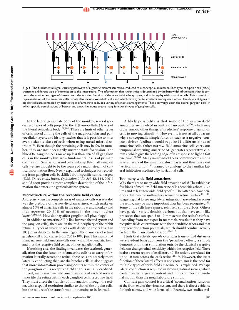
Fig. 6. The fundamental signal-carrying pathways of a generic mammalian retina, reduced to a conceptual minimum. Each type of bipolar cell (black)transmits a different type of information to the inner retina. The information that it transmits is determined by the bandwidth of the cones that it con-tacts, the number and type of those cones, the transfer function of the cone to bipolar synapse, and its interplay with amacrine cells. This is a minimalrepresentation of the amacrine cells, which also include wide-field cells and which have synaptic contacts among each other. The different types ofbipolar cells are contacted by distinct types of amacrine cells, in a variety of synaptic arrangements. These converge upon the retinal ganglion cells, inwhich specific combinations of bipolar and amacrine inputs create many functional types of ganglion cells.
©20
01 N
atu
re P
ub
lish
ing
Gro
up
h
ttp
://n
euro
sci.n
atu
re.c
om
© 2001 Nature Publishing Group http://neurosci.nature.com
884 nature neuroscience • volume 4 no 9 • september 2001
uated temporal contrast adaptation using reverse correlationand flickering checkerboards. They produced evidence for botha mechanism that works on a large spatial scale118 and one thatis extremely local—operating on a scale, in the rabbit, ofapproximately 100 µm, a fraction of the size of the receptivefield center for many ganglion cells119. There is some evidencethat the rate of adaptation is different for different-sized stim-uli. This suggests the existence of multiple, independent formsof contrast adaptation. One form of temporal contrast adapta-tion seems to operate entirely within the bipolar cells them-selves, because it persists in the presence of pharmacologicalagents that should block amacrine cell function. For larger stim-uli, the array of amacrine cells may contain several mechanismsby which the responsiveness of the retina is tuned to the char-acteristics of the visual environment.
What are the fundamental channels of vision?A final question concerns events at the heart of the retina’s design.What are the separate filters represented by the different types ofbipolar cells, and how are they reflected in the information trans-mitted centrally?
The diffuse bipolar cells represent as-yet-undeciphered par-allel channels by which the retina parses the visual input (Fig.6). In some cases, the operation performed by bipolar cells isobvious. The blue bipolar cell acts as a spectral filter tuned towavelengths peaking at about 420 nm, and to moderate spatialfrequencies. The red and green midget bipolars of primates aretuned to their particular wavelengths and to higher spatial fre-quencies. Roughly half the diffuse bipolar cells carry out a signinversion creating the ON and OFF classes of response. Withineach broad class (ON or OFF) of diffuse bipolars, though, thereare at least four specific subtypes of bipolar cells of uncertaintuning. We learn their approximate spatial tuning from theirdendritic spread, but we have only hints from their neurotrans-mitter receptors and channels about their dynamic properties.
From early studies in cold-blooded vertebrates28,29,120, andmore recent studies in mammals, bipolar cells were found to comein sustained (low-pass) and transient (high-pass) varieties. Resultsfrom salamander retina30,121 point to even greater diversity, andthis is also clear in the existing recordings from cone bipolar cellsof mammals25,31,122. Although these experiments are technicallydifficult, a critically important challenge to physiologists is to pre-cisely characterize the behavior of each channel.
Another challenge is to learn how the bipolar channels arerecombined at the level of the ganglion cell (Figs. 4 and 6). Here,modeling techniques may be useful. The central problem is tounderstand, especially in the temporal domain, how the finalresponse of a ganglion cells is created from one or several bipolarcell inputs123. It is unlikely that anyone will soon record simul-taneously from one ganglion cell and two bipolar cells; modelsor simulations may clarify our thinking in this realm.
A higher-order question is how the parallel channels createdby bipolar cells are reflected in the central visual system. The firstlimiting event for scotopic vision is the capture of photons by thecone mosaic. Even though cones’ output is much transformedlater—within the retina and higher in the visual system—vision’soverall sensitivity, chromatic selectivity and resolution dependexactly on the number and spacing of the different types ofcones43,124. The second limiting event in vision is the transmis-sion of signals from the cones to the inner retina by the bipolarcells. Bipolar cells are the mandatory link between cones (or rods)and the rest of the visual system—all visual information mustflow through them. Even though these signals, too, are later
shaped and recombined, it is inescapable that the separate chan-nels inherent in bipolar cell diversity represent fundamentals ofvision, basic building blocks from which all further codings areconstructed. In principle, we should eventually be able to decon-volve the outputs of individual bipolar channels from signalsencountered even deep within the central visual system.
ACKNOWLEDGEMENTSR. Rockhill made the illustrations. B. Boycott and P. Sterling made comments on
the manuscript. The author is a Senior Investigator of Research to Prevent
Blindness.
RECEIVED 15 JUNE 2000; ACCEPTED 19 JULY 2001
1. Vaney, D. I. A quantitative comparison between the ganglion cell populationsand axonal outflows of the visual streak and periphery of the rabbit retina. J. Comp. Neurol. 189, 215–233 (1980).
2. Martin, P. R. & Grünert, U. Spatial density and immunoreactivity of bipolarcells in the macaque monkey retina. J. Comp. Neurol. 323, 269–287 (1992).
3. MacNeil, M. A. & Masland, R. H. Extreme diversity among amacrine cells:implications for function. Neuron 20, 971–982 (1998).
4. MacNeil, M. A., Heussy, J. K., Dacheux, R., Raviola, E. & Masland, R. H. Theshapes and numbers of amacrine cells: matching of photofilled with Golgi-stained cells in the rabbit retina and comparison with other mammalianspecies. J. Comp. Neurol. 413, 305–326 (1999).
5. Masland, R. H. Neuronal diversity in the retina. Curr. Opin. Neurobiol. 11431–436 (2001).
6. Vaney, D. I. in Progress in Retinal Research (eds. Osborne, N. & Chader, G.)49–100 (Pergamon, New York, 1990).
7. Wässle, H. & Boycott, B. B. Functional architecture of the mammalian retina.Physiol. Rev. 71, 447–480 (1991).
8. Sterling, P. in The Synaptic Organization of the Brain Vol. 4 (ed. Shepherd, G. M.), 205–253 (Oxford Univ. Press, New York, 1998).
9. Rodieck, R.W. The First Steps in Seeing (Sinauer Associates, Sunderland,Massachusetts, 1998).
10. Nathans, J. The evolution and physiology of human color vision: insights frommolecular genetic studies of visual pigments. Neuron 24, 299–312 (1999).
11. Boycott, B. B. & Wässle, H. Parallel processing in the mammalian retina: TheProctor Lecture. Invest. Ophthalmol. Vis. Sci. 40, 1313–1327 (1999).
12. Okano, T., Kojima, D., Fukada, Y. & Shichida, Y. Primary structures ofchicken cone visual pigments: vertebrate rhodopsins have evolved out of conevisual pigments. Proc. Natl. Acad. Sci. USA 89, 5932–5936 (1992).
13. Johnson, R. L. et al. Cloning and expression of goldfish opsin sequences.Biochemistry 32, 208–214 (1993).
14. Jacobs, G. H. The distribution and nature of colour vision among themammals. Biol. Rev. 68, 413–471 (1993).
15. Westheimer, G. The maxwellian view. Vision Res. 6, 669–682 (1966).16. Cohen, E. & Sterling, P. Demonstration of cell types among cone bipolar
neurons of the cat retina. Phil. Trans. R. Soc. Lond. B Biol. Sci. 330, 305–321(1990).
17. Boycott, B. B. & Wässle, H. Morphological classification of bipolar cells of theprimate retina. Eur. J. Neurosci. 3, 1069–1088 (1991).
18. Cajal, S. R. The Structure of the Retina (Thomas, Springfield, 1972).19. McGuire, B. A., Stevens, J. K. & Sterling, P. Microcircuitry of bipolar cells in
rat retina. J. Neurosci. 12, 2920–2938 (1984).20. Grünert, U., Martin, P. R. & Wässle, H. Immunocytochemical analysis of
bipolar cells in the macaque monkey retina. J. Comp. Neurol. 348, 607–627(1994).
21. Hartveit, E. et al. Localization and developmental expression of the NMDAreceptor subunit NR2A in the mammalian retina. J. Comp. Neurol. 348,570–582 (1994).
22. Haverkamp, S. & Wässle, H. Immunocytochemical analysis of the mouseretina. J. Comp. Neurol. 424, 1–23 (2000).
23. Greferath, U. et al. GABAA receptor subunits have differential distributions inthe rat retina: in situ hybridization and immunohistochemistry. J. Comp.Neurol. 353, 553–571 (1995).
24. Euler, T. & Wässle, H. Different contributions of GABAA and GABACreceptors to rod and cone bipolar cells in a rat retinal slice preparation. J. Neurophysiol. 79, 1384–1395 (1998).
25. Euler, T. & Masland, R. H. Light-evoked responses of bipolar cells in amammalian retina. J. Neurophysiol. 83, 1817–1829 (2000).
26. Nawy, S. & Jahr, C. E. Suppression by glutamate of cGMP-activatedconductance in retinal bipolar cells. Nature 346, 269–271 (1990).
27. Nawy, S. & Jahr, C. E. cGMP-gated conductance in retinal bipolar cells issuppressed by the photoreceptor transmitter. Neuron 7, 677–683 (1991).
28. Kaneko, A. Physiological and morphological identification of horizontal,bipolar and amacrine cells in goldfish retina. J. Physiol. (Lond.) 207, 623–633(1970).
29. Werblin, F. S. & Dowling, J. E. Organization of the retina of the mudpuppy,
review©
2001
Nat
ure
Pu
blis
hin
g G
rou
p
htt
p:/
/neu
rosc
i.nat
ure
.co
m© 2001 Nature Publishing Group http://neurosci.nature.com

necturus maculosus II. Intracellular recording. J. Neurophysiol. 32, 339–355(1969).
30. Awatramani, G. B. & Slaughter, M. M. Origin of transient and sustainedresponses in ganglion cells of the retina. J. Neurosci. 20, 7087–7095 (2000).
31. DeVries, S. H. Bipolar cells use kainate and AMPA receptors to filter visualinformation into separate channels. Neuron 28, 847–856 (2000).
32. Kouyama, N. & Marshak, D. W. Bipolar cells specific for blue cones in themacaque retina. J. Neurosci. 12, 1233–1252 (1992).
33. Dacey, D. M. & Lee, B. B. The ‘blue-on’ opponent pathway in primate retinaoriginates from a distinct bistratified ganglion cell type. Nature 367, 731–735(1994).
34. Famiglietti, E. V. Jr. & Kolb, H. A bistratified amacrine cell and synaptic circuitryin the inner plexiform layer of the retina. Brain Res. 84, 293–300 (1975).
35. Smith, R. G., Freed, M. A. & Sterling, P. Microcircuitry of the dark-adaptedcat retina: functional architecture of the rod–cone network. J. Neurosci. 6,3505–3517 (1986).
36. Strettoi, E., Dacheux, R. F. & Raviola, E. Synaptic connections of rod bipolarcells in the inner plexiform layer of the rabbit retina. J. Comp. Neurol. 295,449–466 (1990).
37. Strettoi, E., Raviola, E. & Dacheux, R. F. Synaptic connections of the narrow-field, bistratified rod amacrine cell (AII) in the rabbit retina. J. Comp. Neurol.325, 152–168 (1992).
38. Strettoi, E. & Masland, R. H. The organization of the inner nuclear layer ofthe rabbit retina. J. Neurosci. 15, 875–888 (1995).
39. Euler, T. & Wässle, H. Immunocytochemical identification of cone bipolarcells in the rat retina. J. Comp. Neurol. 361, 461–478 (1995).
40. Jeon, C.-J., Strettoi, E. & Masland, R. H. The major cell populations of themouse retina. J. Neurosci. 18, 8936–8946 (1998).
41. Masland, R. H. Processing and encoding of visual information in the retina.Curr. Opin. Neurobiol. 6, 467–474 (1996).
42. Williams, R. W., Cavada, C. & Reinoso-Suarez, F. Rapid evolution of the visualsystem: a cellular assay of the retina and dorsal lateral geniculate nucleus of theSpanish wildcat and the domestic cat. J. Neurosci. 13, 208–228 (1993).
43. Williams, D. R. Seeing through the photoreceptor mosaic. Trends Neurosci. 9193–198 (1986).
44. Nathans, J., Piantanida, T. P., Eddy, R. L., Shows, T. B. & Hogness, D. S.Molecular genetics of inherited variation in human color vision. Science 232,203–210 (1986).
45. Nathans, J., Thomas, D. & Hogness, D. S. Molecular genetics of human colorvision: the genes encoding blue, green, and red pigments. Science 232,193–202 (1986).
46. Mollon, J. D. in Evolution of the Eye and Visual System (eds. Cronly-Dillon, J. R. & Gregory, R. L.) 306–319 (CRC Press, Boca Raton, Florida, 1991).
47. Dacey, D. M. Parallel pathways for spectral coding in primate retina. Annu.Rev. Neurosci. 23, 743–775 (2000).
48. Calkins, D. J. & Sterling, P. Evidence that circuits for spatial and color visionsegregate at the first retinal synapse. Neuron 24, 313–321 (1999).
49. Boycott, B. B., Hopkins, J. M. & Sperling, H. G. Cone connections of thehorizontal cells of the rhesus monkey’s retina. Proc. R. Soc. Lond. B Biol. Sci.229, 345–379 (1987).
50. Hack, I. & Peichl, L. Horizontal cells of the rabbit retina are non-selectivelyconnected to the cones. Eur. J. Neurosci. 11, 2261–2274 (1999).
51. Peichl, L., Sandmann, D. & Boycott, B. B. in Development and Organization ofthe Retina (eds. Chalupa, L. M. & Finlay, B. L.) 147–172 (Plenum, New York,1998).
52. Dacey, D. M., Lee, B. B., Stafford, D. K., Pokorny, J. & Smith, V. C. Horizontalcells of the primate retina: cone specificity without spectral opponency.Science 271, 656–658 (1996).
53. Nelson, R., Lützow, A. V., Kolb, H. & Gouras, P. Horizontal cells in cat retinawith independent dendritic systems. Science 189, 137–139 (1975).
54. Freed, M. A. & Sterling, P. The ON-alpha ganglion cell of the cat retina and itspresynaptic cell types. J. Neurosci. 8, 2303–2320 (1988).
55. Calkins, D. J., Schein, S. J., Tsukamoto, Y. & Sterling, P. M and L cones inmacaque fovea connect to midget ganglion cells by different numbers ofexcitatory synapses. Nature 371, 70–72 (1994).
56. Jacoby, R., Stafford, D., Kouyama, N. & Marshak, D. Synaptic inputs to ONparasol ganglion cells in the primate retina. J. Neurosci. 16, 8041–8056 (1996).
57. Hubel, D. H. Eye, Brain and Vision (Freeman, New York, 1988).58. Nolte, J. The Human Brain (Mosby-Year Book, St. Louis, Missouri, 1988).59. Vaney, D. I. Patterns of neuronal coupling in the retina. Prog. Ret. Eye Res. 13,
301–355 (1994).60. Meister, M., Lagnado, L. & Baylor, D. A. Concerted signaling by retinal
ganglion cells. Science 270, 1207–1210 (1995).61. Meister, M. Multineuronal codes in retinal signaling. Proc. Natl. Acad. Sci.
USA 93, 609–614 (1996).62. Ehinger, B. Functional role of dopamine in the retina. Prog. Ret. Res. 2,
213–232 (1983).63. Dowling, J. E. Dopamine: a retinal neuromodulator? Trends Neurosci. 9,
236–240 (1986).64. Witkovsky, P. & Dearry, A. Functional roles of dopamine in the vertebrate
retina. Prog. Ret. Res. 11, 247–292 (1991).65. Tauchi, M., Madigan, N. M. & Masland, R. H. Shapes and distributions of the
catecholamine-accumulating neurons in the rabbit retina. J. Comp. Neurol.293, 178–189 (1990).
66. Hampson, E. C. G. M., Vaney, D. I. & Weiler, R. Dopaminergic modulation of
gap junction permeability between amacrine cells in mammalian retina. J. Neurosci. 12, 4911–4922 (1992).
67. Piccolino, M., Witkovsky, P. & Trimarchi, C. Dopaminergic mechanismsunderlying the reduction of electrical coupling between horizontal cells of theturtle retina induced by D-amphetamine, bicuculline, and veratridine. J. Neurosci. 7, 2273–2284 (1987).
68. Jensen, R. J. & Daw, N. W. Effects of dopamine antagonists on receptive fieldsof brisk cells and directionally selective cells in the rabbit retina. J. Neurosci. 4,2972–2985 (1984).
69. Jensen, R. J. & Daw, N. W. Effects of dopamine and its agonists andantagonists on the receptive field properties of ganglion cells in the rabbitretina. Neuroscience 17, 837–855 (1986).
70. Bruenner, U. & Burnside, B. Pigment granule migration in isolated cells ofthe teleost retinal pigment epithelium. Invest. Ophthalmol. Vis. Sci. 27,1634–1643 (1986).
71. Gustincich, S., Feigenspan, A., Wu, D. K., Koopman, L. J. & Raviola, E.Control of dopamine release in the retina: a transgenic approach to neuralnetworks. Neuron 18, 723–736 (1997).
72. Puopolo, M., Hochstetler, S. E., Gustincich, S., Wightman, R. M. & Raviola,E. Extrasynaptic release of dopamine in a retinal neuron: activity dependenceand transmitter modulation. Neuron 30, 211–225 (2001).
73. O’Malley, D. M., Sandell, J. H. & Masland, R. H. Co-release of acetylcholineand GABA by the starburst amacrine cells. J. Neurosci. 12, 1394–1408 (1992).
74. Masland, R. H. & Tauchi, M. The cholinergic amacrine cell. Trends Neurosci.9, 218–223 (1986).
75. He, S.-G. & Masland, R. H. Retinal direction selectivity after targeted laserablation of starburst amacrine cells. Nature 389, 378–382 (1997).
76. Yoshida, K. et al. A key role of starburst amacrine cells in originating retinaldirectional selectivity and optokinetic eye movement. Neuron 30, 771–780(2001).
77. Berson, D. M., Isayama, T. & Pu, M. The eta ganglion cell type of the catretina. J. Comp. Neurol. 408, 204–219 (1999).
78. Berson, D. M., Pu, M. & Famiglietti, E. V. The zeta cell: a new ganglion celltype in cat retina. J. Comp. Neurol. 399, 269–288 (1998).
79. Isayama, T., Berson, D. M. & Pu, M. Theta ganglion cell type of the cat retina.J. Comp. Neurol. 417, 32–48 (2000).
80. Pu, M., Berson, D. M. & Pan, T. Structure and function of retinal ganglioncells innervating the cat’s geniculate wing: an in vitro study. J. Neurosci. 14,4338–4358 (1994).
81. Barlow, H. B. & Levick, W. R. The mechanism of directionally selective unitsin rabbit’s retina. J. Physiol. (Lond.) 178, 477–504 (1965).
82. Levick, W. R. Receptive fields and trigger features of ganglion cells in thevisual streak of the rabbit’s retina. J. Physiol. (Lond.) 188, 285–307 (1967).
83. Oyster, C. W., Takahashi, E. & Collewijn, H. Direction selective retinalganglion cells and control of optokinetic nystagmus in the rabbit. Vision Res.12, 183–193 (1972).
84. Simpson, J. I. The accessory optic system. Annu. Rev. Neurosci. 7, 13–41(1984).
85. Oyster, C. W., Simpson, J. I., Takahashi, E. S. & Soodak, R. E. Retinal ganglioncells projecting to the rabbit accessory optic system. J. Comp. Neurol. 190,49–61 (1980).
86. Provencio, I. et al. A novel human opsin in the inner retina. J. Neurosci. 20,600–605 (2000).
87. Pasik, P. & Pasik, T. in The Oculomotor System (ed. Bender, M. B.) 40–80(Harper & Row, New York, 1964).
88. Tauchi, M. & Masland, R. H. Local order among the dendrites of an amacrinecell population. J. Neurosci. 5, 2494–2501 (1985).
89. Yang, G. & Masland, R. H. Direct visualization of the dendritic and receptivefields of directionally selective retinal ganglion cells. Science 258, 1949–1952(1992).
90. Rodieck, R. W. & Watanabe, M. Survey of the morphology of macaque retinalganglion cells that project to the pretectum, superior colliculus and theparvicellular laminae of the lateral geniculate nucleus. J. Comp. Neurol. 338,289–303 (1993).
91. Dacey, D. M. Morphology of a small-field bistratified ganglion cell type in themacaque and human retina. Vis. Neurosci. 10, 1081–1098 (1993).
92. Dacey, D. M. Physiology, morphology and spatial densities of identifiedganglion cell types in the primate retina. Ciba Found. Symp. 184, 12–34(1994).
93. Peterson, B. B. & Dacey, D. M. Morphology of wide-field, monostratifiedganglion cells of the human retina. Vis. Neurosci. 16, 107–120 (1999).
94. Calkins, D. J., Tsukamoto, Y. & Sterling, P. Microcircuitry and mosaic of ablue-yellow ganglion cell in the primate retina. J. Neurosci. 18, 3373–3385(1998).
95. Wiesel, T. N. & Hubel, D. H. Spatial and chromatic interactions in the lateralgeniculate body of the rhesus monkey. J. Neurophysiol. 29, 1115–1156 (1966).
96. Curcio, C. A., Sloan, K. R., Kalina, R. E. & Hendrickson, A. E. Humanphotoreceptor topography. J. Comp. Neurol. 292, 497–523 (1990).
97. Buhl, E. H. & Peichl, L. Morphology of rabbit retinal ganglion cells projectingto the medial terminal nucleus of the accessory optic system. J. Comp. Neurol.253, 163–174 (1986).
98. Ts’o, D. Y. & Gilbert, C. D. The organization of chromatic and spatialinteractions in the primate striate cortex. J. Neurosci. 8, 1712–1727 (1988).
99. Hubel, D. & Livingstone, M. Color puzzles. Cold Spring Harb. Symp. Quant.Biol. 55, 643–649 (1990).
review
nature neuroscience • volume 4 no 9 • september 2001 885
©20
01 N
atu
re P
ub
lish
ing
Gro
up
h
ttp
://n
euro
sci.n
atu
re.c
om
© 2001 Nature Publishing Group http://neurosci.nature.com
886 nature neuroscience • volume 4 no 9 • september 2001
100. Conway, B. R. Spatial structure of cone inputs to color cells in alertmacaque primary visual cortex (V1). J. Neurosci. 21, 2768–2783 (2001).
101. Hendry, S. H. & Yoshioka, T. A neurochemically distinct third channel inthe macaque dorsal lateral geniculate nucleus. Science 264, 575–577(1994).
102. Hendry, S. H. C. & Reid, R. C. The koniocellular pathway in primate vision.Annu. Rev. Neurosci. 23, 127–153 (2000).
103. Saul, A. B. & Humphrey, A. L. Spatial and temporal response properties oflagged and nonlagged cells in cat lateral geniculate nucleus. J. Neurophysiol.64, 206–224 (1990).
104. Menger, N., Pow, D. V. & Wässle, H. Glycinergic amacrine cells of the ratretina. J. Comp. Neurol. 401, 34–46 (1998).
105. Kalloniatis, M., Marc, R. E. & Murry, R. F. Amino acid signatures in theprimate retina. J. Neurosci. 16, 6807–6829 (1996).
106. Shapley, R. & Enroth-Cugell, C. Visual adaptation and retinal gain controls.Prog. Ret. Res. 3, 263–346 (1984).
107. Berry, M. J., Brivanlou, I. H., Jordan, T. A. & Meister, M. Anticipation ofmoving stimuli by the retina. Nature 398, 334–338 (1999).
108. Nelson, R. AII amacrine cells quicken time course of rod signals in the catretina. J. Neurophysiol. 47, 928–947 (1982).
109. Boos, R., Schneider, H. & Wässle, H. Voltage- and transmitter-gatedcurrents of AII-amacrine cells in a slice preparation of the rat retina. J. Neurosci. 13, 2874–2888 (1993).
110. Roska, B. & Werblin, F. Vertical interactions across ten parallel, stackedrepresentations in the mammalian retina. Nature 410, 583–587 (2001).
111. Famiglietti, E. V. Polyaxonal amacrine cells of rabbit retina: size anddistribution of PA1 cells. J. Comp. Neurol. 316, 406–421 (1992).
112. Dacey, D. M. Axon-bearing amacrine cells of the macaque monkey retina. J. Comp. Neurol. 284, 275–293 (1989).
113. Cook, P. B. & Werblin, F. S. Action potentials are propagated by wide-fieldamacrine cells in the tiger salamander retina. J. Neurosci. 14, 3852–3861(1994).
114. Stafford, D. K. & Dacey, D. M. Physiology of the A1 amacrine: a spiking,axon-bearing interneuron of the macaque monkey retina. Vis. Neurosci. 14,507–522 (1997).
115. Taylor, W. R. Response properties of long-range axon-bearing amacrinecells in the dark-adapted rabbit retina. Vis. Neurosci. 13, 599–604 (1996).
116. McIlwain, J. B. Receptive fields of optic tract axons and lateral geniculatecells: peripheral extent and barbituate sensitivity. J. Neurophysiol. 27,1154–1173 (1964).
117. Neuenschwander, S. & Singer, W. Long-range synchronization ofoscillatory light responses in the cat retina and lateral geniculate nucleus.Nature 379, 728–733 (1996).
118. Smirnakis, S. M., Berry, M. J., Warland, D. K., Bialek, W. & Meister, M.Adaptation of retinal processing to image contrast and spatial scale. Nature386, 69–73 (1997).
119. Brown, S. P. & Masland, R. H. Spatial scale and cellular substrate of contrastadaptation in retinal ganglion cell. Nat. Neurosci. 4, 44–51 (2001).
120. Dowling, J. E. The Retina: An Approachable Part of the Brain (Belknap,Cambridge, 1987).
121. Wu, S., Gao, F. & Maple, B. R. Functional architecture of synapses in theinner retina: segregation of visual signals by stratification of bipolar cellaxon terminals. J. Neurosci. 20, 4462–4470 (2000).
122. McBillem, G. S. & Dacheux, R. F. Responses to glutamate ofmorphologically defined rabbit cone bipolar cells. Invest. Ophthalmol. Vis.Sci. (2001).
123. Freed, M. A. Parallel cone bipolar pathways to a ganglion cell use differentrates and amplitudes of quantal excitation. J. Neurosci. 20, 3956–3963(2000).
124. Packer, O. S., Williams, D. R. & Bensinger, D. G. Photopigmenttransmittance imaging of the primate photoreceptor mosaic. J. Neurosci.16, 2251–2260 (1996).
125. Vaney, D. I. Neuronal coupling in rod-signal pathways of the retina. Invest.Ophthalmol. Vis. Sci. 38, 267–273 (1997).
review©
2001
Nat
ure
Pu
blis
hin
g G
rou
p
htt
p:/
/neu
rosc
i.nat
ure
.co
m© 2001 Nature Publishing Group http://neurosci.nature.com