The Fibroblast Growth Factor Family: Neuromodulation of Affective Behavior
15
Neuron Review The Fibroblast Growth Factor Family: Neuromodulation of Affective Behavior Cortney A. Turner, 1, * Stanley J. Watson, 1,2 and Huda Akil 1,2 1 Molecular and Behavioral Neuroscience Institute 2 Department of Psychiatry University of Michigan, 205 Zina Pitcher Place, Ann Arbor, MI 48109, USA *Correspondence: [email protected] http://dx.doi.org/10.1016/j.neuron.2012.08.037 In this review, we propose a broader view of the role of the fibroblast growth factor (FGF) family in modulating brain function. We suggest that some of the FGF ligands together with the FGF receptors are altered in indi- viduals with affective disorder and modulate emotionality in animal models. Thus, we propose that members of the FGF family may be genetic predisposing factors for anxiety, depression, or substance abuse; that they play a key organizing role during early development but continue to play a central role in neuroplasticity in adulthood; and that they work not only over extended time frames, but also via rapid signaling mechanisms, allowing them to exert an ‘‘on-line’’ influence on behavior. Therefore, the FGF family appears to be a prototype of ‘‘switch genes’’ that are endowed with organizational and modulatory properties across the lifespan, and that may represent molecular candidates as biomarkers and treatment targets for affective and addictive disorders. The Role of Growth Factors in Emotionality Our understanding of the role of growth factors has evolved significantly over the last quarter century, with increasing appre- ciation of their pivotal roles in brain function and dysfunction across the life span. Early views emphasized the central role of molecules such as nerve growth factor (NGF) in development, survival, and differentiation particularly in embryonic sensory and sympathetic neurons (Levi-Montalcini, 1987). Even following the discovery of several neurotrophins, including brain-derived neurotrophic factor (BDNF) and the emerging recognition of their coordinate actions as trophic factors in the central nervous system (CNS), much of the emphasis remained on under- standing their role in development. For example, a 1993 review concludes that: ‘‘In the adult, the roles of the same trophic factors are likely to be more restricted, either activated only in specific neuronal populations or, alternatively, only during very specific physiological states of the nervous tissue’’ (Knu ¨ sel and Hefti, 1993). Nevertheless, that era saw an increasing interest in the ability of neurotrophins to promote cell survival and repair following injury or neurodegeneration, and they were proposed as potential therapeutic targets for neurodegenerative disorders (Snider and Johnson, 1989; Thoenen, 1991). By the mid 1990s, additional roles of growth factors in neural function were emerging. For example, NGF was implicated in pain regulation and neuroimmune function (Levi-Montalcini et al., 1995), while neurotrophins were shown to play a role in synapse formation and neuroplasticity (Lu and Figurov, 1997). With the realization that severe and chronic stress can produce significant damage to certain areas of the CNS, such as the hippocampus (Fuchs and Flu ¨ gge, 1998; Magarin ˜ os et al., 1997; McEwen and Magarinos, 1997), the potential role of growth factors in counteracting the effects of stress came into focus. In 1997, it was shown that chronic stress decreases BDNF in conjunction with atrophy of hippocampal neurons (Duman et al., 1997). Given that chronic stress has served as an animal model of clinical depression, the authors suggested that the mode of action of chronic antidepressant therapy might involve activation of neurotrophic factors (Duman et al., 1997; Duman, 1998). This framework represented the first explicit implication of growth factors in a hypothesis related to a psychiatric disorder. As is the case for other growth factors, our views of the functions of the fibroblast growth factor (FGF) family in the brain originally revolved primarily around neural development (Go ´ mez-Pinilla et al., 1994; Riedel et al., 1995; Temple and Qian, 1995; Vaccarino et al., 1999). Subsequent observations implicated the FGF family in neurogenesis both during early development and in adulthood (Bartlett et al., 1994; Cheng et al., 2001; Guillemot and Zimmer, 2011; Tao et al., 1996; Zheng et al., 2004). This paved the way to a greater interest in this family’s role in neuroplasticity. In this review, we suggest that the FGF family plays a lifelong neuromodulatory role in the way an organism responds to and copes with the environment. We propose that the fine-tuning of this family of molecules alters the organism’s propensity to explore a novel environment and modifies anxiety-like and depression-like behavior. Moreover, the FGF system is involved in fear conditioning and the response to stress and plays a role in the vulnerability to drug-taking behavior. Why Link the FGF System to Mood and Affect? Our view on the affective role of the FGF family emerged from studies of postmortem brains of subjects who had died while suffering from severe clinical depression. Major depressive disorder (MDD) is the most debilitating mood disorder in the United States, accounting for the single greatest psychiatric cause of disability. Anxiety disorders run a close second, and these two affective diseases are often comorbid. Thus, relative 160 Neuron 76, October 4, 2012 ª2012 Elsevier Inc.
Transcript of The Fibroblast Growth Factor Family: Neuromodulation of Affective Behavior

Neuron
Review
The Fibroblast Growth Factor Family:Neuromodulation of Affective Behavior
Cortney A. Turner,1,* Stanley J. Watson,1,2 and Huda Akil1,21Molecular and Behavioral Neuroscience Institute2Department of PsychiatryUniversity of Michigan, 205 Zina Pitcher Place, Ann Arbor, MI 48109, USA*Correspondence: [email protected]://dx.doi.org/10.1016/j.neuron.2012.08.037
In this review, we propose a broader view of the role of the fibroblast growth factor (FGF) family in modulatingbrain function. We suggest that some of the FGF ligands together with the FGF receptors are altered in indi-viduals with affective disorder and modulate emotionality in animal models. Thus, we propose that membersof the FGF family may be genetic predisposing factors for anxiety, depression, or substance abuse; that theyplay a key organizing role during early development but continue to play a central role in neuroplasticity inadulthood; and that they work not only over extended time frames, but also via rapid signaling mechanisms,allowing them to exert an ‘‘on-line’’ influence on behavior. Therefore, the FGF family appears to be a prototypeof ‘‘switch genes’’ that are endowed with organizational and modulatory properties across the lifespan, andthat may represent molecular candidates as biomarkers and treatment targets for affective and addictivedisorders.
The Role of Growth Factors in EmotionalityOur understanding of the role of growth factors has evolved
significantly over the last quarter century, with increasing appre-
ciation of their pivotal roles in brain function and dysfunction
across the life span. Early views emphasized the central role of
molecules such as nerve growth factor (NGF) in development,
survival, and differentiation particularly in embryonic sensory
and sympathetic neurons (Levi-Montalcini, 1987). Even following
the discovery of several neurotrophins, including brain-derived
neurotrophic factor (BDNF) and the emerging recognition of
their coordinate actions as trophic factors in the central nervous
system (CNS), much of the emphasis remained on under-
standing their role in development. For example, a 1993 review
concludes that: ‘‘In the adult, the roles of the same trophic
factors are likely to be more restricted, either activated only in
specific neuronal populations or, alternatively, only during very
specific physiological states of the nervous tissue’’ (Knusel and
Hefti, 1993). Nevertheless, that era saw an increasing interest
in the ability of neurotrophins to promote cell survival and repair
following injury or neurodegeneration, and they were proposed
as potential therapeutic targets for neurodegenerative disorders
(Snider and Johnson, 1989; Thoenen, 1991).
By the mid 1990s, additional roles of growth factors in neural
function were emerging. For example, NGF was implicated in
pain regulation and neuroimmune function (Levi-Montalcini
et al., 1995), while neurotrophins were shown to play a role in
synapse formation and neuroplasticity (Lu and Figurov, 1997).
With the realization that severe and chronic stress can produce
significant damage to certain areas of the CNS, such as the
hippocampus (Fuchs and Flugge, 1998; Magarinos et al., 1997;
McEwen and Magarinos, 1997), the potential role of growth
factors in counteracting the effects of stress came into focus.
In 1997, it was shown that chronic stress decreases BDNF in
conjunction with atrophy of hippocampal neurons (Duman
160 Neuron 76, October 4, 2012 ª2012 Elsevier Inc.
et al., 1997). Given that chronic stress has served as an animal
model of clinical depression, the authors suggested that the
mode of action of chronic antidepressant therapy might involve
activation of neurotrophic factors (Duman et al., 1997; Duman,
1998). This framework represented the first explicit implication
of growth factors in a hypothesis related to a psychiatric
disorder.
As is the case for other growth factors, our views of the
functions of the fibroblast growth factor (FGF) family in the
brain originally revolved primarily around neural development
(Gomez-Pinilla et al., 1994; Riedel et al., 1995; Temple and
Qian, 1995; Vaccarino et al., 1999). Subsequent observations
implicated the FGF family in neurogenesis both during early
development and in adulthood (Bartlett et al., 1994; Cheng
et al., 2001; Guillemot and Zimmer, 2011; Tao et al., 1996; Zheng
et al., 2004). This paved the way to a greater interest in this
family’s role in neuroplasticity.
In this review, we suggest that the FGF family plays a lifelong
neuromodulatory role in the way an organism responds to and
copes with the environment. We propose that the fine-tuning
of this family of molecules alters the organism’s propensity to
explore a novel environment and modifies anxiety-like and
depression-like behavior. Moreover, the FGF system is involved
in fear conditioning and the response to stress and plays a role in
the vulnerability to drug-taking behavior.
Why Link the FGF System to Mood and Affect?Our view on the affective role of the FGF family emerged from
studies of postmortem brains of subjects who had died while
suffering from severe clinical depression. Major depressive
disorder (MDD) is the most debilitating mood disorder in the
United States, accounting for the single greatest psychiatric
cause of disability. Anxiety disorders run a close second, and
these two affective diseases are often comorbid. Thus, relative

Neuron
Review
to the general population, an individual who has one of these
disorders has a 25-fold-greater chance of expressing the other
(Kessler et al., 1994), suggesting highly overlapping, if not
common, etiology. A better understanding of the pathophysi-
ology of these diseases is acutely needed given the high rate
of incidence of these diseases (e.g., 25% lifetime incidence of
MDD), and only a 33% response rate to first of the line treatments
(Robins and Regier, 1991).
In 2004, work in the context of the Pritzker Neuropsychiatric
Disorders Research Consortium (http://www.
pritzkerneuropsych.org/) examined alterations in genome-wide
expression profiles in the brains of patients suffering from MDD
relative to normal controls (Evans et al., 2004). This ‘‘discovery’’
approach first focused on areas in the frontal cortex. Data mining
revealed thatmembers of the FGF family were highly significantly
altered in major depression. Moreover, this effect was not
dependent on treatment with the selective-serotonin reuptake
inhibitors (SSRIs). Indeed, a history of SSRI treatment blunted
the dysregulation in FGF gene expression. In that original paper,
FGF1, FGF2, FGFR2, and FGFR3were downregulated in MDD in
the anterior cingulate cortex and/or the dorsolateral prefrontal
cortex. Conversely, FGF9 and FGF12 were upregulated in these
same brain regions. As will be described below, these findings
have since been extended to other brain regions using multiple
analysis platforms, and have led to a series of studies in animal
models that have transformed our understanding of the role of
the FGF family in brain function and dysfunction.
In this review, we will focus primarily on the more recent
evidence relating to the FGF system, emotionality and mood
disorders. We will attempt to answer three main questions
regarding FGF signaling and behavior: (1) What is known about
the FGF system in mood disorders? (2) What are the effects of
the FGF system on other affective behaviors including anxiety,
fear, stress responsivity and substance abuse? and, (3) how
might the FGF system exert these effects? To this end, we will
describe the important ligands and receptors for the FGF family.
We will review the various functions of the FGF system with
a focus on FGF2, the prototypical ligand. We will end with
a discussion of other molecular partners of this system that
suggest pharmacological and clinical strategies with molecules
that are not ‘‘the usual suspects.’’
Molecular Components of the FGF FamilyOverview of the FGF Family
For a review of the literature on the structure and function of the
FGF system prior to 2006, the reader is referred to a previous
review (Turner et al., 2006). To summarize, the FGF system is
comprised of 18 ligands, of which ten are expressed in brain.
There were four previous members, now termed FGF homolo-
gous factors (FHF1-4), that have been removed from the original
list of 22 ligands (Goldfarb et al., 2007). These molecules lack
functional similarity, although they share structural similarity
and remain intracellular.
There are four membrane-bound receptors and a fifth trun-
cated (soluble) receptor with differing affinities for the various
ligands (Reuss and von Bohlen und Halbach, 2003). Many of
the ligands lack signal peptides but are secreted nonetheless.
The receptors are composed of three extracellular Ig-like
domains, a transmembrane domain, and two intracellular kinase
domains (Reuss and von Bohlen und Halbach, 2003). The acid
box region between the first and second Ig-like domain deter-
mines the ligand specificity. There are also multiple splice vari-
ants of the third Ig-like domain resulting in IIIb or IIIc isoforms.
The IIIb isoform is expressed predominantly during early devel-
opment, while the IIIc isoform is expressed predominantly in
adulthood.
The receptors signal primarily through three main pathways,
phospholipase Cg (PLCg), mitogen-activated protein kinase
(MAPK), and AKT to influence gene transcription. This signaling
is akin to other growth factors; however, the strength of the
signaling may vary between growth factors. This is possible,
by analogy, since the strength of the signaling can vary between
FGF receptor homodimers. For example, FGF ligands in different
subfamilies can induce different FGF receptor 1 (FGFR1) homo-
dimer formations andMAPK signaling (Romero-Fernandez et al.,
2011). Moreover, there may be differences in kinase activity de-
pending on which molecule triggered the signal (Ditlevsen et al.,
2008). Finally, the receptors can interact with other neurotrans-
mitter receptors, as will be described in more detail below (see
Beyond the FGFs: Receptor-Interacting Partners).
FGF Ligands
Each of the FGF ligands has a distinct functional profile. We will
focus here on a subset of ligands that are expressed in brain and
appear modulated in mood disorders.
FGF2, also known as basic fibroblast growth factor, was the
first FGF to be cloned in the rat (Kurokawa et al., 1988). It is
the prototypical FGF ligand and has been well-characterized
for its roles in cell proliferation, differentiation, growth, survival,
as well as angiogenesis in various cell models (Ford-Perriss
et al., 2001). This ligand is composed of a b trefoil motif and
has a basic canyon structure allowing heparin sulfate proteogly-
cans to bind in a 2:2:2 stoichiometry with the receptors (Reuss
and von Bohlen und Halbach, 2003). FGF2 exists in multiple
molecular weight isoforms of which only the lowest molecular
weight (18 kDa) is secreted. The higher molecular weight iso-
forms remain in the nucleus and affect nuclear functioning,
such as rRNA transcription.
In early brain development, FGF2 is expressed by the neural
tube and is involved in neural induction (Ford-Perriss et al.,
2001). Later on, FGF2 is expressed in the ventricular region of
the developing cortex and the cortical plate. FGF2 is also ex-
pressed by neural precursor cells throughout development and
promotes the proliferation of neural stem cells (Dono et al.,
1998; Vaccarino et al., 1999). Thus, FGF2 knockout mice have
alterations in the deep layers of the cortex and the hippocampus
compared to wild-type mice (Raballo et al., 2000). These mice
also have a lower number of neural progenitor cells in the sub-
ventricular zone (SVZ), a decreased ability to proliferate in
response to FGF2 in progenitor cells of the subgranular zone
(SGZ) and decreased astroglial differentiation (Zheng et al.,
2004). Together, the results suggest that FGF2 affects both
neuronal and glial output. However, astrocytic expression of
FGF2 only becomes apparent starting at postnatal day (PND)
4–6 (Gomez-Pinilla et al., 1994).
In the adult brain, FGF2 is expressed by both neurons and glial
cells with astrocytes containing the highest levels of FGF2
Neuron 76, October 4, 2012 ª2012 Elsevier Inc. 161
Neuron
Review
(Gonzalez et al., 1995). FGF2 binds with the highest affinity to
FGFR1 (Reuss and von Bohlen und Halbach, 2003). Moreover,
FGF2 is ubiquitously expressed in the adult brain with the highest
expression in the hippocampus and cortical areas (Gomez-
Pinilla et al., 1994).
The regulation of FGF2 expression is complex. An antisense
transcript regulates its expression (Nudt6), functioning as
a repressor (Knee et al., 1997; MacFarlane et al., 2010; MacFar-
lane and Murphy, 2010). There are also various transcription
factors that can bind its promoter elements, such as HoxA10,
AP-1, and SP-1 (Shah et al., 2012; Shibata et al., 1991). More-
over, the role of FGF2 in brain development is influenced by
the existence of an IRES-dependent mechanism for translation
(Audigier et al., 2008). This activity peaks at PND7, remains
elevated in neurons during adulthood, and is regulated by itself
and by electrical activity. Other mechanisms of regulation of
FGF2 expression in the developing brain, be they by epigenetic
or microRNA mechanisms, remain to be elucidated. The effects
of FGF2 on the adult brain will be discussed below.
FGF1, also known as acidic fibroblast growth factor, was
cloned in the rat subsequent to FGF2 (Goodrich et al., 1989).
FGF1 is predominantly expressed by neurons and, in stark
contrast to FGF2, it is expressed relatively little outside of the
nervous system. FGF1 is expressed at low concentrations until
E16 when it rises to adult levels (Alam et al., 1996; Elde et al.,
1991) Culture experiments demonstrated that FGF1 is involved
in the maturation and maintenance of neurons (Ford-Perriss
et al., 2001). However, FGF1 knockout mice show no severe
deficits (Miller et al., 2000). Finally, not much is known about
the effects of FGF1 on the adult brain.
FGF9 is a mitogenic factor expressed predominantly by
neurons with high expression in hippocampal and cortical areas.
FGF9 also has the highest affinity for the astrocytic receptor,
FGFR3, specifically the adult IIIc splice variant (Cinaroglu et al.,
2005; Plotnikov et al., 2001). Given the alterations described
above in the human postmortem cortex, the role of FGF9 is of
great interest. Unfortunately, not much is known about the in vivo
effects of FGF9 in general.
Intracellular fibroblast growth factors (iFGF), also known as
FGF homologous factors, may also play a role in emotionality.
These molecules share structural, but not functional homology
with other FGFs that interact with the extracellular receptors
(Olsen et al., 2003). FGF11–14, are also referred to as FHF1–4.
By remaining intracellular, FGF12 and FGF14 have the ability
to interact directly with and activate voltage-gated sodium
channels (VGSC) (Goetz et al., 2009; Goldfarb et al., 2007).
This would allow these members of the FGF family to exert rapid
effects on numerous intracellular functions. For example, FGF14
is localized to the axon initial segment (Spugnini et al., 2010) and
is thereby in a position to strongly influence neuronal excitability
(Laezza et al., 2009) (cf. Figure 2). What effect this has on mood
and behavior has yet to be determined.
Given the complexities of the FGF ligands, it has proven
difficult to parse their interactions or precisely define the full
range of this family’s contribution to brain function and behavior.
Beyond the sheer number of ligands, the FGF family exhibits
both convergence and divergence. Thus, multiple ligands
converge on a smaller number of membrane receptors, and
162 Neuron 76, October 4, 2012 ª2012 Elsevier Inc.
each ligand is capable of activating more than one of these
receptors. This is further complicated by the existence of
receptor splice variants each with a unique pattern of interac-
tions with the ligands (Zhang et al., 2006). Suffice it to say that
each ligand appears to exhibit a unique profile of action, which
may be worthy of greater investigation in the context of affective
behavior.
FGF Receptors
As previously described, many FGF ligands signal by activating
one or more of the four membrane spanning FGF receptors
R1–R4 (Turner et al., 2006). As noted above, each of these recep-
tors can be alternatively spliced, resulting in additional variants
with distinct profiles of interactions with their various ligands
(Zhang et al., 2006). While all of these receptors are present in
the brain, FGFR4 is only expressed in the habenula and will not
be discussed in this review.
The prototypical receptor, FGFR1, is foundmostly on neurons,
although its expression has also been demonstrated on neural
stem cells (Frinchi et al., 2008). This receptor has been shown
to play a predominant role in both the development of the cortex
and hippocampus, two key regions in MDD. These two regions
are also the output regions of neurogenesis from the subventric-
ular zone and subgranular zone, respectively. This suggests that
FGFR1 is likely necessary for the growth and proliferation of
neural stem cells. Indeed, FGFR1 dominant negative tyrosine
kinase knockout mice exhibit a decrease in the number of pyra-
midal neurons in layer V of the cortex (Shin et al., 2004). Similarly,
conditional knockout of FGFR1 appear to be important in the
development and size of the hippocampus (Ohkubo et al.,
2004). More recent work has demonstrated the critical role of
FGFR1 in hippocampal function, as it modulates: (1) proliferation
of neural progenitor cells, (2) neurogenesis, (3) memory consoli-
dation, and (4) long-term potentiation (LTP), a model of learning
and memory (Zhao et al., 2007).
On the other hand, FGFR2 exhibits primarily a glial pattern of
expression, being expressed predominantly by oligodendro-
cytes, although its expression has also been demonstrated
on neural stem cells. Conditional knockout of FGFR2 in radial
glial cells affects the development of the prefrontal cortex, as
well as its projection areas (Stevens et al., 2010). Moreover,
short-term learning and neurogenesis in the dentate gyrus are
dependent on FGFR2 functioning in the adult hippocampus.
Conversely, long-term learning and the number of parvalbumin
interneurons are dependent on FGFR2 in the embryonic hippo-
campus (Stevens et al., 2012).
Similarly, FGFR3 is predominantly expressed by astrocytes in
the brain. FGFR3 knockout mice exhibit deficits in cortical and
hippocampal volumes (Moldrich et al., 2011). These effects
appear to be the most extreme on GABAergic neurons of the
telencephalon. Moreover, FGFR3 appears to be more important
in the formation of the caudal cortex and resultant projections.
However, the information on the function of this receptor in
the brain is sparse, yet of great interest given its consistent
downregulation in human depression (see below).
Interestingly, there are additional endogenous molecules
that can bind FGF ligands. One such example is FGF binding
protein 3 (FGFBP3), a truncated version of FGFR1 that does
not signal but has the ability to bind FGF ligands, likely acting
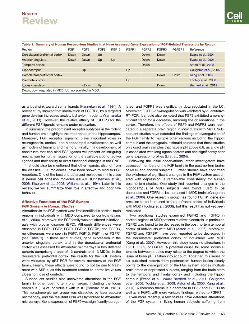
Table 1. Summary of Human Postmortem Studies that Have Assessed Gene Expression of FGF-Related Transcripts by Region
Region FGF1 FGF2 FGF9 FGF12 FGFR1 FGFR2 FGFR3 FGFBP1 Reference
Dorsolateral prefrontal cortex Down Down Up Down Down Evans et al., 2004
Anterior cingulate Down Down Up Up Down Down Evans et al., 2004
Temporal cortex Down Aston et al., 2005
Hippocampus Up Up Gaughran et al., 2006
Dorsolateral prefrontal cortex Down Down Kang et al., 2007
Prefrontal cortex Up Tochigi et al., 2008
Locus coeruleus �Down Up Down Bernard et al., 2011
Down, downregulated in MDD; Up, upregulated in MDD.
Neuron
Review
as a local sink toward some ligands (Hanneken et al., 1994). A
recent study showed that inactivation of FGFBP3, by a targeted
gene deletion increased anxiety behavior in rodents (Yamanaka
et al., 2011). However, the relative affinity of FGFBP3 for the
different FGF ligands remains under evaluation.
In summary, the predominant receptor subtypes in the rodent
and human brain highlight the importance of the hippocampus.
Moreover, FGF receptor signaling plays important roles in
neurogenesis, cortical, and hippocampal development, as well
as models of learning and memory. Finally, the development of
constructs that can bind FGF ligands will present an intriguing
mechanism for further regulation of the available pool of active
ligands and their ability to exert functional changes in the CNS.
It should also be mentioned that other ligands, distinct from
the classical FGF molecules, have been shown to bind to FGF
receptors. One of the best characterized molecules in this class
is neural cell adhesion molecule (NCAM) (Christensen et al.,
2006; Kiselyov et al., 2005; Williams et al., 1994). Later in this
review, we will summarize their role in affective and cognitive
behavior.
Affective Functions of the FGF SystemFGF System in Human Studies
Alterations in the FGF systemwere first identified in cortical brain
regions in individuals with MDD compared to controls (Evans
et al., 2004). Moreover, the FGF family was not altered in individ-
uals with bipolar disorder (BPD). Although differences were
observed in FGF1, FGF2, FGF9, FGF12, FGFR2, and FGFR3,
no differences were seen in FGF7, FGF13, FGF14, or FGFR1
(see Table 1). In these initial studies, gene expression in the
anterior cingulate cortex and in the dorsolateral prefrontal
cortex was assessed by Affymetrix microarrays in two different
cohorts comprising a total of 13 controls and 13 MDDs. In the
dorsolateral prefrontal cortex, the results for the FGF system
were validated by qRT-PCR for several members of the FGF
family. Finally, these effects were found to not be due to treat-
ment with SSRIs, as this treatment tended to normalize values
closer to those of controls.
Subsequent studies also uncovered alterations in the FGF
family in other postmortem brain areas, including the locus
coeruleus (LC) of individuals with MDD (Bernard et al., 2011).
This noradrenergic cell group was dissected by laser capture
microscopy, and the resultant RNA was hybridized to Affymetrix
microarrays. Gene expression of FGF9 was significantly upregu-
lated, and FGFR3 was significantly downregulated in the LC.
Moreover, FGFR3 downregulation was validated by quantitative
RT-PCR. It should also be noted that FGF2 exhibited a nonsig-
nificant trend for a decrease, mirroring the observations in the
cortex. Therefore, the effects of FGF9 and FGFR3 were repli-
cated in a separate brain region in individuals with MDD. Sub-
sequent studies have extended the findings of dysregulation of
the FGF family to multiple other regions including the hippo-
campus and the amygdala. It should be noted that these studies
only used brain samples that have a pH above 6.8, as a low pH
is associated with long agonal factors and can significantly alter
gene expression profiles (Li et al., 2004).
Following the initial observations, other investigators have
assessed members of the FGF family in the postmortem brains
of MDD and control subjects. Further studies have confirmed
the existence of significant changes in the FGF system associ-
ated with depression, a remarkable consistency for human
postmortem studies. One study first reported changes in the
hippocampus of MDD subjects, and found FGF2 to be
decreased and FGFR1 to be increased in MDD brains (Gaughran
et al., 2006). One research group has found FGFR1 gene ex-
pression to be increased in the prefrontal cortex of individuals
with MDD (Tochigi et al., 2008), but this result has not yet been
replicated.
Two additional studies examined FGFR2 and FGFR3 in
cortical regions ofMDDpatients relative to controls. In particular,
FGFR2 was found to be decreased in the postmortem temporal
cortex of individuals with MDD (Aston et al., 2005). Moreover,
FGFR3 and FGFBP1 have been reported to be decreased in
the dorsolateral prefrontal cortex of individuals with MDD
(Kang et al., 2007). However, this study found no alterations in
FGF1, FGF9, or FGFR2. A potential cause for some inconsis-
tencies between studies may relate to the degree to which the
issue of brain pH is taken into account. Together, this series of
six published reports from postmortem human brains clearly
points to the dysregulation of the FGF system across multiple
brain areas of depressed subjects, ranging from the brain stem
to the temporal and frontal cortex and including the hippo-
campus (Evans et al., 2004; Bernard et al., 2011; Gaughran
et al., 2006; Tochigi et al., 2008; Aston et al., 2005; Kang et al.,
2007). A common theme is a decrease in FGF2 and FGFR3 as
well as in FGF2, with more variable findings related to FGFR1.
Even more recently, a few studies have detected alterations
of the FGF system in living human subjects suffering from
Neuron 76, October 4, 2012 ª2012 Elsevier Inc. 163
Neuron
Review
MDD. For example, a single nucleotide polymorphism (SNP)
in FGF2 (rs1048201) was found to be associatedwith side effects
and altered responsiveness to antidepressant treatment in
individuals with MDD (Kato et al., 2009). Other SNPs in FGF2
(rs1449683 and rs308393) were also associated with differential
treatment response to SSRIs. One other study also found serum
levels of FGF2 to be increased in individuals with MDD and
borderline personality disorder (Kahl et al., 2009). These studies
extend the postmortem findings and suggest that the FGF
system may offer potentially valuable biomarkers, be they diag-
nostic or pharmacogenomic, for the diagnosis and treatment of
major depression.
In summary, evidence from several independent research
groups has shown significant alterations in the FGF family across
multiple brain regions of individuals suffering from major de-
pression, see Table 1. More specific hypotheses can now be
generated from these discoveries and tested directly in patients
or patient samples. These observations prompted studies
evaluating the functions of FGF2 in animal models—a case of
‘‘reverse translation.’’
FGF System in Emotionality in Animals
Animal studies have proven pivotal in validating and extending
the discoveries made in human postmortem findings. When
changes are observed in gene expression in human brain, the
findings, even if fully validated, may not be functionally sig-
nificant but rather represent mere side effects of other types of
dysregulation. To attribute functional import to them, it is critical
to manipulate them or test their regulation in the context of
animal models.
Indeed, the first animal studies, predating the human findings,
were pharmacological and suggested a possible role of FGF2
in mediating the actions of antidepressants and anxiolytic drugs.
Thus, chronic antidepressant treatment for three weeks resulted
in an increase in FGF2 24h later (Mallei et al., 2002). Similarly,
FGF2 was increased (6 and 12 hr) following acute treatment
with an anxiolytic (Gomez-Pinilla et al., 2000). This led to the
suggestion that FGF2, like BDNF, may mediate the actions of
these drugs, and was consistent with the observation that
patients on antidepressants expressed a lower degree of dysre-
gulation in their FGF system relative to untreated MDDs.
Following the observation that FGF2 was decreased in the
postmortem brain of MDD subjects, it was important to deter-
mine whether FGF2 was altered in an animal model of depres-
sion-like behavior. This study relied on a subchronic social
defeat stress in the rat as a model of depression, since social
stress activates neural circuits in the rodent that parallel those
altered in human depression (Kollack-Walker et al., 1997,
1999). Moreover, social defeat has ethological validity, in that it
mimics some of the physiological and anhedonic aspects of
depression in humans. This study focused on the hippocampus,
as postmortem studies pointed to this brain region as the most
altered by MDD (unpublished observations). Moreover, this is
an area that is critical in the biology of ‘‘stress-related disorders,’’
including MDD, anxiety, and posttraumatic stress disorder
(PTSD). For example, human brain imaging studies have shown
that the volume of the human hippocampus is negatively corre-
lated with PTSD (Gilbertson et al., 2002), consistent with the view
that this area is highly responsive to stress-related disorders.
164 Neuron 76, October 4, 2012 ª2012 Elsevier Inc.
This work, in fact, suggested that hippocampal volume may be
a predisposing factor in PTSD. Thus, the hippocampal size of
the twin who had not been exposed to combat predicted the
magnitude of PTSD in the combat twin. Since FGF2 can control
the development and size of the hippocampus (Ohkubo et al.,
2004), it was logical to assess FGF2 expression in this region
follow a social stress animal model. It should also be mentioned
that neuroimaging studies have shown a reduced hippocampal
volume in depressed subjects (Campbell et al., 2004). However,
it remains unclear whether this is a result of stress and an ante-
cedent to depression or a consequence of having the disorder.
Following repeated social defeat stress, the expression of
FGF2, as well as one of its receptors, FGFR1, was decreased
in the hippocampus (Turner et al., 2008a), suggesting the
hypothesis that the observed decrease in FGF2 both in human
MDDs and an animal model may contribute to the affective
changes accompanying depression. Could FGF2 be ‘‘an endog-
enous antidepressant,’’ and could its suppression, therefore,
contribute to the negative affect of depressed humans? This
hypothesis was tested by administering FGF2 intracerebroven-
tricularly to adult rats to ascertain its potential antidepressant-
like effects. Following both acute and chronic administration
and across multiple tests of depression-like behavior, such as
the forced swim test and novelty-suppressed feeding, FGF2
proved to have antidepressant properties (Turner et al., 2008c).
Surprisingly, FGFR1 expression was also increased by the
FGF2 treatment. This suggested that FGF2 can prime its own
receptor and further amplify the effects of its administration.
Moreover, other ligands known to bind to and activate FGF
receptors, such as neural cell adhesion molecule (NCAM), also
decreased depression-like behavior following acute intracere-
broventricular administration (Turner et al., 2008c).
While the above animal studies implicated FGF2 in response
to depression in humans and to stress in animals, and pointed
to FGF2 as an ‘‘endogenous antidepressant,’’ they did not
address whether FGF2 could be a predisposing factor to vulner-
ability to anxiety-like behavior. This question was addressed
by relying on a selectively-bred rat line of emotionality. Two lines
of rat were bred on the basis of novelty seeking in a novel envi-
ronment and termed bred high responders (bHR) and bred low
responders (bLRs). These two lines exhibit many differences
across behavior and are proposed as models of externalizing
disorders (bHRs) versus internalizing disorders (bLRs). Thus,
bHRs show lower levels of spontaneous anxiety, greater propen-
sity for risk-taking, sign-tracking, and drug-taking behavior
(Flagel et al., 2008, 2009, 2010; Stead et al., 2006). By contrast,
bLRs exhibit greater anxiety- and depression-like behaviors and
greater responsiveness to stress. It was, therefore, reasonable to
use these two lines to investigate whether FGF2may be a predis-
posing factor to emotional reactivity.
Indeed, the high anxiety bLRs exhibited lower endogenous
levels of FGF2 gene expression in the hippocampus relative to
the low anxiety HRs (Perez et al., 2009). Moreover, repeated
peripheral administration of FGF2 decreased anxiety-like
behavior, and the bLRs benefited more from the treatment
than the bHRs. Similarly, environmental complexity, a manipu-
lation known to decrease anxiety in rodents, increased FGF2
expression in the hippocampus and showed a greater effect in
Neuron
Review
bLRs. Perez et al. (2009) also assessed neurogenesis following
peripheral FGF2 administration and found that chronic adminis-
tration did not influence cell proliferation but increased cell
survival in the dentate gyrus, especially in the bLR rats that
exhibit the greater decrease in anxiety behavior. Although
FGF2 increased the survival of both neurons and glia, the
increase in the number of astrocytes was particularly prominent.
Together, these findings led to the view that FGF2 is both
a genetic predisposing factor that affects basal anxiety levels,
and amodulator of environmental influences on anxiety behavior
in the adult rat.
If FGF2 is indeed not only an endogenous antidepressant but
also an endogenous anxiolytic factor, where does it exert this
influence on behavior? This question was addressed by using
a knockdown strategy to reduce FGF2 expression in the dentate
gyrus and CA3 region by RNA interference, and assess its
impact on behavior in rats (Eren-Kocak et al., 2011). A lentiviral
vector containing a short-hairpin targeting FGF2 was used to
knockdown FGF2, and this treatment resulted in an anxiogenic
effect without altering other behaviors. This suggests that
FGF2 expression in the hippocampus does indeed modulate
the level of spontaneous anxiety. Based on this body of work,
a model was proposed illustrating the importance of hippo-
campal levels of FGF2 in the modulation of allostatic load (Sal-
maso and Vaccarino, 2011).
Having established that FGF2 may modulate both the vulner-
ability and/or resilience to anxiety-like behavior, as well as
mediate environmental changes such as stress and environ-
mental complexity, its role in the development of emotional
circuitry became critical. This question was addressed by
assessing the effects of early life FGF2, administered the day
after birth, on emotionality, hippocampal development and
gene expression (Turner et al., 2011). Remarkably, a single injec-
tion of FGF2 (20 ng/g, subcutaneously) early in life was able
to alter neurogenesis in outbred animals. In adulthood, these
animals exhibited a denser dentate gyrus with more neurons,
consistent with the idea that neurogenesis precedes gliogenesis
in early development (Palmer et al., 1999). Moreover, when the
same early life FGF2 treatment was given to high anxiety animals
(bLRs), FGF2 decreased their spontaneous anxiety (Turner et al.,
2011). This effect was associatedwith altered gene expression in
the dentate gyrus. Laser capture microdissection followed by
microarray analyses identified transcripts that differed between
bLR-VEH and bLR-FGF2 animals. Specifically, molecules previ-
ously associated with anxiety (gad1) were decreased, whereas
molecules associated with cell survival (bcl2-like2) were
increased in the high anxiety bred rats in conjunction with
decreased anxiety by FGF2 treatment. Thus, early life FGF2
treatment altered the developmental trajectory of the dentate
gyrus and had long-term effects on emotionality and gene
expression.
Most recently, a study by Duman’s group extended these
findings to mice and to other models of stress (Elsayed et al.,
2012). Thus, the authors reported that chronic infusion of FGF2
had antidepressant-like effects in both rats and mice. They
also added site-specificity to the antidepressant effects by
infusing FGF2 into the medial prefrontal cortex. Moreover,
FGF2 blocked the effects of chronic unpredictable stress
(CUS) on both depression-like behavior, and the CUS-induced
inhibition of glial proliferation. Treatment with an FGF receptor
antagonist that targets all FGF receptors blocked the effects of
fluoxetine on glial proliferation, as well as the effect of fluoxetine
as an antidepressant. These results suggest that not only is
FGF2 a sufficient antidepressant, it is also necessary for the
antidepressant effects of SSRIs, although the lack of selectivity
of the available FGF antagonists requires caution in the inter-
pretation of these latter results.
Moreover, the study by Elsayed et al. (2012) also hinted at
relatively rapid effects of FGF2 in animal models of depression
and anxiety (5 days after administration). We have also observed
rapid effects of FGF2 in other paradigms. Indeed, some of the
behavior and biochemical effects of FGF2 can be observed
within minutes and certainly within hours, but the mechanism
of these rapid effects needs further exploration. FGFR1 is
required for the electrophysiological correlate of learning and
memory, long-term potentiation (Zhao et al., 2007). Is it possible
that the same glutamatergic mechanisms may underlie the
susceptibility to anxiety and depression? FGFR1, the highest
affinity receptor for FGF2, is located on glutamatergic cells in
the cortex (Shin et al., 2004). Therefore, it is possible that an
increase in glutamate transmission may be responsible for the
antidepressant effects of FGF2.
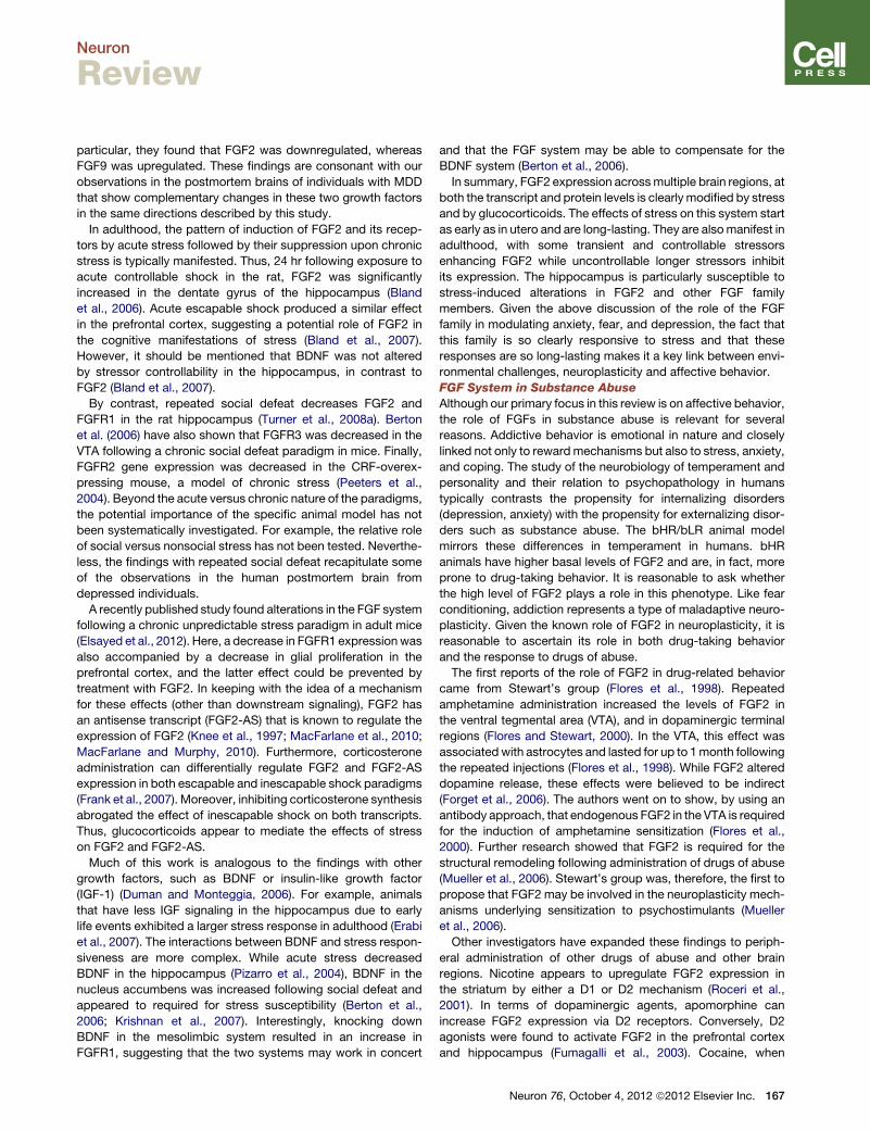
Figure 1 summarizes the body of work that implicates FGF2
in each of the factors thought to modify emotionality. Thus,
genetic differences between the bHR and bLR lines implicate
FGF2 as a genetic factor. The early FGF2 administration studies
demonstrate its critical organizational function in laying down
differences in emotional reactivity. And the various studies with
stress paradigms and environmental complexity demonstrate
its role in mediating changes that result from experience, result-
ing in altered neurogenesis and other types of neuroplasticity.
The hippocampus and prefrontal cortex are two loci of its
actions, as shown in both human and animal studies, with other
loci yet to be identified.
FGF System in Fear Conditioning in Animals
The FGF system has not only been implicated in general anxiety,
but also plays a role in emotional learning and fear conditioning,
suggesting that it may be involved in another affective disorder—
PTSD. This disorder is based on the inability to extinguish
fearful memories under conditions that are presumably ‘‘safe.’’
Initially, FGF2 was implicated in the acquisition phase of fear
conditioning (Graham and Richardson, 2009a). When FGF2
was given subcutaneously immediately prior to conditioning,
it facilitated contextual fear memory in young rats (PND 16,
PND19, or PND22). This group went on to show that systemic
FGF2 can also facilitate extinction if it is on-board during consol-
idation (Graham and Richardson, 2009b). Moreover, when given
immediately after extinction, FGF2 reduced reinstatement and
relapse (Graham and Richardson, 2010b).
FGF2 was also shown to have effects on fear conditioning
when subcutaneously administered to neonatal rats, with testing
conducted 11–18 days after the last injection (Graham and
Richardson, 2010a). However, unlike the effects on anxiety
described above, multiple injections of FGF2 were required
(PND 1–5) to facilitate both fear conditioning and context-
dependent extinction. Taken together, FGF2 may be involved
Neuron 76, October 4, 2012 ª2012 Elsevier Inc. 165
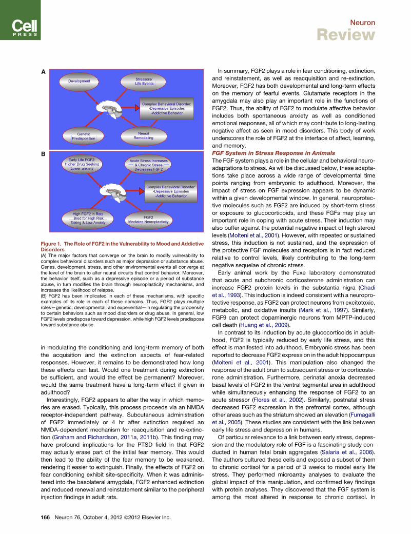
High FGF2 in Rats Bred for High Risk
Taking & Low Anxiety
Complex Behavioral Disorder:
-Depressive Episodes -Addictive Behavior
Acute Stress Increases & Chronic Stress Decreases FGF2
Early Life FGF2: Higher Drug Seeking
Lower anxiety
FGF2 Mediates Neuroplasticity
Genetic Predisposition
Complex Behavioral Disorder:
-Depressive Episodes -Addictive Behavior
Stressors/ Life Events
Development
Neural Remodeling
A
B
Figure 1. TheRole of FGF2 in the Vulnerability toMood andAddictiveDisorders(A) The major factors that converge on the brain to modify vulnerability tocomplex behavioral disorders such as major depression or substance abuse.Genes, development, stress, and other environmental events all converge atthe level of the brain to alter neural circuits that control behavior. Moreover,the behavior itself, such as a depressive episode or a period of substanceabuse, in turn modifies the brain through neuroplasticity mechanisms, andincreases the likelihood of relapse.(B) FGF2 has been implicated in each of these mechanisms, with specificexamples of its role in each of these domains. Thus, FGF2 plays multipleroles—genetic, developmental, and experiential—in regulating the propensityto certain behaviors such as mood disorders or drug abuse. In general, lowFGF2 levels predispose toward depression, while high FGF2 levels predisposetoward substance abuse.
Neuron
Review
in modulating the conditioning and long-term memory of both
the acquisition and the extinction aspects of fear-related
responses. However, it remains to be demonstrated how long
these effects can last. Would one treatment during extinction
be sufficient, and would the effect be permanent? Moreover,
would the same treatment have a long-term effect if given in
adulthood?
Interestingly, FGF2 appears to alter the way in which memo-
ries are erased. Typically, this process proceeds via an NMDA
receptor-independent pathway. Subcutaneous administration
of FGF2 immediately or 4 hr after extinction required an
NMDA-dependent mechanism for reacquisition and re-extinc-
tion (Graham and Richardson, 2011a, 2011b). This finding may
have profound implications for the PTSD field in that FGF2
may actually erase part of the initial fear memory. This would
then lead to the ability of the fear memory to be weakened,
rendering it easier to extinguish. Finally, the effects of FGF2 on
fear conditioning exhibit site-specificity. When it was adminis-
tered into the basolateral amygdala, FGF2 enhanced extinction
and reduced renewal and reinstatement similar to the peripheral
injection findings in adult rats.
166 Neuron 76, October 4, 2012 ª2012 Elsevier Inc.
In summary, FGF2 plays a role in fear conditioning, extinction,
and reinstatement, as well as reacquisition and re-extinction.
Moreover, FGF2 has both developmental and long-term effects
on the memory of fearful events. Glutamate receptors in the
amygdala may also play an important role in the functions of
FGF2. Thus, the ability of FGF2 to modulate affective behavior
includes both spontaneous anxiety as well as conditioned
emotional responses, all of which may contribute to long-lasting
negative affect as seen in mood disorders. This body of work
underscores the role of FGF2 at the interface of affect, learning,
and memory.
FGF System in Stress Response in Animals
The FGF system plays a role in the cellular and behavioral neuro-
adaptations to stress. As will be discussed below, these adapta-
tions take place across a wide range of developmental time
points ranging from embryonic to adulthood. Moreover, the
impact of stress on FGF expression appears to be dynamic
within a given developmental window. In general, neuroprotec-
tive molecules such as FGF2 are induced by short-term stress
or exposure to glucocorticoids, and these FGFs may play an
important role in coping with acute stress. Their induction may
also buffer against the potential negative impact of high steroid
levels (Molteni et al., 2001). However, with repeated or sustained
stress, this induction is not sustained, and the expression of
the protective FGF molecules and receptors is in fact reduced
relative to control levels, likely contributing to the long-term
negative sequelae of chronic stress.
Early animal work by the Fuxe laboratory demonstrated
that acute and subchronic corticosterone administration can
increase FGF2 protein levels in the substantia nigra (Chadi
et al., 1993). This induction is indeed consistent with a neuropro-
tective response, as FGF2 can protect neurons from excitotoxic,
metabolic, and oxidative insults (Mark et al., 1997). Similarly,
FGF9 can protect dopaminergic neurons from MPTP-induced
cell death (Huang et al., 2009).
In contrast to its induction by acute glucocorticoids in adult-
hood, FGF2 is typically reduced by early life stress, and this
effect is manifested into adulthood. Embryonic stress has been
reported to decrease FGF2 expression in the adult hippocampus
(Molteni et al., 2001). This manipulation also changed the
response of the adult brain to subsequent stress or to corticoste-
rone administration. Furthermore, perinatal anoxia decreased
basal levels of FGF2 in the ventral tegmental area in adulthood
while simultaneously enhancing the response of FGF2 to an
acute stressor (Flores et al., 2002). Similarly, postnatal stress
decreased FGF2 expression in the prefrontal cortex, although
other areas such as the striatum showed an elevation (Fumagalli
et al., 2005). These studies are consistent with the link between
early life stress and depression in humans.
Of particular relevance to a link between early stress, depres-
sion and the modulatory role of FGF is a fascinating study con-
ducted in human fetal brain aggregates (Salaria et al., 2006).
The authors cultured these cells and exposed a subset of them
to chronic cortisol for a period of 3 weeks to model early life
stress. They performed microarray analyses to evaluate the
global impact of this manipulation, and confirmed key findings
with protein analyses. They discovered that the FGF system is
among the most altered in response to chronic cortisol. In
Neuron
Review
particular, they found that FGF2 was downregulated, whereas
FGF9 was upregulated. These findings are consonant with our
observations in the postmortem brains of individuals with MDD
that show complementary changes in these two growth factors
in the same directions described by this study.
In adulthood, the pattern of induction of FGF2 and its recep-
tors by acute stress followed by their suppression upon chronic
stress is typically manifested. Thus, 24 hr following exposure to
acute controllable shock in the rat, FGF2 was significantly
increased in the dentate gyrus of the hippocampus (Bland
et al., 2006). Acute escapable shock produced a similar effect
in the prefrontal cortex, suggesting a potential role of FGF2 in
the cognitive manifestations of stress (Bland et al., 2007).
However, it should be mentioned that BDNF was not altered
by stressor controllability in the hippocampus, in contrast to
FGF2 (Bland et al., 2007).
By contrast, repeated social defeat decreases FGF2 and
FGFR1 in the rat hippocampus (Turner et al., 2008a). Berton
et al. (2006) have also shown that FGFR3 was decreased in the
VTA following a chronic social defeat paradigm in mice. Finally,
FGFR2 gene expression was decreased in the CRF-overex-
pressing mouse, a model of chronic stress (Peeters et al.,
2004). Beyond the acute versus chronic nature of the paradigms,
the potential importance of the specific animal model has not
been systematically investigated. For example, the relative role
of social versus nonsocial stress has not been tested. Neverthe-
less, the findings with repeated social defeat recapitulate some
of the observations in the human postmortem brain from
depressed individuals.
A recently published study found alterations in the FGF system
following a chronic unpredictable stress paradigm in adult mice
(Elsayed et al., 2012). Here, a decrease in FGFR1 expression was
also accompanied by a decrease in glial proliferation in the
prefrontal cortex, and the latter effect could be prevented by
treatment with FGF2. In keeping with the idea of a mechanism
for these effects (other than downstream signaling), FGF2 has
an antisense transcript (FGF2-AS) that is known to regulate the
expression of FGF2 (Knee et al., 1997; MacFarlane et al., 2010;
MacFarlane and Murphy, 2010). Furthermore, corticosterone
administration can differentially regulate FGF2 and FGF2-AS
expression in both escapable and inescapable shock paradigms
(Frank et al., 2007). Moreover, inhibiting corticosterone synthesis
abrogated the effect of inescapable shock on both transcripts.
Thus, glucocorticoids appear to mediate the effects of stress
on FGF2 and FGF2-AS.
Much of this work is analogous to the findings with other
growth factors, such as BDNF or insulin-like growth factor
(IGF-1) (Duman and Monteggia, 2006). For example, animals
that have less IGF signaling in the hippocampus due to early
life events exhibited a larger stress response in adulthood (Erabi
et al., 2007). The interactions between BDNF and stress respon-
siveness are more complex. While acute stress decreased
BDNF in the hippocampus (Pizarro et al., 2004), BDNF in the
nucleus accumbens was increased following social defeat and
appeared to required for stress susceptibility (Berton et al.,
2006; Krishnan et al., 2007). Interestingly, knocking down
BDNF in the mesolimbic system resulted in an increase in
FGFR1, suggesting that the two systems may work in concert
and that the FGF system may be able to compensate for the
BDNF system (Berton et al., 2006).
In summary, FGF2 expression acrossmultiple brain regions, at
both the transcript and protein levels is clearly modified by stress
and by glucocorticoids. The effects of stress on this system start
as early as in utero and are long-lasting. They are alsomanifest in
adulthood, with some transient and controllable stressors
enhancing FGF2 while uncontrollable longer stressors inhibit
its expression. The hippocampus is particularly susceptible to
stress-induced alterations in FGF2 and other FGF family
members. Given the above discussion of the role of the FGF
family in modulating anxiety, fear, and depression, the fact that
this family is so clearly responsive to stress and that these
responses are so long-lasting makes it a key link between envi-
ronmental challenges, neuroplasticity and affective behavior.
FGF System in Substance Abuse
Although our primary focus in this review is on affective behavior,
the role of FGFs in substance abuse is relevant for several
reasons. Addictive behavior is emotional in nature and closely
linked not only to rewardmechanisms but also to stress, anxiety,
and coping. The study of the neurobiology of temperament and
personality and their relation to psychopathology in humans
typically contrasts the propensity for internalizing disorders
(depression, anxiety) with the propensity for externalizing disor-
ders such as substance abuse. The bHR/bLR animal model
mirrors these differences in temperament in humans. bHR
animals have higher basal levels of FGF2 and are, in fact, more
prone to drug-taking behavior. It is reasonable to ask whether
the high level of FGF2 plays a role in this phenotype. Like fear
conditioning, addiction represents a type of maladaptive neuro-
plasticity. Given the known role of FGF2 in neuroplasticity, it is
reasonable to ascertain its role in both drug-taking behavior
and the response to drugs of abuse.
The first reports of the role of FGF2 in drug-related behavior
came from Stewart’s group (Flores et al., 1998). Repeated
amphetamine administration increased the levels of FGF2 in
the ventral tegmental area (VTA), and in dopaminergic terminal
regions (Flores and Stewart, 2000). In the VTA, this effect was
associated with astrocytes and lasted for up to 1month following
the repeated injections (Flores et al., 1998). While FGF2 altered
dopamine release, these effects were believed to be indirect
(Forget et al., 2006). The authors went on to show, by using an
antibody approach, that endogenous FGF2 in the VTA is required
for the induction of amphetamine sensitization (Flores et al.,
2000). Further research showed that FGF2 is required for the
structural remodeling following administration of drugs of abuse
(Mueller et al., 2006). Stewart’s group was, therefore, the first to
propose that FGF2 may be involved in the neuroplasticity mech-
anisms underlying sensitization to psychostimulants (Mueller
et al., 2006).
Other investigators have expanded these findings to periph-
eral administration of other drugs of abuse and other brain
regions. Nicotine appears to upregulate FGF2 expression in
the striatum by either a D1 or D2 mechanism (Roceri et al.,
2001). In terms of dopaminergic agents, apomorphine can
increase FGF2 expression via D2 receptors. Conversely, D2
agonists were found to activate FGF2 in the prefrontal cortex
and hippocampus (Fumagalli et al., 2003). Cocaine, when
Neuron 76, October 4, 2012 ª2012 Elsevier Inc. 167

Neuron
Review
administered acutely, can rapidly alter levels of FGF2 in the
prefrontal cortex and striatum, with chronic exposure to cocaine
resulting in enduring elevations of FGF2, especially in the stria-
tum (Fumagalli et al., 2006). Thus, long-lasting changes take
place in regions highly innervated by midbrain dopaminergic
neurons, suggesting that FGF2 is not only involved in the initial
response to drugs of abuse, but also in the long-term neuroa-
daptations.
Interestingly, the selectively bred line of rats that shows
greater propensity to drug seeking behavior (i.e., bHR rats)
exhibit higher basal levels of expression of FGF2 in the hippo-
campus and nucleus accumbens than their bLRs counterparts
that show lower propensity to self administer drugs (Perez
et al., 2009; Clinton et al., 2012). Moreover, a sensitizing treat-
ment with cocaine generally decreased FGFR1 expression in
the hippocampus and increased FGFR1 in the prefrontal cortex
(Turner et al., 2008b). However, the two selectively bred lines
showed a differential effect of the drug. In the hippocampus,
cocaine decreased gene expression in bHRs without affecting
bLRs, whereas in the prefrontal cortex cocaine increased gene
expression in bLRs without affecting bHRs. Thus, cocaine inter-
acted with the novelty-seeking trait to alter gene expression
differentially depending on brain region, furthering the idea that
the FGF system may be involved in the individual differences in
the response to drugs of abuse.
A single administration of FGF2 on PND1 increased cocaine
self-administration in adulthood (Turner et al., 2009). This effect
is selective as there were no associated differences in spatial
or appetitive learning. Moreover, there were no sustained
changes in gene expression in the dopaminergic system seen
in the adult animal. This does not preclude the possibility that
early exposure to FGF2 primed the dopaminergic system, which
in turn led to increased drug-taking behavior in adulthood.
Whether the actions of early life FGF2 are mediated via dopa-
mine or other mechanisms, the ability of this growth factor to
enhance drug-taking behavior identifies it as a molecular ante-
cedent of vulnerability for substance abuse.
Given the fact that drugs of abuse interact with stress, it is
notable that both stress and drugs of abuse converge to modu-
late FGF2 expression. Thus, in the prefrontal cortex, acute stress
potentiated the cocaine-induced increase in FGF2 expression,
whereas prolonged stress prevented the response of FGF2 to
cocaine (Fumagalli et al., 2008). In the striatum, the cocaine-
induced FGF2 response was only increased following repeated
stress.
In summary, FGF2 appears to promote both the initial vulner-
ability and the sequelae of substance abuse. Its administration in
early life enhances the propensity for self-administration of drugs
of abuse in adulthood. In turn, repeated exposure to drugs of
abuse induces FGF2 expression especially in the dopaminergic
system, and this induction is required for the development of
sensitization.
Overall, FGF2, along with FGFR1, can be construed as molec-
ular factors that modulate emotional reactivity—higher FGF2
levels render animals more prone to novelty and drug taking
behavior, while lower FGF2 levels render animals less prone to
drug seeking but more prone to anxiety- and depression-like
behaviors.
168 Neuron 76, October 4, 2012 ª2012 Elsevier Inc.
Beyond the FGFs: The Role of Other Interacting Ligands
Other molecules, such as NCAM, can also interact with the FGF
receptors and appear to play a role in the control of emotionality.
NCAM polymorphisms have been observed in conjunction with
mood disorders in humans (Atz et al., 2007; Vawter, 2000). In
animal models, NCAM responds to stress system activation,
with upregulation of its expression in the cortex following acute
corticosterone injections and downregulation following chronic
corticosterone (Sandi and Loscertales, 1999)—a pattern that
mirrors the regulation of FGF2 by this stress hormone. However,
the isoform of NCAM is also important. For example, exposure
to a stressful situation decreased NCAM-180 levels in the hippo-
campus without affecting the levels of NCAM-140 or NCAM-120
(Sandi et al., 2005). Finally, posttranslational modifications of
NCAM (polysialylation) can also be affected by stress (Cordero
et al., 2005).
Similar to FGF2, FGL, a fragment of the NCAM structure (Car-
afoli et al., 2008; Ditlevsen et al., 2008), has antidepressant
effects when acutely administered intracerebroventricularly to
rodents (Turner et al., 2008c). Conversely, NCAM-deficient
mice exhibit an increase in anxiety- and depression-like
behavior. The latter effect, along with FGFR signaling deficits,
can be restored by treatment with FGL (Aonurm-Helm et al.,
2008; Aonurm-Helm et al., 2010). Similarly, FGL was able to
reverse the chronic stress, as well as NCAM-deficiency-induced
cognitive impairments (Bisaz et al., 2011). The effects of FGL on
fear conditioning and spatial learning have also been assessed,
whereby both the positive and negative effects were enhanced
(Cambon et al., 2004). Additionally, FGL can enhance presyn-
aptic function, promote synaptogenesis, and facilitate memory
(Cambon et al., 2004). Not surprisingly, FGL can also prevent
stress-induced impairments in cognitive function (Borcel et al.,
2008). Two other NCAM-derived peptides, dennexin and plan-
nexin, have been shown to have effects in vivo, modulating
neuroplasticity, and learning (Køhler et al., 2010; Kraev et al.,
2011). For a more thorough discussion of the role of NCAM in
cognition and stress, the reader is referred to other reviews
(Conboy et al., 2010; Sandi and Bisaz, 2007).
Other ligands, such as N-cadherin and pentraxin, are cell
adhesion molecules that can bind to FGF receptors as well as
the cytoskeleton (Hansen et al., 2008; Sanchez-Heras et al.,
2006). Similar to NCAM, N-cadherin binds to the acid box region
of the FGF receptor, which is different than the binding site for
FGF2. Interestingly, peptide moieties of N-cadherin have been
identified that can act as agonists, and one of the main functions
of N-cadherin is to induce neurite outgrowth (Williams et al.,
2002).
In general, non-FGF ligands that interact with the FGF recep-
tors have been identified for the treatment of cognitive deficits.
Given the relevance of the FGF system to fear, anxiety, depres-
sion, and addiction, it will be important to ascertain their potential
as targets for affective disorders.
Beyond the FGFs: Receptor-Interacting Partners
The complexity and the potential functions of the FGF system
are augmented not only by a host of binding molecules but
also by the potential for receptor-receptor interactions.
Recently, FGFR1 has been shown to directly interact with two
different neurotransmitter receptors. The first is the adenosine
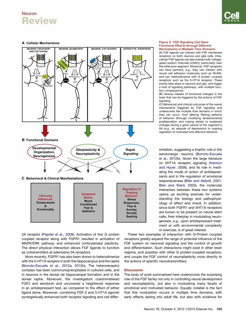
A Cellular Mechanisms
B Functional Domains
C Behavioral & Clinical Manifestations
Life-Long Influences:
Temperament&
Emotionality
SustainedStates:Mood,
ConditionedFear &
Addiction
Regulation ofOngoing Behavior:
StressResponses,
Fear &Anxiety,NoveltySeeking
DevelopmentalOrganization,Neurogenesis
Glioplasticity &Neuroplasticity
Rapid Signaling
ASTROCYTE: FGF9/FGFR3NEURON: FGF2/FGFR1AND FGF12/VGSC
NEURON: NCAM/FGFR1 NEURON: 5-HT1A/FGFR1
FGF2
FGF2
FGFR3
FGF9
FGF9
FGFR1
FGF2
FGF2
FGFR1
PKA
NCAM5-HT
FGF2
FGF2
FGFR1
PKA
NCAM
5-HT1A
CREB CREBAKT
MAPK PLCγ
VGSC
FGF12
AIS
AKT
MAPK PLCγ
AKT
MAPK PLCγ
AKT
MAPK PLCγ
Figure 2. FGF Signaling Can HaveFunctional Effects through DifferentMechanisms in Multiple Time Domains(A) FGF ligands can interact with FGF membranereceptors on both neurons and glial cells. Intra-cellular FGF ligands can also interact with voltage-gated sodium channels (VGSC), particularly nearthe initial axon segment. Moreover, FGF receptorscan have partners, e.g., they can interact withneural cell adhesion molecules such as NCAM,and can heterodimerize with G protein coupledreceptors such as the 5-HT1A receptor. Theseevents take place in neurons and glia, and triggera host of signaling pathways, with multiple func-tion consequences.(B) Various classes of functional changes in thebrain that can be triggered by the actions of FGFsignaling.(C) Behavioral and clinical outcomes of the neuralmechanisms triggered by FGF signaling andunderscores the multiple time domains in whichthey can occur, from altering lifelong patternsof behavior (through modifying temperamentalpredisposition and coping styles) to sustainedchanges during a given period of the organism’slife (e.g., an episode of depression) to ongoingregulation of motivated and affective behavior.
Neuron
Review
2A receptor (Flajolet et al., 2008). Activation of this G protein
coupled receptor along with FGFR1 resulted in activation of
MAPK/ERK pathway and enhanced corticostriatal plasticity.
The direct physical interaction allows FGF ligands to function
as cotransmitters at adenosine 2A receptors.
More recently, FGFR1 has also been shown to heterodimerize
with the 5-HT1A receptor in both the hippocampus and the raphe
(Borroto-Escuela et al., 2012a, 2012b). The heteroreceptor
complex has been coimmunoprecipitated in cultured cells, and
in neurons in the dorsal rat hippocampal formation and in the
dorsal raphe. Moreover, the investigators coadministered
FGF2 and serotonin and uncovered a heightened response
in an antidepressant test, as compared to the effect of either
ligand alone. Moreover, combining FGF-2 and 5-HT1A agonist
synergistically enhanced both receptor signaling and cell differ-
Neuron 76
entiation, suggesting a trophic role in the
serotonergic neurons (Borroto-Escuela
et al., 2012b). Given the large literature
on 5HT1A receptor signaling (Hannon
and Hoyer, 2008), and its role in medi-
ating the mode of action of antidepres-
sants and in the regulation of emotional
responsiveness (Blier and Abbott, 2001;
Blier and Ward, 2003), the molecular
interaction between these two systems
opens up exciting avenues for under-
standing the biology and pathophysi-
ology of affect and mood. In addition,
since both FGFR1 and 5HT1A receptors
are known to be present on neural stem
cells, their interplay in modulating neuro-
genesis, e.g., upon antidepressant treat-
ment or with environmental complexity
or exercise, is of great interest.
These two examples of interaction with G-Protein coupled
receptors greatly expand the range of potential influence of the
FGF system on neuronal signaling and the control of growth
and differentiation. Such interactions might exist in other brain
regions, and possibly with other G protein-coupled receptors,
and couple the FGF control of neuroplasticity more directly to
the actions of specific neurotransmitters.
DiscussionThe body of work summarized here underscores the surprising
role of the FGF family not only in controlling neural development
and neuroplasticity, but also in modulating many facets of
emotional and motivated behavior. Equally notable is the fact
that this modulation occurs in multiple time domains, with
early effects lasting into adult life, but also with evidence for
, October 4, 2012 ª2012 Elsevier Inc. 169
Neuron
Review
‘‘on-line’’ control of signaling and behavioral responsiveness
during adulthood.
It should be mentioned that other growth factors, such as
BDNF and IGF-1, have similar neuromodulatory effects as
FGF2. For example, both molecules promote neurogenesis
and act as antidepressants (Anderson et al., 2002; Hoshaw
et al., 2005; Schmidt and Duman, 2010). BDNF is also up-
regulated following antidepressant drug treatment and has
long-lasting effects on hippocampal function (Monteggia
et al., 2004; Nibuya et al., 1995). However, FGF2 has effects
on glial cells, specifically astrocytes, which have not been
shown for BDNF or IGF-1 (Numakawa et al., 2011). One of
these functions includes upregulating microRNAs, where
BDNF and IGF-1 failed to do so. Given that depression may
be related to a perturbation in glia, this may represent a sig-
nificant difference between growth factor families (Bernard
et al., 2011; Choudary et al., 2005). Finally, FGF receptors
can interact with other neurotransmitters, and this has the
potential for FGF ligands to have multiple and rapid cellular
and behavioral effects.
The FGF family appears to reside at the interface of genetic,
developmental, environmental, and experiential regulation of
mood, affect, and addiction. As depicted in Figure 1, endoge-
nous levels of FGFmolecules are predisposing factors that regu-
late stress responsiveness and the vulnerability or resilience to
anxiety, depression, fear conditioning, and substance abuse.
In turn, as depicted in Figure 2, FGF molecules are effectors of
the impact of experience on brain morphology, neurogenesis,
cell survival, and neuronal signaling. They rely on a host of
mechanisms to alter every phase of neuronal organization and
function, to modify stable patterns of reactivity, and to control
ongoing behavior.
In the context of mood disorders, the role of the FGF family
combines two distinct hypotheses regarding the biological
causes of severe depression—a neurotransmitter-based
hypothesis such as the dysregulation of serotonin signaling
(Sharp and Cowen, 2011) and a stress hypothesis (Akil,
2005), focusing on early developmental adversity, enhanced
vulnerability to stressors and a disrupted neuroendocrine
dysregulation, resulting in a range of negative consequences
on brain structure and function. Our view of the FGF family
synthesizes these hypotheses by placing FGFs at the very
interface of stress regulation, neurotransmitter signaling, and
neural remodeling.
In particular, FGF molecules appear to interact with classical
neurotransmitter molecules at the level of heteroreceptor
complexes, or by direct physical interaction, to control both
cellular morphology and signaling, as shown in Figure 2. In
addition, a host of other molecules modulate this system
including cell adhesion molecules and endogenous molecules.
These factors operate in both neurons and glia and in dif-
ferent combinations across distinct neural circuits. Clearly,
much remains to be learned about the role of the various
members of this complex family in the regulation of affect,
motivation and mood. But the research to date has already
illuminated previously unsuspected roles and pointed to
exciting new targets for the treatment of affective and addictive
disorders.
170 Neuron 76, October 4, 2012 ª2012 Elsevier Inc.
ACKNOWLEDGMENTS
This work was supported by NIMH Conte Center grant P50 MH60398, NIDAP01 DA021633, The Office of Naval Research (ONR) grants N00014-09-1-0598 and N00014-12-1-0366, the Pritzker Neuropsychiatric DisordersResearch Consortium Fund LLC (http://www.pritzkerneuropsych.org), theHope for Depression Research Foundation, NCRR (grant UL1RR024986), aswell as a Rachel Upjohn Clinical Scholars Award to C.T. The authors aremembers of the Pritzker Neuropsychiatric Disorders Research Consortium,which is supported by the Pritzker Neuropsychiatric Disorders ResearchFund L.L.C. A shared intellectual property agreement exists between thisphilanthropic fund and the University of Michigan, Stanford University, theWeill Medical College of Cornell University, the University of California atIrvine, and the HudsonAlpha Institute for Biotechnology to encourage thedevelopment of appropriate findings for research and clinical applications.
REFERENCES
Akil, H. (2005). Stressed and depressed. Nat. Med. 11, 116–118.
Alam, K.Y., Frostholm, A., Hackshaw, K.V., Evans, J.E., Rotter, A., and Chiu,I.M. (1996). Characterization of the 1B promoter of fibroblast growth factor 1and its expression in the adult and developing mouse brain. J. Biol. Chem.271, 30263–30271.
Anderson, M.F., Aberg, M.A., Nilsson, M., and Eriksson, P.S. (2002). Insulin-like growth factor-I and neurogenesis in the adult mammalian brain. BrainRes. Dev. Brain Res. 134, 115–122.
Aonurm-Helm, A., Jurgenson, M., Zharkovsky, T., Sonn, K., Berezin, V., Bock,E., and Zharkovsky, A. (2008). Depression-like behaviour in neural cell adhe-sion molecule (NCAM)-deficient mice and its reversal by an NCAM-derivedpeptide, FGL. Eur. J. Neurosci. 28, 1618–1628.
Aonurm-Helm, A., Berezin, V., Bock, E., and Zharkovsky, A. (2010). NCAM-mimetic, FGL peptide, restores disrupted fibroblast growth factor receptor(FGFR) phosphorylation and FGFR mediated signaling in neural cell adhesionmolecule (NCAM)-deficient mice. Brain Res. 1309, 1–8.
Aston, C., Jiang, L., and Sokolov, B.P. (2005). Transcriptional profiling revealsevidence for signaling and oligodendroglial abnormalities in the temporalcortex from patients with major depressive disorder. Mol. Psychiatry 10,309–322.
Atz, M.E., Rollins, B., and Vawter, M.P. (2007). NCAM1 association study ofbipolar disorder and schizophrenia: polymorphisms and alternatively splicedisoforms lead to similarities and differences. Psychiatr. Genet. 17, 55–67.
Audigier, S., Guiramand, J., Prado-Lourenco, L., Conte, C., Gonzalez-Herrera,I.G., Cohen-Solal, C., Recasens, M., and Prats, A.C. (2008). Potent activationof FGF-2 IRES-dependent mechanism of translation during brain develop-ment. RNA 14, 1852–1864.
Bartlett, P.F., Dutton, R., Likiardopoulos, V., and Brooker, G. (1994). Regula-tion of neurogenesis in the embryonic and adult brain by fibroblast growthfactors. Alcohol Alcohol. Suppl. 2, 387–394.
Bernard, R., Kerman, I.A., Thompson, R.C., Jones, E.G., Bunney, W.E.,Barchas, J.D., Schatzberg, A.F., Myers, R.M., Akil, H., and Watson, S.J.(2011). Altered expression of glutamate signaling, growth factor, and gliagenes in the locus coeruleus of patients withmajor depression.Mol. Psychiatry16, 634–646.
Berton, O., McClung, C.A., Dileone, R.J., Krishnan, V., Renthal, W., Russo,S.J., Graham, D., Tsankova, N.M., Bolanos, C.A., Rios, M., et al. (2006). Essen-tial role of BDNF in the mesolimbic dopamine pathway in social defeat stress.Science 311, 864–868.
Bisaz, R., Schachner, M., and Sandi, C. (2011). Causal evidence for theinvolvement of the neural cell adhesion molecule, NCAM, in chronic stress-induced cognitive impairments. Hippocampus 21, 56–71.
Bland, S.T., Schmid, M.J., Greenwood, B.N., Watkins, L.R., and Maier, S.F.(2006). Behavioral control of the stressor modulates stress-induced changesin neurogenesis and fibroblast growth factor-2. Neuroreport 17, 593–597.
Bland, S.T., Tamlyn, J.P., Barrientos, R.M., Greenwood, B.N., Watkins, L.R.,Campeau, S., Day, H.E., and Maier, S.F. (2007). Expression of fibroblastgrowth factor-2 and brain-derived neurotrophic factor mRNA in the medial
Neuron
Review
prefrontal cortex and hippocampus after uncontrollable or controllable stress.Neuroscience 144, 1219–1228.
Blier, P., and Abbott, F.V. (2001). Putativemechanisms of action of antidepres-sant drugs in affective and anxiety disorders and pain. J. Psychiatry Neurosci.26, 37–43.
Blier, P., andWard, N.M. (2003). Is there a role for 5-HT1A agonists in the treat-ment of depression? Biol. Psychiatry 53, 193–203.
Borcel, E., Perez-Alvarez, L., Herrero, A.I., Brionne, T., Varea, E., Berezin, V.,Bock, E., Sandi, C., and Venero, C. (2008). Chronic stress in adulthoodfollowed by intermittent stress impairs spatial memory and the survival ofnewborn hippocampal cells in aging animals: prevention by FGL, a peptidemimetic of neural cell adhesion molecule. Behav. Pharmacol. 19, 41–49.
Borroto-Escuela, D.O., Romero-Fernandez,W.,Mudo, G., Perez-Alea,M., Cir-uela, F., Tarakanov, A.O., Narvaez, M., Di Liberto, V., Agnati, L.F., Belluardo,N., and Fuxe, K. (2012a). Fibroblast growth factor receptor 1- 5-hydroxytryp-tamine 1A heteroreceptor complexes and their enhancement of hippocampalplasticity. Biol. Psychiatry 71, 84–91.
Borroto-Escuela, D.O., Romero-Fernandez, W., Perez-Alea, M., Narvaez, M.,Tarakanov, A.O., Mudo, G., Agnati, L.F., Ciruela, F., Belluardo, N., and Fuxe,K. (2012b). The existence of FGFR1-5-HT1A receptor heterocomplexes inmidbrain 5-HT neurons of the rat: relevance for neuroplasticity. J. Neurosci.32, 6295–6303.
Cambon, K., Hansen, S.M., Venero, C., Herrero, A.I., Skibo, G., Berezin, V.,Bock, E., and Sandi, C. (2004). A synthetic neural cell adhesion moleculemimetic peptide promotes synaptogenesis, enhances presynaptic function,and facilitates memory consolidation. J. Neurosci. 24, 4197–4204.
Campbell, S., Marriott, M., Nahmias, C., and MacQueen, G.M. (2004). Lowerhippocampal volume in patients suffering from depression: a meta-analysis.Am. J. Psychiatry 161, 598–607.
Carafoli, F., Saffell, J.L., and Hohenester, E. (2008). Structure of the tandemfibronectin type 3 domains of neural cell adhesion molecule. J. Mol. Biol.377, 524–534.
Chadi, G., Rosen, L., Cintra, A., Tinner, B., Zoli, M., Pettersson, R.F., and Fuxe,K. (1993). Corticosterone increases FGF-2 (bFGF) immunoreactivity in thesubstantia nigra of the rat. Neuroreport 4, 783–786.
Cheng, Y., Tao, Y., Black, I.B., and DiCicco-Bloom, E. (2001). A single periph-eral injection of basic fibroblast growth factor (bFGF) stimulates granule cellproduction and increases cerebellar growth in newborn rats. J. Neurobiol.46, 220–229.
Choudary, P.V., Molnar, M., Evans, S.J., Tomita, H., Li, J.Z., Vawter, M.P.,Myers, R.M., Bunney, W.E., Jr., Akil, H., Watson, S.J., and Jones, E.G.(2005). Altered cortical glutamatergic and GABAergic signal transmissionwith glial involvement in depression. Proc. Natl. Acad. Sci. USA 102, 15653–15658.
Christensen, C., Lauridsen, J.B., Berezin, V., Bock, E., and Kiselyov, V.V.(2006). The neural cell adhesion molecule binds to fibroblast growth factorreceptor 2. FEBS Lett. 580, 3386–3390.
Cinaroglu, A., Ozmen, Y., Ozdemir, A., Ozcan, F., Ergorul, C., Cayirlioglu, P.,Hicks, D., and Bugra, K. (2005). Expression and possible function of fibroblastgrowth factor 9 (FGF9) and its cognate receptors FGFR2 and FGFR3 in post-natal and adult retina. J. Neurosci. Res. 79, 329–339.
Clinton, S.M., Turner, C.A., Flagel, S.B., Simpson, D.N., Watson, S.J., and Akil,H. (2012). Neonatal fibroblast growth factor treatment enhances cocainesensitization. Pharmacol. Biochem. Behav. 103, 6–17.
Conboy, L., Bisaz, R., Markram, K., and Sandi, C. (2010). Role of NCAM inemotion and learning. Adv. Exp. Med. Biol. 663, 271–296.
Cordero, M.I., Rodrıguez, J.J., Davies, H.A., Peddie, C.J., Sandi, C., andStewart, M.G. (2005). Chronic restraint stress down-regulates amygdaloidexpression of polysialylated neural cell adhesion molecule. Neuroscience133, 903–910.
Ditlevsen, D.K., Owczarek, S., Berezin, V., and Bock, E. (2008). Relative role ofupstream regulators of Akt, ERK and CREB in NCAM- and FGF2-mediatedsignalling. Neurochem. Int. 53, 137–147.
Dono, R., Texido, G., Dussel, R., Ehmke, H., and Zeller, R. (1998). Impairedcerebral cortex development and blood pressure regulation in FGF-2-deficientmice. EMBO J. 17, 4213–4225.
Duman, R.S. (1998). Novel therapeutic approaches beyond the serotoninreceptor. Biol. Psychiatry 44, 324–335.
Duman, R.S., and Monteggia, L.M. (2006). A neurotrophic model for stress-related mood disorders. Biol. Psychiatry 59, 1116–1127.
Duman, R.S., Heninger, G.R., and Nestler, E.J. (1997). Amolecular and cellulartheory of depression. Arch. Gen. Psychiatry 54, 597–606.
Elde, R., Cao, Y.H., Cintra, A., Brelje, T.C., Pelto-Huikko, M., Junttila, T., Fuxe,K., Pettersson, R.F., and Hokfelt, T. (1991). Prominent expression of acidicfibroblast growth factor in motor and sensory neurons. Neuron 7, 349–364.
Elsayed, M., Banasr, M., Duric, V., Fournier, N.M., Licznerski, P., and Duman,R.S. (2012). Antidepressant effects of fibroblast growth factor-2 in behavioraland cellular models of depression. Biol. Psychiatry 72, 258–265.
Erabi, K., Morinobu, S., Kawano, K., Tsuji, S., and Yamawaki, S. (2007).Neonatal isolation changes the expression of IGF-IR and IGFBP-2 in thehippocampus in response to adulthood restraint stress. Int. J. Neuropsycho-pharmacol. 10, 369–381.
Eren-Kocak, E., Turner, C.A., Watson, S.J., and Akil, H. (2011). Short-hairpinRNA silencing of endogenous fibroblast growth factor 2 in rat hippocampusincreases anxiety behavior. Biol. Psychiatry 69, 534–540.
Evans, S.J., Choudary, P.V., Neal, C.R., Li, J.Z., Vawter, M.P., Tomita, H.,Lopez, J.F., Thompson, R.C., Meng, F., Stead, J.D., et al. (2004). Dysregulationof the fibroblast growth factor system in major depression. Proc. Natl. Acad.Sci. USA 101, 15506–15511.
Flagel, S.B., Watson, S.J., Akil, H., and Robinson, T.E. (2008). Individual differ-ences in the attribution of incentive salience to a reward-related cue: influenceon cocaine sensitization. Behav. Brain Res. 186, 48–56.
Flagel, S.B., Akil, H., and Robinson, T.E. (2009). Individual differences in theattribution of incentive salience to reward-related cues: Implications foraddiction. Neuropharmacology 56 (Suppl 1 ), 139–148.
Flagel, S.B., Robinson, T.E., Clark, J.J., Clinton, S.M., Watson, S.J., Seeman,P., Phillips, P.E., and Akil, H. (2010). An animal model of genetic vulnerability tobehavioral disinhibition and responsiveness to reward-related cues: implica-tions for addiction. Neuropsychopharmacology 35, 388–400.
Flajolet, M., Wang, Z., Futter, M., Shen, W., Nuangchamnong, N., Bendor, J.,Wallach, I., Nairn, A.C., Surmeier, D.J., and Greengard, P. (2008). FGF actsas a co-transmitter through adenosine A(2A) receptor to regulate synapticplasticity. Nat. Neurosci. 11, 1402–1409.
Flores, C., and Stewart, J. (2000). Changes in astrocytic basic fibroblastgrowth factor expression during and after prolonged exposure to escalatingdoses of amphetamine. Neuroscience 98, 287–293.
Flores, C., Rodaros, D., and Stewart, J. (1998). Long-lasting induction of astro-cytic basic fibroblast growth factor by repeated injections of amphetamine:blockade by concurrent treatment with a glutamate antagonist. J. Neurosci.18, 9547–9555.
Flores, C., Samaha, A.N., and Stewart, J. (2000). Requirement of endogenousbasic fibroblast growth factor for sensitization to amphetamine. J. Neurosci.20, RC55.
Flores, C., Stewart, J., Salmaso, N., Zhang, Y., and Boksa, P. (2002).Astrocytic basic fibroblast growth factor expression in dopaminergic regionsafter perinatal anoxia. Biol. Psychiatry 52, 362–370.
Ford-Perriss,M., Abud, H., andMurphy, M. (2001). Fibroblast growth factors inthe developing central nervous system. Clin. Exp. Pharmacol. Physiol. 28,493–503.
Forget, C., Stewart, J., and Trudeau, L.E. (2006). Impact of basic FGF expres-sion in astrocytes on dopamine neuron synaptic function and development.Eur. J. Neurosci. 23, 608–616.
Frank, M.G., Der-Avakian, A., Bland, S.T., Watkins, L.R., and Maier, S.F.(2007). Stress-induced glucocorticoids suppress the antisense molecularregulation of FGF-2 expression. Psychoneuroendocrinology 32, 376–384.
Neuron 76, October 4, 2012 ª2012 Elsevier Inc. 171

Neuron
Review
Frinchi, M., Bonomo, A., Trovato-Salinaro, A., Condorelli, D.F., Fuxe, K.,Spampinato, M.G., and Mudo, G. (2008). Fibroblast growth factor-2 and itsreceptor expression in proliferating precursor cells of the subventricularzone in the adult rat brain. Neurosci. Lett. 447, 20–25.
Fuchs, E., and Flugge, G. (1998). Stress, glucocorticoids and structural plas-ticity of the hippocampus. Neurosci. Biobehav. Rev. 23, 295–300.
Fumagalli, F., Bedogni, F., Maragnoli, M.E., Gennarelli, M., Perez, J., Racagni,G., and Riva, M.A. (2003). Dopaminergic D2 receptor activation modulatesFGF-2 gene expression in rat prefrontal cortex and hippocampus. J. Neurosci.Res. 74, 74–80.
Fumagalli, F., Bedogni, F., Slotkin, T.A., Racagni, G., and Riva, M.A. (2005).Prenatal stress elicits regionally selective changes in basal FGF-2 geneexpression in adulthood and alters the adult response to acute or chronicstress. Neurobiol. Dis. 20, 731–737.
Fumagalli, F., Pasquale, L., Racagni, G., and Riva, M.A. (2006). Dynamic regu-lation of fibroblast growth factor 2 (FGF-2) gene expression in the rat brainfollowing single and repeated cocaine administration. J. Neurochem. 96,996–1004.
Fumagalli, F., Di Pasquale, L., Caffino, L., Racagni, G., and Riva, M.A. (2008).Stress and cocaine interact to modulate basic fibroblast growth factor (FGF-2)expression in rat brain. Psychopharmacology (Berl.) 196, 357–364.
Gaughran, F., Payne, J., Sedgwick, P.M., Cotter, D., and Berry, M. (2006).Hippocampal FGF-2 and FGFR1 mRNA expression in major depression,schizophrenia and bipolar disorder. Brain Res. Bull. 70, 221–227.
Gilbertson, M.W., Shenton, M.E., Ciszewski, A., Kasai, K., Lasko, N.B., Orr,S.P., and Pitman, R.K. (2002). Smaller hippocampal volume predicts patho-logic vulnerability to psychological trauma. Nat. Neurosci. 5, 1242–1247.
Goetz, R., Dover, K., Laezza, F., Shtraizent, N., Huang, X., Tchetchik, D.,Eliseenkova, A.V., Xu, C.F., Neubert, T.A., Ornitz, D.M., et al. (2009). Crystalstructure of a fibroblast growth factor homologous factor (FHF) definesa conserved surface on FHFs for binding and modulation of voltage-gatedsodium channels. J. Biol. Chem. 284, 17883–17896.
Goldfarb, M., Schoorlemmer, J., Williams, A., Diwakar, S., Wang, Q., Huang,X., Giza, J., Tchetchik, D., Kelley, K., Vega, A., et al. (2007). Fibroblast growthfactor homologous factors control neuronal excitability through modulation ofvoltage-gated sodium channels. Neuron 55, 449–463.
Gomez-Pinilla, F., Lee, J.W., and Cotman, C.W. (1994). Distribution of basicfibroblast growth factor in the developing rat brain. Neuroscience 61, 911–923.
Gomez-Pinilla, F., Dao, L., Choi, J., and Ryba, E.A. (2000). Diazepam inducesFGF-2 mRNA in the hippocampus and striatum. Brain Res. Bull. 53, 283–289.
Gonzalez, A.M., Berry, M., Maher, P.A., Logan, A., and Baird, A. (1995). Acomprehensive analysis of the distribution of FGF-2 and FGFR1 in the ratbrain. Brain Res. 701, 201–226.
Goodrich, S.P., Yan, G.C., Bahrenburg, K., and Mansson, P.E. (1989). Thenucleotide sequence of rat heparin binding growth factor 1 (HBGF-1). NucleicAcids Res. 17, 2867.
Graham, B.M., and Richardson, R. (2009a). Acute systemic fibroblast growthfactor-2 enhances long-term memory in developing rats. Neurobiol. Learn.Mem. 91, 424–430.
Graham, B.M., and Richardson, R. (2009b). Acute systemic fibroblast growthfactor-2 enhances long-term extinction of fear and reduces reinstatement inrats. Neuropsychopharmacology 34, 1875–1882.
Graham, B.M., and Richardson, R. (2010a). Early-life exposure to fibroblastgrowth factor-2 facilitates context-dependent long-term memory in devel-oping rats. Behav. Neurosci. 124, 337–345.
Graham, B.M., and Richardson, R. (2010b). Fibroblast growth factor-2enhances extinction and reduces renewal of conditioned fear. Neuropsycho-pharmacology 35, 1348–1355.
Graham, B.M., and Richardson, R. (2011a). Fibroblast growth factor-2 altersthe nature of extinction. Learn. Mem. 18, 80–84.
172 Neuron 76, October 4, 2012 ª2012 Elsevier Inc.
Graham, B.M., and Richardson, R. (2011b). Memory of fearful events: the roleof fibroblast growth factor-2 in fear acquisition and extinction. Neuroscience189, 156–169.
Guillemot, F., and Zimmer, C. (2011). From cradle to grave: the multiple rolesof fibroblast growth factors in neural development. Neuron 71, 574–588.
Hanneken, A., Ying, W., Ling, N., and Baird, A. (1994). Identification of solubleforms of the fibroblast growth factor receptor in blood. Proc. Natl. Acad. Sci.USA 91, 9170–9174.
Hannon, J., and Hoyer, D. (2008). Molecular biology of 5-HT receptors. Behav.Brain Res. 195, 198–213.
Hansen, S.M., Berezin, V., and Bock, E. (2008). Signaling mechanisms ofneurite outgrowth induced by the cell adhesion molecules NCAM andN-cadherin. Cell. Mol. Life Sci. 65, 3809–3821.
Hoshaw, B.A., Malberg, J.E., and Lucki, I. (2005). Central administration ofIGF-I and BDNF leads to long-lasting antidepressant-like effects. Brain Res.1037, 204–208.
Huang, J.Y., Hong, Y.T., and Chuang, J.I. (2009). Fibroblast growth factor 9prevents MPP+-induced death of dopaminergic neurons and is involved inmelatonin neuroprotection in vivo and in vitro. J. Neurochem. 109, 1400–1412.
Kahl, K.G., Bens, S., Ziegler, K., Rudolf, S., Kordon, A., Dibbelt, L., andSchweiger, U. (2009). Angiogenic factors in patients with current majordepressive disorder comorbid with borderline personality disorder. Psycho-neuroendocrinology 34, 353–357.
Kang, H.J., Adams, D.H., Simen, A., Simen, B.B., Rajkowska, G., Stockmeier,C.A., Overholser, J.C., Meltzer, H.Y., Jurjus, G.J., Konick, L.C., et al. (2007).Gene expression profiling in postmortem prefrontal cortex of major depressivedisorder. J. Neurosci. 27, 13329–13340.
Kato, M., Okugawa, G., Wakeno, M., Takekita, Y., Nonen, S., Tetsuo, S.,Nishida, K., Azuma, J., Kinoshita, T., and Serretti, A. (2009). Effect of basicfibroblast growth factor (FGF2) gene polymorphisms on SSRIs treatmentresponse and side effects. Eur. Neuropsychopharmacol. 19, 718–725.
Kessler, R.C., McGonagle, K.A., Zhao, S., Nelson, C.B., Hughes, M., Eshle-man, S., Wittchen, H.U., and Kendler, K.S. (1994). Lifetime and 12-monthprevalence of DSM-III-R psychiatric disorders in the United States. Resultsfrom the National Comorbidity Survey. Arch. Gen. Psychiatry 51, 8–19.
Kiselyov, V.V., Soroka, V., Berezin, V., and Bock, E. (2005). Structural biologyof NCAM homophilic binding and activation of FGFR. J. Neurochem. 94, 1169–1179.
Knee, R., Li, A.W., and Murphy, P.R. (1997). Characterization and tissue-specific expression of the rat basic fibroblast growth factor antisense mRNAand protein. Proc. Natl. Acad. Sci. USA 94, 4943–4947.
Knusel, B., and Hefti, F. (1993). Multiple and interactive responses of centralneurons to neurotrophic factors. Semin. Neurosci. 5, 259–267.
Køhler, L.B., Christensen, C., Rossetti, C., Fantin, M., Sandi, C., Bock, E., andBerezin, V. (2010). Dennexin peptides modeled after the homophilic bindingsites of the neural cell adhesion molecule (NCAM) promote neuronal survival,modify cell adhesion and impair spatial learning. Eur. J. Cell Biol. 89, 817–827.
Kollack-Walker, S., Watson, S.J., and Akil, H. (1997). Social stress in hamsters:defeat activates specific neurocircuits within the brain. J. Neurosci. 17, 8842–8855.
Kollack-Walker, S., Don, C., Watson, S.J., and Akil, H. (1999). Differentialexpression of c-fos mRNA within neurocircuits of male hamsters exposed toacute or chronic defeat. J. Neuroendocrinol. 11, 547–559.
Kraev, I., Henneberger, C., Rossetti, C., Conboy, L., Kohler, L.B., Fantin, M.,Jennings, A., Venero, C., Popov, V., Rusakov, D., et al. (2011). A peptidemimetic targeting trans-homophilic NCAM binding sites promotes spatiallearning and neural plasticity in the hippocampus. PLoS ONE 6, e23433.
Krishnan, V., Han, M.H., Graham, D.L., Berton, O., Renthal, W., Russo, S.J.,Laplant, Q., Graham, A., Lutter, M., Lagace, D.C., et al. (2007). Molecularadaptations underlying susceptibility and resistance to social defeat in brainreward regions. Cell 131, 391–404.
Neuron
Review
Kurokawa, T., Seno, M., and Igarashi, K. (1988). Nucleotide sequence of ratbasic fibroblast growth factor cDNA. Nucleic Acids Res. 16, 5201.
Laezza, F., Lampert, A., Kozel, M.A., Gerber, B.R., Rush, A.M., Nerbonne,J.M., Waxman, S.G., Dib-Hajj, S.D., and Ornitz, D.M. (2009). FGF14 N-terminalsplice variants differentially modulate Nav1.2 and Nav1.6-encoded sodiumchannels. Mol. Cell. Neurosci. 42, 90–101.
Levi-Montalcini, R. (1987). The nerve growth factor 35 years later. Science 237,1154–1162.
Levi-Montalcini, R., Dal Toso, R., della Valle, F., Skaper, S.D., and Leon, A.(1995). Update of the NGF saga. J. Neurol. Sci. 130, 119–127.
Li, J.Z., Vawter, M.P., Walsh, D.M., Tomita, H., Evans, S.J., Choudary, P.V.,Lopez, J.F., Avelar, A., Shokoohi, V., Chung, T., et al. (2004). Systematicchanges in gene expression in postmortem human brains associated withtissue pH and terminal medical conditions. Hum. Mol. Genet. 13, 609–616.
Lu, B., and Figurov, A. (1997). Role of neurotrophins in synapse developmentand plasticity. Rev. Neurosci. 8, 1–12.
MacFarlane, L.A., and Murphy, P.R. (2010). Regulation of FGF-2 by an endog-enous antisense RNA: effects on cell adhesion and cell-cycle progression.Mol. Carcinog. 49, 1031–1044.
MacFarlane, L.A., Gu, Y., Casson, A.G., andMurphy, P.R. (2010). Regulation offibroblast growth factor-2 by an endogenous antisense RNA and by argo-naute-2. Mol. Endocrinol. 24, 800–812.
Magarinos, A.M., Verdugo, J.M., and McEwen, B.S. (1997). Chronic stressalters synaptic terminal structure in hippocampus. Proc. Natl. Acad. Sci.USA 94, 14002–14008.
Mallei, A., Shi, B., and Mocchetti, I. (2002). Antidepressant treatments inducethe expression of basic fibroblast growth factor in cortical and hippocampalneurons. Mol. Pharmacol. 61, 1017–1024.
Mark, R.J., Keller, J.N., Kruman, I., andMattson, M.P. (1997). Basic FGF atten-uates amyloid beta-peptide-induced oxidative stress, mitochondrial dysfunc-tion, and impairment of Na+/K+-ATPase activity in hippocampal neurons.Brain Res. 756, 205–214.
McEwen, B.S., and Magarinos, A.M. (1997). Stress effects on morphology andfunction of the hippocampus. Ann. N Y Acad. Sci. 821, 271–284.
Miller, D.L., Ortega, S., Bashayan, O., Basch, R., and Basilico, C. (2000).Compensation by fibroblast growth factor 1 (FGF1) does not account for themild phenotypic defects observed in FGF2 null mice. Mol. Cell. Biol. 20,2260–2268.
Moldrich, R.X., Mezzera, C., Holmes, W.M., Goda, S., Brookfield, S.J., Rankin,A.J., Barr, E., Kurniawan, N., Dewar, D., Richards, L.J., et al. (2011). Fgfr3 regu-lates development of the caudal telencephalon. Dev. Dyn. 240, 1586–1599.
Molteni, R., Fumagalli, F., Magnaghi, V., Roceri, M., Gennarelli, M., Racagni,G., Melcangi, R.C., and Riva, M.A. (2001). Modulation of fibroblast growthfactor-2 by stress and corticosteroids: from developmental events to adultbrain plasticity. Brain Res. Brain Res. Rev. 37, 249–258.
Monteggia, L.M., Barrot, M., Powell, C.M., Berton, O., Galanis, V., Gemelli, T.,Meuth, S., Nagy, A., Greene, R.W., and Nestler, E.J. (2004). Essential role ofbrain-derived neurotrophic factor in adult hippocampal function. Proc. Natl.Acad. Sci. USA 101, 10827–10832.
Mueller, D., Chapman, C.A., and Stewart, J. (2006). Amphetamine inducesdendritic growth in ventral tegmental area dopaminergic neurons in vivo viabasic fibroblast growth factor. Neuroscience 137, 727–735.
Nibuya, M., Morinobu, S., and Duman, R.S. (1995). Regulation of BDNF andtrkB mRNA in rat brain by chronic electroconvulsive seizure and antidepres-sant drug treatments. J. Neurosci. 15, 7539–7547.
Numakawa, T., Yamamoto, N., Chiba, S., Richards, M., Ooshima, Y., Kishi, S.,Hashido, K., Adachi, N., and Kunugi, H. (2011). Growth factors stimulateexpression of neuronal and glial miR-132. Neurosci. Lett. 505, 242–247.
Ohkubo, Y., Uchida, A.O., Shin, D., Partanen, J., and Vaccarino, F.M. (2004).Fibroblast growth factor receptor 1 is required for the proliferation of hippo-campal progenitor cells and for hippocampal growth in mouse. J. Neurosci.24, 6057–6069.
Olsen, S.K., Garbi, M., Zampieri, N., Eliseenkova, A.V., Ornitz, D.M., Goldfarb,M., and Mohammadi, M. (2003). Fibroblast growth factor (FGF) homologousfactors share structural but not functional homology with FGFs. J. Biol.Chem. 278, 34226–34236.
Palmer, T.D., Markakis, E.A., Willhoite, A.R., Safar, F., and Gage, F.H. (1999).Fibroblast growth factor-2 activates a latent neurogenic program in neuralstem cells from diverse regions of the adult CNS. J. Neurosci. 19, 8487–8497.
Peeters, P.J., Fierens, F.L., van den Wyngaert, I., Goehlmann, H.W., Swage-makers, S.M., Kass, S.U., Langlois, X., Pullan, S., Stenzel-Poore, M.P., andSteckler, T. (2004). Gene expression profiles highlight adaptive brain mecha-nisms in corticotropin releasing factor overexpressing mice. Brain Res. Mol.Brain Res. 129, 135–150.
Perez, J.A., Clinton, S.M., Turner, C.A., Watson, S.J., and Akil, H. (2009). A newrole for FGF2 as an endogenous inhibitor of anxiety. J. Neurosci. 29, 6379–6387.
Pizarro, J.M., Lumley, L.A., Medina, W., Robison, C.L., Chang, W.E., Alagap-pan, A., Bah, M.J., Dawood, M.Y., Shah, J.D., Mark, B., et al. (2004). Acutesocial defeat reduces neurotrophin expression in brain cortical and subcorticalareas in mice. Brain Res. 1025, 10–20.
Plotnikov, A.N., Eliseenkova, A.V., Ibrahimi, O.A., Shriver, Z., Sasisekharan, R.,Lemmon, M.A., and Mohammadi, M. (2001). Crystal structure of fibroblastgrowth factor 9 reveals regions implicated in dimerization and autoinhibition.J. Biol. Chem. 276, 4322–4329.
Raballo, R., Rhee, J., Lyn-Cook, R., Leckman, J.F., Schwartz, M.L., andVaccarino, F.M. (2000). Basic fibroblast growth factor (Fgf2) is necessary forcell proliferation and neurogenesis in the developing cerebral cortex. J. Neuro-sci. 20, 5012–5023.
Reuss, B., and von Bohlen und Halbach, O. (2003). Fibroblast growthfactors and their receptors in the central nervous system. Cell Tissue Res.313, 139–157.
Riedel, B., Friauf, E., Grothe, C., and Unsicker, K. (1995). Fibroblast growthfactor-2-like immunoreactivity in auditory brainstem nuclei of the developingand adult rat: correlation with onset and loss of hearing. J. Comp. Neurol.354, 353–360.
Robins, L.N., and Regier, D.A. (1991). In Psychiatric Disorders in America: theEpidemiologic Catchment Area Study (New York: The Free Press).
Roceri, M., Molteni, R., Fumagalli, F., Racagni, G., Gennarelli, M., Corsini, G.,Maggio, R., and Riva, M. (2001). Stimulatory role of dopamine on fibroblastgrowth factor-2 expression in rat striatum. J. Neurochem. 76, 990–997.
Romero-Fernandez, W., Borroto-Escuela, D.O., Tarakanov, A.O., Mudo, G.,Narvaez, M., Perez-Alea, M., Agnati, L.F., Ciruela, F., Belluardo, N., andFuxe, K. (2011). Agonist-induced formation of FGFR1 homodimers andsignaling differ among members of the FGF family. Biochem. Biophys. Res.Commun. 409, 764–768.
Salaria, S., Chana, G., Caldara, F., Feltrin, E., Altieri, M., Faggioni, F., Dome-nici, E., Merlo-Pich, E., and Everall, I.P. (2006). Microarray analysis of culturedhuman brain aggregates following cortisol exposure: implications for cellularfunctions relevant to mood disorders. Neurobiol. Dis. 23, 630–636.
Salmaso, N., and Vaccarino, F.M. (2011). Toward a novel endogenousanxiolytic factor, fibroblast growth factor 2. Biol. Psychiatry 69, 508–509.
Sanchez-Heras, E., Howell, F.V., Williams, G., and Doherty, P. (2006). Thefibroblast growth factor receptor acid box is essential for interactions withN-cadherin and all of the major isoforms of neural cell adhesion molecule.J. Biol. Chem. 281, 35208–35216.
Sandi, C., and Loscertales, M. (1999). Opposite effects onNCAMexpression inthe rat frontal cortex induced by acute vs. chronic corticosterone treatments.Brain Res. 828, 127–134.
Sandi, C., and Bisaz, R. (2007). A model for the involvement of neural celladhesion molecules in stress-related mood disorders. Neuroendocrinology85, 158–176.
Sandi, C., Woodson, J.C., Haynes, V.F., Park, C.R., Touyarot, K., Lopez-Fer-nandez, M.A., Venero, C., and Diamond, D.M. (2005). Acute stress-inducedimpairment of spatial memory is associated with decreased expression of
Neuron 76, October 4, 2012 ª2012 Elsevier Inc. 173
Neuron
Review
neural cell adhesion molecule in the hippocampus and prefrontal cortex. Biol.Psychiatry 57, 856–864.
Schmidt, H.D., and Duman, R.S. (2010). Peripheral BDNF produces antide-pressant-like effects in cellular and behavioral models. Neuropsychopharma-cology 35, 2378–2391.
Shah, C.A., Bei, L., Wang, H., Platanias, L.C., and Eklund, E.A. (2012). HoxA10protein regulates transcription of gene encoding fibroblast growth factor 2(FGF2) in myeloid cells. J. Biol. Chem. 287, 18230–18248.
Sharp, T., and Cowen, P.J. (2011). 5-HT and depression: is the glass half-full?Curr. Opin. Pharmacol. 11, 45–51.
Shibata, F., Baird, A., and Florkiewicz, R.Z. (1991). Functional characterizationof the human basic fibroblast growth factor gene promoter. Growth Factors 4,277–287.
Shin, D.M., Korada, S., Raballo, R., Shashikant, C.S., Simeone, A., Taylor, J.R.,and Vaccarino, F. (2004). Loss of glutamatergic pyramidal neurons in frontaland temporal cortex resulting from attenuation of FGFR1 signaling is associ-ated with spontaneous hyperactivity in mice. J. Neurosci. 24, 2247–2258.
Snider, W.D., and Johnson, E.M., Jr. (1989). Neurotrophic molecules. Ann.Neurol. 26, 489–506.
Spugnini, E.P., Citro, G., and Fais, S. (2010). Proton pump inhibitors as antivacuolar-ATPases drugs: a novel anticancer strategy. J. Exp. Clin. CancerRes. 29, 44.
Stead, J.D., Clinton, S., Neal, C., Schneider, J., Jama, A., Miller, S., Vazquez,D.M., Watson, S.J., and Akil, H. (2006). Selective breeding for divergence innovelty-seeking traits: heritability and enrichment in spontaneous anxiety-related behaviors. Behav. Genet. 36, 697–712.
Stevens, H.E., Smith, K.M., Maragnoli, M.E., Fagel, D., Borok, E., Shanab-rough, M., Horvath, T.L., and Vaccarino, F.M. (2010). Fgfr2 is required forthe development of themedial prefrontal cortex and its connectionswith limbiccircuits. J. Neurosci. 30, 5590–5602.
Stevens, H.E., Jiang, G.Y., Schwartz, M.L., and Vaccarino, F.M. (2012).Learning and memory depend on fibroblast growth factor receptor 2 func-tioning in hippocampus. Biol. Psychiatry 71, 1090–1098.
Tao, Y., Black, I.B., and DiCicco-Bloom, E. (1996). Neurogenesis in neonatalrat brain is regulated by peripheral injection of basic fibroblast growth factor(bFGF). J. Comp. Neurol. 376, 653–663.
Temple, S., and Qian, X. (1995). bFGF, neurotrophins, and the control orcortical neurogenesis. Neuron 15, 249–252.
Thoenen, H. (1991). The changing scene of neurotrophic factors. TrendsNeurosci. 14, 165–170.
Tochigi, M., Iwamoto, K., Bundo, M., Sasaki, T., Kato, N., and Kato, T. (2008).Gene expression profiling of major depression and suicide in the prefrontalcortex of postmortem brains. Neurosci. Res. 60, 184–191.
174 Neuron 76, October 4, 2012 ª2012 Elsevier Inc.
Turner, C.A., Akil, H., Watson, S.J., and Evans, S.J. (2006). The fibroblastgrowth factor system and mood disorders. Biol. Psychiatry 59, 1128–1135.
Turner, C.A., Calvo, N., Frost, D.O., Akil, H., and Watson, S.J. (2008a). Thefibroblast growth factor system is downregulated following social defeat.Neurosci. Lett. 430, 147–150.
Turner, C.A., Flagel, S.B., Clinton, S.M., Akil, H., and Watson, S.J. (2008b).Cocaine interacts with the novelty-seeking trait to modulate FGFR1 geneexpression in the rat. Neurosci. Lett. 446, 105–107.
Turner, C.A., Gula, E.L., Taylor, L.P., Watson, S.J., and Akil, H. (2008c). Antide-pressant-like effects of intracerebroventricular FGF2 in rats. Brain Res. 1224,63–68.
Turner, C.A., Capriles, N., Flagel, S.B., Perez, J.A., Clinton, S.M., Watson, S.J.,and Akil, H. (2009). Neonatal FGF2 alters cocaine self-administration in theadult rat. Pharmacol. Biochem. Behav. 92, 100–104.
Turner, C.A., Clinton, S.M., Thompson, R.C., Watson, S.J., Jr., and Akil, H.(2011). Fibroblast growth factor-2 (FGF2) augmentation early in life altershippocampal development and rescues the anxiety phenotype in vulnerableanimals. Proc. Natl. Acad. Sci. USA 108, 8021–8025.
Vaccarino, F.M., Schwartz, M.L., Raballo, R., Nilsen, J., Rhee, J., Zhou, M.,Doetschman, T., Coffin, J.D., Wyland, J.J., and Hung, Y.T. (1999). Changesin cerebral cortex size are governed by fibroblast growth factor during embryo-genesis. Nat. Neurosci. 2, 848.
Vawter, M.P. (2000). Dysregulation of the neural cell adhesion molecule andneuropsychiatric disorders. Eur. J. Pharmacol. 405, 385–395.
Williams, E.J., Furness, J., Walsh, F.S., and Doherty, P. (1994). Activation ofthe FGF receptor underlies neurite outgrowth stimulated by L1, N-CAM, andN-cadherin. Neuron 13, 583–594.
Williams, G., Williams, E.J., and Doherty, P. (2002). Dimeric versions of twoshort N-cadherin binding motifs (HAVDI and INPISG) function as N-cadherinagonists. J. Biol. Chem. 277, 4361–4367.
Yamanaka, Y., Kitano, A., Takao, K., Prasansuklab, A., Mushiroda, T., Yama-zaki, K., Kumada, T., Shibata, M., Takaoka, Y., Awaya, T., et al. (2011). Inacti-vation of fibroblast growth factor binding protein 3 causes anxiety-relatedbehaviors. Mol. Cell. Neurosci. 46, 200–212.
Zhang, X., Ibrahimi, O.A., Olsen, S.K., Umemori, H., Mohammadi, M., and Or-nitz, D.M. (2006). Receptor specificity of the fibroblast growth factor family.The complete mammalian FGF family. J. Biol. Chem. 281, 15694–15700.
Zhao, M., Li, D., Shimazu, K., Zhou, Y.X., Lu, B., and Deng, C.X. (2007). Fibro-blast growth factor receptor-1 is required for long-term potentiation, memoryconsolidation, and neurogenesis. Biol. Psychiatry 62, 381–390.
Zheng, W., Nowakowski, R.S., and Vaccarino, F.M. (2004). Fibroblast growthfactor 2 is required for maintaining the neural stem cell pool in the mouse brainsubventricular zone. Dev. Neurosci. 26, 181–196.