The effect of Vesicular stomatitis virus and Herpes simplex virus
54
The effect of Vesicular stomatitis virus and Herpes simplex virus infection on the expression patterns of p63 and Bax in epithelial cell lines László Orosz M.D. Ph. D. Thesis 2010.
Transcript of The effect of Vesicular stomatitis virus and Herpes simplex virus
The effect of Vesicular stomatitis virus and Herpes simplex virus infection on
the expression patterns of p63 and Bax in epithelial cell lines
László Orosz M.D.
Ph. D. Thesis
2010.
The effect of Vesicular stomatitis virus and Herpes simplex virus infection on the
expression patterns of p63 and Bax in different epithelial cell lines
László Orosz M.D.
Ph.D. thesis
Department of Medical Microbiology and Immunobiology,
University of Szeged, Faculty of Medicine
2010.

3
LIST OF PUBLICATIONS
Full papers cited in the thesis
I. Megyeri, K., Orosz, L., Kemény, L. (2007). Vesicular stomatitis virus infection
triggers apoptosis associated with decreased ∆Np63α and increased Bax levels in the
immortalized HaCaT keratinocyte cell line. Biomed. Pharmacother. 61, 254-260.
IF: 1.526
II. Megyeri, K., Orosz, L., Kormos, B., Pásztor, K., Seprényi, G., Mándi, Y., Bata-
Csörgı, Z., Kemény, L. (2009). The Herpes simplex virus-induced demise of
keratinocytes is associated with a dysregulated pattern of p63 expression. Microb.
Infect. 11, 785-794. IF: 2.51
III. Orosz, L., Gallyas, É., Kemény, L., Mándi, Y., Facskó, A., Megyeri, K. (2010).
Involvement of p63 in the Herpes simplex virus-1-induced demise of corneal cells. J.
Biomed. Sci. 17:47.
4
ABBREVIATIONS
ABTS 2,2'-azino-bis(3-ethylbenzthiazoline-6-sulphonic acid)
ATM ataxia teleangiectasia mutated pathway
ATR ataxia teleangiectasia mutated- and rad3-related pathway
Bax Bcl-2-associated X protein
Bcl-2 B-cell lymphoma 2
BH Bcl-2 homology
BSA bovine serum albumin
DBD DNA-binding domain
DDR DNA damage response
DNA deoxyribonucleic acid
EDTA ethylenediaminetetraacetic acid
EF enrichment factor
ELISA enzyme-linked immunosorbent assay
FACS fluorescence-activated cell sorter
FITC fluorescein isothiocyanate
gD glycoprotein D
hpi hour(s) postinfection
HSV Herpes simplex virus
IFN interferon
kDa kiloDalton
LAT latency-associated transcript
5
M matrix protein
MAb monoclonal antibody
MOI multiplicity of infection
MTT 3-(4,5-dimethylthiazol-2-yl)2,5-diphenyltetrazolium bromide
N nucleocapsid
Oct octamer binding protein
OD oligomerization domain
p53mt mutated p53
p53wt wild-type p53
PAGE polyacrylamide gel electrophoresis
PBS phosphate-buffered saline
PFU plaque forming unit
PI propidium iodide
PIKK phosphatidylinositol 3-kinase-like kinase
PKR double-stranded RNA-dependent protein kinase
RNA ribonucleic acid
SAM sterile alpha motif
SD standard deviation
SDS sodium dodecyl sulfate
SIRC Staatens Seruminstitute Rabbit Cornea cell line
SiRNA short interfering RNA or silencing RNA
TA transactivation
TID transactivation inhibitory domain
α-TIF α-trans-inducing factor

6
UV ultraviolet radiation
vhs virion-associated host shutoff protein
VSV Vesicular stomatitis virus
7
INTRODUCTION
During the course of their replication, viruses perturb many strictly monitored cellular
processes, and the profound structural and functional damage eventually kills the infected
cells [1-5]. The demise of virus-infected cells may play a pivotal role in the pathogenesis of
diseases by destroying the structural and functional integrity of human tissues. Moreover, the
cytopathogenicity of viruses defined as viral oncolytic therapy agents can be exploited in the
treatment of malignant tumors [6-9]. The tissue damage triggered by viruses involves various
forms of cell death, including necrosis, apoptosis, anoikis, pyroptosis, necroptosis and
autophagy [1, 2, 10-18].
Necrotic cell death is characterized by cytoplasmic and organelle swelling, followed
by the loss of cell membrane integrity and release of the cellular contents into the surrounding
extracellular space. The activation of necrosis is important for virus-induced inflammation
and innate immune control of viral infections [19].
Apoptosis is a form of cell death characterized by cell shrinkage, chromatin
condensation, chromosomal DNA fragmentation, plasma membrane blebbing and formation
of apoptotic bodies. These morphological changes are consequences of the activation of
specific enzymes, called caspases, which mediate the execution phase of apoptosis [19].
Apoptosis of host cells during viral infection functions as a defense mechanism by destroying
the site of pathogen replication [19, 20]. Nevertheless, apoptosis is subverted by many viruses
to ensure their replication [19-21].
Anoikis is programmed cell death induced by a loss of cell adhesion [19, 22].
Extracellular matrix receptors of the integrin family play an important role in anoikis
suppression [22]. The cells respond to detachment from the extracellular matrix by disruption
of the actin skeleton, leading to cell rounding and the triggering of anoikis via activation of
pro-apoptotic Bcl-2 family proteins and the mitochondria-mediated apoptotic pathway [19,
22]. This kind of apoptosis following the loss of cell anchorage is important for development,
tissue homeostasis and several diseases, such as cancer [19, 22].
Pyroptosis is a more recently recognized form of regulated cell death, with
morphological and biochemical properties distinct from necrosis and apoptosis [19, 23].
Active caspase-1, the central executor of pyroptotic cell death, acts mainly by inducing the
formation of discretely sized ion-permeable pores in the plasma membrane. The resulting
osmotic pressure leads to water influx, cell swelling and ultimately cell lysis. Furthermore,
caspase-1 activation initiates an inflammatory response by cleavage of the proinflammatory
cytokines pro-interleukin-1β and pro-interleukin-18, which are released by the cell upon their
8
activation [19]. Pyroptosis plays an important role in cell death during the course of infectious
diseases [23].
Necroptosis is a process of regulated cell death displaying necrotic morphology, which
can be induced by death domain receptors through receptor-interacting protein-1 kinase
activity [19, 24]. Although necroptosis is activated by the same stimuli as those that initiate
apoptosis, the morphological features of this kind of cell death (organelle swelling, rapid
mitochondrial dysfunction, plasma membrane permeabilization and a lack of nuclear
fragmentation) are characteristic of pathological necrosis, which is presumed to be
unregulated death caused by overwhelming stress [19, 24].
Autophagy is an evolutionarily conserved catabolic pathway that allows eukaryotes to
degrade and recycle cellular components [19]. Proteins and organelles are sequestered in
specialized double-membrane vesicles, designated autophagosomes [19]. Many viruses have
been shown to evade, subvert or exploit autophagy, seemingly to ensure their own replication
or survival advantage [25]. Autophagy in virus-infected cells may be accompanied by other
modes of cell death, or it may be involved in the sensitization of infected cells to apoptosis or
exert an inhibitory effect on apoptotic cell death evoked by viral infection [19].
I. Herpes simplex viruses
The Herpes simplex viruses (HSV-1 and HSV-2), which belong in the Herpesviridae
family, are 120-200 nm in size. The virions are composed of a double-stranded DNA genome
of about 150 kbp, a capsid shell with 162 capsomers, a protein layer termed the tegument on
the outside of the capsid, and an outer envelope composed of viral membrane proteins and
glycoproteins embedded in a lipid bilayer [1]. The genome contains two covalently linked
components, one long and one short, with unique sequences such as unique long and unique
short, flanked by large inverted repeats. These viruses encode at least 84 different
polypeptides, which serve several hundred functions [1, 2, 26].
To initiate infection, virions attach to different classes of cell surface molecules,
including heparan sulfate chains on proteoglycans, a member of the tumor necrosis factor
receptor family (herpesvirus entry mediator) and two members of the immunoglobulin
superfamily (nectin-1 and nectin-2) [27]. Thereafter, the virions fuse their envelope with the
plasma membrane. Once inside the cell, HSVs appear to use the intracellular transport
machinery to accomplish targeting to the nucleus. After the nucleocapsid reaches the nuclear
pores, the viral DNA translocates into the nucleoplasm [28]. The HSVs then replicate by three
rounds of transcription and translation, such as the production of immediate early (IE)
proteins that mainly regulate viral replication; the early proteins that synthesize and package

9
DNA; and the late proteins, most of which are part of the virions structure [1, 29]. In the
course of this process, there is de novo synthesis and maturation of virions, and ultimately the
progeny virus is transported to the plasma membrane for viral egress. The mature virions are
released and are able to infect other nearby cells [1, 29, 30].
HSVs invade the body through the cells of the skin, the mucous membranes and the
ocular surface [1, 29]. The primary infection of the epithelia causes a lytic infection and
extensive cell death, the mechanism of which is complex, involving necrosis, apoptosis and
autophagy/xenophagy [13-18, 31-37]. After the initial virus replication, progeny virions pass
through the sensory nerve endings, are transported to sensory ganglia by retrograde axonal
flow, and establish lifelong latency within the neuronal cells of the ganglia, brain stem,
olfactory bulbs and temporal lobe [1, 29, 30]. During latent infection, viral nucleic acid is
present in neurons, and the latency-associated transcripts are the only abundant viral RNAs
expressed [1, 29, 30]. Following the establishment of a latent HSV infection in the nervous
system, the inhibition of apoptosis predominates and maintains cell survival. However,
systemic and local stressors can interrupt the latency and induce viral reactivation, leading to
recrudescent infections [1, 29, 30, 38].
HSV-1 and HSV-2 have been identified as causative agents of various mild and even
life-threatening diseases, including herpes simplex labialis, herpetic gingivostomatitis, genital
herpes and keratitis [1, 29, 30, 39-43]. Primary herpetic oral, genital and ocular diseases are
the most common manifestations of HSV infections [1, 29, 30, 40]. The majority of HSV-
induced primary orofacial infections are subclinical and therefore unrecognized [40]. Herpetic
gingivostomatitis, the most common orofacial manifestation of HSV infection, is preceded by
a sensation of burning or paresthesia at the site of inoculation, cervical and submandibular
lymphadenopathy, fever, malaise, myalgia, loss of appetite, dysphagia and headache. A few
days later, numerous transient vesicles appear on the oral mucosa and rapidly rupture to cause
painful, superficial ulcerations in and around the oral cavity [26, 29, 40]. Although both HSV-
1 and HSV-2 may lead to primary oral infection, the majority of the oral herpetic infections
are caused by HSV-1 [40]. Genital herpetic disease is most commonly caused by HSV-2, but
the frequency of infections due to HSV-1 is currently increasing [40, 44, 45]. The appearance
of genital herpetic lesions is often preceded by a prodrome of localized pain, tingling or a
burning sensation. Within a few days of sexual contact, vesicles of varying sizes erupt on the
genitals. These vesicles gradually rupture to form irregular ulcers and erosions which crust
over and heal without scarring. Inguinal and femoral lymphadenopathy and cervicitis
frequently accompany the primary infection. Complications of genital herpetic disease include
aseptic meningitis, extragenital lesions and an autonomic dysfunction such as urinary
10
retention [40, 44]. In healthy individuals, primary infection has an excellent prognosis, with
recovery expected within 10 to 14 days [40]. A wide variety of internal and external triggers
may lead to reactivation of the virus. These include fever, immunosuppression, exposure to
sunlight, psychological stress or local tissue trauma [40]. In most cases, the recurrent episodes
are milder and shorter in duration [1, 29, 40]. Herpetic eye involvement may manifest
clinically as blepharitis, conjunctivitis, keratitis, iridocyclitis and acute retinal necrosis [39,
46]. Primary herpetic ocular surface disease can develop directly via ‘front-door’ route
infection by droplet spread, or via a ‘back-door’ route, which involves the indirect spread of
HSV to the eye from a non-ocular site [39]. Serious ocular herpetic infection may affect all
three corneal layers, leading to epithelial, stromal and endothelial keratitis [39, 47]. Epithelial
keratitis, due to the direct cytopathic effect of HSV, can be characterized by the appearance of
branching dendritiform or enlarged geographic ulcers [39, 46]. The underlying mechanisms
that contribute to the development of stromal keratitis and endothelitis are complex, involving
tissue damage triggered by HSV multiplication and indirect, immune-mediated events. HSV
invasion of the corneal stroma induces an influx of innate immune cells [48-51]. The chronic
immune-inflammatory reaction, together with the HSV-induced cytopathogenicity, can result
in stromal scarring, thinning, neovascularization, severe iridocyclitis and an elevated
intraocular pressure [39]. Most cases of corneal ulceration will eventually resolve, though
these factors impair the corneal function and can lead to vision impairment [39, 46].
Although herpetic diseases are frequent and may lead to serious consequences, the
molecular events implicated in the direct cytopathic effect of the HSVs remain unclear.
II. Vesicular stomatitis virus
The Vesicular stomatitis virus (VSV) is a member of the Vesiculovirus genus of the
Rhabdoviridae family. The virion is enveloped; bullet-shaped in structure and typically
100-400 nm long and 45–100 nm in diameter [52]. VSV comprises an 11-kilobase, negative-
sense RNA genome that encodes for only five proteins, referred to as the nucleocapsid (N)
protein, the phosphoprotein (P), the large (L) protein, the matrix (M) protein and the
glycoprotein (G) [52]. The N, P and L proteins, in conjunction with specific host proteins, are
responsible for both viral transcription and replication [52]. Protein G is a major antigen
responsible for type specificity as it is a target for neutralizing antibody, and is additionally
responsible for binding to host cells [52-57]. The M protein binds to the nucleocapsid core
and exerts multiple functions. It has a crucial role in several processes, including virus
assembly and budding. The M protein is required to shut off cellular mRNA synthesis and to
11
inhibit mRNA export. This protein is also an important mediator of apoptotic cellular
responses triggered by VSV [52, 58-62].
In the course of its replication, VSV attaches to receptors on the surface of the host
cell by the G protein [63, 64]. Thereafter, the virus penetrates the plasma membrane and
uncoats to release the ribonucleoprotein particles [65]. After endocytosis, a drop in pH within
the endosome causes membrane fusion, which releases the viral cores into the cytoplasm [65].
The L and P polymerase proteins, which are carried in with the virus, bind to the 3’-end of the
genome and sequentially synthesize the five individual subgenomic mRNAs encoding N, P,
M, G and L [52, 66]. The polymerases are also responsible for the replication of full-length
viral genomes that are packaged into progeny virions [52, 66]. Newly synthesized N, P and L
proteins associate in the cytoplasm and form ribonucleoprotein cores which bind to regions of
the plasma membrane that are rich in both M and G proteins. The VSV particles are then
formed and released by budding or through cellular lysis [52].
VSV has a broad host range and can cause epizootics among horses, cattle and swine.
The rare VSV infections of humans are frequently asymptomatic or mild, characterized by
fever, vesicular lesions in the mouth, lips and nose, pharyngitis, headache, retroorbital pain,
nausea and vomiting [52, 67].
Interesting studies have demonstrated that VSV possesses powerful inherent oncolytic
activity that can be exploited in the therapy of malignant tumors [7, 8, 66, 68-71]. The
replication of VSV in immortalized cells is highly efficient, while in normal cells with a
functional interferon (IFN) system it is restricted [72-74]. The finding that the IFN pathway is
defective in the majority of transformed cell lines tested indicates that this signaling cascade
is important in the regulation of cell growth, and it is dysregulated in cancer cells [75-79]. The
double-stranded RNA-dependent protein kinase (PKR) is an IFN-inducible serine/threonine
protein kinase that undergoes autophosphorylation following interaction with dsRNA [80-83].
Its most circumstantially characterized physiological substrate is the alpha subunit of
eukaryotic initiation factor 2 (eIF2α) [80-83]. Phosphorylated eIF2α effectively sequesters
eIF2B, a rate-limiting component in the cell, and subsequently causes a dramatic inhibition in
the initiation of translation. Thus, in normal cells, activation of PKR inhibits viral protein
synthesis [80-83]. Tumors with a dysregulated IFN pathway could be considered defective in
cellular defense responses and plausibly susceptible to VSV-mediated oncolysis [74]. A
number of cell lines derived from lung, renal, colorectal, conjunctival, ovarian, breast,
endometrial, prostate, central nervous system, melanoma and hematologic tumors have been
demonstrated to be permissive to VSV [8, 9, 68-74, 84-87]. Nevertheless, the susceptibilities
of other cell types have not yet been determined.
12
The mechanism of VSV-mediated oncolysis is linked to apoptotic mechanisms [73,
87, 88]. The M protein is known to be implicated in the apoptotic process triggered by VSV
[58, 59]. The infection disrupts the mitochondrial transmembrane potential, leading to the
death of infected cells [60]. It has also been established that VSV infection may induce a pro-
apoptotic shift in the level of the Bcl-2 family member proteins [60, 89]. In certain
experimental systems, the infected cells display decreased levels of some anti-apoptotic
proteins, including Bcl-2 or Bcl-XL, and increased levels of some pro-apoptotic proteins,
including p18 Bax [60, 89]. Furthermore, the over-expression of Bcl-2 or Bcl-XL confers
significant protection against the pro-apoptotic effect of VSV [90].
These noteworthy studies have demonstrated that VSV infection triggers both the
intrinsic and extrinsic pathways of apoptosis in cancer cells. However, the underlying
mechanisms involved in the oncolytic effects of this virus have not yet been fully defined.
III. The transcription factor p63 and its isoforms in epithelial cells
The transcription factor p63 is a member of the p53 family that also includes p73
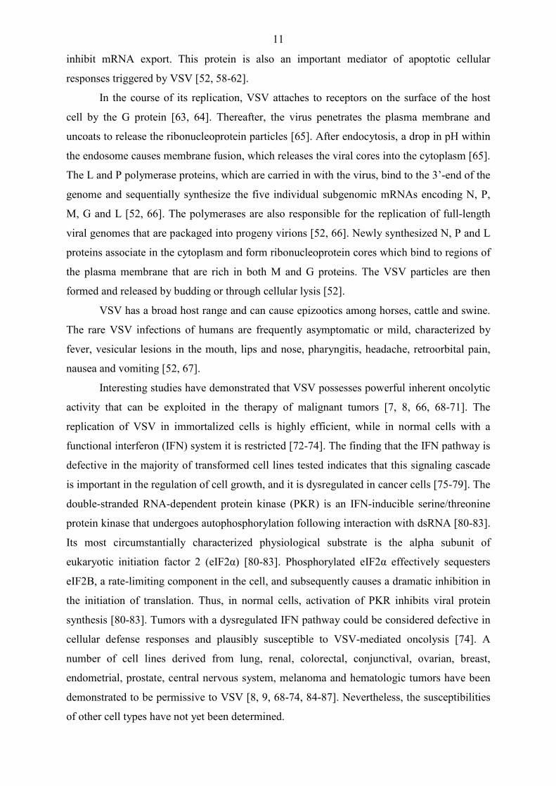
proteins [91]. The human p63 gene resides on chromosome 3q27–29, and consists of 15 exons
spread over about 270 kbp, with introns up to 100 kbp in length (Fig. 1) [91, 92]. There are
different p63 protein isoforms, which can be expressed from two distinct promoters, one
immediately preceding the first exon and the second one lying in the third intron (Fig. 1) [91-
97]. Transcription from the first and second promoters gives rise to transactivating (TA) or
amino terminally truncated (∆N) variations of p63, respectively [91-97]. The TA isoforms
possess an N-terminal acidic transactivation domain, while the ∆Np63 proteins lack this
domain (Fig. 1) [91-97]. Both TA and ∆N transcripts can undergo alternative splicing, leading
to the formation of three C-terminal variants, denoted α, β and γ, which further increase the
diversity of the p63 proteins (Fig. 1) [91-97].
All p63 isoforms contain a DNA-binding domain (DBD) and an oligomerization
domain (OD). In addition, the α isoforms contain a sterile alpha motif (SAM) and a
transactivation inhibitory domain (TID). The former is a protein-protein interaction domain,
while the latter is an inhibitory domain that blocks transactivation by masking a few residues
on the N-terminal of the TA domain, and in this way could be responsible for oligomerization
between different p63 isoforms [93]. The β variants lack exon 13 and consequently the SAM
and the TID domains (Fig. 1). The γ isoforms lack the C-terminal exons 11, 12, 13 and 14, but
incorporate an additional sequence of exon 15 (Fig. 1) [98].
13
Fig. 1. (A) Gene architecture of human p63. The alternative promoters and splicing events used to generate the various p63 isoforms are indicated. (B) Domain structure of the various p63 proteins. The transcription activation domain (TAD), DNA binding domain (DBD), oligomerization domain (OD), sterile α motif (SAM) and transinhibitor domain (TID) are depicted. The molecular size of each isoform is indicated on the right. (Not drawn to scale; adapted from [91-97] .) aa, amino acid
TAp63 and ∆Np63 isoforms have the ability to regulate a number of genes and possess
opposing regulatory effects [91-111]. As sequence-specific transcription factors, the TAp63
isoforms bind p53-responsive elements (p53-RE), stimulate the expression of p53 target
genes, such as the bax gene, and exert biological functions that partially overlap with those of
p53. The TAp63 isoforms also interact with the p63 DNA consensus motif, which is not
recognized by p53. Thus, a unique set of genes, which contains p63-RE, but lacks p53-RE in
its regulatory region, exhibits specific responsiveness to p63 [91, 93]. The TAp63 proteins
have been reported to induce growth arrest and apoptosis in a manner consistent with their
transactivation capabilities [92, 99-101, 104-108]. The various TA isoforms display widely
differing transcriptional efficiencies; proteins with β and γ C-termini exhibit higher
transactivation potentials than that of TAp63α, which contains TID. In contrast, the ∆Np63
isoforms may exert dominant-negative activities by antagonizing the target gene induction

14
triggered by TAp63 isoforms and p53 [99, 105, 107]. Moreover, these isoforms can actively
repress or activate transcription, possibly in consequence of the presence of two cryptic
transactivation domains [111, 112]. The ∆Np63 isoforms have been shown to inhibit
apoptosis and exert oncogenic properties [91, 94, 99, 100, 103, 108].
The six p63 isoforms regulate a wide array of cellular functions, including cell cycle
progression, proliferation, adhesion, senescence and apoptosis; thereby, they play important
roles in embryonic development, tumor progression and certain physiological processes and
pathological conditions that affect the epithelial tissues [91-108]. Noteworthy studies have
clearly demonstrated that p63 is instrumental in the development and maintenance of
epithelial tissues [91-101]. Its role has been elucidated in large part through the analysis of
p63-deficient mice, which display developmental abnormalities including the complete lack
of limbs, stratified epithelia and derivative structures such as skin, breast, prostate and hair
follicles [95, 96]. A range of human syndromes characterized by ectodermal dysplasia have
been linked to a diversity of heterozygous p63 mutations [113]. Although p63 does not
conform to Knudson’s two-hit hypothesis, the dysregulated expression of p63 has been shown
to contribute to the pathogenesis of cancers [97, 114]. Previous studies have also
demonstrated that p63 isoforms are involved in the control of the epithelial cell fate and in the
regulation of the differentiation program of the skin and the eye [91, 93-97].
Within the mature epidermis, ∆Np63α is the predominant isoform, expressed in high
levels in the basal layer of the skin [91-94]. ∆Np63α is indispensable for maintenance of the
viability and proliferative potential of basal keratinocytes, and it is also essential to prevent
the premature entry of cells into the differentiation program. The expression of ∆Np63α is
downregulated as keratinocytes commit to the process of differentiation, while the TAp63
isoforms are required to achieve terminal differentiation [91-94].
Within the ocular surface epithelia, no expression of the TAp63 isoforms can be
detected. In contrast, ∆Np63α is detected in high levels within the basal to intermediate layers
of the limbal and conjunctival epithelia [115]. This p63 isoform seems to be indispensable for
the viability and proliferative potential of the ocular surface epithelial stem cell population
[106-120]. Neither ∆Np63β nor ∆Np63γ has been shown to be present in substantial amounts
within the conjunctiva, the limbus or the cornea [115]. However, following injury, dramatic
increases are detected in the low-level constitutive expression of these p63 isoforms within
the regenerating corneal tissue [120].
15
IV. The Bcl-2–associated X protein (Bax) and its apoptotic regulatory functions
The Bcl-2–associated X protein, or Bax, is a product of the Bcl-2 gene family. Bcl-2 is
an oncogene, which is frequently linked in follicular lymphoma to an immunoglobulin locus
by the chromosome translocation t(14:18) [121]. It was the first example of an oncogene that
inhibits cell death rather than promotes proliferation. When homologs of Bcl-2 were
identified, it became apparent that these proteins can be defined by the presence of conserved
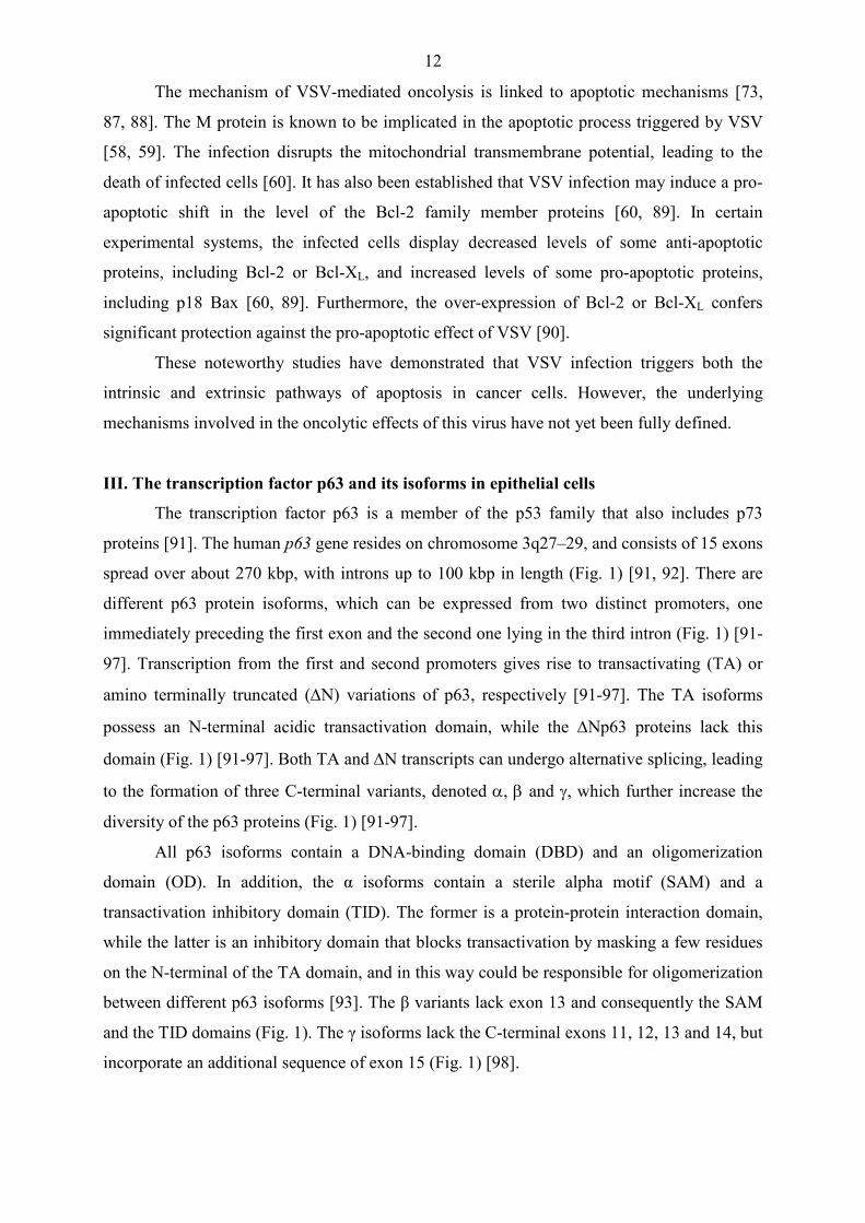
sequence motifs known as Bcl-2 homology domains (BH1 to BH4) [122]. In mammals, up to
30 relatives have been described, some of which belong in an anti-apoptotic group and others
in a pro-apoptotic group [123]. Besides Bcl-2 itself, there are several other anti-apoptotic
proteins (Bcl-w, Mcl-1, Bcl-XL and A1/Bfl-1), which all possess the domains BH1, BH2,
BH3 and BH4. The pro-apoptotic group of the Bcl-2 family can be divided into two
subgroups. The multi-domain pro-apoptotic subgroup consists of Bax, Bak and Bok, which all
possess the domains BH1, BH2 and BH3, whereas the BH3-only proteins (Bid, Bim, Bik,
Bad, Bmf, Hrk, Noxa, Puma, Blk, BNIP3 and Spike) have only the short BH3 motif, an
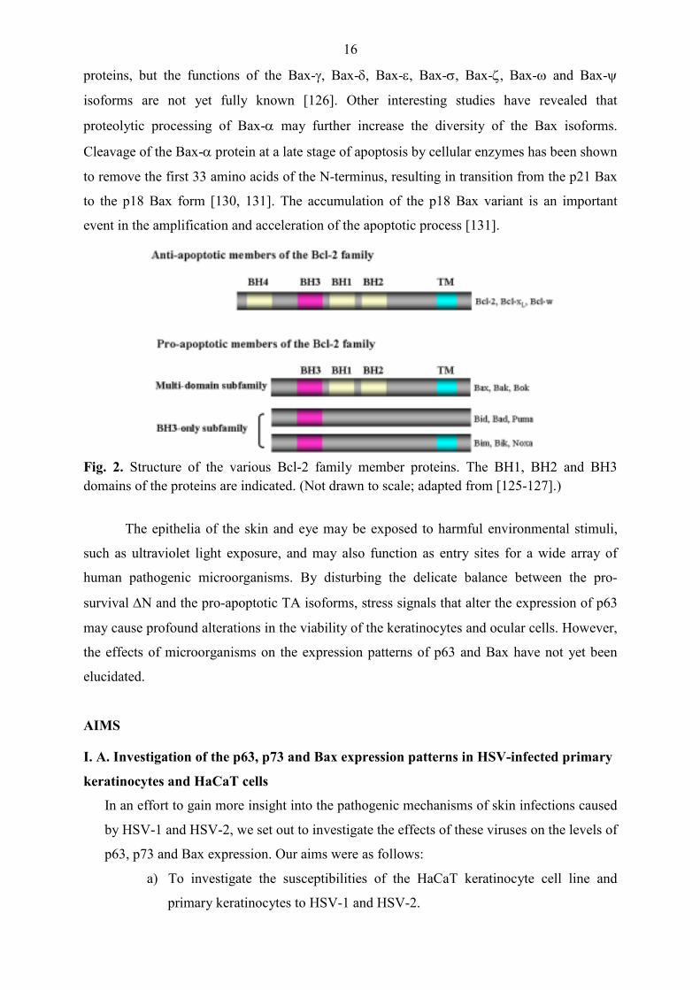
interaction domain that is both necessary and sufficient for their pro-apoptotic effect (Fig. 2)
[122, 123, 124].
The bax gene encodes multiple splice variants [125]. The alternatively splicing
patterns of the bax gene are highly complex. Nine Bax isoforms have been identified thus far,
including Bax-α, Bax-β, Bax-γ, Bax-δ, Bax-ε, Bax-σ, Bax-ζ, Bax-ω and Bax-ψ [126]. The
mRNA for Bax-α encodes a 21-kDa protein, while the mRNA for Bax-β encodes a 24-kDa
protein (Fig. 2) [126]. It is well documented that Bax-α is a central component of apoptosis
induction [111, 124, 127]. Bax-α can be found as a cytosolic monomer in viable cells. During
apoptosis, Bax-α changes its conformation, integrates into the outer mitochondrial membrane
and oligomerizes [122, 123]. Although the mechanism is controversial, Bax-α and Bak
oligomers are believed to provoke or contribute to the permeabilization of the outer
mitochondrial membrane, either by forming channels themselves or by interacting with
components of the permeability transition pore [122, 123]. This results in the release of
cytochrome c and other pro-apoptotic factors from the mitochondria, leading to the activation
of caspases and apoptosis [128]. Recent studies have clarified the function of Bax-β [126,
129]. The Bax-β protein is expressed constitutively in several human cell types, and its level
is controlled by proteasomal degradation. Similarly to Bax-α, Bax-β has the capability to
trigger apoptosis via the mitochondrial pathway. Moreover, Bax-β facilitates Bax-α activation
[126]. In this way, both proteins play important roles in the intrinsic pathway of apoptosis. A
few data suggest that most alternatively spliced Bax variants are active as pro-apoptotic
16
proteins, but the functions of the Bax-γ, Bax-δ, Bax-ε, Bax-σ, Bax-ζ, Bax-ω and Bax-ψ
isoforms are not yet fully known [126]. Other interesting studies have revealed that
proteolytic processing of Bax-α may further increase the diversity of the Bax isoforms.
Cleavage of the Bax-α protein at a late stage of apoptosis by cellular enzymes has been shown
to remove the first 33 amino acids of the N-terminus, resulting in transition from the p21 Bax
to the p18 Bax form [130, 131]. The accumulation of the p18 Bax variant is an important
event in the amplification and acceleration of the apoptotic process [131].
Fig. 2. Structure of the various Bcl-2 family member proteins. The BH1, BH2 and BH3 domains of the proteins are indicated. (Not drawn to scale; adapted from [125-127].)
The epithelia of the skin and eye may be exposed to harmful environmental stimuli,
such as ultraviolet light exposure, and may also function as entry sites for a wide array of
human pathogenic microorganisms. By disturbing the delicate balance between the pro-
survival ∆N and the pro-apoptotic TA isoforms, stress signals that alter the expression of p63
may cause profound alterations in the viability of the keratinocytes and ocular cells. However,
the effects of microorganisms on the expression patterns of p63 and Bax have not yet been
elucidated.
AIMS
I. A. Investigation of the p63, p73 and Bax expression patterns in HSV-infected primary
keratinocytes and HaCaT cells
In an effort to gain more insight into the pathogenic mechanisms of skin infections caused
by HSV-1 and HSV-2, we set out to investigate the effects of these viruses on the levels of
p63, p73 and Bax expression. Our aims were as follows:
a) To investigate the susceptibilities of the HaCaT keratinocyte cell line and
primary keratinocytes to HSV-1 and HSV-2.
17
b) To investigate the role of apoptosis in the cell demise triggered by HSV-1 and
HSV-2.
c) To analyze the expression levels of p63, p73 and Bax in HSV-1- or HSV-2-
infected HaCaT keratinocytes.
I. B. Investigation of the p63 and Bax expression patterns in HSV-1-infected SIRC
corneal cell line
In an effort to gain more insight into the pathogenic mechanism of herpetic ocular surface
disease, we set out to investigate the effects of HSV-1 on the levels of p63 and Bax
expression. Our aims were as follows:
a) To investigate the susceptibility of the Staatens Seruminstitute Rabbit Cornea
cell line (SIRC) to HSV-1.
b) To investigate the role of apoptosis in the cell demise triggered by HSV-1.
c) To analyze the expression levels of p63 and Bax in HSV-1-infected SIRC
cells.
II. Investigation of the p63 and Bax expression patterns in VSV-infected HaCaT
keratinocyte cell line
In an effort to evaluate the potential oncolytic activity of VSV in epithelial-derived skin
cancers, we set out to investigate the cytopathogenicity of this virus in the immortalized
HaCaT keratinocyte cell line. Our aims were as follows:
a) To investigate the susceptibility of the HaCaT cell line to VSV.
b) To investigate the role of apoptosis in the cell demise triggered by VSV.
c) To analyze the expression levels of p63 and Bax in VSV-infected HaCaT cells.
MATERIALS AND METHODS
Cell cultures
HaCaT cells: The HaCaT cell line, kindly provided by Dr. N. E. Fusenig (Heidelberg,
Germany), originally derived from the distant periphery of a melanoma located on the upper
half of the back of a 62-year-old male patient. The line is clonal in origin as indicated by
specific and stable cytogenetic markers, has a transformed phenotype in vitro but is not
tumorigenic, and is noninvasive in vivo, however it expresses mutated p53 (p53mt) [132]. The
cells were grown at 37 °C in a 5% CO2 atmosphere in Dulbecco's modified Eagle's minimal
essential medium (Sigma Chemical Co., St. Louis, MO, USA) supplemented with 10% fetal
calf serum (Gibco/BRL, Grand Island, NY, USA).
Primary keratinocytes: The normal human primary keratinocytes, kindly provided by
Prof. Dr. Lajos Kemény (Department of Dermatology and Allergology, University of Szeged,
18
Hungary), were cultured at 37 °C in a 5% CO2 atmosphere in keratinocyte growth medium
(Gibco/BRL).
SIRC cell line: The SIRC cell line was obtained from the European Collection of Cell
Cultures [(ECACC) (Health Protection Agency Culture Collections, Porton Down, UK)].
Cells were grown in Dulbecco's modified Eagle's minimal essential medium (Sigma)
supplemented with 10% fetal bovine serum (Gibco/BRL) at 37 °C in a 5% CO2 atmosphere.
Viruses
Herpes simplex viruses: The KOS strain of HSV-1 and the wild-type HSV-2 were
propagated at an MOI of 0.001 PFU per cell in Vero cell cultures for 3 days at 37 °C. The
culture fluids of HSV-1- or HSV-2-infected Vero cells were harvested, stored at -70 °C, and
used as the infecting stock of the virus.
Vesicular stomatitis virus: The Indiana strain of VSV was propagated at a multiplicity
of infection (MOI) of 0.001 plaque forming unit (PFU) per cell in L929 cell cultures for 3
days at 37 °C. The culture fluid of VSV-infected L929 cells was harvested, stored at -70 °C,
and used as the infecting stock of the virus.
Methods used to detect virus replication and host cell viability
Indirect immunofluorescence assay: Cytospin cell preparations were fixed in methanol-
acetone (1:1) for 15 minutes (min) at -20 °C. Slides were incubated with a 1:500 dilution of
VSV G protein-specific monoclonal antibody (MAb) (Sigma) or 1:200 dilution of HSV gD-
specific MAb (Santa Cruz Biotechnology Inc., Cambridge, MA, USA) for 1 hour (h) at 37 °C.
After washing with phosphate-buffered saline (PBS), the samples were reacted with
fluorescein isothiocyanate (FITC)-conjugated anti-mouse antibody (1:160) (Sigma) and
incubated for 1 h at 37 °C. After washing with PBS, the slides were visualized by confocal
microscopy. The ratio of positive to negative cells was determined after counting 1000 cells in
random fields.
Quantification of virus replication by plaque titration: Virus plaque assays were
performed on confluent monolayers of Vero cells inoculated with HSV or VSV for 1 h at
37 °C and overlaid with 0.5% agarose (FMC, Rockland, ME) in phenol red-free Eagle’s
minimum essential medium supplemented with 7.5% fetal bovine serum and 2 mM L-
glutamine. After 2 days of culturing at 37 °C, a second agarose overlay containing 0.005%
neutral red was added. Plaque titers were determined at 3 days after infection.
Quantification of cell viability by MTT assay: The viability of virus-infected cells was
measured with the colorimetric MTT [3-(4,5-dimethylthiazol-2-yl)2,5-diphenyltetrazolium
19
bromide] assay Tox-1 kit (Sigma). In this assay, the cells were seeded in 96-well plates at a
density of 1x10⁴/well. The cultures were infected with HSV or VSV at different MOIs. At 24
or 48 hours postinfection (hpi) at 37 oC, 10 µl MTT reagent (5 mg/ml) was added to each
well. After 2 h incubation, MTT solvent containing 0.1 M HCl and isopropanol was added for
15 h. Absorbance was measured at 545 and 630 nm. The ratio of living cells was calculated
via the following formula: percentage viability = [(absorbance of infected cells – blank) /
(absorbance of corresponding mock-infected control cells – blank)] x 100.
Inhibition of viral DNA replication: To inhibit the DNA replication of HSV-1 and
HSV-2, 9-[(2-Hydroxyethoxy)methyl]guanine [(ACG) (Sigma)] was used at various
concentrations when indicated.
Methods used to detect apoptosis
Quantification of apoptosis by enzyme-linked immunosorbent assay (ELISA): The
cells were washed in PBS and the cell pellet was processed in a cell death detection ELISA kit
(Roche Diagnostics GmbH, Penzberg, Germany) based on the measurement of histones
complexed with mono- and oligonucleosome fragments formed during cell death. For this
assay, the cells were incubated in lysis buffer for 30 min and centrifuged at 12,000 rpm for 10
min. The supernatants were transferred into a streptavidin-coated microplate and incubated
with biotin-conjugated anti-histone and peroxidase-conjugated anti-DNA monoclonal
antibodies for 2 h. After washing, substrate solution 2,2'-azino-bis(3-ethylbenzthiazoline-6-
sulphonic acid) (ABTS) was added to each well for 15 min. Absorbance was measured at 405
and 490 nm. The specific enrichment of mono- and oligonucleosomes was calculated as the
enrichment factor (EF) = absorbance of infected cells/absorbance of corresponding non-
infected control cells.
Quantification of apoptosis by annexin V staining: The cells were stained with
FITC-labeled annexin V and propidium iodide (PI) (Bender MedSystems Inc., Burlingame,
CA, USA) according to the manufacturer’s instructions. The fluorescence intensities of
annexin-FITC and PI were determined with a FACStar Plus flow cytometer (BD Biosciences,
San Diego, CA, USA) by using the WinMDI software. The percentages of apoptotic cells
were calculated by sorting the cells that were positive only for annexin V (early apoptotic
stage) or positive for both annexin V and PI (late apoptotic and necrotic stages).
Methods used to identify proteins
Western blot assays: Cells (1×10⁷) were homogenized in ice-cold lysis buffer containing
150 mM NaCl, 10 mM Tris⋅HCl, pH 7.6, 5 mM EDTA, 1% (v/v) Nonidet P-40, 0.1% SDS,
20
1% sodium deoxycholate and protease inhibitor cocktail (Sigma), and the mixture was then
centrifuged at 10,000 g for 10 min to remove cell debris. Protein concentrations of cell lysates
were determined by using the Bio-Rad protein assay (Bio-Rad, Hercules, CA, USA).
Supernatants were mixed with Laemmli's sample buffer and boiled for 3 min. Aliquots of the
supernatants, containing 50 µg of total protein to detect HSV gD, VSV G protein, p53, p63,
p73, Bax and β-actin, were resolved by SDS-polyacrylamide gel electrophoresis (PAGE) and
electrotransferred onto nitrocellulose filters (Amersham, Buckinghamshire, UK). Preblocked
blots were reacted with specific antibodies to VSV G protein (Sigma), HSV gD (Sigma), p63
(clone 4A4) detecting all of the various p63 isoforms (Santa Cruz), p40 detecting the ∆Np63
isoforms (Merck KGaA, Darmstadt, Germany), p53 (Serotec Inc. Raleigh, NC), p73 (clone H-
79) detecting all of the various p73 isoforms (Santa Cruz), β-actin (Sigma), and Bax
(PharMingen, SanDiego, CA) for 4 h in PBS containing 0.05% (v/v) Tween 20, 1% (w/v)
dried non-fat milk (Difco Laboratories, Detroit, MI) and 1% (w/v) BSA (fraction V; Sigma).
Blots were then incubated for 2 h with species-specific secondary antibodies coupled to
peroxidase [peroxidase-conjugated anti-mouse antibody (DakoCytomation, Carpinteria, CA,
USA), or peroxidase-conjugated anti-rabbit antibody (DakoCytomation)]. Filters were washed
five times in PBS−Tween for 5 min after each step and were developed by using a
chemiluminescence detection system (Amersham). The autoradiographs were scanned with a
GS-800 densitometer (Bio-Rad), and the relative band intensities were quantified by use of
the ImageQuant software (Amersham).
Gene silencing by small interfering RNA (siRNA): Chemically synthetized siRNA
targeting TAp63 (Silencer siRNA 4798) and non-silencing control siRNA (Silencer negative
control #2 siRNA 4613) were obtained from Ambion Inc. (Austin, TX, USA). Transient
transfections were performed by using the siPORT amine reagent (Ambion) according to the
manufacturer’s protocol, with a final siRNA concentration of 50 nM. The transfected HaCaT
cells were incubated at 37 °C in a humidified atmosphere of 5% CO2 for 48 h. The effect of
silencing was analyzed at the protein level by Western blot assay.
Statistical analysis
All values are expressed as means ± standard deviation (SD). Student's unpaired t test
was used for comparisons and P values < 0.05 were considered statistically significant. The
one-way ANOVA test with the Bonferroni post-test was used for pairwise multiple
comparisons, and P values < 0.05 were considered statistically significant.

21
RESULTS
I. A. The effects of HSV infection on the expression patterns of p63, p73 and Bax in
HaCaT cells and primary keratinocytes
I. A. 1. The HaCaT cell line is permissive for HSV-1 replication
The HaCaT cells were infected with the KOS strain of HSV-1 at various multiplicities
and maintained for different periods of time. The production of progeny virus was determined
by plaque titration of the culture supernatants taken from HaCaT cells at 6, 12, 24 or 48 hpi.
Depending on the infectious dose, the level of HSV-1 production varied between
<5×10² and 1×10⁴ PFU/ml at 6 hpi (Table 1). The virus titers thereafter increased, and ranged
from 6×10⁶ and 3.3×10⁸ PFU/ml at 48 h after inoculation. Accordingly, the maximum yield at
0.001, 0.01, 0.1, 1 and 10 MOI corresponded to 30, 550, 600, 1650 and 1500 PFU/cell,
respectively. These data demonstrate that HSV-1 replicates efficiently in the HaCaT
keratinocyte cell line.
Table 1. Viral titers in HSV-1-infected HaCaT cells
MOI Titer of HSV-1 (PFU/ml)
6 h 12 h 24 h 48 h
0.001 <5x10² <5x10² 1.7x10⁴ 6x10⁶
0.01 <5x10² <5x10² 1.6x10⁶ 1.1x10⁸
0.1 5x10² 1x10³ 8x10⁶ 1.2x10⁸
1.0 2x10³ 3.5x10³ 1.7x10⁸ 3.3x10⁸
10 1x10⁴ 4.7x10⁵ 1.5x10⁸ 3x10⁸
I. A. 2. HSV-1 and HSV-2 trigger cell death in the HaCaT keratinocyte cell line
The cytopathogenicity of HSV-1 and HSV-2 was determined by the MTT assay.
HSV-1-infected cells displayed decreased viability at 24 hpi; the proportions of dead cells
were 9, 28, 38 and 50% at MOIs of 0.01, 0.1, 1 and 10, respectively (Fig. 3A). HSV-2-
infected cells likewise exhibited decreased viability; at 24 h after inoculation the proportions
of dead cells were 9, 21, 42 and 50% at MOIs of 0.01, 0.1, 1 and 10, respectively (Fig. 3A).
To examine the ability of HSV-1 and HSV-2 to induce apoptosis in HaCaT cells, the
extent of apoptosis was measured by annexin V binding assay at 24 hpi. The proportions of
annexin V-single-positive (early apoptotic) and double-positive (early apoptotic and necrotic)
cells in cultures infected with HSV-1 at an MOI of 10 were 21 and 16%, respectively
(Fig. 3B). In contrast, the proportions of annexin V-single-positive and double-positive cells
in cultures infected with HSV-2 at an MOI of 10 were 12 and 25%, respectively (Fig. 3B).

22
These results indicate that HSV-1 and HSV-2 trigger different types of cell death in HaCaT
cultures.
Fig. 3. HSV-1 and HSV-2 induce cell death in the HaCaT keratinocyte cell line. Cellular viability was measured by the MTT assay at 24 hpi (A). Apoptosis was measured by annexin V staining (B). *P < 0.05; ***P < 0.001.
I. A. 3. HSV-1 and HSV-2 alter the levels of Bax, p63 and p73 in a serotype-specific
manner
To determine whether HSV-1 and HSV-2 can alter the expressions of Bax, p63 and
p73 in the HaCat cell line, the steady-state levels of these proteins were determined by
Western blot analysis. Experiments to investigate the kinetics of HSV-1 replication revealed
the presence of gD in cultures infected with HSV-1 as early as 6 hpi (Fig.4; lane 18). The
level of gD thereafter increased, and its expression was highly upregulated in every culture
infected with HSV-1 at 48 h after inoculation (Fig. 4; lanes 32-36).
Mock-infected HaCaT cells displayed the endogenous expression of Bax-α, which
remained constant during the 48 h of culturing (Fig. 4; lanes 1, 7, 13, 19, 25 and 31).
Interestingly, the analysis revealed the presence of a Bax isoform corresponding to Bax-β in
HSV-1-infected cultures as early as 6 hpi (Fig. 4; lane 18). The level of Bax-β thereafter

23
increased, and its expression was highly upregulated in every culture infected with HSV-1 by
48 hpi (Fig. 4; lanes 32-36).
The expression pattern of p63 was determined by using an antibody preparation,
which recognizes all of the various p63 isoforms. The analysis demonstrated that the
predominant isoform in the HaCaT cell line is a p63 protein migrating near 68 kDa. HSV-1
triggered an impressive reduction in the level of this 68 kDa p63 isoform. The HSV-1-
infected cells exhibited decreased levels of this protein as early as 12 hpi (Fig. 4; lanes 23-24).
The expression of the 68 kDa p63 isoform was downregulated in cells infected at MOIs of
0.1, 1 and 10 at 48 h after inoculation (Fig. 4; lanes 32-36). Interestingly, a p63 isoform
migrating between 51 and 62 kDa was also detected in HSV-1-infected cells as early as 6 hpi
(Fig. 4; lane 18). The level of the 51-62 kDa p63 isoform thereafter increased, and the
expression of this protein was highly upregulated in every culture infected with HSV-1 by 48
hpi (Fig. 4; lanes 32-36).
Given the high number of possible, functionally diverse p63 isoforms, an exact
assignment of the isoforms to the proteins detected by Western blot analysis through use of
the pan-p63-specific antibody is difficult. Thus, to identify the p63 isoforms, the steady-state
levels of these proteins were also determined by Western blot analysis, using a polyclonal
antiserum which reacts only with the ∆N forms. The ∆Np63-specific antibody preparation
detected the 68 kDa p63 isoform, but failed to recognize the 51-62 kDa p63 isoform in
HSV-1-infected cultures. This result indicates that the 68 kDa isoform belongs in the ∆N
subclass and might be identical with ∆Np63α, while the 51-62 kDa isoform is a member of
the TA subclass and corresponds to TAp63γ. Furthermore, these experiments confirmed that
the level of ∆Np63α was decreased, while the expression of TAp63γ was highly increased
following HSV-1 infection (Fig. 4; lanes 1-36).
The expression pattern of p73 was determined by using an antibody preparation which
recognizes all of the various p73 isoforms. Mock-infected HaCaT cells expressed two p73
isoforms, migrating near 50 and 44.5 kDa, the levels of which remained constant during the
48 h of culturing (Fig.4; lanes 1, 7, 13, 19, 25 and 31). The HSV-1-infected cells exhibited a
decreased level of the 50 kDa p73 isoform at 24 h after virus inoculation (Fig. 4; lanes 26-30).
In every infected culture, the expression of this protein was likewise downregulated by 48 hpi
(Fig. 4; lanes 32-36). The HSV-1-infected cells displayed an increased level of the 44.5 kDa
p73 isoform at 24 hpi (Fig. 4; lanes 26-30). Similarly, the level of this protein was
upregulated by 48 hpi in cultures infected with HSV-1 at MOIs of 0.01, 0.1, 1 and 10 (Fig. 4;
lanes 33-36).
24
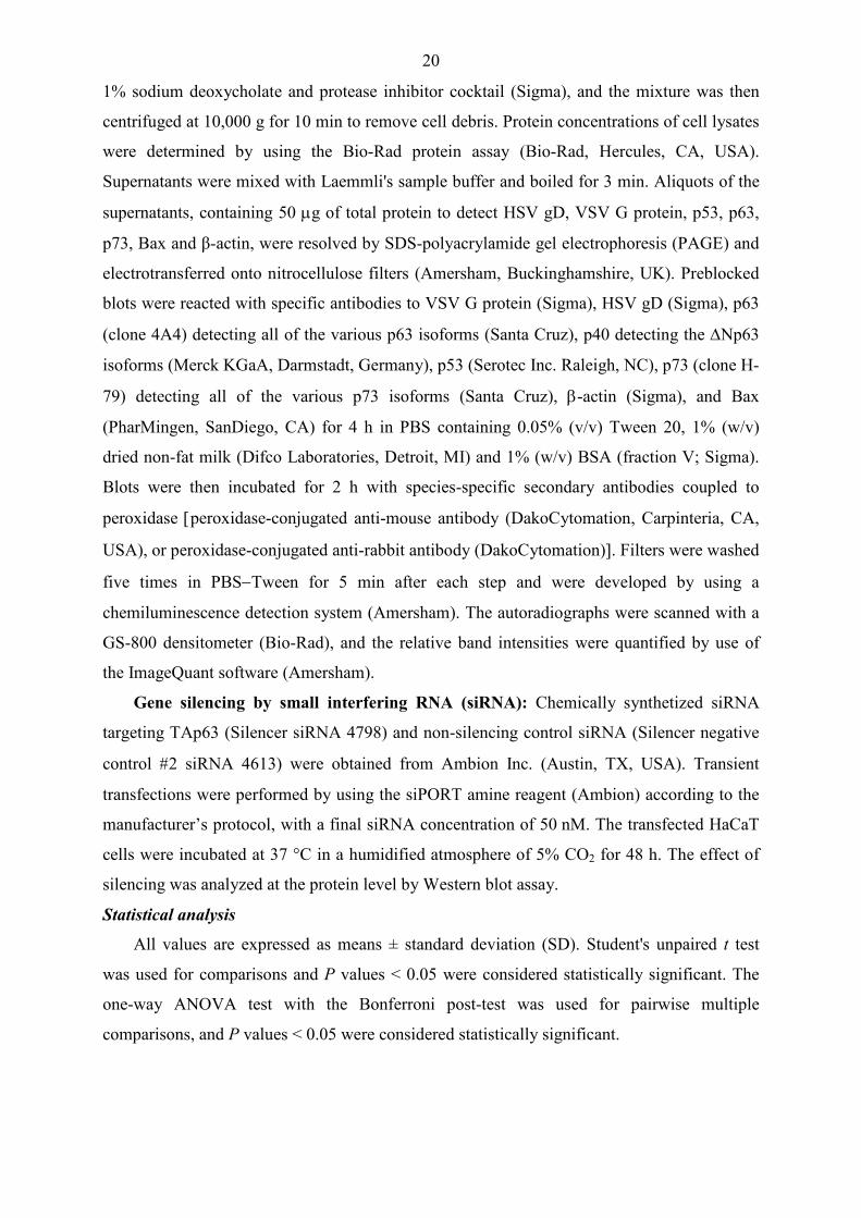
Experiments to investigate the replication of HSV-2 revealed the presence of gD in
cultures infected with HSV-2 at 24 hpi (Fig. 4; lanes 40-42). The levels of the 50 and 44.5
kDa p73 isoforms and ∆Np63α were decreased, Bax-α and TAp63γ remained unaffected,
while the expression of Bax-β was slightly increased at 24 h after inoculation in HSV-2-
infected HaCaT cells (Fig. 4; lanes 38-42). These findings suggest that HSV-1 and HSV-2
alter the levels p63, p73 and Bax in a type-specific manner in HaCaT epithelial cell cultures.
Fig. 4. HSV-1 and HSV-2 differentially modulate the levels of Bax, p63 and p73 in the HaCaT keratinocyte cell line. The steady-state levels of proteins were analyzed by Western blot assay. To quantify protein levels in HSV-infected cells, band intensities were determined by densitometric analysis with the ImageQuant software. The numbers indicate the relative quantities of each band, normalized to the control cells at each time point. I. A. 4. HSV-1 alters the levels of Bax and p63 in primary keratinocytes
To determine whether HSV-1 can dysregulate the expressions of Bax and p63 in
primary keratinocytes, the steady-state levels of these proteins were determined by Western
blot analysis. Primary keratinocytes were infected at an MOI of 1, and the kinetics of HSV-1
replication was investigated. The experiments revealed the presence of gD in cultures infected
25
with HSV-1 as early as 6 hpi, and its level was highly increased at 12 and 24 h after
inoculation (Fig. 5; lanes 6, 8, 10).
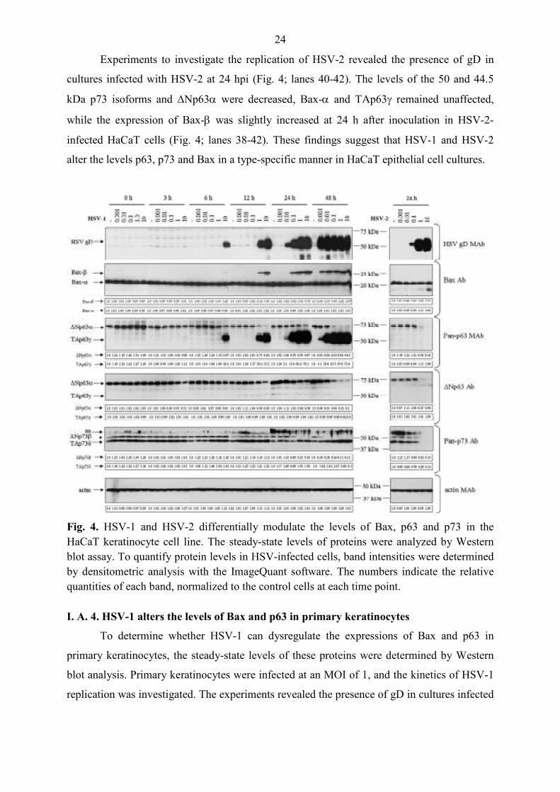
The mock-infected cells displayed the endogenous expression of the wild-type p53
protein (p53wt), ∆Np63α and Bax-α (Fig. 5; lanes 1, 3, 5, 7, 9). The level of ∆Np63α was
decreased, p53wt and Bax-α remained unaffected, while the expressions of the Bax-β and
TAp63γ were highly increased by 12 hpi in HSV-1-infected primary keratinocytes (Fig. 5;
lane 8). These data indicate that HSV-1 alters the levels of Bax and p63 in primary
keratinocytes.
Fig. 5. HSV-1 alters the levels of Bax and p63 in primary keratinocytes. The steady-state levels of proteins were analyzed by Western blot assay. To quantify protein levels in HSV-infected cells, band intensities were determined by densitometric analysis with the ImageQuant software. The numbers indicate the relative quantities of each band, normalized to the control cells at each time point.
I. A. 5. HSV-1-mediated TAp63γ expression requires viral DNA replication
To investigate the basis of the HSV-1-induced accumulation of TAp63γ, HaCaT cells
were infected in the presence or absence of the viral DNA replication inhibitor ACG. The
cells were analyzed for the presence of p63 and Bax by Western blot analysis.
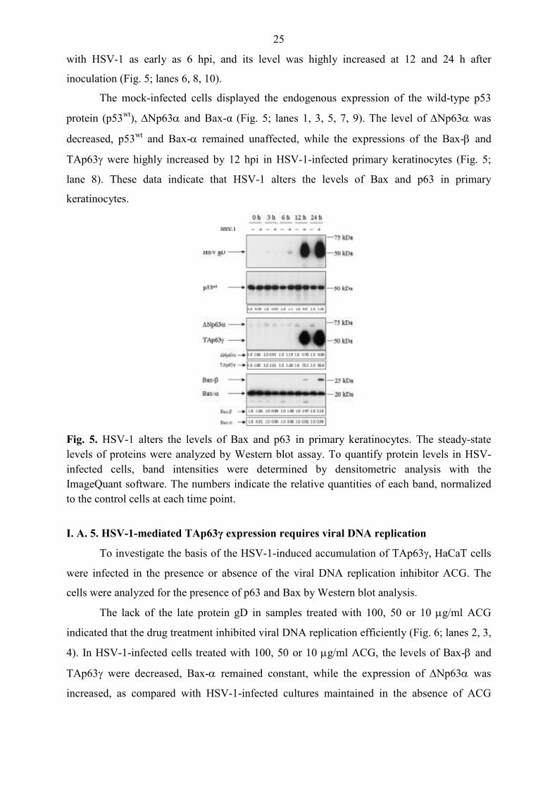
The lack of the late protein gD in samples treated with 100, 50 or 10 µg/ml ACG
indicated that the drug treatment inhibited viral DNA replication efficiently (Fig. 6; lanes 2, 3,
4). In HSV-1-infected cells treated with 100, 50 or 10 µg/ml ACG, the levels of Bax-β and
TAp63γ were decreased, Bax-α remained constant, while the expression of ∆Np63α was
increased, as compared with HSV-1-infected cultures maintained in the absence of ACG
26
(Fig. 6; lanes 2, 3, 4 and 1). These findings demonstrate that the HSV-1-mediated TAp63γ
expression requires viral DNA replication.
Fig. 6. HSV-1-mediated TAp63γ expression requires viral DNA replication. HaCaT cells were infected with the KOS strain of HSV-1 at an MOI of 1 and maintained for 24 h in the presence or in the absence of ACG. The steady-state levels of Bax and p63 isoforms were analyzed by Western blot assay. To quantify protein levels in HSV-1-infected cells, band intensities were determined by densitometric analysis with the ImageQuant software. The numbers indicate the relative quantities of each band, normalized to the control cells at each time point.
I. A. 6. Knockdown of HSV-1-induced TAp63 expression increases the viability of
infected HaCaT cells
To evaluate the biological effects of the accumulation of TAp63γ in HSV-1-infected
cells, siRNA technology was used. The delivery of TAp63-specific siRNA resulted in an 85%
reduction in HSV-1-induced TAp63γ expression as compared with cultures treated with a
negative control siRNA preparation (Fig. 7A). In the presence of the TAp63-specific siRNA,
the viability of HSV-1-infected cells was increased by about 15% at 24 hpi (Fig. 7B). These
results confirm that the 51-62 kDa p63 isoform corresponds to the TA subclass, and suggest
that TAp63γ may play some role in the cytopathogenicity of HSV-1.
27
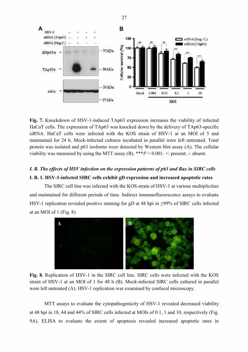
Fig. 7. Knockdown of HSV-1-induced TAp63 expression increases the viability of infected HaCaT cells. The expression of TAp63 was knocked down by the delivery of TAp63-specific siRNA. HaCaT cells were infected with the KOS strain of HSV-1 at an MOI of 5 and maintained for 24 h. Mock-infected cultures incubated in parallel were left untreated. Total protein was isolated and p63 isoforms were detected by Western blot assay (A). The cellular viability was measured by using the MTT assay (B). ***P < 0.001. +: present; -: absent.
I. B. The effects of HSV infection on the expression patterns of p63 and Bax in SIRC cells
I. B. 1. HSV-1-infected SIRC cells exhibit gD expression and increased apoptotic rates
The SIRC cell line was infected with the KOS strain of HSV-1 at various multiplicities
and maintained for different periods of time. Indirect immunofluorescence assays to evaluate
HSV-1 replication revealed positive staining for gD at 48 hpi in ≥99% of SIRC cells infected
at an MOI of 1 (Fig. 8).
Fig. 8. Replication of HSV-1 in the SIRC cell line. SIRC cells were infected with the KOS strain of HSV-1 at an MOI of 1 for 48 h (B). Mock-infected SIRC cells cultured in parallel were left untreated (A). HSV-1 replication was examined by confocal microscopy.
MTT assays to evaluate the cytopathogenicity of HSV-1 revealed decreased viability
at 48 hpi in 18, 44 and 44% of SIRC cells infected at MOIs of 0.1, 1 and 10, respectively (Fig.
9A). ELISA to evaluate the extent of apoptosis revealed increased apoptotic rates in

28
HSV-1-infected SIRC cells at 48 hpi; the EFs measured at MOIs of 0.1, 1 and 10 were 1.42,
4.35 and 5.8, respectively (Fig. 9B).
Fig. 9. HSV-1 induces cell death in the SIRC cell line. SIRC cells were infected with HSV-1 at different MOIs for 48 h. Mock-infected cells cultured in parallel were left untreated. The cell viability was measured by using the MTT assay (A). Apoptosis was detected by measuring the specific enrichment of mono- and oligonucleosomes in the cytoplasm by ELISA (B). aP < 0.001 vs. mock; bP < 0.001 vs. 0.1 MOI; ns = nonsignificant vs. mock.
Together, these data demonstrate the expression of HSV-1 gD protein that is
consistent with efficient viral replication. Moreover, these results reveal that HSV-1 elicits a
strong cytopathic effect in the SIRC cell line, and apoptosis plays an important role in the
demise of the infected cells.
I. B. 2. HSV-1 alters the levels of Bax and p63 proteins in SIRC cells
To determine whether HSV-1 can alter the expressions of Bax and p63, the
steady-state levels of these proteins were determined by Western blot analysis. First, the
kinetics of HSV-1 gD expression was investigated. The presence of gD was observed in the
SIRC cell cultures infected with HSV-1 at an MOI of 10 at 12 hpi (Fig. 10; lane 20). The gD
protein accumulated in the cultures infected with HSV-1 at MOIs of 0.1, 1 and 10 at 24 hpi
(Fig. 10; lanes 23-25). High-level expression of the gD protein was also revealed in every
culture infected with HSV-1 by 48 hpi (Fig. 10; lanes 27-30).
The analysis revealed the presence of a Bax isoform corresponding to Bax-β in HSV-
1-infected SIRC cultures at 12 hpi (the relative quantity of Bax-β in cells infected at an MOI
of 10 was 1.67) (Fig. 10; lane 20). At the 24-h time point, the expression of the Bax-β protein
in the HSV-1-infected SIRC cultures was upregulated (the relative quantities of Bax-β in cells
infected at MOIs of 1 and 10 were 6.42 and 8.31, respectively) (Fig. 10; lanes 24 and 25). At
the 48-h time point, the HSV-1-infected SIRC cultures displayed elevated levels of Bax-β (the

29
relative quantities of Bax-β in cells infected at MOIs of 0.01, 0.1, 1 and 10 were 9.27, 9.93,
7.57 and 6.62, respectively) (Fig. 10; lanes 27-30).
The expression pattern of p63 was determined by using an antibody preparation which
recognizes all of the various p63 isoforms. The analysis revealed the constitutive expression
of a p63 protein migrating near 68 kDa in the mock-infected SIRC cells (lanes 1, 6, 11, 16, 21
and 26 in Fig. 10). Previously published data demonstrated that the 68 kDa protein possibly
corresponds to ∆Np63α [92, 133]. At 12 hpi, the expression of ∆Np63α in the HSV-1-
infected SIRC cultures was downregulated (the relative quantity of ∆Np63α in cells infected
at an MOI of 10 was 0.87) (Fig. 10; lane 20). At the 24-h time point, HSV-1 triggered an
impressive reduction in the level of ∆Np63α in the SIRC cells (the relative quantities in cells
infected at MOIs of 0.1, 1 and 10 were 0.89, 0.43 and 0.41, respectively) (Fig. 10; lanes 23-
25). At the 48-h time point, the HSV-1-infected SIRC cultures exhibited decreased levels of
∆Np63α (the relative quantities in cells infected at MOIs of 0.01, 0.1, 1 and 10 were 0.36,
0.22, 0.19 and 0.17, respectively) (Fig. 10; lanes 27-30).
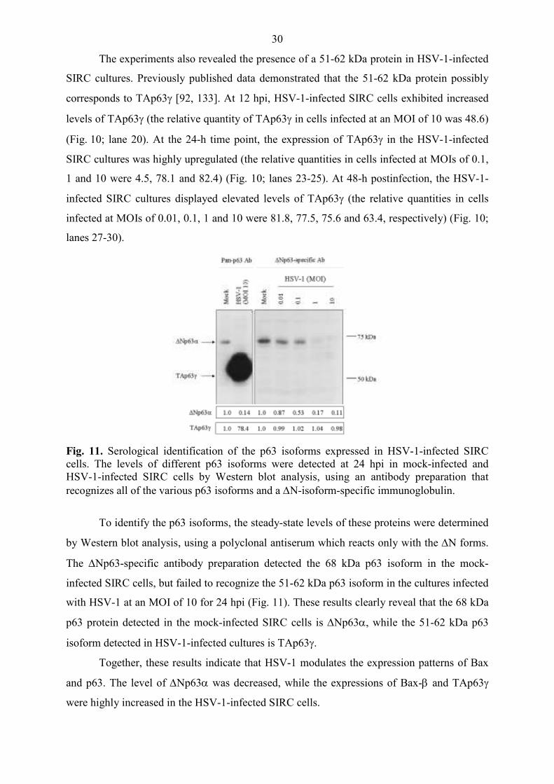
Fig. 10. HSV-1 infection alters the levels of p63 and Bax-β in the SIRC cell line. The steady-state levels of proteins were analyzed by Western blot assay. To quantify protein levels in HSV-1-infected cells, band intensities were determined by densitometric analysis with the ImageQuant software. The numbers indicate the relative quantities of each band, normalized to the control cells at each time point.
30
The experiments also revealed the presence of a 51-62 kDa protein in HSV-1-infected
SIRC cultures. Previously published data demonstrated that the 51-62 kDa protein possibly
corresponds to TAp63γ [92, 133]. At 12 hpi, HSV-1-infected SIRC cells exhibited increased
levels of TAp63γ (the relative quantity of TAp63γ in cells infected at an MOI of 10 was 48.6)
(Fig. 10; lane 20). At the 24-h time point, the expression of TAp63γ in the HSV-1-infected
SIRC cultures was highly upregulated (the relative quantities in cells infected at MOIs of 0.1,
1 and 10 were 4.5, 78.1 and 82.4) (Fig. 10; lanes 23-25). At 48-h postinfection, the HSV-1-
infected SIRC cultures displayed elevated levels of TAp63γ (the relative quantities in cells
infected at MOIs of 0.01, 0.1, 1 and 10 were 81.8, 77.5, 75.6 and 63.4, respectively) (Fig. 10;
lanes 27-30).
Fig. 11. Serological identification of the p63 isoforms expressed in HSV-1-infected SIRC cells. The levels of different p63 isoforms were detected at 24 hpi in mock-infected and HSV-1-infected SIRC cells by Western blot analysis, using an antibody preparation that recognizes all of the various p63 isoforms and a ∆N-isoform-specific immunoglobulin.
To identify the p63 isoforms, the steady-state levels of these proteins were determined
by Western blot analysis, using a polyclonal antiserum which reacts only with the ∆N forms.
The ∆Np63-specific antibody preparation detected the 68 kDa p63 isoform in the mock-
infected SIRC cells, but failed to recognize the 51-62 kDa p63 isoform in the cultures infected
with HSV-1 at an MOI of 10 for 24 hpi (Fig. 11). These results clearly reveal that the 68 kDa
p63 protein detected in the mock-infected SIRC cells is ∆Np63α, while the 51-62 kDa p63
isoform detected in HSV-1-infected cultures is TAp63γ.
Together, these results indicate that HSV-1 modulates the expression patterns of Bax
and p63. The level of ∆Np63α was decreased, while the expressions of Bax-β and TAp63γ
were highly increased in the HSV-1-infected SIRC cells.
31
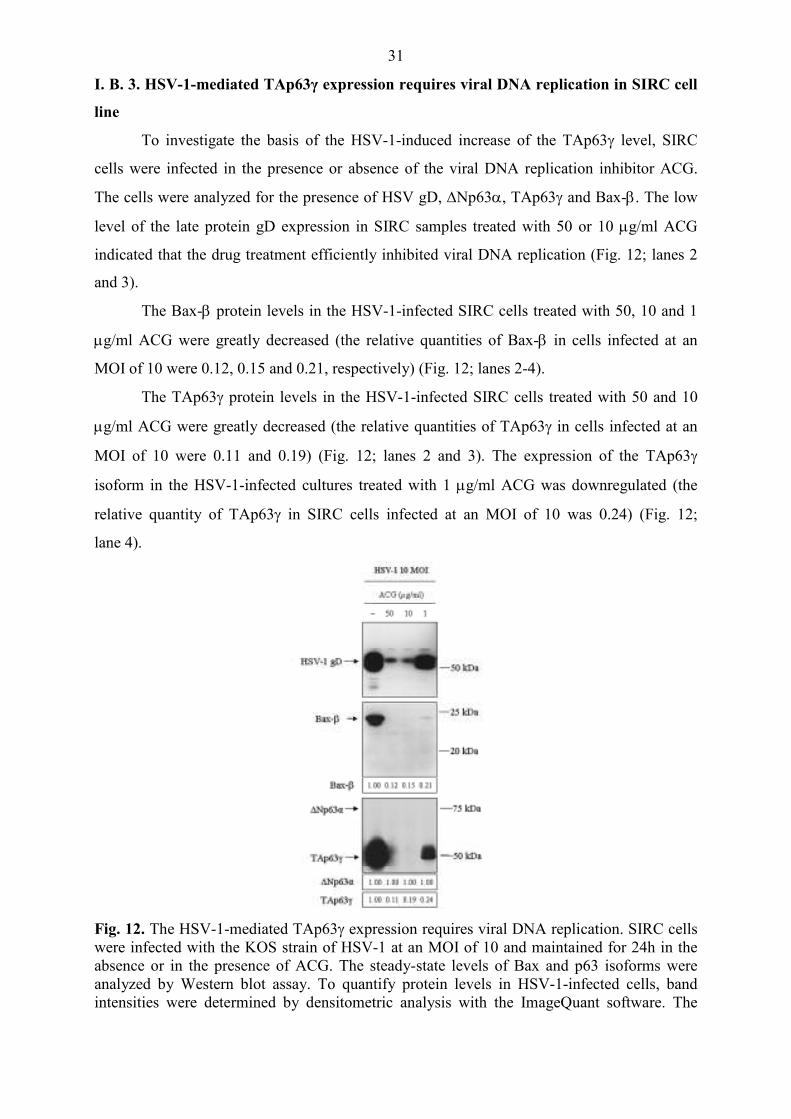
I. B. 3. HSV-1-mediated TAp63γ expression requires viral DNA replication in SIRC cell
line
To investigate the basis of the HSV-1-induced increase of the TAp63γ level, SIRC
cells were infected in the presence or absence of the viral DNA replication inhibitor ACG.
The cells were analyzed for the presence of HSV gD, ∆Np63α, TAp63γ and Bax-β. The low
level of the late protein gD expression in SIRC samples treated with 50 or 10 µg/ml ACG
indicated that the drug treatment efficiently inhibited viral DNA replication (Fig. 12; lanes 2
and 3).
The Bax-β protein levels in the HSV-1-infected SIRC cells treated with 50, 10 and 1
µg/ml ACG were greatly decreased (the relative quantities of Bax-β in cells infected at an
MOI of 10 were 0.12, 0.15 and 0.21, respectively) (Fig. 12; lanes 2-4).
The TAp63γ protein levels in the HSV-1-infected SIRC cells treated with 50 and 10
µg/ml ACG were greatly decreased (the relative quantities of TAp63γ in cells infected at an
MOI of 10 were 0.11 and 0.19) (Fig. 12; lanes 2 and 3). The expression of the TAp63γ
isoform in the HSV-1-infected cultures treated with 1 µg/ml ACG was downregulated (the
relative quantity of TAp63γ in SIRC cells infected at an MOI of 10 was 0.24) (Fig. 12;
lane 4).
Fig. 12. The HSV-1-mediated TAp63γ expression requires viral DNA replication. SIRC cells were infected with the KOS strain of HSV-1 at an MOI of 10 and maintained for 24h in the absence or in the presence of ACG. The steady-state levels of Bax and p63 isoforms were analyzed by Western blot assay. To quantify protein levels in HSV-1-infected cells, band intensities were determined by densitometric analysis with the ImageQuant software. The
32
numbers indicate the relative quantities of each band, normalized to the control cells at each time point. II. The effects of VSV infection on the expression patterns of p63 and Bax in HaCaT cells
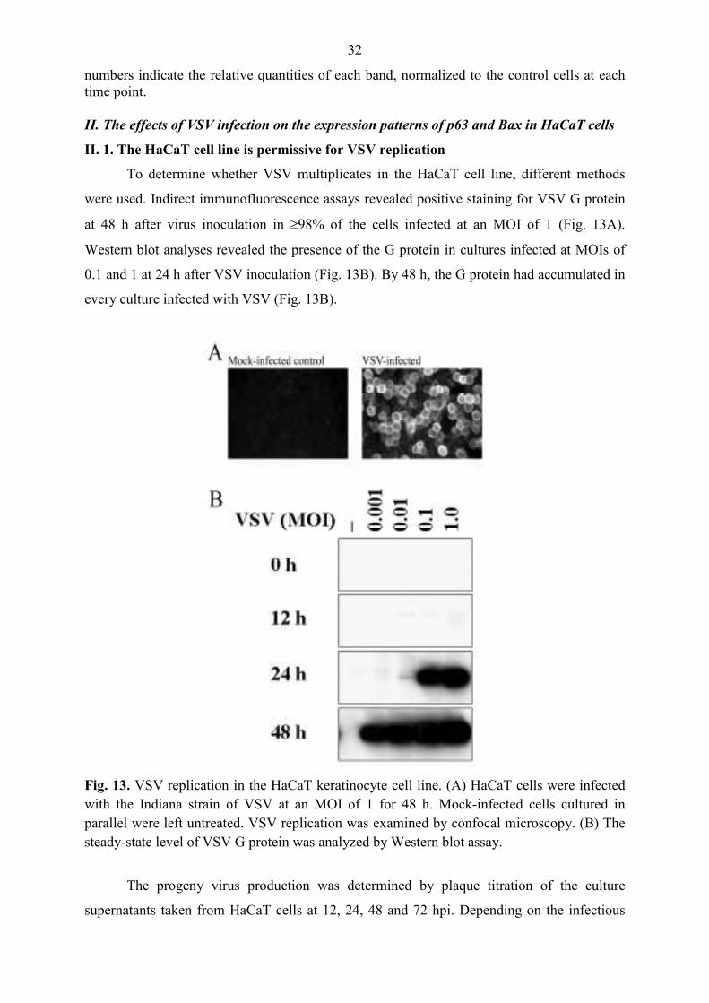
II. 1. The HaCaT cell line is permissive for VSV replication
To determine whether VSV multiplicates in the HaCaT cell line, different methods
were used. Indirect immunofluorescence assays revealed positive staining for VSV G protein
at 48 h after virus inoculation in ≥98% of the cells infected at an MOI of 1 (Fig. 13A).
Western blot analyses revealed the presence of the G protein in cultures infected at MOIs of
0.1 and 1 at 24 h after VSV inoculation (Fig. 13B). By 48 h, the G protein had accumulated in
every culture infected with VSV (Fig. 13B).
Fig. 13. VSV replication in the HaCaT keratinocyte cell line. (A) HaCaT cells were infected with the Indiana strain of VSV at an MOI of 1 for 48 h. Mock-infected cells cultured in parallel were left untreated. VSV replication was examined by confocal microscopy. (B) The steady-state level of VSV G protein was analyzed by Western blot assay.
The progeny virus production was determined by plaque titration of the culture
supernatants taken from HaCaT cells at 12, 24, 48 and 72 hpi. Depending on the infectious
33
dose, the level of VSV production varied between 3.0 x 10³ and 1.9 x 10⁶ PFU/ml at 12 hpi
(Table 2). The virus titers thereafter increased, and ranged from 4.5 x 10⁵ to 4.6 x 10⁷ PFU/ml
at 24 h after virus inoculation (Table 2). The level of virus production varied between 2.6 x
107 and 6.4 x 10⁷ PFU/ml at 48 hpi (Table 2). The VSV production of cells infected with
various MOIs rose to titers of about 2 x 10⁸ PFU/ml after 72 h of culturing (Table 2).
Accordingly, the maximum yield at 0.001, 0.01, 0.1 and 1 MOI corresponded to 1300,
1400, 1200, and 1000 PFU/cell, respectively. Together, these data clearly demonstrate that
VSV replicates efficiently in the HaCaT keratinocyte cell line.
Table 2. Viral titers in VSV-infected HaCaT cells
MOI Titer of VSV (PFU/ml)
12 h 24 h 48 h 72 h
0.001 3.0 x 10³ 4.5 x 10⁵ 2.6 x 10⁷ 2.6 x 10⁸
0.01 1.5 x 10⁴ 3.6 x 10⁶ 2.8 x 10⁷ 2.8 x 10⁸
0.1 1.5 x 10⁵ 1.7 x 10⁷ 3.8 x 10⁷ 2.4 x 10⁸
1.0 1.9 x 10⁶ 4.6 x 10⁷ 6.4 x 10⁷ 2.0 x 10⁸
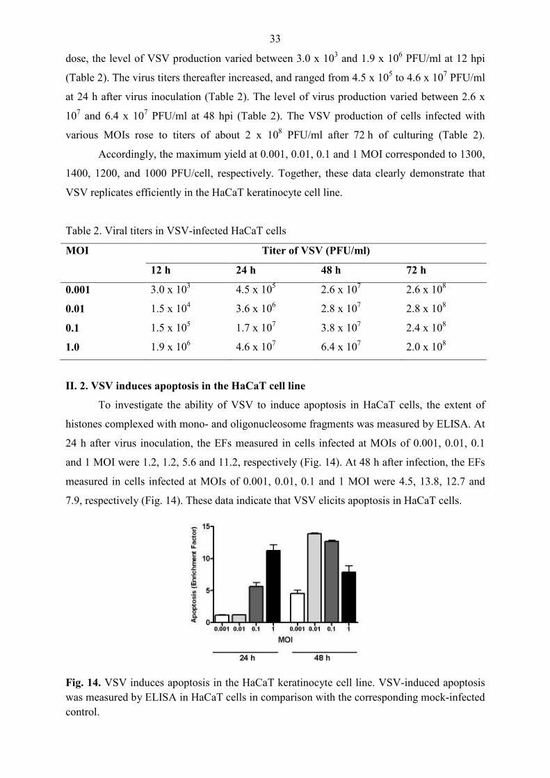
II. 2. VSV induces apoptosis in the HaCaT cell line
To investigate the ability of VSV to induce apoptosis in HaCaT cells, the extent of
histones complexed with mono- and oligonucleosome fragments was measured by ELISA. At
24 h after virus inoculation, the EFs measured in cells infected at MOIs of 0.001, 0.01, 0.1
and 1 MOI were 1.2, 1.2, 5.6 and 11.2, respectively (Fig. 14). At 48 h after infection, the EFs
measured in cells infected at MOIs of 0.001, 0.01, 0.1 and 1 MOI were 4.5, 13.8, 12.7 and
7.9, respectively (Fig. 14). These data indicate that VSV elicits apoptosis in HaCaT cells.
Fig. 14. VSV induces apoptosis in the HaCaT keratinocyte cell line. VSV-induced apoptosis was measured by ELISA in HaCaT cells in comparison with the corresponding mock-infected control.

34
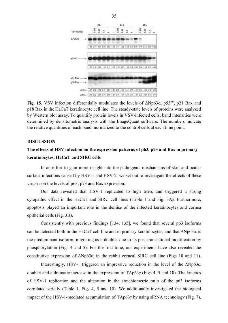
II. 3. VSV infection alters the levels of ∆Np63α, p53mt, p21 Bax and p18 Bax proteins in
the HaCaT cell line
To determine whether VSV infection can alter the expressions of proteins involved in
apoptosis, the steady-state levels of ∆Np63α, p53mt, p21 Bax and p18 Bax were measured by
Western blot assay. The analysis revealed the endogenous expression of ∆Np63α, p53mt and
p21 Bax in mock-infected HaCaT cells (Fig. 15; lanes 1, 6 and 11). The endogenous
expression of ∆Np63α in mock-infected cells remained constant throughout the 48 h of
culturing (Fig. 15; lanes 1, 6 and 11).
The VSV-infected cells exhibited decreased levels of ∆Np63α at 24 h after virus
inoculation. The relative quantities of ∆Np63α were 0.84 and 0.7 in cells infected at MOIs of
0.1 and 1, respectively. (Fig. 15; lanes 9 and 10). The expression of ∆Np63α protein in the
VSV-infected cultures at the 48 h time point was downregulated. The relative quantities of
∆Np63α were 0.44, 0.12, 0.08 and 0.08 in cells infected at MOIs of 0.001, 0.01, 0.1 and 1,
respectively (Fig. 15; lanes 12-15).
The endogenous expression of p53mt in mock-infected cells similarly remained
constant throughout 48 h of culturing (Fig. 15; lanes 1, 6, and 11). No quantitative differences
between the VSV-infected and control cultures were observed in the level of expression of
p53mt protein at the 24 h time point (Fig. 15; lanes 7-10 and 6, respectively). The expression
of p53mt in VSV-infected cultures at 48 h after VSV inoculation was downregulated (the
relative quantities of p53mt were 0.83 and 0.4 in cells infected at MOIs of 0.1 and 1,
respectively) (Fig. 15; lanes 14 and 15).
The endogenous expression of p21 Bax in mock-infected cells likewise remained
constant in the course of the 48 h of culturing (Fig. 15; lanes 1, 6, and 11). No quantitative
differences between the VSV-infected and control cultures were displayed in the level of
expression of p21 Bax protein at the 24 h time point (Fig. 15; lanes 7-10 and 6, respectively).
The expression of p21 Bax in VSV-infected cells at 48 h after inoculation was upregulated
(the relative quantities of p21 Bax were 1.33 and 1.53 in cells infected at MOIs of 0.1 and 1,
respectively) (Fig. 15; lanes 14 and 15).
Furthermore, VSV-infected cells exhibited increased levels of p18 Bax at 48 h after
inoculation (the relative quantities of p18 Bax were 2.32, 8.64, 14.8 and 17.7 in cells infected
at MOIs of 0.001, 0.01, 0.1 and 1, respectively) (Fig. 15; lanes 12-15). Together, these data
indicate that the expressions of ∆Np63α, p53mt, p21 Bax and p18 Bax are differentially
modulated by VSV.
35
Fig. 15. VSV infection differentially modulates the levels of ∆Np63α, p53mt, p21 Bax and p18 Bax in the HaCaT keratinocyte cell line. The steady-state levels of proteins were analyzed by Western blot assay. To quantify protein levels in VSV-infected cells, band intensities were determined by densitometric analysis with the ImageQuant software. The numbers indicate the relative quantities of each band, normalized to the control cells at each time point.
DISCUSSION
The effects of HSV infection on the expression patterns of p63, p73 and Bax in primary
keratinocytes, HaCaT and SIRC cells
In an effort to gain more insight into the pathogenic mechanisms of skin and ocular
surface infections caused by HSV-1 and HSV-2, we set out to investigate the effects of these
viruses on the levels of p63, p73 and Bax expression.
Our data revealed that HSV-1 replicated to high titers and triggered a strong
cytopathic effect in the HaCaT and SIRC cell lines (Table 1 and Fig. 3A). Furthermore,
apoptosis played an important role in the demise of the infected keratinocytes and cornea
epithelial cells (Fig. 3B).
Consistently with previous findings [134, 135], we found that several p63 isoforms
can be detected both in the HaCaT cell line and in primary keratinocytes, and that ∆Np63α is
the predominant isoform, migrating as a doublet due to its post-translational modification by
phosphorylation (Figs 4 and 5). For the first time, our experiments have also revealed the
constitutive expression of ∆Np63α in the rabbit corneal SIRC cell line (Figs 10 and 11).
Interestingly, HSV-1 triggered an impressive reduction in the level of the ∆Np63α
doublet and a dramatic increase in the expression of TAp63γ (Figs 4, 5 and 10). The kinetics
of HSV-1 replication and the alteration in the stoichiometric ratio of the p63 isoforms
correlated strictly (Table 1, Figs 4, 5 and 10). We additionally investigated the biological
impact of the HSV-1-mediated accumulation of TAp63γ by using siRNA technology (Fig. 7).
36
Our experiments revealed that the knockdown of TAp63 expression increases the viability of
infected HaCaT cells (Fig. 7B), suggesting that TAp63γ may play some role in the complex
mechanisms involved in the cytopathogenicity of HSV-1. Noteworthy previous studies raise
the possibility that HSV-1 may alter the expression of p63 via multiple mechanisms [136-
144]. Certain viral proteins may have the potential to alter the transcription of p63 or to affect
the stability and activity of the p63 isoforms via the induction of their posttranslational
modifications [136-144]. The virion-associated host shutoff protein [(vhs), also known as
UL41], which causes the degradation of cellular and viral RNA [136, 137], may evoke a
decrease in the level of ∆Np63α mRNA. The α-trans-inducing factor [(α-TIF), also known as
VP16 or UL48], which stimulates the transcription of IE genes via cellular transcription
factors, such as the POU homeodomain protein Oct-1 (where Oct stands for octamer binding
protein) and the host cell factor [138-140], may elicit an increase in the level of TAp63γ. The
infected cell protein (ICP) 0, which controls the stability of cellular proteins and leads to the
disruption of promyelocytic leukemia (PML) nuclear bodies [also known as PODs (PML
oncogenic domains) and ND10 (nuclear domain 10)] [141-144], may dysregulate the
expression pattern of p63. Other interesting recent studies have also revealed that HSV
genome synthesis activates the cellular DNA damage response [145-149]. While HSV
disrupts the ataxia teleangiectasia mutated- and rad3-related (ATR) pathway, it activates and
even exploits the ataxia teleangiectasia mutated (ATM)-dependent signaling cascade to
support its own replication [147, 149]. Both ATM and ATR belong in the family of
phosphatidylinositol 3-kinase-like kinases (PIKKs) and function as part of a complex
response to DNA damage [149]. Via phosphorylation, PIKKs activate multiple proteins,
including the p53 family members [149]. Recent observations indicate that genotoxic stress
induces the accelerated degradation of ∆Np63 and the accumulation of TAp63 isoforms; in
turn, these function as downstream mediators of the cellular DNA damage response, to
provide time for repair or to kill cells bearing irreparable DNA damage and unstable genome
by inducing apoptotic demise [112, 150-152]. Our experiments have demonstrated that the
viral DNA replication inhibitor ACG completely abolished the HSV-1-mediated induction of
TAp63γ in both HaCaT and SIRC cells, indicating that replication of viral DNA is necessary
for the accumulation of TAp63γ (Figs 6 and 12). This observation strongly supports the view
that the dysregulation of p63 expression depends on the cellular DDR, but does not exclude
the role of HSV-1-encoded proteins. Thus, additional studies are required to elucidate the
potential contributions of vhs, α-TIF, ICP0 and other viral proteins to the development of the
HSV-1-mediated dysregulation of p63 expression.
37
In line with these data, we next investigated the expression of the p53 family member
p73. Similar to p63, the p73 gene has two transcription start sites, producing two p73
subclasses: the TA and ∆N isoforms. In addition to these amino-terminal differences,
alternative splicing generates seven carboxy-terminal variants, denoted α, β, γ, δ, ε, ζ and η.
The TAp73 isoforms transactivate a variety of p53 and p73 target genes and induce apoptosis,
while the ∆Np73 isoforms possess little transcriptional activity, display dominant negative
behavior and inhibit apoptosis [153]. Our studies have shown that the level of a 50 kDa p73
isoform was decreased, while the expression of a 44.5 kDa p73 protein was increased in
HaCaT cells following HSV-1 infection (Fig. 4). On the basis of previously published data we
suggest that the p73 isoforms migrating near 50 and 44.5 kDa may correspond to ∆Np73β and
TAp73δ, respectively [106, 153]. However, further studies are required for the clear-cut
identification of the p73 isoforms detected in HaCaT keratinocytes. Together, these data
demonstrate that HSV-1 dysregulates the expression pattern of p73 in the HaCaT cell line.
In order to gain more insight into the stress response triggered by HSV-1, we also
studied the expression of Bax, which is known to be upregulated by TAp63α and TAp63γ
[99, 105]. Previous studies have demonstrated the existence of several Bax isoforms [125]. It
is well documented that Bax-α is a central component of apoptosis induction [111]. In
response to apoptotic stimuli, Bax-α becomes activated, translocates to the mitochondria and
triggers the release of cytochrome c and caspase-9, which in turn results in the irreversible
execution of the apoptotic program [154]. It has also been reported that the Bax-β protein is
expressed constitutively in several human cell types, and its level is controlled by proteasomal
degradation [126]. Various stressors inhibit ubiquitination of the Bax-β protein and thereby
prevent its proteasomal degradation, leading to the accumulation of this Bax isoform [126].
Similarly to Bax-α, Bax-β has the capability to trigger apoptosis via the mitochondrial
pathway [126, 127]. Moreover, Bax-β associates with and promotes Bax-α activation [127].
Our experiments revealed no alterations in the expression of Bax-α (Figs 4, 5 and 10).
Interestingly, we observed a dramatic rise in the level of Bax-β in HSV-1-infected HaCaT and
SIRC cultures (Figs 4 and 10). Following the demonstration of an altered Bax expression
pattern in the HaCaT and SIRC cells, we postulate an important role for Bax-β in the
apoptotic responsiveness of keratinocytes and corneal epithelial cells infected with HSV-1.
Other interesting recent data have proved that HSVs encode ubiquitinating and
deubiquitinating enzymes, which can modify the ubiquitination status of both viral and host
cell proteins [155, 156]. In view of these observations, it is reasonable to infer that the Bax-β
protein may be a novel target of HSV-1-mediated deubiquitinating events. However, the

38
precise molecular mechanisms responsible for stabilization of the Bax-β protein in HSV-1-
infected cells remain to be elucidated.
A great body of experimental evidence indicates that the cellular responses triggered
by HSV-1 or HSV-2 display both various similarities and profound differences [1, 3, 5]. It has
been clearly proved that HSV-1 and HSV-2 differ in their nucleotide sequences and rates of
reactivation, and also in the cellular transcriptional responses and spectrum of diseases they
evoke. Interesting previous studies have also revealed fundamental differences between HSV-
1 and HSV-2 in their apoptosis-modulating effect [157]. Accordingly, we examined the
expression patterns of the p63, p73 and Bax isoforms and determined the proportion of
apoptotic HaCaT keratinocytes after infection with HSV-2. Although the proportions of dead
cells were comparable in the HSV-1- and HSV-2-infected cultures, the early apoptotic
population was larger in the cultures infected with HSV-1 than in those infected with HSV-2
(Fig. 3). These data raise the possibility that HSV-1-infected keratinocytes, displaying highly
elevated TAp63γ levels, may be prone to commit apoptosis, while HSV-2-infected cells may
rather be disposed to die by way of necrosis or autophagy. Similarly as in the case of HSV-1,
HSV-2-infected HaCaT cultures exhibited impressive reductions in the expressions of
∆Np63α and the 50 kDa p73 isoform; unexpectedly, however, the level of TAp63γ remained
unaffected and the expression of a 44.5 kDa p73 isoform was decreased in HSV-2-infected
HaCaT keratinocytes (Fig. 4). HSV-2 infection further resulted in a very slight increase in the
expression of Bax-β, the magnitude of which proved to be much lower than that observed
after HSV-1 infection (Fig. 4).
Taken together, our results indicate that both HSV-1 and HSV-2 replicate efficiently
and elicit powerful cytopathogenicity, and apoptosis plays an important role in the demise of
the infected keratinocytes and corneal epithelial cells. For the first time, our data also
demonstrate that HSV-1 and HSV-2 modulate the patterns of p63, p73 and Bax expression in
a serotype-specific manner. The dysregulated pattern of p63 expression observed in HSV-
infected HaCaT and SIRC cultures may comprise part of a mechanism by which these viruses
perturb the functions of epithelial cells and lead to their demise. These data may bear on the
pathogenic mechanisms of diseases caused by HSV-1 and HSV-2, as p63 isoforms play a
pivotal role in the epithelial homeostasis.
The effects of VSV infection on the expression patterns of p63 and Bax in HaCaT cells
In an effort to evaluate the potential oncolytic activity of VSV in epithelial-derived
skin cancers, we set out to investigate the cytopathogenicity of this virus in the immortalized
HaCaT keratinocyte cell line.
39
Our data revealed that VSV was able to establish an infection, affecting virtually all of
the cells and yielding high titers of progeny virus (Fig. 13 and Table 2). VSV infection
elicited a strong cytopathic effect (data not shown) and apoptosis (Fig. 14), leading to the
demise of cultures within 72 h. For the first time, our data clearly demonstrated the
susceptibility of immortalized keratinocytes to the deadly infection caused by VSV.
Interestingly, we observed an impressive reduction of the ∆Np63α level of VSV-
infected cells (Fig. 15). It is important that the kinetics of VSV replication, apoptosis and
suppression of ∆Np63α expression correlated strictly (Figs 13-15 and Table 2). This suggests
that the VSV-induced decrease in ∆Np63α levels is a key event in the apoptotic response of
the infected keratinocytes.
Consistent with previous results [158], our experiments have shown that the HaCaT
cell line accumulates high amounts of p53mt (Fig. 15). Our further experiments revealed that
VSV infection decreases the level of p53mt (Fig. 15). Since apoptosis was detected in infected
cultures displaying unaffected levels of p53mt (Figs 14 and 15), the downregulation of p53mt
expression does not seem to be involved in the induction of apoptosis; it may rather operate in
the executional phase and contribute to the inevitable death of heavily infected cells.
In line with these data, we investigated the expression of Bax isoforms. Our
experiments revealed the endogenous expression of a ∼21 kDa Bax isoform in the HaCaT cell
line (Fig. 15). Previously published data suggest that p21 Bax is identical with Bax-α [159].
Interestingly, we observed high increases in the levels of Bax-α and p18 Bax in VSV-infected
HaCaT cultures (Fig. 15). Recent data have shown that the activity of Bax-α is subject to
regulation by calpain-mediated proteolytic cleavage in cells exposed to stress, such as
irradiation, etoposide or cisplatin treatment [131, 160]. Cleavage of Bax-α was shown to yield
a p18 Bax product, which behaves like a sensitizer type of BH3-only proteins [141]. The p18
truncated form of Bax-α is more potent in disrupting mitochondrial integrity and inducing
apoptosis [130, 131, 160]. Thus, the dramatic rises detected in the levels of Bax-α and p18
Bax in HaCaT cells following VSV infection are indicative of a pro-apoptotic shift and may
be of importance in the amplification of the apoptotic process, and contribute to the powerful
cytopathogenicity of this virus.
Taken together, our results demonstrate for the first time that VSV replicates
efficiently and triggers apoptosis in the immortalized HaCaT keratinocyte cell line. The VSV-
mediated alterations in the expressions of ∆Np63α and Bax may be implicated in the
apoptotic demise of infected cells and may also sensitize to other apoptotic stimuli. Our
findings extend the known spectrum of cell types susceptible for the powerful oncolytic
40
activity of VSV to immortalized keratinocytes. These observations may stimulate further
studies aimed at the development of VSV-based virotherapy into an effective modality for the
treatment of epithelial-derived tumors of the skin.
SUMMARY
In light of the critical role of p63 in the epithelial cell fate, we considered the question
of whether HSV-1, HSV-2 and VSV affect the patterns of p63 and Bax expression.
Our results have revealed that both HSV-1 and HSV-2 replicated efficiently, and
elicited a strong cytopathic effect, and apoptosis played an important role in the demise of the
infected primary keratinocytes, HaCaT and SIRC cells. The level of ∆Np63α was decreased,
Bax-α remained unaffected, while the expressions of the Bax-β and TAp63γ isoforms were
highly increased in HSV-1-infected primary keratinocytes, HaCaT and SIRC cells. In
contrast, in response to HSV-2 infection the level of ∆Np63α was decreased, Bax-α and
TAp63γ remained unaffected, while the expression of Bax-β was slightly increased in HaCaT
keratinocytes. The knockdown of TAp63 expression enhanced the viability of HSV-1-infected
cells. Thus, HSV-1 and HSV-2 modulate the patterns of p63 and Bax expression in a
serotype-specific manner. The dysregulated pattern of p63 expression observed in HSV-
infected HaCaT and SIRC cultures may comprise part of a mechanism by which these viruses
perturb the functions of epithelial cells and lead to their demise. By disturbing the delicate
balance between the pro-survival ∆N and the pro-apoptotic TA isoforms, HSV-1 and HSV-2
may cause profound alterations in the tissue homeostasis of the skin and the ocular surface.
Our results have demonstrated that VSV replicated efficiently, and elicited a strong
cytopathic effect leading to the demise of immortalized HaCaT keratinocytes. The
cytopathogenicity evoked by VSV was linked to apoptotic mechanisms. The levels of
∆Np63α and p53mt were decreased, while the expressions of Bax-α and p18 Bax were
increased in VSV-infected HaCaT cells. The VSV-mediated alterations in the expressions of
∆Np63α and Bax may be implicated in the apoptotic demise of infected cells and may also
sensitize to other apoptotic stimuli. Our findings extend the known spectrum of cell types
susceptible for the powerful oncolytic activity of VSV to immortalized keratinocytes. These
observations may stimulate further studies aimed at the development of VSV-based
virotherapy into an effective modality for the treatment of epithelial-derived tumors of the
skin.

41
ACKNOWLEDGEMENTS
This work has been carried out at the Department of Medical Microbiology and
Immunobiology, Faculty of Medicine, University of Szeged.
I am deeply indebted to my supervisor, Associate Professor Klára Megyeri, who has helped
me with good sense, unfailing efficiency and friendly encouragement. She has made the
enterprise of research work a challenge, as well as an education for me. I am also very
grateful to her for useful advice and for critical reading of the manuscript.
I greatly acknowledge Professor Yvette Mándi, Head of the Department of Medical
Microbiology and Immunobiology for providing working facilities and for her support and
advice.
My warmest thanks are due to Professor Lajos Kemény for the significant help.
I would like to acknowledge the support of Professor Norbert E. Fusenig.
I also thank Gyöngyi Ábrahám for her excellent technical assistance and advice.
I owe much to my colleagues, especially Dr. Éva Gallyas, Dr. Andrea Facskó, Prof.
Zsuzsanna Bata-Csörgı, Dr. György Seprényi, Katalin Pásztor, Dr. Béla Taródi, Dr.
Imre Ocsovszki and Bernadett Kormos for pleasant cooperation.
I thank all my colleagues at the Department of Medical Microbiology and Immunobiology
for their support and for creating a pleasant work-environment.
I record my gratitude to my family for their love, support and understanding.
The financial support received from grants OTKA/T043144 by the Hungarian Scientific
Research Fund and ETT/398/2003 by the Hungarian Ministry of Health, Social and Family
Affairs is gratefully acknowledged.
42
REFERENCES
1. Roizman, B., Sears, A. E. (1996). Herpes simplex viruses and their replication. In:
Fields, B. N., Knipe, D. M., Howley, P. M., Chanock, R. M., Melnick, J. L., Monath,
T. P., Roizman, B., Straus S. E. (Eds.), “Fields Virology”, 3rd ed., pp. 2231-2295.
Lippincott-Raven, Philadelphia.
2. Taylor, T. J., Brockman, M. A., McNamee, E. E., Knipe, D. M. (2002). Herpes
simplex virus. Front. Biosci. 7, 752-764.
3. Taddeo, B., Esclatine, A., Roizman, B. (2002). The patterns of accumulation of
cellular RNAs in cells infected with a wild-type and a mutant herpes simplex virus 1
lacking the virion host shutoff gene. P. Natl. Acad. Sci. USA 99, 17031-17036.
4. Roizman, B., Gu, H., Mandel, G. (2005). The first 30 minutes in the life of a virus:
unrest in the nucleus. Cell Cycle 4, 1019-1021.
5. Kamakura, M., Nawa, A., Ushijima, Y. Goshima, F., Kawaguchi, Y., Kikkawa, F.,
Nishiyama, Y. (2008). Microarray analysis of transcriptional responses to infection by
herpes simplex virus types 1 and 2 and their US3-deficient mutants. Microbes Infect.
10, 405-413.
6. Biederer, C., Ries, S., Brandts, C. H., McCormick, F. (2002). Replication-selective
viruses for cancer therapy. J. Mol. Med. 80, 163-175.
7. Ring, C. J. A. (2002). Cytolytic viruses as potential anti-cancer agents. J. Gen. Virol.
83, 491-502.
8. Balachandran, S., Porosnicu, M., Barber, G. N. (2001). Oncolytic activity of vesicular
stomatitis virus is effective against tumors exhibiting aberrant p53, Ras, or Myc
function and involves the induction of apoptosis. J. Virol. 75, 3474-3479.
9. Fernandez, M., Porosnicu, M., Markovic, D., Barber, G. N. (2002). Genetically
engineered vesicular stomatitis virus in gene therapy: application for treatment of
malignant disease. J. Virol. 76, 895-904.
10. Biswas, P. S., Rouse, B. T. (2005). Early events in HSV keratitis–setting the stage for
a blinding disease. Microbes Infect. 7, 799-810.
11. Koyama, A. H., Adachi, A. (1997). Induction of apoptosis by herpes simplex virus
type 1. J. Gen. Virol. 78, 2909-2912.
12. Koyama, A. H., Akari, H., Adachi, A., Goshima, F., Nishiyama, Y. (1998). Induction
of apoptosis in Hep-2 cells by infection with herpes simplex virus type 2. Arch. Virol.
143, 2435-2441.
43
13. Galvan, V., Roizman, B. (1998). Herpes simplex virus 1 induces and blocks apoptosis
at multiple steps during infection and protects cells from exogenous inducers in a cell-
type-dependent manner. P. Natl. Acad. Sci. USA 95, 3931-3936.
14. Aubert, M., O’Toole, J., Blaho, J. A. (1999). Induction and prevention of apoptosis in
human Hep-2 cells by herpes simplex virus type 1. J. Virol. 73, 10359-10370.
15. Aubert, M., Blaho, J. A. (2001). Modulation of apoptosis during herpes simplex virus
infection in human cells, Microbes Infect. 3, 859-866.
16. McLean, J. E., Ruck, A., Shirazian, A., Pooyaei-Mehr, F., Zakeri, Z. F. (2008). Viral
manipulation of cell death. Curr. Pharm. Des. 14, 198-220.
17. Megyeri, K. (2007). Modulation of apoptotic pathways by herpes simplex viruses. In:
Minarovits, J., Gonczol, E., Valyi-Nagy T. (Eds.), “Latency Strategies of
Herpesviruses”, 1st ed., pp. 37-54. Springer, New York.
18. Orvedahl, A., Levine, B. (2008). Autophagy and viral neurovirulence. Cell. Microbiol.
10, 1747-1756.
19. Duprez, L., Wirawan, E., Vanden Berghe, T., Vandenabeele, P. (2009). Major cell
death pathways at a glance. Microbes Infect. 11, 1050-1062.
20. Roulston, A., Marcellus, R. C., Branton, P. E. (1999). Viruses and apoptosis. Annu.
Rev. Microbiol. 53, 577-628.
21. Hay, S., Kannourakis, G. (2002). A time to kill: viral manipulation of the cell death
program. J. Gen. Virol. 83, 1547-1564.
22. Chiarugi, P., Giannoni, E. (2008). Anoikis: a necessary death program for anchorage-
dependent cells. Biochem. Pharmacol. 76, 1352-1364.
23. Labbé, K., Saleh, M. (2008). Cell death in the host response to infection. Cell Death
Differ. 15, 1339-1349.
24. Galluzzi, L., Kroemer, G. (2008). Necroptosis: a specialized pathway of programmed
necrosis. Cell 135, 1161-1163.
25. Espert, L., Codogno, P., Biard-Piechaczyk, M. (2007). Involvement of autophagy in
viral infections: antiviral function and subversion by viruses. J. Mol. Med. 85, 811-
823.
26. Whitley, R. J., Roizman, B. (2001). Herpes simplex virus infections. Lancet 357,
1513-1518.
27. Spear, P. G. (2004). Herpes simplex virus: receptors and ligands for cell entry. Cell.
Microbiol. 6, 401–410.
28. Garner, J. A. (2003). Herpes simplex virion entry into and intracellular transport
within mammalian cells. Adv. Drug Deliver. Rev. 55, 1497– 1513.
44
29. Whitley, R. J. (1996). Herpes simplex viruses. In: Fields, B. N., Knipe, D. M.,
Howley, P. M., Chanock, R. M., Melnick, J. L., Monath, T. P., Roizman, B., Straus S.
E. (Eds.), “Fields Virology”, 3rd ed., pp. 2297-2342. Lippincott-Raven, Philadelphia.
30. Roizman, B., Pellett, P. E. (2001). The family Herpesviridae: a brief introduction. In:
Knipe, D. M., Howley, P. M. (Eds.), “Fields Virology”, 4th ed., pp. 2381-2397.
Lippincott Williams & Wilkins, Philadelphia.
31. Nguyen, M. L., Blaho, J. A. (2007). Apoptosis during herpes simplex virus infection.
Adv. Virus Res. 69, 67-97.
32. Galvan, V., Brandimarti, R., Roizman, B. (1999). Herpes simplex virus 1 blocks
caspase-3-independent and caspase-dependent pathways to cell death. J. Virol. 73,
3219-3226.
33. Wu, N., Watkins, S. C., Schaffer P. A., DeLuca, N. A. (1996). Prolonged gene
expression and cell survival after infection by a herpes simplex virus mutant defective
in the immediate-early genes encoding ICP4, ICP27, and ICP22. J. Virol. 70, 6358–
6369.
34. Lia, H., Baskaran, R., Krisky, D. M., Bein, K., Grandi, P., Cohen, J. B., Joseph C.
Glorioso, J. C. (2008). Chk2 is required for HSV-1 ICP0-mediated G2/M arrest and
enhancement of virus growth. Virology 375, 13–23.
35. Hobbs, W. E., DeLuca, N. A. (1999). Perturbation of cell cycle progression and
cellular gene expression as a function of herpes simplex virus ICP0. J. Virol. 73, 8245-
8255.
36. Aubert, M., Pomeranz, L. E., Blaho, J. A. (2007). Herpes simplex virus blocks
apoptosis by precluding mitochondrial cytochrome c release independent of caspase
activation in infected human epithelial cells. Apoptosis 12, 19-35.
37. Tallóczy, Z., Virgin, W. H., Levine, B. (2006). PKR-dependent autophagic
degradation of Herpes simplex virus type 1. Autophagy 2:24-29.
38. Deshmane, S. L., Fraser, N. W. (1989). During latency, herpes simplex virus type 1
DNA is associated with nucleosomes in a chromatin structure. J. Virol. 63, 943-947.
39. Kaye, S., Choudhary, A. (2006). Herpes simplex keratitis. Prog. Retin. Eye Res. 25,
355-380.
40. Fatahzadeh, M., Schwartz, R. A. (2007). Human herpes simplex virus infections:
Epidemiology, pathogenesis, symptomatology, diagnosis, and management. J. Am.
Acad. Dermatol. 57, 737-763.
41. Rouse, B. T., Kaistha, A. (2006). A tale of two α-herpesviruses: lessons for
vaccinologists. Clin. Infect. Dis. 42, 810-817.
45
42. Rudnick, C., Hoekzema, G. (2002). Neonatal herpes simplex virus infections. Am.
Fam. Physician. 65, 1138-1142.
43. Whitley, R. J. (1997). Herpes simplex virus. In: Scheld, W. M., Whitley, R. J., Durack,
D. T. (Eds.), “Infections of the central nervous system”, 2nd ed., pp. 73-89. Lippincott-
Raven, Philadelphia.
44. Gupta, R., Warren, T., Wald, A. (2007). Genital herpes. Lancet 370, 2127-2137.
45. Lafferty, W. E. (2002). The changing epidemiology of HSV-1 and HSV-2 and
implications for serological testing. Herpes 9, 51-55.
46. Holdeman, N. R. (2005). Herpes simplex virus: ocular manifestations. In: Onofrey, B.
E., Skorin, L., Holdeman, N. R. (Eds.), “Ocular Therapeutics Handbook: A Clinical
Manual”. 2nd ed., pp. 208-210. Lippincott Williams & Wilkins, Philadelphia.
47. Kaye, S. B., Baker, K., Bonshek, R., Maseruka, H., Grinfeld, E., Tullo, A., Easty, D.
L., Hart, C. A. (2000). Human herpesviruses in the cornea. Br. J. Ophthalmol. 84, 563-
571.
48. Keadle, T. L., Morris, J. L., Pepose, J. S., Stuart, P. M. (2002). CD4+ and CD8+ cells
are key participants in the development of recurrent herpetic stromal keratitis in mice.
Microb. Pathogen. 32, 255-262.
49. Jirmo, A. C., Nagel, C. H., Bohnen, C., Sodeik, B., Behrens, G. M. (2009).
Contribution of direct and cross-presentation to CTL immunity against herpes simplex
virus 1. J. Immunol. 182, 283-292.
50. Sarangi, P. P., Sehrawat, S., Suvas, S., Rouse, B. T. (2008). IL-10 and natural
regulatory T cells: two independent anti-inflammatory mechanisms in herpes simplex
virus-induced ocular immunopathology. J. Immunol. 180, 6297-6306.
51. Stumpf, T. H., Shimeld, C., Easty, D. L., Hill, T. J. (2001). Cytokine production in a
murine model of recurrent herpetic stromal keratitis. Invest. Ophthalmol. Vis. Sci. 42,
372-378.
52. Rose, J. K., Whitt, M. A. (2001). Rhabdoviridae: the viruses and their replication. In:
Fields, B. N., Knipe, D. M., Howley, P. M., Chanock, R. M., Melnick, J. L., Monath,
T. P., Roizman, B., Straus S. E. (Eds.), “Fields Virology”, 4th ed., pp. 1221-1242
Philadelphia: Lippincott Williams & Wilkins; 1221-1242. Lippincott Williams &
Wilkins, Philadelphia.
53. Kelley, J. R., Emerson, S. U., Wagner, R. R. (1972). The glycoprotein of vesicular
stomatitis virus is the antigen that gives rise to and reacts with neutralizing antibody. J.
Virol. 10, 1231-1235.
46
54. Lefrancois, L., Lyles, D. S. (1983). Antigenic determinants of vesicular stomatitis
virus: analysis with antigenic variants. J. Immunol. 130, 394–398.
55. Vandepol, S. B., Lefrancois, L., Holland, J. J. (1986). Sequences of the major antibody
binding epitopes of the Indiana serotype of vesicular stomatitis virus. Virology 148,
312–325.
56. Luo, L. H., Li, Y., Snyder, R. M., Wagner, R. R. (1988). Point mutations in
glycoprotein gene of vesicular stomatitis virus (New Jersey serotype) selected by
resistance to neutralization by epitope-specific monoclonal antibodies. Virology 163,
341–348.
57. Roche S, Albertini AA, Lepault J, Bressanelli S, Gaudin Y. (2008). Structures of
vesicular stomatitis virus glycoprotein: membrane fusion revisited. Cell. Mol. Life Sci.
65, 1716-28.
58. Kopecky, S. A., Willingham, M. C., Lyles, D. S. (2001). Matrix protein and another
viral component contribute to induction of apoptosis in cells infected with vesicular
stomatitis virus. J. Virol. 75, 12169-12181.
59. Gaddy, D. F., Lyles, D. S. (2005). Vesicular stomatitis viruses expressing wild-type or
mutant M proteins activate apoptosis through distinct pathways. J. Virol. 79, 4170-
4179.
60. Gadaleta, P., Perfetti, X., Mersich, S., Coulombié, F. (2005). Early activation of the
mitochondrial apoptotic pathway in vesicular stomatitis virus-infected cells. Virus Res.
109, 65-69.
61. Kopecky, S. A., Lyles, S. D. (2003). Contrasting effects of matrix protein on apoptosis
in HeLa and BHK cells infected with vesicular stomatitis virus are due to inhibition of
host gene expression. J. Virol. 77, 4658-4669.
62. Faria, P. A., Chakraborty, P., Levay, A., Barber, G. N., Ezelle, H. J., Enninga, J.,
Arana, C., van Deursen, J., Fontoura, B. M. (2005). VSV disrupts the Rae1/mrnp41
mRNA nuclear export pathway. Mol. Cell 17, 93–102.
63. Schlegel, R., Tralka, T. S., Willingham, M. C., Pastan, I. (1983). Inhibition of VSV
binding and infectivity by phosphatidylserine: is phosphatidylserine a VSV-binding
site? Cell 32, 639–646.
64. Coil, D. A., Miller, A. D. (2004). Phosphatidylserine is not the cell surface receptor for
vesicular stomatitis virus. J. Virol. 78, 10920–10926.
65. Carneiro, F. A., Bianconi, M. L., Weissmüller, G., Stauffer, F., Da Poian, A. T.
(2002). Membrane recognition by vesicular stomatitis virus involves enthalpy-driven
protein-lipid interactions. J. Virol. 76, 3756–3764.
47
66. Barber, G. N. (2004). Vesicular stomatitis virus as an oncolytic vector. Viral.
Immunol. 17, 516-527.
67. Rodríguez, L. L. (2002). Emergence and re-emergence of vesicular stomatitis in the
United States. Virus Res. 85, 211-219.
68. Lichty, B. D., Power, A. T., Stojdl, D. F., Bell, J. C. (2004). Vesicular stomatitis virus:
re-inventing the bullet. Trends Mol. Med. 10, 210-216.
69. Lichty, B. D., Stojdl, D. F., Taylor, R. A., Miller, L., Frenkel, I., Atkins, H., Bell, J. C.
(2004). Vesicular stomatitis virus: a potential therapeutic virus for the treatment of
hematologic malignancy. Hum. Gene Ther. 15, 821-831.
70. Balachandran, S., Barber, G. N. (2000). Vesicular stomatitis virus (VSV) therapy of
tumors. IUBMB Life 50, 135-138.
71. Balachandran, S., Barber, G. N. (2004). Defective translational control facilitates
vesicular stomatitis virus oncolysis. Cancer Cell 5, 51–65.
72. Stojdl, D. F., Lichty, B., Knowles, S., Marius, R., Atkins, H., Sonenberg, N., Bell, J.
C. (2000). Exploiting tumor-specific defects in the interferon pathway with a
previously unknown oncolytic virus. Nat. Med. 6, 821-825.
73. Masters, P. S., Samuel, C. E. (1983). Mechanism of interferon action: Inhibition of
vesicular stomatitis virus replication in human amnion U cells by cloned human
leukocyte interferon. II. Effect on viral macromolecular synthesis. J. Biol. Chem. 258,
12026-12033.
74. Barber, G. N. (2005). VSV-tumor selective replication and protein translation.
Oncogene 24, 7710-7719.
75. Brady, C. S., Bartholomew, J. S., Burt, D. J., Duggan-Keen, M. F., Glenville, S.,
Telford, N., Little, A. M., Davidson, J. A., Jimenez, P., Ruiz-Cabello, F., Garrido, F.,
Stern, P. L. (2000). Multiple mechanisms underlie HLA dysregulation in cervical
cancer. Tissue Antigens 55, 401-411.
76. Brender, C., Nielsen, M., Kaltoft, K., Mikkelsen, G., Zhang, Q., Wasik, M., Billestrup,
N., Odum, N. (2001). STAT3-mediated constitutive expression of SOCS-3 in
cutaneous T-cell lymphoma. Blood 97, 1056-1062.
77. Lensch, M. W., Rathbun, R. K., Olson, S. B., Jones, G. R., Bagby, G. C. Jr. (1999).
Selective pressure as an essential force in molecular evolution of myeloid leukemic
clones: a view from the window of Fanconi anemia. Leukemia 13, 1784-1789.
78. Moriyama, Y., Nishiguchi, S., Tamori, A., Koh, N., Yano, Y., Kubo, S., Hirohashi, K.,
Otani, S. (2001). Tumor-suppressor effect of interferon regulatory factor-1 in human
hepatocellular carcinoma. Clin. Cancer Res. 7, 1293-1298.
48
79. Xi, H., Blanck, G. (2000). Interferon regulatory factor-2 point mutations in human
pancreatic tumors. Int. J. Cancer 87, 803-808.
80. Balachandran, S., Roberts, P. C., Brown, L. E., Truong, H., Pattnaik, A. K., Archer, D.
R., Barber, G. N. (2000). Essential role for the dsRNA-dependent protein kinase PKR
in innate immunity to viral infection. Immunity 13, 129-141.
81. Meurs, E., Chong, K., Galabru, J., Thomas, N. S., Kerr, I. M., Williams, B. R.,
Hovanessian, A. G. (1990). Molecular cloning and characterization of the human
double-stranded RNA-activated protein kinase induced by interferon. Cell 62, 379-
390.
82. Balachandran, S., Roberts, P. C., Kipperman, T., Bhalla, K. N., Compans, R. W.,
Archer, D. R., Barber, G. N. (2000). Alpha/beta interferons potentiate virus-induced
apoptosis through activation of the FADD/Caspase-8 death signaling pathway. J.
Virol. 74, 1513–1523.
83. Abraham, N., Stojdl, D. F., Duncan, P. I., Méthot, N., Ishii, T., Dubé, M.,
Vanderhyden, B. C., Atkins, H. L., Gray, D. A., McBurney, M. W., Koromilas, A. E.,
Brown, E. G., Sonenberg, N., Bell J. C. (1999). Characterization of transgenic mice
with targeted disruption of 523 the catalytic domain of the double-stranded RNA-
dependent protein kinase, PKR. J. Biol. Chem. 274, 5953–5962.
84. Stojdl, D. F., Lichty, B. D., tenOever, B. R., Paterson, J. M., Power, A. T., Knowles,
S., Marius, R., Reynard, J., Poliquin, L., Atkins, H., Brown, E. G., Durbin, R. K.,
Durbin, J. E., Hiscott, J., Bell, J. C. (2003). VSV strains with defects in their ability to
shutdown innate immunity are potent systemic anticancer agents. Cancer Cell 4, 263-
275.
85. Obuchi, M., Fernandez, M., Barber, G. N. (2003). Development of recombinant
vesicular stomatitis viruses that exploit defects in host defense to augment specific
oncolytic activity. J. Virol. 77, 8843-8856.
86. Wollmann, G., Rogulin, V., Simon, I., Rose, J. K., van den Pol, A. N. (2010). Some
attenuated variants of vesicular stomatitis virus show enhanced oncolytic activity
against human glioblastoma cells relative to normal brain cells. J. Virol. 84, 1563-
1573.
87. Stojdl, D. F., Abraham, N., Knowles, S., Marius, R., Brasey, A., Lichty, B. D., Brown,
E. G., Sonenberg, N., Bell, J. C. (2000). The murine double-stranded RNA-dependent
protein kinase PKR is required for resistance to vesicular stomatitis virus. J. Virol. 74,
9580-9585.

49
88. Hobbs, J. A., Hommel-Berrey, G., Brahmi, Z. (2003). Requirement of caspase-3 for
efficient apoptosis induction and caspase-7 activation but not viral replication or cell
rounding in cells infected with vesicular stomatitis virus. Hum. Immunol. 64, 82-92.
89. Gallyas, É., Seprényi, G., Sonkoly, E., Mándi, Y., Kemény, L., Megyeri, K. (2006).
Vesicular stomatitis virus induces apoptosis in the Wong-Kilbourne derivative of the
Chang conjunctival cell line. Graefe’s Arch. Clin. Exp. Ophthalmol. 244, 717-724.
90. Kopecky, S. A., Lyles, D. S. (2003). The cell-rounding activity of the vesicular
stomatitis virus matrix protein is due to the induction of cell death. J. Virol. 77, 5524-
5528.
91. Barbieri, C. E., Pietenpol, J. A. (2006). p63 and epithelial biology. Exp. Cell Res. 312,
695-706.
92. Yang, A., Kaghad, M., Wang, Y., Gillet, E., Fleming, M., Dotsch, V., Andrews, N.,
Caput, D., McKeon, F. (1998). p63, a p53 homolog at 3q27–29, encodes multiple
products with transactivating death-inducing, and dominant-negative activities. Mol.
Cell 2, 305–316.
93. Candi, E., Cipollone, R., Rivetti di Val Cervo, P., Gonfloni, S., Melino, G., Knight, R.
(2008). p63 in epithelial development. Cell. Mol. Life Sci. 65, 3126-3133.
94. Koster, M. I., Roop, D. R. (2004). The role of p63 in development and differentiation
of the epidermis. J. Dermatol. Sci. 34, 3-9.
95. Mills, A. A., Zheng, B., Wang X. J., Vogel, H., Roop, D. R., Bradley A. (1999). p63 is
a p53 homologue required for limb and epidermal morphogenesis. Nature 398, 708-
713.
96. Yang, A., Schweitzer, R., Sun, D., Kaghad, M., Walker, N., Bronson, R. T., Tabin, C.,
Sharpe, A., Caput, D., Crum, C., McKeon, F. (1999). p63 is essential for regenerative
proliferation in limb, craniofacial and epithelial development. Nature 398, 714-718.
97. Moll, U. M., Slade, N. (2004). p63 and p73: Roles in development and tumor
formation. Mol. Cancer Res. 2, 371-386.
98. Strano, S., Rossi, M., Fontemaggi, G., Munarriz, E., Soddu, S., Sacchi, A., Blandino,
G. (2001). From p63 to p53 across p73. FEBS Lett. 490, 163-170.
99. Candi, E., Dinsdale, D., Rufini, A., Salomoni, P., Knight, R. A., Mueller, M.,
Krammer, P. H., Melino, G. (2007). TAp63 and ∆Np63 in cancer and epidermal
development. Cell Cycle 6, 274-285.
100. Trink, B., Osada, M., Ratovitski, E., Sidransky, D. (2007). p63 transcriptional
regulation of epithelial integrity and cancer. Cell Cycle 6, 240-245.
50
101. Tomkova, V., Tomka, M., Zajac, V. (2008). Contribution of p53, p63 and p73 to the
developmental diseases and cancer. Neoplasma 55, 177-181.
102. Bénard, J., Douc-Rasy, S., Ahomadegbe, J-C. (2003). TP53 family members and
human cancers. Hum. Mutat. 21, 182-191.
103. Flores, E. R. (2007). The roles of p63 in cancer. Cell Cycle 6, 300-304.
104. Levrero, M., De Laurenzi, V., Costanzo, A., Sabatini, S., Gong, J., Wang, J. Y. J.
(2000). The p53/p63/p73 family of transcription factors: overlapping and distinct
functions. J. Cell Sci. 113, 1661-1670.
105. Gressner, O., Schilling, T., Lorenz, K., Schleithoff, E. S., Koch, A., Schulze-
Bergkamen, H., Lena, A. M., Candi, E., Terrinoni, A., Catani, M. V., Oren, M.,
Melino, G., Krammer, P. H., Stremmel, W., Müller, M. (2005). TAp63α induces
apoptosis by activating signaling via death receptors and mitochondria. EMBO J 24,
2458-2471.
106. Müller, M., Schleithoff, E. S., Stremmel, W., Melino, G., Krammer, P. H., Schilling,
T. (2007). One, two, three – p53, p63, p73 and chemosensitivity. Drug Resist. Update.
9, 288-306.
107. Finlan, L. E., Hupp, T. R. (2007). p63: the phantom of the tumor suppressor. Cell
Cycle 6, 1062-1071.
108. Dohn, M., Zhang, S., Chen, X. (2001). p63α and ∆Np63α can induce cell cycle arrest
and apoptosis and differentially regulate p53 target genes. Oncogene 20, 3193–3205.
109. Sasaki, Y., Morimoto, I., Ishida, S., Yamashita, T., Imai, K., Tokino, T. (2001).
Adenovirus-mediated transfer of the p53 family genes, p73 and p51/p63 induces cell
cycle arrest and apoptosis in colorectal cancer cell lines: potential application to gene
therapy of colorectal cancer. Gene Ther. 8, 1401-1408.
110. Buzás, K., Miczák, A., Degré, M., Megyeri, K. (2004). Rubella virus infection
dysregulates the pattern of p63 expression. APMIS 112, 656-662.
111. Dietrich, J. B. (1997). Apoptosis and anti-apoptosis genes in the Bcl-2 family. Arch.
Physiol. Biochem. 105, 125-135.
112. Katoh, I., Aisaki, K-I., Kurata, S-I., Ikawa, S., Ikawa, Y. (2000). p51A (TAp63γ), a
p53 homolog, accumulates in response to DNA damage for cell regulation. Oncogene
19, 3126-3130.
113. Rinne, T., Brunner, H. G., van Bokhoven H. (2007). p63-associated disorders. Cell
Cycle 6, 262-268.
114. Irwin, M. S., Kaelin, W. G. (2001). p53 family update: p73 and p63 develop their own
identities. Cell Growth Differ. 12, 337-349.

51
115. Kawasaki, S., Tanioka, H., Yamasaki, K., Connon, C. J,, Kinoshita, S. (2006).
Expression and tissue distribution of p63 isoforms in human ocular surface epithelia.
Exp. Eye Res. 82, 293-299.
116. Arpitha, P., Prajna, N. V., Srinivasan, M., Muthukkaruppan, V. (2008). A subset of
human limbal epithelial cells with greater nucleus-to-cytoplasm ratio expressing high
levels of p63 possesses slow-cycling property. Cornea 27, 1164-1170.
117. Arpitha, P., Prajna, N. V., Srinivasan, M., Muthukkaruppan, V. (2005). High
expression of p63 combined with a large N/C ratio defines a subset of human limbal
epithelial cells: implications on epithelial stem cells. Invest. Ophthalmol. Vis. Sci. 46,
3631-3636.
118. Epstein, S. P., Wolosin, J. M., Asbell, P. A. (2005). p63 expression levels in side
population and low light scattering ocular surface epithelial cells. Trans. Am.
Ophthalmol. Soc. 103, 187-199.
119. Wang, D. Y., Cheng, C. C., Kao, M. H., Hsueh, Y. J., Ma, D. H., Chen, J. K. (2005).
Regulation of limbal keratinocyte proliferation and differentiation by TAp63 and
∆Np63 transcription factors. Invest. Ophthalmol. Vis. Sci. 46, 3102-3108.
120. Di Iorio, E., Barbaro, V., Ruzza, A., Ponzin, D., Pellegrini, G., De Luca, M. (2005).
Isoforms of ∆Np63 and the migration of ocular limbal cells in human corneal
regeneration. P. Natl. Acad. Sci. USA 102, 9523-9528.
121. Vaux, D. L., Cory, S., Adams, J. M. (1988). Bcl-2 gene promotes haemopoietic cell
survival and cooperates with c-myc to immortalize pre-B cells. Nature 335, 440-442.
122. Chao, D. T., Korsmeyer, S. J. (1998). BCL-2 family: regulators of cell death. Annu.
Rev. Immunol. 16, 395-419.
123. Borner, C. (2003). The Bcl-2 protein family: sensors and checkpoints for life-or-death
decisions. Mol. Immunol. 39, 615-647.
124. Oltvai, Z. N., Milliman, C. L., Korsmeyer, S. J. (1993). Bcl-2 heterodimerizes in vivo
with a conserved homolog, Bax, that accelerates programmed cell death. Cell 74, 609–
619.
125. Zhou, M., Demo, S. D., McClure, T. N., Crea, R., Bitler, C. M. (1998). A novel splice
variant of the cell death-promoting protein BAX. J. Biol. Chem. 273, 1193-11936.
126. Fu, N. Y., Sukumaran, S. K., Kerk, S. Y., Yu, V. C. (2009). Baxβ: A constitutively
active human Bax isoform that is under tight regulatory control by the proteosomal
degradation mechanism. Mol. Cell 33, 15-29.
52
127. Bargou, R. C. (1996). Overexpression of the death-promoting gene Bax-alpha which is
downregulated in breast cancer restores sensitivity to different apoptotic stimuli and
reduces tumor growth in SCID mice. J. Clin. Invest. 97, 2651-2659.
128. Youle, R. J., Strasser, A. (2008). The BCL-2 protein family: opposing activities that
mediate cell death. Nat. Rev. Mol. Cell Biol. 9, 47-59.
129. Kim, H., Hsieh, J. J., Cheng, E. H. (2009). Deadly splicing: Bax becomes Almighty.
Mol. Cell 33, 145-146.
130. Cartron, P. F., Oliver, L., Juin, P., Meflah, K., Vallette, F. M. (2004). The p18
truncated form of Bax behaves like a Bcl-2 homology domain 3-only protein. J. Biol.
Chem. 279, 11503-11512.
131. Cao, X., Deng, X., May, W. S. (2003). Cleavage of Bax to p18 Bax accelerates stress-
induced apoptosis, and a cathepsin-like protease may rapidly degrade p18 Bax. Blood
102, 2605-2614.
132. Boukamp, P., Petrussevska, R. T., Breitkreutz, D., Hornung, J., Markham, A., Fusenig,
N. E. (1988). Normal keratinization in a spontaneously immortalized aneuploid human
keratinocyte cell line. J. Cell Biol. 106: 761-771.
133. van Bokhoven, H., Brunner, H. G. (2002). Splitting p63. Am. J. Hum. Genet. 71, 1-13.
134. Urist, M. J., Di Como, C. J., Lu, M. L., Charytonowicz, E., Verbel, D., Crum, C. P.,
Ince, T. A., McKeon, F. D., Cordon-Cardo, C. (2002). Loss of p63 expression is
associated with tumor progression in bladder cancer. Am. J. Pathol. 161, 1199-1206.
135. Westfall, M. D., Mays, D. J., Sniezek, J. C., Pietenpol JA. (2003). The ∆Np63α
phosphoprotein binds the p21 and 14-3-3σ promoters in vivo and has transcriptional
repressor activity that is reduced by Hay-Wells syndrome-derived mutations. Mol.
Cell. Biol. 23, 2264-2276.
136. Taddeo, B., Zhang, W., Roizman, B. (2006). The UL41 protein of herpes simplex virus
1 degrades RNA by endonucleolytic cleavage in absence of other cellular or viral
proteins. P. Natl. Acad. Sci. USA 103, 2827-2832.
137. Matis, J., Kúdelová, M. (2001). Early shutoff of host protein synthesis in cells infected
with Herpes simplex viruses. Acta Virol. 45, 269-277.
138. Wysocka, J., Herr, W. (2003). The herpes simplex virus VP16-induced complex: the
makings of a regulatory switch. Trends Biochem. Sci. 28, 294-304.
139. Nogueira, M. L., Wang, V. E. H., Tantin, D., Sharp, P. A., Kristie, T. M. (2004).
Herpes simplex virus infections are arrested in Oct-1-deficient cells. P. Natl. Acad.
Sci. USA 101, 1473-1478.

53
140. Narayanan, A., Nogueira, M. L., Ruyechan, W. T., Kristie, T. M. (2005).
Combinatorial transcription of herpes simplex and varicella zoster virus immediate
early genes is strictly determined by the cellular coactivator HCF-1. J. Biol. Chem.
280, 1369-1375.
141. Everett, R. D. (2000). ICP0, a regulator of Herpes simplex virus during lytic and late
infection. BioEssays 22, 761-770.
142. Boutell, C., Sadis, S., Everett, R. D. (2002). Herpes simplex virus type 1 immediate
early protein ICP0 and its isolated RING finger domain act as ubiquitin E3 ligases in
vitro. J. Virol. 76, 841-850.
143. Everett, R. D., Murray, J., Orr, A., Preston, C. M. (2007). Herpes simplex virus type 1
genomes are associated with ND10 nuclear substructures in quiescently infected
human fibroblasts. J. Virol. 81, 10991-11004.
144. Wilcock, D., Lane, D. P. (1991). Localization of p53, retinoblastoma and host
replication proteins at sites of viral replication in herpes-infected cells. Nature 349,
429-431.
145. de Bruyn Kops, A., Knipe, D. M. (1988). Formation of DNA replication structures in
herpes virus-infected cells requires a viral DNA binding protein. Cell 55, 857–868.
146. Lilley, C. E., Carson, C. T., Muotri, A. R., Gage, F. H., Weitzman, M. D. (2005).
DNA repair proteins affect the lifecycle of herpes simplex virus 1. P. Natl. Acad. Sci.
USA 102, 5844-5849.
147. Shirata, N., Kudoh, A., Daikoku, T., Tatsumi, Y., Fujita, M., Kiyono, T., Sugaya, Y.,
Isomura, H., Ishizaki, K., Tsurumi, T. (2005). Activation of ataxia teleangiectasia-
mutated DNA damage checkpoint signal transduction elicited by herpes simplex virus
infection. J. Biol. Chem. 280, 30336-30341.
148. Boutell, C., Everett, R. D. (2004). Herpes simplex virus type 1 infection induces the
stabilization of p53 in a USP7- and ATM-independent manner. J. Virol. 78, 8068-
8077.
149. Wilkinson, D. E., Weller, S. K. (2006). Herpes simplex virus type I disrupts the ATR-
dependent DNA-damage response during lytic infection. J. Cell Sci. 119, 2695-7203.
150. Roos, W. P., Kaina, B. (2006). DNA damage-induced cell death by apoptosis. Trends
Mol. Med. 12, 440-450.
151. Okada, Y., Osada, M., Kurata, S., Sato, S., Aisaki, K., Kageyama, Y., Kihara, K.,
Ikawa, Y., Katoh, I. (2002). p53 gene family p51(p63)-encoded, secondary
transactivator p51B(TAp63alpha) occurs without forming an immunoprecipitable
54
complex with MDM2, but responds to genotoxic stress by accumulation. Exp. Cell
Res. 276, 194-200.
152. Petitjean, A., Ruptier, C., Tribollet, V., Hautefeuille, A., Chardon, F., Cavard, C.,
Puisieux, A., Hainaut, P., Caron de Fromentel, C. (2008). Properties of the six
isoforms of p63: p53-like regulation in response to genotoxic stress and cross talk with
∆Np73. Carcinogenesis 29, 273-281.
153. Murray-Zmijewski, F., Lane, D. P., Bourdon, J-C. (2006). p53/p63/p73 isoforms: an
orchestra of isoforms to harmonise cell differentiation and response to stress. Cell
Death Differ. 13, 962-972.
154. Schlottmann, K., Schölmerich, J. (2000). BCL-2 family members and mitochondria.
In: Schunkert, H., Riegger, G. A. J. (Eds.), “Apoptosis in cardiac biology”, 1st ed., pp.
71-91. Springer, Berlin.
155. Diao, L., Zhang, B., Fan, J., Gao, X., Sun, S., Yang, K., Xin, D., Jin, N., Geng, Y.,
Wang, C. (2005). Herpes virus proteins ICP0 and BICP0 can activate NF-kappaB by
catalyzing IkappaBalpha ubiquitination. Cell. Signal. 17, 217-229.
156. Meulmeester E, Maurice MM, Boutell C. (2005). Loss of HAUSP-mediated
deubiquitination contributes to DNA damage-induced destabilization of Hdmx and
Hdm2. Mol. Cell 18, 565-576.
157. Yedowitz, J. C., Blaho, J. A. (2005). Herpes simplex virus 2 modulates apoptosis and
stimulates NF-kappaB nuclear translocation during infection in human epithelial
Hep-2 cells. Virology 342, 297-310.
158. Fusenig, N. E., Boukamp, P. (1998). Multiple stages and genetic alterations in
immortalization, malignant transformation, and tumor progression of human skin
keratinocytes. Mol. Carcinog. 23, 144-158.
159. Itoh, M., Chiba, H., Noutomi, T., Takada, E., Mizuguchi, J. (2000). Cleavage of Bax-
alpha and Bcl-x(L) during carboplatin-mediated apoptosis in squamous cell carcinoma
cell line. Oral Oncol. 36, 277-285.
160. Tavolari, S., Bonafè, M., Marini, M., Ferreri, C., Bartolini, G., Brighenti, E., Manara,
S., Tomasi, V., Laufer, S., Guarnieri, T. (2008). Licofelone, a dual COX/5-LOX
inhibitor, induces apoptosis in HCA-7 colon cancer cells through the mitochondrial
pathway independently from its ability to affect the arachidonic acid cascade.
Carcinogenesis 29, 371-380.