Interactions Vesicular Stomatitis Virus Proteins...
7
JOURNAL OF VIROLOGY, Jan. 1994, p. 441-447 Vol. 68, No. 1 0022-538X/94/$04.00+0 Copyright © 1994, American Society for Microbiology Interactions of Normal and Mutant Vesicular Stomatitis Virus Matrix Proteins with the Plasma Membrane and Nucleocapsids LISA D. CHONG AND JOHN K. ROSE* Departments of Pathology and Cell Biology, Yale University School of Medicine, New Haven, Connecticut 06510 Received 17 March 1993/Accepted 4 October 1993 We demonstrated recently that a fraction of the matrix (M) protein of vesicular stomatitis virus (VSV) binds tightly to cellular membranes in vivo when expressed in the absence of other VSV proteins. This membrane- associated M protein was functional in binding purified VSV nucleocapsids in vitro. Here we show that the membrane-associated M protein is largely associated with a membrane fraction having the density of plasma membranes, indicating membrane specificity in the binding. In addition, we analyzed truncated forms of M protein to identify regions responsible for membrane association and nucleocapsid binding. Truncated M protein lacking the amino-terminal basic domain still associated with cellular membranes, although not as tightly as wild-type M protein, and could not bind nucleocapsids. In contrast, deletion of the carboxy-terminal 14 amino acids did not disrupt stable membrane association or nucleocapsid interaction. These results suggest that the amino terminus of M protein either interacts directly with membranes and nucleocapsids or stabilizes a conformation that is required for M protein to mediate both of these interactions. Vesicular stomatitis virus (VSV) is an enveloped negative- strand RNA virus containing five proteins. The genomic RNA is encapsidated by the nucleoprotein (N) and is tightly associ- ated with the viral RNA polymerase proteins L and NS(P) (11). This core complex is surrounded by a lipid bilayer derived from the plasma membrane and contains the viral transmem- brane glycoprotein (G). The matrix (M) protein of VSV lies beneath the lipid bilayer and presumably bridges between the viral envelope and the nucleocapsid core. The presence of M protein at the inner surface of the plasma membrane of infected cells (4, 23, 27) and the rapid association of a fraction of M protein with membranes after synthesis on cytosolic polyribosomes (1, 10, 19) have been well docu- mented, but it was not clear whether these were membranes already involved in viral assembly. We recently used subcellu- lar fractionation studies of transfected HeLa cells to show that M protein can associate tightly with membranes in vivo, in the absence of other VSV proteins, but the nature of the mem- branes to which M protein bound was not determined in that study. Furthermore, membrane-associated M protein was functional in binding nucleocapsid cores to membranes in vitro. On the basis of these findings, we proposed a VSV assembly pathway that could be initiated by membrane-asso- ciated M protein (9). M protein lacks a distinct stretch of hydrophobic residues indicative of a membrane-associating region, and modification by fatty acid has not been demonstrated (22, 29). However, labeling studies with a hydrophobic reagent suggested that the amino-terminal domain penetrates the bilayer (22). Interest- ingly, a trypsin-resistant core of M protein lacking the amino- terminal 43 residues could also associate with artificial lipo- somes, suggesting that other regions of M protein may be involved in membrane interaction (26). Here, we report studies using subcellular fractionation to show that M protein ex- pressed in cells binds largely to a plasma membrane fraction rather than nonspecifically to all cellular membranes. Addi- * Corresponding author. Mailing address: Department of Pathology, Yale University School of Medicine, New Haven, CT 06510. Phone: (203) 785-6184. Fax: (203) 785-7467. tional studies on truncated M proteins show that the amino- terminal basic domain of M is not required for binding M protein to membranes but is required for stable membrane association. In addition, we demonstrate that this domain is required for membrane-associated M protein to bind nucleo- capsid cores in vitro. We propose that this amino-terminal region either interacts directly with membranes and nucleo- capsid cores or stabilizes a conformation which is required for M protein to support these interactions. MATERUILS AND METHODS Viruses and cell culture. Preparation of radiolabeled VSV (Indiana serotype, San Juan strain) was performed in baby hamster kidney (BHK-21) cells as described previously (9). The recombinant vaccinia virus vTF7-3 (14) was prepared as described by Whitt et al. (35). Plasmid construction and antibodies. Plasmids pBSG, pBSM, and pBSN, encoding the G, M, and N proteins of VSV (Indiana serotype, San Juan strain), are described elsewhere (9). Plasmid pBSsCD4KDEL containing DNA encoding sCD4KDEL has been described previously (7). Rabbit anti- VSV serum (9) and anti-sCD4KDEL antibody (7) have been described elsewhere. Plasmids pBSMN15 and pBSMC14 were generated by the PCR method, using synthetic oligonucleotide primers and plasmid pARM. Plasmid pARM was generated by cloning a BamHI fragment containing the M protein gene from plasmid pMZ10 (10) into the unique BamHI site of pAR-2529 (14, 30). Plasmid pBSMN15 was made by using one primer (5' -GCGGGATCCATCATGAGTAAGAAATTAGGGA TCGCA-3') containing a BamHI restriction site (underlined) and an initiation codon (in boldface) followed by sequence corresponding to the DNA encoding residues 18 to 23 of M protein and a second primer (5'-CAACTCAGCTTlCCTIf CGGGC-3') corresponding to downstream vector sequence in pAR-2529. The PCR product was digested with BamHI and ligated into the unique BamHI site of pBS-SK(+) (Stratagene, La Jolla, Calif.). Plasmid pBSMC14 was made by using one primer (5'-ATCGGATCCTCACTTIV1llCTCGACAATCAG GCCAAACATTAAGGC-3') corresponding to DNA encod- ing residues 205 to 215 of M protein and introducing a stop 441 on May 12, 2018 by guest http://jvi.asm.org/ Downloaded from
Transcript of Interactions Vesicular Stomatitis Virus Proteins...

JOURNAL OF VIROLOGY, Jan. 1994, p. 441-447 Vol. 68, No. 10022-538X/94/$04.00+0Copyright © 1994, American Society for Microbiology
Interactions of Normal and Mutant Vesicular Stomatitis Virus MatrixProteins with the Plasma Membrane and Nucleocapsids
LISA D. CHONG AND JOHN K. ROSE*Departments of Pathology and Cell Biology, Yale University School of Medicine, New Haven, Connecticut 06510
Received 17 March 1993/Accepted 4 October 1993
We demonstrated recently that a fraction of the matrix (M) protein of vesicular stomatitis virus (VSV) bindstightly to cellular membranes in vivo when expressed in the absence of other VSV proteins. This membrane-associated M protein was functional in binding purified VSV nucleocapsids in vitro. Here we show that themembrane-associated M protein is largely associated with a membrane fraction having the density of plasmamembranes, indicating membrane specificity in the binding. In addition, we analyzed truncated forms of Mprotein to identify regions responsible for membrane association and nucleocapsid binding. Truncated Mprotein lacking the amino-terminal basic domain still associated with cellular membranes, although not astightly as wild-type M protein, and could not bind nucleocapsids. In contrast, deletion of the carboxy-terminal14 amino acids did not disrupt stable membrane association or nucleocapsid interaction. These results suggestthat the amino terminus ofM protein either interacts directly with membranes and nucleocapsids or stabilizesa conformation that is required for M protein to mediate both of these interactions.
Vesicular stomatitis virus (VSV) is an enveloped negative-strand RNA virus containing five proteins. The genomic RNAis encapsidated by the nucleoprotein (N) and is tightly associ-ated with the viral RNA polymerase proteins L and NS(P)(11). This core complex is surrounded by a lipid bilayer derivedfrom the plasma membrane and contains the viral transmem-brane glycoprotein (G). The matrix (M) protein of VSV liesbeneath the lipid bilayer and presumably bridges between theviral envelope and the nucleocapsid core.The presence ofM protein at the inner surface of the plasma
membrane of infected cells (4, 23, 27) and the rapid associationof a fraction of M protein with membranes after synthesis oncytosolic polyribosomes (1, 10, 19) have been well docu-mented, but it was not clear whether these were membranesalready involved in viral assembly. We recently used subcellu-lar fractionation studies of transfected HeLa cells to show thatM protein can associate tightly with membranes in vivo, in theabsence of other VSV proteins, but the nature of the mem-branes to which M protein bound was not determined in thatstudy. Furthermore, membrane-associated M protein wasfunctional in binding nucleocapsid cores to membranes invitro. On the basis of these findings, we proposed a VSVassembly pathway that could be initiated by membrane-asso-ciated M protein (9).M protein lacks a distinct stretch of hydrophobic residues
indicative of a membrane-associating region, and modificationby fatty acid has not been demonstrated (22, 29). However,labeling studies with a hydrophobic reagent suggested that theamino-terminal domain penetrates the bilayer (22). Interest-ingly, a trypsin-resistant core of M protein lacking the amino-terminal 43 residues could also associate with artificial lipo-somes, suggesting that other regions of M protein may beinvolved in membrane interaction (26). Here, we report studiesusing subcellular fractionation to show that M protein ex-pressed in cells binds largely to a plasma membrane fractionrather than nonspecifically to all cellular membranes. Addi-
* Corresponding author. Mailing address: Department of Pathology,Yale University School of Medicine, New Haven, CT 06510. Phone:(203) 785-6184. Fax: (203) 785-7467.
tional studies on truncated M proteins show that the amino-terminal basic domain of M is not required for binding Mprotein to membranes but is required for stable membraneassociation. In addition, we demonstrate that this domain isrequired for membrane-associated M protein to bind nucleo-capsid cores in vitro. We propose that this amino-terminalregion either interacts directly with membranes and nucleo-capsid cores or stabilizes a conformation which is required forM protein to support these interactions.
MATERUILS AND METHODS
Viruses and cell culture. Preparation of radiolabeled VSV(Indiana serotype, San Juan strain) was performed in babyhamster kidney (BHK-21) cells as described previously (9).The recombinant vaccinia virus vTF7-3 (14) was prepared asdescribed by Whitt et al. (35).Plasmid construction and antibodies. Plasmids pBSG,
pBSM, and pBSN, encoding the G, M, and N proteins of VSV(Indiana serotype, San Juan strain), are described elsewhere(9). Plasmid pBSsCD4KDEL containing DNA encodingsCD4KDEL has been described previously (7). Rabbit anti-VSV serum (9) and anti-sCD4KDEL antibody (7) have beendescribed elsewhere. Plasmids pBSMN15 and pBSMC14 weregenerated by the PCR method, using synthetic oligonucleotideprimers and plasmid pARM. Plasmid pARM was generated bycloning a BamHI fragment containing the M protein gene fromplasmid pMZ10 (10) into the unique BamHI site of pAR-2529(14, 30). Plasmid pBSMN15 was made by using one primer(5'-GCGGGATCCATCATGAGTAAGAAATTAGGGATCGCA-3') containing a BamHI restriction site (underlined)and an initiation codon (in boldface) followed by sequencecorresponding to the DNA encoding residues 18 to 23 of Mprotein and a second primer (5'-CAACTCAGCTTlCCTIfCGGGC-3') corresponding to downstream vector sequence inpAR-2529. The PCR product was digested with BamHI andligated into the unique BamHI site of pBS-SK(+) (Stratagene,La Jolla, Calif.). Plasmid pBSMC14 was made by using oneprimer (5'-ATCGGATCCTCACTTIV1llCTCGACAATCAGGCCAAACATTAAGGC-3') corresponding to DNA encod-ing residues 205 to 215 of M protein and introducing a stop
441
on May 12, 2018 by guest
http://jvi.asm.org/
Dow
nloaded from

442 CHONG AND ROSE
codon (bold letters) to truncate M protein at residue 215,followed by a BamHI restriction site (underlined), and asecond primer (5'-TAATACGACTCACTATAGGG-3') over-lapping the sequence encoding part of the T7 RNA polymerasepromoter in pAR-2529 located upstream from the BamHIcloning site. The PCR product was digested with BamHI andligated into the unique BamHI site of pBS-SK(+). DNAsequences were confirmed by dideoxynucleotide sequencing(31) using Sequenase (U.S. Biochemical Corp., Cleveland,Ohio).
Expression, radiolabeling, and immunoprecipitation of Mproteins. Procedures have been described elsewhere (9).Briefly, HeLa cells (106 cells per 3.5-cm-diameter plate) wereinfected for 30 min at 37°C with vTF7-3 at a multiplicity ofinfection of 10 in 100 RI of Dulbecco's modified Eagle'smedium (DMEM) with the cationic liposome reagent Trans-fectACE (Bethesda Research Laboratories, Gaithersburg,Md.). At 4 h postinfection, cells were labeled for 1 h with 50,uCi of [35S]methionine per ml of methionine-free DMEM.When indicated, labeling medium was replaced with 2 ml ofDMEM supplemented with 2.5 mM unlabeled methionine.Proteins were immunoprecipitated from cell lysates with anti-VSV serum (28). Labeled proteins were resolved by polyacryl-amide gel electrophoresis (PAGE) on 10% polyacrylamidegels containing sodium dodecyl sulfate (SDS) (21) and visual-ized by fluorography (6).
Analysis of membrane-associated M proteins. Procedureshave been described elsewhere (9). Briefly, HeLa cells (5 x10⁶ cells) were infected with vTF7-3 at a multiplicity ofinfection of 10 for 30 min at 37°C and then transfected with 10,ug of plasmid DNA per 2.5 x 10⁶ cells in 2 ml of DMEM for4 h. Cells were then labeled for 1 h with [35S]methionine (50,uCi/ml) and chased for 1 h in DMEM supplemented with 2.5mM unlabeled methionine. Cells were harvested and disruptedin a Dounce homogenizer (9). The postnuclear lysate wasadjusted to 80% sucrose and fractionated on an 80%-65%-10% (wt/wt) sucrose step gradient by centrifugation at 35,000rpm for 18 h at 4°C. Fractions were collected from the top,diluted with detergent solution, and immunoprecipitated withanti-VSV serum (28). Labeled proteins were analyzed bySDS-PAGE (10% gel).
Reconstitution of RNP cores with membranes. Total cellularmembranes from transfected and radiolabeled HeLa cells (1.5x 10⁷ cells) containing associated matrix protein were pre-pared according to the sucrose membrane flotation gradientmethod described previously (9). One unit of ribonucleopro-tein (RNP) cores, prepared from radiolabeled VSV virions (9),was incubated with the indicated membranes for 1 h at 37°Cwith periodic shaking. The suspension was then subjected tobuoyant density analysis by centrifugation on a continuous 10to 70% (wt/wt) sucrose gradient at 150,000 x g for 45 mim at0°C. Fractions were collected from the bottom, diluted withdetergent solution, and immunoprecipitated with anti-VSVserum as described above. Labeled proteins were analyzed bySDS-PAGE (10% gel).
Subcellular fractionation and assays. Total cellular mem-branes were isolated from the indicated transfected and radio-labeled HeLa cells (1.5 x 10⁷ cells) by the sucrose membraneflotation gradient (9) and then further fractionated accordingto a method modified from that of Frangioni et al. (13).Isolated membranes were resuspended in 200 pLI of 0.25 Msucrose buffer containing 10 mM Tris-HCl (pH 7.4), 5 mMKCl, 1 mM EDTA, and 100 kallikren units of aprotinin per ml.The membrane suspension was applied to the top of a sucrosestep gradient consisting of 2.0 M sucrose (bottom), 1.2 Msucrose (center), and 0.25 M sucrose (top), and the gradient
TopFraction Number
Bot2 4 6 8 10
CD4KDEL
VSV-G
VSV-M
0
'64--X VSV-G-*--VSV-M--s CD4KDEL- mannosidase II
2 4 6 8 10Top Bot
Fraction NumberFIG. 1. Fractionation of cellular membranes. Total cellular mem-
branes were isolated from 1.5 x 10⁷ transfected and radiolabeledHeLa cells expressing sCD4KDEL, VSV G protein, or VSV M proteinand fractionated on a sucrose gradient by isopycnic centrifugation asdescribed in Materials and Methods. Gradient fractions were collectedfrom the top and divided in half. One half of the gradient fractions wasdiluted with detergent solution, immunoprecipitated with the appro-priate antibody (28), and analyzed by SDS-PAGE (10% gel). Quanti-tation of total labeled protein in each fraction was carried out byscanning densitometry of autoradiographs. The other half of thegradient fractions was analyzed for cx-mannosidase II activity by themethod of Storrie and Madden (33). The graph was generated fromdata collected from two experiments of cells expressing VSV G orsCD4KDEL protein, three experiments of cells expressing VSV Mprotein, and seven gradients for oa-mannosidase II activity. Fractionsare numbered from the top (fraction 1) to the bottom (fraction 10).
was centrifuged at 100,000 x g for 2.5 h at 4°C. Fractions werecollected from the top. Each fraction was divided in half; onehalf was immunoprecipitated with the indicated antibody, andlabeled proteins were resolved by SDS-PAGE (10% gel).Quantitation of the amount of labeled protein in each fractionwas carried out by scanning densitometry of autoradiographs.The other half of the gradient fractions was assayed forcx-mannosidase II activity by the method of Storrie and Mad-den (33).
RESULTS
M protein associates with a plasma membrane-enrichedmembrane fraction in vivo. Previously, we expressed wild-typeM protein in transfected HeLa cells by using a recombinantvaccinia virus/T7 RNA polymerase-based expression system(14) and reported its association with cellular membranes (9).To determine whether there was specificity to the membraneassociation in vivo, total cellular membranes containing radio-labeled M protein were isolated from transfected HeLa cells byequilibrium membrane flotation in a sucrose gradient (9) andthen further separated by isopycnic centrifugation with appro-priate markers (13). This fractionation protocol was chosenbecause it is known to give good separation of membranes
IMPRIA
J. VIROL.
on May 12, 2018 by guest
http://jvi.asm.org/
Dow
nloaded from
VSV M PROTEIN INTERACTIONS 443
NH MSSLKKILGLKGKGKKSKKLGIAIPPPI / EKKASGAWVLDSISHFKFC
MN15 N I M S K K L G Al P P P //
MC14 NI MSSLKKILGLKGKGKKSKKLGIAI PPP
BL "
z! u
04 m mni O >
EKKASGAWVLDSISHFKF C
213 215
/f EKKF C
chase time(hours)
-L M protein
-G MC14
-N/NSMN15
-M
1 2 3 4 .
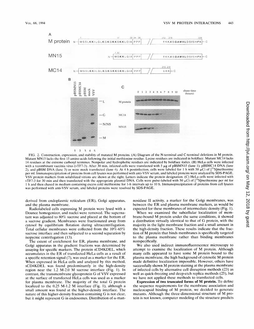
FIG. 2. Construction, expression, and stability of mutated M proteins. (A) Diagram of the N-terminal and C-terminal deletions in M protein.Mutant MN15 lacks the first 15 amino acids following the initial methionine residue. Lysine residues are indicated in boldface. Mutant MC14 lacks14 residues at the extreme carboxyl terminus. Nonpolar and hydrophobic residues are indicated by boldface italics. (B) HeLa cells were infectedwith a recombinant vaccinia virus (vTF7-3). After 30 min, infected cells were transfected with 5 ,ug of pBSMN15 (lane 1), pBSMC14 DNA (lane2), and pBSM DNA (lane 3) or were mock transfected (lane 4). At 4 h postinfection, cells were labeled for 1 h with 50 ,uCi of [35S]methionineper ml. Immunoprecipitation of proteins from cell lysates was performed with anti-VSV serum, and labeled proteins were analyzed by SDS-PAGE.VSV protein markers from solubilized virions are shown at the right. Letters indicate the protein designation. (C) HeLa cells were infected withvTF7-3 for 30 min and then transfected with the appropriate plasmid DNA. Cells were pulse-labeled with 50 ,uCi of [35S]methionine per ml forI h and then chased in medium containing excess cold methionine for 1-h intervals up to 10 h. Immunoprecipitation of proteins from cell lysateswas performed with anti-VSV serum, and labeled proteins were resolved by SDS-PAGE.
derived from endoplasmic reticulum (ER), Golgi apparatus,and the plasma membrane.
Radiolabeled cells expressing M protein were lysed with a
Dounce homogenizer, and nuclei were removed. The superna-
tant was adjusted to 80% sucrose and placed at the bottom ofa sucrose gradient. Membranes were fractionated away fromcytosol by equilibrium flotation during ultracentrifugation.Total cellular membranes were collected from the 10O%-65%sucrose interface and then subjected to a second separation byisopycnic centrifugation (13).The extent of enrichment for ER, plasma membrane, and
Golgi apparatus in the gradient fractions was determined byassaying for specific markers. The protein sCD4KDEL, whichaccumulates in the ER of transfected HeLa cells as a result ofa specific retention signal (7), was used as a marker for the ER.When expressed in HeLa cells and analyzed by this method,sCD4KDEL was found predominantly in the high-densityregion near the 1.2 M-2.0 M sucrose interface (Fig. 1). Incontrast, the transmembrane glycoprotein G of VSV expressedat the surface of transfected HeLa cells was used as a markerfor plasma membrane. The majority of the total G proteinlocalized to the 0.25 M-1.2 M interface (Fig. 1), although a
small amount was found at the higher-density interface. Thenature of this higher-density fraction containing G is not clear,but it might represent G in endosomes. Distribution of ox-man-
nosidase II activity, a marker for the Golgi membranes, was
between the ER and plasma membrane markers, as would beexpected for these membranes of intermediate density (Fig. 1).When we examined the subcellular localization of mem-
brane-bound M protein under the same conditions, it showeda distribution virtually identical to that of G protein, with themajority in the light membrane fraction and a small amount inthe high-density fraction. These results indicate that the frac-tion of M protein that binds membranes is specifically targetedto the plasma membrane rather than binding membranesnonspecifically.We also used indirect immunofluorescence microscopy to
attempt to examine the localization of M protein. Althoughmany cells appeared to have some M protein bound to theplasma membrane, the high background of cytosolic M proteinmade definitive localization impossible. However, others havesuccessfully shown M protein staining at the plasma membraneof infected cells by alternative cell disruption methods (23) as
well as quick-freezing and deep-etch replica methods (25), butwe have not applied these methods to transfected cells.
Expression of two truncated forms of M protein. To definethe sequence requirements for the membrane association andnucleocapsid binding of M protein, we decided to generatemutants. Although the three-dimensional structure of M pro-tein is not known, computer modeling of the structure predicts
A
M protein
C0 1 2 3 4 5 6 7 8 9 10
VOL. 68, 1994
on May 12, 2018 by guest
http://jvi.asm.org/
Dow
nloaded from

444 CHONG AND ROSE
A Top Bot B MN15 MC14
M
Top Bot Top
MN15
Bot
= .-Untreated
MC14
5 11 FractionNumber 2M KCI
50 mM EDTA
pH 11.05 *- _ _ 1_
5 11 1 5 11
Fraction FractionNumber Number
FIG. 3. Analysis of the membrane association of wild-type M protein, mutant MN15, and mutant MC14 by sucrose flotation gradients. (A)Lysates from 5 x 10⁶ transfected and radiolabeled HeLa cells were prepared as described in Materials and Methods. Lysates were made to 80%sucrose, placed at the bottom of a Beckman SW41 centrifuge tube, and overlaid with 65% (wt/wt) (5 ml) and 10% (wt/wt) (2.5 ml) sucrose layers.The step gradients were then centrifuged to equilibrium at 35,000 rpm for 18 h at 4°C. Fractions were collected from the top, diluted with detergentsolution, and immunoprecipitated with rabbit anti-VSV serum, and labeled proteins were analyzed by SDS-PAGE (10% gel). Shown are gradientsfrom cells expressing wild-type M protein, MN15, or MC14. Fractions are numbered from the top (fraction 1) to the bottom (fraction 11). (B) Totalcellular membranes from 1.5 x 10⁷ HeLa cells containing associated mutant MN15 or mutant MC14 from transfected and [35S]methionine-labeledHeLa cells were prepared by the sucrose flotation method as described in Materials and Methods. Membranes were treated with 2 M KCI-10 mMTris-HCl (pH 7.4) or 50 mM EDTA-Tris-HCl (pH 7.4) for 1 h at 25°C, extracted with carbonate buffer (pH 11.0) for 30 min at 0°C (15), or leftuntreated. Samples were made to 80% sucrose, and membranes were reisolated on a second sucrose flotation gradient. Fractions were collectedfrom the top, diluted with detergent solution, immunoprecipitated with rabbit anti-VSV serum, and analyzed by SDS-PAGE. Fractions arenumbered from the top (fraction 1) to the bottom (fraction 11).
a globular core with very little secondary structure at its aminoor carboxyl terminus. We reasoned that if both termini werefree, they might constitute domains interacting with the nucle-ocapsid and the membrane. The amino-terminal 19 residuescontain eight lysines and are followed by a triple-prolineregion, suggesting that they define a separate domain (29). Thecarboxyl terminus is somewhat hydrophobic when analyzed bythe program of Kyte and Doolittle (20). We therefore con-structed two plasmids, pBSMN15 and pBSMC14, which dif-fered from the complete M gene clone pBSM by deletions of15 amino acids at the amino terminus and 14 amino acids atthe carboxyl terminus of M protein. A schematic representa-tion of the truncated proteins is presented in Fig. 2A.Both mutant proteins were expressed in HeLa cells, as
shown by immunoprecipitation from lysates of transfected andradiolabeled cells with anti-VSV serum (Fig. 2B). Both mutantMN15 and mutant MC14 had mobilities on SDS-PAGE con-sistent with the deletion sizes (Fig. 2B, lanes 1 and 2). Tocompare the stability of mutant proteins MN15 and MC14 withthat of wild-type M protein, a pulse-chase experiment wasconducted in which transfected HeLa cells were labeled with[35S]methionine for 30 min and then incubated in mediumsupplemented with unlabeled methionine for a period of 1 h to10 h (Fig. 2C). Wild-type M protein was stable through 10 h ofchase time. However, mutant MC14 exhibited a half-life of 5 to6 h, and mutant MN15 exhibited a half-life of 3 to 4 h. A chasetime of 1 h was chosen in subsequent experiments so thatsimilar levels of labeled wild-type and mutant proteins could bestudied.Membrane association of mutants MN15 and MC14. We
expressed mutants MN15 and MC14 in HeLa cells and ana-lyzed membrane association by subcellular fractionation. Totalcell lysates were prepared from transfected and radiolabeledcells expressing M protein, mutant MN15, or mutant MC14,and membranes were fractionated from cytosolic material byequilibrium flotation during ultracentrifugation as previouslydescribed (9). Approximately 10% of the total M protein,MN15 protein, or MC14 protein was associated with cellularmembranes (Fig. 3A). This result indicated that despite dele-tions at either terminus, both mutant proteins were capable ofmembrane association.The amino terminus of M protein is required for stable
membrane association. Membranes containing radiolabeledmutant MN15 or MC14 were isolated from transfected HeLacells by the sucrose flotation gradient method as previouslydescribed (9). Membrane samples were then extracted with 2M KCl, 50 mM EDTA, or pH 11.0 carbonate buffer andsubjected to fractionation on a sucrose flotation gradient (Fig.3B). All of these conditions removed mutant MN15 proteinfrom cellular membranes, and the dislodged MN15 protein wasdetected at the bottom of the gradient. However, all of theseconditions failed to release mutant MC14 from membranes,similar to what was previously observed with membrane-associated wild-type M protein (9).The amino terminus is required for membrane-associated
M protein to bind RNP cores. To determine whether thedeletions affected the ability of MN15 or MC14 to bind RNPcores in vitro, membranes containing radiolabeled M protein,mutant MN15, or mutant MC14 were isolated from transfectedHeLa cells by the sucrose flotation gradient method. Mem-
J. VIROL.
qoow
.10ii, -
on May 12, 2018 by guest
http://jvi.asm.org/
Dow
nloaded from
VSV M PROTEIN INTERACTIONS 445
B Bot
aidSE:
5L-07
D Bot
N/NS -
10
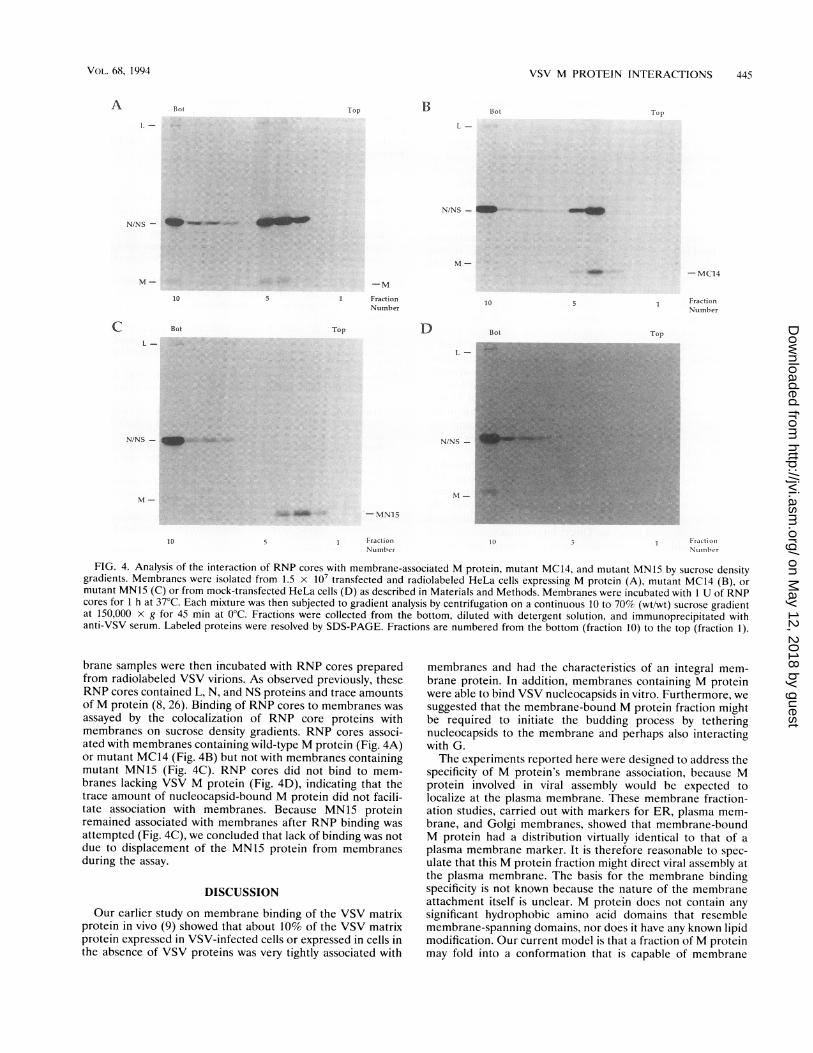
FIG. 4. Analysis of the interaction of RNP cores with membrane-associated M protein, mutant MC14, and mutant MN15 by sucrose densitygradients. Membranes were isolated from 1.5 x 10⁷ transfected and radiolabeled HeLa cells expressing M protein (A), mutant MC14 (B), or
mutant MN15 (C) or from mock-transfected HeLa cells (D) as described in Materials and Methods. Membranes were incubated with 1 U of RNPcores for 1 h at 37°C. Each mixture was then subjected to gradient analysis by centrifugation on a continuous 10 to 70% (wt/wt) sucrose gradientat 150,000 x g for 45 min at 0°C. Fractions were collected from the bottom, diluted with detergent solution, and immunoprecipitated withanti-VSV serum. Labeled proteins were resolved by SDS-PAGE. Fractions are numbered from the bottom (fraction 10) to the top (fraction 1).
brane samples were then incubated with RNP cores preparedfrom radiolabeled VSV virions. As observed previously, theseRNP cores contained L, N, and NS proteins and trace amountsof M protein (8, 26). Binding of RNP cores to membranes wasassayed by the colocalization of RNP core proteins withmembranes on sucrose density gradients. RNP cores associ-ated with membranes containing wild-type M protein (Fig. 4A)or mutant MC14 (Fig. 4B) but not with membranes containingmutant MN15 (Fig. 4C). RNP cores did not bind to mem-branes lacking VSV M protein (Fig. 4D), indicating that thetrace amount of nucleocapsid-bound M protein did not facili-tate association with membranes. Because MN15 proteinremained associated with membranes after RNP binding was
attempted (Fig. 4C), we concluded that lack of binding was notdue to displacement of the MN15 protein from membranesduring the assay.
DISCUSSION
Our earlier study on membrane binding of the VSV matrixprotein in vivo (9) showed that about 10% of the VSV matrixprotein expressed in VSV-infected cells or expressed in cells inthe absence of VSV proteins was very tightly associated with
membranes and had the characteristics of an integral mem-brane protein. In addition, membranes containing M proteinwere able to bind VSV nucleocapsids in vitro. Furthermore, wesuggested that the membrane-bound M protein fraction mightbe required to initiate the budding process by tetheringnucleocapsids to the membrane and perhaps also interactingwith G.The experiments reported here were designed to address the
specificity of M protein's membrane association, because Mprotein involved in viral assembly would be expected tolocalize at the plasma membrane. These membrane fraction-ation studies, carried out with markers for ER, plasma mem-brane, and Golgi membranes, showed that membrane-boundM protein had a distribution virtually identical to that of aplasma membrane marker. It is therefore reasonable to spec-ulate that this M protein fraction might direct viral assembly atthe plasma membrane. The basis for the membrane bindingspecificity is not known because the nature of the membraneattachment itself is unclear. M protein does not contain anysignificant hydrophobic amino acid domains that resemblemembrane-spanning domains, nor does it have any known lipidmodification. Our current model is that a fraction of M proteinmay fold into a conformation that is capable of membrane
Bot Top TopA
. -
N/NS -
M-
CL-
N/NS -
Bot
-M
1 FractionNumber
Top
-MC14
1 FractionNumber
Top
10
-MN15
FractionNumber
FractionNCtniber
VOL. 68, 1994
on May 12, 2018 by guest
http://jvi.asm.org/
Dow
nloaded from

446 CHONG AND ROSE
insertion and perhaps also interact with a plasma membranecomponent.The other studies described here were designed to identify
membrane and nucleocapsid binding domains in M protein.We have shown that neither the amino-terminal 15 residuesnor the carboxyl-terminal 14 residues of the VSV M proteinare essential for a normal extent of M protein binding tomembranes in vivo. However, the presence of the amino-terminal residues is required to achieve the normal stability ofmembrane association seen with wild-type M protein. It ispossible that membrane association is stabilized by penetrationof the lipid bilayer with the amino terminus (22). Other studiesdemonstrated that removal of the initial 43 amino acids of Mprotein by trypsin did not prevent the trypsin-resistant corefrom associating with artificial liposomes in vitro, although thenature of this interaction was not defined (26). It is possiblethat a region in the trypsin-resistant core of M proteinmediates an electrostatic interaction with the lipid bilayerwhile the amino terminus acts as a membrane anchor. Such amechanism has recently been proposed for the membraneassociation of the neuronal protein synapsin I with synapticvesicles (3). It is also possible that the amino terminus forms amembrane-associating domain with another region of M pro-tein. A mutation in measles virus matrix protein outside of thepredicted hydrophobic carboxyl terminus affected membraneassociation, suggesting that other regions in measles matrixprotein may be involved in membrane binding (16).Membrane-bound M protein lacking the amino-terminal 15
residues lost the ability to bind nucleocapsids in vitro. Thesimplest interpretation of these data is that the amino-terminaldomain is required for nucleocapsid binding. However, therole of the amino terminus of M protein in interaction withnucleocapsids is not yet clear. Direct interaction was suggestedfrom the ability of a synthetic oligopeptide corresponding tothe first 20 amino acids of M protein to prevent transcriptioninhibition by M protein in vitro (32). Kaptur et al. (18)suggested that the amino terminus of M protein is exposedwhen bound to nucleocapsids because protease could cleavenucleocapsid-bound M protein at positions 19 and 20. How-ever, it is not clear from that experiment whether residuespreceding position 19 interact with the nucleocapsid. Theinability of MN15 protein to bind nucleocapsids would also beconsistent with the suggestion by Kaptur et al. (18) thatproteolytic cleavage at the amino terminus may cause aconformational change that is disruptive to a downstreamnucleocapsid binding region (26, 32). Removal of criticalphosphorylation sites in the amino terminus or the alterationof other phosphorylation sites in M protein due to a confor-mational change may affect nucleocapsid and/or membraneassociation (2, 17).Although the carboxyl-terminal 14 residues ofM protein are
slightly hydrophobic, their removal had no effect on the extentof stability of membrane binding in vivo or nucleocapsidbinding by membranes containing the truncated M protein.This region therefore may not be critical to assembly, althoughother studies have indicated that the C terminus may berequired for the cytopathic effects caused by M protein (5, 24).The association of M protein with the plasma membrane in
vivo and the ability of membrane-bound M protein to interactwith nucleocapsid cores in vitro (9), taken together, support amodel of VSV assembly in which membrane-bound M proteincould nucleate sites for viral assembly at the cell surface (9).The cell-free system in which nucleocapsid cores can bind tomembrane-bound M protein may therefore be useful forfurther investigation of the budding process.
ACKNOWLEDGMENTSWe thank B. Crise, M. Whitt, C. Hammond, and all other members
of the laboratory for advice during the course of this work.This work was supported by grant A124345 from the National
Institutes of Health.
REFERENCES
1. Atkinson, P. H., S. A. Moyer, and D. F. Summers. 1976. Assemblyof vesicular stomatitis virus glycoprotein and matrix protein intoHeLa cell plasma membranes. J. Mol. Biol. 102:613-631.
2. Beckes, J. D., L. C. Childers, and J. Perrault. 1989. Phosphoryla-tion of vesicular stomatitis virus M protein: evidence for a secondvirion-associated protein serine kinase activity. Virology 169:161-171.
3. Benefanati, F., P. Greengard, J. Brunner, and M. Bahler. 1989.Electrostatic and hydrophobic interactions of synapsin I andsynapsin I fragments with phospholipid bilayers. J. Cell Biol.108:1851-1862.
4. Bergmann, J. E., and P. J. Fusco. 1988. The M protein of vesicularstomatitis virus associates specifically with the basolateral mem-branes of polarized epithelial cells independently of the G protein.J. Cell Biol. 107:1707-1715.
5. Blondel, D., G. G. Harmison, and M. Schubert. 1990. Role ofmatrix protein in cytopathogenesis of vesicular stomatitis virus. J.Virol. 64:1716-1725.
6. Bonner, W. M., and R. A. Laskey. 1974. A film detection methodfor tritium-labelled proteins and nucleic acids in polyacrylamidegels. Eur. J. Biochem. 46:83-88.
7. Buonocore, L., and J. K. Rose. 1990. Prevention of HIV-1 glyco-protein transport by soluble CD4 retained in the endoplasmicreticulum. Nature (London) 345:625-628.
8. Carroll, A. R., and R. R. Wagner. 1979. Role of the membrane (M)protein in endogenous inhibition of in vitro transcription byvesicular stomatitis virus. J. Virol. 29:134-142.
9. Chong, L. D., and J. K. Rose. 1993. Membrane association offunctional vesicular stomatitis virus matrix protein in vivo. J. Virol.67:407-414.
10. David, A. E. 1973. Assembly of the vesicular stomatitis envelope:incorporation of viral polypeptides into the host cell plasmamembrane. J. Mol. Biol. 76:135-148.
11. Emerson, S. U., and Y. H. Yu. 1975. Both NS and L proteins arerequired for in vitro RNA synthesis by vesicular stomatitis virus. J.Virol. 23:708-716.
12. Florkiewicz, R., and J. K. Rose. Unpublished data.13. Frangioni, J. V., P. H. Beahm, V. Shifrin, C. A. Jost, and B. G.
Neel. 1992. The nontransmembrane tyrosine phosphatase PTP-1Blocalizes to the endoplasmic reticulum via its 35 amino acidC-terminal sequence. Cell 68:545-560.
14. Fuerst, T. R., E. G. Niles, F. W. Studier, and B. Moss. 1986.Eukaryotic transient expression system based on recombinantvaccinia virus that synthesizes bacteriophage T7 RNA polymerase.Proc. Natl. Acad. Sci. USA 83:8122-8126.
15. Fujiki, Y., A. L. Hubbard, S. Fowler, and P. B. Lazarow. 1982.Isolation of intracellular membranes by means of sodium carbon-ate treatment: application to endoplasmic reticulum. J. Cell Biol.93:97-102.
16. Hirano, A., A. H. Wang, A. F. Gombart, and T. C. Wong. 1992. Thematrix proteins of neurovirulent subacute sclerosing panencepha-litis virus and its acute measles progenitor are functionally differ-ent. Proc. Natl. Acad. Sci. USA 89:8745-8749.
17. Kaptur, P. E., B. J. McCreedy, and D. S. Lyles. 1992. Sites of invivo phosphorylation of vesicular stomatitis virus matrix protein. J.Virol. 66:5384-5392.
18. Kaptur, P. E., R. B. Rhodes, and D. S. Lyles. 1991. Sequences ofthe vesicular stomatitis virus matrix protein involved in binding tonucleocapsids. J. Virol. 65:1057-1065.
19. Knipe, D. M., D. Baltimore, and H. F. Lodish. 1977. Separatepathways of maturation of the major structural proteins of vesic-ular stomatitis virus. J. Virol. 21:1128-1139.
20. Kyte, J., and R. F. Doolittle. 1982. A simple method for displayingthe hydrophobic character of a protein. J. Mol. Biol. 157:105-132.
21. Laemmli, U. K. 1970. Cleavage of structural proteins during the
J. VIROL.
on May 12, 2018 by guest
http://jvi.asm.org/
Dow
nloaded from

VSV M PROTEIN INTERACTIONS 447
assembly of the head of bacteriophage T4. Nature (London)227:680-685.
22. Lenard, J., and R. Vanderoef. 1990. Localization of the mem-brane-associated region of vesicular stomatitis virus M protein atthe N terminus, using the hydrophobic photoreactive probe'25I-TID. J. Virol. 64:3486-3491.
23. McCreedy, B. J., and D. S. Lyles. 1989. Distribution of M proteinand nucleocapsid protein of vesicular stomatitis virus in infectedcell plasma membranes. Virus Res. 14:189-206.
24. Moyer, S. A., S. C. Baker, and J. L. Lessard. 1986. Tubulin: anecessary factor for the synthesis of both Sendai virus andvesicular stomatitis virus RNAs. Proc. Natl. Acad. Sci. USA83:5405-5409.
25. Odenwald, W. F., H. Arnheiter, M. Dubois-Dalcq, and R. A.Lazzarini. 1986. Stereo images of vesicular stomatitis virus assem-bly. J. Virol. 57:922-932.
26. Ogden, J. R., R. Pal, and R. R. Wagner. 1986. Mapping regions ofthe matrix protein of vesicular stomatitis virus which bind toribonucleocapsids, liposomes, and monoclonal antibodies. J. Virol.58:860-868.
27. Ohno, S., and N. Ohtake. 1987. Immunocytochemical study of theintracellular localization of M protein of vesicular stomatitis virus.Histochem. J. 19:297-306.
28. Rose, J. K., and J. E. Bergmann. 1983. Altered cytoplasmicdomains affect intracellular transport of the vesicular stomatitis
virus glycoprotein. Cell 34:513-524.29. Rose, J. K., and C. L. Gallione. 1981. Nucleotide sequences of the
mRNAs encoding the vesicular stomatitis virus G and M proteinsdetermined from the cDNA clones containing the completecoding regions. J. Virol. 39:519-529.
30. Rosenberg, A. H., B. N. Lade, D.-C. Chiu, S.-W. Lin, J. J. Dunn,and F. W. Studier. 1987. Vectors for selective expression of clonedDNAs by T7 RNA polymerase. Gene 56:125-135.
31. Sanger, F., A. R. Coulson, B. J. Barrell, A. J. H. Smith, and B. A.Roe. 1980. Cloning in single-stranded bacteriophage as an aid torapid DNA sequencing. J. Mol. Biol. 143:161-178.
32. Shipley, J. B., R. Pal, and R. R. Wagner. 1988. Antigenicity,function, and conformation of synthetic oligopeptides correspond-ing to amino-terminal sequences of wild-type and mutant matrixproteins of vesicular stomatitis virus. J. Virol. 62:2569-2577.
33. Storrie, B., and E. A. Madden. 1990. Isolation of subcellularorganelles. Methods Enzymol. 182:203-225.
34. Van der Sluijs, P., M. Hull, L. A. Huber, P. Male, B. Goud, and I.Mellman. 1992. Reversible phosphorylation-dephosphorylationdetermines the localization of rab4 during the cell cycle. EMBO J.11:4379-4389.
35. Whitt, M. A., L. Chong, and J. K. Rose. 1989. Glycoproteincytoplasmic domain sequences required for rescue of a vesicularstomatitis virus glycoprotein mutant. J. Virol. 63:3569-3578.
VOL. 68, 1994
on May 12, 2018 by guest
http://jvi.asm.org/
Dow
nloaded from