The Effect of Hydroxyethyl Starch 130/0.4 On Canine ...€¦ · It was unknown if HES 130/0.4...
97
1 The Effect of Hydroxyethyl Starch 130/0.4 On Canine Platelet Function Duana McBride BVSc DACVECC MRCVS College of Veterinary Medicine School of Veterinary and Life Sciences Murdoch University Western Australia This thesis is presented for the degree Research Masters with Training (RMT) 2014
Transcript of The Effect of Hydroxyethyl Starch 130/0.4 On Canine ...€¦ · It was unknown if HES 130/0.4...
1
The Effect of Hydroxyethyl Starch 130/0.4
On Canine Platelet Function
Duana McBride
BVSc DACVECC MRCVS
College of Veterinary Medicine
School of Veterinary and Life Sciences
Murdoch University
Western Australia
This thesis is presented for the degree Research Masters with Training (RMT) 2014
2
Declaration
I declare that this thesis is my own account of my research and contains as its main content work
which has not previously been submitted for a degree at any tertiary education institution.
Chapters three and four are for publication in scientific journals in which publication are in
conjunction with the principle supervisors and other co-authors. The majority of study designs,
experimental research and writing for publication were undertaken by myself as primary author with
guidance from the principle supervisor and other co-authors.
Chapter three has been published in the American Journal of Veterinary Research, and copyright for
publication in this thesis has been granted. Chapter four has been submitted to a scientific journal,
and this chapter is consistent with the most recent copy submitted at the time of thesis submission.
As a result of this, abbreviations may differ to the remaining thesis, and spelling in these two
chapters are in American English.
Ethical approval for the experimental research in chapter three and four in this thesis has been
granted by Murdoch University Animal Ethics Committee with approval number R2324/10 and
R2398/11 respectively.
Duana McBride BVSc DACVECC MRCVS
Principle Author
Lisa Smart BVSc (Hons) DACVECC
Principle Supervisor
3
Abstract
Hydroxyethyl starch (HES) 130/0.4 is an artificial colloid solution commonly used to treat shock in
dogs. Adverse effects from this solution have been reported in people, including platelet
dysfunction. It was unknown if HES 130/0.4 causes platelet dysfunction in dogs. Therefore, we
investigated the in vitro and in vivo effects of HES 130/0.4 on platelet function in dogs. We used
closure time (CT) using the platelet function analyser-100 and adenosine diphosphate-collagen
cartridges as a platelet agonist, to measure platelet function in this report. In the in vitro study, two
solutions were compared: HES 130/0.4 and HES 200/0.5. Citrated blood from ten healthy dogs was
diluted 1:9 and 1:3 with HES 130/0.4, HES 200/0.5 and 0.9% sodium chloride (NaCl). Closure time of
the diluted blood was measured. Only HES 200/0.5 increased the CT beyond the dilutional effect at
the 1:3 dilution, to a median CT of 125 seconds (interquartile range, 117.5 to 139.5 seconds),
suggesting that HES 200/0.5, but not HES 130/0.4, caused platelet dysfunction at this dilution. No
effect of HES or dilution on CT was identified at the 1:9 dilution. In the in vivo study, haemorrhagic
shock was induced by removing 48ml/kg of blood from eleven greyhounds under general
anaesthesia. Dogs were randomised to receive either 20mL/kg of HES 130/0.4 or 80mL/kg of 0.9%
NaCl intravenously over 20 minutes. Both the HES 130/0.4 and 0.9% NaCl group had a significantly
increased mean CT after fluid administration to 91.4 seconds (95% CI 69.3-113.4) and 95.5 seconds
(95% CI 78.2-112.8), respectively. The magnitude of change was significantly greater for the 0.9%
NaCl group than the HES 130/0.4 group, indicating the increase in CT with HES 130/0.4 is most likely
due to a dilutional effect. In this study we also investigated the effect of shock on platelet function
by measuring CT before and after inducing haemorrhagic shock. We found haemorrhagic shock did
not significantly change CT. This report suggests HES 130/0.4 does not cause significant platelet
dysfunction in dogs.
4
TABLE OF CONTENTS
Declaration……………………………………………………………………………………………………………………………….……….2
Abstract………………………………………………………………………………………………………………………………….………….3
Acknowledgements………………………………………………………………………………………………………………….………..6
List of tables and figures………..………………………………………………………………………………………………………….7
1. Chapter 1: Introduction, objective, hypothesis……………………………………………………………………………8
1.1. Introduction…………………………………………………………………………………………………………………………8
1.2. Objective……………………………………………………………………………………………………………………………..8
1.3. Hypothesis…………………………………………………………………………………………………………………………..8
2. Chapter 2: Literature review……………………………………………………………………………………………………….9
2.1. Platelet function………………………………………………………………………………………………………………….9
2.2. Platelet function analysis……………………………………………………………………………………………………11
2.2.1. Platelet function analyser-100 ……………………………………………………………………………..…..11
2.2.2. Other platelet function tests…………………………………………………………………………………..…14
2.2.2.1. Buccal mucosal bleeding time……………………………………………………………………14
2.2.2.2. Platelet aggregometry……………………………………………………………………………….14
2.2.2.3. Flow cytometry………………………………………………………………………………………….16
2.2.2.4. Thromboelastography……………………………………………………………………………….16
2.2.2.5. Sonoclot…………………………………………………………………………………………………….18
2.2.2.6. Cone and plate(let) analyser………………………………………………………………………18
2.2.3. Summary………………………………………………………………………..…………………………………………18
2.3. Hydroxyethyl starch solutions……………………………………………………………………………………………19
2.3.1. Properties of hydroxyethyl starch solutions…………………..………………………………………….19
2.3.2. Pharmacokinetics………………………………………………………..…………………………………………….22
2.3.3. Pharmacodynamics……………………………………………………………………………………………………22
2.4. Indications of use of hydroxyethyl starch solutions……………………………………………………………23
2.4.1. Use of hydroxyethyl starch solutions in the treatment of shock………………………………..24
2.4.2. Other indications for the use of hydroxyethyl starch solutions………………………………….26
2.5. Adverse effects of hydroxyethyl starch solutions……………………………………………………………….27
2.5.1. Introduction………………………………………………………………………………………………………………27
2.5.2. Platelet dysfunction…………………………………………………………………………………………………..28
2.5.2.1. Platelet dysfunction in people……………………………………………………………….…..28
2.5.2.2. Platelet dysfunction in dogs……………………………………………………………………….29
2.5.3. Effect on von Willebrand factor and factor VIII………………………………………………………….29
5
2.5.4. Fibrin formation and fibrinolysis……………………………………………………………………………....30
2.5.5. Acute kidney injury…………………………………………………………………………………………………...30
2.6. Clinical use of hydroxyethyl starch 130/0.4………………………………………………………………………..32
2.7. Reference list for literature review…………………………………………………………………………………….35
3. Chapter 3: The effect of hydroxyethyl starch 130/0.4 and 200/0.5 on canine platelet function in
vitro…………………………………………………………………………………………………………………………………………..49
3.1. Abstract……………………………………………………………………………………………………………………………..49
3.2. Introduction……………………………………………………………………………………………………………………….49
3.3. Material and methods……………………………………………………………………………………………………….51
3.4. Results……………………………………………………………………………………………………………………………….53
3.5. Discussion………………………………………………………………………………………………………………………….54
3.6. Footnotes…………………………………………………………………………………………………………………………..58
3.7. Reference…………………………………………………………………………………………………………………………..59
4. Chapter 4: Platelet function in dogs with haemorrhagic shock treated with hydroxyethyl starch
130/0.4 or 0.9% NaCl…………………………………………………………………………………………………………………64
4.1. Abstract……………………………………………………………………………………………………………………………..64
4.2. Introduction……………………………………………………………………………………………………………………….65
4.3. Material and methods……………………………………………………………………………………………………….66
4.4. Results……………………………………………………………………………………………………………………………….70
4.5. Discussion………………………………………………………………………………………………………………………….74
4.6. Footnotes…………………………………………………………………………………………………………………………..77
4.7. Reference…………………………………………………………………………………………………………………………..78
5. Summary and Conclusion………………………………………………………………………………………………………….84
APPENDICIES……………………………………………………………………………………………………………………………………87
Appendix A: Chapter 3 raw data – closure times……………………………………………………………………………..87
Appendix B: Chapter 3 raw data – packed cell volumes and platelet counts…………………………………….94
Appendix C: Chapter 4 raw data – closure times………………………………………………………………………………95
Appendix D: Chapter 4 raw data – packed cell volumes and platelet counts……………………………………97
Appendix E: Chapter 4 raw data for measured and calculated values required for oxygen extraction
ratio determination………………………………………………………………………………………………………………………….98
6
Acknowledgements
I would like to sincerely thank my principle supervisor Dr Lisa Smart for her extended commitment in
supporting me throughout this project. She has sacrificed her own personal time in order to help me
reach big and small mile stones which I am forever grateful for. She has taught me precision in
scientific writing, endurance to achieve my goals, and has supported me generously in scientific
presentations. I would not be where I am professionally and personally without her dedication.
This thesis would also not be possible without the help and expertise of Professor Giselle Hosgood. I
could not express how fortunate I was to have her guidance in producing this thesis. Her experience
in research is phenomenal, where she has taught me precision in scientific writing with a purest
approach and an ability to self-critique my work.
I also could not have come this far without support from the many of my residency supervisors
including (and in no particular order), Dr Katrin Swindells, Dr Melissa Claus, and Dr Ryan Ong. You
have all been amazing people who have given me so much support in many ways. And to also to Dr
Anthea Raisis for the many hours we spent together during the experimental process. I will always
remember those Friday mornings.
I would also extend my gratitude to the Clinical Pathology department of Murdoch University for
their assistance with this project; my residency mates Dr Rachel Peacock and Dr Mark Haworth who
were also an imminent part of this journey; and to all the staff who are my friends at Murdoch
University Veterinary Hospital for making this masters, residency program, and Perth a fond memory
for me.
7
List of Tables and Figures
Table 3-1: Median (IQR) PCV and platelet count for 1:3 and 1:9 dilutions with 0.9% NaCl solution of
whole blood samples from 10 healthy dogs…………………………………………………………………………………….53
Table 3-2: Median (IQR) platelet CT for 1:3 and 1:9 dilutions with 0.9% NaCl solution, HES 130/0.4,
and HES 200/0.5 of whole blood samples from 10 healthy dogs………………………………………………………54
Figure 4-1: Time points for platelet closure time measurements: T0, at base line during general
anesthesia immediately prior to hemorrhage; T1, during hemorrhagic shock, which was immediately
after removing 48 mL/kg of blood; and T2, 40 minutes after completion of fluid administration of
either hydroxyethyl starch (HES) 13/0.4 or 0.9% sodium chloride (NaCl)……………………………….………..69
Table 4-1: Formulae for calculated variables……………………………………………………………………………………70
Table 4-2: Mean (95% confidence interval) packed cell volume, platelet count, oxygen extraction
ratio (O2ER) and platelet closure time in greyhounds under general anesthesia (T0) and thirty
minutes after 43mL/kg of blood loss (T1). ……………………………………………………………………………………….72
Table 4-3: Mean (95% confidence interval) of packed cell volume, platelet count, oxygen extraction
ratio (O2ER) and platelet closure time in greyhounds in hemorrhagic shock (T1), under general
anesthesia, that then received either 20ml/kg of hydroxyethyl starch (HES) 130/0.4 or 80ml/kg of
0.9% sodium chloride (NaCl) (T2)………………………………………………………………………………………….…………73
8
1. Chapter 1: INTRODUCTION, OBJECTIVES AND HYPOTHESES
1.1. Introduction
The aim of this thesis was to investigate the effect of two commonly used hydroxyethyl starch
solutions, HES 130/0.4 and HES 200/0.5, on canine platelet function. The first study compared the
two solutions in vitro and the second study focused on the effect of HES 130/0.4 in vivo. The second
study utilised a shock model, therefore, we also had to determine the effect of shock on platelet
function. Our assessment of platelet function was based on closure time (CT), as measured by the
Platelet Function Analyser 100 (PFA-100).
1.2. Objectives
1. To determine if HES 130/0.4 and HES 200/0.5 increases CT in vitro, beyond a dilutional effect
(Chapter 3).
2. To determine if haemorrhagic shock in anaesthetised dogs alters CT (Chapter 4).
3. To determine if HES 130/0.4 increases CT, beyond any effect of shock or haemodilution
(Chapter 4).
1.3. Hypothesis
We hypothesised that HES 130/0.4 would increase CT in vitro and in vivo, as measured by the PFA-
100. We also hypothesised that haemorrhagic shock in dogs would alter CT.
9
2. Chapter 2: LITERATURE REVIEW
2.1. Platelet Function
Haemostasis is a complex mechanism resulting in thrombus formation. Traditionally, haemostasis
has been divided into separate stages, including primary haemostasis, secondary haemostasis and
fibrinolysis. However, it has been found more recently to be a complex mechanism with all stages
occurring synergistically, involving tissue factor bearing cells, platelets and the endothelium.1 The
new theory is referred to as the cell-based model of coagulation. Endothelial injury is the most
common initiator of coagulation, caused by surgical incision or venepuncture, infectious disease
processes, severe acidaemia, hypoxaemia, inflammation, hypotension and even during normal
endothelial cell turn over.2
The first step in forming a thrombus is the adhesion of platelets to the site of injury.3 Platelets are
synthesised in bone marrow and smooth muscle cells, and, in small amounts, in the liver, spleen and
kidneys.2 Its production is activated by thrombopoietin, which is produced in the liver and kidneys.
The average lifespan of platelets in circulation in the dog is approximately 6 days.4 Platelets achieve
primary haemostasis by forming a platelet plug in three interrelated steps. This includes adhesion of
platelets to endothelial surfaces,5 activation of platelets, and platelet aggregation.6 The mechanism
of platelet function can differ in high shear stress conditions within small and medium sized arteries
compared to low shear stress conditions within large arteries and in veins.5
Endothelial injury initially exposes subendothelial collagen. As a result, platelets adhere to the
extracellular matrix and surrounding endothelial cells, which is mediated by von Willebrand Factor
(vWF), fibronectin, laminin and thrombospondin. Von Willebrand factor is an adhesive protein
synthesised by platelet precursors, megakaryocytes. After synthesis, vWF is stored within alpha
granules in platelets. In addition, they are also produced and stored in endothelial cells.7 Von
Willebrand factor release is stimulated by thrombin, fibrin, vasopressin, collagen, platelet-activating
factor, epinephrine or histamine.8 Once in circulation, it can bind to exposed collagen at the site of
10
endothelial injury. Platelets then adhere to the site of injury by binding to vWF, mediated by integrin
αIIbβ3 receptors and glycoprotein (GP) Ibα, which is a component of the GP Ib-IX-V complex found on
platelet surfaces.5 During high shear stress conditions, binding of vWF on collagen and loose binding
of vWF to GP Ibα receptors result in platelet rolling along endothelial surfaces, facilitated by P-
selectin expression. During low shear stress conditions, fibronectin and laminin mediate adhesion.
During the adhesion process, factor (F) VIII is released, and vWF forms a complex with FVIII to avoid
rapid clearance from plasma.9
Once platelets are adhered to the subendothelial collagen, they are further activated by agonists
such as collagen, vWF, thrombin, adenosine diphosphate (ADP), and thromboxane A2 (TXA2), as well
as being activated by shear stress conditions.10 Activation causes degranulation of α granules and
dense bodies, increased cytosolic calcium concentration, conformational change of platelets, release
of ADP, serotonin and epinephrine, and activation of protein kinase C.6 Under high shear stress
conditions, ADP is the primary promoter of platelet activation; while under low shear stress
conditions, TXA2 promotes platelet activation.11 Thrombin plays a key role as it activates platelets
under all shear conditions via the protease-activated receptors (PAR)1 and PAR4, as well as the GP
Ib-V-IX complex .5 Although activation of platelets usually follows adhesion, it can also occur
independent of adhesion, for example when thrombin is generated during inflammation, which can
promote pathologic thrombus formation.
Platelet activation allows platelet aggregation to occur which leads to the formation of platelet
plugs. Aggregation occurs by binding of platelet surface receptors, primarily integrin αIIbβ3 as well as
GP Ib-IX-V, mediated by vWF, fibrinogen, thrombin, ADP and TXA2, in order to form a platelet plug.6
Thromboxane A2, produced by the arachidonic acid cascade in platelets, is one of the most
important facilitators of aggregation by activating integrin αIIbβ3 receptors. In high shear stress
conditions, vWF is the major ligand for platelet aggregation, while in low shear stress conditions
fibrinogen is the major ligand.12 Once a platelet plug is formed, a cross-linked fibrin meshwork can
11
then be formed to stabilise the platelet plug. The fibrin meshwork has been historically described as
secondary haemostasis.
Concurrent to platelet plug formation, adjacent endothelium releases prostacyclin and nitrous oxide,
which causes vasodilation and inhibits platelet aggregation. This prevents excessive thrombus
formation.13
2.2. Platelet Function Analysis
There are several methods to assess platelet function in people and in dogs. Due to the complexity
of primary haemostasis, and the importance of platelet interactions with cellular surfaces in vivo, no
single platelet function test is without limitations. The different methods of platelet function analysis
reviewed here are available for clinical and research use, and the understanding of the function and
limitations of each test is important for interpretation of results. The PFA-100 is described in detail
below, followed by other tests validated for use in veterinary medicine.
2.2.1. Platelet Function Analyser - 100
The PFA-100 is a point-of-care, bench top platelet analyser, which measures platelet function in high
shear stress conditions in vitro. A small volume of citrated whole blood (0.8 mL) is placed in a
reservoir in a disposable test cartridge, which is then aspirated by a vacuum through a stainless steel
capillary. This mimics the high shear stress conditions that platelets encounter in small and medium
sized arteries. The sample reaches a small aperture (150 µm diameter) with a membrane coated
with collagen, and either ADP or epinephrine, depending on the type of cartridge selected. The high
shear stress conditions combined with the platelet agonists result in platelet adhesion, activation
and aggregation followed by platelet plug formation, which occludes the small aperture. The time
from the onset of aspiration to the time of aperture closure is measured in seconds, and is recorded
as the closure time (CT).14
12
The validation of the PFA-100 in dogs is based on several studies.15-17 Reference intervals reported in
these studies with the use of ADP and collagen cartridges were 53 – 98 seconds,15 47 - 81 seconds,16
and 52 – 86 seconds.17 Reference intervals reported in these studies with the use of epinephrine and
collagen cartridges were 92 - >300 seconds,15 67 – 210 seconds,16 and 97 - >300 seconds.17 Closure
times with epinephrine and collagen activating cartridges resulted in greater variability in results,17
making this cartridge less reliable in dogs. The variability is thought to be due to the lack of ability for
epinephrine to induce significant platelet aggregation in dogs compared with people.18
The PFA-100 has also been validated for use in greyhounds. This is important because greyhounds
have different haematological reference intervals compared to non-greyhound breed dogs. A
bleeding tendency during surgery has been reported in some greyhounds, which is thought to be
due to increased fibrinolysis, although this mechanism is not completely understood. A study by
Couto et al measured CT with ADP and collagen coated cartridges as well as epinephrine and
collagen coated cartridges, which ranged from 63 to 92 seconds and 87 to 238 seconds
respectively.19 Following this study, the same investigators measured CT in greyhounds with clinical
postoperative haemorrhage, which found no significant difference in CT between greyhounds with
clinical evidence of haemorrhage and those without noticeable haemorrhage, indicating normal
platelet function in greyhounds with bleeding tendencies.20
Several individual-related factors have shown to influence CT in dogs. Firstly a decrease in platelet
count (< 100,000 platelets/µL17) can increase CT, due to the decreased availability of platelets. A
decrease in haematocrit, or packed cell volume (PCV) (< 2516 – 30%15,17) can also increase CT. A low
haematocrit can affect platelet function due to changes in blood flow dynamics altering platelet
interaction with the endothelium,21 which may also hold true for contact with the PFA-100 aperture.
Another proposed reason is due to decreased ADP release as a result of fewer available red blood
cells.22 Although a defined minimum PCV is difficult to specify, CT results should be interpreted in
line with a concurrent PCV. Von Willebrand factor can also influence CT as it is essential in primary

13
haemostasis and platelet function.15,17 Therefore, the PFA-100 has the clinical utility to diagnose von
Willebrand Disease (vWD) in dogs,17,23 and vWD should be considered if an increase in CT is detected
in a clinical scenario.
As described above, the PFA-100 has been used for the diagnosis of vWD, with improved sensitivity
and specificity compared with the traditionally used buccal mucosal bleeding time,17 as well as
contributing to the investigation of the mechanism of bleeding in greyhounds. It has also been used
in dogs to monitor efficacy of aspirin therapy,15 Scott’s syndrome,24 as well as to assess the effect of
HES solutions on platelet function.25,26 Unlike other platelet function tests such as flow cytometry,
platelet aggregometry, and thromboelastography with platelet mapping, the PFA-100 has the added
advantage of mimicking high shear stress conditions, which is more applicable to physiological
conditions. However, as it is an in vitro test, it lacks platelet interaction with true endothelium and is
limited by the lack of cellular release of factors and proteins which normally occurs during in vivo
haemostasis. From our experience, although the use of this machine is technically simple, results can
be variable at times, particularly if the CT is not measured immediately, venepuncture technique is
poor, the sample is left to sit without adequate rocking, or the citrated blood sample is not kept at a
standard temperature.
There are limited studies on the use of the PFA-100 for detecting increased platelet reactivity or
evaluating the risk of bleeding. One study in people found that CT was associated with increased
perioperative red blood cell transfusion requirements in patients with aortic valve stenosis,27
however there are no other studies which specifically address the association between CT and risk of
haemorrhage. One study measured CT and platelet aggregometry immediately following trauma,
which found increased platelet reactivity measured by aggregometry and this correlated with a
decreased CT. Further studies are warranted to investigate the significance of a decreased CT and
the use of CT to predict clinical bleeding.28
14
2.2.2. Other platelet function tests
2.2.2.1. Buccal mucosal bleeding time
Buccal mucosal bleeding time (BMBT) is a simple in vivo method for diagnosing primary haemostatic
disorders, which can differentiate from secondary haemostatic disorders.29 A small incision is made
on the mucosa of the upper lip with a standardized instrument, while the lip is being reflected
dorsally with a gauze bandage tied over the muzzle. While gently removing blood from the mucosa
with a filter paper, the time from when the incision is made until the time taken for bleeding to
cease is measured in seconds.30 Two studies have reported normal BMBT in dogs with mean and
standard deviations of 2.73 +/- 0.77 minutes (range 1.83 – 4.75 minutes),32 and 2.62 +/- 0.49 minutes
(range 1.68 – 4.14 minutes).30 A prolonged BMBT was found in dogs with vWD, thrombocytopenia,
thrombocytopathia, severe uraemia, and with aspirin therapy.30 Buccal mucosal bleeding time in
greyhounds has also been reported with a mean reference interval of 2.15 minutes +/- 0.73 minutes
(range 0.88 – 4.12 minutes), which is different to non-greyhound breeds.32 This study described one
of the major limitations which are inter-observer and intra-observer variability which can both range
up to approximately 2 minutes within the same dog. Another limitation of BMBT is that it does not
predict surgical bleeding in dogs.30
2.2.2.2. Platelet Aggregometry
Platelet aggregometry is considered the gold standard method for detecting platelet dysfunction.
There are two methods of platelet aggregometry, light transmission aggregometry using platelet rich
plasma or electrical impedance aggregometry using whole blood, with both methods being validated
for use in dogs.33-35 Platelet aggregometery measures platelet aggregation in response to various
agonists added to platelet rich plasma or whole blood samples, depending on the method utilised.
Platelet agonists that have been used include ADP, collagen, arachadonic acid, thombin and
epinephrine.36 With light transmission aggregometry, the sample becomes more translucent over
time as platelet aggregate, and therefore light transmission through the sample increases.33 Light
15
transmission is recorded graphically, which can evaluate the time to maximum aggregation and
overall rate of aggregation.36 The more recently developed method of electrical impedance has the
advantage of being able to use whole blood samples, which eliminates the process of producing
platelet rich plasma for analysis. With this method platinum electrodes are placed in the whole
blood sample, and the blood is gently stirred.34-35 Once the agonist is added, platelets form a
monolayer over the electrodes, which impede electrical current between the two electrodes. The
transmitted current is then traced into a graph similar to light transmission aggregometry.
One of the major benefits of platelet aggregometry is its ability to quantitatively measure platelet
function with the addition of varied platelet agonists. This technology has been used to measure
platelet function in dogs after being given various therapies that may affect platelet function.
Platelet aggregometry showed no significant change in platelet aggregation after HES 670/0.75
administration to dogs, despite a significant decrease in platelet count.37 However it should be noted
that the major limitation of this study is the small sample size. Various studies have also utilised this
technology to investigate the effect of pimobendan,38 lipid emulsions,39 non-steroidal anti-
inflammatory drugs,40 aspirin,41 and clopidogrel on platelet function in dogs.41 Platelet aggregation
has also been used to demonstrate decreased platelet function in dogs with infections,42 and
increased platelet function in dogs with malignancies,43 which also demonstrates its benefits in being
able to measure platelet hyper-reactivity. Experimentally, this technology has also been used to
analyse platelet function in frozen canine platelet concentrates.44 In people this technology has been
used for diagnosing vWD,45 although it has not been used in dogs for this purpose. Future scope for
this technology for clinical use to diagnose primary haemostatic disorders, including vWD and to
monitor drug therapy, are promising but its use is currently limited by availability.
Limitations to light transmission platelet aggregometry include the requirements of sample
preparation in order to produce platelet rich plasma. It can be time consuming and require
expertise, the centrifugation process can activate platelets, and the temperature of blood and the
16
time elapsed between sample collection and processing can also affect results.46 Other limitations of
aggregometry are that it does not mimic physiological conditions such as shear stress, and with light
transmission aggregometry, the lack of red blood cells may also alter results.
2.2.2.3. Flow cytometry
Flow cytometry is a platelet function test validated for use in dogs,47,48 which has the ability to assess
platelet membrane glycoproteins, ligands, and platelet derived microparticles in platelets before and
after being stimulated by platelet agonists including ADP, collagen, thrombin and epinephrine.36
Whole blood, platelet rich plasma or washed platelet samples can be used. Platelets are
fluorescently labelled and light scatter is detected to quantify the type and number of receptors, and
platelet surface antibodies expressed on platelet surfaces. One of the benefits of this platelet
function test is that it can be used to investigate the mechanism of platelet dysfunction by labelling
platelet receptors,49,50 and has been crucial in the development of our understanding of the
mechanism of effects of different drugs on platelet function.51 In dogs, flow cytometry has been
used to investigate the function of platelets during inflammation,52,53 and in dogs with immune
mediated haemolytic anaemia.54,55 It has also been used to investigate the effect of different
therapeutics, such as aspirin56 and cyclosporine,51 on platelet function. More recent investigations
have used flow cytometry to assess the viability of platelets in canine platelet transfusion
products.57,58 One of the major limitations of flow cytometry is that it is a bench top platelet function
test limited to laboratory use, and in addition does not mimic in vivo platelet function under shear
stress conditions.
2.2.2.4. Thromboelastography
Thromboelastography has been employed to assess hypercoagulability and hypocoagulability in
dogs. However, thromboelastography alone is a global coagulation function test and, therefore, does
not specifically assess platelet function. More recently however, thromboelastography with platelet
17
mapping has become available, which measures the influence of platelet function on
thromboelastography.
Thromboelastography involves adding a small amount of whole blood to a small cup with a pin
suspended in the middle. Depending on the machine utilised, either the cup rotates
(thromboelastography) or the pin rotates (thromboelastometry). As only thromboelastography has
been used for platelet mapping, the following discussion will be based on thromboelastography
alone. With thromboelastography platelets can be activated by different activators, such as tissue
factor, activator F, kaolin, arachidonic acid and ADP. Torque is created by rotation of the pin, which
creates a graph reflecting generation of a clot. Several measurements can be made from the graph
including reaction time (R), which is the time to initiate fibrin formation; K-time (K), which is the time
to form a clot strength of 20 mm; alpha-angle (α), which describes the speed of fibrin build up and
cross-linkage; maximum amplitude (MA), which is the maximum clot strength; and clot lysis after 30
minutes (LY30) and 60 minutes (LY60) after MA.
Thromboelastography with platelet mapping requires three samples run concurrently. The first
sample is citrated blood with kaolin activation (MAthrombin), which measures the maximum clot
strength for global coagulation. The second sample is heparinised blood activated with activator F
(MAfibrin), which isolates the fibrin contribution to the clot. The third sample is the MAfibrin sample
with either ADP (MAADP) or arachidonic acid (MAAA) as an additional agonist. The formula (MAADP/AA –
MAFibrin)/(MAThrombin – MAFibrin) x 100 is used to determine the platelet inhibition response.
Thromboelastography with platelet mapping has shown to be associated with platelet aggregation in
people.59 Thromboelastography has been used in dogs to assess the effect of clopidogrel41 and non-
steroidal anti-inflammatory drugs on platelet aggregation60 which also showed association with
platelet aggregometry. Thromboelastography has also been utilised in the assessment for platelet
function in dogs with hyperadrenocorticism.61 Thromboelastography has the advantage of being able
to assess the global formation of a clot, including hypercoagulability and hypocoagulability, and now
18
with platelet mapping, includes specific assessment of platelet function. However one of the major
disadvantages of thromboelastography with platelet mapping is the expense of requiring two
thromboelastography machines to run concurrently, and currently there is minimal literature
validating its use in dogs.
2.2.2.5. Sonoclot
Sonoclot is a global coagulation test that also has capability to measure platelet function. It has been
validated for use in dogs40 and has been shown to be useful in monitoring unfractionated heparin
doses in dogs.62 It has a probe with a sensor that detects torque as blood clots within a cuvette. The
sensor detects the change in torque during clot formation which is displayed in a graph. The graph
represents sonoclot activated clotting time (ACT), which is the time in seconds till change in torque is
detected; clot rate, which is the rate of fibrin formation; and platelet function (PF), which is
calculated by the software from the peak strength of the clot and the time to peak clot formation.
Despite the name, PF, this calculated value also involves fibrin formation, therefore is not specific to
platelet function alone. Compared to platelet aggregometry, this test is limited to thrombin-
stimulated platelet activation, therefore Sonoclot cannot be used to determine the effect of certain
platelet inhibitors such as clopidogrel and aspirin.63
2.2.2.6. Cone and plate(let) analyser
The cone and plate(let) analyser is another bed-side platelet function test that uses whole blood in
high shear stress conditions like the PFA-100, and has the advantage of being able to measure
increase and decrease in platelet function. However this test in only validated for research use in
people, and has not been validated for use in dogs.36
2.2.3. Summary
The role of platelet function in haemostasis is complicated by multiple factors contributing to its
activation, adhesion to endothelium, and aggregation resulting in final clot formation. Therefore,

19
despite the multiple methods available in order to investigate platelet function, there is no single
test which provides all the information required to assess platelet function. An ideal test would be
readily accessible, easy to use with minimal inter-observer and intra-observer variability, have the
ability to measure variable components of platelet function, and in addition, mimic physiological
conditions. The PFA-100 fulfils some of these criteria as it is easy to perform, produces a
measurement which is easily compared, has been validated for use in experimental research and is a
bed-side test which mimics physiological shear stress conditions. However using multiple methods
of platelet function analysis would be able to provide more information on the mechanisms of
platelet function.
2.3. Hydroxyethyl Starch Solutions
2.3.1. Properties of Hydroxyethyl Starch solutions
Hydroxyethyl starch (HES) solutions are artificial colloid solutions that are used to treat shock by
expanding blood volume. The colloid molecules are made by modification of natural polymers of
amylopectin made from waxy maize starch.64 There are a variety of HES solutions available, which
are characterised by their physical and chemical characteristics including mean molecular weight
(MW), degree of substitution (MS), and hydroxyethylation ratio at carbon position C2 and C6 (C2/C6
ratio). These properties influence the rate of degradation and separate the solutions into slowly
degradable solutions such as HES 670/0.75, rapidly degradable solutions such as HES 130/0.4, or
solutions having a medium rate of degradation such as HES 200/0.5. Slowly degradable products
tend to have greater adverse effects. Other important properties include the carrier solution type in
which the starch molecules are suspended in, such as 0.9% NaCl or a balanced crystalloid solution,
and the concentration of starch molecules within the solution, most commonly 6% or 10%.64 These
properties are discussed in more detail in this chapter.
20
The MW of HES solutions describe the mean MW of a polydispersed solution, meaning, there are a
broad range of HES molecules with different MWs in one solution.64 The MW is often described as
being high (> 400 kDa), medium (200 – 400 kDa), or low (< 200 kDa). The MW can influence the rate
of degradation of HES solutions; with higher MW solutions having a slower rate of degradation, and
lower MW solutions having a faster rate of degradation. The mean MW can be calculated by two
different methods; the number average MW and the weight average (or mass average) MW.65 If
there are larger molecules in a solution, the weight average MW is influenced to a greater degree
than the number average MW. The ratio of weight average MW to the number average MW will give
an index of the degree of polydispersity of the solution.65
Lederer et al reported that previous methods of MW determination for currently labelled HES
solutions are inaccurate.66 Using size exclusion chromatography coupled with low-angle laser light
scattering techniques, HES labelled 450/0.7 was determined to have a MW of 670 KDa and HES
200/0.5, a MW of 240 KDa.66 However HES 130/0.4 was found to have an accurate mean MW of 130
KDa as determined by low angle laser light scattering technique.67 However, despite further accuracy
on size, the in vitro molecular weight described by the manufacturer differs to the eventual in vivo
MW. This is because when HES solutions are administered intravascularly, the small molecules (< 50
kDa) are rapidly excreted by the kidneys.68 The larger molecules are hydrolysed by amylase into
smaller molecules, as discussed in the pharmacokinetics chapter, resulting in an increase in the
number of molecules in vivo. As a result the in vivo MW is lower than the in vitro MW.64 Metabolism
of the molecules actually improves the colloid osmotic pressure, as it is the number of molecules,
not absolute MW, that determines colloid osmotic pressure.
The MS describes the average number of substitutions of hydroxyl groups with hydroxyethyl
residuals at carbon position 2 (C2) and carbon position 6 (C6) on each glucose subunit within the
hydroxyethyl starch polymer.64 Solutions can be highly substituted (0.62 – 0.75), have a medium
substitution (0.5) or low substitution (0.4). The MS can be calculated by the formula, MS = W/(1-W) x

21
162/44; where W is the weight fraction of hydroxyethyl groups in the polymer, 162 is the mass of
the starch, and 44 is the mass of the hydroxyethyl group.65 The MS determines the ability of amylase
to cleave HES molecules in vivo into smaller molecules, resulting in a smaller in vivo MW.67 Higher
MS solutions are more difficult to cleave by amylase, resulting in reduced enzymatic degradation and
prolongation of intravascular retention.65
The C2/C6 ratio is the ratio of hydroxyethylation at the C2 position compared to the C6 position. This
property is also important as there is greater difficulty in cleaving HES molecules at C2 compared to
C6. Hence, HES products with a higher C2/C6 ratio have a slower rate of degradation.69
Another important characteristic of HES solutions is the solvent in which HES molecules are
suspended in. Commonly used solvents include 0.9% NaCl or balanced crystalloid solutions. The
characteristics of the solvents can influence acid-base and electrolyte balance. It has been shown
that 0.9% NaCl as a solvent causes a significant decrease in blood pH, and an increase in chloride
concentration and base deficit, compared to a balanced crystalloid solution.12 The concentration of
hydroxyethyl starch molecules in the solvent is commonly 6% or 10%, depending on the product.
The concentration of the solution affects the resulting plasma concentration,70 altering the rate of
excretion and contributing to adverse effects, as well as the volume expansion effects.
The two solutions most commonly used in people are 10% HES 200/0.5 and 6% HES 130/0.4. The use
of high MW HES 670/0.75 has been associated with significant adverse effects and its use has fallen
out of favour. Hydroxyethyl starch 200/0.5 has a medium mean MW of 200 kDa, a medium MS of
0.5, and a C2/C6 ratio of 5:1, and is commonly described as a pentastarch solution. Due to these
properties, HES 200/0.5 has a medium rate of degradation when compared to other HES solutions.70
Hydroxyethyl starch 130/0.4 has a low mean MW of 130 kDa, a low MS of 0.4 and a high C2:C6 ratio,
and is commonly described as a tetrastarch solution. Despite the high C2/C6 ratio, the low MW and
MS have a greater influence on the behaviour of this solution, resulting in a rapid rate of
degradation.65 The HES 130/0.4 solution under investigation described in this thesis has a
22
concentration of 6% suspended in a 0.9% NaCl solvent, although HES 130/0.4 in 10% solutions and
balanced electrolyte solvents are also available.
2.3.2. Pharmacokinetics
Once polydispersed HES solutions are administered intravenously, HES molecules with a MW lower
than the renal threshold (40 - 60 KDa) are excreted by the kidneys.67,72 Larger molecules are cleaved
by amylase, increasing the number of molecules contributing to colloid osmotic pressure.67,72 There
is also a small increase in MS in vivo as the more highly substituted molecules resist cleavage. A
small proportion of HES molecules are stored in the tissues and are excreted following
redistribution.67
Because 6% HES 130/0.4 has a low MW and low MS, it has the highest plasma clearance rate (31.4
mL/min) and shorter half-life in people (12.1 hours), compared to the higher MW higher MS HES
solutions, after a single dose administration of 500mLs.70 The maximum plasma concentration
achieved was 3.7mg/mL. By 24 hours after administration, plasma concentrations decreased to less
than 0.5 mg/ml. Compared to this, 6% HES 670/0.75 (which has a high MW of 670 KDa and a high MS
of 0.75) and 10% HES 200/0.5 had a much slower rate of plasma clearance of 0.98 mL/min and 9.24
mL/min respectively; a longer half-life of 46.4 hours and 30.6 hours respectively; and higher
maximum plasma concentration of 13mg/mL and 8mg/mL respectively.67
2.3.3. Pharmacodynamics
The pharmacodynamic property of interest for HES solutions is the effect of these solutions on blood
volume expansion. A study by Silverstein et al73 showed that administration of 20mL/kg of HES
670/0.75 immediately increased blood volume by 40% and was able to maintain an increase of 30%
for 240 minutes. In comparison, 80ml/kg of 0.9% NaCl was able to increase blood volume by 76%,
however rapidly decreased to 20% blood volume expansion, despite the larger volume administered.
This effect can be explained by Starling’s principle, which states that net fluid flow across a vessel
23
wall is dependent on the filtration coefficient, reflection coefficient, the balance between capillary
and interstitial hydrostatic pressure and the balance between the capillary and interstitial colloid
osmotic pressure. This can be described by the following equation:
Jv = Kf[(Pc – Pi) – σ(πc – πi)]
where Jv is the net fluid flow across a vessel wall, Pc is the capillary hydrostatic pressure, Pi is the
interstitial hydrostatic pressure, πc is the capillary oncotic pressure, πi is the interstitial oncotic
pressure, Kf is the filtration coefficient, and σ is the reflection coefficient.74,75 Therefore,
administration of HES solutions, which have a higher MW and therefore higher oncotic pressure
compared to crystalloid solutions, will result in increased intravascular oncotic pressure, resulting in
prevention of extravasation of fluid, compared to a crystalloid solution.76-78 However it is also
important to remember that artificial colloids such as HES will also cause an increase in hydrostatic
pressure, resulting in some degree of extravasation of fluid into the interstitial space. The degree of
extravasation of fluid, and hence an HES solution’s ability to expand blood volume, is also altered by
patient factors such as albumin balance between the capillary and interstitium and capillary
permeability, which can both be altered in diseased states.
Although there are no studies on the pharmacodynamics of 6% HES 130/0.4 or 10% HES 200/0.5 in
dogs, several studies have assessed the pharmacodynamics of these solutions in people.
Administration of 500mL of HES 130/0.4 over 15 minutes resulted in a plasma volume increase of
380mls over 4 to 6 hours, which returned to baseline within 8 to 24 hours. Despite the lower plasma
concentration and shorter half-life of HES 130/0.4, there was no difference in plasma volume effect
compared to 10% HES 200/0.5 or 6% HES 670/0.75.79,80 Also, there was no blood volume expansion
effect 24 hours after a single dose despite the persistence of HES solution in plasma.79,81
2.4. Indications for use of hydroxyethyl starch solutions
24
Hydroxyethyl starch solutions are used in the treatment of shock to rapidly increase and maintain
intravascular blood volume. There are several advantages of artificial colloid solutions compared to
crystalloid solutions. One advantage is that it can minimise development of interstitial oedema by
maintaining colloid osmotic pressure.82,83 As less of the solution redistributes outside of the vascular
space, it provides longer intravascular volume expansion effects compared with administration of
crystalloid solutions,73 therefore requiring smaller volumes.84 Additional benefits may include the
ability to reduce capillary permeability,85,86 and anti-inflammatory effects.87-89
2.4.1 Use of hydroxyethyl starch solutions in the treatment of shock
Shock can be defined as inadequate oxygen delivery (DO2) to tissues resulting in failure to meet
oxygen demands, impairing oxidative metabolism.90 Failure of the cardiovascular system to deliver
oxygen in order to meet oxygen demands is defined as circulatory shock.90 To maintain organ
function, the cardiovascular system distributes oxygen to tissues to facilitate energy production,
which is generated within mitochondria as adenosine triphosphate (ATP).91 Adenosine triphosphate
is hydrolysed to ADP for normal cellular function. Adenosine triphosphate production also occurs
during glycolysis, where 2 ATP are produced per glucose molecule, an anaerobic process. When
there is adequate oxygen available, pyruvate, which is an end product of glycolysis, enters the Kreb’s
cycle resulting in oxidative phosphorylation and production of an additional 36 ATP per glucose
molecule. During shock, aerobic metabolism can no longer occur and pyruvate converts to lactate
instead of entering the Kreb’s cycle. This produces nicotinamide adenine dinucleotide, a reducing
substrate, which is used to feed back into glycolysis and continue the small production of 2 ATP per
glucose molecule. This eventually results in inadequate energy production for cellular function
resulting in cellular and organ dysfunction.
The primary indication for the use of HES solutions is for rapid blood volume expansion in patients
with circulatory shock. Some clinicians prefer using HES solutions for fluid resuscitation instead of
crystalloid solutions, as colloid solutions have the benefit of maintaining a longer blood volume

25
expansion effect following administration.73 Although natural colloids, such as human albumin, are
preferable in people due to potential risks associated with HES solutions, HES solutions are still
widely utilised in dogs due to the limited availability of canine albumin, the risks of human albumin
use in dogs, in addition to the costs associated with natural products.92 There is also some risk to
rapidly infusing any natural product due to potential immunological incompatibility.
Silverstein et al demonstrated that the blood volume expansion effects of 80mL/kg of crystalloid
solution was no different to 20mL/kg of an artificial colloid solution.73 Four dogs weighing 20kg were
administered different resuscitative fluids one week apart, including an isotonic crystalloid solution
of 0.9% NaCl, a hypertonic crystalloid solution of 7.5% NaCl, and two artificial colloid solutions
Dextran-70 and 6% HES 670/0.75, as well as a control group of no resuscitative fluid administration.
In-line haematocrit monitoring was used to determine real-time changes in blood volume during and
after fluid administration for up to 240 minutes. For each fluid type, the degree of blood volume
expansion was related to the volume of fluid administered. Immediately after the completion of
crystalloid administration, the peak blood volume rapidly declined, while immediately after colloid
administration the blood volume continued to increase, indicating intravascular fluid shift from
other compartments. The cumulative blood volume expansion was greatest with colloids. However,
this study did not specifically investigate more modern HES solutions, 6% HES 130/0.4 or 10% HES
200/0.5.
Gandhi et al compared volume efficacy of 6% HES 130/0.4 with 6% HES 670/0.75 in a controlled
double blinded multicentre trial in people.81 Efficacy of HES solutions was measured by the volume
required for intraoperative volume replacement, as guided by central venous pressure and arterial
blood pressure. They found that the volume required for intraoperative volume replacement was
not significantly different between the two solutions. Gallandat et al compared volume efficacy
between 6% HES 130/0.4 and 10% HES 200/0.5 in a prospective randomised double-blinded
multicentre study in people.93 Fifty nine people undergoing coronary artery bypass grafting were
26
enrolled in the study. The efficacy as a volume expander was measured by the required volumes of
fluid, hemodynamic parameters (heart rate, mean arterial pressure, central venous pressure,
pulmonary capillary wedge pressure, cardiac index), and colloid osmotic pressure. These values were
not significantly different, suggesting 6% HES 130/0.4 and 10% HES 200/0.5 have similar volume
replacement/expansion properties to each other. It is likely that the reason for these similarities in
volume expansion is the similar number of HES molecules in vivo, which determine the colloid
osmotic pressure, not the size of the molecules. A recent study by Barros et al compared the effect
of a 1:1 volume replacement dose of 6% HES 130/0.4 to 3:1 volume replacement dose of lactated
Ringers solution (LRS) administered to splenectomised dogs with haemorrhagic shock. Six percent
HES 130/0.4 provided greater blood volume expansion effect compared to LRS, 45 minutes and 90
minutes after administration. This study also measured oxygen extraction ratio (O2ER), which is an
objective measure of shock. Haemorrhagic shock increased O2ER significantly from base line, and
both solutions decreased O2ER back to base line 5 minutes after fluid administration. However, 45
minutes after administration of LRS, the O2ER increased significantly from base line values, while
O2ER remained the same 45 minutes after 6% HES 130/0.4 administration. This study suggests that
one third of a dose of 6% HES 130/0.4 compared to LRS is superior for the treatment of
haemorrhagic shock in splenectomised dogs.
2.4.2. Other indications for the use of hydroxyethyl starch solutions
Hydroxyethyl starch solutions are also used as a continuous infusion to maintain colloid osmotic
pressure in dogs with hypoalbuminaemia.15,16 Hypoalbuminaemia results in a decrease in colloid
osmotic pressure,82 as albumin accounts for the majority of the plasma colloid osmotic pressure in a
non-linear relationship.94,95 It has been shown by Smiley and Garvey,83 that the administration of 6%
HES 670/0.75 (dose ranging from 9 to 27mL/kg), was associated with a subjective improvement in
clinical signs of peripheral oedema in 18 of 26 dogs, and resulted in a significant increase in colloid
osmotic pressure in all 13 dogs in which colloid osmotic pressure was measured. However this study
27
did not have a control population, therefore improvement in peripheral oedema and colloid osmotic
pressure may have occurred with administration of a crystalloid solution or no solution at all.
Another study by Moore and Garvey82 found 6% HES 670/0.75 improved colloid osmotic pressure
immediately after administration of a single dose, but decreased back to base line 12 hours after
administration in dogs with hypoalbuminaemia.
In people, there was no difference in colloid osmotic pressure after 6% HES 200/0.5 or 6% HES
130/0.4 administration.67,93 Currently, there are no studies which determine the colloid osmotic
pressures in dogs after administration of 6% HES 130/0.4 or 10% HES 200/0.5 as a bolus or as a
continuous rate infusion.
Another interesting finding specific to 6% HES 130/0.4 is its potential anti-inflammatory effects and
capability of decreasing capillary permeability. Recent experimental studies on septic animal models
have shown that HES solutions, compared with crystalloid and gelatine-based colloid solutions, can
decrease capillary permeability,85,86 down regulate the expression of adhesion molecules, inhibit
neutrophil recruitment, and decrease cytokine production.87-89,96,97 In particular, 6% HES 130/0.4 has
been shown to be superior to 10% HES 200/0.5 in regards to these effects.85 Research in this area is
still in its early phases, and recommendations for the use of 6% HES 130/0.4 specifically to decrease
capillary permeability and inflammation cannot be recommended.
2.5. Adverse Effects of hydroxyethyl starch solutions
2.5.1. Introduction
Despite the many advantages of HES solutions, the use of HES solutions is controversial due to an
association with adverse effects.98-100 One of the major adverse effects is the risk of blood loss and
increased transfusion requirements, mainly due to development of platelet dysfunction. In addition,
there is evidence for risk of developing acute kidney injury (AKI) in people with sepsis.101 Other rare
28
complications include allergic reactions, development of pruritus, as well as metabolic acidosis,
depending on the carrier solution.100 Due to these adverse effects that HES solutions can cause, and
with the additional costs of these solutions compared with use of crystalloid solutions, a complex
and ongoing debate is continuing in the human medical literature.
2.5.2. Platelet dysfunction
It is well documented that HES solutions can cause both human50,102-105 and canine25,26,37 platelet
dysfunction. Proposed mechanisms include binding of vWF,28,29 HES molecules coating platelet
surfaces, blocking access to platelet surface protein integrin αIIbβ3, preventing platelet adhesion and
aggregation.108
2.5.2.1. Platelet dysfunction in people
Stogermuller et al found that 10% HES 200/0.5 decreased expression of integrin αIIbβ3, in vitro and in
vivo in healthy volunteers, but not P-selectin or glycoprotein Ib receptor expression.109 Franz et al
used platelet flow cytometry to examine the effect of various HES solutions (HES 130/0.4, 200/0.6,
70/0.5 and 400/0.8) on agonist induced activation of integrin αIIbβ3 complex and P-selectin, in
healthy volunteers.49 There was decreased expression of integrin αIIbβ3 receptors, but not P-selectin,
with all but 6% HES 130/0.4 administration. These findings suggest that HES molecules may cause
platelet dysfunction by decreasing the availability of functional receptors required for normal
adhesion and aggregation. Interestingly, although a variety of HES solutions affected integrin αIIbβ3
receptor expression in this study, 6% HES 130/0.4 did not, suggesting it may be a superior solution to
other HES solutions if platelet dysfunction was of concern.
Another proposed mechanism is a non-specific binding of HES molecules to platelet surfaces. Deusch
et al found using flow cytometry that HES molecules with fluorescent labelling were found to coat
platelet surfaces independent of integrin αIIbβ3 binding.108 Although the exact mechanism is
unknown, this mechanism of platelet dysfunction is often described as platelet coating. Although
29
impaired intracellular calcium signalling due to HES administration was previously thought to
contribute to platelet dysfunction, this has been more recently disproven.110
2.5.2.2. Platelet dysfunction in dogs
There are four studies concerning the association of HES solutions with platelet dysfunction in dogs.
Two published studies assessed the effect of 6% HES 670/0.75 on canine platelet function using the
PFA-100, in vitro and in vivo.25,26 Canine blood diluted at a ratio of 1:3 with 6% HES 670/0.75 in vitro
increased CT,26 and a 20 mL/kg dose of 6% HES 670/0.75 administered to healthy dogs in vivo also
increased CT.25,26 Both of these studies support that 6% HES 670/0.75 causes canine platelet
dysfunction, although the effect of dilution alone on CT in the second study was not specifically
addressed. One other study assessed the effect of 10mL/kg of 6% HES 670/0.75 on platelet function
in anaesthetised dogs, using platelet aggregometry, and found no difference in platelet aggregation
compared to administration of 10ml/kg of LRS.37
More recently, 6% HES 130/0.42 was compared with saline solution in vitro.111 This particular
solution is a low MW, low MS HES solution with a slightly higher MS than 6% HES 130/0.4, and
derived from potato starch instead of maize. This study found that platelet dysfunction occurred
with 6% HES 130/0.42 at a 1:3 dilution with blood as measured by the PFA-100. A direct comparison
of all fluid solutions would be ideal, however this is difficult due to different availability of products
in different countries. In addition, in vivo studies are warranted for further investigation of 6% HES
130/0.42 on platelet function in dogs.
No studies have investigated the effect of 6% HES 130/0.4 and 10% HES 200/0.5 specifically on
canine platelet function.
2.5.3. Effect on von Willebrand Factor and Factor VIII
In people HES solutions cause a decrease in circulating vWF and FVIII in healthy volunteers and
clinical patients.67,106,107,112 Although dilution alone can cause a decrease in any factor
30
concentration,113 the decrease in vWF and FVIII was found to be beyond a dilutional effect.106,107 The
cause of this dilution-independent decrease in vWF is unclear; some authors have suggested that it
may be due to binding of vWF to HES molecules, resulting in increased clearance of vWF.71,114 The
decrease in FVIII is likely due to the decrease in vWF, which is a carrier protein for FVIII. Decrease in
vWF and FVIII results in impaired ristocetin cofactor activity and prolongation of activated partial
thromboplastin time.112 Increase in CT, as measured by the PFA-100, has been attributed to a
decrease in vWF in people115 and in dogs.23
Despite these earlier findings with the use of slowly degradable HES solutions in people, 6% HES
130/0.4 was found not to decrease vWF or FVIII in clinical patients.93,116 One limited study in
anesthetised dogs found that 10ml/kg administration of 6% HES 130/0.4 resulted in no significant
change in FVIII or vWF, although this finding may be due to the low dose administered.37
2.5.4. Fibrin formation and fibrinolysis
Hydroxyethyl starch solutions were found to impair thrombin-fibrinogen interactions and fibrin clot
formation.117,118 The effect of HES solutions on fibrinolysis is controversial. Some studies showed
increased fibrinolysis in vitro119 and in vivo,120 while others found no effect.112 A recent study
showed that 6% HES 130/0.4, when added to canine blood in vitro, resulted in hypocoagulable
parameters on TEG consistent with dose-dependent alteration in fibrinogen concentration and
inhibition of platelet function.121 Further studies are warranted to investigate the effect of 6% HES
130/0.4 on fibrinolysis.
2.5.5. Acute kidney Injury
Hydroxyethyl starch solutions, including HES 130/0.4, are known to cause acute kidney injury (AKI) in
people. There are very few studies which have investigated the exact mechanism of AKI secondary
to HES 130/0.4 administration.122-124 One of the most commonly suggested mechanisms is the
development of osmotic nephrosis-like lesions, which includes proximal tubular cell swelling and
31
vacuolisation, due to accumulation of proximal tubular vacuoles via pinocytosis. This was
demonstrated in a septic rat model where renal histopathology was compared after HES 130/0.4,
gelatine or 0.9% NaCl administration.122 This study found that septic rats which received HES 130/0.4
had greater dilation of peritubular capillaries, increased renal tubular cell death (verified by cell
swelling and fragmentation of cell nuclei), and large numbers of vesicles within the proximal tubular
epithelial cells, compared to rats which received 0.9% NaCl. This study also demonstrated increased
neutrophil gelatinase-associated lipocalin, which is an early marker of kidney injury in rats which
received HES 130/0.4 compared with 0.9% NaCl. This same group of investigators also performed an
in vitro study where different concentrations of HES 130/0.4 were applied to harvested proximal
tubular cells and incubated over 21 hours.123 Hydroxyethyl starch 130/0.4 caused tubular cell injury
in a dose dependent manner, when compared to 0.9% NaCl, and fluorescent images revealed small
vesicular-like structures and large agglomerates which was similar to the earlier study’s histological
findings. Contrary to these findings, however, a similar in vitro model demonstrated that HES
blunted the damage induced by incubation with tumour necrosis factor α.124
A porcine study with histologic examination of kidneys after HES 130/0.42 administration also
showed tubular epithelial cell vacuolisation suggestive of osmotic nephrosis-like lesions.125 This study
also demonstrated significantly increased macrophage infiltration and tubular damage with HES
200/0.5, but not HES 130/0.4, when compared to the control solution of lactated Ringer’s. These
results may indicate some advantages of using a lower MW and MS solution. In an ovine
endotoxaemic shock model, intraluminal protein precipitates and cellular injury was present after
HES 200/0.5, HES 130/0.4 and 0.9% NaCl administration, however was significantly greater with HES
200/0.5 administration, also supporting the advantages of lower MW and MS solutions.126
The only in vivo study in people which investigated the mechanism of AKI with HES administration
found that after administration of HES to brain-dead kidney donors, histopathology demonstrated
osmotic-nephrosis-like lesions in the tubules.127 However only three kidneys were examined, and the
32
type of HES solution was not specified. Therefore, although it is known that HES solutions can cause
kidney injury in people, there is limited understanding of the mechanism of this process. There are
currently no studies which demonstrate kidney injury in dogs, therefore, although precautions
should be taken given the current literature, it is unknown if kidney injury occurs in dogs with HES
administration.
2.6. Clinical use of hydroxyethyl starch 130/0.4
The use of HES 130/0.4 has become controversial in people due to adverse effects described in the
earlier chapters. In 2013, HES 130/0.4 was taken off the market in the United Kingdom by the
Medicines and Healthcare Products Regulatory Agency, and the United States Food and Drug
Administration has recommended against its use in critically ill patients or those with pre-existing
renal dysfunction. In addition, the most recent 2012 Surviving Sepsis Campaign Guidelines
recommends ‘against the use of HES for fluid resuscitation of severe sepsis and septic shock’.128
These changes in guidelines have evolved from four studies.101,129-131 The first of these, the Efficacy of
Volume Substitution and Insulin Therapy in Severe Sepsis (VISEP) trial, was a multicentre randomised
study comparing 10% HES 200/0.5 with a balanced crystalloid solution, Ringers lactate. This study
was also investigating insulin therapy in severe sepsis, and was terminated early due to
hypoglycaemia in patients with strict insulin control. The results therefore included 537 patients
with severe sepsis in a multidisciplinary intensive care unit. They found that administration of HES
200/0.5 resulted in a less positive fluid balance, greater increase in central venous pressure (CVP)
and central venous oxygen saturation; however the multivariate analysis found HES 200/0.5 was an
independent predictor for renal replacement therapy requirement and 90 day mortality in a dose
dependent manner.130 However, these results are not directly applicable to HES 130/0.4 due to
difference in concentration, MW and MS.
33
Following this, The Scandinavian Starch for Severe Sepsis/Septic Shock (6S) trial was published,
which was a multicentre blinded trial, including 798 patients with septic shock randomised to receive
HES 130/0.42 in a balanced solution or Ringer’s acetate for blood volume expansion within the first 3
days of the trial.129 Thereafter, both groups received open-labelled HES 130/0.42 and or Ringer’s
acetate at the attending clinician’s discretion. The HES 130/0.42 group was associated with greater
90 mortality rates, a greater number of patients dependent on renal replacement therapy, and
significantly more transfusions.
The largest study, The Crystalloid versus Hydroxyethyl Starch Trial (CHEST),132 included 7000 medical
and surgical ICU human patients randomised to receive fluid resuscitation with either HES 130/0.4 or
0.9% NaCl. The median dose of HES 130/0.4 administered was 5ml/kg/day for 4 days. This study, as
with the VISEP study, found a greater increase in CVP and lower positive fluid balance in the HES
130/0.4 group. However, this group received greater volumes of blood products, had a greater need
of renal replacement therapy, and had greater adverse effects, mostly pruritus. This study however
did not show a difference in mortality, possibly because people who were unlikely to survive were
excluded, and the mortality was less than the calculated power required to find significant difference
between the two groups. It should be noted as well that this study included a general intensive care
unit population, not a septic population like the other studies.
The CRYSTMAS study randomised 174 people with severe sepsis to receive either HES 130/0.4 or
0.9% NaCl for fluid resuscitation.131 The mean volume of HES 130/0.4 administered was 1379mls
over a four day period. There was no difference in blood volume expansion, need for renal
replacement therapy or mortality in this study. The lack of significant difference may be due to the
low numbers of people enrolled, and hence lack of power to detect significant difference. Despite
these results, there was no benefit in the use of HES 130/0.4 and with the additional costs of this
product compared with a crystalloid solution its use was not supported by the results of this study.

34
Although several meta-analyses have also supported these findings of greater transfusion
requirements, increased risk of acute kidney injury and increased risk of mortality with low MW and
MS HES solutions,133-134 there are limited studies investigating the safety of these products in other
patient populations such as hypovolaemic shock, trauma or perioperative patients.134 The only study
specifically investigating people sustaining trauma is by James et al, in which 115 people with severe
blunt or penetrating trauma were randomised to receive HES 130/0.4 or 0.9% NaCl for fluid
resuscitation.135 In people with penetrating trauma, the volume administered was less, lactate
clearance was faster, and there was a lower incidence of kidney injury with HES 130/0.4
administration. In the blunt trauma group, there were greater transfusion requirements in the HES
130/0.4 group, however, the HES 130/0.4 group also had higher trauma scores. This study was
discontinued early due to slow recruitment of cases and did not measure mortality as a primary end
point.
The major concern for the use of HES 130/0.4 in the perioperative period is the risk of inducing
coagulopathy and higher transfusion requirement. There are minimal studies investigating the effect
of HES solutions in the perioperative and postoperative period. A pooled analysis of randomised
clinical trials including people undergoing orthopaedic, cardiac and urologic surgery compared the
effect of HES 130/0.4 and HES 200/0.5 on coagulation parameters, blood loss and transfusion
requirements.136 Two hundred and twenty eight people were included in the HES 130/0.4 group and
221 people were included in the HES 200/0.5 group. This study found a greater prolongation in
activated partial thromboplastin time, lower vWF concentration, lower platelet count, and greater
blood loss and transfusion requirements in the HES 200/0.5 group, despite the HES 200/0.5 receiving
a lower volume of the study fluid. This study suggests that there is less risk of haemorrhage with the
use of HES 130/0.4 compared with HES 200/0.5, however, this study did not compare the effect to
crystalloid solutions. A meta-analysis of randomised trials compared the risk of postoperative
bleeding between use of HES 130/0.4 with albumin administration in people undergoing
cardiopulmonary bypass. This study included 18 trials with 970 people. They found that HES 130/0.4
35
increased the risk of postoperative blood loss by 33.3%, and also resulted in a significantly greater
number of re-operations and increased transfusion requirements; however there was no difference
in mortality between the two groups.
Although there are some advantages in using HES 130/0.4 for blood volume expansion, the adverse
effects such as AKI, haemorrhage and increased transfusion requirements should be considered.
Despite the significant amount of literature in people describing these adverse effects, the exact
mechanism causing these adverse effects are still under investigation. Currently there is minimal
evidence for or against the use of HES 130/0.4 in dogs, although it should be used with caution.
Further knowledge is required to determine if these adverse effects occur in dogs and if they are
clinically relevant, in addition to a better understanding of the mechanisms contributing to these
adverse effects.
2.7 Reference list for literature review
1. Smith SA. The cell-based model of coagulation. J Vet Emerg Crit Care (San Antonio)
2009;19:3-10.
2. Goggs R, Poole AW. Platelet signaling-a primer. J Vet Emerg Crit Care (San Antonio)
2012;22:5-29.
3. Rivera J, Lozano ML, Navarro-Nunez L, et al. Platelet receptors and signaling in the
dynamics of thrombus formation. Haematologica 2009;94:700-711.
4. Heilmann E, Friese P, Anderson S, et al. Biotinylated platelets: a new approach to the
measurement of platelet life span. Br J Haematol 1993;85:729-735.
5. Jackson SP, Nesbitt WS, Kulkarni S. Signaling events underlying thrombus formation.
J Thromb Haemost 2003;1:1602-1612.
6. Thomas S. Platelet membrane glycoproteins in haemostasis. Clin Lab 2002;48:247-
262.
36
7. Meyer D, Pietu G, Fressinaud E, et al. von Willebrand factor: structure and function.
Mayo Clin Proc 1991;66:516-523.
8. Bailey SR, Adair HS, Reinemeyer CR, et al. Plasma concentrations of endotoxin and
platelet activation in the developmental stage of oligofructose-induced laminitis. Vet Immunol
Immunopathol 2009;129:167-173.
9. Hindriks G, Ijsseldijk MJ, Sonnenberg A, et al. Platelet adhesion to laminin: role of
Ca2+ and Mg2+ ions, shear rate, and platelet membrane glycoproteins. Blood 1992;79:928-935.
10. Ruggeri ZM. Von Willebrand factor, platelets and endothelial cell interactions. J
Thromb Haemost 2003;1:1335-1342.
11. Konstantopoulos K, Wu KK, Udden MM, et al. Flow cytometric studies of platelet
responses to shear stress in whole blood. Biorheology 1995;32:73-93.
12. Base EM, Standl T, Lassnigg A, et al. Efficacy and safety of hydroxyethyl starch 6%
130/0.4 in a balanced electrolyte solution (Volulyte) during cardiac surgery. J Cardiothorac Vasc
Anesth 2011;25:407-414.
13. Loscalzo J. Nitric oxide insufficiency, platelet activation, and arterial thrombosis. Circ
Res 2001;88:756-762.
14. Hayward CP, Harrison P, Cattaneo M, et al. Platelet function analyzer (PFA)-100
closure time in the evaluation of platelet disorders and platelet function. J Thromb Haemost
2006;4:312-319.
15. Mischke R, Keidel A. Influence of platelet count, acetylsalicylic acid, von Willebrand's
disease, coagulopathies, and haematocrit on results obtained using a platelet function analyser in
dogs. Vet J 2003;165:43-52.
16. Keidel A, Mischke R. [Clinical evaluation of platelet function analyzer PFA-100 in
dogs]. Berl Munch Tierarztl Wochenschr 1998;111:452-456.
17. Callan MB, Giger U. Assessment of a point-of-care instrument for identification of
primary hemostatic disorders in dogs. Am J Vet Res 2001;62:652-658.
37
18. Feingold HM, Pivacek LE, Melaragno AJ, et al. Coagulation assays and platelet
aggregation patterns in human, baboon, and canine blood. Am J Vet Res 1986;47:2197-2199.
19. Couto CG, Lara A, Iazbik MC, et al. Evaluation of platelet aggregation using a point-
of-care instrument in retired racing Greyhounds. J Vet Intern Med 2006;20:365-370.
20. Lara-Garcia A, Couto CG, Iazbik MC, et al. Postoperative bleeding in retired racing
greyhounds. J Vet Intern Med 2008;22:525-533.
21. Turitto VT, Baumgartner HR. Platelet interaction with subendothelium in flowing
rabbit blood: effect of blood shear rate. Microvasc Res 1979;17:38-54.
22. Alkhamis TM, Beissinger RL, Chediak JR. Artificial surface effect on red blood cells
and platelets in laminar shear flow. Blood 1990;75:1568-1575.
23. Burgess HJ, Woods JP, Abrams-Ogg AC, et al. Use of a questionnaire to predict von
Willebrand disease status and characterize hemorrhagic signs in a population of dogs and evaluation
of a diagnostic profile to predict risk of bleeding. Can J Vet Res 2009;73:241-251.
24. Brooks MB, Randolph J, Warner K, et al. Evaluation of platelet function screening
tests to detect platelet procoagulant deficiency in dogs with Scott syndrome. Vet Clin Pathol
2009;38:306-315.
25. Smart L, Jandrey KE, Kass PH, et al. The effect of Hetastarch (670/0.75) in vivo on
platelet closure time in the dog. J Vet Emerg Crit Care (San Antonio) 2009;19:444-449.
26. Wierenga JR, Jandrey KE, Haskins SC, et al. In vitro comparison of the effects of two
forms of hydroxyethyl starch solutions on platelet function in dogs. Am J Vet Res 2007;68:605-609.
27. Sucker C, Zotz RB, Kurt M, et al. Platelet-function analyzer closure times indicate
shear stress-induced hemostatic abnormalities in patients with aortic valve stenosis and correlate
with perioperative transfusion requirements. Perfusion 2010;25:153-158.
28. Jacoby RC, Owings JT, Holmes J, et al. Platelet activation and function after trauma. J
Trauma 2001;51:639-647.

38
29. Brooks M, Catalfamo J. Buccal mucosa bleeding time is prolonged in canine models
of primary hemostatic disorders. Thromb Haemost 1993;70:777-780.
30. Jergens AE, Turrentine MA, Kraus KH, et al. Buccal mucosa bleeding times of healthy
dogs and of dogs in various pathologic states, including thrombocytopenia, uremia, and von
Willebrand's disease. Am J Vet Res 1987;48:1337-1342.
31. Forsythe LT, Willis SE. Evaluating oral mucosa bleeding times in healthy dogs using a
spring-loaded device. Can Vet J 1989;30:344-345.
32. Sato I, Anderson GA, Parry BW. An interobserver and intraobserver study of buccal
mucosal bleeding time in Greyhounds. Res Vet Sci 2000;68:41-45.
33. Callan MB, Shofer FS, Catalfamo JL. Effects of anticoagulant on pH, ionized calcium
concentration, and agonist-induced platelet aggregation in canine platelet-rich plasma. Am J Vet Res
2009;70:472-477.
34. Kalbantner K, Baumgarten A, Mischke R. Measurement of platelet function in dogs
using a novel impedance aggregometer. Vet J 2010;185:144-151.
35. Marschner CB, Kristensen AT, Spodsberg EH, et al. Evaluation of platelet
aggregometry in dogs using the Multiplate platelet analyzer: impact of anticoagulant choice and
assay duration. J Vet Emerg Crit Care (San Antonio) 2012;22:107-115.
36. Jandrey KE. Assessment of platelet function. J Vet Emerg Crit Care (San Antonio)
2012;22:81-98.
37. Chohan AS, Greene SA, Grubb TL, et al. Effects of 6% hetastarch (600/0.75) or
lactated Ringer's solution on hemostatic variables and clinical bleeding in healthy dogs anesthetized
for orthopedic surgery. Vet Anaesth Analg 2011;38:94-105.
38. Shipley EA, Hogan DF, Fiakpui NN, et al. In vitro effect of pimobendan on platelet
aggregation in dogs. Am J Vet Res 2013;74:403-407.
39. Tonkin LR, Parnell NK, Hogan DF. In vitro effects of lipid emulsion on platelet
function and thromboelastography in canine blood samples. Am J Vet Res 2013;74:567-571.
39
40. Babski DM, Brainard BM, Krimer PM, et al. Sonoclot evaluation of whole blood
coagulation in healthy adult dogs. J Vet Emerg Crit Care (San Antonio) 2012;22:646-652.
41. Brainard BM, Epstein KL, LoBato D, et al. Effects of clopidogrel and aspirin on
platelet aggregation, thromboxane production, and serotonin secretion in horses. J Vet Intern Med
2011;25:116-122.
42. Ferkau A, Gillmann HJ, Mischke R, et al. Infection-associated platelet dysfunction of
canine platelets detected in a flow chamber model. BMC Vet Res 2013;9:112.
43. McNiel EA, Ogilvie GK, Fettman MJ, et al. Platelet hyperfunction in dogs with
malignancies. J Vet Intern Med 1997;11:178-182.
44. Guillaumin J, Jandrey KE, Norris JW, et al. Analysis of a commercial dimethyl-
sulfoxide-stabilized frozen canine platelet concentrate by turbidimetric aggregometry. J Vet Emerg
Crit Care (San Antonio) 2010;20:571-577.
45. Valarche V, Desconclois C, Boutekedjiret T, et al. Multiplate whole blood impedance
aggregometry: a new tool for von Willebrand disease. J Thromb Haemost 2011;9:1645-1647.
46. Breddin HK. Can platelet aggregometry be standardized? Platelets 2005;16:151-158.
47. Moritz A, Walcheck BK, Weiss DJ. Flow cytometric detection of activated platelets in
the dog. Vet Clin Pathol 2003;32:6-12.
48. Wills TB, Wardrop KJ, Meyers KM. Detection of activated platelets in canine blood by
use of flow cytometry. Am J Vet Res 2006;67:56-63.
49. Franz A, Braunlich P, Gamsjager T, et al. The effects of hydroxyethyl starches of
varying molecular weights on platelet function. Anesth Analg 2001;92:1402-1407.
50. Sossdorf M, Marx S, Schaarschmidt B, et al. HES 130/0.4 impairs haemostasis and
stimulates pro-inflammatory blood platelet function. Crit Care 2009;13:R208.
51. Thomason J, Lunsford K, Stokes J, et al. The effects of cyclosporine on platelet
function and cyclooxygenase expression in normal dogs. J Vet Intern Med 2012;26:1389-1401.
40
52. Moritz A, Walcheck BK, Weiss DJ. Evaluation of flow cytometric and automated
methods for detection of activated platelets in dogs with inflammatory disease. Am J Vet Res
2005;66:325-329.
53. Tarnow I, Kristensen AT, Krogh AK, et al. Effects of physiologic agonists on canine
whole blood flow cytometry assays of leukocyte-platelet aggregation and platelet activation. Vet
Immunol Immunopathol 2008;123:345-352.
54. Weiss DJ, Brazzell JL. Detection of activated platelets in dogs with primary immune-
mediated hemolytic anemia. J Vet Intern Med 2006;20:682-686.
55. Ridyard AE, Shaw DJ, Milne EM. Evaluation of platelet activation in canine immune-
mediated haemolytic anaemia. J Small Anim Pract 2010;51:296-304.
56. Thomason J, Lunsford K, Mullins K, et al. Platelet cyclooxygenase expression in
normal dogs. J Vet Intern Med 2011;25:1106-1112.
57. Segawa K, Kondo T, Kimura S, et al. Effects of prostaglandin E1 on the preparation of
platelet concentrates in dogs. J Vet Intern Med 2012;26:370-376.
58. Helmond SE, Catalfamo JL, Brooks MB. Flow cytometric detection and procoagulant
activity of circulating canine platelet-derived microparticles. Am J Vet Res 2013;74:207-215.
59. Bochsen L, Wiinberg B, Kjelgaard-Hansen M, et al. Evaluation of the TEG platelet
mapping assay in blood donors. Thromb J 2007;5:3.
60. Brainard BM, Meredith CP, Callan MB, et al. Changes in platelet function,
hemostasis, and prostaglandin expression after treatment with nonsteroidal anti-inflammatory
drugs with various cyclooxygenase selectivities in dogs. Am J Vet Res 2007;68:251-257.
61. Park FM, Blois SL, Abrams-Ogg AC, et al. Hypercoagulability and ACTH-dependent
hyperadrenocorticism in dogs. J Vet Intern Med 2013;27:1136-1142.
62. Babski DM, Brainard BM, Ralph AG, et al. Sonoclot(R) evaluation of single- and
multiple-dose subcutaneous unfractionated heparin therapy in healthy adult dogs. J Vet Intern Med
2012;26:631-638.
41
63. McMichael MA, Smith SA. Viscoelastic coagulation testing: technology, applications,
and limitations. Vet Clin Pathol 2011;40:140-153.
64. Treib J, Baron JF, Grauer MT, et al. An international view of hydroxyethyl starches.
Intensive Care Med 1999;25:258-268.
65. Westphal M, James MF, Kozek-Langenecker S, et al. Hydroxyethyl starches: different
products different effects. Anesthesiology 2009;111:187-202.
66. Lederer K, Huber C, Dunky M, et al. Studies on hydroxyethyl starch. Part I: Molecular
characterization by size exclusion chromatography coupled with low-angle laser light scattering.
Arzneimittelforschung 1985;35:610-614.
67. Jungheinrich C, Sauermann W, Bepperling F, et al. Volume efficacy and reduced
influence on measures of coagulation using hydroxyethyl starch 130/0.4 (6%) with an optimised in
vivo molecular weight in orthopaedic surgery : a randomised, double-blind study. Drugs R D
2004;5:1-9.
68. Ferber HP, Nitsch E, Forster H. Studies on hydroxyethyl starch. Part II: Changes of the
molecular weight distribution for hydroxyethyl starch types 450/0.7, 450/0.5, 450/0.3, 300/0.4,
200/0.7, 200/0.5, 200/0.3 and 200/0.1 after infusion in serum and urine of volunteers.
Arzneimittelforschung 1985;35:615-622.
69. Jung F, Koscielny J, Mrowietz C, et al. [The effect of molecular structure of
hydroxyethyl starch on the elimination kinetics and fluidity of blood in human volunteers].
Arzneimittelforschung 1993;43:99-105.
70. Waitzinger J, Bepperling F, Pabst G, et al. Pharmacokinetics and Tolerability of a New
Hydroxyethyl Starch (HES) Specification [HES (130/0.4)] after Single-Dose Infusion of 6% or 10%
Solutions in Healthy Volunteers. Clin Drug Investig 1998;16:151-160.
71. Kozek-Langenecker SA. Effects of hydroxyethyl starch solutions on hemostasis.
Anesthesiology 2005;103:654-660.
42
72. Yacobi A, Stoll RG, Sum CY, et al. Pharmacokinetics of hydroxyethyl starch in normal
subjects. J Clin Pharmacol 1982;22:206-212.
73. Deborah C. Silverstein JA, Steve C. Haskins, Kenneth J. Drobatz, Larry D. Cowgill.
Assessment of changes in blood volume in
response to resuscitative fluid administration in
dogs. Journal of Veterinary Emergency and Critical Care 2005;15:185 - 192.
74. Pappenheimer JR, Soto-Rivera A. Effective osmotic pressure of the plasma proteins
and other quantities associated with the capillary circulation in the hindlimbs of cats and dogs. Am J
Physiol 1948;152:471-491.
75. Levick JR, Michel CC. Microvascular fluid exchange and the revised Starling principle.
Cardiovasc Res 2010;87:198-210.
76. Jones PA, Tomasic M, Gentry PA. Oncotic, hemodilutional, and hemostatic effects of
isotonic saline and hydroxyethyl starch solutions in clinically normal ponies. Am J Vet Res
1997;58:541-548.
77. Rackow EC, Falk JL, Fein IA, et al. Fluid resuscitation in circulatory shock: a
comparison of the cardiorespiratory effects of albumin, hetastarch, and saline solutions in patients
with hypovolemic and septic shock. Crit Care Med 1983;11:839-850.
78. Lamke LO, Liljedahl SO. Plasma volume changes after infusion of various plasma
expanders. Resuscitation 1976;5:93-102.
79. James MF, Latoo MY, Mythen MG, et al. Plasma volume changes associated with
two hydroxyethyl starch colloids following acute hypovolaemia in volunteers. Anaesthesia
2004;59:738-742.
80. Kasper SM, Meinert P, Kampe S, et al. Large-dose hydroxyethyl starch 130/0.4 does
not increase blood loss and transfusion requirements in coronary artery bypass surgery compared
with hydroxyethyl starch 200/0.5 at recommended doses. Anesthesiology 2003;99:42-47.
43
81. Gandhi SD, Weiskopf RB, Jungheinrich C, et al. Volume replacement therapy during
major orthopedic surgery using Voluven (hydroxyethyl starch 130/0.4) or hetastarch. Anesthesiology
2007;106:1120-1127.
82. Moore LE, Garvey MS. The effect of hetastarch on serum colloid oncotic pressure in
hypoalbuminemic dogs. J Vet Intern Med 1996;10:300-303.
83. Smiley LE, Garvey MS. The use of hetastarch as adjunct therapy in 26 dogs with
hypoalbuminemia: a phase two clinical trial. J Vet Intern Med 1994;8:195-202.
84. Chan DL. Colloids: current recommendations. Vet Clin North Am Small Anim Pract
2008;38:587-593, xi.
85. Marx G, Pedder S, Smith L, et al. Attenuation of capillary leakage by hydroxyethyl
starch (130/0.42) in a porcine model of septic shock. Crit Care Med 2006;34:3005-3010.
86. Feng X, Yan W, Wang Z, et al. Hydroxyethyl starch, but not modified fluid gelatin,
affects inflammatory response in a rat model of polymicrobial sepsis with capillary leakage. Anesth
Analg 2007;104:624-630.
87. Di Filippo A, Ciapetti M, Prencipe D, et al. Experimentally-induced acute lung injury:
the protective effect of hydroxyethyl starch. Ann Clin Lab Sci 2006;36:345-352.
88. Lv R, Zhou ZQ, Wu HW, et al. Hydroxyethyl starch exhibits antiinflammatory effects
in the intestines of endotoxemic rats. Anesth Analg 2006;103:149-155, table of contents.
89. Xie J, Lv R, Yu L, et al. Hydroxyethyl starch 130/0.4 exerts its anti-inflammatory effect
in endotoxemic rats by inhibiting the TLR4/NF-kappaB signaling pathway. Ann Clin Lab Sci
2010;40:240-246.
90. Weil MH, Henning RJ. New concepts in the diagnosis and fluid treatment of
circulatory shock. Thirteenth annual Becton, Dickinson and Company Oscar Schwidetsky Memorial
Lecture. Anesth Analg 1979;58:124-132.
91. Guyton JEHAC. Textbook of Medical Physiology. 11th ed: Elsevier - Health Sciences
Division, 2005.
44
92. Cohn LA, Kerl ME, Lenox CE, et al. Response of healthy dogs to infusions of human
serum albumin. Am J Vet Res 2007;68:657-663.
93. Gallandat Huet RC, Siemons AW, Baus D, et al. A novel hydroxyethyl starch (Voluven)
for effective perioperative plasma volume substitution in cardiac surgery. Can J Anaesth
2000;47:1207-1215.
94. Thomas LA, Brown SA. Relationship between colloid osmotic pressure and plasma
protein concentration in cattle, horses, dogs, and cats. Am J Vet Res 1992;53:2241-2244.
95. Navar PD, Navar LG. Relationship between colloid osmotic pressure and plasma
protein concentration in the dog. Am J Physiol 1977;233:H295-298.
96. Matharu NM, Butler LM, Rainger GE, et al. Mechanisms of the anti-inflammatory
effects of hydroxyethyl starch demonstrated in a flow-based model of neutrophil recruitment by
endothelial cells. Crit Care Med 2008;36:1536-1542.
97. Feng X, Ren B, Xie W, et al. Influence of hydroxyethyl starch 130/0.4 in pulmonary
neutrophil recruitment and acute lung injury during polymicrobial sepsis in rats. Acta Anaesthesiol
Scand 2006;50:1081-1088.
98. Perel P, Roberts I. Colloids versus crystalloids for fluid resuscitation in critically ill
patients. Cochrane Database Syst Rev 2011:CD000567.
99. Reinhart K, Perner A, Sprung CL, et al. Consensus statement of the ESICM task force
on colloid volume therapy in critically ill patients. Intensive Care Med 2012;38:368-383.
100. Hartog CS, Bauer M, Reinhart K. The efficacy and safety of colloid resuscitation in the
critically ill. Anesth Analg 2011;112:156-164.
101. Myburgh JA, Finfer S, Bellomo R, et al. Hydroxyethyl starch or saline for fluid
resuscitation in intensive care. N Engl J Med 2012;367:1901-1911.
102. Raja SG, Akhtar S, Shahbaz Y, et al. In cardiac surgery patients does Voluven(R)
impair coagulation less than other colloids? Interact Cardiovasc Thorac Surg 2011;12:1022-1027.
45
103. Matsota P, Politou M, Kalimeris K, et al. Do different substitution patterns or plant
origin in hydroxyethyl starches affect blood coagulation in vitro? Blood Coagul Fibrinolysis
2010;21:448-451.
104. Liu FC, Liao CH, Chang YW, et al. Hydroxyethyl starch interferes with human blood ex
vivo coagulation, platelet function and sedimentation. Acta Anaesthesiol Taiwan 2009;47:71-78.
105. Scharbert G, Deusch E, Kress HG, et al. Inhibition of platelet function by hydroxyethyl
starch solutions in chronic pain patients undergoing peridural anesthesia. Anesth Analg 2004;99:823-
827.
106. de Jonge E, Levi M, Buller HR, et al. Decreased circulating levels of von Willebrand
factor after intravenous administration of a rapidly degradable hydroxyethyl starch (HES 200/0.5/6)
in healthy human subjects. Intensive Care Med 2001;27:1825-1829.
107. Jamnicki M, Bombeli T, Seifert B, et al. Low- and medium-molecular-weight
hydroxyethyl starches: comparison of their effect on blood coagulation. Anesthesiology
2000;93:1231-1237.
108. Deusch E, Gamsjager T, Kress HG, et al. Binding of hydroxyethyl starch molecules to
the platelet surface. Anesth Analg 2003;97:680-683.
109. Stogermuller B, Stark J, Willschke H, et al. The effect of hydroxyethyl starch 200 kD
on platelet function. Anesth Analg 2000;91:823-827.
110. Gamsjager T, Gustorff B, Kozek-Langenecker SA. The effects of hydroxyethyl starches
on intracellular calcium in platelets. Anesth Analg 2002;95:866-869.
111. Classen J, Adamik KN, Weber K, et al. In vitro effect of hydroxyethyl starch 130/0.42
on canine platelet function. Am J Vet Res 2012;73:1908-1912.
112. Conroy JM, Fishman RL, Reeves ST, et al. The effects of desmopressin and 6%
hydroxyethyl starch on factor VIII:C. Anesth Analg 1996;83:804-807.
113. Fenger-Eriksen C, Tonnesen E, Ingerslev J, et al. Mechanisms of hydroxyethyl starch-
induced dilutional coagulopathy. J Thromb Haemost 2009;7:1099-1105.
46
114. Treib J, Haass A, Pindur G. Coagulation disorders caused by hydroxyethyl starch.
Thromb Haemost 1997;78:974-983.
115. Favaloro EJ, Facey D, Henniker A. Use of a novel platelet function analyzer (PFA-100)
with high sensitivity to disturbances in von Willebrand factor to screen for von Willebrand's disease
and other disorders. Am J Hematol 1999;62:165-174.
116. Langeron O, Doelberg M, Ang ET, et al. Voluven, a lower substituted novel
hydroxyethyl starch (HES 130/0.4), causes fewer effects on coagulation in major orthopedic surgery
than HES 200/0.5. Anesth Analg 2001;92:855-862.
117. Fenger-Eriksen C, Anker-Moller E, Heslop J, et al. Thrombelastographic whole blood
clot formation after ex vivo addition of plasma substitutes: improvements of the induced
coagulopathy with fibrinogen concentrate. Br J Anaesth 2005;94:324-329.
118. Strauss RG, Stump DC, Henriksen RA, et al. Effects of hydroxyethyl starch on
fibrinogen, fibrin clot formation, and fibrinolysis. Transfusion 1985;25:230-234.
119. Jamnicki M, Zollinger A, Seifert B, et al. Compromised blood coagulation: an in vitro
comparison of hydroxyethyl starch 130/0.4 and hydroxyethyl starch 200/0.5 using
thrombelastography. Anesth Analg 1998;87:989-993.
120. Strauss RG, Pennell BJ, Stump DC. A randomized, blinded trial comparing the
hemostatic effects of pentastarch versus hetastarch. Transfusion 2002;42:27-36.
121. Falco S, Bruno B, Maurella C, et al. In vitro evaluation of canine hemostasis following
dilution with hydroxyethyl starch (130/0.4) via thromboelastometry. J Vet Emerg Crit Care (San
Antonio) 2012;22:640-645.
122. Schick MA, Isbary TJ, Schlegel N, et al. The impact of crystalloid and colloid infusion
on the kidney in rodent sepsis. Intensive Care Med 2010;36:541-548.
123. Neuhaus W, Schick MA, Bruno RR, et al. The effects of colloid solutions on renal
proximal tubular cells in vitro. Anesth Analg 2012;114:371-374.
47
124. Wittlinger M, Schlapfer M, De Conno E, et al. The effect of hydroxyethyl starches
(HES 130/0.42 and HES 200/0.5) on activated renal tubular epithelial cells. Anesth Analg
2010;110:531-540.
125. Huter L, Simon TP, Weinmann L, et al. Hydroxyethylstarch impairs renal function and
induces interstitial proliferation, macrophage infiltration and tubular damage in an isolated renal
perfusion model. Crit Care 2009;13:R23.
126. Ertmer C, Kohler G, Rehberg S, et al. Renal effects of saline-based 10% pentastarch
versus 6% tetrastarch infusion in ovine endotoxemic shock. Anesthesiology 2010;112:936-947.
127. Cittanova ML, Leblanc I, Legendre C, et al. Effect of hydroxyethylstarch in brain-dead
kidney donors on renal function in kidney-transplant recipients. Lancet 1996;348:1620-1622.
128. Dellinger RP, Levy MM, Rhodes A, et al. Surviving sepsis campaign: international
guidelines for management of severe sepsis and septic shock, 2012. Intensive Care Med
2013;39:165-228.
129. Perner A, Haase N, Guttormsen AB, et al. Hydroxyethyl starch 130/0.42 versus
Ringer's acetate in severe sepsis. N Engl J Med 2012;367:124-134.
130. Brunkhorst FM, Engel C, Bloos F, et al. Intensive insulin therapy and pentastarch
resuscitation in severe sepsis. N Engl J Med 2008;358:125-139.
131. Guidet B, Martinet O, Boulain T, et al. Assessment of hemodynamic efficacy and
safety of 6% hydroxyethylstarch 130/0.4 vs. 0.9% NaCl fluid replacement in patients with severe
sepsis: The CRYSTMAS study. Crit Care 2012;16:R94.
132. Myburgh J, Li Q, Heritier S, et al. Statistical analysis plan for the Crystalloid Versus
Hydroxyethyl Starch Trial (CHEST). Crit Care Resusc 2012;14:44-52.
133. Haase N, Perner A. Hydroxyethyl starch for resuscitation. Curr Opin Crit Care
2013;19:321-325.

48
134. Zarychanski R, Abou-Setta AM, Turgeon AF, et al. Association of hydroxyethyl starch
administration with mortality and acute kidney injury in critically ill patients requiring volume
resuscitation: a systematic review and meta-analysis. JAMA 2013;309:678-688.
135. James MF, Michell WL, Joubert IA, et al. Resuscitation with hydroxyethyl starch
improves renal function and lactate clearance in penetrating trauma in a randomized controlled
study: the FIRST trial (Fluids in Resuscitation of Severe Trauma). Br J Anaesth 2011;107:693-702.
136. Kozek-Langenecker SA, Jungheinrich C, Sauermann W, et al. The effects of
hydroxyethyl starch 130/0.4 (6%) on blood loss and use of blood products in major surgery: a pooled
analysis of randomized clinical trials. Anesth Analg 2008;107:382-390.
49
3. Chapter 3: EFFECT OF HYDROXYETHYL STARCH 130/0.4 AND 200/0.5 SOLUTIONS ON CANINE
PLATELET FUNCTION IN VITRO
3.1. Abstract
Objective - To determine whether dilution of blood samples from healthy dogs with 2 hydroxyethyl
starch (HES) solutions, HES 130/0.4 and HES 200/0.5, would result in platelet dysfunction as
Measured by closure time (Ct) beyond a dilutional effect.
Sample - Citrated blood samples from 10 healthy dogs with a Ct within reference limits (52 to 86
seconds).
Procedures - Blood samples were diluted 1:9 and 1:3 with 6% HES 130/0.4 and 10% HES 200/0.5
solutions and saline (0.9% NaCl) solution. Dilutions at 1:9 and 1:3 mimicked 10 mL/kg and 30 mL/kg
doses, respectively, ignoring in vivo redistribution. Closure time was measured with a platelet
function analyzer and compared among dilutions.
Results - A dilutional effect on Ct was evident for the 1:3 dilution, compared with the 1:9 dilution,
but only HES 200/0.5 increased the Ct beyond the dilutional effect at the 1:3 dilution, to a median Ct
of 125 seconds (interquartile range, 117.5 to 139.5 seconds). No effect of HES or dilution on Ct was
identified at the 1:9 dilution.
Conclusions and Clinical Relevance - 1:3 dilution of blood samples from healthy dogs with HES
200/0.5 but not HES 130/0.4 significantly increased Ct beyond the dilutional effect, suggesting that
IV administration of HES 200/0.5 in dogs might cause platelet dysfunction.
3.2. Introduction
50
Hydroxyethyl starch solutions are artificial colloid solutions used in dogs to treat shock by expanding
blood volume. The use of HES is controversial because of its association with platelet dysfunction in
people1–5 and dogs,6–8 and in human medicine, its use has been associated with an increase in
intraoperative blood loss and transfusion requirements.1,9–12 This effect is particularly relevant to
solutions with a high molecular weight and molar substitution,1,9,10 such as the commonly used
solution HES 670/0.75, which has a high molecular weight of 670 kDa and a high molar substitution
of 0.75. A platelet function analyzer can be used to assess platelet function in high shear stress
conditions.13 The device measures closure time (Ct), which is the time taken in seconds for a small
aperture, coated with a platelet agonist, to be closed by platelet plug formation.13 An increase in Ct
beyond the reference interval of 52 to 86 seconds,14 through use of cartridges coated with collagen
and ADP as a platelet agonist, indicates platelet dysfunction in dogs. Dilution of blood that results in
a decrease in Hct or PCV (< 25%15 to 30%14,16) and platelet count (< 100 x 109 cells/L14) can also
increase Ct. Two studies6,7 assessed the effect of HES 670/0.75 on canine platelet function by use of
the PFA-100. Canine blood samples diluted at a ratio of 1:3 with HES 670/0.75 solution led to an
increase in Ct,6 and HES 670/0.75 administered IV to healthy dogs at a dose of 20 mL/kg also
resulted in an increase in Ct.6,7 The findings of both studies support the hypothesis that use of HES
670/0.75 can cause platelet dysfunction in dogs, although the effect of dilution alone on Ct in the
second study was not specifically addressed. In another study8 in which HES 670/0.75 was
administered to anesthetized dogs at a dose of 10 mL/kg IV, platelet aggregometry revealed no
difference in platelet function, compared with platelet function after administration of lactated
Ringer’s solution. Because of the complications associated with the use of HES 670/0.75 in people,
HES solutions with lower molecular weights and molar substitutions, such as HES 130/0.4 and HES
200/0.5, were developed to minimize adverse effects, such as platelet dysfunction. Evidence for a
difference between these 2 HES solutions, however, is mixed in regard to their effect on platelet
function in people. Whereas some studies2,17,18 have indicated that administration of HES 130/0.4
does not cause platelet dysfunction, others have shown that use of HES 130/0.4 causes platelet
51
dysfunction to the same4 or lesser5 degree than that caused by HES 200/0.5. No consensus has been
reached on the clinical implication of using an HES solution of lower molecular weight and molar
substitution. Although a study19 has shown some differences in blood loss and transfusion
requirements among people treated with HES solutions of different molecular weights and molar
substitutions, several meta-analyses have failed to show a significant difference.1,20,21
Little research has been conducted to investigate the effect of HES solutions of low molecular weight
and molar substitution on platelet function in dogs. One study22 revealed that dilution of canine
blood samples with HES 130/0.42 at a ratio of 1:3 resulted in an increase in Ct beyond a dilutional
effect.22 The product HES 130/0.4, which has a low molecular weight and molar substitution and is
commercially available in North America and Australia, has different properties from HES 130/0.42.
The product HES 200/0.5 is also available in Australia. Whether use of HES 130/0.4 causes platelet
dysfunction in dogs is unknown, as is whether use of HES 200/0.5 would cause worse platelet
dysfunction because of its higher molecular weight and molar substitution than HES 130/0.4. The
purpose of the study reported here was to measure Ct in samples of canine whole blood diluted with
HES 130/0.4 and HES 200/0.5 in vitro and to compare the results with those of whole blood diluted
with saline (0.9% NaCl) solution. We hypothesized that dilution alone would result in a dilution-
dependent increase in the Ct and that whole blood samples diluted with HES 200/0.5 would have a
longer Ct than those diluted with HES 130/0.4, beyond any dilutional effect.
3.3 Material and methods
Animals—Ten healthy adult dogs (6 males and 4 females) owned by hospital staff members were
included in the study after owner consent was obtained. The median age of the dogs was 4 years
(IQR, 2 to 8 years), and the median body weight was 28.2 kg (IQR, 16.2 to 31.2 kg). All dogs were
considered healthy on the basis of the absence of the following factors: disease or illness within 1
52
month prior to study inclusion; history of coagulopathy or disease that may affect hemostasis;
treatment with NSAIDs, propofol, synthetic colloids, or antimicrobials within 1 month prior to study
inclusion; and abnormal findings during physical examination. Each dog had a PCV > 40%; platelet
count > 200 X 109 cells/L as measured with a hematologic analyzera and verified by manual count;
mean Ct within the reference interval of 52 to 86 seconds,14 as measured in duplicate with the
platelet function analyzera by use of cartridges coated with collagen and ADPb; and results of
automated CBC analysisc within reference intervals. The study protocol was approved by the Animal
Ethics Committee of Murdoch University.
Blood sample collection and processing—The skin over a jugular vein was aseptically prepared for
venipuncture in all dogs. A 21-gauge butterfly catheter was placed in the prepared vein through a
stab incision and connected to allow blood collection (4.5 mL) into each of 4 evacuated tubes
containing 3.2% trisodium citrate. The first tube in each set was discarded, and the remaining 13.5
mL of citrated whole blood was used for the experiment.
Each citrated whole blood sample was diluted with 6% HES 130/0.4,d 10% HES 200/0.5,e and saline
solution at a ratio of 1:9 (0.2 mL of solution to 1.8 mL of whole blood) and 1:3 (0.5 mL of solution to
1.5 mL of whole blood) and was maintained at 38.0°C in a water bath. Within 2 hours after sample
collection, Ct for each diluted blood sample was measured with the platelet function analyzer and
collagen and ADP–coated cartridges in duplicate. When duplicate measurements had a covariance >
15%, or when the analyzer indicated an error such as flow obstruction, a new sample at the same
dilution was created, and a repeated measurement was made. Results that had a covariance > 15%
were then excluded from analysis.
Statistical analysis—Closure time was used as the response variable of interest, and values were
assessed for a normal distribution with the Shapiro-Wilk test. Because the test revealed that the
values were not normally distributed, Cts were reported as median (IQR). Reciprocal transformation
resulted in a normal distribution, so 1/Ct was used for the response variable in statistical tests. A
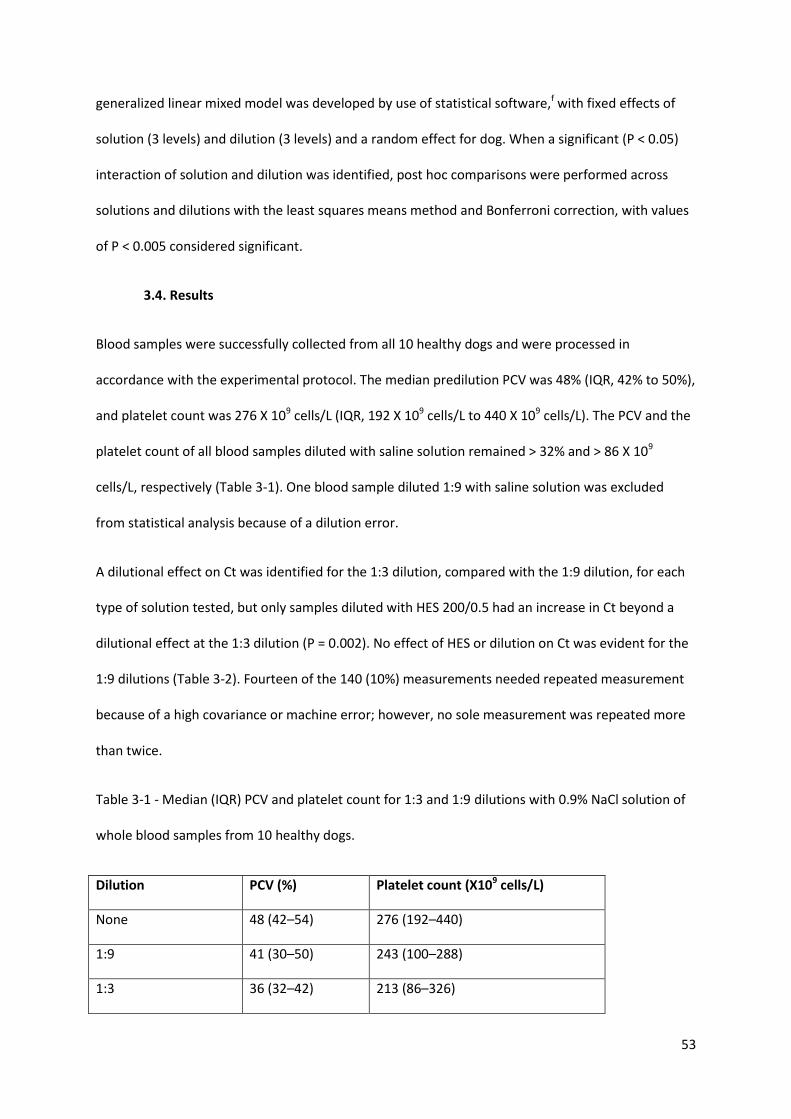
53
generalized linear mixed model was developed by use of statistical software,f with fixed effects of
solution (3 levels) and dilution (3 levels) and a random effect for dog. When a significant (P < 0.05)
interaction of solution and dilution was identified, post hoc comparisons were performed across
solutions and dilutions with the least squares means method and Bonferroni correction, with values
of P < 0.005 considered significant.
3.4. Results
Blood samples were successfully collected from all 10 healthy dogs and were processed in
accordance with the experimental protocol. The median predilution PCV was 48% (IQR, 42% to 50%),
and platelet count was 276 X 109 cells/L (IQR, 192 X 109 cells/L to 440 X 109 cells/L). The PCV and the
platelet count of all blood samples diluted with saline solution remained > 32% and > 86 X 109
cells/L, respectively (Table 3-1). One blood sample diluted 1:9 with saline solution was excluded
from statistical analysis because of a dilution error.
A dilutional effect on Ct was identified for the 1:3 dilution, compared with the 1:9 dilution, for each
type of solution tested, but only samples diluted with HES 200/0.5 had an increase in Ct beyond a
dilutional effect at the 1:3 dilution (P = 0.002). No effect of HES or dilution on Ct was evident for the
1:9 dilutions (Table 3-2). Fourteen of the 140 (10%) measurements needed repeated measurement
because of a high covariance or machine error; however, no sole measurement was repeated more
than twice.
Table 3-1 - Median (IQR) PCV and platelet count for 1:3 and 1:9 dilutions with 0.9% NaCl solution of
whole blood samples from 10 healthy dogs.
Dilution PCV (%) Platelet count (X109 cells/L)
None 48 (42–54) 276 (192–440)
1:9 41 (30–50) 243 (100–288)
1:3 36 (32–42) 213 (86–326)
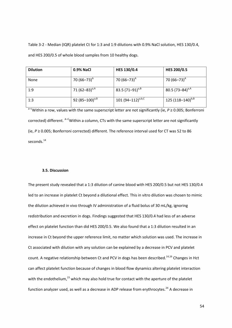
54
Table 3-2 - Median (IQR) platelet Ct for 1:3 and 1:9 dilutions with 0.9% NaCl solution, HES 130/0.4,
and HES 200/0.5 of whole blood samples from 10 healthy dogs.
Dilution 0.9% NaCl HES 130/0.4 HES 200/0.5
None 70 (66–73)A 70 (66–73)A 70 (66–73)A
1:9 71 (62–83)a,A 83.5 (71–91)a,B 80.5 (73–84)a,A
1:3 92 (85–100)a,B 101 (94–112)a,b,C 125 (118–140)b,B
a–cWithin a row, values with the same superscript letter are not significantly (ie, P ≥ 0.005; Bonferroni
corrected) different. A–CWithin a column, CTs with the same superscript letter are not significantly
(ie, P ≥ 0.005; Bonferroni corrected) different. The reference interval used for CT was 52 to 86
seconds.14
3.5. Discussion
The present study revealed that a 1:3 dilution of canine blood with HES 200/0.5 but not HES 130/0.4
led to an increase in platelet Ct beyond a dilutional effect. This in vitro dilution was chosen to mimic
the dilution achieved in vivo through IV administration of a fluid bolus of 30 mL/kg, ignoring
redistribution and excretion in dogs. Findings suggested that HES 130/0.4 had less of an adverse
effect on platelet function than did HES 200/0.5. We also found that a 1:3 dilution resulted in an
increase in Ct beyond the upper reference limit, no matter which solution was used. The increase in
Ct associated with dilution with any solution can be explained by a decrease in PCV and platelet
count. A negative relationship between Ct and PCV in dogs has been described.14,16 Changes in Hct
can affect platelet function because of changes in blood flow dynamics altering platelet interaction
with the endothelium,23 which may also hold true for contact with the aperture of the platelet
function analyzer used, as well as a decrease in ADP release from erythrocytes.24 A decrease in
55
platelet count may have also contributed to the increase in Ct in the diluted samples in the present
study, which has been identified in previous studies14,16 involving dogs. This effect may have been
enhanced by dilution of von Willebrand factor.25 Several theories may explain the mechanism by
which HES 200/0.5 dilution caused platelet dysfunction in the present study, including binding of von
Willebrand factor,26,27 HES molecules coating platelet surfaces, and a decrease in expression of
platelet surface protein integrin αIIbβ3, which all may reduce platelet adhesion and aggregation.28
An in vitro study18 of human platelet function revealed a decrease in expression of integrin αIIbβ3
receptor with dilutions involving HES 200/0.5 but not HES 130/0.4.18 The differences identified
between HES 200/0.5 and HES 130/0.4 might be related to solution-specific pharmacological
properties, in particular molecular weight and molar substitution.27,29 The molecular weight of an
HES solution is the mean manufactured molecular weight of the polydispersed solution, and the
molar substitution is the number of hydroxyl groups substituted with hydroxyethyl groups on the
glucose subunit of HES molecules.29 Hydroxyethyl starch solutions of higher molecular weight and
molar substitution have a slower rate of degradation and excretion in vivo than do other HES
solutions, and this phenomenon has commonly been cited as the main reason that treatment with
these solutions causes greater platelet dysfunction.29,30 However, these pharmacokinetic properties
may not be relevant to in vitro studies. It is possible that cleaving of HES molecules by amylase may
still occur in vitro, as it does in vivo, decreasing the HES molecular weight within the sample;
therefore the rate of degradation may still be relevant. Some evidence exists that high molar
substitution can considerably affect the degree of whole blood coagulation in vitro, independent of
molecular weight.31 But there is little evidence regarding the effect of differences in molecular
weight and molar substitution, independent of each other, on platelet function specifically in vitro.
Another property related to a slower rate of HES degradation and greater platelet dysfunction is a
high C2:C6 ratio; 31 however, this was unlikely to be applicable in the present study given that HES
200/0.5 has a lower C2:C6 ratio (5:1) than does HES 130/0.4 (9:1). The present study did not
56
elucidate the exact mechanisms of HES-induced platelet dysfunction, given that it was designed only
to determine whether platelet dysfunction occurred, as detected by the platelet function analyzer.
One additional difference between the 2 HES solutions evaluated was the concentration of HES. The
HES 200/0.5 solution had a greater concentration of HES molecules (10%), compared with HES
130/0.4 (6%). The 10% solution was chosen for comparison because it is the solution available for
clinical use in Australia. Although it is unknown which solution had the greater number of HES
molecules because of the polydiversity among brands and individual bags of solution, HES 200/0.5
may have provided more individual HES molecules, enhancing platelet dysfunction. Such differences
between solutions also create variations in the degree of redistribution in vivo, such as differences in
plasma volume expansion, degrees of dilution of blood constituents, and rates of clearance.32,33
These variations contribute to the challenge of designing in vivo studies, not only in interpreting
differences between solutions but also in the ability to select a valid control solution for comparing
the effect of dilution, given that crystalloid solutions have a different pattern of distribution.
Therefore, in vitro studies such as the present study, although limited by many factors including the
absence of any platelet interaction with the endothelium, also have an advantage in that the effect
of various solutions can be compared without the complicating factors of redistribution and
metabolism. Other complicating factors common to in vivo studies designed to assess the effect of
HES solutions on platelet function include variability in doses used, differences in the disease status
of patient populations, and choice of method of platelet function analysis, making comparisons of
findings across studies difficult. We chose 1:9 and 1:3 dilutions with the aim of indirectly comparing
results of the present study with those of another study,6 which demonstrated that whole blood
diluted with HES 670/0.75 led to an increase in CT beyond a dilutional effect when a 1:3 but not 1:9
dilution was used. The 1:9 dilution mimicked a 10 mL/kg bolus dose and the 1:3 dilution mimicked a
30 mL/kg bolus dose administered IV to a dog with an estimated blood volume of 90 mL/kg (ie,
euvolemia). The effect of the same in vitro dilutions on Ct in dogs was investigated in another
57
study22 involving an HES solution of low molecular weight and molar substitution, HES 130/0.42,
which has some differences in properties compared with HES 130/0.4. A significant increase in Ct
was identified with HES 130/0.42 diluted 1:3 with canine whole blood beyond a dilutional effect,
which differs from our findings, despite the 2 solutions being very similar. Several factors could
account for this difference. Hydroxyethyl starch 130/0.4 is a waxy, maize based colloid, whereas HES
130/0.42 is a potato-based colloid. The 2 solutions also differ in molar substitution, which may
account for the difference in Ct. A conservative value of P < 0.005 was used for pairwise comparisons
made in our study to reduce type I error, which may have precluded detection of more differences
than were identified. Differences in study subjects and subject numbers as well as a lower
predilution Ct in our study may have also contributed to different results. For comparison, an in vitro
study3 in humans compared the effect on coagulation between these 2 solutions through use of
thromboelastography and platelet aggregometry and found no difference between the solutions,
but HES 130/0.4 and HES 130/0.42 have not been compared directly in dogs.
The clinical relevance of the effect of HES solutions with low molecular weight and molar
substitution on platelet function in humans remains controversial despite the abundance of
literature on the topic. Early in vitro studies indicated that blood samples diluted with HES 130/0.4
caused less platelet dysfunction as measured through various techniques such as platelet
aggregometry,4,34 Ct analysis,4,5,18 and flow cytometry,18 compared with that caused by HES solutions
of higher molecular weight and molar substitution. However, several clinical trials1,20,21 comparing
the effects of HES 130/0.4 with HES 200/0.5 on blood loss and transfusion requirements in
orthopedic surgery, cardiopulmonary bypass patients, and intensive care patients have yielded
conflicting evidence, compared with the in vitro studies. Meta-analyses and literature reviews1,20,21
have shown that no conclusion can be made regarding the benefit of administration of HES 130/0.4
versus HES 200/0.5 or crystalloid solutions in humans because of the small sample sizes and the
quality of these human trials and because several reports were retracted for scientific misconduct. In
addition, although the manufacturers of HES 130/0.4 have recommended a dose limit of 50 mL/kg/d,
58
the European Society of Intensive Care Medicine released a consensus statement, in which no dose
recommendations for HES 130/0.4 could be made given the minimal evidence for its safety
regarding blood loss, acute kidney injury, and risk of death.35 Most recently, the Surviving Sepsis
Campaign consensus statement has recommended against the use of HES solutions in septic human
patients because of the lack of clear benefit and evidence of harm caused to patients,36 but large
clinical trials have yet to be performed in veterinary medicine.
The present study demonstrated that dilution of canine blood samples 1:3 with saline solution, HES
130/0.4, and HES 200/0.5 led to an increase in Ct attributable to a dilutional effect alone.
Hydroxyethyl starch 200/0.5, however, led to an increase in Ct beyond a dilutional effect when
diluted 1:3 with blood, consistent with platelet dysfunction. Interestingly, we found that HES 130/0.4
did not cause platelet dysfunction at this same dilution, which suggests that this solution may be a
superior choice when platelet dysfunction is of concern. However, in vivo studies are warranted to
investigate the risk of clinical use of this solution in dogs.
3.6. Footnotes
a. Platelet Function Analyzer-100, Dade Boehring Inc, Miami, Fla.
b. Dade PFA Collagen/ADP Test Cartridge, Siemens Healthcare, Marburg, Germany.
c. ADVIA 120 hematology system, Bayer Diagnostics Mfg Ltd, Dublin, Ireland.
d. Voluven 6% (hydroxyethyl starch 130/0.4), Fresenius Kabi Deutschland GmbH, Friedberg,
Germany.
e. Starquin 200 10%, Biomed Ltd, Point Chevalier, Auckland, New Zealand.
f. SAS, version 9.3, SAS Institute Inc, Cary, NC.

59
3.7 References
1. Raja SG, Akhtar S, Shahbaz Y, et al. In cardiac surgery patients does Voluven(R)
impair coagulation less than other colloids? Interact Cardiovasc Thorac Surg 2011;12:1022–1027.
2. Sossdorf M, Marx S, Schaarschmidt B, et al. HES 130/0.4 impairs haemostasis and
stimulates pro-inflammatory blood platelet function. Crit Care [serial online]. 2009;13:R208.
Available at: ccforum.com/content/13/6/R208. Accessed June 6, 2011.
3. Matsota P, Politou M, Kalimeris K, et al. Do different substitution patterns or plant
origin in hydroxyethyl starches affect blood coagulation in vitro? Blood Coagul Fibrinolysis
2010;21:448–451.
4. Liu FC, Liao CH, Chang YW, et al. Hydroxyethyl starch interferes with human blood ex
vivo coagulation, platelet function and sedimentation. Acta Anaesthesiol Taiwan 2009;47:71–78.
5. Scharbert G, Deusch E, Kress HG, et al. Inhibition of platelet function by hydroxyethyl
starch solutions in chronic pain patients undergoing peridural anesthesia. Anesth Analg
2004;99:823–827.
6. Wierenga JR, Jandrey KE, Haskins SC, et al. In vitro comparison of the effects of two
forms of hydroxyethyl starch solutions on platelet function in dogs. Am J Vet Res 2007;68:605–609.
7. Smart L, Jandrey KE, Kass PH, et al. The effect of hetastarch (670/0.75) in vivo on
platelet closure time in the dog. J Vet Emerg Crit Care 2009;19:444–449.
8. Chohan AS, Greene SA, Grubb TL, et al. Effects of 6% hetastarch (600/0.75) or
lactated Ringer’s solution on hemostatic variables and clinical bleeding in healthy dogs anesthetized
for orthopedic surgery. Vet Anaesth Analg 2011;38:94–105.
9. Gandhi SD, Weiskopf RB, Jungheinrich C, et al. Volume replacement therapy during
major orthopedic surgery using Voluven (hydroxyethyl starch 130/0.4) or hetastarch. Anesthesiology
60
2007;106:1120–1127.
10. Kasper SM, Meinert P, Kampe S, et al. Large-dose hydroxyethyl starch 130/0.4 does
not increase blood loss and transfusion requirements in coronary artery bypass surgery compared
with hydroxyethyl starch 200/0.5 at recommended doses. Anesthesiology 2003;99:42–47.
11. Schramko AA, Suojaranta-Ylinen RT, Kuitunen AH, et al. Rapidly degradable
hydroxyethyl starch solutions impair blood coagulation after cardiac surgery: a prospective
randomized trial. Anesth Analg 2009;108:30–36.
12. Schramko AA, Suojaranta-Ylinen RT, Kuitunen AH, et al. Comparison of the effect of
6% hydroxyethyl starch and gelatine on cardiac and stroke volume index: a randomized, controlled
trial after cardiac surgery. Perfusion 2010;25:283–291.
13. Hayward CP, Harrison P, Cattaneo M, et al. Platelet function analyser (PFA)-100
closure time in the evaluation of platelet disorders and platelet function. J Thromb Haemost
2006;4:312–319.
14. Callan MB, Giger U. Assessment of a point-of-care instrument for identification of
primary hemostatic disorders in dogs. Am J Vet Res 2001;62:652–658.
15. Keidel A, Mischke R. Clinical evaluation of platelet function analyzer PFA-100 in dogs
[in German]. Berl Munch Tierarztl Wochenschr 1998;111:452–456.
16. Mischke R, Keidel A. Influence of platelet count, acetylsalicylic acid, von Willebrand’s
disease, coagulopathies, and haematocrit on results obtained using a platelet function analyser in
dogs. Vet J 2003;165:43–52.
17. Liang H, Yang CX, Li H, et al. The effects of preloading infusion with hydroxyethyl
starch 200/0.5 or 130/0.4 solution on hypercoagulability and excessive platelet activation of patients
with colon cancer. Blood Coagul Fibrinolysis 2010;21:406–413.

61
18. Franz A, Braunlich P, Gamsjager T, et al. The effects of hydroxyethyl starches of
varying molecular weights on platelet function. Anesth Analg 2001;92:1402–1407.
19. Kozek-Langenecker SA, Jungheinrich C, Sauermann W, et al. The effects of
hydroxyethyl starch 130/0.4 (6%) on blood loss and use of blood products in major surgery: a pooled
analysis of randomized clinical trials. Anesth Analg 2008;107:382–390.
20. Navickis RJ, Haynes GR, Wilkes MM. Effect of hydroxyethyl starch on bleeding after
cardiopulmonary bypass: a meta-analysis of randomized trials. J Thorac Cardiovasc Surg
2012;144:223–230.
21. Gattas DJ, Dan A, Myburgh J, et al. Fluid resuscitation with 6% hydroxyethyl starch
(130/0.4) in acutely ill patients: an updated systematic review and meta-analysis. Anesth Analg
2012;114:159–169.
22. Classen J, Adamik KN, Weber K, et al. In vitro effect of hydroxyethyl starch 130/0.42
on canine platelet function. Am J Vet Res 2012;73:1908–1912.
23. Turitto VT, Baumgartner HR. Platelet interaction with subendothelium in flowing
rabbit blood: effect of blood shear rate. Microvasc Res 1979;17:38–54.
24. Alkhamis TM, Beissinger RL, Chediak JR. Artificial surface effect on red blood cells
and platelets in laminar shear flow. Blood 1990;75:1568–1575.
25. Fenger-Eriksen C, Tonnesen E, Ingerslev J, et al. Mechanisms of hydroxyethyl starch-
induced dilutional coagulopathy. J Thromb Haemost 2009;7:1099–1105.
26. de Jonge E, Levi M, Buller HR, et al. Decreased circulating levels of von Willebrand
factor after intravenous administration of a rapidly degradable hydroxyethyl starch (HES 200/0.5/6)
in healthy human subjects. Intensive Care Med 2001;27:1825–1829.
62
27. Jamnicki M, Bombeli T, Seifert B, et al. Low- and medium molecular- weight
hydroxyethyl starches: comparison of their effect on blood coagulation. Anesthesiology
2000;93:1231–1237.
28. Deusch E, Gamsjager T, Kress HG, et al. Binding of hydroxyethyl starch molecules to
the platelet surface. Anesth Analg 2003;97:680–683.
29. Kozek-Langenecker SA. Effects of hydroxyethyl starch solutions on hemostasis.
Anesthesiology 2005;103:654–660.
30. Treib J, Baron JF, Grauer MT, et al. An international view of hydroxyethyl starches.
Intensive Care Med 1999;25:258–268.
31. von Roten I, Madjdpour C, Frascarolo P, et al. Molar substitution and C2/C6 ratio of
hydroxyethyl starch: influence on blood coagulation. Br J Anaesth 2006;96:455–463.
32. Lamke LO, Liljedahl SO. Plasma volume changes after infusion of various plasma
expanders. Resuscitation 1976;5:93–102.
33. Waitzinger J, Bepperling F, Pabst G, et al. Hydroxyethyl starch (HES) [130/0.4], a new
HES specification: pharmacokinetics and safety after multiple infusions of 10% solution in healthy
volunteers. Drugs R D 2003;4:149–157.
34. Winterhalter M, Malinski P, Danzeisen O, et al. Prospective observational study for
perioperative volume replacement with 6% HES 130/0,42, 4% gelatin and 6% HES 200/0,5 in cardiac
surgery. Eur J Med Res 2010;15:383–389.
35. Reinhart K, Perner A, Sprung CL, et al. Consensus statement of the ESICM task force
on colloid volume therapy in critically ill patients. Intensive Care Med 2012;38:368–383.

63
36. Dellinger RP, Levy MM, Rhodes A, et al. Surviving sepsis campaign: international
guidelines for management of severe sepsis and septic shock, 2012. Intensive Care Med 2013;
39:165–228.
64
4. Chapter 4: PLATELET CLOSURE TIME IN ANESTHETIZED DOGS WITH HEMORRHAGIC SHOCK
TREATED WITH HYDROXYETHYL STARCH 130/0.4 OR 0.9% NACL
4.1 Abstract
Objective - Hydroxyethyl starch (HES) 130/0.4 is used to treat shock in dogs. This study measured
platelet closure time (PCT) in dogs during hemorrhagic shock and after HES 130/0.4 or 0.9% NaCl
administration.
Animals - Eleven healthy greyhounds
Procedure - Dogs were anesthetized and 48mL/kg of blood removed to induce hemorrhagic shock.
Dogs received 20 mL/kg of HES 130/0.4 (n=6) or 80 mL/kg of 0.9% sodium chloride (NaCl) (n=5)
intravenously over 20 minutes. Platelet closure time was measured using the Platelet Function
Analyser-100 with collagen and adenosine-diphosphate cartridges at: T0 = 60 minutes after
induction of anesthesia prior to hemorrhage, T1 = during hemorrhagic shock and T2 = 40 minutes
after completion of fluid bolus. Packed cell volume (PCV) and platelet count were concurrently
measured.
Results - Hemorrhagic shock did not significantly change PCT, with no difference between T0 and T1.
Both the HES 130/0.4 and 0.9% NaCl group had a significantly increased mean PCT at T2 to 91.4
seconds (95% CI 69.3-113.4) and 95.5 seconds (95% CI 78.2-112.8), respectively, compared to T1.
The magnitude of change was significantly greater for the 0.9% NaCl group than the HES 130/0.4
group. There was no difference in the magnitude of change in PCV and platelet count between the
two groups. The PCV and platelet count were > 25% and > 100,000/µL respectively in all dogs, except
for dogs in the HES 130/0.4 group at T90 where platelet counts were <100,000/µL.
Conclusion and Clinical Relevance - Hemorrhagic shock in greyhounds under anesthesia did not
cause a significant change in PCT. Both HES 130/0.4 and 0.9% NaCl administration after induction of
65
shock increased PCT. These results do not support that HES 130/0.4 causes relevant platelet
dysfunction beyond hemodilution, however, this study did not assess risk of bleeding.
4.2 Introduction
The use of hydroxyethyl starch (HES) solutions for the treatment of shock is controversial because of
adverse effects reported on both human1-5 and canine6-8 platelet function, and its association with
increased intra-operative blood loss and transfusion requirements in people.1,9-12 Many studies
support that these complications are associated with HES solutions of high molecular weight (MW)
and high molar substitution (MS).5,6,13 Therefore, the use of HES 130/0.4, with a low MW of 130 KDa
and a low MS of 0.4, has become more commonplace in people due to its minimal effect on platelet
function.2,13,14 However, there is some contradicting evidence that platelet dysfunction still occurs
with HES 130/0.4.4,5
Studies investigating the use of HES solutions in dogs have most commonly used the Platelet
Function Analyzer-100 (PFA-100) to measure platelet function.6,7,15 It measures platelet closure time
(PCT), which is the time taken in seconds for a small aperture, to be closed by platelet plug
formation.16 An increased PCT indicates platelet dysfunction, however, dilution of blood that
decreases hematocrit (< 25% – 30%17-19) and platelet count (< 100,000 platelets/µL19) can also
increase PCT.
There have been two recent in vitro studies using the PFA-100 to assess the effect of low MW and
low MS HES solutions on canine platelet function.15,20 One study using HES 130/0.42 found that it
caused a mild prolongation in PCT20 whereas one using HES 130/0.4 found no effect.15 As in vitro
studies cannot assess the pharmacokinetic and pharmacodynamic effects of these fluids, and
exclude any platelet interaction with the endothelium, it is unknown to what degree HES 130/0.4
may have on canine platelet function in vivo. Also, these solutions are usually used in dogs suffering
66
from shock and it has been found in people that trauma-induced shock can cause platelet
dysfunction itself.21-24 Therefore, shock may further enhance any platelet dysfunction that HES
130/0.4 causes.
The objective of this study was to determine if hemorrhagic shock increases PCT above baseline in
anesthetized greyhounds. The secondary objective was to determine if 20 mL/kg of HES 130/0.4
increases PCT beyond the effect of hemorrhagic shock or hemodilution.
4.3 Materials and methods
This study was approved by the university’s Animal Ethics Committee. Eleven healthy adult
greyhound dogs were included in this study. Dogs were healthy based on physical examination and
complete blood count. The dogs were not fasted and had free access to water. Each dog was
randomized to receive either HES 130/0.4 (treated group, n = 6) or 0.9% sodium chloride (NaCl)
(control group, n = 5) according to an assigned sealed envelope.
The dogs were premedicated with 0.2mg/kg of methadonea given intramuscularly. Thirty minutes
after premedication, a 20-gauge 1-inch over-the-needle catheter was aseptically placed in the right
cephalic vein, and anesthesia was induced with 2.5mg/kg of alfaxaloneb given intravenously. Animals
were placed in left lateral recumbency, orotracheally intubated and anesthesia was maintained with
isofluranec and 100% oxygen via a circle breathing system. Vaporiser settings were adjusted to
achieve an end-tidal isoflurane concentration of 1.4% measured using an agent monitor.d
Mechanical ventilation was provided using a volume controlled time cycled ventilatore at a rate of 10
breaths per minute with an initial tidal volume of 20 mL/kg, which was subsequently adjusted if
required during the instrumentation period to achieve a PaCO2 of 35-40 mmHg. No further
adjustment was made throughout the remainder of anesthesia. Hartmann’s solutionf was
administered via the cephalic catheter at a rate of 10 ml/kg/hr until the time of administration of the
67
treatment or control solutions. A 12-gauge 6-inch over-the-needle catheter was aseptically placed in
the right jugular vein, with the distal end measured beyond the thoracic inlet. A 14-gauge 4-inch
over-the-needle catheter was surgically placed in the left femoral artery. The catheters were
connected via fluid filled transducersg to a multivariable monitorh and calibrated to atmospheric
pressure. Heart rate (HR), electrocardiogram, central venous pressure, and pulse oximetry were
monitored with a multivariable monitor throughout the experiment. Body temperature was
maintained between 36°C and 38°C by applying warm air to the upper surfaces of the dog.i
Sixty minutes after induction of anesthesia, hemorrhagic shock was induced by removing 48 mL/kg
of blood over a 30 minute period from the femoral arterial catheter. A pilot study in greyhounds
using the same anesthetic protocol was performed prior to this study, which determined that
removal of 48 mL/kg of blood induced shock, as assessed by an increase in HR, a direct MAP of less
than 60 mmHg, and an increase in oxygen extraction ratio (O2ER).
Immediately after 48 mL/kg of blood was removed, intravenous Hartmann’s solution administration
was discontinued and the dogs received either 20 mL/kg of HES 130/0.4j or 80 mL/kg of 0.9% NaCl
over 20 minutes. Hydroxyethyl starch 130/0.4 was delivered by a pressure bag via the cephalic
catheter and 0.9% NaCl was delivered by 2 pressure bags via the cephalic and jugular catheters. The
pressure applied to the pressure bags was monitored throughout to ensure the fluid was delivered
continuously during the 20 minute period.
Cardiac output (CO) was measured by the lithium dilution cardiac outputk technique as previously
described,25 with modification of the dose of lithium chloride to 0.005 mmol/kg and withdrawing
blood via the femoral artery. Measurements were made in duplicate. When two results varied more
than 15%, repeated measurements were made and the results varying more than 15% were
excluded from analysis.

68
Blood was collected simultaneously from the femoral arterial and jugular venous catheters into
lithium heparin syringes, and blood gas analysisl was performed within 5 minutes of blood collection.
Venous blood was collected from the jugular venous catheter into sodium citrate tubes for the
measurement of PCT. Platelet closure time was measured in duplicate using the PFA-100,m with
collagen and adenosine diphosphate cartridgesn and co-variance of less than 15% between
measurements were accepted. Where duplicate measurements had a co-variance of greater than
15%, or if the analyzer indicated an error such as flow obstruction, measurement was repeated using
the same sample and the two closest samples with a CV < 15% were included. Platelet closure time
was measured at three time points: T0, at base line during general anesthesia immediately prior to
hemorrhage; T1, during hemorrhagic shock, which was immediately after removing 48 mL/kg of
blood; and T2, 40 minutes after completion of fluid administration (Figure 1).
Data also collected simultaneously at these time points included HR, direct MAP, arterial and venous
blood gas, hemoglobin concentration (Hb), PCV, and CO. At the conclusion of data collection,
euthanasia was performed using intravenous pentobarbital in accordance with the ethics committee
approved protocol. The O2ER was calculated at each time point using the formulae in Table 1.
Statistical analysis - For assessment of the effect of shock on PCV, platelet count, O2ER and PCT,
paired comparisons were made across all greyhounds for data at T0 vs T1 using a paired t-test.
For assessment of the effect of fluid therapy on PCV, platelet count, O2ER and PCT, comparisons
were made across both treatment groups for data at T1 vs T2 using a paired t-test. Where both
treatments had a similar result (i.e. increase or decrease), the magnitude of the change was
compared between treatments using an unpaired t-test with a Satterthwaite adjustment for unequal
variances when indicated. Significance was determined at p < 0.05 for all tests. SAS v 9.3 was used
for the analysis (SAS Institute, Cary, NC).
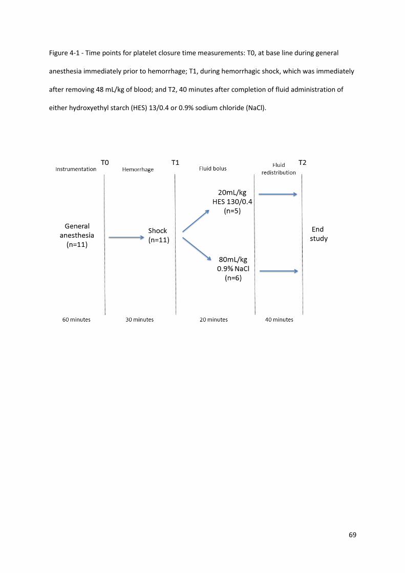
69
Figure 4-1 - Time points for platelet closure time measurements: T0, at base line during general
anesthesia immediately prior to hemorrhage; T1, during hemorrhagic shock, which was immediately
after removing 48 mL/kg of blood; and T2, 40 minutes after completion of fluid administration of
either hydroxyethyl starch (HES) 13/0.4 or 0.9% sodium chloride (NaCl).
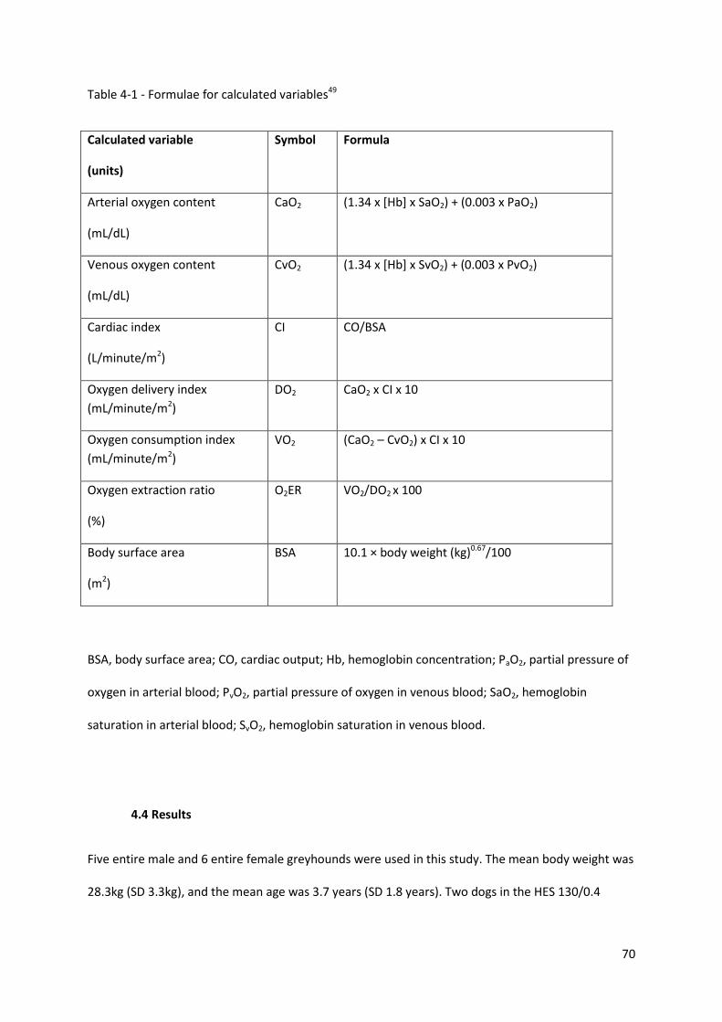
70
Table 4-1 - Formulae for calculated variables49
Calculated variable
(units)
Symbol Formula
Arterial oxygen content
(mL/dL)
CaO2 (1.34 x [Hb] x SaO2) + (0.003 x PaO2)
Venous oxygen content
(mL/dL)
CvO2 (1.34 x [Hb] x SvO2) + (0.003 x PvO2)
Cardiac index
(L/minute/m2)
CI CO/BSA
Oxygen delivery index
(mL/minute/m2)
DO2 CaO2 x CI x 10
Oxygen consumption index
(mL/minute/m2)
VO2 (CaO2 – CvO2) x CI x 10
Oxygen extraction ratio
(%)
O2ER VO2/DO2 x 100
Body surface area
(m2)
BSA 10.1 × body weight (kg)0.67/100
BSA, body surface area; CO, cardiac output; Hb, hemoglobin concentration; PaO2, partial pressure of
oxygen in arterial blood; PvO2, partial pressure of oxygen in venous blood; SaO2, hemoglobin
saturation in arterial blood; SvO2, hemoglobin saturation in venous blood.
4.4 Results
Five entire male and 6 entire female greyhounds were used in this study. The mean body weight was
28.3kg (SD 3.3kg), and the mean age was 3.7 years (SD 1.8 years). Two dogs in the HES 130/0.4
71
group had PCTs and platelet count at T2 excluded from analysis due to platelet clumping in the
sample.
The first objective was evaluated across all dogs. There was no significant change in PCT at T1,
compared to T0 (Table 2). There was a significant decrease in PCV and platelet count, and a
significant increase in O2ER, at T1 compared to T0 (Table 2), consistent with hemorrhagic shock.
The second objective was evaluated across treatment groups. Both groups had a significant increase
in PCT at T2, compared to T1 (Table 3). The magnitude of change was significantly greater in the
0.9% NaCl group compared to the HES 130/0.4 group. Both groups had a significant decrease in PCV,
platelet count and O2ER at T2, compared to T1 (Table 2), however, the magnitude of change was not
significantly different between the two groups.
The PCV of all dogs at each time point was > 25%. All platelet counts where > 100,000/µL, except for
dogs in the HES 130/0.4 group at T2 where platelet counts were < 100,000/µL.

72
Table 4-2 - Mean (95% confidence interval) packed cell volume, platelet count, oxygen extraction
ratio (O2ER) and platelet closure time in greyhounds under general anesthesia (T0) and thirty
minutes after 43mL/kg of blood loss (T1).
Variable T0 T1 P value*
Packed cell volume (%)
49.3
(47.5 - 51.0)
46.9
(45.4 - 48.5)
0.0125
Platelet count
(X109 cells/L)
172
(158 - 186)
149.5
(131.3 - 167.8)
0.0001
O2ER (%)
8.80
(6.90 - 10.5)
60.0
(48.1 - 70.9)
<0.0001
Platelet closure time+
(seconds)
87.7
(69.6 - 105.8)
70.4
(61.7 - 79.0)
0.0805
* Values less than 0.05 were considered significant
+The greyhound reference interval for platelet closure time is 63 to 92 seconds48
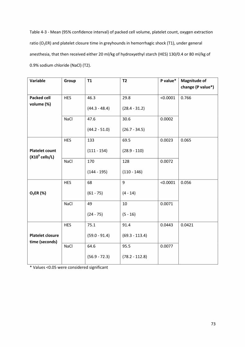
73
Table 4-3 - Mean (95% confidence interval) of packed cell volume, platelet count, oxygen extraction
ratio (O2ER) and platelet closure time in greyhounds in hemorrhagic shock (T1), under general
anesthesia, that then received either 20 ml/kg of hydroxyethyl starch (HES) 130/0.4 or 80 ml/kg of
0.9% sodium chloride (NaCl) (T2).
Variable Group T1 T2 P value* Magnitude of
change (P value*)
Packed cell
volume (%)
HES 46.3
(44.3 - 48.4)
29.8
(28.4 - 31.2)
<0.0001 0.766
NaCl 47.6
(44.2 - 51.0)
30.6
(26.7 - 34.5)
0.0002
Platelet count
(X109 cells/L)
HES 133
(111 - 154)
69.5
(28.9 - 110)
0.0023 0.065
NaCl 170
(144 - 195)
128
(110 - 146)
0.0072
O2ER (%)
HES 68
(61 - 75)
9
(4 - 14)
<0.0001 0.056
NaCl 49
(24 - 75)
10
(5 - 16)
0.0071
Platelet closure
time (seconds)
HES 75.1
(59.0 - 91.4)
91.4
(69.3 - 113.4)
0.0443 0.0421
NaCl 64.6
(56.9 - 72.3)
95.5
(78.2 - 112.8)
0.0077
* Values <0.05 were considered significant
74
4.5 Discussion
This study demonstrated no change in PCT during shock but a significant increase in PCT after
administration of 20 mL/kg of 6% HES 130/0.4 and 80 mL/kg of 0.9% NaCl. Interestingly, we found
that the magnitude of change in PCT was greater after 0.9% NaCl administration, compared to HES
130/0.4 administration. This result does not support that HES 130/0.4 causes platelet dysfunction
beyond the effect of hemodilution.
The lack of significant change in PCT during shock in this study is contrary to the majority of recent
studies in people that found platelet dysfunction to occur during hemorrhagic shock secondary to
trauma.21-24,26,27 However, one study in people showed a decrease in PCT immediately following
traumatic hemorrhage, which corresponded with increased platelet reactivity measured by flow
cytometry.28 Blood samples were collected immediately following trauma in this one study while
blood samples in most previous studies were collected later in the emergency room. Our study also
chose an early time point in shock to measure PCT, however the fluctuation in PCT that we observed
was not statistically significant.
We used O2ER as an objective measure of shock, as several studies have demonstrated a significant
increase in O2ER during severe shock.26,29,30 An increase in O2ER reflects decreasing oxygen delivery,
despite oxygen consumption staying relatively constant. An O2ER greater than 60% is generally
considered a marker of severe shock, as this is the time at which oxygen consumption starts to
depend on oxygen delivery, depending on the underlying cause of shock. In our study, there was a
moderate to marked increase in O2ER, and all dogs were tachycardic and hypotensive, supporting
the presence of shock. The increase in O2ER was not as marked as expected in all individuals, given
the volume of blood loss. This may be due to individual variability, or due to measuring the
parameter in early shock, where good cardiovascular compensation is still intact. Another reason for
this difference may be due to the greyhound breed utilized in our study. Although O2ER has not been
75
compared between greyhounds and non-greyhound breed dogs, cardiovascular function31 and
oxygen carrying capacity32 are known to be different.
We found a minor, but significant, increase in PCT with both types of fluid administration, which can
be explained by hemodilution. A negative relationship between PCT and PCV has been previously
described in dogs.18,19 Changes in hematocrit can affect platelet function due to changes in blood
flow dynamics altering platelet interaction with the endothelium,33 as well as decreased ADP release
from red blood cells.34 The effect of dilution on PCT may also be due to dilution of von Willebrand
Factor.35 In our study, a decrease in platelet count may have also contributed to the increased PCT,
which has been described previously in dogs.18,19 It is interesting that the magnitude of change in
PCT was greater in the 0.9% NaCl group, which seems unlikely to be due to greater hemodilution, as
there was no significant difference in PCV or platelet count between the groups. However, dilution
of other factors that may affect PCT, such as von Willebrand Factor, were not measured.
In contrast to previous studies using higher MW and MS HES solutions,6,7 our study found that HES
130/0.4 did not significantly increase PCT, compared to 0.9% NaCl. Hydroxyethyl starch solutions
with a high MW and MS are known to have a direct adverse effect on platelet function in people, the
mechanisms of which have been described elsewhere.14,36,37 Previous studies in dogs showed HES
670/0.75 to cause platelet dysfunction as demonstrated by an increased PCT in vitro6 and in vivo.7 In
another study, HES 200/0.5 (medium MW and MS) diluted 1:3 with canine whole blood increased
PCT in vitro, however HES 130/0.4 at this same dilution did not increase PCT beyond dilutional
effect. 15 This finding supports our result. In contrast, HES 130/0.42 diluted 1:3 with canine whole
blood increased PCT beyond dilutional effect in vitro.20 However, the different properties of HES
130/0.42 make direct comparisons with HES 130/0.4 difficult.
The failure to cause a significant change in PCT with HES 130/0.4, beyond hemodilution, may be due
to use of a 20 mL/kg dose in our study. A 1:4 dilution of HES 130/0.4 with canine whole blood caused
greater abnormalities in coagulation, as measured by thromboelastometry, compared with a 1:10
76
dilution, which suggests a dose dependent effect on coagulation.38 This study did not assess platelet
function specifically. We chose a dose of 20 mL/kg of HES 130/0.4, attempting to match the 80
mL/kg dose of isotonic crystalloid solution,39,40 as it is a common large bolus dose for the treatment
of shock in dogs.41 As we chose this single dose we cannot comment if HES 130/0.4 has a dose
dependent adverse effect on PCT.
There are some limitations to this study. The shock model used in this study induced hemorrhagic
shock with minimal trauma by removing a fixed volume of blood in a controlled manner. This model
is applicable to situations of large volumes of blood loss under general anesthesia. The model is not
directly applicable to animals with systemic inflammatory response syndrome, sepsis or trauma
where there is likely cytokine release, which may affect the result.
The results of this study may only be referent to greyhounds. Greyhounds are known to have a
higher PCV, resulting in higher blood viscosity, and a lower platelet count.42-44 Some greyhounds are
also known to have significant post-operative bleeding, related to increased fibrinolysis45 or reduced
fibrin cross link formation and clot strength, as measured by thromboelastography.46,47 Despite
postoperative bleeding, PCTs were found to be normal in one study.46 Platelet closure time has also
been validated in greyhounds with their own established reference interval.48 These findings suggest
that the greyhound breed-specific variability in coagulation should not heavily influence the PCT.
We did not have a control group that did not receive fluid resuscitation, therefore we do not know if
PCT would have changed over time under general anesthesia or due to prolonged shock. We cannot
determine if the changes we saw in PCT after treatment with fluid resuscitation were due to
hemodilution or the late effects of shock. Inclusion of a group that were resuscitated with auto-
transfused fresh whole blood may have answered this question.
In conclusion, we demonstrated that hemorrhagic shock did not cause platelet dysfunction, as
measured by PCT, in greyhounds under general anesthesia. We found that 20 mL/kg of HES 130/0.4

77
and 80 mL/kg of 0.9% NaCl administered over a 20 minute period significantly increased PCT. The
magnitude in increase of PCT was greater with 0.9% NaCl administration, indicating HES 130/0.4
does not cause platelet dysfunction beyond a dilutional effect. Although the changes in PCT were
significant, they were minor, therefore may not be clinically relevant. However, this study did not
assess the risk of bleeding due to platelet dysfunction.
4.6. Footnotes
a Methone, Parnell Australia Pty Ltd, NSW, Australia
b Alfaxan, Jurox Pty Ltd, NSW, Australia
c ISO Veterinary Companies of Australia, NSW, Australia
d Capnomac Ultima, Datex-Ohmeda Medical supplies Australia, NSW, Australia
e Model TH-1 Beijing Read Eagle Technology Co Ltd, Beijing, China
f Baxter Healthcare Pty Ltd Australia, NSW, Australia
g DTX PlusTM Argon Critical Care Systems, Singapore
h Surgivet V9203 Sound Medical, VIC, Australia
I Bair Hugger warming unit Model 505, Critical Assist, VIC, Australia
j Voluven 6% (hydroxyethyl starch 130/0.4), Fresenius Kabi Deutschland GmbH, Friedberg,
Germany
k LiDCO Cardiac Sensor Systems, LiDCO Ltd, London, UK
l ABL 725 Radiometer Pacific Pty Ltd, VIC, Australia

78
m Platelet Function Analyzer-100, Dade BoehringInc, Miami, Florida, USA
n Dade PFA Collagen /ADP Test Cartridge, Siemens Healthcare, Marburg, Germany
4.7. References
1. Raja SG, Akhtar S, Shahbaz Y, et al. In cardiac surgery patients does Voluven(R)
impair coagulation less than other colloids? Interact Cardiovasc Thorac Surg 2011;12:1022-1027.
2. Sossdorf M, Marx S, Schaarschmidt B, et al. HES 130/0.4 impairs haemostasis and
stimulates pro-inflammatory blood platelet function. Crit Care 2009;13:R208.
3. Matsota P, Politou M, Kalimeris K, et al. Do different substitution patterns or plant
origin in hydroxyethyl starches affect blood coagulation in vitro? Blood Coagul Fibrinolysis
2010;21:448-451.
4. Liu FC, Liao CH, Chang YW, et al. Hydroxyethyl starch interferes with human blood ex
vivo coagulation, platelet function and sedimentation. Acta Anaesthesiol Taiwan 2009;47:71-78.
5. Scharbert G, Deusch E, Kress HG, et al. Inhibition of platelet function by hydroxyethyl
starch solutions in chronic pain patients undergoing peridural anesthesia. Anesth Analg 2004;99:823-
827, table of contents.
6. Wierenga JR, Jandrey KE, Haskins SC, et al. In vitro comparison of the effects of two
forms of hydroxyethyl starch solutions on platelet function in dogs. Am J Vet Res 2007;68:605-609.
7. Smart L, Jandrey KE, Kass PH, et al. The effect of Hetastarch (670/0.75) in vivo on
platelet closure time in the dog. J Vet Emerg Crit Care (San Antonio) 2009;19:444-449.
79
8. Chohan AS, Greene SA, Grubb TL, et al. Effects of 6% hetastarch (600/0.75) or
lactated Ringer's solution on hemostatic variables and clinical bleeding in healthy dogs anesthetized
for orthopedic surgery. Vet Anaesth Analg 2011;38:94-105.
9. Gandhi SD, Weiskopf RB, Jungheinrich C, et al. Volume replacement therapy during
major orthopedic surgery using Voluven (hydroxyethyl starch 130/0.4) or hetastarch. Anesthesiology
2007;106:1120-1127.
10. Kasper SM, Meinert P, Kampe S, et al. Large-dose hydroxyethyl starch 130/0.4 does
not increase blood loss and transfusion requirements in coronary artery bypass surgery compared
with hydroxyethyl starch 200/0.5 at recommended doses. Anesthesiology 2003;99:42-47.
11. Schramko AA, Suojaranta-Ylinen RT, Kuitunen AH, et al. Rapidly degradable
hydroxyethyl starch solutions impair blood coagulation after cardiac surgery: a prospective
randomized trial. Anesth Analg 2009;108:30-36.
12. Schramko AA, Suojaranta-Ylinen RT, Kuitunen AH, et al. Comparison of the effect of
6% hydroxyethyl starch and gelatine on cardiac and stroke volume index: a randomized, controlled
trial after cardiac surgery. Perfusion 2010;25:283-291.
13. Liang H, Yang CX, Li H, et al. The effects of preloading infusion with hydroxyethyl
starch 200/0.5 or 130/0.4 solution on hypercoagulability and excessive platelet activation of patients
with colon cancer. Blood Coagul Fibrinolysis 2010;21:406-413.
14. Franz A, Braunlich P, Gamsjager T, et al. The effects of hydroxyethyl starches of
varying molecular weights on platelet function. Anesth Analg 2001;92:1402-1407.
15. McBride D, Hosgood GL, Mansfield CS, et al. Effect of hydroxyethyl starch 130/0.4
and 200/0.5 solutions on canine platelet function in vitro. Am J Vet Res 2013;74:1133-1137.
80
16. Hayward CP, Harrison P, Cattaneo M, et al. Platelet function analyzer (PFA)-100
closure time in the evaluation of platelet disorders and platelet function. J Thromb Haemost
2006;4:312-319.
17. Keidel A, Mischke R. [Clinical evaluation of platelet function analyzer PFA-100 in
dogs]. Berl Munch Tierarztl Wochenschr 1998;111:452-456.
18. Mischke R, Keidel A. Influence of platelet count, acetylsalicylic acid, von Willebrand's
disease, coagulopathies, and haematocrit on results obtained using a platelet function analyser in
dogs. Vet J 2003;165:43-52.
19. Callan MB, Giger U. Assessment of a point-of-care instrument for identification of
primary hemostatic disorders in dogs. Am J Vet Res 2001;62:652-658.
20. Classen J, Adamik KN, Weber K, et al. In vitro effect of hydroxyethyl starch 130/0.42
on canine platelet function. Am J Vet Res 2012;73:1908-1912.
21. Kutcher ME, Redick BJ, McCreery RC, et al. Characterization of platelet dysfunction
after trauma. J Trauma Acute Care Surg 2012;73:13-19.
22. Carroll RC, Craft RM, Langdon RJ, et al. Early evaluation of acute traumatic
coagulopathy by thrombelastography. Transl Res 2009;154:34-39.
23. Wohlauer MV, Moore EE, Thomas S, et al. Early platelet dysfunction: an
unrecognized role in the acute coagulopathy of trauma. J Am Coll Surg 2012;214:739-746.
24. Pareti FI, Capitanio A, Mannucci L, et al. Acquired dysfunction due to the circulation
of "exhausted" platelets. Am J Med 1980;69:235-240.
25. Shih A, Maisenbacher HW, Bandt C, et al. Assessment of cardiac output
measurement in dogs by transpulmonary pulse contour analysis. J Vet Emerg Crit Care (San Antonio)
2011;21:321-327.
81
26. Nelson DP, Beyer C, Samsel RW, et al. Pathological supply dependence of O2 uptake
during bacteremia in dogs. J Appl Physiol 1987;63:1487-1492.
27. Samsel RW, Nelson DP, Sanders WM, et al. Effect of endotoxin on systemic and
skeletal muscle O2 extraction. J Appl Physiol 1988;65:1377-1382.
28. Jacoby RC, Owings JT, Holmes J, et al. Platelet activation and function after trauma. J
Trauma 2001;51:639-647.
29. Schlichtig R, Kramer DJ, Pinsky MR. Flow redistribution during progressive
hemorrhage is a determinant of critical O2 delivery. J Appl Physiol (1985) 1991;70:169-178.
30. Van der Linden P, Schmartz D, De Groote F, et al. Critical haemoglobin concentration
in anaesthetized dogs: comparison of two plasma substitutes. Br J Anaesth 1998;81:556-562.
31. Cox RH, Peterson LH, Detweiler DK. Comparison of arterial hemodynamics in the
mongrel dog and the racing greyhound. Am J Physiol 1976;230:211-218.
32. Zaldivar-Lopez S, Chisnell HK, Couto CG, et al. Blood gas analysis and cooximetry in
retired racing Greyhounds. J Vet Emerg Crit Care (San Antonio) 2011;21:24-28.
33. Turitto VT, Baumgartner HR. Platelet interaction with subendothelium in flowing
rabbit blood: effect of blood shear rate. Microvasc Res 1979;17:38-54.
34. Alkhamis TM, Beissinger RL, Chediak JR. Artificial surface effect on red blood cells
and platelets in laminar shear flow. Blood 1990;75:1568-1575.
35. Fenger-Eriksen C, Tonnesen E, Ingerslev J, et al. Mechanisms of hydroxyethyl starch-
induced dilutional coagulopathy. J Thromb Haemost 2009;7:1099-1105.
36. Stogermuller B, Stark J, Willschke H, et al. The effect of hydroxyethyl starch 200 kD
on platelet function. Anesth Analg 2000;91:823-827.
82
37. Deusch E, Gamsjager T, Kress HG, et al. Binding of hydroxyethyl starch molecules to
the platelet surface. Anesth Analg 2003;97:680-683.
38. Falco S, Bruno B, Maurella C, et al. In vitro evaluation of canine hemostasis following
dilution with hydroxyethyl starch (130/0.4) via thromboelastometry. J Vet Emerg Crit Care (San
Antonio) 2012;22:640-645.
39. Deborah C. Silverstein JA, Steve C. Haskins, Kenneth J. Drobatz, Larry D. Cowgill.
Assessment of changes in blood volume in
response to resuscitative fluid administration in
dogs. Journal of Veterinary Emergency and Critical Care 2005;15:185 - 192.
40. Lamke LO, Liljedahl SO. Plasma volume expansion after infusion of 5%, 20% and 25%
albumin solutions in patients. Resuscitation 1976;5:85-92.
41. Courtice FC. The blood volume of normal animals. J Physiol 1943;102:290-305.
42. Shiel RE, Brennan SF, Omodo-Eluk AJ, et al. Thyroid hormone concentrations in
young, healthy, pretraining greyhounds. Vet Rec 2007;161:616-619.
43. Porter JA, Jr., Canaday WR, Jr. Hematologic values in mongrel and greyhound dogs
being screened for research use. J Am Vet Med Assoc 1971;159:1603-1606.
44. Sullivan PS, Evans HL, McDonald TP. Platelet concentration and hemoglobin function
in greyhounds. J Am Vet Med Assoc 1994;205:838-841.
45. Lanevschi A, Kramer JW, Greene SA, et al. Fibrinolytic activity in dogs after surgically
induced trauma. Am J Vet Res 1996;57:1137-1140.
46. Lara-Garcia A, Couto CG, Iazbik MC, et al. Postoperative bleeding in retired racing
greyhounds. J Vet Intern Med 2008;22:525-533.
83
47. Saavedra PV, Stingle N, Iazbik C, et al. Thromboelastographic changes after
gonadectomy in retired racing greyhounds. Vet Rec 2011;169:99.
48. Couto CG, Lara A, Iazbik MC, et al. Evaluation of platelet aggregation using a point-
of-care instrument in retired racing Greyhounds. J Vet Intern Med 2006;20:365-370.
49. Haskins S, Pascoe PJ, Ilkiw JE, et al. Reference cardiopulmonary values in normal
dogs. Comp Med 2005;55:156-161.
84
5. Chapter 5: SUMMARY AND CONCLUSION
We found that HES 200/0.5 causes canine platelet dysfunction, as measured by the PFA-100 in vitro
at a 1:3 dilution with saline, however HES 130/0.4 did not. We also found that in a greyhound
haemorrhagic shock model, 20 ml/kg of HES 130/0.4 administered rapidly to treat shock did not
cause platelet dysfunction beyond the effects of dilution. This is consistent with the reports from
people, where higher MW and MS solutions has a greater risk of causing platelet dysfunction
compared with lower MW and MS solutions.48
Our findings are an important addition to the literature as they are specific to canine platelet
function. Previous studies have shown that HES 670/0.75 causes platelet dysfunction in dogs in vitro
and in vivo25,26 and, although direct comparisons cannot be made with our study, our findings
suggest that HES 130/0.4 would be a more ideal solution to use if platelet dysfunction and
haemorrhage were of concern.
Some of the limitations of our studies are that only one platelet function test, CT measured by the
PFA-100, was utilised, and multiple methods of platelet function analysis would be ideal in order to
investigate the mechanisms of platelet dysfunction. In addition, there were some difficulties in
measuring platelet function with the PFA-100, due to mechanical errors and result variability. We
minimised these errors by ensuring duplicate measurements were made with minimal covariance,
however, it became obvious during the study that use of this equipment requires excellent sample
handling. This was especially a problem with the in vitro study, where 14% of samples had to be
repeated. One of the reasons for this may have been that samples for the in vitro study required
more delay before analysis, to allow mixing with solutions, which may have led to platelet clumping.
Another possibility is that, as the in vivo study was performed after the in vitro study, the technique
for sample handling may have improved. In future studies, the research group will be analysing
samples in triplicate in order to avoid having to repeat samples when there is a mechanical error and
85
improve accuracy of results. This will also avoid the bias of discarding the result that has the larger
covariance.
It is unclear how our results apply to dogs requiring colloid support for disease. The first study was in
vitro and may not replicate the clinical situation. The second study was an in vivo study, but is
specific to a haemorrhagic shock model under general anaesthesia. Therefore, the results from this
study may be applicable to haemorrhage associated with surgery but may not apply to animals
sustaining trauma in which tissue injury and inflammation may interfere with platelet function. It is
also unknown what the effect of anaesthetic agents such as isoflurane have on platelet function in
dogs. An additional limitation of the in vivo study is the use of greyhounds. Although the PFA-100
has been validated in greyhounds,19 as discussed in chapter 3, results may differ from those in non-
greyhound breed dogs.
Further studies are warranted to investigate the clinical applicability of our results. Unfortunately,
there is no single platelet function or coagulation test that predicts bleeding in dogs or people and
the volume of bleeding can be difficult to determine. Therefore, it would be difficult to determine if
bleeding occurs with the use of HES 200/0.5, or even HES 130/0.4, despite the findings of our results.
Large scale clinical trials would be required to determine the risk of haemorrhage in dogs with
trauma, surgery, and those at risk of coagulopathies with the use of HES 130/0.4.
It is also important to address other potential risks of HES solutions in dogs; most importantly acute
kidney injury, which is a serious concern in people with sepsis.100,127-129 Future studies are justified to
determine if HES solutions cause acute kidney injury in dogs, and if this risk is clinically relevant. If it
does, the canine model may be of use in determining the mechanism of kidney injury, as this
information is currently sparse in the human literature also.
Despite our limited understanding of the effects of HES solutions in dogs, based on the results of our
study and those to date in the literature, there should be some caution using HES solutions in dogs

86
at risk of coagulopathy and kidney injury, and in dogs in septic shock. If choosing a HES solution, our
findings suggest there would be some benefit in using a lower MW and MS solution such as HES
130/0.4, as it appears to cause less platelet dysfunction, which may reduce the risk of ongoing
haemorrhage.
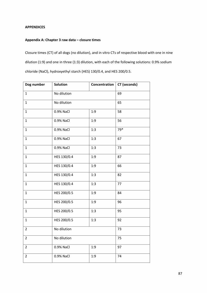
87
APPENDICES
Appendix A: Chapter 3 raw data – closure times
Closure times (CT) of all dogs (no dilution), and in vitro CTs of respective blood with one in nine
dilution (1:9) and one in three (1:3) dilution, with each of the following solutions: 0.9% sodium
chloride (NaCl), hydroxyethyl starch (HES) 130/0.4, and HES 200/0.5.
Dog number Solution Concentration CT (seconds)
1 No dilution 69
1 No dilution 65
1 0.9% NaCl 1:9 58
1 0.9% NaCl 1:9 56
1 0.9% NaCl 1:3 79*
1 0.9% NaCl 1:3 67
1 0.9% NaCl 1:3 73
1 HES 130/0.4 1:9 87
1 HES 130/0.4 1:9 66
1 HES 130/0.4 1:3 82
1 HES 130/0.4 1:3 77
1 HES 200/0.5 1:9 84
1 HES 200/0.5 1:9 96
1 HES 200/0.5 1:3 95
1 HES 200/0.5 1:3 92
2 No dilution 73
2 No dilution 75
2 0.9% NaCl 1:9 97
2 0.9% NaCl 1:9 74
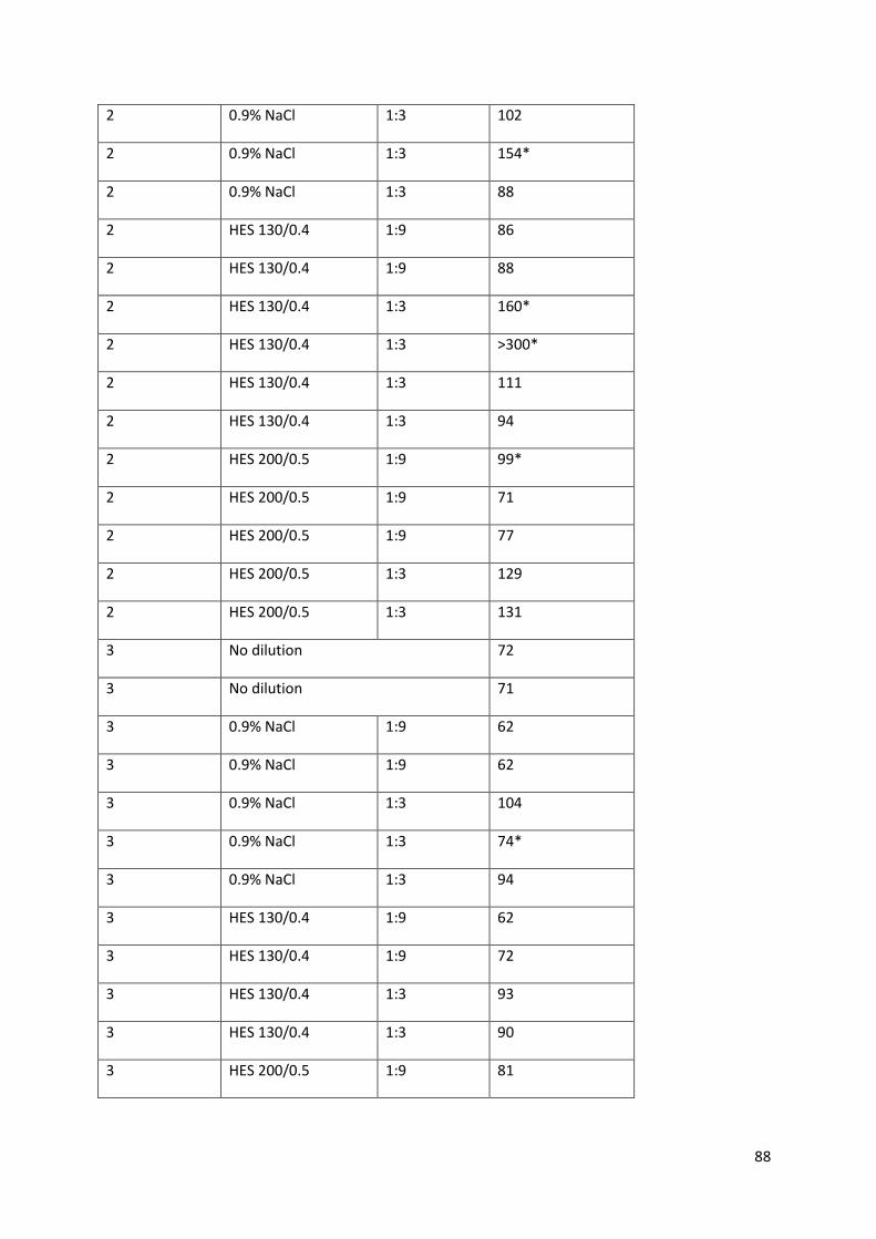
88
2 0.9% NaCl 1:3 102
2 0.9% NaCl 1:3 154*
2 0.9% NaCl 1:3 88
2 HES 130/0.4 1:9 86
2 HES 130/0.4 1:9 88
2 HES 130/0.4 1:3 160*
2 HES 130/0.4 1:3 >300*
2 HES 130/0.4 1:3 111
2 HES 130/0.4 1:3 94
2 HES 200/0.5 1:9 99*
2 HES 200/0.5 1:9 71
2 HES 200/0.5 1:9 77
2 HES 200/0.5 1:3 129
2 HES 200/0.5 1:3 131
3 No dilution 72
3 No dilution 71
3 0.9% NaCl 1:9 62
3 0.9% NaCl 1:9 62
3 0.9% NaCl 1:3 104
3 0.9% NaCl 1:3 74*
3 0.9% NaCl 1:3 94
3 HES 130/0.4 1:9 62
3 HES 130/0.4 1:9 72
3 HES 130/0.4 1:3 93
3 HES 130/0.4 1:3 90
3 HES 200/0.5 1:9 81
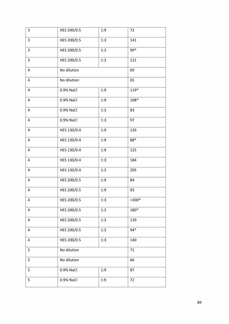
89
3 HES 200/0.5 1:9 72
3 HES 200/0.5 1:3 141
3 HES 200/0.5 1:3 99*
3 HES 200/0.5 1:3 121
4 No dilution 69
4 No dilution 65
4 0.9% NaCl 1:9 119*
4 0.9% NaCl 1:9 108*
4 0.9% NaCl 1:3 83
4 0.9% NaCl 1:3 97
4 HES 130/0.4 1:9 126
4 HES 130/0.4 1:9 88*
4 HES 130/0.4 1:9 125
4 HES 130/0.4 1:3 184
4 HES 130/0.4 1:3 205
4 HES 200/0.5 1:9 84
4 HES 200/0.5 1:9 93
4 HES 200/0.5 1:3 >300*
4 HES 200/0.5 1:3 180*
4 HES 200/0.5 1:3 139
4 HES 200/0.5 1:3 94*
4 HES 200/0.5 1:3 140
5 No dilution 71
5 No dilution 66
5 0.9% NaCl 1:9 87
5 0.9% NaCl 1:9 72
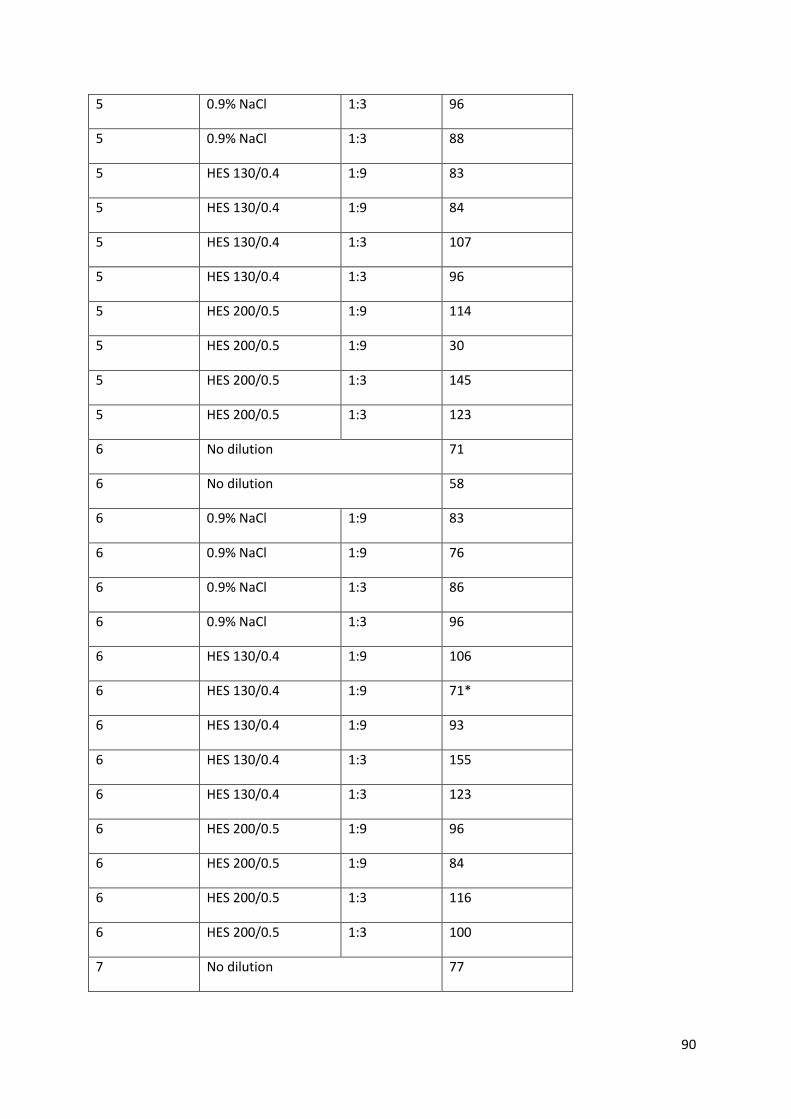
90
5 0.9% NaCl 1:3 96
5 0.9% NaCl 1:3 88
5 HES 130/0.4 1:9 83
5 HES 130/0.4 1:9 84
5 HES 130/0.4 1:3 107
5 HES 130/0.4 1:3 96
5 HES 200/0.5 1:9 114
5 HES 200/0.5 1:9 30
5 HES 200/0.5 1:3 145
5 HES 200/0.5 1:3 123
6 No dilution 71
6 No dilution 58
6 0.9% NaCl 1:9 83
6 0.9% NaCl 1:9 76
6 0.9% NaCl 1:3 86
6 0.9% NaCl 1:3 96
6 HES 130/0.4 1:9 106
6 HES 130/0.4 1:9 71*
6 HES 130/0.4 1:9 93
6 HES 130/0.4 1:3 155
6 HES 130/0.4 1:3 123
6 HES 200/0.5 1:9 96
6 HES 200/0.5 1:9 84
6 HES 200/0.5 1:3 116
6 HES 200/0.5 1:3 100
7 No dilution 77
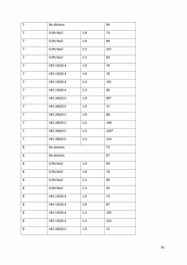
91
7 No dilution 66
7 0.9% NaCl 1:9 73
7 0.9% NaCl 1:9 89
7 0.9% NaCl 1:3 107
7 0.9% NaCl 1:3 83
7 HES 130/0.4 1:9 74
7 HES 130/0.4 1:9 70
7 HES 130/0.4 1:3 101
7 HES 130/0.4 1:3 96
7 HES 200/0.5 1:9 99*
7 HES 200/0.5 1:9 71
7 HES 200/0.5 1:9 80
7 HES 200/0.5 1:3 198
7 HES 200/0.5 1:3 100*
7 HES 200/0.5 1:3 154
8 No dilution 73
8 No dilution 67
8 0.9% NaCl 1:9 69
8 0.9% NaCl 1:9 70
8 0.9% NaCl 1:3 90
8 0.9% NaCl 1:3 94
8 HES 130/0.4 1:9 73
8 HES 130/0.4 1:9 87
8 HES 130/0.4 1:3 105
8 HES 130/0.4 1:3 103
8 HES 200/0.5 1:9 71
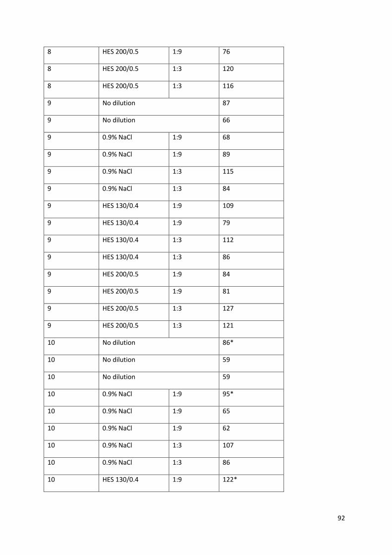
92
8 HES 200/0.5 1:9 76
8 HES 200/0.5 1:3 120
8 HES 200/0.5 1:3 116
9 No dilution 87
9 No dilution 66
9 0.9% NaCl 1:9 68
9 0.9% NaCl 1:9 89
9 0.9% NaCl 1:3 115
9 0.9% NaCl 1:3 84
9 HES 130/0.4 1:9 109
9 HES 130/0.4 1:9 79
9 HES 130/0.4 1:3 112
9 HES 130/0.4 1:3 86
9 HES 200/0.5 1:9 84
9 HES 200/0.5 1:9 81
9 HES 200/0.5 1:3 127
9 HES 200/0.5 1:3 121
10 No dilution 86*
10 No dilution 59
10 No dilution 59
10 0.9% NaCl 1:9 95*
10 0.9% NaCl 1:9 65
10 0.9% NaCl 1:9 62
10 0.9% NaCl 1:3 107
10 0.9% NaCl 1:3 86
10 HES 130/0.4 1:9 122*
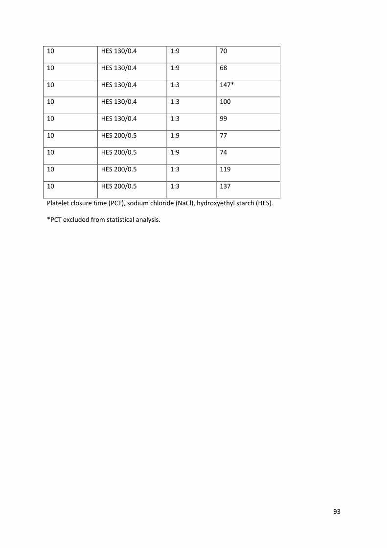
93
10 HES 130/0.4 1:9 70
10 HES 130/0.4 1:9 68
10 HES 130/0.4 1:3 147*
10 HES 130/0.4 1:3 100
10 HES 130/0.4 1:3 99
10 HES 200/0.5 1:9 77
10 HES 200/0.5 1:9 74
10 HES 200/0.5 1:3 119
10 HES 200/0.5 1:3 137
Platelet closure time (PCT), sodium chloride (NaCl), hydroxyethyl starch (HES).
*PCT excluded from statistical analysis.
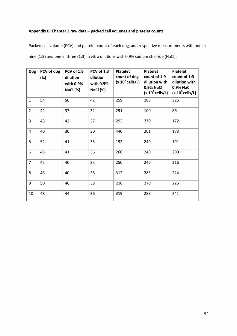
94
Appendix B: Chapter 3 raw data – packed cell volumes and platelet counts
Packed cell volume (PCV) and platelet count of each dog, and respective measurements with one in
nine (1:9) and one in three (1:3) in vitro dilutions with 0.9% sodium chloride (NaCl).
Dog PCV of dog
(%)
PCV of 1:9
dilution
with 0.9%
NaCl (%)
PCV of 1:3
dilution
with 0.9%
NaCl (%)
Platelet count of dog (x 109 cells/L)
Platelet count of 1:9 dilution with 0.9% NaCl (x 109 cells/L)
Platelet count of 1:3 dilution with 0.9% NaCl (x 109 cells/L)
1 54 50 42 259 288 326
2 42 37 32 291 100 86
3 48 42 37 292 270 172
4 40 30 30 440 201 173
5 52 41 35 192 240 191
6 48 41 36 260 240 209
7 42 40 33 250 246 216
8 46 40 38 312 283 224
9 50 46 38 216 270 225
10 48 44 36 319 288 241
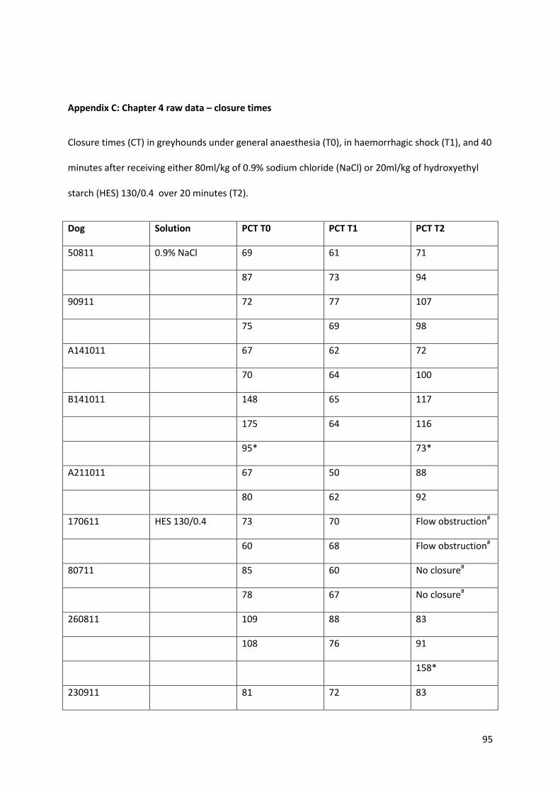
95
Appendix C: Chapter 4 raw data – closure times
Closure times (CT) in greyhounds under general anaesthesia (T0), in haemorrhagic shock (T1), and 40
minutes after receiving either 80ml/kg of 0.9% sodium chloride (NaCl) or 20ml/kg of hydroxyethyl
starch (HES) 130/0.4 over 20 minutes (T2).
Dog Solution PCT T0 PCT T1 PCT T2
50811 0.9% NaCl 69 61 71
87 73 94
90911 72 77 107
75 69 98
A141011 67 62 72
70 64 100
B141011 148 65 117
175 64 116
95* 73*
A211011 67 50 88
80 62 92
170611 HES 130/0.4 73 70 Flow obstruction#
60 68 Flow obstruction#
80711 85 60 No closure#
78 67 No closure#
260811 109 88 83
108 76 91
158*
230911 81 72 83
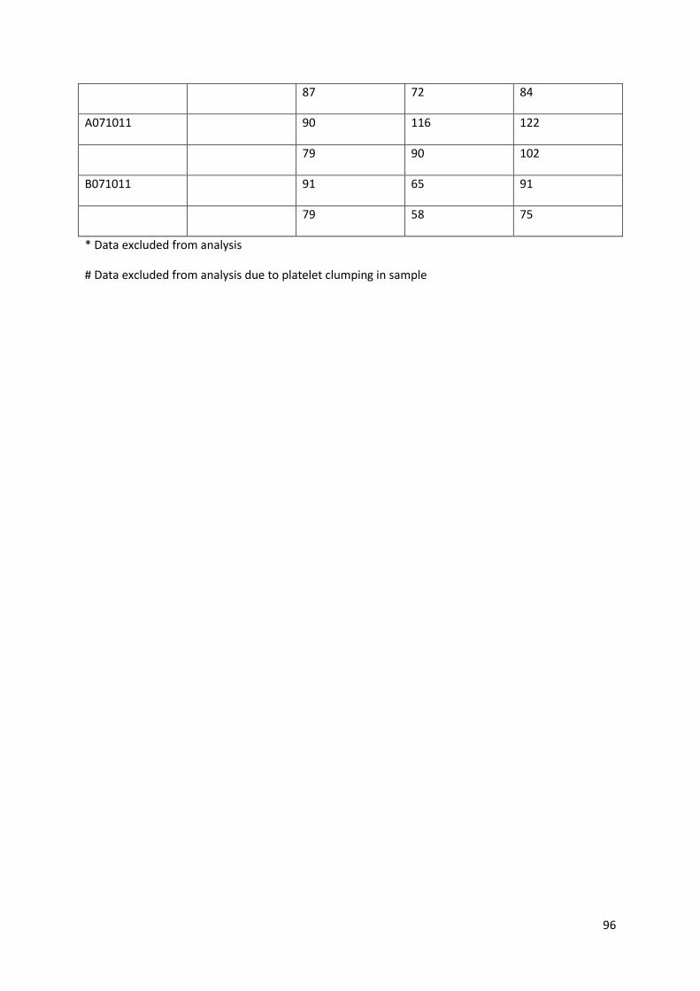
96
87 72 84
A071011 90 116 122
79 90 102
B071011 91 65 91
79 58 75
* Data excluded from analysis
# Data excluded from analysis due to platelet clumping in sample
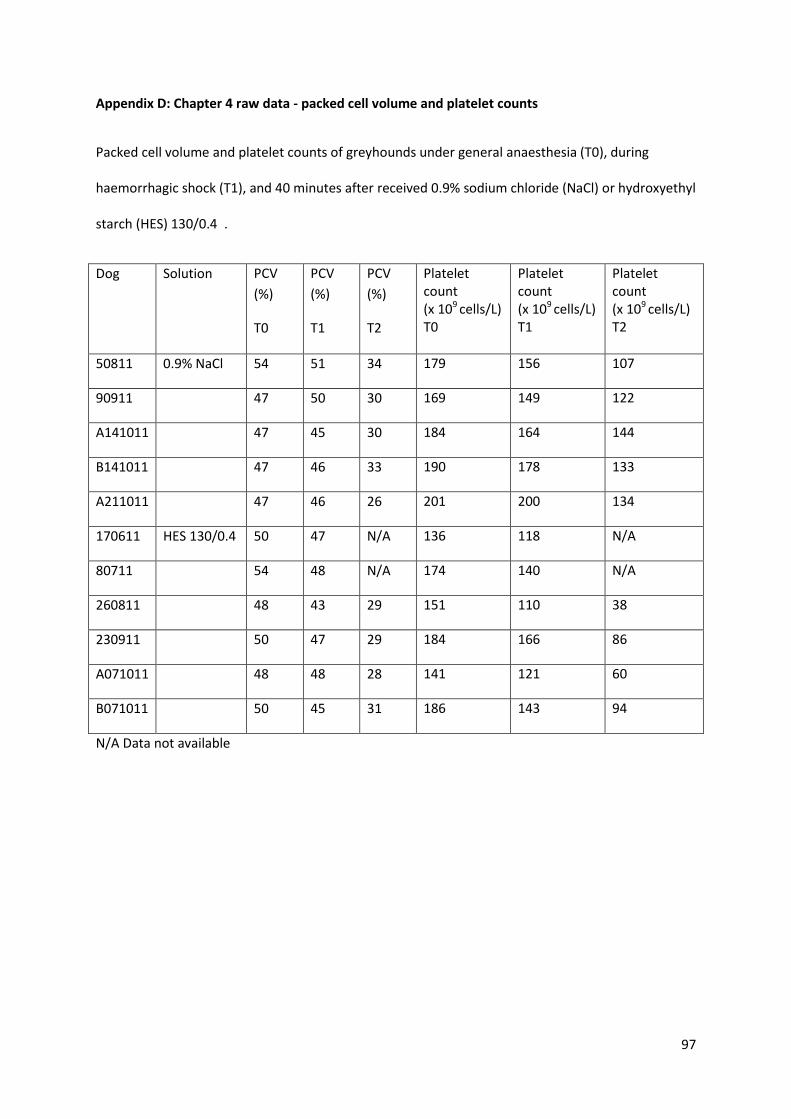
97
Appendix D: Chapter 4 raw data - packed cell volume and platelet counts
Packed cell volume and platelet counts of greyhounds under general anaesthesia (T0), during
haemorrhagic shock (T1), and 40 minutes after received 0.9% sodium chloride (NaCl) or hydroxyethyl
starch (HES) 130/0.4 .
Dog Solution PCV
(%)
T0
PCV
(%)
T1
PCV
(%)
T2
Platelet count (x 109 cells/L) T0
Platelet count (x 109 cells/L) T1
Platelet count (x 109 cells/L) T2
50811 0.9% NaCl 54 51 34 179 156 107
90911 47 50 30 169 149 122
A141011 47 45 30 184 164 144
B141011 47 46 33 190 178 133
A211011 47 46 26 201 200 134
170611 HES 130/0.4 50 47 N/A 136 118 N/A
80711 54 48 N/A 174 140 N/A
260811 48 43 29 151 110 38
230911 50 47 29 184 166 86
A071011 48 48 28 141 121 60
B071011 50 45 31 186 143 94
N/A Data not available


![[Product Monograph Template - Schedule C] · PRODUCT MONOGRAPH. VOLUVEN ® 6% hydroxyethyl starch 130/0.4 in 0.9% sodium chloride injection . Plasma Volume Expander . Fresenius Kabi](https://static.fdocuments.in/doc/165x107/5ccabd8c88c99364298bd45c/product-monograph-template-schedule-c-product-monograph-voluven-6-hydroxyethyl.jpg)