Structural traits of some species of Hydrocotyle (Araliaceae
THE EFFECT OF Hydrocotyle sibthorpioides Lam.
EXTRACT ON DENGUE VIRUS 2 INHIBITION In Vitro
FITRIEN BINTI HUSIN
UNIVERSITI SAINS MALAYSIA
2015
THE EFFECT OF Hydrocotyle sibthorpioides Lam.
EXTRACT ON DENGUE VIRUS 2 INHIBITION In Vitro
by
FITRIEN BINTI HUSIN
Thesis submitted in fulfilment of the requirements for the Degree of
Master of Science
(Microbiology)
September 2015
ii
ACKNOWLEDGEMENT
In the name of Allah, the most Merciful and Compassionate, had it not been due to His
will and favour, the completion of this study would not been possible.
First and foremost, I would like to express my deepest gratitude and greatest appreciation
to my supervisor, Dr. Rafidah Hanim Shueb, whom I cannot thank enough for her
continuous assistance and advice during the whole study, co researchers, Prof. Siti Amrah
Sulaiman, Prof. Gan Siew Hwa, Associate Prof. Dr. Chan Yean Yean, and Dr. Nabilah
Ismail for their time and continuous supports.
To my beloved parent, family and husband (Mohd Salihin Selamat) whose irreplaceable,
no words can describe on how much I adore their love, never ending concerns and supports
throughout my life.
My sincere and special thanks to staff of Department of Medical Microbiology &
Parasitology and Department of Pharmacology, especially Mr. Chan and Mr. Owi who
helps me in offering the facilities for the cell culture work.
Lastly, for my colleagues, Nurul Izzati, Tg. Ahmad Akram, Om Prakash, Mohd Lukman,
Nik Zuraina and Adila, Amalina, Izzah and other students, bunch thanks for their
immeasurable assistance, cooperation and encouragement. May Allah bless them all.
Ameen.
I hope this thesis will be a useful reference in the future in assisting other study or planning
further work.
iii
TABLE OF CONTENT
PAGE
ACKNOWLEDGEMENT II
TABLE OF CONTENTS III
LIST OF TABLES XI
LIST OF FIGURES XII
LIST OF ABBREVIATIONS XV
ABSTRAK XVII
ABSTRACT XX
CHAPTER 1: LITERATURE REVIEW
1.1 Overview of dengue 1
1.1.1 Disease burden 1
1.1.2 Epidemiology 4
1.1.3 Dengue virus 6
1.1.4 Genome structure and viral protein 6
iv
1.1.4.1 NS-1 protein 8
1.1.5 Virus replication 9
1.2 Dengue transmission 12
1.2.1 The vector 13
1.3 Clinical features of dengue 15
1.4 Dengue case classification 16
1.4.1 The 1997 dengue case classification 17
1.4.2 The 2009 dengue case classification 19
1.5 Host immune response 21
1.5.1 Primary and secondary dengue infections 22
1.6 Laboratory diagnosis of dengue 24
1.7 Management and prevention of dengue 24
1.8 Antiviral study 26
1.8.1 Model of infection 27
1.8.1.1 Animal model 28
v
1.8.1.2 Cell culture 29
1.9 Medicinal plants for treatment of dengue 31
1.9.1 Phytochemicals with anti-dengue activity 37
1.9.2 Hydrocotyle sibthorpioides Lam. 40
1.10 Rationale and objectives of the study 41
CHAPTER 2: MATERIALS AND METHODS
2.1 Chemicals and reagents 43
2.2 Preparation of plant extracts 44
2.2.1 Plant collection 44
2.2.2 Plant extraction 44
2.3 Preparation of media 46
2.3.1 Growth media 46
2.3.2 Foetal bovine serum (FBS) 46
2.3.3 Tryptose phosphate broth (TPB) 47
2.3.4 Trypsin EDTA 47
vi
2.3.5 Antibiotic 47
2.3.6 Phosphate buffer saline (PBS) 47
2.3.7 L15 complete growth media 48
2.3.8 DMEM complete growth media 48
2.3.9 Overlay medium 48
2.3.10 Cryopreservation medium 49
2.4 Cell lines preparation 49
2.4.1 C6/36 cells 50
2.4.2 Vero cells 50
2.4.3 HepG2 cells 51
2.4.4 PscloneD cells 51
2.5 Cryopreservation of cells 51
2.6 Retrieval of cell lines from liquid nitrogen 52
2.7 Cell counting 52
2.8 Dengue virus 53
vii
2.8.1 Virus propagation 53
2.8.2 Multiplicity of infection (m.o.i) 54
2.8.3 Viral growth characteristics 54
2.9 Antiviral assay 55
2.9.1 Determination of maximum non-toxic dose (MNTD) 56
2.9.2 Pre-treatment of cells prior to DENV-2 infection 57
2.9.3 Post treatment of cells after DENV-2 infection 57
2.9.4 Virucidal treatment 58
2.9.5 Concurrent treatment 58
2.10 Microscopy observation 58
2.11 Plaque assay 59
2.11.1 Preparation of methylene blue staining 60
2.12 Immunofluorescence assay 60
2.12.1 Preparation of antibody 60
2.13 NS-1 ELISA 61
viii
2.13.1 Standard curve for NS-1 protein concentration 61
2.14 RNA extraction 62
2.14.1 Isolation of viral RNA from cell culture 62
2.14.2 Isolation of viral RNA from cell culture supernatants 62
2.15 Quantitative RT-PCR 63
2.16 Primers 64
CHAPTER 3: RESULTS
3.1 Morphology of healthy cell lines 65
3.2 Presentation of CPE in various cell lines 67
3.3 Kinetics of DENV replication in C6/36 and Vero cells 70
3.4 Determination of MNTD of H.sibthorpioides water and methanol
extracts on cell lines 77
3.5 Antiviral activity of H.sibthorpioides extracts on Vero and C6/36 cells 82
3.5.1 Pre-treatment prophylactic effect of H.sibthorpioides plant
extracts on Vero and C6/36 cells 82
3.5.1.1 Morphological changes of Vero and C6/36 cells 83
ix
3.5.1.2 IFA analysis 86
3.5.1.3 Dengue viral titres 90
3.5.2 Post treatment therapeutic effect of H.sibthorpioides
plant extracts on Vero and C6/36 cells 96
3.5.2.1 Morphological changes of Vero and C6/36 cells 96
3.5.2.2 IFA analysis 100
3.5.2.3 Dengue viral titres 103
3.6 Antiviral of H.sibthorpioides plant extracts on HepG2 cells 110
3.6.1 NS-1 protein expression 110
3.6.2 Morphological changes of HepG2 cells 114
3.6.3 Dengue viral titres 116
3.6.3.1 Virucidal effect of H.sibthorpioides plant extracts
on HepG2 cells 116
3.6.3.2 Concurrent treatment effect of H.sibthorpioides
plant extracts on HepG2 cells 119
3.6.3.3 Post treatment therapeutic effect of
H.sibthorpioides plant extracts on HepG2 cells 121
x
CHAPTER 4: DISCUSSION 124
CHAPTER 5: CONCLUSION 135
REFERENCES 136
APPENDICES 152
xi
LIST OF TABLES
Table 1.1 Estimated burden of dengue in 2010, by continent 3
Table 1.2 Traditional plants that had been identified to have
anti-dengue activity 33
Table 1.3 Overview of some phytochemicals having anti-dengue
properties along with their sources 34
Table 1.4 Plant classification of Hydrocotyle sibthorpioides Lam. 43
Table 2.1 Chemicals and reagents used in the study 43
Table 3.1 Morphological changes in DENV-2 infected Vero cells
pre-treated with H.sibthorpioides water and methanol
extracts 84
Table 3.2 Morphological changes in DENV-2 infected C6/36 cells
pre-treated with H.sibthorpioides water and methanol
extracts 85
Table 3.3 Morphological changes in DENV-2 infected Vero cells
post treated with H.sibthorpioides water and methanol
extracts 98
Table 3.4 Morphological changes in DENV-2 infected C6/36 cells
post treated with H.sibthorpioides water and methanol
extracts 99
Table 3.5 Morphological changes in DENV-2 infected HepG2 cells
treated with H.sibthorpioides water and methanol extracts 115
xii
LIST OF FIGURES
Figure 1.1 Countries/ areas at risk of DENV transmission
(dark-grey shading) 5
Figure 1.2 Schematic representation of the organisation of the
genes for Flavivirus 7
Figure 1.3 Dengue virus replication 11
Figure 1.4 Life cycle of Aedes aegypti from egg to adult mosquito 14
Figure 1.5 Manifestations of dengue virus infection 18
Figure 1.6 The 2009 revised dengue case classification 20
Figure 1.7 An immune response to dengue infection 23
Figure 1.8 Images of densely populated Hydrocotyle sibthorpioides
showing long creeping stems and scalloped leaf edges 39
Figure 2.1 Schematic view of Soxhlet apparatus 45
Figure 3.1 Morphology of confluent healthy (A) C6/36 cells
(B) Vero cells (C) HepG2 cell and (D) PscloneD
cells monolayer grown in T25cm2 tissue culture flask
viewed under inverted microscope (220 x magnification) 66
Figure 3.2 Changes in C6/36 cell morphology following DENV-2
Infection 68
Figure 3.3 Replication of different DENV serotypes in C6/36 cells 73
Figure 3.4 Replication of different DENV serotypes in Vero cells 75
xiii
Figure 3.5 (A) Morphology of Vero cells from incubation within
MNTD concentrations range and (B) the toxicity effect
of H.sibthorpioides extract on Vero cells above the MNTD
range of concentrations 79
Figure 3.6 Determination of maximum non toxicity dose of
H.sibthorpioides water and methanolic extracts in (A) Vero
cells (B) C6/36 cells and (C) HepG2 cells as determined
by MTS assay 80
Figure 3.7 Effect of H.sibthorpioides extracts on DENV-2
replication in pre-treatment assay 88
Figure 3.8 Effect of H.sibthorpioides extracts on DENV-2
replication in pre-treatment assay 89
Figure 3.9 DENV-2 infectivity in Vero cells pre-treated with
(A) water extract and (B) methanolic extract
of H.sibthorpioides 93
Figure 3.10 DENV-2 infectivity in C6/36 cells pre-treated with
(A) water extract and (B) methanolic extract of
H.sibthorpioides 95
Figure 3.11 Effect of H.sibthorpioides extract on DENV-2 replication
in post treatment assay 101
Figure 3.12 Effect of H.sibthorpioides extract on DENV-2 replication
in post treatment assay 102
Figure 3.13 DENV-2 infectivity in Vero cells post treated with
(A) water extract and (B) methanolic extract of
H.sibthorpioides 106
Figure 3.14 DENV-2 infectivity in C6/36 cells post treated with
(A) water extract and (B) methanolic extract of
H.sibthorpioides 109
Figure 3.15 Standard curve for NS-1 protein level 112
Figure 3.16 NS-1 protein expression in post treated HepG2 cells with
H.sibthorpioides (A) water extract and (B) methanolic
extract 113
xiv
Figure 3.17 Virucidal activity of H.sibthorpioides extracts on
DENV-2 infectivity 118
Figure 3.18 Concurrent treatment of H.sibthorpioides extracts
on DENV-2 infectivity 120
Figure 3.19 Post treatment effect of H.sibthorpioides extracts
on DENV-2 infectivity 123
xv
LIST OF ABBREVIATIONS
CMC carboxymethyl cellulose
CO2 carbon dioxide
CPE cytopathic effect
DENV dengue virus
DF dengue fever
DHF dengue haemorrhagic fever
DMEM Dulbecco’s modified eagle medium
DMSO dimethyl sulfoxide
DSS dengue shock syndrome
EC50 effective concentration
FBS foetal bovine serum
h hour
HBV hepatitis B virus
HSV herpes simplex virus
IC50 inhibitory concentration
IFN interferon
L15 leibovitz media
m.o.i multiplicity of infection
OD optical density
PBS phosphate buffer saline
PES polyethersulfone
xvi
pfu particle forming unit
RNA ribonucleic acid
TNF tumor necrosis factor
TPB tryptose phosphate broth
WHO World Health Organisation
xvii
KESAN EKSTRAK Hydrocotyle sibthorpioides Lam. TERHADAP PERENCATAN
DENGGI VIRUS 2 SECARA In Vitro
ABSTRAK
Selama beribu tahun, amalan perubatan tradisional menggunakan tumbuh-
tumbuhan dan herba telah berkesan dalam merawat pelbagai jangkitan peyakit. Walau
bagaimanapun, setelah berdekad usaha pencarian dijalankan, tiada antivirus yang terbukti
berkesan untuk merawat jangkitan denggi virus (DENV) dan usaha ke arah pembangunan
vaksin denggi terhalang oleh beberapa faktor utama seperti jenis serotip yang berbeza.
Oleh itu, tujuan penyelidikan ini dijalankan adalah untuk mengkaji potensi antivirus di
dalam tumbuhan Hydrocotyle sibthorpioides Lam. (H.sibthorpioides) terhadap jangkitan
DENV-2 secara in-vitro. Tiga sel telah dipilih untuk tujuan kajian iaitu sel Vero, sel
C6/36, dan sel HepG2.
H.sibthorpioides telah diekstrak menggunakan dua pelarut berbeza, air dan
metanol. Ujian ketoksikan telah dijalankan untuk menilai dan menentukan dos tertinggi
ekstrak H.sibthorpioides yang mampu diterima oleh sel-sel yg dikaji. Hasil ujian
menunjukkan ekstrak methanol H.sibthorpioides adalah lebih toksik berbanding ekstrak
air H.sibthorpioides. Sel Vero mempunyai daya toleransi yang lebih luas terhadap ekstrak
H.sibthorpioides diikuti oleh sel C6/36 dan sel HepG2. Asai antivirus telah dijalankan
dalam dua peringkat rawatan yang berbeza, rawatan sebelum dan selepas infeksi untuk
menilai kesan pencegahan dan terapeutik H.sibthorpioides terhadap replikasi DENV-2.
Pemerhatian dijalankan terhadap perubahan morfologi dan kesan sitopatik (CPE), diikuti
xviii
dengan pengesanan virus antigen menggunakan IFA dan seterusnya penilaian titer virus
menggunakan asai plak dan qRT-PCR.
Hasil kajian menunjukkan bahawa ekstrak H.sibthorpioides mempunyai aktiviti
pencegahan yang rendah terhadap replikasi DENV-2 di dalam sel Vero tetapi tidak di
dalam sel C6/36. Pra rawatan sel Vero menunjukkan ekstrak air H.sibthorpioides
mempunyai 2 % - 44 % kesan perlindungan terhadap DENV-2 manakala 18 % - 30 %
kesan perlindungan dilihat apabila ekstrak methanol H.sibthorpioides digunakan.
Sebaliknya, secara umum, pra rawatan H.sibthorpioides ke atas sel C6/36 mempunyai
kesan perlindungan yang minimum terhadap infeksi DENV-2. Hanya beberapa kepekatan
H.sibthorpioides memberikan 5 % - 13 % kesan perencatan yang tidak signifikan pada sel
C6/36.
Rawatan selepas infeksi sel Vero yang dijangkiti dengan DENV-2 menggunakan
ekstrak methanol H.sibthorpioides mempunyai kesan terapeutik yang lebih tinggi
berbanding rawatan ekstrak air H.sibthorpioides. Replikasi DENV-2 direncatkan
sebanyak 6 % - 31 % apabila pelbagai kepekatan ekstrak air H.sibthorpioides digunakan,
manakala perencatan titer sebanyak 2 % - 42 % dicapai dengan rawatan ekstrak metanol
H.sibthorpioides. Yang mengejutkan, rawatan selepas infeksi ke atas sel C6/36
menyebabkan peningkatan replikasi DENV-2. Peningkatan replikasi DENV-2 sebanyak
7 % - 59 % dilihat dalam sel C6/36 yang dijangkiti dan telah dirawat dengan ekstrak air
dan metanol H.sibthorpioides.
xix
Asai antivirus telah dilanjutkan di dalam sel HepG2 dan hasil kajian menunjukkan
bahawa ekstrak metanol H.sibthorpioides adalah lebih baik berbanding ekstrak air
H.sibthorpioides dalam mengurangkan replikasi DENV-2. Rawatan selepas infeksi
terhadap DENV-2 menggunakan ekstrak H.sibthorpioides menunjukkan potensi antivirus
yang lebih baik berbanding rawatan pembunuhan dan rawatan serentak dalam sel HepG2.
Kesimpulannya, H.sibthorpioides mempunyai kesan yang berbeza terhadap replikasi
DENV-2 bergantung kepada jenis rawatan, jenis pelarut dan jenis sel yang digunakan; dan
ini memberikan pemahaman lanjut tentang sifat kegunaan perubatan tumbuhan ini
terhadap replikasi DENV-2. Kajian lanjut harus dijalankan untuk mengenalpasti kompaun
yang terlibat dan bagaimana aktiviti antivirus itu berlaku.
xx
THE EFFECT OF Hydrocotyle sibthorpioides Lam. EXTRACT ON DENGUE
VIRUS 2 INHIBITION In Vitro
ABSTRACT
For thousands of years the practice of traditional medicine, using plant and herbs,
has been effective in the treatment of various infections. Despite decades of efforts, there
is no proven effective antiviral for DENV infection and attempts at vaccine development
have been hampered by several major obstacles e.g different serotypes. The aim of this
study therefore was to investigate the potential antiviral activity of Hydrocotyle
sibthorpioides Lam. (H.sibthorpioides) towards DENV-2 infection in-vitro in three cell
lines, Vero cells, C6/36 cells and HepG-2 cells.
The H.sibthorpioides was extracted using two different solvents, water and
methanol. Toxicity test were initially performed to evaluate and determine the highest
tolerable dose of H.sibthorpioides extracts in the tested cell lines. The H.sibthorpioides
methanolic extract was found to be more toxic than the water extract. Vero cells had a
wider tolerance range to H.sibthorpioides extracts followed by C6/36 and HepG-2 cells.
Antiviral assays were performed in two different stages, pre- and post-treatment to
evaluate the H.sibthorpioides prophylactic and therapeutic effects on DENV-2 replication.
The activities were scored by observing the morphological changes and CPE appearances,
followed by detection of viral antigen by IFA and quantitation of viral titres by either
plaque assay or qRT-PCR.
xxi
The results demonstrated that H.sibthorpioides extracts possess mild prophylactic
activity against DENV-2 replication in Vero cells but not in C6/36 cells. Pre-treatment of
Vero cells showed that H.sibthorpioides pretreatment had 2 % - 44 % protective effect
against DENV-2 in Vero cells using water extract, while 18 % - 30 % protective effect
were seen when H.sibthorpioides methanolic extract was used. On the contrary, in general,
H.sibthorpioides pre-treatment on C6/36 cells had low to no cellular protective effect
against DENV-2 infection. Only few concentrations of H.sibthorpioides caused 5 % - 13
% inconsequential inhibition on C6/36 cells
The post treatment of DENV-2 infected-Vero cells with H.sibthorpioides
methanolic extract presented higher therapeutic effect when compared with the
H.sibthorpioides water extract. The DENV-2 viral replication was inhibited by 6 % - 31
% when various concentrations of H.sibthorpioides water extract were used, while 2 % –
42 % of DENV-2 titre reduction was seen in Vero cells post treated with H.sibthorpioides
methanolic extract. Surprisingly, post treatment of C6/36 cells resulted in an enhancement
of DENV-2 replication. An enhancement effect of DENV-2 by 7 % - 59 % was seen in
infected-C6/36 cells post treated with H.sibthorpioides water and methanolic extracts.
The antiviral assay was further extended in HepG-2 cells and the results
demonstrated that the methanolic extract of H.sibthorpioides was better than the water
extract in reducing DENV-2 replication. Post treatment of H.sibthorpioides extracts
against DENV-2 showed more potent antiviral activity followed by virucidal treatment
and concurrent treatment in HepG2 cells. As a conclusion, H.sibthorpioides had variable
effects on DENV-2 replication, depending on treatment types, solvent types and cell lines
xxii
used; providing important novel insight on the phytomedicinal properties of the plant on
DENV. Further studies are needed to verify which compounds could be responsible and
how they exert their antiviral effects
1
CHAPTER ONE
LITERATURE REVIEW
1.1 Overview of dengue
Dengue fever (DF), the most prevalent arthropod-borne viral illness in humans, is
caused by dengue virus (DENV). Dengue is a one disease entity with different clinical
presentations and often with unpredictable clinical evolution and outcomes (WHO, 2009).
Originally, dengue virus infections occurred mainly as epidemics in tropical and
subtropical countries, but over time, with increasing globalisation and the geographic
spread of inhabitants of Aedes aegypti and Aedes albopictus mosquitos, dengue virus
infection has steadily penetrated every corner of the world (Noisakran et al., 2010).
1.1.1 Disease burden
Dengue is a worsening global health problem. In 19th century, dengue was
considered a sporadic disease that caused epidemics at long intervals, a reflection of the
slow pace of transport and limited travel at that time and today in contrast, dengue ranks
as the most important mosquito-borne viral disease in the world (WHO, 2012). According
to the World Health Organization report, there are 50 million dengue infections and 500
000 cases of dengue haemorrhagic fever (DHF) leading to hospitalisation each year.
In Malaysia, dengue cases have risen alarmingly across the country this year, with
data showing a 269% rise in the disease from January till May 2014 as compared to the
same period last year (KKM, 2014). The number of cases reported annually to WHO
ranged from 0.4 to 1.3 million in the decade 1996 – 2005. Underreporting and
misdiagnoses are major obstacles to understanding the full burden of dengue and as an
2
infectious disease, the number of cases varies substantially from year to year (WHO,
2009).
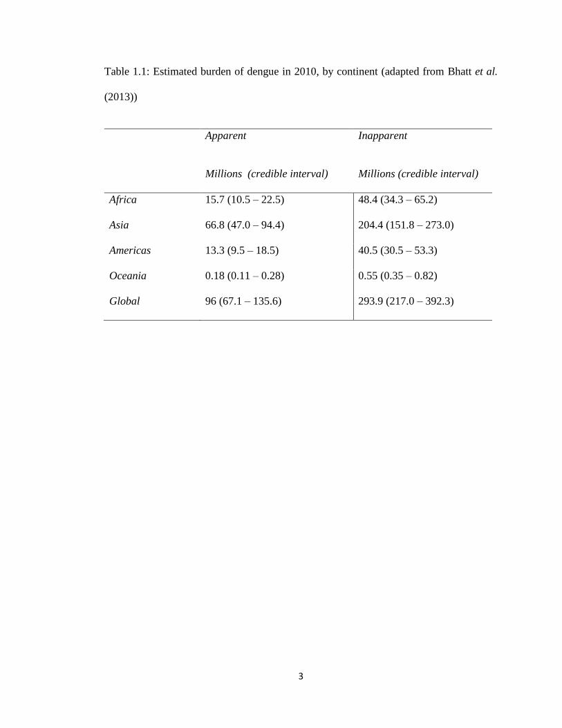
Bhatt et al. (2013) estimated that there were 96 million apparent dengue infections
in 2010 (Table 1.1) with Asia bore 70% of this burden, and is characterised by large
swathes of densely populated regions coinciding with very high suitability for disease
transmission. Recent studies have estimated annual economic burden of dengue in specific
countries of Southeast Asia using the average reported cases between 2001 – 2005, in
Cambodia, Malaysia and Thailand were at least US$ 3.1 (± 0.2), US$ 42.4 (± 4.3), and
US$ 53.1 (± 11.4) million respectively (Shepard et al., 2013). Dengue inflicts all levels of
society but the burden may be higher among the poorest who grow up in communities
with inadequate water supply and solid waste infrastructure (WHO, 2009).
3
Table 1.1: Estimated burden of dengue in 2010, by continent (adapted from Bhatt et al.
(2013))
Apparent
Millions (credible interval)
Inapparent
Millions (credible interval)
Africa 15.7 (10.5 – 22.5) 48.4 (34.3 – 65.2)
Asia 66.8 (47.0 – 94.4) 204.4 (151.8 – 273.0)
Americas 13.3 (9.5 – 18.5) 40.5 (30.5 – 53.3)
Oceania 0.18 (0.11 – 0.28) 0.55 (0.35 – 0.82)
Global 96 (67.1 – 135.6) 293.9 (217.0 – 392.3)
4
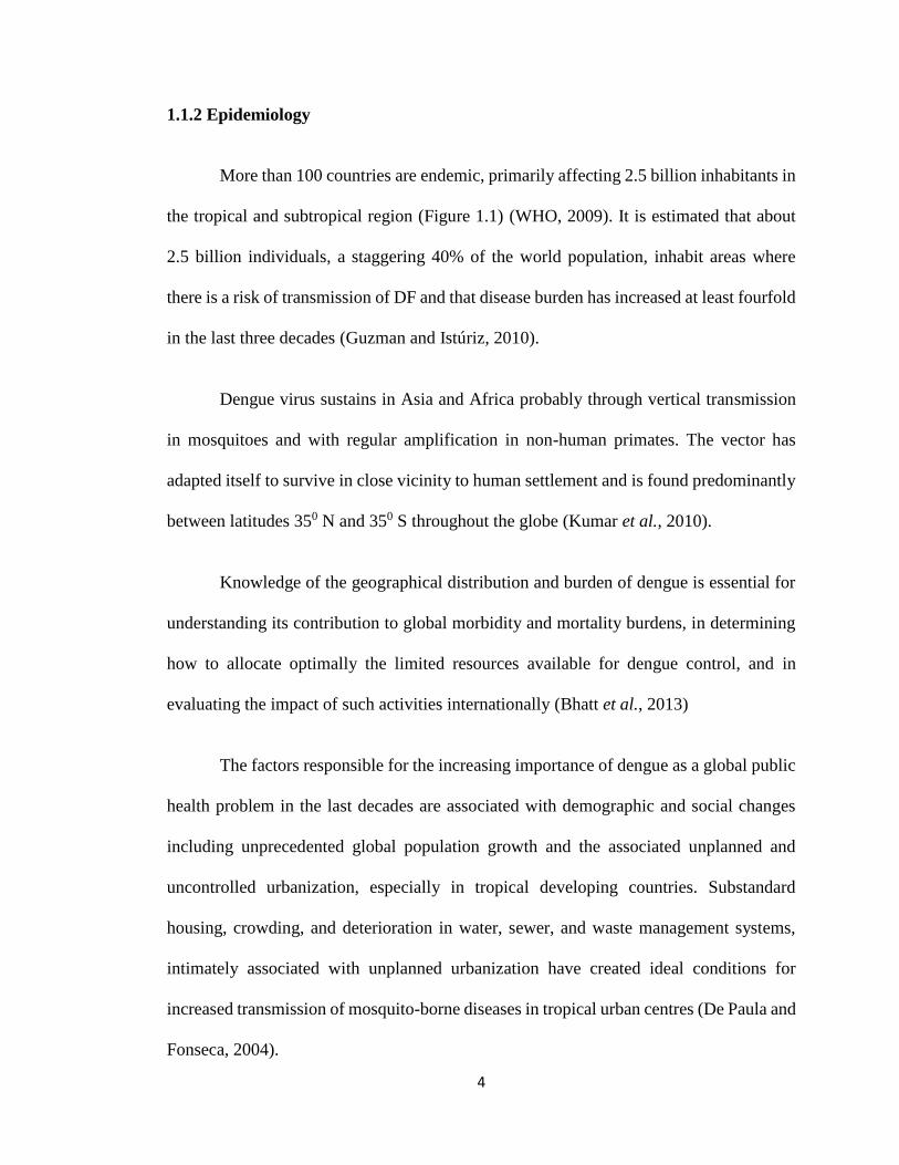
1.1.2 Epidemiology
More than 100 countries are endemic, primarily affecting 2.5 billion inhabitants in
the tropical and subtropical region (Figure 1.1) (WHO, 2009). It is estimated that about
2.5 billion individuals, a staggering 40% of the world population, inhabit areas where
there is a risk of transmission of DF and that disease burden has increased at least fourfold
in the last three decades (Guzman and Istúriz, 2010).
Dengue virus sustains in Asia and Africa probably through vertical transmission
in mosquitoes and with regular amplification in non-human primates. The vector has
adapted itself to survive in close vicinity to human settlement and is found predominantly
between latitudes 350 N and 350 S throughout the globe (Kumar et al., 2010).
Knowledge of the geographical distribution and burden of dengue is essential for
understanding its contribution to global morbidity and mortality burdens, in determining
how to allocate optimally the limited resources available for dengue control, and in
evaluating the impact of such activities internationally (Bhatt et al., 2013)
The factors responsible for the increasing importance of dengue as a global public
health problem in the last decades are associated with demographic and social changes
including unprecedented global population growth and the associated unplanned and
uncontrolled urbanization, especially in tropical developing countries. Substandard
housing, crowding, and deterioration in water, sewer, and waste management systems,
intimately associated with unplanned urbanization have created ideal conditions for
increased transmission of mosquito-borne diseases in tropical urban centres (De Paula and
Fonseca, 2004).
5
Figure 1.1: Countries/areas at risk of DENV transmission (dark-grey shading). The
contour lines indicate the potential geographical limits of the northern and southern
hemispheres for year-round survival of Aedes aegypti, the principal mosquito vector of
DENVs (adapted from WHO (2009)).
6
1.1.3 Dengue virus
DENV, the causative agent of dengue is a member of the family Flaviviridae with
four distinct serotypes, dengue serotype 1, 2, 3 and 4 (DENV 1-4). It is a small, enveloped
virus that contains a single-stranded, positive sense (messenger) RNA genome packaged
inside a core protein, which is surrounded by an icosahedral scaffold and encapsidated by
a lipid envelope (Noisakran et al., 2010). All four serotypes of DENV can be found
worldwide.
The relationships between the serotypes and transmission efficiency or disease
expression are uncertain, but DENV-2 and DENV-3 are likely to contribute the most to
disease severity and mortality (Guzman and Istúriz, 2010). Studies on the outbreaks in
endemic areas, such as South East Asia revealed that a primary infection with DENV-1 or
DENV-3 frequently resulted in a more severe disease than if DENV-2 or DENV-4 were
the primary infection (Tang et al., 2012). Dengue virus also causes a wide range of clinical
manifestations ranging from inapparent or mild febrile illness to severe and fatal
haemorrhagic disease.
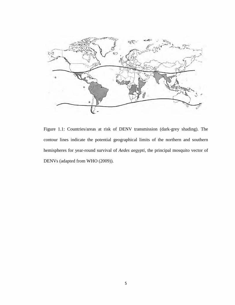
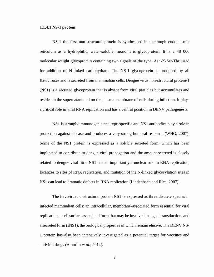
1.1.4 Genome structure and viral proteins
The distinction between dengue serotypes is based on their antigenicity. Flavivirus
particles are spherical in shape, with a lipid envelope approximately 50 nm in diameter
(Halstead, 2008). The genome is approximately 11 000 base pairs long, with 5’ capped
and 3’-end usually not polyadenylated. The 5’ and 3’ non-coding regions are important
for regulating viral replication. The termini of the genome contain untranslated regions
(UTRs) that have key roles in the regulation of translation and genome replication. The 5’
7
UTR is relatively short and has a type І cap structure; the 3’ UTR contains several
conserved RNA structures and lacks a terminal polyadenate tract (Murphy and Whitehead,
2011). The gene order for structural proteins from the 5’ terminus is C-prM-E (Figure
1.2).
There are three structural proteins encoded in the 5’-one third of the viral genome:
the capsid (C) protein forms the nucleocapsid shell protecting the viral genome, and the
premembrane (prM), and envelope (E) proteins, both virion surface proteins embedded in
the virion envelope. The main biological properties of the viruses are located in the E
protein, including receptor binding, haemagglutination of erythrocytes, neutralising
antibody induction, protective immune response, membrane fusion and virion assembly
(Schoub and Blackburn, 2004). The M- protein, which consists of seven antiparallel β-
strands, is important in the formation and maturation of the viral particles (Lai et al.,
2008).
Seven non-structural (NS) proteins are encoded in the 3’-two thirds of the viral
genome: NS1, NS2a, NS2b, NS3, NS4a, NS4b, and NS5 (WHO, 2007).The structural
proteins form the viral particle while the non-structural proteins participate in the
replication of the RNA genome, virion assembly and invasion of innate immune response.
Figure 1.2: Schematic representation of the organisation of the genes for Flavivirus
8
1.1.4.1 NS-1 protein
NS-1 the first non-structural protein is synthesised in the rough endoplasmic
reticulum as a hydrophilic, water-soluble, monomeric glycoprotein. It is a 48 000
molecular weight glycoprotein containing two signals of the type, Asn-X-Ser/Thr, used
for addition of N-linked carbohydrate. The NS-1 glycoprotein is produced by all
flaviviruses and is secreted from mammalian cells. Dengue virus non-structural protein-1
(NS1) is a secreted glycoprotein that is absent from viral particles but accumulates and
resides in the supernatant and on the plasma membrane of cells during infection. It plays
a critical role in viral RNA replication and has a central position in DENV pathogenesis.
NS1 is strongly immunogenic and type-specific anti NS1 antibodies play a role in
protection against disease and produces a very strong humoral response (WHO, 2007).
Some of the NS1 protein is expressed as a soluble secreted form, which has been
implicated to contribute to dengue viral propagation and the amount secreted is closely
related to dengue viral titre. NS1 has an important yet unclear role in RNA replication,
localizes to sites of RNA replication, and mutation of the N-linked glycosylation sites in
NS1 can lead to dramatic defects in RNA replication (Lindenbach and Rice, 2007).
The flavivirus nonstructural protein NS1 is expressed as three discrete species in
infected mammalian cells: an intracellular, membrane-associated form essential for viral
replication, a cell surface associated form that may be involved in signal transduction, and
a secreted form (sNS1), the biological properties of which remain elusive. The DENV NS-
1 protein has also been intensively investigated as a potential target for vaccines and
antiviral drugs (Amorim et al., 2014).
9
1.1.5 Virus replication
The initial step of viral replication is the attachment of infectious viral particle to
host cell. Recognition of the appropriate target cell and binding is mediated by viral
surface or envelope proteins interacting with one or more cellular membrane proteins or
other attachment factors on the plasma membrane. There are two methods by which
attached virus may penetrate the host cells, the virion envelope may fuse with the plasma
membrane with immediate deposition of the nucleocapsid into the cytoplasm, or the
plasma membrane may invaginate, forming an endocytic vesicle around the still
enveloped virus (Halstead, 2008).
Since dengue viral genome can function as mRNA, if the viral RNA can be
delivered into a cell’s cytoplasm through biologically active vesicles, translation and
genome synthesis can occur accordingly (Noisakran et al., 2010). Two conditions are
necessary for dengue virus fusion; an acidic environment and the presence of a negatively
charged membrane (Zaitseva et al., 2010). Acidic conditions have been shown to activate
a fusion protein, which leads to the deposition of the nucleocapsid within the cytoplasm.
Since the dengue virus RNA genome has a positive sense, it must first be translated
to make the RNA polymerase required for its replication. The polymerase transcribes the
positive-strand RNA to negative-strand RNA, which then serves as template for additional
positive strands. Viral RNA is then translated into a polyprotein that is processed by viral
and cellular proteases and the viral NS proteins replicate the genome RNA (van Cleef et
al., 2013)
10
Flavivirus assembly takes place at the endoplasmic reticulum (ER) where
translation of the viral RNA and production of the viral polypeptide occurred. The
structural glycoproteins prM and E localize to the luminal side of the ER and form an
immature particle with prM and E in a heterodimeric complex. The cell’s protein synthesis
produces new viral proteins that replicate the viral RNA and begin to form viral particles.
Furin-mediated proteolysis of prM in the trans-Golgi network triggers rearrangement,
homodimerization of E, and formation of the mature viral particle before release from the
infected cell (Whitby et al., 2005).
The new mature viruses bud on the surface of the infected cell and are released by
exocytosis and they are able to enter other white blood cells, such as monocytes and
macrophages (Rodenhuis-Zybert et al., 2010). Released virus contain little, if any, prM;
therefore, cleavage of prM must occur before or during exit from the cell. As the virus is
transported through exocytic vesicle, and immediately prior to release, the prM protein is
cleaved by a furin like protease to its mature M protein form, thus allowing the formation
of E protein homodimer and activating the E protein for the pH dependent conformational
changes which occur during subsequent attachment and entry into cells (Halstead et al.,
2005). Immature, prM containing flavivirions are about 60-fold less infectious than
mature virus. PrM may maintain the virion in a highly stable but relatively inert state. The
final cleavage step makes the virus competent for infection but more labile. The processes
of dengue viral replication have been simplified as shown in Figure 1.3.
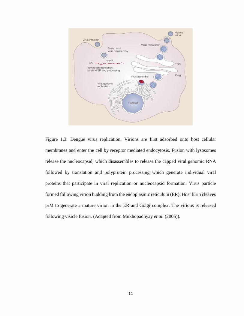
11
Figure 1.3: Dengue virus replication. Virions are first adsorbed onto host cellular
membranes and enter the cell by receptor mediated endocytosis. Fusion with lysosomes
release the nucleocapsid, which disassembles to release the capped viral genomic RNA
followed by translation and polyprotein processing which generate individual viral
proteins that participate in viral replication or nucleocapsid formation. Virus particle
formed following virion budding from the endoplasmic reticulum (ER). Host furin cleaves
prM to generate a mature virion in the ER and Golgi complex. The virions is released
following visicle fusion. (Adapted from Mukhopadhyay et al. (2005)).

12
1.2 Dengue transmission
Principle hosts for dengue viruses are human and mosquito; mosquito remains
infected throughout its life but human develops illness once they are infected. The four
dengue viruses originated in monkeys and independently jumped to humans in Africa or
Southeast Asia between 100 and 800 years ago (Halstead, 2008).
Dengue viruses can be transmitted through the bite of female mosquitoes of Aedes
aegypti, Aedes albopictus, Aedes polynesiensis and several species of the Aedes scutellaris
complex. Each of these species has a particular ecology, behaviour and geographical
distribution. Aedes aegypti is one of the most efficient vectors for arboviruses because it
is highly anthropophilic, frequently bites several times before completing oogenesis, and
thrives in close proximity to humans (WHO, 2009).
In mosquito, after ingestion of a blood meal containing virus, the virus infects the
epithelial cells lining the midgut, then escapes from the midgut epithelium into the
haemocele and infects the salivary gland. The virus is secreted in the saliva, causing
infection during probing (Chawla et al., 2014). The mosquito’s saliva containing dengue
virus are transferred through the bite site when the infected mosquito is taking a blood
meal and then spreads to possible target tissues such as lymph nodes, spleen, bone marrow
and liver (Halstead, 2008).
A female mosquito that takes blood meal from a person infected with dengue fever,
during the initial 2-10 days febrile period becomes itself infected with the virus in the cells
lining its gut (Vassil St. Goergiev, 2009). The mosquito may be infected with 2 different
viruses without affecting the yield of either virus.
13
1.2.1 The vector
Generally, mosquitoes spend the aquatic phase in immature stages and the
terrestrial phase in the adult stage during which the events of mating, blood feeding and
ovipositing take place. The life span of the adult mosquito usually depends on several
factors: temperature, humidity, sex of the mosquito and time of the year (Alameda, 2001).
The adult life of Aedes aegypti can range from 2 weeks to a month depending on
environmental conditions and the life cycle can be completed within one and a half to
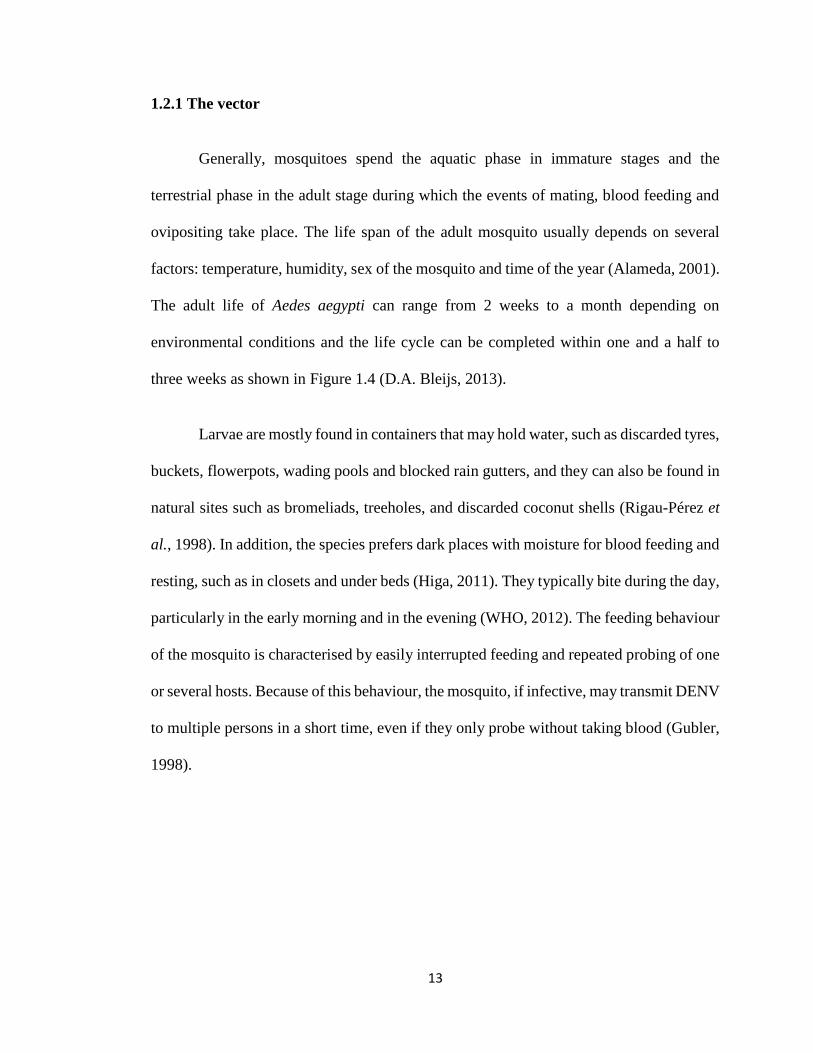
three weeks as shown in Figure 1.4 (D.A. Bleijs, 2013).
Larvae are mostly found in containers that may hold water, such as discarded tyres,
buckets, flowerpots, wading pools and blocked rain gutters, and they can also be found in
natural sites such as bromeliads, treeholes, and discarded coconut shells (Rigau-Pérez et
al., 1998). In addition, the species prefers dark places with moisture for blood feeding and
resting, such as in closets and under beds (Higa, 2011). They typically bite during the day,
particularly in the early morning and in the evening (WHO, 2012). The feeding behaviour
of the mosquito is characterised by easily interrupted feeding and repeated probing of one
or several hosts. Because of this behaviour, the mosquito, if infective, may transmit DENV
to multiple persons in a short time, even if they only probe without taking blood (Gubler,
1998).
14
Figure 1.4: Life cycle of Aedes aegypti from egg to adult mosquito (adapted from D.A.
Bleijs (2013)).
15
1.3 Clinical features of dengue
Infection with a DENV can produce a spectrum of clinical illness which ranges
from a non-specific viral symptom to a severe and fatal haemorrhagic disease. Upon
DENV infection, some individuals may develop mild disease with flu-like symptoms,
whereas a few individuals may develop severe disease. Typically, people infected with
dengue virus are asymptomatic or only have mild symptoms such as an uncomplicated
fever. Young children may have an undifferentiated febrile disease with maculopapular
rash while older children and adults have either a mild febrile syndrome or the classical
incapacitating disease.
After incubation period, the illness begins abruptly followed by the phases of
febrile, clinical and recovery. The acute febrile phase of the illness, characterized by fever
and myalgia, lasts 2-7 days and is often accompanied by facial flushing, skin erythema,
generalised body ache and headache. Back pain, arthralgias and conjunctivitis may also
occur (Whitehorn and Farrar, 2011). Fever, chills and malaise are common but nonspecific
(Rothman, 2011). The earliest abnormality in the full blood count is a progressive decrease
in total white cell count, which should alert the physician to a high probability of dengue.
Around the time of defervescence, an increase in the capillary permeability in
parallel with increasing haematocrit levels may occur, and this marks the beginning of the
critical phase. Progressive leukopenia followed by a rapid decrease in platelet count
usually precedes plasma leakage. The degree of increase above the baseline haematocrit
often reflects the severity of plasma leakage. Shock occurs when a critical volume of
plasma is lost through leakage and it often preceded by warning signs (WHO, 2009).
16
A gradual reabsorption of extravascular compartment fluid takes place in the
following 48-72 hours if the patient survives the critical phase. The patient’s general
wellbeing improves, appetite returns, gastrointestinal symptoms abate and haemodynamic
status stabilizes. The haematocrit stabilizes or may be lower due to the dilution effect of
reabsorbed fluid. White blood count usually starts to rise soon after defervescence but the
recovery of platelet count is typically later than that of white blood cell count (WHO,
2009). Convalescence is accompanied by asthenia, and a full recovery often takes several
weeks (Lim et al., 2013).
1.4 Dengue case classification
Clinical information from outbreaks of fatal dengue hemorrhagic fever in
Southeast Asia children during the late 1960s, formed the basis for a dengue clinical
classification published in World Health Organisation (WHO) guideline in 1975 and
updated in 1997 (WHO, 1975; WHO, 1997).
Symptomatic dengue virus infections were grouped into undifferentiated fever,
dengue fever (DF) and dengue haemorrhagic fever (DHF). Difficulties in applying the
criteria for DHF in the clinical situation, together with the increase in clinically severe
dengue cases which did not fulfil the strict criteria of DHF, has led to the request for the
classification to be considered (WHO, 2009).
17
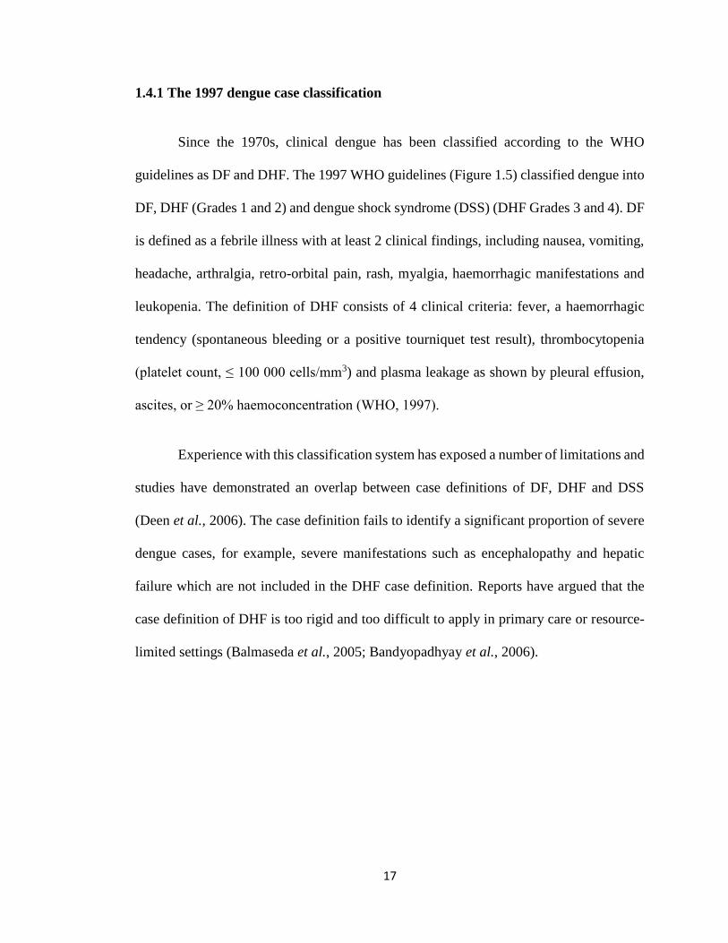
1.4.1 The 1997 dengue case classification
Since the 1970s, clinical dengue has been classified according to the WHO
guidelines as DF and DHF. The 1997 WHO guidelines (Figure 1.5) classified dengue into
DF, DHF (Grades 1 and 2) and dengue shock syndrome (DSS) (DHF Grades 3 and 4). DF
is defined as a febrile illness with at least 2 clinical findings, including nausea, vomiting,
headache, arthralgia, retro-orbital pain, rash, myalgia, haemorrhagic manifestations and
leukopenia. The definition of DHF consists of 4 clinical criteria: fever, a haemorrhagic
tendency (spontaneous bleeding or a positive tourniquet test result), thrombocytopenia
(platelet count, ≤ 100 000 cells/mm3) and plasma leakage as shown by pleural effusion,
ascites, or ≥ 20% haemoconcentration (WHO, 1997).
Experience with this classification system has exposed a number of limitations and
studies have demonstrated an overlap between case definitions of DF, DHF and DSS
(Deen et al., 2006). The case definition fails to identify a significant proportion of severe
dengue cases, for example, severe manifestations such as encephalopathy and hepatic
failure which are not included in the DHF case definition. Reports have argued that the
case definition of DHF is too rigid and too difficult to apply in primary care or resource-
limited settings (Balmaseda et al., 2005; Bandyopadhyay et al., 2006).

18
Figure 1.5: Manifestations of dengue virus infection (adapted from WHO (1997)).
19
1.4.2 The 2009 dengue case classification
It then became apparent that the 1997 classification system is not universally
applicable for appropriate clinical management, and in the 2006, the WHO Dengue
Scientific Working Group recommended additional research into dengue diagnostics and
triaging of patients for optimised clinical management and this has led to the re-
classification of dengue into dengue with and without warning signs and severe cases
published in 2009 (Barniol et al., 2011; Hadinegoro, 2012)
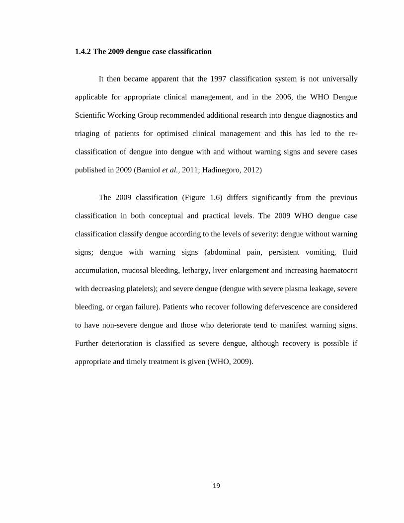
The 2009 classification (Figure 1.6) differs significantly from the previous
classification in both conceptual and practical levels. The 2009 WHO dengue case
classification classify dengue according to the levels of severity: dengue without warning
signs; dengue with warning signs (abdominal pain, persistent vomiting, fluid
accumulation, mucosal bleeding, lethargy, liver enlargement and increasing haematocrit
with decreasing platelets); and severe dengue (dengue with severe plasma leakage, severe
bleeding, or organ failure). Patients who recover following defervescence are considered
to have non-severe dengue and those who deteriorate tend to manifest warning signs.
Further deterioration is classified as severe dengue, although recovery is possible if
appropriate and timely treatment is given (WHO, 2009).
20
Figure 1.6: The 2009 revised dengue case classification (adapted from WHO (2009)).
21
1.5 Host immune response
To establish infection and replication in the hosts, flaviviruses have evolved a
variety of strategies to modulate the host’s immune responses (Ye et al., 2013). They must
evade or inhibit important elements of the innate immune system, namely the type І
interferon (IFN) response, which negatively influences the subsequent development of
antigen-specific adaptive immunity against those viruses.
Innate immunity and type І IFN responses function as the first line of defence
against viral infections, and responds by immediate protective defence mechanisms
(Morrison et al., 2012). It involves the rapid recognition of pathogen associated molecular
patterns (PAMP) in non-immune cells or cells of the innate immune system such as
monocytes or macrophages, dendritic cells (DC), and natural killer (NK) cells. Hence the
first barrier to overcome for successful viral infection is the rapid innate immune
responses of the host, including type І IFNs, inflammatory cytokine, complement
response, NK cell immunity, apoptosis and autophagy (Ye et al., 2013). Most viruses
target these important elements to avoid being sensed or recognised in infected cells and
to efficiently establish infection in the host.
Adaptive immunity is triggered when a pathogen evades the innate immune system
and generates a threshold level of antigen. The system consists of the humoral immune
response (production of antibodies by B cells) and the cellular immune response (activities
carried out by CD4+ and CD8+ T cells) (Ye et al., 2013). The lymphocytes of the adaptive
immune system evolved to provide a more versatile means of defence which, in addition,
provides increased protection against subsequent reinfection with the same pathogen.
22
Conversely, it may also play a critical role in the enhancement of disease severity in most
patients with DHF/DSS (Murphy and Whitehead, 2011).
The cells of the innate immune system, however, also play a crucial part in the
initiation and subsequent direction of adaptive immune responses, as well as participating
in the removal of pathogens that have been targeted by an adaptive immune response.
1.5.1 Primary and secondary dengue infections
Two types of infections are caused by DENV, primary and secondary infection.
Primary infection causes acute febrile illness known as DF and secondary infection causes
more severe cases and results in DHF (Idrees and Ashfaq, 2013). Primary infection with
any of the four serotypes results in a lifelong immunity to that serotypes, and temporary
immunity to the others. However this temporary immunity usually wanes after 6 months,
at which point an individual is susceptible to the other three DENV serotypes. Subsequent
infections often leads to more severe secondary infection in the presence of heterologous
dengue antibodies, which attributed to antibody dependent enhancement (Murrell et al.,
2011).
The diagnosis of dengue fever can be made serologically by detecting anti-dengue
IgM and/or IgG antibodies. Primary infections are characterized by an increase in dengue-
specific IgM antibodies four to five days after the onset of fever and by an increase in IgG
antibodies (Chawla et al., 2014). In patients experiencing a primary infection, anti-dengue
antibodies, initially of the IgM class, evolve relatively slowly. The presence of IgM
without IgG is suggestive of DENV primary infection. After a primary infection IgG
reaches peak levels in the blood after 14-21 days.
23
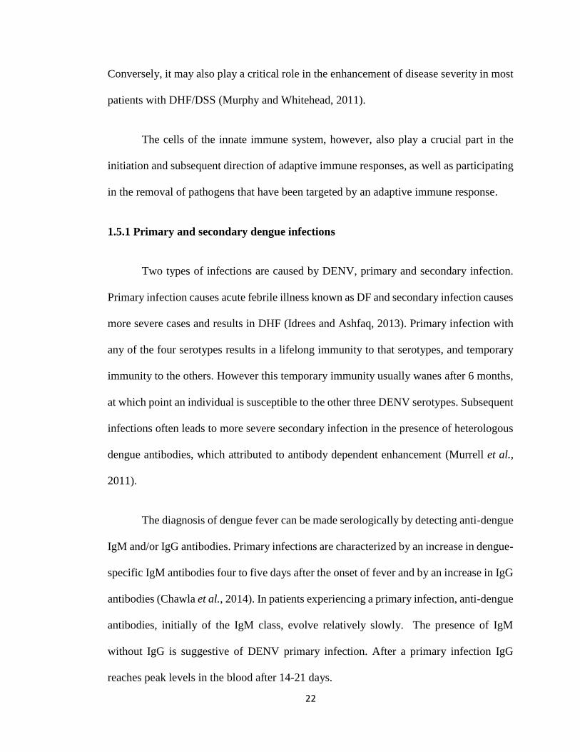
Secondary responses are IgG antibodies which appear early, often during the
febrile period, rise rapidly and are referred to as anamnestic (memory) responses. In
subsequent re-infections, level peak earlier and the titres are usually higher. Both IgM and
IgG provide protective immunity to the infecting serotype of the virus. IgM antibody is
transient and generally disappears 30-90 days after onset of illness in primary infections
while IgG antibody by contrast, persists for at least 60 years and probably for the life of
the patient (Gubler, 2010).
Figure 1.7: An Immune response to dengue infection (adapted from Guzman et al.
(2010))
24
1.6 Laboratory diagnosis of dengue
Currently, dengue diagnosis is based on serology, viral isolation and viral RNA
detection. Proof of a dengue infection depends on confirmatory reverse transcriptase-
polymerase chain reaction (RT-PCR), dengue serology, specific NS-1 antigen detection
or viral isolation, if available (Whitehorn and Farrar, 2011). During the early stages of the
disease, virus isolation, nucleic acid or antigen detection can be used to diagnose the
infection. At the end of the acute phase of infection, serology is the method of choice for
diagnosis. Enzyme linked immunosorbent assays (ELISA) are still the most widely used
technique for serological diagnosis (De Paula and Fonseca, 2004).
The differential diagnosis is extensive and varies depending on where the patient
is seen, but would include malaria, typhoid, leptospirosis, scrub and murine typhus,
septicaemia, other viral haemorrhagic fevers (eg. Ebola, Lassa fever), Chikungunya, and
Rift Valley fever (usually without a rash) (Whitehorn and Farrar, 2011).
1.7 Management and prevention of dengue
To date, there are no available vaccine, chemoprophylactic or effective antiviral
treatment for dengue. Thus far, the current prevention for dengue virus is by the prevention
of its vector which is mosquito while medical supportive care is the recommended primary
treatment for infected patients. Patients with DF require rest, oral fluids to compensate for
losses via diarrhoea or vomiting, analgesics, and antipyretics for high fever.
Emergency control measures are based primarily on application of insecticides,
and it is essential to monitor periodically the vector’s susceptibility to the insecticides