The descending respiratory,pathway in man
13
J. Neurol. Neurosurg. Psychiat., 1963, 26, 487 The descending respiratory, pathway in man P. W. NATHAN' From the National Hospital for Nervous Diseases, Queen Square, London The descending respiratory pathway may be con- sidered to be the most important tract of the spinal cord; yet its position in man is unknown. It is important to work out the location of these descend- ing fibres not only to complete anatomical knowledge but also to provide those practising surgery on the cervical cord with essential information. The absence of this knowledge contributed to the death of some of our patients after cordotomy and this has also been a cause of death in patients from other series of cordotomies. Following Stookey's (1943) report of his experience of high cervical cordotomies, Penfield (1943) said: 'I have had a patient in whom I sectioned the pain tract at the second cervical segment under local anaesthesia. Suddenly, as the wound was being sutured, while he was talking and in the midst of a sentence, he died . . . Autopsy showed no explanation of it.' Stookey replied that he had never had such an experience following unilateral cordotomy but he had with the bilateral operation. He added: 'In bilateral cordotomy, I think that sectioning of the anterior lateral columns probably interferes with the higher respiratory pathway coming down from the medulla to the diaphragm ... The difficulties I have had have been with bilateral cordotomies and I felt they were respiratory deaths, due to interruption of an upper respiratory path- way.' Undoubtedly many neurosurgeons have had the same experience: patients having had unilateral and more commonly bilateral cordotomies in the cervical region may die suddenly, often in their sleep, and at necropsy no explanation has been forthcoming. It will be shown in this paper that an adequate high cervical cordotomy will damage most of the descending fibres controlling respiratory movements. In 1830, Sir Charles Bell made the suggestion that the tract for controlling respiratory movements lies in the lateral part of the cord, somewhere between the anterior and posterior roots. Although since then there has been work on animals that confirms Sir Charles Bell's conjecture, there has been, as far as I can ascertain, no evidence produced for man. In 1895, Porter looked for the location of the pathway running 'from the bulb to the phrenic 'Member external scientific staff, Medical Research Council. nuclei' in the dog and the rabbit. In a single experi- ment on the dog he cut through the cord at the first cervical segment, leaving intact 'a part of the lateral column' on one side; 'the dog continued to breathe, somewhat feebly'. In a rabbit he cut the right half of the cord at the second cervical segment; he then cut the left half of the medulla oblongata 'close behind the calamus scriptorius' to 1 mm. from the lateral surface. Following a period of artificial respiration, the animal started to breathe again spontaneously. Dividing the remaining region of the medulla permanently stopped respiration. He concluded that 'the respiratory impulse descends in the lateral column.' Allen (1927) also carried out partial sections of the cord in the cat and in the guinea-pig. His main purpose was to find out whether a 'visceral bulbospinal pathway' exists; he also made some observations on the pathway for impulses to the motor neurones of the diaphragm and intercostal muscles. He concluded that 'the medullospinal respiratory impulses were conducted chiefly in the ventral part of the lateral columns and in the lateral part of the anterior columns.' Tosatti (1939a, 1939b), who also carried out partial sections of the cord cranial to the phrenic nuclei in the dog and the rabbit, came to the conclusion that there are two pathways, one main tract and one reserve pathway that comes into action only when the main tract is blocked. The main tract runs in the lateral column and the reserve tract in the anterior column. Pitts (1940), also using the method of partial sections of the spinal cord in the cat, concluded that the respiratory pathway lies in the anterior column and in the anterior part of the lateral column; he found it to be mainly uncrossed. Gardner and Haddad (1953) found in the cat that breathing continues as long as 'but one ventrolateral region of the cord' is left intact, the rest of the cord being divided. Hukuhara, Nakayama, and Okada (1954) traced the descending fibres in the cat and the dog by seeking for the action potentials of the respiratory discharge that originates in the medulla oblongata; they traced the location of this discharge in the cervical cord to a region of the white matter lying very close to the grey matter, near the substantia intermedia lateralis. 487
-
Upload
trinhnguyet -
Category
Documents
-
view
222 -
download
2
Transcript of The descending respiratory,pathway in man

J. Neurol. Neurosurg. Psychiat., 1963, 26, 487
The descending respiratory, pathway in manP. W. NATHAN'
From the National Hospital for Nervous Diseases, Queen Square, London
The descending respiratory pathway may be con-sidered to be the most important tract of the spinalcord; yet its position in man is unknown. It isimportant to work out the location of these descend-ing fibres not only to complete anatomical knowledgebut also to provide those practising surgery on thecervical cord with essential information. Theabsence of this knowledge contributed to the deathof some of our patients after cordotomy and thishas also been a cause of death in patients from otherseries of cordotomies. Following Stookey's (1943)report of his experience of high cervical cordotomies,Penfield (1943) said: 'I have had a patient in whom Isectioned the pain tract at the second cervicalsegment under local anaesthesia. Suddenly, as thewound was being sutured, while he was talking andin the midst of a sentence, he died . . . Autopsyshowed no explanation of it.' Stookey replied that hehad never had such an experience following unilateralcordotomy but he had with the bilateral operation.He added: 'In bilateral cordotomy, I think thatsectioning of the anterior lateral columns probablyinterferes with the higher respiratory pathwaycoming down from the medulla to the diaphragm ...The difficulties I have had have been with bilateralcordotomies and I felt they were respiratory deaths,due to interruption of an upper respiratory path-way.' Undoubtedly many neurosurgeons have hadthe same experience: patients having had unilateraland more commonly bilateral cordotomies in thecervical region may die suddenly, often in theirsleep, and at necropsy no explanation has beenforthcoming. It will be shown in this paper that anadequate high cervical cordotomy will damage mostof the descending fibres controlling respiratorymovements.
In 1830, Sir Charles Bell made the suggestionthat the tract for controlling respiratory movementslies in the lateral part of the cord, somewhere betweenthe anterior and posterior roots. Although sincethen there has been work on animals that confirmsSir Charles Bell's conjecture, there has been, as faras I can ascertain, no evidence produced for man.
In 1895, Porter looked for the location of thepathway running 'from the bulb to the phrenic'Member external scientific staff, Medical Research Council.
nuclei' in the dog and the rabbit. In a single experi-ment on the dog he cut through the cord at the firstcervical segment, leaving intact 'a part of the lateralcolumn' on one side; 'the dog continued to breathe,somewhat feebly'. In a rabbit he cut the right half ofthe cord at the second cervical segment; he then cutthe left half of the medulla oblongata 'close behindthe calamus scriptorius' to 1 mm. from the lateralsurface. Following a period of artificial respiration,the animal started to breathe again spontaneously.Dividing the remaining region of the medullapermanently stopped respiration. He concludedthat 'the respiratory impulse descends in the lateralcolumn.' Allen (1927) also carried out partialsections of the cord in the cat and in the guinea-pig.His main purpose was to find out whether a 'visceralbulbospinal pathway' exists; he also made someobservations on the pathway for impulses to themotor neurones of the diaphragm and intercostalmuscles. He concluded that 'the medullospinalrespiratory impulses were conducted chiefly in theventral part of the lateral columns and in thelateral part of the anterior columns.' Tosatti (1939a,1939b), who also carried out partial sections of thecord cranial to the phrenic nuclei in the dog and therabbit, came to the conclusion that there are twopathways, one main tract and one reserve pathwaythat comes into action only when the main tract isblocked. The main tract runs in the lateral columnand the reserve tract in the anterior column. Pitts(1940), also using the method of partial sectionsof the spinal cord in the cat, concluded that therespiratory pathway lies in the anterior column andin the anterior part of the lateral column; he foundit to be mainly uncrossed. Gardner and Haddad(1953) found in the cat that breathing continues aslong as 'but one ventrolateral region of the cord' isleft intact, the rest of the cord being divided.Hukuhara, Nakayama, and Okada (1954) tracedthe descending fibres in the cat and the dog byseeking for the action potentials of the respiratorydischarge that originates in the medulla oblongata;they traced the location of this discharge in thecervical cord to a region of the white matter lyingvery close to the grey matter, near the substantiaintermedia lateralis.
487
P. W. Nathan
Thus the evidence obtained from dogs, rabbits,guinea-pigs, and cats shows that the descendingfibres run in the anterior and/or the lateral columnsof the cord.
MATERIALS AND METHODS
The material for this study consists of eight main casesand five confirmatory cases; in all of them the operationof antero-lateral cordotomy for the pain of cancer wasperformed in the cervical segments of the cord. In theeight main cases, full histological examination of theaffected region in the cervical cords was carried out byDr. Marion Smith. No post-mortem examination waspossible in the five confirmatory cases; in them theposition of the lesion made at operation could be de-duced from the region of loss of pain sensibility causedby this operation.For many years I was hoping that an opportunity
would occur of studying the effect on respiratory move-ments of cutting the lateral cortico-spinal tract but asthis operation is no longer performed, there seemed nopoint in waiting any longer for the chance of investigatingsuch a case. Accordingly, Oliver's (1953) cases in whichthe lateral cortico-spinal tract was cut are quoted ascontrol cases, for in them there was no disturbance ofrespiratory movements; in our cases of cordotomy thesemovements were seriously disturbed.
Before the first and representative case is given in detail,it is essential for the argument to take note of Pitts' (1940)findings in his work on the location of this pathway inthe cat. He found that 'even with hemisection of thecord', there was always some return of ipsilateral res-piratory movements within the survival period of 10days. It is clear from Pitts' work that this recovery mustbe due to fibres descending in the opposite side of thecord; no other explanation seems to be possible. Similarlyin the cases reported here, respiratory movements werenever permanently stopped by the operation. It is thusclear that no long-lasting paralysis of respiratory move-ments on the side of the lesion is to be expected fromcutting the relevant ipsilateral descending fibres: re-covery from the paralysis does not mean that all therelevant ipsilateral fibres have not been divided.
HISTOLOGICAL METHODS
Transverse sections of the cord through the region ofoperation stained by a wide variety of methods, detailedin previous papers (Nathan and Smith, 1951, 1953), were
examined to establish the exact location of the operativelesion. On the basis of the location and extent of theincision, the cases have been divided into groups;
photographs of sections of the cords of four cases willbe shown, each section typical of each group of cases.
METHODS OF EXAMINING RESPIRATION
Day-to-day observations were made on respiration byroutine clinical methods. In addition the diaphragm wasexamined by radiological screening, the patient usually
being in the upright position, and the excursion of thetwo halves of the diaphragm was measured on thescreen; sometimes screening after the operation had to bedone with the patient lying on his back. During screeningafter the operation, various procedures were carried outto try and induce respiration of the paralysed side. Theactivity of the intercostal muscles was examined electro-myographically with co-axial needle electrodes, samplesfrom the two sides being taken simultaneously. In fiveof the patients changes in respiration were recordedduring the operation; in one patient volume changes ofthe right and left lungs were recorded independently bymeans of a volumetric recorder through a Carlen'sendobronchial tube; in the other four, the movementswere recorded mechanically.
MAIN CASES
Only one case will be presented in full detail.
CASE 841 Before the operation of high cervical cordotomyfor the pain due to a sarcoma of the right scapula, allrespiratory movements were seen to be normal onclinical examination. Electromyographical recordingfrom the intercostal muscles showed the presence ofactive units in phase with inspiration and expirationwherever they were looked for. Radiologically theexcursions of the two halves of the diaphragm were seento be of normal extent.The operation was carried out on the left side of the
cord, midway between the second and third cervicalroots; the purpose was to obtain an upper level ofanalgesia as near to the level of the incision as possibleand this necessitated making an incision that extendedfar forwards and medially. This was successful and thereresulted a detectable change in pain and thermal sensi-bility up to the distribution of the mandibular division ofthe fifth nerve, a loss of pain sensibility to a pin of 60 g.weight up to the third cervical dermatome, and a loss ofall pain and thermal sensibility up to the fifth cervicaldermatome.
Illustrations from the respiratory records made duringthe operation are shown in Figure 1. The records fromthe left lung, the side of the cordotomy incision, areshown as the upper records, and those from the rightlung as the lower records. Figure la is the control record,showing the tidal volume of the two lungs before thespinal cord was incised. An incision was first made intothe spinal cord at the second cervical segment, the pointof the knife being inserted into the cord posterior to theline of attachment of the denticulate ligament to a depthof 6 mm. and then withdrawn through the anterior partof the lateral column. As can be seen from Fig. lb, thisincision did not affect respiration for more than a fewseconds, the tidal volume remaining essentially un-changed: the average length of 20 excursions of the leftrecord was 30 mm. before and 29 mm. after the incision,a change of 3-3 %, whereas that of the right record was45 mm. before and 42 mm. afterwards, a change of 6-6 %.'The case numbers are those used throughout the series ofcordotomiesreported on previously by Nathan and Smith (1951, 1953, 1958).
488

The descending respiratory pathway in man
L
200ml.
I m'n.I.. I m.
I
I knife in
post-root cut
re- cut Incisionenlarged
L
FIG. la. Control record oftidal volume; upper tracing ofleft lung, lower tracingof right lung.
R FIG. lb. Records madeduring two incisions into theleft side of the cord atthe second cervical segment;
Ii upper tracing of left lung;lower tracing of right lung.
FIG. la *t
{ n nre-cutknifeinFIG. Ilb divided it
From previous operations in the cervical segments, we
knew that an incision not affecting respiratory movementswould not give an adequately cranial level of analgesiczone; to bring the cranial extent of the analgesic zone
higher up, the incision had to be more anterior. Asecond incision was then made, the knife being insertedto its full depth and being brought out as far mediallyand anteriorly as possible. This is shown in Fig. lb, as
're-cut, incision enlarged'. Following this incision, theaverage excursion of the record on the left was 14 mm.,
which is 47% of that before the incisions into the cord;the average excursion on the right was 43 mm., which is95% of that before the incisions. It was thus clear that thetidal volume had been considerably reduced on the left.It remained to ascertain if the volume changes on theleft were in fact due to active respiratory movements ofthe left half of the chest and diaphragm or if they were
due to movements transmitted from the right half of thechest. To determine this, the first manoeuvre was toconstrict the tube between the apparatus and the rightbronchus. This manoeuvre, designed to increase thenegative pressure in the left side of the thorax, causedno increase in the excursions of the left side, and indicatedthat there was no mediastinal flap. To ascertain if any ofthe excursion of the left side was being transmitted fromthe movements of the right side, succinylcholine (Scoline)was then given intravenously. When all the activerespiratory movements had been completely paralysedbilaterally, artificially induced respiratory pressurechanges were applied to the right lung alone. It was thenseen from the record that this artificially induced re-
spiration gave a greater excursion of the right recorder4
than did the natural respiration, and this greater excursionwas associated with only a small excursion on the left.It could be shown that when the right lung was beinginflated artificially, the movement transmitted from theright caused the left to be deflated. It was shown that4 to 5 mm. of the excursion of the left side was due tomovements transmitted from the right. Thus it followedthat 14 minus 4 mm. (10 mm.) was the true excursion onthe left side.
It was concluded that the incision in the left side ofthe cord had greatly reduced the tidal air ipsilaterally; itdid not stop all ipsilateral respiratory movements com-pletely. It is interesting to note that whereas severalobservers agreed that no signs of respiratory movementscould be detected clinically on the left, nevertheless abouta third of the normal volume changes were still con-tinuing.When the patient was re-examined one hour after the
incision in the cord had been made, almost no movementsof the left side of the thorax were seen; there was a traceof movement visible antero-laterally in the most caudalpart of the left hemithorax. Over the next hour the leftside of the chest became very taut, becoming immobilein the inspiratory position.
Electromyographic sampling of the intercostal musclesof the two sides was proceeded with from one to sevenhours after the incision into the cord. Before the recordingwas started, it was obvious that when the needles wereinserted into the intercostal muscles, the shaft of theneedle on the right moved with each respiratory excur-sion, whereas that on the left was motionless. Recordstaken during this period are shown in Figure 2. Extensive
48g
P. W. Nathan
ssec.
L
W Ij
FIG. 2a
FIG. 2a. Electromyogram of intercostalmuscles of the two sides madeone to two hours after the incisioninto the cord; upper tracing of rightside; lower tracing of left side.
FIG. 2b. Electromyogram of intercostalmuscles of the two sides, made six toseven hours after the incision into thecord; upper tracing of left side; lowertracing of right side.
R
sec.
sampling of the intercostal muscles on the left wasneeded before any active units in phase with respirationcould be encountered; this was very different from the.right side and from the left side before operation. Thisamount of activity, an example of which is shown inFig. 2a, was insufficient, it will be noted, to causeenough movement of the left side of the thorax to bevisible to any observer. Three to four hours after theincision into the cord, activity in other intercostalmuscles on the left was again sought, but none wasfound. Six to seven hours after the incision into the cord,a further search was made; again no activity on the sideipsilateral to the lesion was found; this was repeated30 hours after the incision into the cord, activity beinglooked for in the sixth and seventh intercostal muscles;none was found. A sample of the records made at thistime is shown in Figure 2b. However, on this occasionsome activity was found bilaterally in the parasternalregion, the region where Taylor (1960) was usually ableto find inspiratory activity.By one week after the operation there was much
restitution of respiratory movements of the left side;these movements were mainly abdominal, thus diaphrag-matic. Three days later, when the patient was screened,it was observed that the movements of the left half of
FIG. 2b
the diaphragm were occurring slightly later than theright on each respiratory movement and that theexcursion was less on the left. For the next two weeksthe movements of the left side of the chest, as judgedclinically, remained far less than those of the right.On the twenty-first day after the operation the patientwas given 5% carbon dioxide and 95% oxygen tobreathe to see if the resulting hyperpnoea would increasethe respiratory movements on the left to equal thoseon the right; it did not do so, although it greatly in-creased the rate and the extent of respiratory movementsbilaterally. Oxygen lack, induced by rebreathing, thecarbon dioxide being absorbed, had no effect onrespiratory movements. Screening at this time, withthe patient lying supine, showed normal excursion of thediaphragm on both sides. When the patient coughed orsniffed to order, there was a very slight delay of the move-ment of the left diaphragm compared to that of the right;the same delay was then observed when the patienttook a deep breath in or out, to order. The excursionof the two halves of the diaphragm was measuredwith the patient standing. Before operation it hadbeen 5 cm. on the right, 3-5 cm. on the left; after theoperation it was 3-8 cm. on the right, 1 9 cm. on the left.Thus, although on the post-operative examination, the
490
The descending respiratory pathway in man
right was 76% of its previous value and the left was 54%of its previous value, the excursion of the left half of thediaphragm had been reduced to about a half of that of theright. Thirty-six days after the operation the two sidesof the chest showed equal movements on clinical examina-tion.With regard to the state of motility of the limbs, it was
observed that when the patient awoke from the generalanaesthetic, he could not move the right limbs, and theywere flaccid. A trace of movement on command returnedseven hours after the making of the incision. The upperlimb eventually recovered well; the lower limb recoveredless well and less rapidly, walking never being normalagain during the remaining 68 days of the patient's life.
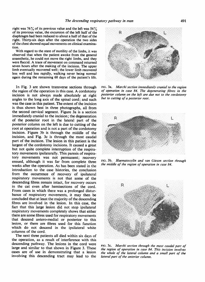
In Fig. 3 are shown transverse sections throughthe region of the operation in this case. A cordotomyincision is not always made absolutely at rightangles to the long axis of the spinal cord; and suchwas the case in this patient. The extent of the incisionis thus shown best in three photographs, all fromthe second cervical segment. Figure 3a is a sectionimmediately cranial to the incision; the degenerationof the posterior root in the lateral part of theposterior column on the left is due to cutting of theroot at operation and is not a part of the cordotomyincision. Figure 3b is through the middle of theincision, and Fig. 3c is through the most caudalpart of the incision. The lesion in this patient is thelargest of the cordotomy incisions. It caused a greatbut not quite complete interruption of the respira-tory movements ipsilaterally. This paresis of respira-tory movements was not permanent; recoveryensued, although it was far from complete threeweeks after the operation. As has been stated in theintroduction to the case histories, the conclusionfrom the occurrence of recovery of ipsilateralrespiratory movements is not that some of thedescending fibres remain intact, for recovery occursin the cat even after hemisections of the cord.From cases in which there was a prolonged distur-bance of respiratory movements, it may then beconcluded that at least the majority of the descendingfibres are involved in the lesion. In this case, thefact that this large lesion did not stop ipsilateralrespiratory movements completely shows that eitherthere are some fibres used for respiratory movementsthat descend antero-medial or posterior to thislesion, or there are fibres used for this functionwhich do not descend in the ipsilateral whitecolumns of the cord.The next three patients all died within six days of
the operation, as a result of interference with thisdescending pathway. The lesions in the cord werelarge and similar to that shown in Figure 3. Thesecases are of use in demonstrating that a lesioninvolving this descending tract may lead to the
FIG. 3a. Marchi section immediately cranial to the regionof operation in case 84. The degenerating fibres in theposterior column on the left are due not to the cordotomybut to cutting of a posterior root.
R_"L
..
FIG. 3b. Haematoxylin and van Gieson section throughthe middle of the region of operation in case 84.
Rs .i
,' ; oss' 'Ii4 -- t e
L
I.W-r f 1
_i
,..- - .+
FIG. 3c. Marchi section through the most caudal part ofthe region of operation in case 84. This incision involvesthe whole of the lateral column and a small part of thelateral part of the anterior column.
R
491
I
P. W. Nathan
patient's death, particularly when it is combinedwith carcinoma of the opposite lung.
CASE 64 Before the operation of high cervical cordotomyfor the pain due to carcinoma of the left lung, the move-ments of the two sides of the chest were equal; however,screening of the chest showed that the left half of thediaphragm was paralysed; this was attributed to thecarcinomatous tissue of the upper lobe of the left lunginvolving the left phrenic nerve. The operation wascarried out on the right side of the cord between thesecond and third cervical segments. This caused loss ofpain and thermal sensibility up to the distribution of themandibular division of the fifth nerve. Immediately afterthe incision respiratory movements of the right side ofthe chest were very greatly diminished. The impressionwas gained that what little movement was still present onthe right was a passive movement transmitted from theleft side of the chest. On deep inspiration in the supineposition no movement was seen of the upper abdominalwall on the right. After recovering from the generalanaesthetic the patient could move the right limbs onlyslightly; active movements returned in the lower limb by15 hours after the operation. Twenty-four hours after theoperation active movements of the right side of the chestwere still not seen. By 30 hours after the operation itappeared that diaphragmatic movements of the rightside had returned, as judged by observation of theabdominal respiratory movements; but no activity wasseen in the intercostal muscles on the right. The patienthad become dyspnoeic and there was increased secretionof mucus in the bronchi and trachea. Signs of broncho-pneumonia then developed, and 36 hours later the patientdied.The lesion in the cord was a large one, similar to that
shown in Figure 3.
CASE 65 Before the operation of high cervical cordotomyfor the pain due to carcinoma of the left lung, movementsof the two sides of the chest were normal and screeningshowed that the two halves of the diaphragm movednormally. The operation was carried out on the right sideof the cord at the second cervical segment. It caused aloss of pain and thermal sensibility, the upper level beinghigh in the neck. Immediately after the incision into thecord, the respiratory movements of the right side of thechest ceased. Six hours after the incision into the cord,the right side of the chest did not move and the leftmoved inadequately. The patient could move all fourlimbs to command, the right slightly less than the left.Although he was placed in a respirator, he died 30 hoursafter the operation.The lesion in the cord was a large one, similar to that
shown in Figure 3.
CASE 42 Before the operation of high cervical cordotomyfor the pain due to carcinoma of the left lung, the move-ments of the left side of the chest were diminished com-pared to those of the right. There was complete consoli-dation and collapse of the left lower lobe, the neoplasmobstructing the left lower bronchus. On screening, theleft half of the diaphragm was shown to be raised and
immobile. The operation was carried out on the rightside of the cord between the first and second cervicalsegments. This caused a loss of pain and thermal sensi-bility below the sixth cervical dermatome. Immediatelyafter the incision and for the following hour the patientwas markedly cyanosed; there were no movements ofthe right side of the chest found on clinical examinationand no activity of the intercostal muscles. Within threehours of the incision into the cord, slight respiratorymovements had re-started ipsilaterally. These movementsremained greatly diminished, until the patient died duringhis sleep five days after the operation.
This incision in the cervical cord caused no hemiparesis.The lesion in the cord was a large one, similar to thatshown in Figure 3. Anteriorly it involved the anteriorroots and posteriorly the anterior part of the lateralcortico-spinal tract.
CASE 44 Before the operation of high cervical cordotomyfor the pain due to carcinoma of the left lung, there wasdiminution of the respiratory movements of the left sideof the chest and considerable consolidation of the leftlung. After the patient had been resting in bed for twohours, the respiratory rate was 18 per minute. Theoperation was carried out on the right side of the cord,the knife being inserted at the point of emergence of thefirst cervical root and carried anteriorly to the midline.It caused a loss of pain and thermal sensibility up to thedistribution of the mandibular division of the fifth nerve.Immediately after this incision, respiratory movementsof the right side were much diminished. On the followingday the patient was cyanosed; both sides of the chestwere moving, the right side far less than the left. Nineteendays after the operation it was seen radiographically thatmovements of the two halves of the diaphragm wereequal; they were less in amplitude than they had beenbefore the operation.
This incision in the cervical cord caused no paresis ordisturbance of motility of the limbs.
The lesion is shown in Fig. 4. It is one of themost anterior of the series. This lesion when com-pared to those of the cases discussed above is seento be a small incision, and it does not involve thelateral cortico-spinal tract, nor the posterior part ofthe lateral column. It caused a great reduction ofrespiratory movements ipsilaterally. Recovery en-sued, and the diaphragmatic movements werenormal in 20 days. From the great diminution in theipsilateral respiratory movements, it may be con-cluded that the majority of the descending fibresconcerned with respiratory movements were involvedin this lesion.
CASE 39 Before the operation of high cervical cordotomyfor the pain due to carcinoma of the right lung, respira-tory movement were equal on the two sides and bothhalves of the diaphragm showed normal excursions.The operation was carried out on the left side of the
cord in the more caudal part of the third cervical segment.
492

The descending respiratory pathway in man
R2r r . s
\ .->. .-a
'-e f i'iN0. .^
3t ..e .e e s.;-
.. e
....
::
. ....
fi
FIG. 4. Marchi section through the region of operation incase 44. This incision involves t,he whole of the anteriorcolumn and the most anterior part of the lateral column.
It caused a loss of pain and thermal sensibility up to thethird cervical dermatome. The record of chest movementsmade during operation showed that following the incisionthere were no respiratory movements of the left side.Twenty-four hours after the operation no respiratorymovements were seen or felt on the left, and none wereinduced by re-breathing, anoxia produced by carbondioxide being absorbed, nor by the administration of 5%carbon dioxide and 95% oxygen. By 48 hours afteroperation some respiratory movements had returned tothe left side. Forty-eight hours later good and equalrespiratory movements were recorded in the lower halvesof both sides of the chest, although the movements ofthe left side were not quite as extensive as on the right.Fourteen days after the operation, screening of thediaphragm showed respiratory movements of both halvesof the diaphragm, although the excursion of the right wasfar less than that of the left. When coughing and sniffingto command the patient could control expiratory andinspiratory movements normally and bilaterally. Oneweek later, screening again showed less excursion of theleft diaphragm, and it could be seen that movements ofthe left diaphragm were slightly delayed compared withthose of the right; this was so, both for regular respira-tory movements and for the movements induced bysudden sniffing. This examination was repeated two dayslater, the same results being obtained.
This operative incision in the cervical cord caused noactual paresis of the ipsilateral limbs, though the leftlower limb felt subjectively heavier to move, and objec-tively it could be passively flopped more easily than theright when tone was examined.
The lesion in the cord was an anterior one,though less anterior than that shown in Fig. 4; itinvolved the most anterior part of the lateralcortico-spinal tract. It caused as much disturbanceof ipsilateral respiratory activity as the large lesionsof cases 84, 64, and 65, all of which involved a greatpart of this tract.
movements and excursions of the diaphragm were seento be equal and normal and respiratory movements wereequal on the two sides of the chest. The operation wascarried out on the left side of the cord, the knife beinginserted just anterior to the denticulate ligament at thelevel of the third cervical roots. It caused a loss of painand thermal sensibility below the fifth cervical dermatome.Immediately after the incision, all respiratory movementsof the left side of the chest had ceased and no breathsounds could be heard on the left, and none could beinduced by the administration of 5% carbon dioxide,by oxygen lack, or on command to breathe deeply.Twelve hours after the operation there was no changebut by 24 hours after the operation respiratory move-ments had returned on the left, although they wereslighter in extent than those on the right. On the fourthday after the operation screening of the diaphragmshowed equal and normal movements on both sides, andthe movements of the chest wall had also returned tonormal. Three weeks after the operation screening of thechest showed normal and equal movements of thediaphragm and of the chest walls. This incision into thecord caused no paresis or disturbance of movement ofthe ipsilateral limbs.
The lesion is shown in Fig. 5 and illustrates alateral cordotomy incision. It caused a completeinterruption of the respiratory movements ipsilater-ally. Recovery ensued, so that the movements werenormal in 20 days. It may be concluded that themajority of the descending fibres concerned withrespiratory movements were involved in this lesion.
CASE 20 Before the operation of high cervical cordo-tomy for the pain due to carcinoma of the right lung,movements of the right side of the chest were found tobe less than those of the left; this was most marked in theupper part; there was consolidation of the right upperlobe. The operation was carried out on the left side ofthe cord at the third cervical segment. It caused a lossof pain and thermal sensibility below the first thoracicdermatome. Immediately after the incision there wereno movements of the left half of the chest. Within five..Rk Li. . .. . . .
-.iT
,. < .f bP-
.,
*' i_-
i.-F ^_
E s; z
,, /,-
*. ..r g,;: z ,,i**.; o -;;
CASE 37 Before the operation of cervical cordotomy for FIG. 5. Marchi section throlugh the region of operation inthe pain due to carcinoma of the ascending colon, the case 37. This incision is restricted to the lateral column.
493
*^ iLLSmaiy..
r
-414ml

days of the operation, respiratory movements of the lefthalf of the chest had recovered to some extent, althoughon clinical examination they were still obviously lessthan those of the right; also the respiratory movementsof the abdomen were less on the left. Three weeks afterthe operation, screening showed that the movements ofboth halves of the diaphragm were normal. This incisioncaused no paresis of the ipsilateral limbs.
The lesion in this case is shown in Figure 6. Itwas one of the smallest lesions of the series. As thislesion, involving only the most anterior part of thelateral cortico-spinal tract and not involving theanterior column, caused as much paralysis ofipsilateral respiratory movements as any of theother cases, it may be concluded that most of thedescending fibres run in the region covered by thislesion.
CORRELATION OF THE LESIONS IN
THE MAIN CASES
The large lesions, of which a typical example isshown in Fig. 3, involved the ipsilateral descend-ing fibres concerned with respiratory movements.This lesion in fact consists of two incisions madeinto the cord: the first one was made in the posteriorpart of the lateral column, and had no effect onrespiratory movements; the second one was madeanteriorly, and involved respiratory movements. Insupport of the deduction from this case that it is themore anterior part of the region involved in Fig. 3that is particularly important for the ipsilateralmovements of respiration, the lesions shown inFigs. 4, 5, and 6 must be considered. The cases withthe anterior and with the lateral lesions exhibitedthe same complete or all but complete paralysis ofipsilateral respiratory movements followed byrecovery, as was shown by the cases with the large
lesions. It may be concluded that the fibres con-cerned are involved in the smaller lesions; they aresituated in the anterior parts of the lateral columnof the cord. Further, the lesion shown in Fig. 6 alsoinvolved all or most of the fibres. The regioncommon to the lesions shown in Figs. 4 and 5 isshown in Fig. 7; this region was implicated in thelesion in all the cases with respiratory paralysis onthe side of the lesion. It is concluded that it iswithin this area that most of the descending fibresconcerned with respiration are located. This regionof the cord is the most anterior part of the lateralcolumn, that is, that region of the white matter lyingimmediately lateral to the emergence of the anteriorroots.As the lesions in some of the cases were large
enough to cut through the anterior horn, it mightbe objected that such incisions damaged the cellsof the anterior horn as well as involving thesedescending fibres. But such a lesion would in allcases have been cranial to the location of the cells
FIG. 6. Marchi section through the region of operationin case 20. This incision is the smallest of the series; it is FIG. 7. Diagram of the two areas of cases 37 and 44restricted to the lateral column. superimposed and of the area common to these two cases.
P. W. Nathan494

The descending respiratory pathway in man
innervating the diaphragm, and in most cases itwould have been lateral to these cells, which aresituated along the medial border of the anteriorhorn. The disturbance of respiration was notmarkedly different in the various cases, whether thelesion was away from or near to the fourth cervicalsegment and whether the lesion involved the greymatter of the anterior horn or not.
In all operations at least some of the fibres of theipsilateral posterior root of the segment in whichthe incision was made were purposely divided orwere damaged when the cord was rotated to enablethe incision to be made. It has been shown by Nathanand Sears (1960) that cutting the fourth cervicalposterior root causes a paralysis of the diaphragmpersisting for weeks. It could be thought that theposterior rhizotomy performed in these cases mighthave had the same effect. This is unlikely, for in nocase were the fourth cervical roots damaged; andfurther, the effects of the operation on respirationwere no different whether the operation was per-formed at the first or the third cervical segments;and the operations also affected the intercostalmuscles whereas a posterior rhizotomy at the thirdcervical segment could not have affected the motilityof these muscles.
Similar incisions made at more caudal levels thanthe fourth cervical segment probably divide thefibres descending to the motor neurones of theintercostal muscles. But such cases have not beenstudied sufficiently to be reported upon.None of the four patients with the smaller lesions
had a hemiparesis; and none of these patients hadthe lateral cortico-spinal tract involved. It is thusapparent that a lesion causing paralysis or paresisof the ipsilateral respiratory movements does notnecessarily cause hemiplegia or hemiparesis.
Unfortunately, although we have waited, theopportunity has never occurred of investigating apatient in whom an operation for the division ofthe lateral cortico-spinal tract in the cord wasindicated. Oliver (1953) has carried out this operationin patients with Parkinsonism but by now thisoperation has been superseded by operations on thebrain. Oliver was aware that division of the lateralcortico-spinal tract might cause disturbances ofrespiration. He found that breathing was notaffected by the operation in four patients in whombilateral pyramidotomy was performed in the cervicalcord.
CONFIRMATORY CASES
If these descending fibres in the cervical cord are inthe location that has been worked out, then the lossof pain sensibility associated with a lesion dividingthese fibres will be throughout the cervical and upper
thoracic segments. On the other hand, if there is alesion posterior to the most anterior part of thelateral column where the descending fibres havebeen shown to run, then the loss of pain and thermalsensibility would be throughout the more caudalsegments of the body, and might not include theupper thoracic and cervical segments. Further, alesion made in the anterior part of the lateral columnmight well cause no disturbance of motility in theipsilateral limbs, whereas a lesion made moreposteriorly might involve the lateral cortico-spinaltract and might cause at least a temporary paresisof the limbs on the side of the incision.
Five of our cases of cordotomy, in which no post-mortem confirmation of the lesion was obtained,can now be considered, to see how far this correlationof loss of sensibility and disturbance of respirationholds.
CASE 60 Before the operation of high cervical cordotomyfor the pain due to a carcinoma of the breast invadingthe brachial plexus, the movements and excursions ofthe diaphragm were seen to be equal and normal andrespiratory movements were equal on the two sides of thechest. The operation was performed on the right side ofthe cord at the second cervical segment. It caused a lossof pain and thermal sensibility up to the second cervicaldermatome; it did not affect any form of sensibility belowthe level of the umbilicus. It caused paralysis of theipsilateral diaphragm and the ipsilateral movements ofthe chest. It caused no paresis of the limbs; indeed,movements of the previously painful upper limbs wereimproved, doubtless due to the loss of pain.
CASE 99 Before the operation of high cervical cordotomyfor the pain due to a carcinoma of the breast invading thebrachial plexus, the movements and excursions of thediaphragm were seen to be equal and normal and respira-tory movements were equal on the two sides of the chest.The operation was performed on the right side of thecord between the roots of the second and third cervicalsegments. It caused a loss of pain and thermal sensibilitythroughout the left side of the body below the thirdcervical segment. It caused no weakness of the limbs.The operation caused paralysis of the respiratory move-ments of the right side of the chest. There was a greatreduction in the tidal air; after the incision the averagerespiratory rate was 24 per min., whereas before it hadbeen 18. Forty-eight hours after the operation there werestill no respiratory movements seen or felt on the rightside of the chest, though the right side of the upperabdomen moved with respiration, albeit less than the left.By the sixth day after the operation respiratory movementswere equal on the two sides of the chest and of theabdomen. One month after the operation screeningshowed equal movements of the two halves of the dia-phragm.
CASE 100. Before the operation of high cervical cordo-tomy for the pain due to a carcinoma of the breast in-
495

P. W. Nathan
volving the brachial plexus, the left half of the diaphragmwas found to be paralysed. The operation was performedon the right side of the cord at the second cervical seg-ment. It caused a loss of pain and thermal sensibilitythroughout the left side of the body below the fourthcervical dermatome and a diminution in the second andthird cervical dermatomes. It caused no change in themotility of the upper limb, but weakness in the lowerlimb, which recovered in a few weeks. The operationcaused almost complete paralysis of the intercostalmuscles on the side of the incision. The patient wascyanosed. Twenty-four hours after the operation it waspossible to screen the patient; diaphragmatic movementswere diminished on the side of the incision, the maximalexcursion being 2 5 cm. Slight movements of the righthemithorax were then seen; on auscultation air entrywas found to be greatly diminished. The patient did notfeel short of breath. By the sixth day after the cordotomysome cyanosis was still present but the movements ofthe right side of the chest had recovered to such anextent that they exceeded those of the left side.
CASE 102 Before the operation of high cervical cordo-tomy for the pain due to carcinoma of the bronchus, itwas found that the movements of the right side of thediaphragm were much diminished, being less than 2 5 cm.in extent when the patient was standing. The operationwas performed on the left side of the cord at the firstcervical dermatome. It caused a loss of pain and thermalsensibility throughout the right side of the body belowthe mandibular division of the fifth cranial nerve.Immediately after the operation the left limbs were weakbut by seven days almost full power had returned to theupper limb and some power to the lower limb; sixmonths after the operation no abnormality in the leftlimbs was found. The operation caused paralysis ofipsilateral respiratory movements; it could be shown thatthe little movement that was present was transmittedfrom the other side. Twenty-four hours after the operationthere was some return of movements of the left half ofthe chest, not transmitted from the right; but it wasapparent that at each breath the movement of the rightside of the chest preceded that of the left. Forty-eighthours after the operation the patient complained offeeling short of breath, particularly after swallowing;he then had to make a conscious effort to breathe. Therewas still far less movement of the left chest than of theright and less movement on the left below the costalmargin. Forty-eight hours later the two sides of thechest and the abdomen moved equally with respiration.Three weeks after the operation screening showed thatthe diaphragmatic movements on the left were normal inextent, and exceeded those of the right.
CASE 101 Before the operation of high cervical cordo-tomy for the pain due to carcinoma of the right bronchusthe movements of the two sides of the chest were equal.The operation was performed on the left side of the cordbetween the roots of the second and third cervical seg-ments. It caused a loss of pain and thermal sensibilitythroughout the right side of the body below the thirdcervical segment and a very slight weakness of the left
upper limb. By a week after the operation the strengthof the upper limb had recovered. The operation causedparalysis of the respiratory movements of the left half ofthe chest. Screening 24 hours after the operation showedno movements of the left half of the diaphragm. Re-covery of the movements of the chest started within 48hours of the operation.
EVIDENCE FROM THE CONFIRMATORY CASES
From our knowledge of the location of the fibresfrom the various segments of the body in the spino-thalamic tract, and of the relation between damageto the lateral cortico-spinal tract and disturbancesof motility of the limbs, it can be surmised wherethe lesions were in these five cases. In case 50, thelesion must have been restricted to the anteriorcolumn and to the most anterior part of the lateralcolumn; in case 99, the lesion must have been similarin location, and would have included the entirelateral column situated anterior to the denticulateligament; in cases 100, 102, and 101 the lesion musthave had the same anterior extent, and it must haveextended further posteriorly, undoubtedly involvingsome of the lateral cortico-spinal tract. All theselesions involved the descending respiratory fibres;small anterior lesions involved them just as much asthe large lesions, which involved the whole of theanterior column and also most of the lateral column.From a purely anatomical point of view, it would
have been important to have had a lesion involvingonly the posterior part of the lateral column. But,as has been mentioned above, such lesions are nolonger made.
DISCUSSION
It has been shown that a lesion involving the mostanterior part of the lateral column of the cervicalcord temporarily stops or greatly reduces ipsilateralrespiratory movements. It is concluded that themajority of the descending fibres subserving themovements of respiration lie here. These descendingfibres remain on the same side of the cord betweenthe first cervical segment and their distribution tothe ipsilateral motor neurones. Although a knifecut through this region causes a great diminution ora stopping of respiratory movements ipsilaterally,it may not be said that the integrity of these descend-ing fibres is essential for respiratory movements ofthe diaphragm and intercostal muscles for recoveryfrom the effects of this lesion soon ensues.Although the evidence needs further confirmation,
it seems clear that the fibres subserving respiratorymovements do not descend in the cortico-spinaltract. This may be the reason why patients in theadvanced stages of amyotrophic lateral sclerosis
496

The descending respiratory pathway in man
continue to live after they have lost all movements ofthe limbs.
Sir Charles Bell's surmise is seen to be correct:the fibres in man run in the same region as they werefound to run in the dog, cat, guinea-pig, and rabbit,as shown by the work of Pitts (1940), Allen (1927),and Gardner and Haddad (1953). The resultsobtained in man after cordotomy are strikingly likethose obtained by Pitts in the cat. He found thatthere was invariably some recovery during the 10-day survival period of the experiments; and thiswas so when the cuts into the cord were so large thatin fact they constituted hemisections.
It is true that it is common to find some recoveryof function after most lesions in the central nervoussystem; but the amount of the recovery and itsearly onset are striking features of the recovery ofrespiratory movements following this lesion in thelateral column, as has been mentioned above. Toaccount for it, the following possibilities may beconsidered. Pathways might be made available thatare not normally used for respiratory movements.Or it might be that routes which had always beenavailable but which were previously used only underconditions of great need might become usedroutinely. Or it might be that the trauma of cuttinga large proportion of the descending fibres somehowhas the effect of temporarily stopping conduction inother fibres that normally conduct in associationwith the cut fibres; eventually the remaining un-injured fibres might start to conduct impulses again.If fibres are used for the function of respirationthat normally are not used for respiratory move-ments, this constitutes an example of vicariousfunctioning. If the sudden withdrawal of a usualsource of excitation stopped conduction in pathwayshabitually used for this function, this would be anexample of diaschisis, which is a transient state ofdiminished or abolished functioning of a part, notprimarily involved by a lesion, being anatomicallyremote from it, but functionally related to thedamaged part.
If alternative pathways are available it seemslikely that they must be located contralaterally, forlesions as large as that shown in Fig. 3 can leave fewfibres intact in the anterior column. If this is so,then each side of the cord has descending fibrespotentially capable of innervating the respiratorymuscles of both sides of the body. Tosatti foundfrom his partial sections of the cord in the rabbit thatthe recovery of function depends on the existence ofboth crossed and uncrossed fibres in the anteriorcolumn. It is also possible that pathways in the greymatter, consisting of chains of neurones, might beavailable as alternative routes. Tosatti (1939a) wasbrought to the conclusion that there are two tracts
available from the respiratory centre to the motorneurones of the phrenic nerve on each side of thecord: 'una via "principale" decorrente nel cordonelaterale del midollo cervicale ed une via "di reserva"decorrente nel cordone anteriore ... la quale entrain funzione solamente quando siano state bloccatale vie principali decorrenti nei cordoni laterali'. Hebelieves that the anterior tract contains both crossedand uncrossed fibres and that the lateral tractcontains only crossed fibres. In a further study(1939b) he showed that the reserve tract is destinedmainly for the motor neurones innervating theintercostal muscles and the motor neurones supply-ing those parts of the diaphragm innervated by theintercostal nerves.The impression was gained from the cases studied
here that the amount of respiratory activity duringrecovery on the side ipsilateral to the surgical lesionis to some extent related to the general level ofexcitation of central nervous activity and in parti-cular to consciousness and the conscious awarenessof the need to breathe. This statement is made inspite of the fact that induced oxygen lack or excesscarbon dioxide in the inspired air did not induceipsilateral respiration in the early stages after theoperation. It was noticed that the respiratory activityreturned intermittently, so that on one occasionintercostal muscle activity might be recorded, andon another occasion, particularly if the patient wastired or drowsy, none could be found. Two of thepatients noted that they were breathing more, ormore deeply, when they concentrated on theirbreathing; this voluntary attention to respirationhelped an activity which is usually an automaticfunction. In other words, it seemed that the recoveryof respiratory activity was to a certain extentrelated to the amount of excitation reaching therespiratory centre; and consciousness and effortcontributed to the total level of excitability.Some neurosurgeons perform bilateral high cer-
vical cordotomies. In view of the importance of thedescending respiratory fibres in this part of the cord,it is surprising that they are able to do this. Some-times death does follow: for instance one surgeonreported that in two of his patients in whom theoperation was performed in one stage, respiratoryfailure occurred during the second incision, and inboth death ensued 24 hours later. In one the cordo-tomy was performed at the Cl and C2 segments, andin the other at the C2 and C3 segments. It seemsthat many surgeons do not usually make bothincisions cranial to the fourth cervical segment, andso diaphragmatic breathing carries on unilaterallyduring the time respiratory movements are suspendedon the other side. It is probable that at whateverlevel of the cervical cord the operation is performed,
497

both incisions do not involve that region in which ithas been shown that these descending fibres run.The reason for putting forward this suggestion isthat in most cases a bilateral region of total analgesiais not obtained up to the segment of the incisions.As the fibres of the spino-thalamic tract issuing fromthe grey matter into the antero-lateral column lienear the region where this descending tract lies, anincision involving this tract is likely to cause im-mediate, complete analgesia up to the segmentallevel of the incision into the cord; and conversely,where there is not such analgesia up to the seg-mental level of the incision in the cord, this tractmay well not be involved. Apart from such profferedsuggestions why the fibres of the descending tractmay not be involved bilaterally in high cervicalcordotomies, it is possible that sometimes they areinvolved; on these occasions, the mechanisms givingrise to recovery of function, whatever they are,might be induced to come into play unusually early.
It has previously been reported by Nathan andSears (1960) that cutting the posterior roots throughwhich the phrenic nerve establishes afferent con-nexion with the spinal cord causes a temporaryparalysis of the diaphragm. The recovery from theparalysis of the diaphragm occurs more quicklyafter cutting the descending fibres in the cord than itdoes after cutting the afferent nerves in the posteriorroot-which is a somewhat surprising finding.The next question to be considered is whether the
localization of the descending fibres as assessedaccording to function corresponds with the locationof any recognized descending tract or system. Themost important work on the descending tractsin this region comes from the Leiden school ofneuroanatomists; most relevant are the theses byvan Beusekom (1955), Busch (1961), and Staal (1961)in the cat and by Sie (1956) in man. Van Beusekomfound that reticulospinal fibres are not united intocompact tracts; there are no bundles consistingchiefly of reticulo-spinal fibres; the fibres lie every-where in the anterior and antero-lateral columns.There was, however, 'a certain degree of concentra-tion of reticulo-spinal fibres . .. between the mediallongitudinal fascicle and the vestibulo-spinal tract,and within the medial longitudinal fascicle itself'.The area in which the descending fibres have herebeen shown to run in man is that where the vestibulo-spinal tract runs in the cat, as shown by vanBeusekom and Staal; and this region containsmany reticulo-spinal fibres. Busch and Staal dividedthe reticulo-spinal system of the cat into four groups:uncrossed medial reticulo-spinal fibres, originatingin the caudal pons and rostral medulla, and descend-ing in the anterior column; uncrossed lateral bulbo-spinal fibres (Thomas' fascicle) scattered in the
anterior column largely outside the sulcomarginalzone; crossed ponto-spinal fibres descending in theposterior part of the lateral column near the rubro-spinal tract; and crossed bulbo-spinal fibres descend-ing contralaterally in the anterior column. Assumingthat the arrangement in man is similar to that in thecat, then the region of the cord common to thelesions made here might include the uncrossedmedial reticulo-spinal, the uncrossed lateral bulbo-spinal, and the crossed bulbo-spinal fibres. Sie foundin man that the reticulo-spinal fibres do not formwell-defined tracts; however. 'in the high cervicalcord the majority of the ventral reticular fibresystems with a part of the dorsal reticular fibresystem form the lateral part of the ventral funiculus'.He found that the reticulo-spinal fibres are com-pletely intermingled with the fibres of the lateralvestibulo-spinal tract and of the medial longitudinalfascicle.
It will be noted that in the cat the reticulo-spinalfibres, except for those of the crossed ponto-spinalgroup, terminate along the medial border of theanterior horn. The motor neurones innervating thediaphragm are situated in the medial part of theanterior horn, as are those innervating the inter-costal muscles, for this is the location of the motorneurones innervating all trunk muscles. It must bestated, however, that most workers have found thatthe reticulo-spinal tracts continue throughout thelength of the spinal cord; and so if it is with reticulo-spinal fibres with which we are here concerned, onlysome of the fibres of this important system must beheld to subserve the movements of the respiratorymusculature. One other tract should be mentioned,the olivo-spinal tract. Although it is known that itsfibres originate in the olive, it is possible that somemay originate in the reticular substance near theolive, and these might include the neurones of therespiratory centre. This tract descends in the regionwhere the evidence shows that the main descendingpathway for the respiratory musculature runs; andthe bulk of evidence concerning this tract shows thatit does not descend further caudally than the fourthor fifth cervical segments.
It is of interest to consider the crossed phrenicphenomenon in relation to these cases. This pheno-menon, as far as I can ascertain, has not yet beennoted in man. In the cat a hemisection of the cordperformed in the cervical region causes paralysis ofthe ipsilateral diaphragm. The contralateral phrenicnerve is then cut, causing paralysis of the contra-lateral diaphragm. This has the immediate effect ofmaking the previously paralysed, ipsilateral dia-phragm start working again.
This is not the place to discuss all aspects of thismost interesting phenomenon. But as some of our
P. W. Nathan498
The descending respiratory pathway in man
patients had lesions, naturally occurring andsurgically induced, which in effect approximated tothe situation of the crossed phrenic phenomenon, itis of interest to consider the phenomenon as itaffects man. It was found in the cat and the rabbit byRosenblueth, Klopp, and Simeone (1938) that whena phrenic nerve was first cut and then the contra-lateral hemisection of the cord was carried outseveral weeks later, the hemisection did not thencause paralysis of the diaphragm on the side of thehemisection. A similar situation to this occurred infour of the patients reported here (cases 64, 42, 100,and 102). In these four patients, the diaphragm onone side was paralysed before the cordotomy owingto involvement of the phrenic nerve in the neoplasm.The contralateral cordotomy did cause an immediatestoppage of respiratory movements in cases 64 and42 and a great diminution of them in case 100.In cases 64 and 42 some recovery from the paralysisof respiratory movements ensued; in these two itwas insufficient to maintain life. In cases 100 and 102recovery occurred, and a week after operation incase 100 and three weeks after operation in case 102respiratory movements were better on the side of thecordotomy than on the side of the neoplasm.
It had already been found by Rosenblueth andOrtiz (1936) that species differ in whether theyexhibit this phenomenon or not: dogs, cats, rabbits,and woodchucks show it; guinea-pigs and monkeysdo not. It may now be stated that in the form of thephenomenon in which the phrenic nerve is firstdivided, man does not show the typical crossedphrenic phenomenon, for division of the descendingfibres in the cord for innervation of the diaphragmdoes cause paralysis of the ipsilateral diaphragm.The crossed phrenic phenomenon, the recovery
of respiratory activity in all the patients reportedhere who lived long enough to show it, and the factthat the ipsilateral respiratory activity seemed to beinfluenced by the patient's efforts to breathe and thegeneral level of neurological activity, these phenom-ena make one realize that neural routes are madeavailable according to need. Pathways are openedup or used in relation to the total activity and needsof the animal; there is not one and only one routeavailable; when the main or the usual pathway isdivided, the impulses can reach the final commonpath via subsidiary, unusual, or new paths, which
appear to be made available so that the necessaryfunctional activity can be maintained.
SUMMARY
Cases of high cervical cordotomy were investigatedto determine the effect of this operation on respira-tion. It was found that a lesion of the most anteriorpart of the lateral column of the first three cervicalsegments of the cord causes paralysis or a very greatreduction in the ipsilateral respiratory movements.Recovery from the effects of this lesion ensues;within a few weeks of the lesion being made, respira-tory movements become equal and normal on thetwo sides.
It is concluded that many or all of the fibres ofthe descending respiratory tract lie in this region;and that these fibres may form a part of the reticulo-spinal tract or they may run with the olivo-spinaltract.I would like to thank Mr. Wylie McKissock and Drs.A. J. H. Hewer, T. A. Sears, and Marion C. Smith forthe generous help they gave me, and Dr. E. A. Carmichaelfor the encouragement and the opportunities he gave mein carrying out this work.
REFERENCES
Allen, W. F. (1927). J. comp. Neurol., 42, 393.Bell, C. (1830). The Nervous System of the Human Body. Longman,
Rees, Orme, Brown, and Green, London.Beusekom, G. T. van (1955). Fibre Analysis ofthe Anterior and Lateral
Funiculi of the Cord in the Cat. Ijdo, Leiden.Busch, H. F. M. (1961). An Anatomical Analysis of the White Matter
in the Brain Stem of the Cat. Van Gorcum, Assen.Hukuhara, T., Nakayama, S., and Okada, H. (1954). Jap. J. Physiol.,
4, 145.Gardner, E., and Haddad, B. (1953). Amer. J. Physiol., 172, 475.Nathan, P. W., and Sears, T. A. (1960). J. Neurol. Neurosurg. Psychiat.,
23, 10., and Smith M. C. (1951). Ibid., 14, 262.
(1953). Ibid., 16, 245.- (1958). Ibid., 21, 177.
Oliver, L. C. (1953). Parkinson's Disease and its Surgical Treatment.Lewis, London.
Penfield, W. (1943). Ass. Res. nerv. Dis. Proc., 23, 431.Pitts, R. F. (1940). J. comp. Neurol., 72, 605.Porter, W. T. (1895). J. Physiol. (Lond.), 17, 455.Rosenblueth, A., and Ortiz, T. (1936). Amer. J. Physiol., 117, 495.
, Klopp, C. T, and Simeone, F. A. (1938).J. Neurophysiol., 1, 508.Sie Pek Giok (1956). Localization of Fibre Systems within the White
Matter of the Medulla Oblongata and the Cervical Cord in Man.Ijdo, Leiden.
Staal, A. (1961). Subcortical Projections on the Spinal Grey Matter oftheCat. Leiden.
Stookey, B. (1943). Ass. Res. nerv. Dis. Proc., 23, 416.Taylor, A. (1960). J. Physiol. (Lond.), 151, 390.Tosatti, E. (1939a). Arch. Fisiol., 38, 533.
(1939b). Boll. Soc. ital. Biol. sper., 14, 615 and 677.
499