The CXC Chemokine Receptor 2, CXCR2, Is the Putative ... · The CXC Chemokine Receptor 2, CXCR2, Is...
10
of July 1, 2018. This information is current as Chemokine-Induced Angiogenic Activity CXC + the Putative Receptor for ELR The CXC Chemokine Receptor 2, CXCR2, Is Alfred Walz, Ann Richmond and Robert M. Strieter Hua Liu, Jan E. Ehlert, Ying Ying Xue, Linda Buechi, Christina L. Addison, Thomas O. Daniel, Marie D. Burdick, http://www.jimmunol.org/content/165/9/5269 doi: 10.4049/jimmunol.165.9.5269 2000; 165:5269-5277; ; J Immunol References http://www.jimmunol.org/content/165/9/5269.full#ref-list-1 , 28 of which you can access for free at: cites 50 articles This article average * 4 weeks from acceptance to publication Fast Publication! • Every submission reviewed by practicing scientists No Triage! • from submission to initial decision Rapid Reviews! 30 days* • Submit online. ? The JI Why Subscription http://jimmunol.org/subscription is online at: The Journal of Immunology Information about subscribing to Permissions http://www.aai.org/About/Publications/JI/copyright.html Submit copyright permission requests at: Email Alerts http://jimmunol.org/alerts Receive free email-alerts when new articles cite this article. Sign up at: Print ISSN: 0022-1767 Online ISSN: 1550-6606. Immunologists All rights reserved. Copyright © 2000 by The American Association of 1451 Rockville Pike, Suite 650, Rockville, MD 20852 The American Association of Immunologists, Inc., is published twice each month by The Journal of Immunology by guest on July 1, 2018 http://www.jimmunol.org/ Downloaded from by guest on July 1, 2018 http://www.jimmunol.org/ Downloaded from
-
Upload
nguyenhanh -
Category
Documents
-
view
222 -
download
0
Transcript of The CXC Chemokine Receptor 2, CXCR2, Is the Putative ... · The CXC Chemokine Receptor 2, CXCR2, Is...
of July 1, 2018.This information is current as
Chemokine-Induced Angiogenic Activity CXC+the Putative Receptor for ELR
The CXC Chemokine Receptor 2, CXCR2, Is
Alfred Walz, Ann Richmond and Robert M. StrieterHua Liu, Jan E. Ehlert, Ying Ying Xue, Linda Buechi, Christina L. Addison, Thomas O. Daniel, Marie D. Burdick,
http://www.jimmunol.org/content/165/9/5269doi: 10.4049/jimmunol.165.9.5269
2000; 165:5269-5277; ;J Immunol
Referenceshttp://www.jimmunol.org/content/165/9/5269.full#ref-list-1
, 28 of which you can access for free at: cites 50 articlesThis article
average*
4 weeks from acceptance to publicationFast Publication! •
Every submission reviewed by practicing scientistsNo Triage! •
from submission to initial decisionRapid Reviews! 30 days* •
Submit online. ?The JIWhy
Subscriptionhttp://jimmunol.org/subscription
is online at: The Journal of ImmunologyInformation about subscribing to
Permissionshttp://www.aai.org/About/Publications/JI/copyright.htmlSubmit copyright permission requests at:
Email Alertshttp://jimmunol.org/alertsReceive free email-alerts when new articles cite this article. Sign up at:
Print ISSN: 0022-1767 Online ISSN: 1550-6606. Immunologists All rights reserved.Copyright © 2000 by The American Association of1451 Rockville Pike, Suite 650, Rockville, MD 20852The American Association of Immunologists, Inc.,
is published twice each month byThe Journal of Immunology
by guest on July 1, 2018http://w
ww
.jimm
unol.org/D
ownloaded from
by guest on July 1, 2018
http://ww
w.jim
munol.org/
Dow
nloaded from
The CXC Chemokine Receptor 2, CXCR2, Is the PutativeReceptor for ELR1 CXC Chemokine-Induced AngiogenicActivity 1
Christina L. Addison,* Thomas O. Daniel,†‡ Marie D. Burdick, § Hua Liu, †‡ Jan E. Ehlert,§
Ying Ying Xue,§ Linda Buechi,¶ Alfred Walz, ¶ Ann Richmond,‡i and Robert M. Strieter2§
We have previously shown that members of the ELR1 CXC chemokine family, including IL-8; growth-related oncogenesa, b, andg; granulocyte chemotactic protein 2; and epithelial neutrophil-activating protein-78, can mediate angiogenesis in the absence ofpreceding inflammation. To date, the receptor on endothelial cells responsible for chemotaxis and neovascularization mediated bythese ELR1 CXC chemokines has not been determined. Because all ELR1 CXC chemokines bind to CXC chemokine receptor 2(CXCR2), we hypothesized that CXCR2 is the putative receptor for ELR1 CXC chemokine-mediated angiogenesis. To test thispostulate, we first determined whether cultured human microvascular endothelial cells expressed CXCR2. CXCR2 was detectedin human microvascular endothelial cells at the protein level by both Western blot analysis and immunohistochemistry usingpolyclonal Abs specific for human CXCR2. To determine whether CXCR2 played a functional role in angiogenesis, we determinedwhether this receptor was involved in endothelial cell chemotaxis. We found that microvascular endothelial cell chemotaxis inresponse to ELR1 CXC chemokines was inhibited by anti-CXCR2 Abs. In addition, endothelial cell chemotaxis in response toELR1 CXC chemokines was sensitive to pertussis toxin, suggesting a role for G protein-linked receptor mechanisms in thisbiological response. The importance of CXCR2 in mediating ELR1 CXC chemokine-induced angiogenesis in vivo was alsodemonstrated by the lack of angiogenic activity induced by ELR1 CXC chemokines in the presence of neutralizing Abs to CXCR2in the rat corneal micropocket assay, or in the corneas of CXCR22/2 mice. We thus conclude that CXCR2 is the receptorresponsible for ELR1 CXC chemokine-mediated angiogenesis.The Journal of Immunology,2000, 165: 5269–5277.
T he CXC family of chemokines is a chemotactic group ofcytokines, defined by the presence of four conserved cys-teine amino acid residues in the amino terminus of the
protein, in which the first two conserved cysteine amino acid res-idues are separated by one nonconserved amino acid residue(hence the CXC designation). This family of molecules can befurther subdivided based on the presence or absence of the aminoacid sequence Glu-Leu-Arg (ELR) preceding the first conservedcysteine amino acid residue in the primary structure of these pro-teins. The ELR motif has been shown to play a role in ligand/receptor interactions on neutrophils (1, 2). We have previouslyshown that the ELR motif is also the structural/functional domain
important in the regulation of CXC chemokine-induced angiogen-esis (3). It has been demonstrated that CXC chemokines that con-tain the ELR motif (ELR1), such as IL-8; growth-related onco-genes (GRO)3 a, b, and g; granulocyte chemotactic protein-2(GCP-2); and epithelial neutrophil-activating protein 78 (ENA-78), are potent inducers of angiogenesis in vivo (3). In contrast,CXC chemokines that lack the ELR motif (ELR2), such as plateletfactor 4, IFN-inducible protein-10, and monokine induced byIFN-g, are potent angiostatic factors, and can inhibit neovascular-ization mediated by ELR1 CXC chemokines (3, 4). Furthermore,the ELR2 CXC chemokines have also been shown to inhibit neo-vascularization induced by the classical angiogenic factors, basicfibroblast growth factor (bFGF) and vascular endothelial cellgrowth factor (VEGF) (3). The receptor responsible for the induc-tion of angiogenesis by ELR1 CXC chemokines has not yet beenelucidated.
The CXC chemokines have been shown to interact with theCXC chemokine receptor (CXCR) family of molecules. These re-ceptors are members of the rhodopsin-like seven-transmembraneG protein-coupled receptor family (5–8). The chemokine receptorsrange in size from 339 to 373 aa, and possess 25–80% homologyto one another on the amino acid level (6, 7, 9, 10). To date, fiveCXC receptors have been identified in various human cell lines.CXCR1, which is also known as IL-8RA, binds IL-8 with high
*Department of Internal Medicine, University of Michigan Medical School, AnnArbor, MI 48109;†Center for Vascular Biology and‡Departments of Medicine andCell Biology, Vanderbilt University Medical Center, Nashville, TN 37232;§Divisionof Pulmonary and Critical Care Medicine, Department of Medicine, University ofCalifornia, Los Angeles School of Medicine, Los Angeles, CA 90095;¶TheodorKocher Institute, Universitat Bern, Bern, Switzerland; andiDepartment of Veteran’sAffairs, Vanderbilt University Medical Center, Nashville, TN 37232
Received for publication December 28, 1999. Accepted for publication July 31, 2000.
The costs of publication of this article were defrayed in part by the payment of pagecharges. This article must therefore be hereby markedadvertisementin accordancewith 18 U.S.C. Section 1734 solely to indicate this fact.1 This work has been supported, in part, by grants from the National Institutes ofHealth (HL66027 to R.M.S. and DK38517 to T.O.D.). This work has also been sup-ported by funds from the National Cancer Institute (CA87879 to R.M.S., CA68485 toT.O.D., and CA34590 to A.R.). T.O.D. is supported by funds from the T. J. MartellFoundation, and A.R. holds a Career Scientist and Merit award from the Departmentof Veteran’s Affairs. C.L.A. is a research fellow of the National Cancer Institute ofCanada supported with funds provided by the Terry Fox Run. J.E.E. is recipient offellowship EH 188/1-1 from the Deutsche Forschungsgemeinschaft.2 Address correspondence and reprint requests to Dr. Robert M. Strieter, Division ofPulmonary and Critical Care Medicine, University of California, Los Angeles Schoolof Medicine, 900 Veteran Avenue, 14-154 Warren Hall, Box 711922, Los Angeles,CA 90095. E-mail address: [email protected]
3 Abbreviations used in this paper: GRO, growth-related oncogene; bFGF, basic fi-broblast growth factor; CXCR, CXC chemokine receptor; DARC, Duffy Ag receptorfor chemokines; ENA, epithelial neutrophil-activating protein; GCP, granulocyte che-motactic protein; HMVEC, human microvascular endothelial cell; HMVEC-D, hu-man dermal microvascular endothelial cell; HMVEC-L, human lung microvascularendothelial cell; HPF, high power field; KLH, keyhole limpet hemocyanin; MIP,macrophage-inflammatory protein; NAP, neutrophil-activating protein; PTx, pertussistoxin; TFA, trifluoroacetic acid; VEGF, vascular endothelial cell growth factor.
Copyright © 2000 by The American Association of Immunologists 0022-1767/00/$02.00
by guest on July 1, 2018http://w
ww
.jimm
unol.org/D
ownloaded from
affinity (8), and more recently has been shown to also bind GCP-2with a somewhat reduced affinity (11). CXCR2, also known asIL-8RB, possesses 78% identity with CXCR1 on the amino acidlevel with the majority of divergence between the two proteinsbeing located in the amino terminus, the carboxyl terminus, andwithin the second extracellular loop (6). This divergence in theextracellular regions (the amino terminus and the second extracel-lular loop) may partly explain why CXCR2 shows less selectivityin chemokine binding than does CXCR1. CXCR2 has been shownto bind all of the ELR1 CXC chemokines, including IL-8; ENA-78; GRO-a, -b, and -g; neutrophil-activating protein-2 (NAP-2);and GCP-2 with high affinity (6, 8, 9, 12). CXCR3 has been shownto bind the ELR2 CXC chemokines IFN-inducible protein-10 andmonokine induced by IFN-g (5). CXCR4 has recently been thefocus of a number of studies, as it has been shown to be a cofactorfor HIV infection of T lymphocytes by T cell tropic viruses (13).To date, stromal cell-derived factor-1 is the only known ligand forCXCR4, and this ELR2 CXC chemokine can inhibit HIV infectionby competing with lymphotropic HIV virus for binding of CXCR4(14). CXCR5 is found mostly on B cells and is responsible for Bcell chemotaxis mediated by B cell-attracting chemokine 1 (15).There is a sixth receptor that has been shown to bind CXC che-mokines, DARC or the Duffy Ag receptor for chemokines (16, 17).IL-8, GRO-a, and NAP-2 have all been shown to bind to DARC;however, ligand binding of this receptor is not restricted to theCXC chemokines, and various CC chemokines, such as RANTESand monocyte chemotactic protein-1, also bind to DARC with highaffinity (18). Moreover, DARC does not appear to demonstrateligand-receptor signal coupling (19). Another receptor whose li-gand binding is restricted to the CXC chemokines is encoded by anopen reading frame from Herpesvirus saimiri (20). The ECRF3open reading frame has been shown to encode a seven-transmem-brane receptor, and this receptor has been shown to bind CXCchemokines, in particular the ELR1 CXC chemokines IL-8,GRO-a, and NAP-2 (20). This receptor is most homologous toCXCR2; however, this homology is only 30% at the amino acidlevel, even though the CXC chemokine-binding repertoire is es-sentially identical (20).
In this study, we investigated the identity of the chemokine re-ceptor responsible for mediating angiogenesis induced by theELR1 CXC chemokines. As mentioned, the ELR1 CXC chemo-kine IL-8 can bind both CXCR1 and CXCR2 with high affinity,while the ELR1 CXC chemokines GRO-a, -b, -g, and ENA-78have been shown to bind only CXCR2 with high affinity (8, 21).Because all of these ELR1 CXC chemokines only bind CXCR2 incommon, and all induce angiogenesis in vivo, we hypothesizedthat CXCR2 is the putative receptor present on endothelial cellsthat mediates angiogenesis induced by ELR1 CXC chemokines invivo. In this study, we demonstrate that CXCR2 is expressed bycultured human microvascular endothelial cells (HMVEC), andthat not only can neutralizing Abs to CXCR2 inhibit endothelialcell chemotaxis in vitro, but also inhibit neovascularization in-duced by the ELR1 CXC chemokines in the rat CMP assay invivo. Furthermore, corneal neovascularization induced by ELR1
CXC chemokines in CXCR22/2 mice is impaired as comparedwith that induced in wild-type mice.
Materials and MethodsCell lines and isolation
Human neutrophils were isolated from heparinized venous blood collectedfrom healthy volunteers, by mixing 1:1 with 0.9% saline and separationfrom mononuclear cells by Ficoll-Hypaque density-gradient centrifugation.Human neutrophils were then isolated by sedimentation in 5% dextran in0.9% saline (Sigma, St. Louis, MO) and separated from erythrocytes by
hypotonic lysis. After washing twice, neutrophils were suspended in HBSSwith calcium/magnesium (Life Technologies, Grand Island, NY) at a con-centration of 23 106 cells/ml. Neutrophils were.99% viable, as deter-mined by trypan blue exclusion. Human dermal microvascular endothelialcells (HMVEC-D) and human lung microvascular endothelial cells(HMVEC-L) were both obtained from Cell Systems (Kirkland, WA), andwere propagated in CS-C complete media on attachment factor-coatedtissue culture flasks according to the manufacturer’s directions. All endo-thelial cell lines were stained for factor VIII-related Ag to confirm theiridentity as being of endothelial origin. Both the HMVEC-L- and HMVEC-D-cultured cells were positive for the expression of factor VIII-related Ag,while human neutrophils were negative following immunohistochemicalstaining.
To generate 293 cells that expressed human CXCR2, the cDNA forhuman CXCR2 was amplified by RT-PCR from total RNA isolated fromhuman neutrophils using the Access PCR kit (Promega, Madison, WI). Theprimers used for amplification were: forward, 59-GTC AGG ATC CAAGTT TAC CTC AAA AAT GG-39, and reverse, 59-CTT AGG TCG ACGGTC TTA GAG AGT AGT GG-39. The reverse-transcription reaction wasperformed at 42°C for 45 min, followed by denaturation at 94°C for 2 min.PCR was then performed for 40 cycles as follows: 94°C denaturation for 1min, 55°C annealing for 1 min, and 68°C elongation for 2 min. The re-sulting PCR was subjected to electrophoresis on an agarose gel, and a bandof ;1.1 kb was removed and purified using the Wizard PCR DNA puri-fication kit (Promega). The PCR product was ligated into the T-overhangplasmid pTARGET according to the manufacturer’s directions kit (Pro-mega). This resulted in human CXCR2 expression under control of thehuman CMV promoter and allowed for generation of stably transfectedmammalian cells by the presence of a neomycin resistance gene. The293 cells were transfected with 5mg of CXCR2 plasmid DNA or controlpTARGET DNA by calcium phosphate transfection, as previously de-scribed (22). G418-resistant colonies were expanded, and expression ofCXCR2 was confirmed by FACS analysis using a mAb against humanCXCR2 (R&D Systems, Minneapolis MN).
Abs and Ab generation
A rabbit polyclonal Ab to the carboxyl terminus of human CXCR2 waspurchased from Santa Cruz Biotechnologies (Santa Cruz CA). Anti-rabbitIgG Abs conjugated to HRP were purchased from Bio-Rad (Hercules CA).For Ab generation, peptides specific to the amino-terminal region of humanor murine CXCR2 were generated by solid-phase peptide synthesis per-formed on a Millipore 9050 continuous flow peptide synthesizer (Milli-pore, Milford, MA) using Fmoc chemistry. Briefly, 1 g of polyethyleneglycol polystyrene-graft copolymer peptide synthesis support (PEG-PSresin) and 0.8 mM of each Fmoc-amino acid active ester were used. Cleav-age and deprotection were conducted in 88% trifluoroacetic acid (TFA),5% liquefied phenol, 2% triisopropylsilane, and 5% water for 2–12 h atroom temperature. The free peptide was then precipitated and washed re-peatedly with ice-cold ether and dried under vacuum. The peptide wasresuspended in water, acidified with TFA, and purified by preparative re-versed-phase HPLC in a C4 column (253 100 mm, 15 mm, 300A,D-Pak;Waters, Millipore, Bedford, MA). The column was eluted at 5 ml/min witha gradient of 0–60% acetonitrile in 0.1% TFA at an increment of 1.3% permm. Fractions were lyophilized and analyzed by analytical reversed-phaseHPLC and mass spectroscopy.
A 17-mer peptide constituting the amino terminus of the mouse CXCR2(MGEFKVDKFNIEDFFSG) and a 21-mer peptide constituting the aminoterminus of human CXCR2 (CMEDFNMESDSFEDFWKGEDL) weresynthesized as described above. The peptides contained an additional ala-nine residue at the carboxyl terminus, and a cysteine residue at the aminoterminus for conjugation with keyhole limpet hemocyanin (KLH). Conju-gation with Imject maleimide-activated KLH was conducted according tothe manufacturer (Pierce, Rockford, IL). Polyclonal antiserum against ei-ther murine CXCR2 or human CXCR2 was generated following s.c. or i.m.injections of 100mg of the KLH-conjugated peptide in CFA, followed byat least three boosters of 100mg of KLH-conjugated peptide in IFA. DirectELISA was used to evaluate antisera titers, and sera were drawn when titershad reached greater than 1:1,000,000.
The specificity of the Ab to human CXCR2 was confirmed followingreceptor neutralization studies on stably transfected cells. This Ab specif-ically recognized human CXCR2 and prevented binding of IL-8 to 293cells transfected to overexpress human CXCR2. This Ab did not cross-react with CXCR1, nor did it prevent binding of IL-8 to human CXCR1,when used to detect receptor expression in 293 cells overexpressing humanCXCR1. To determine the specificity of the anti-mouse CXCR2 Abs, wetested the ability of the Ab to inhibit neutrophil recruitment in vivo. CBA/Jmice or Fischer rats were injected i.p. with 80 ng of recombinant KC (K
5270 CXCR2 IS RESPONSIBLE FOR ANGIOGENESIS MEDIATED BY ELR1 CXC CHEMOKINES
by guest on July 1, 2018http://w
ww
.jimm
unol.org/D
ownloaded from
and C coordinates on the autorad from intial cloning (23)) in combinationwith either 0.5 ml of the anti-murine CXCR2 antisera or 0.5 ml of normalrabbit serum. Four hours later, animals were sacrificed and peritoneal la-vage was performed using 3 ml of PBS with 5 mM EDTA. The concen-tration of cells within the lavage fluid was counted using a hemacytometer;equal numbers of cells were subjected to cytocentrifugation; and the slideswere fixed and stained using the Diff-Quik kit (Baxter Diagnostics, McGawPark, IL), followed by cell differential determination. It was found that thepresence of anti-murine CXCR2 Abs specifically inhibited neutrophil mi-gration in response to KC, while having no effect on monocyte infiltrationin both rodent systems.
RT-PCR for CXCR2 gene expression
Cells were isolated as described above. Total RNA was extracted fromcells using Trizol reagent (Life Technologies, Grand Island, NY), accord-ing to manufacturer’s instructions. RT-PCR was performed using anAccess RT-PCR kit (Promega, Madison, WI).b-actin was used as a house-keeping gene. Forb-actin, the sense primer used was 59-GTGGGGCGCCCCAGGCACCA; the antisense primer was 59-GCTCGGCCGTGGTGGTGAAGC (550 bp). For human CXCR2, the sense primer used was 59-CCGGGCGTGGTGGTGAG; the antisense primer was 59-TCTGCCTTTTGGGTCTTGTGAATA (385-bp product). PCR products were visualizedby agarose gel electrophoresis. To exclude genomic DNA contamination,PCR was performed in the presence or absence of a preceding step thatincluded reverse-transcriptase reaction with the isolated RNA.
Western blot analysis
Total protein extracts were made by scraping endothelial or 293 cell mono-layers into TNE lysis buffer (20 mM Tris-HCl, pH 8, 150 mM NaCl, 1%Nonidet P-40, 2.5 mM EDTA) supplemented with 2 ng/ml aprotinin and 35ng/ml PMSF or by resuspending isolated neutrophils into supplementedTNE lysis buffer. Cell extracts were incubated on ice for 30 min, followedby centrifugation at 4°C for 30 min. Supernatants were then removed andassayed for total protein content using bicinchoninic acid protein assayreagents (Pierce, Rockford, IL) and comparison with known amounts ofBSA. A total of 40mg of total protein was loaded in each well of a 12%polyacrylamide gel, and extracts were subjected to SDS-PAGE. The sep-arated proteins were transferred to polyvinylidene fluoride membrane(Pierce) by electrophoretic transfer overnight in Tris-glycine buffer (20mM Tris, 150 mM glycine, pH 8, methanol added to a final concentrationof 20% (v/v)). Blots were blocked in 5% skim milk in TBST buffer (10 mMTris-HCl, pH 8, 150 mM NaCl, 0.05% Tween 20) for 2 h at room tem-perature, followed by incubation in rabbit primary Ab sera against humanCXCR2 diluted 1/1000 in blocking solution for 2 h at room temperature.Blots were washed for three 10-min washes in TBST and were incubatedfor 1 h at room temperature in goat anti-rabbit HRP-conjugated secondaryAb (Bio-Rad, Hercules, CA) at a 1/20,000 dilution. Blots were againwashed for four 10-min washes in TBST, and proteins were visualizedfollowing incubation of the blots in SuperSignal chemiluminescent sub-strate solution according to the manufacturer’s protocol (Pierce) and ex-posure to XAR-5 film (Kodak, Rochester, NY).
Immunohistochemistry
Immunolocalization of CXCR2 was performed as previously described(24). Briefly, cytospins of human peripheral blood neutrophils, or Tissue-Teks (Fisher, Pittsburgh, PA) of 90% confluent unstimulated endothelialcell monolayers were fixed in 4% paraformaldehyde in PBS for 10 min,then rinsed twice with PBS. Before staining, the slides were again fixed for30 min in the presence or absence of 1:1 absolute methanol and 3% H2O2,and rinsed in PBS, and then nonspecific binding sites were blocked with a1/50 dilution of normal goat serum by incubation at room temperature for30 min. Following the blocking step, a 1/500 dilution of either control(rabbit) or rabbit anti-human CXCR2 serum was added as a primary Ab,and slides were incubated for 30 min at room temperature. Slides were thenrinsed with PBS, overlaid with biotinylated goat anti-rabbit IgG (VectorABC Elite Kit; Vector Laboratories, Burlingame, CA), and incubated foran additional 30 min. Slides were again rinsed two times with PBS, andwere then treated with streptavidin-conjugated peroxidase (Vector ABCElite Kit; Vector Laboratories) for 30 min at room temperature. Followingthree washes with PBS, the slides were subjected to colorimetric detectionusing the substrate chromogen 3,39-diaminobenzidine (Vector Laborato-ries). Slides were incubated for 5–10 min in 3,39-diaminobenzidine solu-tion at room temperature to allow color development, and rinsed with dis-tilled water to quench the reaction. Mayer’s hematoxylin was used as acounterstain.
Endothelial cell chemotaxis
Endothelial cell chemotaxis assays were performed essentially as previ-ously described (3). HMVEC-L cells were harvested by trypsinization,resuspended in CS-C medium without growth factors with 2% FBS added(Cell Systems), and preincubated with either anti-human CXCR2 anti-serum or normal rabbit serum at a final dilution of 1/250 at 37°C for 1 h.Alternatively, endothelial cells were preincubated in 2.5 mM pertussistoxin (PTx; Sigma) at 37°C for 1 h. Following preincubation, an aliquot of160 ml containing 53 105 cells/ml was added to each of the lower wellsof a 12-well chemotaxis chamber (Neuro Probe, Cabin John, MD). Thechambers were assembled (by placing 0.1% gelatin-coated 5-mm pore-sizefilters over the lower wells, followed by a gasket and the upper chamber)and incubated at 37°C in a CO2 incubator for 2 h in an inverted position.The chambers were then turned upright, and 100-ml aliquots of variouschemotactic agents in solution were added to the upper wells of the cham-ber in the following concentrations: 80 ng/ml IL-8, ENA-78, or GRO-a; 40ng/ml bFGF or VEGF. The chambers were placed in a 37°C CO2 incubatorfor 2 h, at which time the filters were removed, the bottom of the filterswere scraped to remove cells that did not undergo chemotaxis, and then thefilters were subjected to Diff-Quik (Baxter) staining. The number of endo-thelial cells that had migrated through the filters into the upper chamberswas calculated by counting the total number of cells in 15 separate fieldsof view under340 power. Results were expressed as the number of en-dothelial cells that migrated per high power field (HPF) after subtractingthe background (unstimulated control) to demonstrate specific migration.Experiments were repeated independently at least three times.
Rat corneal micropocket assay of angiogenesis
To address the ability of neutralizing CXCR2 Abs to inhibit angiogenesisin vivo, the corneal micropocket assay was performed in the rat eye, aspreviously described (3). Human IL-8, human ENA-78, murine KC, ormurine macrophage-inflammatory protein (MIP)-2 was diluted in PBS plus0.25% human serum albumin to a final concentration of 80 ng per pellet.VEGF or bFGF was diluted as above to a final concentration of 50 ng perpellet. Rabbit anti-murine CXCR2 antiserum or normal rabbit serum wasadded at a 1/1000 dilution to each of the above chemokines and cytokines,and mixed with an equal volume of Hydron casting solution (Hydro MedSciences, New Brunswick, NJ). Five-microliter aliquots were pipetted ontothe flat surface of a sterile polypropylene specimen container and wereallowed to polymerize overnight under UV light in a laminar flow hood.Before implantation, the pellets were rehydrated with normal saline. Ani-mals were given 150 mg/kg ketamine and 250mg/kg atropine as an anes-thetic, and the rat corneas were anesthetized with 0.5% proparacaine hy-drochloride ophthalmic solution, followed by implantation of the Hydronpellet into an intracorneal pocket (1–2 mm from the limbus). Six days afterimplantation, animals received heparin (1000 U) and ketamine (150 mg/kg)i.p. 30 min before sacrifice, followed by perfusion with 10 ml of colloidalcarbon via the left ventricle. Corneas were then harvested and photo-graphed. No inflammatory response was observed in any of the corneastreated with the above cytokines. Sustained directional ingrowth of capil-lary sprouts and hairpin loops toward the implant were considered positiveneovascularization responses. Negative responses were characterized byeither no vessel growth or by the presence of only an occasional hairpinloop or sprout that displayed no evidence of sustained growth.
Mouse corneal angiogenesis assay
Hydron pellets incorporating sucralfate with either vehicle alone, bFGF (3pmol/pellet, gift from Scios, Sunnyvale, CA), murine rMIP-2 (20 pmol/pellet, gift from Elias Lolis at Yale University School of Medicine, NewHaven, CT), or human rIL-8 (20 pmol/pellet, purchased from R&D Sys-tems, Minneapolis, MN) were made as described (25). Pellets were surgi-cally implanted into corneal stromal micropockets created 1 mm medial tothe lateral corneal limbus of C57BL/6 CXCR22/2 and CXCR21/1 mice(9–10 wk old, derived from the strain developed at Genentech (South SanFrancisco, CA) by Cacalano et al. (26), a gift from Dr. Robert Terkeltaub,and maintained by Ann Richmond). Five days postimplantation, corneaswere photographed at an incipient angle of 35–50o from the polar axis inthe meridian containing the pellet, using a Zeiss split lamp. Images weredigitalized and processed by subtractive color filters (Adobe Photoshop4.0; Adobe Systems, Mountain View, CA): the fraction of the total cornealimage that was vascularized (vascular area), and the ratio of pixels markingneovascular capillaries, both within the vascularized region (regional vas-cular density) and within the total corneal image (total vascular density),were calculated using the Bioquant software (Nashville, TN).
5271The Journal of Immunology
by guest on July 1, 2018http://w
ww
.jimm
unol.org/D
ownloaded from
Statistical analysis
Data were analyzed on a PC computer using the Statview 5.0 statisticalpackage (Abacus Concepts, Berkeley, CA). Comparisons were made usingthe unpairedt test or the Mann-Whitney test for nonparametric data. Datawere considered statistically significant ifp values were 0.05 or less.
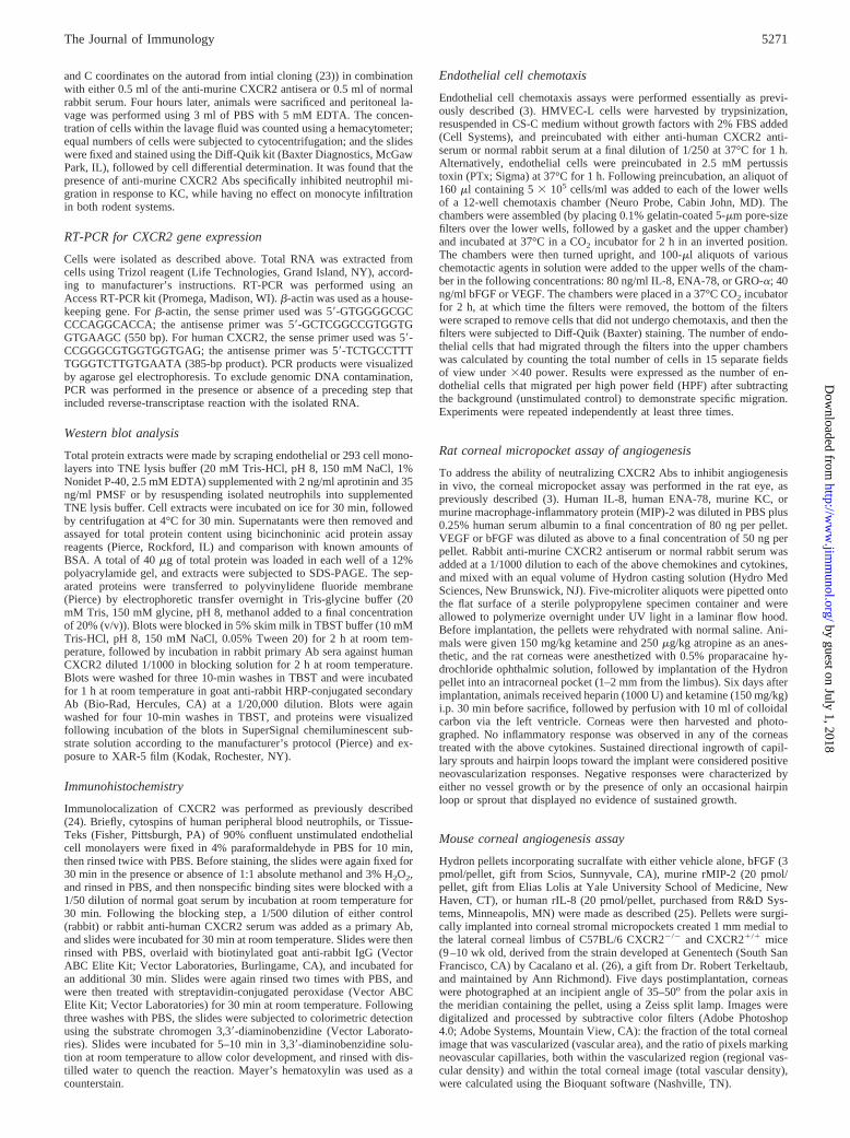
ResultsCXCR2 is expressed by microvascular endothelial cells
We sought to determine the mechanism by which the ELR1 CXCchemokines mediate the angiogenic activity of endothelial cells, byexamining the presence and the role of the chemokine receptorCXCR2, in angiogenesis. Because all ELR1 CXC chemokines in-duce angiogenic activity, and CXCR2 on neutrophils appears tobind all of these chemokines, we first evaluated whether CXCR2mRNA was expressed in HMVEC, as compared with neutrophils.Total RNA was extracted from HMVEC-L and HMVEC-D, aswell as neutrophils. RT-PCR analysis demonstrated the presenceof an appropriate band for CXCR2 mRNA by gel electrophoresisin both HMVEC lines, as well as in neutrophils (Fig. 1,IA). b-ac-tin served as an internal control (Fig. 1,IB). No bands were visu-alized using the same primers for CXCR2 under conditions inwhich reverse transcriptase was excluded from the reaction beforePCR (Fig. 1,IC). This finding supported the notion that the RT-PCR product/bands seen in Fig. 1,IA, was not due to genomicDNA contamination of the specimens before PCR. To further con-firm that this mRNA was transcribed into protein, we used an Abspecific for human CXCR2 in Western blot analysis to determinewhether this receptor was expressed in microvascular endothelialcells at the protein level. The molecular mass of CXCR2 has beenreported to be a 44- to 46-kDa band in Western blot analysis byother investigators (27, 28). Total protein extracts of human neu-trophils, HMVEC-D, HMVEC-L, control-transfected 293 cells,and CXCR2-transfected 293 cells were made in TNE lysis buffer.An aliquot containing 50mg of total protein was subjected to SDS-PAGE and transferred to polyvinylidene fluoride membrane forWestern blot analysis using a rabbit polyclonal Ab specific to hu-man CXCR2. A protein band of molecular mass of;50 kDa wasspecifically recognized by the human CXCR2 Ab in lysates fromhuman neutrophils and CXCR2-transfected 293 cells (positivecontrols), while this band was absent in control-transfected 293cells (Fig. 1,II). A protein band of similar molecular mass was alsorecognized by the Ab in both HMVEC-D and HMVEC-L proteinextracts (Fig. 1,II). The presence of this;50-kDa protein speciesin endothelial cell extracts was also confirmed by Western blotanalysis using a second commercially available Ab to humanCXCR2 (data not shown).
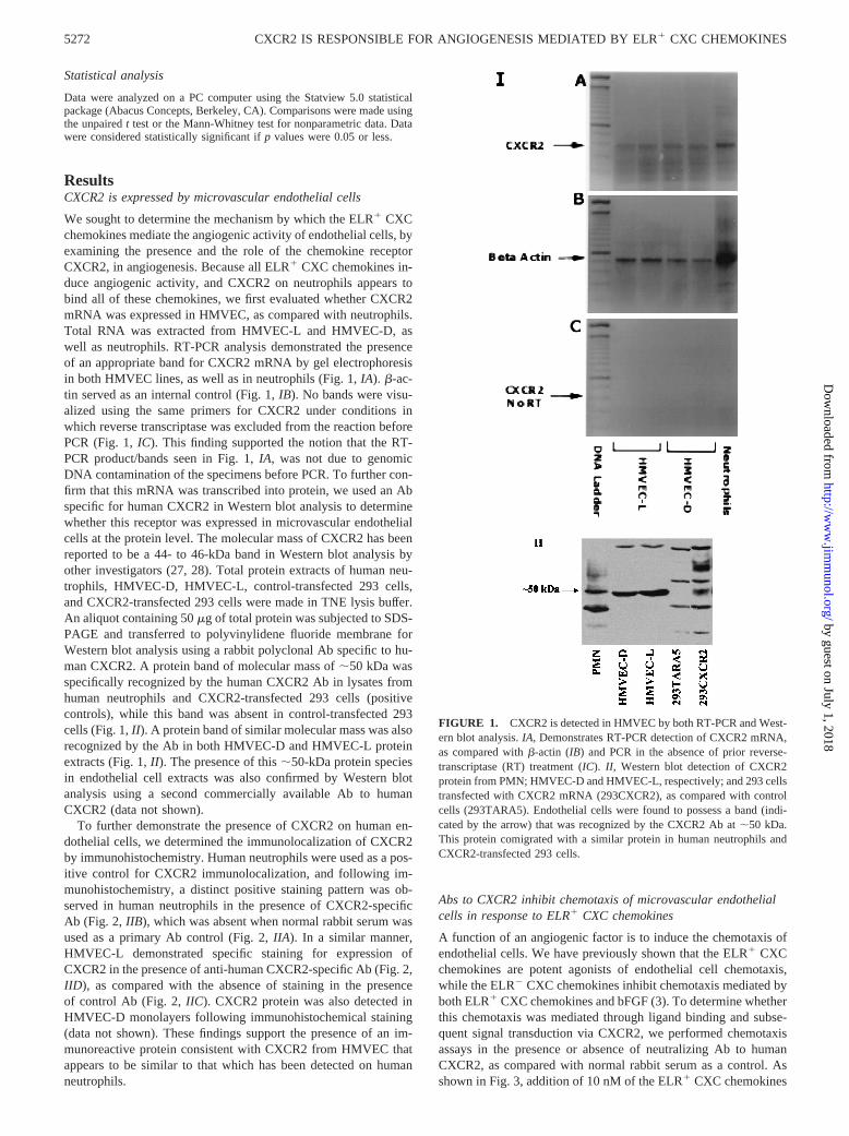
To further demonstrate the presence of CXCR2 on human en-dothelial cells, we determined the immunolocalization of CXCR2by immunohistochemistry. Human neutrophils were used as a pos-itive control for CXCR2 immunolocalization, and following im-munohistochemistry, a distinct positive staining pattern was ob-served in human neutrophils in the presence of CXCR2-specificAb (Fig. 2, IIB), which was absent when normal rabbit serum wasused as a primary Ab control (Fig. 2,IIA). In a similar manner,HMVEC-L demonstrated specific staining for expression ofCXCR2 in the presence of anti-human CXCR2-specific Ab (Fig. 2,IID), as compared with the absence of staining in the presenceof control Ab (Fig. 2,IIC). CXCR2 protein was also detected inHMVEC-D monolayers following immunohistochemical staining(data not shown). These findings support the presence of an im-munoreactive protein consistent with CXCR2 from HMVEC thatappears to be similar to that which has been detected on humanneutrophils.
Abs to CXCR2 inhibit chemotaxis of microvascular endothelialcells in response to ELR1 CXC chemokines
A function of an angiogenic factor is to induce the chemotaxis ofendothelial cells. We have previously shown that the ELR1 CXCchemokines are potent agonists of endothelial cell chemotaxis,while the ELR2 CXC chemokines inhibit chemotaxis mediated byboth ELR1 CXC chemokines and bFGF (3). To determine whetherthis chemotaxis was mediated through ligand binding and subse-quent signal transduction via CXCR2, we performed chemotaxisassays in the presence or absence of neutralizing Ab to humanCXCR2, as compared with normal rabbit serum as a control. Asshown in Fig. 3, addition of 10 nM of the ELR1 CXC chemokines
FIGURE 1. CXCR2 is detected in HMVEC by both RT-PCR and West-ern blot analysis.IA, Demonstrates RT-PCR detection of CXCR2 mRNA,as compared withb-actin (IB) and PCR in the absence of prior reverse-transcriptase (RT) treatment (IC).II, Western blot detection of CXCR2protein from PMN; HMVEC-D and HMVEC-L, respectively; and 293 cellstransfected with CXCR2 mRNA (293CXCR2), as compared with controlcells (293TARA5). Endothelial cells were found to possess a band (indi-cated by the arrow) that was recognized by the CXCR2 Ab at;50 kDa.This protein comigrated with a similar protein in human neutrophils andCXCR2-transfected 293 cells.
5272 CXCR2 IS RESPONSIBLE FOR ANGIOGENESIS MEDIATED BY ELR1 CXC CHEMOKINES
by guest on July 1, 2018http://w
ww
.jimm
unol.org/D
ownloaded from

IL-8 or ENA-78 induced specific HMVEC-L chemotaxis (118.4615.6 and 108.16 9.6 cells/HPF, respectively). bFGF (5 nM) wasalso shown to induce similar levels of endothelial cell chemotaxisin our assay system to those observed with the ELR1 CXC che-mokines (119.46 16.1 cells/HPF). To determine whether CXCR2was mediating the endothelial cell chemotactic effect of ELR1
CXC chemokines, we addressed whether specific Abs to humanCXCR2 inhibited HMVEC-L chemotaxis. HMVEC-L were prein-cubated in the presence of anti-human CXCR2 or normal controlAbs, and then used in the chemotaxis assay. As compared withcells incubated with angiogenic stimuli alone, HMVEC-L chemo-taxis in response to IL-8 or ENA-78 was attenuated by 97% or99%, respectively (p , 0.001), by the presence of neutralizing Abto human CXCR2. In contrast, preincubation of HMVEC-L withcontrol Abs yielded chemotaxis results similar to those obtained inthe presence of the ELR1 CXC chemokines alone (123.86 17.7and 101.76 14.4 cells/HPF for IL-8 and ENA-78, respectively). Asimilar inhibition of HMVEC-L chemotaxis in response to theELR1 CXC chemokine GRO-a was also observed in the presenceof anti-CXCR2 Abs (data not shown). Moreover, preincubation ofendothelial cells with Abs to human CXCR2 had no significanteffect on HMVEC-L chemotaxis in response tobFGF (Fig. 3). Asimilar inhibition of chemotaxis by anti-human CXCR2 in responseto ELR1 CXC chemokines was also found when HMVEC-D wereused (data not shown).
To exclude that CXCR1 might be contributing to the endothelialcell chemotactic response to IL-8, we investigated the role ofCXCR1 in the chemotactic response of endothelial cells to theELR1 CXC chemokines. HMVEC-L were preincubated with ei-ther anti-human CXCR1 or control Abs, and the specific migrationin response to IL-8, ENA-78, bFGF, or VEGF was determined. Weobserved no difference in endothelial cell-specific migration in re-sponse to any of the angiogenic factors tested in the presence ofAbs to human CXCR1, as compared with the specific migrationobserved in the presence of control Abs (data not shown). Theseresults support that CXCR2 is the primary CXCR responsible forendothelial cell migration induced by the ELR1 CXC chemokines.
Endothelial cell chemotaxis in response to ELR1 CXCchemokines is inhibited by PTx
To confirm that signal transduction through CXCR2, a G protein-linked receptor, was responsible for the endothelial cell chemotac-tic response to the ELR1 CXC chemokines, we tested the abilityof PTx to inhibit this specific migration. The chemotactic responseof HMVEC-L to either IL-8, ENA-78, bFGF, or VEGF in thepresence or absence of PTx is shown in Fig. 4. Endothelial cellchemotaxis in response to IL-8 or ENA-78 was attenuated by 97%and 91%, respectively (p , 0.001), in the presence of 2.5 mMPTx. In contrast, HMVEC-L chemotaxis in response to bFGF orVEGF was unaltered in the presence of PTx (p 5 0.25 and 0.28,
FIGURE 2. Immunohistochemical staining in both human neutrophilsand cultured microvascular endothelial cells. Cytospins of human periph-eral neutrophils (Aand B) or monolayers of human lung microvascularendothelial cells (CandD) were subjected to immunohistochemical stain-ing using either normal rabbit serum (AandC) or a polyclonal Ab specificto human CXCR2 (BandD) as a primary Ab. Both human neutrophils andendothelial cells were found to stain positive for the presence of CXCR2.
FIGURE 3. Endothelial cell chemotaxis induced by the ELR1 CXCchemokines is inhibited by Ab to human CXCR2. Endothelial cell chemo-taxis (cells/HPF;3400) was determined by the average number of cellsthat had migrated in response to IL-8 (80 ng/ml) or ENA-78 (80 ng/ml), orof 50 ng/ml for bFGF in the presence or absence of anti-CXCR2, as com-pared with control Abs. Each bar is a representation of the mean and SEMdetermined from the average counts of three independent experiments.p,p , 0.05.
FIGURE 4. Endothelial cell chemotaxis induced by the ELR1 CXCchemokines is PTx sensitive. Endothelial cell chemotaxis (cells/HPF;3400) was determined by the average number of cells that had migrated inresponse to IL-8 (80 ng/ml) or ENA-78 (80 ng/ml), or of 50 ng/ml forbFGF and VEGF in the presence or absence of PTx. Each bar is a repre-sentation of the mean and SEM determined from the average counts ofthree independent experiments.p, p , 0.05.
5273The Journal of Immunology
by guest on July 1, 2018http://w
ww
.jimm
unol.org/D
ownloaded from
respectively). These data indicate that HMVEC-L chemotaxis inresponse to ELR1 CXC chemokines, but not to bFGF or VEGF, ismediated by a signal transduction pathway associated with PTx-sensitive G proteins. These data further support CXCR2 as thecandidate receptor mediating endothelial cell chemotaxis to ELR1
CXC chemokines.
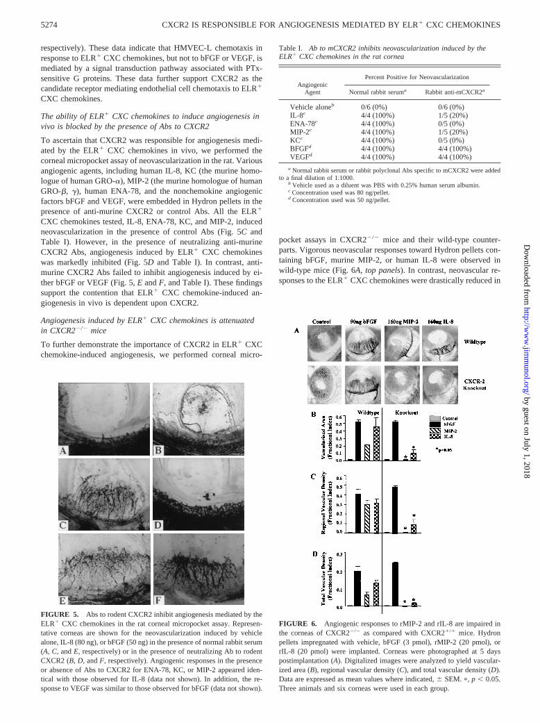
The ability of ELR1 CXC chemokines to induce angiogenesis invivo is blocked by the presence of Abs to CXCR2
To ascertain that CXCR2 was responsible for angiogenesis medi-ated by the ELR1 CXC chemokines in vivo, we performed thecorneal micropocket assay of neovascularization in the rat. Variousangiogenic agents, including human IL-8, KC (the murine homo-logue of human GRO-a), MIP-2 (the murine homologue of humanGRO-b, g), human ENA-78, and the nonchemokine angiogenicfactors bFGF and VEGF, were embedded in Hydron pellets in thepresence of anti-murine CXCR2 or control Abs. All the ELR1
CXC chemokines tested, IL-8, ENA-78, KC, and MIP-2, inducedneovascularization in the presence of control Abs (Fig. 5C andTable I). However, in the presence of neutralizing anti-murineCXCR2 Abs, angiogenesis induced by ELR1 CXC chemokineswas markedly inhibited (Fig. 5D and Table I). In contrast, anti-murine CXCR2 Abs failed to inhibit angiogenesis induced by ei-ther bFGF or VEGF (Fig. 5,E andF, and Table I). These findingssupport the contention that ELR1 CXC chemokine-induced an-giogenesis in vivo is dependent upon CXCR2.
Angiogenesis induced by ELR1 CXC chemokines is attenuatedin CXCR22/2 mice
To further demonstrate the importance of CXCR2 in ELR1 CXCchemokine-induced angiogenesis, we performed corneal micro-
pocket assays in CXCR22/2 mice and their wild-type counter-parts. Vigorous neovascular responses toward Hydron pellets con-taining bFGF, murine MIP-2, or human IL-8 were observed inwild-type mice (Fig. 6A,top panels). In contrast, neovascular re-sponses to the ELR1 CXC chemokines were drastically reduced in
FIGURE 5. Abs to rodent CXCR2 inhibit angiogenesis mediated by theELR1 CXC chemokines in the rat corneal micropocket assay. Represen-tative corneas are shown for the neovascularization induced by vehiclealone, IL-8 (80 ng), or bFGF (50 ng) in the presence of normal rabbit serum(A, C, andE, respectively) or in the presence of neutralizing Ab to rodentCXCR2 (B,D, andF, respectively). Angiogenic responses in the presenceor absence of Abs to CXCR2 for ENA-78, KC, or MIP-2 appeared iden-tical with those observed for IL-8 (data not shown). In addition, the re-sponse to VEGF was similar to those observed for bFGF (data not shown).
Table I. Ab to mCXCR2 inhibits neovascularization induced by theELR1 CXC chemokines in the rat cornea
AngiogenicAgent
Percent Positive for Neovascularization
Normal rabbit seruma Rabbit anti-mCXCR2a
Vehicle aloneb 0/6 (0%) 0/6 (0%)IL-8c 4/4 (100%) 1/5 (20%)ENA-78c 4/4 (100%) 0/5 (0%)MIP-2c 4/4 (100%) 1/5 (20%)KCc 4/4 (100%) 0/5 (0%)BFGFd 4/4 (100%) 4/4 (100%)VEGFd 4/4 (100%) 4/4 (100%)
a Normal rabbit serum or rabbit polyclonal Abs specific to mCXCR2 were addedto a final dilution of 1:1000.
b Vehicle used as a diluent was PBS with 0.25% human serum albumin.c Concentration used was 80 ng/pellet.d Concentration used was 50 ng/pellet.
FIGURE 6. Angiogenic responses to rMIP-2 and rIL-8 are impaired inthe corneas of CXCR22/2 as compared with CXCR21/1 mice. Hydronpellets impregnated with vehicle, bFGF (3 pmol), rMIP-2 (20 pmol), orrIL-8 (20 pmol) were implanted. Corneas were photographed at 5 dayspostimplantation (A). Digitalized images were analyzed to yield vascular-ized area (B), regional vascular density (C), and total vascular density (D).Data are expressed as mean values where indicated,6 SEM. p, p , 0.05.Three animals and six corneas were used in each group.
5274 CXCR2 IS RESPONSIBLE FOR ANGIOGENESIS MEDIATED BY ELR1 CXC CHEMOKINES
by guest on July 1, 2018http://w
ww
.jimm
unol.org/D
ownloaded from
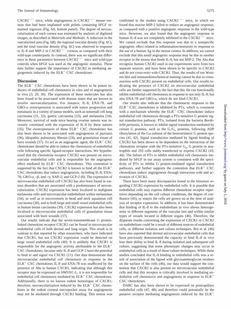
CXCR22/2 mice, while angiogenesis in CXCR22/2 mouse cor-neas that had been implanted with pellets containing bFGF re-mained vigorous (Fig. 6A,bottom panel). The degree of neovas-cularization of each cornea was estimated by analysis of digitizedimages, as described inMaterials and Methods. A reduction in thevascularized area (Fig. 6B), the regional vascular density (Fig. 6C),and the total vascular density (Fig. 6C) was observed in responseto IL-8 and MIP-2 in CXCR22/2 corneas as compared with theirwild-type counterparts. In contrast, there was no significant differ-ence in these parameters between CXCR22/2 mice and wild-typecontrols when bFGF was used as the angiogenic stimulus. Thesedata further support the importance of CXCR2 in mediating an-giogenesis induced by the ELR1 CXC chemokines.
DiscussionThe ELR1 CXC chemokines have been shown to be potent in-ducers of endothelial cell chemotaxis in vitro and of angiogenesisin vivo (3, 29, 30). The expression of these molecules has alsobeen found to be associated with disease states that are known toinvolve neovascularization. For instance, IL-8, ENA-78, andGRO-a overexpression is associated with tumor progression andmetastasis in a variety of tumor models, such as nonsmall cell lungcarcinoma (31, 32), gastric carcinoma (33), and melanoma (34).Moreover, survival of nude mice bearing ovarian tumors was in-versely correlated with the expression of IL-8 by these tumors(35). The overexpression of these ELR1 CXC chemokines hasalso been shown to be associated with angiogenesis of psoriasis(36), idiopathic pulmonary fibrosis (24), and granulation tissue ofburn wounds (37). To act as an angiogenic agent, the ELR1 CXCchemokines should be able to induce the chemotaxis of endothelialcells following specific ligand-receptor interactions. We hypothe-sized that the chemokine receptor CXCR2 was present on micro-vascular endothelial cells and is responsible for the angiogeniceffect mediated by ELR1 CXC chemokines. This contention issupported by the fact that CXCR2 is known to bind all the ELR1
CXC chemokines that induce angiogenesis, including IL-8; ENA-78; GRO-a, -b, and -g; NAP-2; and GCP-2 (8). The expression ofmicrovascular endothelial cell CXCR2 has also been found in var-ious disorders that are associated with a predominance of neovas-cularization. CXCR2 expression has been localized to malignantmelanoma cells and microvascular endothelium within melanomas(34), as well as to microvessels in head and neck squamous cellcarcinoma (38), and to both large and small vessel endothelial cellsin human breast carcinoma (39). CXCR2 expression has also beenlocalized to microvascular endothelial cells of granulation tissueassociated with burn wounds (37).
Our results indicate that the seven-transmembrane G protein-linked chemokine receptor CXCR2 can be found in microvascularendothelial cells of both dermal and lung origin. This result is incontrast to that reported by other researchers, who have indicatedthat CXCR1, but not CXCR2 expression could be detected onlarge vessel endothelial cells (40). It is unlikely that CXCR1 isresponsible for the angiogenic activity attributable to the ELR1
CXC chemokines, because only IL-8 and GCP-2 have the potentialto bind to and signal via CXCR1 (11). Our data demonstrate thatmicrovascular endothelial cell chemotaxis in response to theELR1 CXC chemokines IL-8 and ENA-78 was unaffected by thepresence of Abs to human CXCR1, indicating that although thisreceptor may be expressed on HMVEC-L, it is not responsible forendothelial cell chemotaxis mediated by ELR1 CXC chemokines.Additionally, there is no known rodent homologue of CXCR1;therefore, neovascularization induced by the ELR1 CXC chemo-kines in the rodent corneal micropocket assay for angiogenesismay not be mediated through CXCR1 binding. This notion was
confirmed in the studies using CXCR22/2 mice, in which wefound that murine MIP-2 failed to induce an angiogenic response,as compared with a positive angiogenic response in the wild-typemice. However, we also found that the angiogenic response tohuman IL-8 was not completely inhibited in the CXCR22/2 mice.We cannot exclude that this response was due to a nonspecificangiogenic effect related to inflammation/immunity in response tothe use of a human Ag in the mouse cornea. In addition, we cannotexclude that this small angiogenic response may be due to anotherreceptor in the mouse that binds IL-8, but not MIP-2. The Abs thatrecognize human CXCR2 used in our experiments were from twoseparate sources, and have been shown to be specific to CXCR2and do not cross-react with CXCR1. Thus, the results of our West-ern blot and immunohistochemical staining cannot be due to cross-reaction with CXCR1 present on endothelial cells. Our results in-dicating the presence of CXCR2 on microvascular endothelialcells are further supported by the fact that this Ab can functionallyinhibit endothelial cell chemotaxis in response to not only IL-8, butalso ENA-78 and GRO-a, which do not bind CXCR1.
Our results also indicate that the chemotactic response to theELR1 CXC chemokines is inhibited by PTx, which is consistentwith a mechanism whereby the ELR1 CXC chemokines induceendothelial cell chemotaxis through a PTx-sensitive G protein sig-nal transduction pathway. PTx, isolated from the bacteriaBorde-tella pertussis, is known to inhibit signal transduction mediated bycertain G proteins, such as the Gi/Go proteins, following ADPribosylation of the Ga subunit of the heterotrimeric G protein spe-cies (41, 42). Signal transduction mediated by ligand binding ofCXCR2 has been shown to be dependent on the interaction of thechemokine receptor with the PTx-sensitive Gi2 G protein in neu-trophils and 293 cells stably transfected to express CXCR2 (43,44). The failure of PTx to inhibit endothelial cell chemotaxis me-diated by bFGF in our assay system is consistent with the speci-ficity of PTx to inhibit G protein-mediated signal transductionpathways, and further supports the contention that ELR1 CXCchemokines induce angiogenesis through interaction with and ac-tivation of CXCR2.
There have been many discrepancies found in the literature re-garding CXCR2 expression by endothelial cells. It is possible thatendothelial cells may express different chemokine receptor reper-toires depending on the cell culture conditions, the degree of con-fluence (45), or matrix the cells are grown on at the time of anal-ysis of receptor expression. In addition, it has been demonstratedthat binding of IL-8 to the endothelium in vivo shows heteroge-neity in different segments of the vascular tree and within similartypes of vessels located in different organs (46). Therefore, thedisparate results concerning the expression of CXCR1 or CXCR2on endothelium could be a result of different sources of endothelialcells, or different isolation and culture techniques. Rot et al. (46)have also reported that dermal microvascular endothelial cells thathave previously demonstrated the capacity to bind IL-8 in vivolose their ability to bind IL-8 during isolation and subsequent cellculture, suggesting that some phenotypic changes may occur inendothelial cells as a result of these culture techniques. While thesestudies concluded that IL-8 binding to endothelial cells was a re-sult of association of the ligand with glycosaminoglycan residueson the surface of the cells (46), our data would support the con-tention that CXCR2 is also present on microvascular endothelialcells and that this receptor is critically involved in mediating en-dothelial cell chemotaxis and angiogenesis in response to ELR1
CXC chemokines.DARC has also been shown to be expressed on postcapillary
endothelial cells (47, 48), and therefore could potentially be theputative receptor mediating angiogenesis induced by the ELR1
5275The Journal of Immunology
by guest on July 1, 2018http://w
ww
.jimm
unol.org/D
ownloaded from
CXC chemokines. This is unlikely, because although this receptordoes bind all the ELR1 CXC chemokines known to induce angio-genesis, it also binds other chemokines of the CC family, which todate have no known angiogenic properties. In addition, there havebeen no indications that DARC mediates signal transductionevents following ligand binding of chemokines (19), and becauseof this observation, it has been proposed to act as a sponge thatbinds excess free chemokines during inflammatory responses.Moreover, our finding for the fundamental role of CXCR2 in me-diating endothelial cell migration in vitro and angiogenesis in vivowould suggest that DARC is unlikely to play a direct role in en-dothelial cell chemotaxis to ELR1 CXC chemokines. Similarly,although CXCR4 is readily expressed on endothelial cells (45, 49,50), CXCR4 is not known to bind the ELR1 CXC chemokines andcan thus be excluded as the receptor responsible for ELR1 CXCchemokine-induced angiogenesis.
The identification of CXCR2 as the receptor responsible for me-diating angiogenesis induced by the ELR1 CXC chemokines hasimportant implications. As mentioned, CXCR2 expression hasbeen shown to be associated with neovascularization in the contextof certain diseases such as psoriasis and tumorigenesis. Antago-nists specific for CXCR2 could be used therapeutically to inhibitELR1 CXC chemokine-dependent neovascularization, and im-prove the prognosis of patients with diseases that are associatedwith marked angiogenesis. Inhibition of neovascularization hasbeen shown to drastically inhibit the progression and metastasis ofvarious cancers; thus, inhibition of ELR1 CXC chemokine-medi-ated angiogenesis could also be advantageous as adjuvant cancertherapy. We have shown that neutralization of IL-8 or ENA-78using specific neutralizing Abs to these molecules can inhibit thegrowth of nonsmall cell lung carcinoma in SCID mice (31, 32).These treatments, however, only reduced tumor growth by 40–60%. This is most likely the result of the fact that these tumorsproduce multiple ELR1 CXC chemokine ligands contributing totheir tumorigenicity. Inhibition of ligand binding to CXCR2, orinhibition of the signal transduction events following activation ofthe CXCR2 receptor would in essence simultaneously inhibit thebiological effect of multiple ELR1 CXC chemokines. Future workfrom our laboratory will focus on the inhibition of tumor growthand metastasis by antagonizing the function of the CXCR2receptor.
AcknowledgmentsWe thank Logan Qian and Jianguo Du for their assistance in the mainte-nance and genotyping of the CXCR22/2 mice.
References1. Hebert, C. A., R. V. Vitangcol, and J. B. Baker. 1991. Scanning mutagenesis of
interleukin-8 identifies a cluster of residues required for receptor binding.J. Biol.Chem. 266:18989.
2. Clark-Lewis, I., B. Dewald, T. Geiser, B. Moser, and M. Baggiolini. 1993. Plate-let factor 4 binds to interleukin 8 receptors and activates neutrophils when its Nterminus is modified with Glu-Leu-Arg.Proc. Natl. Acad. Sci. USA 90:3574.
3. Strieter, R. M., P. J. Polverini, S. L. Kunkel, D. A. Arenberg, M. D. Burdick,J. Kasper, J. Dzuiba, J. V. Damme, A. Walz, D. Marriott, et al. 1995. The func-tional role of the ‘ELR’ motif in CXC chemokine-mediated angiogenesis.J. Biol.Chem. 270:27348.
4. Maione, T. E., G. S. Gray, J. Petro, A. J. Hunt, A. L. Donner, S. I. Bauer,H. F. Carson, and R. J. Sharpe. 1990. Inhibition of angiogenesis by recombinanthuman platelet factor-4 and related peptides.Science 247:77.
5. Loetscher, M., B. Gerber, P. Loetscher, S. A. Jones, L. Piali, I. Clark-Lewis,M. Baggiolini, and B. Moser. 1996. Chemokine receptor specific for IP-10 andMig: structure, function, and expression in activated T-lymphocytes.J. Exp. Med.184:963.
6. Murphy, P. M. 1994. The molecular biology of leukocyte chemoattractant recep-tors.Annu. Rev. Immunol. 12:593.
7. Murphy, P. M., M. Baggiolini, I. F. Charo, C. A. Hebert, R. Horuk,K. Matsushima, L. H. Miller, J. J. Oppenheim, and C. A. Power. 2000. Interna-tional union of pharmacology. XXII. Nomenclature for chemokine receptors.Pharmacol. Rev. 52:145.
8. Lee, J., R. Horuk, G. C. Rice, G. L. Bennett, T. Camerato, and W. I. Wood. 1992.Characterization of two high affinity human interleukin-8 receptors.J. Biol.Chem. 267:16283.
9. Murphy, P. M., and H. L. Tiffany. 1991. Cloning of complementary DNA en-coding a functional human interleukin-8 receptor.Science 253:1280.
10. Premack, B. A., and T. J. Schall. 1996. Chemokine receptors: gateways to in-flammation and infection.Nat. Med. 2:1174.
11. Wolf, M., M. B. Delgado, S. A. Jones, B. Dewald, I. Clark-Lewis, andM. Baggiolini. 1998. Granulocyte chemotactic protein 2 acts via both IL-8 re-ceptors, CXCR1 and CXCR2.Eur. J. Immunol. 28:164.
12. Ahuja, S. K., and P. M. Murphy. 1996. The CXC chemokines growth-regulatedoncogene (GRO)a, GROb, GROg, neutrophil-activating peptide-2, and epithe-lial cell-derived neutrophil-activating peptide-78 are potent agonists for the typeB, but not the type A, human interleukin-8 receptor.J. Biol. Chem. 271:20545.
13. Feng, Y., C. C. Broder, P. E. Kennedy, and E. A. Berger. 1996. HIV-1 entrycofactor: functional cDNA cloning of a seven-transmembrane, G protein-coupledreceptor.Science 272:872.
14. Oberlin, E., A. Amara, F. Bachelerie, C. Bessia, J.-L. Virelizier,F. Arenzana-Seisdedos, O. Schwartz, J.-M. Heard, I. Clark-Lewis, D. F. Legler,et al. 1996. The CXC chemokine SDF-1 is the ligand for LESTR/fusin and pre-vents infection by T-cell-line-adapted HIV-1.Nature 382:833.
15. Legler, D. F., M. Loetscher, R. S. Roos, I. Clark-Lewis, M. Baggiolini, andB. Moser. 1998. B cell-attracting chemokine 1, a human CXC chemokine ex-pressed in lymphoid tissues, selectively attracts B lymphocytes via BLR1/CXCR5.J. Exp. Med. 187:655.
16. Horuk, R. 1994. The interleukin-8-receptor family from chemokines to malaria.Immunol. Today 15:169.
17. Horuk, R. 1994. Molecular properties of the chemokine receptor family.TrendsPharmacol. Sci. 15:159.
18. Lu, Z. H., Z. X. Wang, R. Horuk, J. Hesselgesser, Y. C. Lou, T. J. Hadley, andS. C. Peiper. 1995. The promiscuous chemokine binding profile of the Duffyantigen/receptor for chemokines is primarily localized to sequences in the amino-terminal domain.J. Biol. Chem. 270:26239.
19. Neote, K., J. Y. Mak, L. F. Kolakowski Jr., and T. J. Schall. 1994. Functional andbiochemical analysis of the cloned Duffy antigen: identity with the red blood cellchemokine receptor.Blood 84:44.
20. Ahuja, S. K., and P. M. Murphy. 1993. Molecular piracy of mammalian inter-leukin-8 receptor type B by herpesvirus saimiri.J. Biol. Chem. 268:20691.
21. Cerretti, D. P., C. J. Kozlosky, T. Vanden Bos, N. Nelson, D. P. Gearing, andM. P. Beckmann. 1993. Molecular characterization of receptors for human in-terleukin-8, GRO/melanoma growth-stimulatory activity and neutrophil activat-ing peptide-2.Mol. Immunol. 30:359.
22. Addison, C. L., D. A. Arenberg, S. B. Morris, Y. Y. Xue, M. D. Burdick,M. S. Mulligan, M. D. Iannettoni, and R. M. Strieter. 2000. The CXC chemokine,monokine induced by interferon-g, inhibits non-small cell lung carcinoma tumorgrowth and metastasis.Hum. Gene Ther. 11:247.
23. Cochran, B. H., A. C. Reffel, and C. D. Stiles. 1983. Molecular cloning of genesequences regulated by platelet-derived growth factor.Cell 33:939.
24. Keane, M. P., D. A. Arenberg, J. P. Lynch III, R. I. Whyte, M. D. Iannettoni,M. D. Burdick, C. A. Wilke, S. B. Morris, M. C. Glass, B. DiGiovine, et al. 1997.The CXC chemokines, IL-8 and IP-10, regulate angiogenic activity in idiopathicpulmonary fibrosis.J. Immunol. 159:1437.
25. Kenyon, B. M., E. E. Voest, C. C. Chen, E. Flynn, J. Folkman, and R. J.D’Amato. 1996. A model of angiogenesis in the mouse cornea.Invest. Ophthal-mol. Visual Sci. 37:1625.
26. Cacalano, G., J. Lee, K. Kikly, A. M. Ryan, S. Pitts-Meek, B. Hultgren,W. I. Wood, and M. W. Moore. 1994. Neutrophil and B cell expansion in micethat lack the murine IL-8 receptor homolog.Science 265:682.
27. Ben-Baruch, A., M. Grimm, K. Bengali, G. A. Evans, O. Chertov, J. M. Wang,O. M. Z. Howard, N. Mukaida, K. Matsushima, and J. I. Oppenheim. 1997. Thedifferential ability of IL-8 and neutrophil-activating peptide-2 to induce attenu-ation of chemotaxis is mediated by their divergent capabilities to phosphorylateCXCR2 (IL-8 receptor B).J. Immunol. 158:5927.
28. Mueller, S. G., J. R. White, W. P. Schraw, V. Lam, and A. Richmond. 1997.Ligand-induced desensitization of the human CXC chemokine receptor-2 is mod-ulated by multiple serine residues in the carboxyl-terminal domain of the recep-tor. J. Biol. Chem. 272:8207.
29. Koch, A. E., P. J. Polverini, S. L. Kunkel, L. A. Harlow, L. A. DiPietro,V. M. Elner, S. G. Elner, and R. M. Strieter. 1992. Interleukin-8 as a macrophage-derived mediator of angiogenesis.Science 258:1798.
30. Strieter, R. M., S. L. Kunkel, V. M. Elner, C. L. Martonyl, A. E. Koch,P. J. Polverini, and S. G. Elner. 1992. Interleukin-8: a corneal factor that inducesneovascularization.Am. J. Pathol. 141:1279.
31. Arenberg, D. A., M. P. Keane, B. DiGiovine, S. L. Kunkel, S. B. Morris,Y. Y. Xue, M. D. Burdick, M. C. Glass, M. D. Iannettoni, and R. M. Strieter.1998. Epithelial-neutrophil activating peptide (ENA-78) is an important angio-genic factor in non-small cell lung cancer.J. Clin. Invest. 102:465.
32. Arenberg, D. A., S. L. Kunkel, P. J. Polverini, M. Glass, M. D. Burdick, andR. M. Strieter. 1996. Inhibition of interleukin-8 reduces tumorigenesis of humannon-small cell lung cancer in SCID mice.J. Clin. Invest. 97:2792.
33. Kitadai, Y., K. Haruma, K. Sumii, S. Yamamoto, T. Ue, H. Yokozaki, W. Yasui,Y. Ohmoto, G. Kajiyama, I. J. Fidler, and E. Tahara. 1998. Expression of inter-leukin-8 correlates with vascularity in human gastric carcinomas.Am. J. Pathol.152:93.
34. Luan, J., R. Shattuck-Brandt, H. Haghnegahdar, J. D. Owen, R. Strieter,M. Burdick, C. Nirodi, D. Beauchamp, K. N. Johnson, and A. Richmond. 1997.Mechanism and biological significance of constitutive expression of MGSA/GRO
5276 CXCR2 IS RESPONSIBLE FOR ANGIOGENESIS MEDIATED BY ELR1 CXC CHEMOKINES
by guest on July 1, 2018http://w
ww
.jimm
unol.org/D
ownloaded from
chemokines in malignant melanoma tumor progression.J. Leukocyte Biol. 62:588.
35. Yoneda, J., H. Kuniyasu, M. A. Crispens, J. E. Price, C. D. Bucana, andI. J. Fidler. 1998. Expression of angiogenesis-related genes and progression ofhuman ovarian carcinomas in nude mice.J. Natl. Cancer Inst. 90:447.
36. Nickoloff, B. J., R. S. Mitra, J. Varani, V. M. Dixit, and P. J. Polverini. 1994.Aberrant production of interleukin-8 and thrombospondin-1 by psoriatic keratin-ocytes mediates angiogenesis.Am. J. Pathol. 144:820.
37. Nanney, L. B., S. G. Mueller, R. Bueno, S. C. Peiper, and A. Richmond. 1995.Distributions of melanoma growth stimulatory activity or growth-related geneand the interleukin-8 receptor type B in human wound repair.Am. J. Pathol.147:1248.
38. Richards, B. L., R. J. Eisma, J. D. Spiro, R. L. Lindquist, and D. L. Kreutzer.1997. Coexpression of interleukin-8 receptors in head and neck squamous cellcarcinoma.Am. J. Surg. 174:507.
39. Miller, L. J., S. H. Kurtzman, Y. Wang, K. H. Anderson, R. R. Lindquist, andD. L. Kreutzer. 1998. Expression of interleukin-8 receptors on tumor cells andvascular endothelial cells in human breast cancer tissue.Anticancer Res. 18:77.
40. Schonbeck, U., E. Brandt, F. Petersen, H. Flad, and H. Loppnow. 1995. IL-8specifically binds to endothelial but not to smooth muscle cells.J. Immunol.154:2375.
41. Nurnberg, B., T. Gudermann, and G. Schultz. 1995. Receptors and G proteins asprimary components of transmembrane signal transduction. II. G proteins: struc-ture and function. [Published erratum appears in1995 J. Mol. Med. 73:379.]J. Mol. Med. 73:123.
42. Post, G. R., and J. H. Brown. 1996. G protein-coupled receptors and signalingpathways regulating growth responses.FASEB J. 10:741.
43. Damaj, B. B., S. R. McColl, K. Neote, C. A. Hebert, and P. H. Naccache. 1996.Diverging signal transduction pathways activated by interleukin 8 (IL- 8) and
related chemokines in human neutrophils: IL-8 and Gro-a differentially stimulatecalcium influx through IL-8 receptors A and B.J. Biol. Chem. 271:20540.
44. Damaj, B. B., S. R. McColl, W. Mahana, M. F. Crouch, and P. H. Naccache.1996. Physical association of Gi2a with interleukin-8 receptors.J. Biol. Chem.271:12783.
45. Feil, C., and H. G. Augustin. 1998. Endothelial cells differentially express func-tional CXC-chemokine receptor-4 (CXCR-4/fusin) under the control of autocrineactivity and exogenous cytokines.Biochem. Biophys. Res. Commun. 247:38.
46. Rot, A., E. Hub, J. Middleton, F. Pons, C. Rabeck, K. Thierer, J. Wintle, B. Wolff,M. Zsak, and P. Dukor. 1996. Some aspects of IL-8 pathophysiology. III. Che-mokine interaction with endothelial cells.J. Leukocyte Biol. 59:39.
47. Hadley, T. J., Z. Lu, K. Wasniowska, S. C. Peiper, J. Hesselgesser, and R. Horuk.1994. Postcapillary venule endothelial cells in kidney express a multispecificchemokine receptor that is structurally and functionally identical to the erythroidisoform, which is the Duffy bloodgroup antigen.J. Clin. Invest. 94:985.
48. Peiper, S. C., Z. X. Wang, K. Neote, A. W. Martin, H. J. Showell, M. J. Conklyn,K. Ogborne, T. J. Hadley, Z. H. Lu, J. Hesselgesser, et al. 1995. The Duffyantigen/receptor for chemokines (DARC) is expressed in endothelial cells ofDuffy negative individuals who lack the erythrocyte receptor.J. Exp. Med. 181:1311.
49. Volin, M. V., L. Joseph, M. S. Shockley, and P. F. Davies. 1998. Chemokinereceptor CXCR4 expression in endothelium.Biochem. Biophys. Res. Commun.242:46.
50. Gupta, S. K., P. G. Lysko, K. Pillarisetti, E. Ohlstein, and J. M. Stadel. 1998.Chemokine receptors in human endothelial cells: functional expression ofCXCR4 and its transcriptional regulation by inflammatory cytokines.J. Biol.Chem. 273:4282.
5277The Journal of Immunology
by guest on July 1, 2018http://w
ww
.jimm
unol.org/D
ownloaded from