The Concordance Rates between LV Hypertrophy and RV ...
10
World Journal of Cardiovascular Diseases, 2015, 5, 171-180 Published Online July 2015 in SciRes. http://www.scirp.org/journal/wjcd http://dx.doi.org/10.4236/wjcd.2015.57020 How to cite this paper: Amin, N.N., Grant, S.B., Yamrozik, J.A., Williams, R.B., Thompson, D.V., Doyle, M., Shah, M. and Bi- ederman, R.W.W. (2015) The Concordance Rates between LV Hypertrophy and RV Hypertrophy in Patients with Hyper- trophic Cardiomyopathy as Diagnosed by Cardiovascular MRI with Fibrosis Imaging. World Journal of Cardiovascular Dis- eases, 5, 171-180. http://dx.doi.org/10.4236/wjcd.2015.57020 The Concordance Rates between LV Hypertrophy and RV Hypertrophy in Patients with Hypertrophic Cardiomyopathy as Diagnosed by Cardiovascular MRI with Fibrosis Imaging Nessim N. Amin * , Saundra B. Grant, June A. Yamrozik, Ronald B. Williams, Diane V. Thompson, Mark Doyle, Moneal Shah, Robert W. W. Biederman Gerald McGinnis Cardiovascular Institute, Allegheny General Hospital, Pittsburgh, Pennsylvania, USA Email: * [email protected] Received 29 March 2015; accepted 21 July 2015; published 24 July 2015 Copyright © 2015 by authors and Scientific Research Publishing Inc. This work is licensed under the Creative Commons Attribution International License (CC BY). http://creativecommons.org/licenses/by/4.0/ Abstract Introduction: CMR has become the leading modality to define the clinical impact of hypertrophic cardiomyopathy (HCM). Late gadolinium enhancement (LGE) accurately identifies regions of myocardial fibrosis. It is well known that myocardial fibrosis can occur in patients with HCM and is independently linked to a poorer prognosis than those without fibrosis by CMR. Hypothesis: We hypothesize that there is significant RV involvement in HCM when incorporates a CMR analysis for RV hypertrophy and fibrosis. Methods: A retrospective review of all patients referred for HCM was performed. SSFP/LGE techniques were used to diagnose patients with HCM, using gadolinium ad- ministration (0.15 mmol/kg). Post-injection (10 minutes) LGE images were obtained using manual T1-weighted, IR-preparations. Regions of myocardium with LGE signals were visually designated as fibrotic. LV/RV mass indices (LVMI/RVMI) and ejection fractions were calculated. Results: Via 72 patients referred for HCM, 47(65%) were CMR confirmed. The mean LVMI was 108 ± 44 g/m 2 while the mean RVMI was 30 ± 21 g/m 2 . As well, 34/47 (72%) had evidence of LV fibrosis while 24/47 (51%) had evidence for RV fibrosis. Of the RVH positive patients, 26/34 (76%) patients were LV LGE positive and 18/34 (52%) were RV LGE positive. Conclusion: The high frequency of RVH and RV fibrosis in the setting of HCM is surprising in that this phenomenon is rarely de- scribed. However, there is no reason to expect the phenotypic expression should be limited to the LV. Interestingly, as for the LV, the presence or absence of RV fibrosis has little predictive power towards the systolic function. * Corresponding author.
Transcript of The Concordance Rates between LV Hypertrophy and RV ...

World Journal of Cardiovascular Diseases, 2015, 5, 171-180 Published Online July 2015 in SciRes. http://www.scirp.org/journal/wjcd http://dx.doi.org/10.4236/wjcd.2015.57020
How to cite this paper: Amin, N.N., Grant, S.B., Yamrozik, J.A., Williams, R.B., Thompson, D.V., Doyle, M., Shah, M. and Bi-ederman, R.W.W. (2015) The Concordance Rates between LV Hypertrophy and RV Hypertrophy in Patients with Hyper-trophic Cardiomyopathy as Diagnosed by Cardiovascular MRI with Fibrosis Imaging. World Journal of Cardiovascular Dis-eases, 5, 171-180. http://dx.doi.org/10.4236/wjcd.2015.57020
The Concordance Rates between LV Hypertrophy and RV Hypertrophy in Patients with Hypertrophic Cardiomyopathy as Diagnosed by Cardiovascular MRI with Fibrosis Imaging Nessim N. Amin*, Saundra B. Grant, June A. Yamrozik, Ronald B. Williams, Diane V. Thompson, Mark Doyle, Moneal Shah, Robert W. W. Biederman Gerald McGinnis Cardiovascular Institute, Allegheny General Hospital, Pittsburgh, Pennsylvania, USA Email: *[email protected] Received 29 March 2015; accepted 21 July 2015; published 24 July 2015
Copyright © 2015 by authors and Scientific Research Publishing Inc. This work is licensed under the Creative Commons Attribution International License (CC BY). http://creativecommons.org/licenses/by/4.0/
Abstract Introduction: CMR has become the leading modality to define the clinical impact of hypertrophic cardiomyopathy (HCM). Late gadolinium enhancement (LGE) accurately identifies regions of myocardial fibrosis. It is well known that myocardial fibrosis can occur in patients with HCM and is independently linked to a poorer prognosis than those without fibrosis by CMR. Hypothesis: We hypothesize that there is significant RV involvement in HCM when incorporates a CMR analysis for RV hypertrophy and fibrosis. Methods: A retrospective review of all patients referred for HCM was performed. SSFP/LGE techniques were used to diagnose patients with HCM, using gadolinium ad-ministration (0.15 mmol/kg). Post-injection (10 minutes) LGE images were obtained using manual T1-weighted, IR-preparations. Regions of myocardium with LGE signals were visually designated as fibrotic. LV/RV mass indices (LVMI/RVMI) and ejection fractions were calculated. Results: Via 72 patients referred for HCM, 47(65%) were CMR confirmed. The mean LVMI was 108 ± 44 g/m2 while the mean RVMI was 30 ± 21 g/m2. As well, 34/47 (72%) had evidence of LV fibrosis while 24/47 (51%) had evidence for RV fibrosis. Of the RVH positive patients, 26/34 (76%) patients were LV LGE positive and 18/34 (52%) were RV LGE positive. Conclusion: The high frequency of RVH and RV fibrosis in the setting of HCM is surprising in that this phenomenon is rarely de-scribed. However, there is no reason to expect the phenotypic expression should be limited to the LV. Interestingly, as for the LV, the presence or absence of RV fibrosis has little predictive power towards the systolic function.
*Corresponding author.

N. N. Amin et al.
172
Keywords Cardiac MRI, Right Ventricle, Hypertrophic Cardiomyopathy, Fibrosis Imaging, Hypertrophy
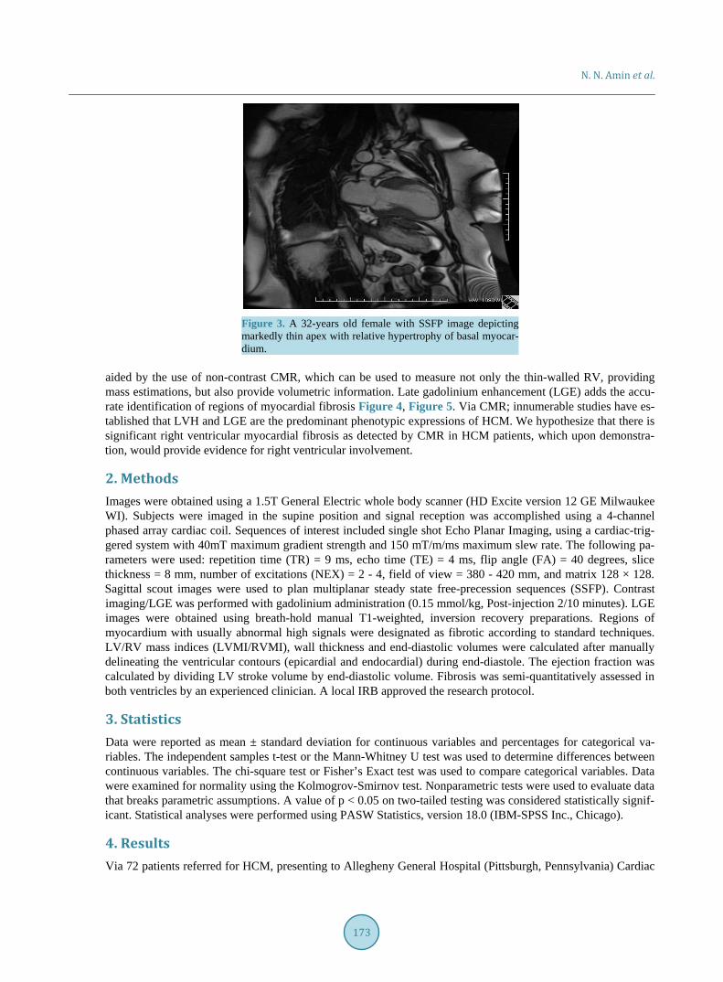
1. Introduction Hypertrophic cardiomyopathy (HCM) is a genetically determined heart muscle disease most often (60 to 70 percent) caused by mutations in one of several sarcomere genes which encode components of the contractile apparatus [1]-[5]. HCM is characterized by left ventricular hypertrophy of various morphologies, with a wide array clinical manifestations and hemodynamic abnormalities [6] [7]. Infrequently, however, right ventricular (RV) involvement is described in reports of HCM. Other cardiac imaging modalities such as echocardiography and nuclear imaging have well known difficulties in assessment of the RV morphological abnormalities in HCM [8]-[10]. This has led to Cardiovascular MRI (CMR) becoming the leading modality to define the structural and functional as well as the clinical impact of HCM providing complete coverage of both ventricles with high spa-tial resolution Figures 1-3. Moreover, assessment of RV diseases, and specifically cardiomyopathies, is greatly
Figure 1. A 52 years old woman with SCD with CMR depict-ing SSFP images that demonstrate markedly thickened anterior wall to a maximum of 33 mm.
Figure 2. A 52 years old woman (same in Figure 1) with SSFP images demonstrating markedly thickened septal wall to a maximum of 37 mm.
N. N. Amin et al.
173
Figure 3. A 32-years old female with SSFP image depicting markedly thin apex with relative hypertrophy of basal myocar-dium.
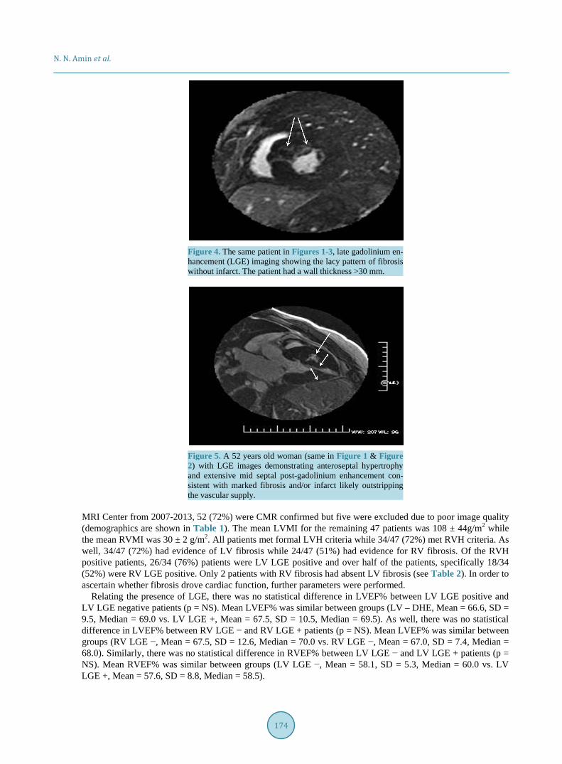
aided by the use of non-contrast CMR, which can be used to measure not only the thin-walled RV, providing mass estimations, but also provide volumetric information. Late gadolinium enhancement (LGE) adds the accu-rate identification of regions of myocardial fibrosis Figure 4, Figure 5. Via CMR; innumerable studies have es-tablished that LVH and LGE are the predominant phenotypic expressions of HCM. We hypothesize that there is significant right ventricular myocardial fibrosis as detected by CMR in HCM patients, which upon demonstra-tion, would provide evidence for right ventricular involvement.
2. Methods Images were obtained using a 1.5T General Electric whole body scanner (HD Excite version 12 GE Milwaukee WI). Subjects were imaged in the supine position and signal reception was accomplished using a 4-channel phased array cardiac coil. Sequences of interest included single shot Echo Planar Imaging, using a cardiac-trig- gered system with 40mT maximum gradient strength and 150 mT/m/ms maximum slew rate. The following pa-rameters were used: repetition time (TR) = 9 ms, echo time (TE) = 4 ms, flip angle (FA) = 40 degrees, slice thickness = 8 mm, number of excitations (NEX) = 2 - 4, field of view = 380 - 420 mm, and matrix 128 × 128. Sagittal scout images were used to plan multiplanar steady state free-precession sequences (SSFP). Contrast imaging/LGE was performed with gadolinium administration (0.15 mmol/kg, Post-injection 2/10 minutes). LGE images were obtained using breath-hold manual T1-weighted, inversion recovery preparations. Regions of myocardium with usually abnormal high signals were designated as fibrotic according to standard techniques. LV/RV mass indices (LVMI/RVMI), wall thickness and end-diastolic volumes were calculated after manually delineating the ventricular contours (epicardial and endocardial) during end-diastole. The ejection fraction was calculated by dividing LV stroke volume by end-diastolic volume. Fibrosis was semi-quantitatively assessed in both ventricles by an experienced clinician. A local IRB approved the research protocol.
3. Statistics Data were reported as mean ± standard deviation for continuous variables and percentages for categorical va-riables. The independent samples t-test or the Mann-Whitney U test was used to determine differences between continuous variables. The chi-square test or Fisher’s Exact test was used to compare categorical variables. Data were examined for normality using the Kolmogrov-Smirnov test. Nonparametric tests were used to evaluate data that breaks parametric assumptions. A value of p < 0.05 on two-tailed testing was considered statistically signif-icant. Statistical analyses were performed using PASW Statistics, version 18.0 (IBM-SPSS Inc., Chicago).
4. Results Via 72 patients referred for HCM, presenting to Allegheny General Hospital (Pittsburgh, Pennsylvania) Cardiac
N. N. Amin et al.
174
Figure 4. The same patient in Figures 1-3, late gadolinium en- hancement (LGE) imaging showing the lacy pattern of fibrosis without infarct. The patient had a wall thickness >30 mm.
Figure 5. A 52 years old woman (same in Figure 1 & Figure 2) with LGE images demonstrating anteroseptal hypertrophy and extensive mid septal post-gadolinium enhancement con-sistent with marked fibrosis and/or infarct likely outstripping the vascular supply.
MRI Center from 2007-2013, 52 (72%) were CMR confirmed but five were excluded due to poor image quality (demographics are shown in Table 1). The mean LVMI for the remaining 47 patients was 108 ± 44g/m2 while the mean RVMI was 30 ± 2 g/m2. All patients met formal LVH criteria while 34/47 (72%) met RVH criteria. As well, 34/47 (72%) had evidence of LV fibrosis while 24/47 (51%) had evidence for RV fibrosis. Of the RVH positive patients, 26/34 (76%) patients were LV LGE positive and over half of the patients, specifically 18/34 (52%) were RV LGE positive. Only 2 patients with RV fibrosis had absent LV fibrosis (see Table 2). In order to ascertain whether fibrosis drove cardiac function, further parameters were performed.
Relating the presence of LGE, there was no statistical difference in LVEF% between LV LGE positive and LV LGE negative patients (p = NS). Mean LVEF% was similar between groups (LV – DHE, Mean = 66.6, SD = 9.5, Median = 69.0 vs. LV LGE +, Mean = 67.5, SD = 10.5, Median = 69.5). As well, there was no statistical difference in LVEF% between RV LGE − and RV LGE + patients (p = NS). Mean LVEF% was similar between groups (RV LGE −, Mean = 67.5, SD = 12.6, Median = 70.0 vs. RV LGE −, Mean = 67.0, SD = 7.4, Median = 68.0). Similarly, there was no statistical difference in RVEF% between LV LGE − and LV LGE + patients (p = NS). Mean RVEF% was similar between groups (LV LGE −, Mean = 58.1, SD = 5.3, Median = 60.0 vs. LV LGE +, Mean = 57.6, SD = 8.8, Median = 58.5).
N. N. Amin et al.
175
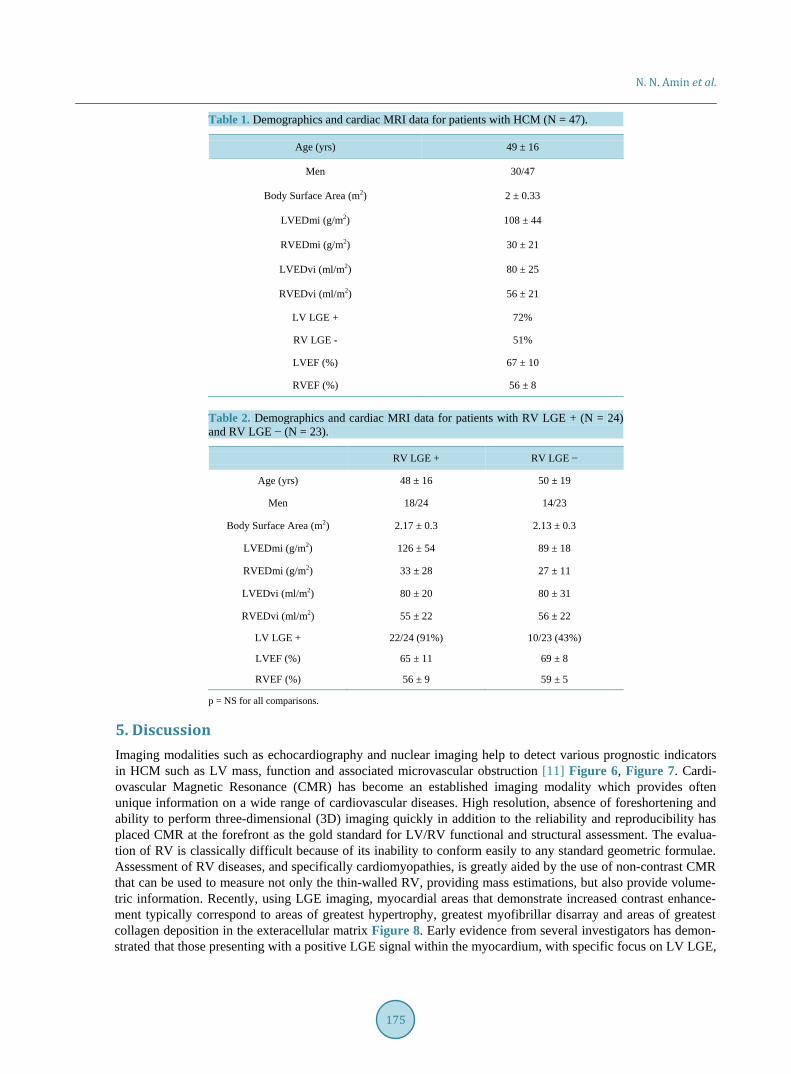
Table 1. Demographics and cardiac MRI data for patients with HCM (N = 47).
Age (yrs) 49 ± 16
Men 30/47
Body Surface Area (m2) 2 ± 0.33
LVEDmi (g/m2) 108 ± 44
RVEDmi (g/m2) 30 ± 21
LVEDvi (ml/m2) 80 ± 25
RVEDvi (ml/m2) 56 ± 21
LV LGE + 72%
RV LGE - 51%
LVEF (%) 67 ± 10
RVEF (%) 56 ± 8
Table 2. Demographics and cardiac MRI data for patients with RV LGE + (N = 24) and RV LGE − (N = 23).
RV LGE + RV LGE −
Age (yrs) 48 ± 16 50 ± 19
Men 18/24 14/23
Body Surface Area (m2) 2.17 ± 0.3 2.13 ± 0.3
LVEDmi (g/m2) 126 ± 54 89 ± 18
RVEDmi (g/m2) 33 ± 28 27 ± 11
LVEDvi (ml/m2) 80 ± 20 80 ± 31
RVEDvi (ml/m2) 55 ± 22 56 ± 22
LV LGE + 22/24 (91%) 10/23 (43%)
LVEF (%) 65 ± 11 69 ± 8
RVEF (%) 56 ± 9 59 ± 5
p = NS for all comparisons.
5. Discussion Imaging modalities such as echocardiography and nuclear imaging help to detect various prognostic indicators in HCM such as LV mass, function and associated microvascular obstruction [11] Figure 6, Figure 7. Cardi-ovascular Magnetic Resonance (CMR) has become an established imaging modality which provides often unique information on a wide range of cardiovascular diseases. High resolution, absence of foreshortening and ability to perform three-dimensional (3D) imaging quickly in addition to the reliability and reproducibility has placed CMR at the forefront as the gold standard for LV/RV functional and structural assessment. The evalua-tion of RV is classically difficult because of its inability to conform easily to any standard geometric formulae. Assessment of RV diseases, and specifically cardiomyopathies, is greatly aided by the use of non-contrast CMR that can be used to measure not only the thin-walled RV, providing mass estimations, but also provide volume-tric information. Recently, using LGE imaging, myocardial areas that demonstrate increased contrast enhance-ment typically correspond to areas of greatest hypertrophy, greatest myofibrillar disarray and areas of greatest collagen deposition in the exteracellular matrix Figure 8. Early evidence from several investigators has demon-strated that those presenting with a positive LGE signal within the myocardium, with specific focus on LV LGE,

N. N. Amin et al.
176
Figure 6. The same patient in Figure 3 with a parasternal long axis view that reveals the asymmetric septal hypertrophy of the left ventricle.
Figure 7. The same patient in Figure 3 with a 5-chamber-view via echo-cardiography demonstrating a similar pattern, albeit less definitive of apic-al thinning with relative basal hypertrophy.
Figure 8. A 48 years old woman with documented HCM and LGE demon-strating diffuse global fibrosis indicative of gross LV fibrosis.

N. N. Amin et al.
177
possess an adverse cardiovascular outcome. Four single-center studies focused on the use of LGE as a prognostic factor for HCM to predict ventricular
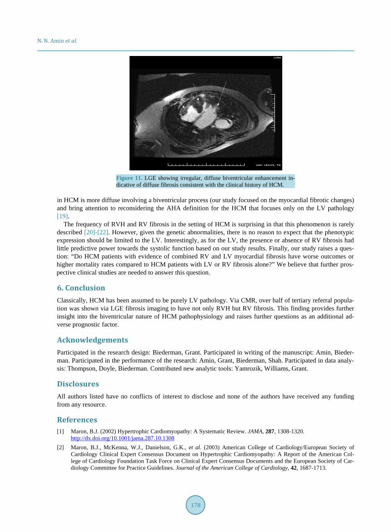
tachyarrhythmia, atrial fibrillation, heart failure and sudden cardiac death demonstrated variable predictive power for VT/VF and SCD dependent on presence or absence of LGE [12]-[15]. More recently, a meta-analysis done by Green et al. incorporating these four studies in 2012 showed that there are significant predictable rela-tionships between LGE and cardiovascular mortality, heart failure death, and all-cause mortality in HCM 16. We had performed an earlier systematic review demonstrating similar finding in which LGE, for the first time in 1064 patients did predict SCD with χ2 of 6.6 and an odds ratio of 3.3 [17]. To date, however, minimal focus has been concentrated on the RV fibrosis and LGE (in only one autopsy-based HCM report in the form of increased wall thickness) [8]. The significant high concordance rates between LV hypertrophy and RV hypertrophy in pa-tients with HCM was previously described in an interesting study, conducted by Maron et al. in 2007, to eva-luate RV wall thickness and mass indices via high spatial resolution CMR [18]. This group concluded that dif-fuse RV wall hypertrophy was present in high number of patients with HCM (54%) while only one subject had CMR evidence of RV fibrosis Figures 9-11. Herein we demonstrate with high sensitivity detection, a high inci-dence of the RV fibrosis (52%) in HCM patients referred to a tertiary referral center using standard LGE tech-niques. Our results provide support for and emphasize the hypothesis that the genetic cardiomyopathic pathology
Figure 9. Late gadolinium enhancement (LGE) demonstrating diffuse en-hancement of the LV and RV. Note, more RV outflow tract LGE is present than LV LGE in a proven HCM patient.
Figure 10. LGE of both ventricles is more evident in a near transmural but irregular RV free wall enhancement pattern from a young man with a fa-ther as the HCM proband.
N. N. Amin et al.
178
Figure 11. LGE showing irregular, diffuse biventricular enhancement in-dicative of diffuse fibrosis consistent with the clinical history of HCM.
in HCM is more diffuse involving a biventricular process (our study focused on the myocardial fibrotic changes) and bring attention to reconsidering the AHA definition for the HCM that focuses only on the LV pathology [19].
The frequency of RVH and RV fibrosis in the setting of HCM is surprising in that this phenomenon is rarely described [20]-[22]. However, given the genetic abnormalities, there is no reason to expect that the phenotypic expression should be limited to the LV. Interestingly, as for the LV, the presence or absence of RV fibrosis had little predictive power towards the systolic function based on our study results. Finally, our study raises a ques-tion: “Do HCM patients with evidence of combined RV and LV myocardial fibrosis have worse outcomes or higher mortality rates compared to HCM patients with LV or RV fibrosis alone?” We believe that further pros-pective clinical studies are needed to answer this question.
6. Conclusion Classically, HCM has been assumed to be purely LV pathology. Via CMR, over half of tertiary referral popula-tion was shown via LGE fibrosis imaging to have not only RVH but RV fibrosis. This finding provides further insight into the biventricular nature of HCM pathophysiology and raises further questions as an additional ad-verse prognostic factor.
Acknowledgements Participated in the research design: Biederman, Grant. Participated in writing of the manuscript: Amin, Bieder-man. Participated in the performance of the research: Amin, Grant, Biederman, Shah. Participated in data analy-sis: Thompson, Doyle, Biederman. Contributed new analytic tools: Yamrozik, Williams, Grant.
Disclosures All authors listed have no conflicts of interest to disclose and none of the authors have received any funding from any resource.
References [1] Maron, B.J. (2002) Hypertrophic Cardiomyopathy: A Systematic Review. JAMA, 287, 1308-1320.
http://dx.doi.org/10.1001/jama.287.10.1308 [2] Maron, B.J., McKenna, W.J., Danielson, G.K., et al. (2003) American College of Cardiology/European Society of
Cardiology Clinical Expert Consensus Document on Hypertrophic Cardiomyopathy: A Report of the American Col-lege of Cardiology Foundation Task Force on Clinical Expert Consensus Documents and the European Society of Car-diology Committee for Practice Guidelines. Journal of the American College of Cardiology, 42, 1687-1713.
N. N. Amin et al.
179
http://dx.doi.org/10.1016/S0735-1097(03)00941-0 [3] Elliott, P.M., Poloniecki, J., Dickie, S., et al. (2000) Sudden Death in Hypertrophic Cardiomyopathy: Identification of
High Risk Patients. Journal of the American College of Cardiology, 36, 2212-2218. http://dx.doi.org/10.1016/S0735-1097(00)01003-2
[4] Spirito, P., Bellone, P., Harris, K.M., Bernabo, P., Bruzzi, P. and Maron, B.J. (2000) Magnitude of Left Ventricular Hypertrophy and Risk of Sudden Death in Hypertrophic Cardiomyopathy. The New England Journal of Medicine, 342, 1778-1785. http://dx.doi.org/10.1056/NEJM200006153422403
[5] Wigle, E.D., Rakowski, H., Kimball, B.P. and Williams, W.G. (1995) Hypertrophic Cardiomyopathy: Clinical Spec-trum and Treatment. Circulation, 92, 1680-1692. http://dx.doi.org/10.1161/01.CIR.92.7.1680
[6] Seidman, J.G. and Seidman, C. (2001) The Genetic Basis for Cardiomyopathy: From Mutation Identification to Me-chanistic Paradigms. Cell, 104, 557-567. http://dx.doi.org/10.1016/S0092-8674(01)00242-2
[7] Richard, P., Charron, P., Carrier, L., et al. (2003) EUROGENE Heart Failure Project. Hypertrophic Cardiomyopathy: Distribution of Disease Genes, Spectrum of Mutations, and Implications for a Molecular Diagnosis Strategy. Circula-tion, 107, 2227-2232. http://dx.doi.org/10.1161/01.CIR.0000066323.15244.54
[8] Mozaffarian, D. and Caldwell, J.H. (2001) Right Ventricular Involvement in Hypertrophic Cardiomyopathy: A Case Report and Literature Review. Clinical Cardiology, 24, 2-8. http://dx.doi.org/10.1002/clc.4960240102
[9] McKenna, W.J., Kleinebenne, A., Nihoyannopoulos, P. and Foale, R. (1988) Echocardiographic Measurements of Right Ventricular Wall Thickness in Hypertrophic Cardiomyopathy: Relation to Clinical and Prognostic Features. Journal of the American College of Cardiology, 11, 351-358. http://dx.doi.org/10.1016/0735-1097(88)90101-5
[10] Severino, S., Caso, P., Cicala, S., et al. (2001) Involvement in Right Ventricle in Left Ventricular Hypertrophy: Analy-sis by Pulsed Doppler Tissue Imaging. European Journal of Echocardiography, 1, 281-288. http://dx.doi.org/10.1053/euje.2000.0043
[11] Noureldin, R.A., Liu, S., Nacif, M.S., et al. (2012) The Diagnosis of Hypertrophic Cardiomyopathy by Cardiovascular Magnetic Resonance. Journal of Cardiovascular Magnetic Resonance, 14, 17. http://dx.doi.org/10.1186/1532-429X-14-17
[12] Papavassiliu, T., Germans, T., Flüchter, S., et al. (2009) CMR Findings in Patients with Hypertrophic Cardiomyopathy and Atrial Fibrillation. JCMR, 11, 34.
[13] Petkow-Dimitrow, P., Klimeczek, P., Vliegenthart, R., Pasowicz, M., Miszalski-Jamka, T., Oudkerk, M., Podolec, P., Dubiel, J.S. and Tracz, W. (2009) Late Gadolinium Enhancement in Cardiovascular Magnetic Resonance in Patients with Hypertrophic Cardiomyopathy Complicated by Life-Threatening Ventricular Tachyarrhythmia. Kardiologia Polska, 67, 964-969.
[14] Maron, M.S. (2012) Clinical Utility of Cardiovascular Magnetic Resonance in Hypertrophic Cardiomyopathy. Journal of Cardiovascular Magnetic Resonance, 14, 13.
[15] Alla, V.M., Koneru, S., Hunter, C. and Mooss, A. (2012) LGE and the Risk of Sudden Death in HCM. JACC: Cardi-ovascular Imaging, 5, 761-762. http://dx.doi.org/10.1016/j.jcmg.2012.05.004
[16] Green, J.J., Berger, J.S., Kramer, C.M. and Salerno, M. (2012) Prognostic Value of Late Gadolinium Enhancement in Clinical Outcomes for Hypertrophic Cardimyopathy. JACC: Cardiovascular Imaging, 5, 370-377. http://dx.doi.org/10.1016/j.jcmg.2011.11.021
[17] Silva, N., Paladino, A., Doyle, M., Reddy, S.T., et al. (2012) A Systematic Review for Sudden Cardiac Death in Hypertrophic Cardiomyopathy Patients with Myocardial Fibrosis: A CMR LGE Study. Circulation, 124, Article ID: A15932.
[18] Maron, M.S., Hauser, T.H., Dubrow, E., et al. (2007) Right Ventricular Involvement in Hypertrophic Cardiomyopathy. American Journal of Cardiology, 100, 1293-1298.
[19] Maron, B.J., Towbin, J.A., Thiene, G., et al. (2006) American Heart Association; Council on Clinical Cardiology, Heart Failure and Transplantation Committee; Quality of Care and Outcomes Research and Functional Genomics and Translational Biology Interdisciplinary Working Groups; Council on Epidemiology and Prevention. Contemporary de-finitions and classification of the cardiomyopathies: An American Heart Association scientific statement from the Council on Clinical Cardiology, Heart Failure and Transplantation Committee; Quality of Care and Outcomes Re-search and Functional Genomics and Translational Biology Interdisciplinary Working Groups; and Council on Epide-miology and Prevention. Circulation, 113, 1807-1816. http://dx.doi.org/10.1161/CIRCULATIONAHA.106.174287
[20] Chan, R.H., Maron, B.J., Olivotto, I., et al. (2015) Significance of Late Gadolinium Enhancement at Right Ventricular Attachment to Ventricular Septum in Patients with Hypertrophic Cardiomyopathy. American Journal of Cardiology, 116, 436-441.
[21] Zhang, S., Yang, Z.G., Sun, J.Y., et al. (2014) Assessing Right Ventricular Function in Patients with Hypertrophic Cardiomyopathy with Cardiac MRI: Correlation with the New York Heart Function Assessment (NYHA) Classifica-

N. N. Amin et al.
180
tion. PLoS ONE, 9, e104312. [22] Bravo, P.E., Luo, H.C., Pozios, I., et al. (2015) Late Gadolinium Enhancement Confined to the Right Ventricular In-
sertion Points in Hypertrophic Cardiomyopathy: An Intermediate Stage Phenotype? European Heart Journal—Cardi- ovascular Imaging (In Press). http://dx.doi.org/10.1093/ehjci/jev154