The Clinical Relevance of Vertebral Artery Hypoplasia would contribute to symptomatic...
7
1 Acta Neurologica Taiwanica Vol 21 No 1 March 2012 Abstract Congenital vertebral artery (VA) hypoplasia is an uncommon embryonic variation of posterior circulation. The frequency of this congenital variation was reported to be 2-6% from autopsy and angiograms. Is it a congenital risk factor of ischemic stroke? In this review, we gave an overview of the literature concerning vertebral artery hypoplasia. VA hypoplasia served as an independent factor of a reduction of the posterior circulation blood flow velocity. VA hypoplasia can play a negative role in cases of occlusion of a major brain vessel since it limits the potential of compensatory blood circulation. VA hypoplasia may also lead to regional hypoperfusion and complex neurovascular consequences which correspond to vestibular neuronitis and migraine pathogenesis. Key words: Vertebral artery hypoplasia, fetal, posterior circulation, vestibular neuronitis, migraine Acta Neurol Taiwan 2012;21:1-7 INTRODUCTION Vertebral artery hypoplasia (VAH) was described in the 19th century (1) . Congenital vertebral artery (VA) hypoplasia is an uncommon embryonic variation of pos- terior circulation (1,2) . The frequency of this congenital variation was reported to be 2-6% from autopsy and angiograms (1-3) . Is it a congenital risk factor of ischemic stroke? VAH with caliber discrepancies of more than 1:1.7 were observed in up to 10% of normal individu- als (3) . There is no general agreement as to the definition of VAH. Operational definitions of VAH vary between diameters of less than 2 to less than 3 mm or an asym- metry ratio of equal or greater than 1:1.7 (3-6) Given the importance of these vascular variations, it is astonishing how little is known about their clinical relevance. In other words, is VAH a risk factor for strokes in the posterior cerebral circulation? Does a hypoplastic VA impair cerebral perfusion and manifest a hidden disease? It has been reported that, during extreme cervical injury, cerebral ischemia would occur in the territory of a hypoplastic vertebrobasilar system (7) . We wondered if this congenital variation played a role similar to spontaneous brainstem/cerebellum ischemic stroke. Recently, congenital VA hypoplasia could be easily defined by duplex color-coded ultra- sonography (2,5) . The normal range of net VA flow volume has been established. And consequently diagnostic crite- ria of vertebrobasilar insufficiency with net VA flow volume <100 ml/min have also been determined (4) . In From the 1 Department of Neurology, Far-Eastern Memorial Hospital and 2 Taipei Veterans General Hospital. 3 School of Medicine, National Yang-Ming University, Taipei, Taiwan. Received and Accepted August 25, 2011. Correspondence to: Yu-Ming Chuang, MD, PhD. Stroke Center and Department of Neurology, Far-Eastern Memorial Hospital. No. 21, Sec. 2, Nanya S. Rd., Banciao Dist., New Taipei City 220, Taiwan (R.O.C.) E-mail: [email protected] The Clinical Relevance of Vertebral Artery Hypoplasia Yu-Ming Chuang 1,2,3 , Lung Chan 1 , Hung-Ming Wu 1 , Siu-Pak Lee 1 , Yiu-Tong Chu 1 Review Article
-
Upload
duongduong -
Category
Documents
-
view
225 -
download
5
Transcript of The Clinical Relevance of Vertebral Artery Hypoplasia would contribute to symptomatic...

1
Acta Neurologica Taiwanica Vol 21 No 1 March 2012
AbstractCongenital vertebral artery (VA) hypoplasia is an uncommon embryonic variation of posterior circulation.The frequency of this congenital variation was reported to be 2-6% from autopsy and angiograms. Is it acongenital risk factor of ischemic stroke? In this review, we gave an overview of the literature concerningvertebral artery hypoplasia. VA hypoplasia served as an independent factor of a reduction of the posteriorcirculation blood flow velocity. VA hypoplasia can play a negative role in cases of occlusion of a majorbrain vessel since it limits the potential of compensatory blood circulation. VA hypoplasia may also lead toregional hypoperfusion and complex neurovascular consequences which correspond to vestibular neuronitisand migraine pathogenesis.
Key words: Vertebral artery hypoplasia, fetal, posterior circulation, vestibular neuronitis, migraine
Acta Neurol Taiwan 2012;21:1-7
INTRODUCTION
Vertebral artery hypoplasia (VAH) was described inthe 19th century(1). Congenital vertebral artery (VA)hypoplasia is an uncommon embryonic variation of pos-terior circulation(1,2). The frequency of this congenitalvariation was reported to be 2-6% from autopsy andangiograms(1-3). Is it a congenital risk factor of ischemicstroke? VAH with caliber discrepancies of more than1:1.7 were observed in up to 10% of normal individu-als(3). There is no general agreement as to the definitionof VAH. Operational definitions of VAH vary betweendiameters of less than 2 to less than 3 mm or an asym-metry ratio of equal or greater than 1:1.7(3-6)
Given the importance of these vascular variations, it
is astonishing how little is known about their clinicalrelevance. In other words, is VAH a risk factor forstrokes in the posterior cerebral circulation? Does ahypoplastic VA impair cerebral perfusion and manifest ahidden disease? It has been reported that, duringextreme cervical injury, cerebral ischemia would occurin the territory of a hypoplastic vertebrobasilar system(7). We wondered if this congenital variation played arole similar to spontaneous brainstem/cerebellumischemic stroke. Recently, congenital VA hypoplasiacould be easily defined by duplex color-coded ultra-sonography(2,5). The normal range of net VA flow volumehas been established. And consequently diagnostic crite-ria of vertebrobasilar insufficiency with net VA flowvolume <100 ml/min have also been determined(4). In
From the 1Department of Neurology, Far-Eastern MemorialHospital and 2Taipei Veterans General Hospital. 3School ofMedicine, National Yang-Ming University, Taipei, Taiwan.Received and Accepted August 25, 2011.
Correspondence to: Yu-Ming Chuang, MD, PhD. Stroke Centerand Department of Neurology, Far-Eastern Memorial Hospital.No. 21, Sec. 2, Nanya S. Rd., Banciao Dist., New Taipei City220, Taiwan (R.O.C.)E-mail: [email protected]
The Clinical Relevance of Vertebral Artery Hypoplasia
Yu-Ming Chuang1,2,3, Lung Chan1, Hung-Ming Wu1, Siu-Pak Lee1, Yiu-Tong Chu1
Review Article
2
Acta Neurologica Taiwanica Vol 21 No 1 March 2012
the early 1990s, a study that analyzed MR angiogramsfound symptomatic or asymptomatic pontine infarctionsto be twice as frequent in patients with VAH as inpatients with symmetric vertebral arteries(8). The samegroup(9) found in an autopsy study VAH to be more fre-quent in cases with basilar artery occlusions than incases with nonbrainstem infarctions.
Recently, VAH has been considered as risk factor ofposterior circulation ischemia(2), and VAH has been asso-ciated with migraine with aura and vestibular neuroni-tis(6,10). The aim of the current study was to evaluatewhether VAH is a possible risk factor for posterior circu-lation stroke.
Association between Vertebral artery hypoplasiaand Stroke
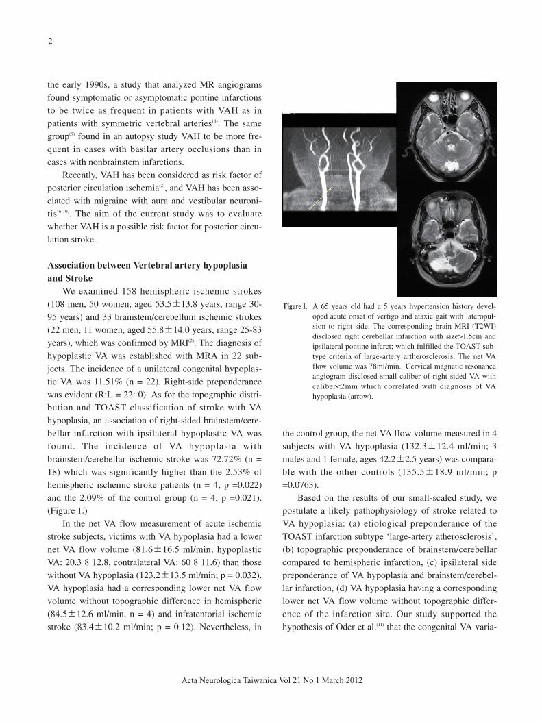
We examined 158 hemispheric ischemic strokes(108 men, 50 women, aged 53.5 13.8 years, range 30-95 years) and 33 brainstem/cerebellum ischemic strokes(22 men, 11 women, aged 55.8 14.0 years, range 25-83years), which was confirmed by MRI(2). The diagnosis ofhypoplastic VA was established with MRA in 22 sub-jects. The incidence of a unilateral congenital hypoplas-tic VA was 11.51% (n = 22). Right-side preponderancewas evident (R:L = 22: 0). As for the topographic distri-bution and TOAST classification of stroke with VAhypoplasia, an association of right-sided brainstem/cere-bellar infarction with ipsilateral hypoplastic VA wasfound. The incidence of VA hypoplasia withbrainstem/cerebellar ischemic stroke was 72.72% (n =18) which was significantly higher than the 2.53% ofhemispheric ischemic stroke patients (n = 4; p =0.022)and the 2.09% of the control group (n = 4; p =0.021).(Figure 1.)
In the net VA flow measurement of acute ischemicstroke subjects, victims with VA hypoplasia had a lowernet VA flow volume (81.6 16.5 ml/min; hypoplasticVA: 20.3 8 12.8, contralateral VA: 60 8 11.6) than thosewithout VA hypoplasia (123.2 13.5 ml/min; p = 0.032).VA hypoplasia had a corresponding lower net VA flowvolume without topographic difference in hemispheric(84.5 12.6 ml/min, n = 4) and infratentorial ischemicstroke (83.4 10.2 ml/min; p = 0.12). Nevertheless, in
the control group, the net VA flow volume measured in 4subjects with VA hypoplasia (132.3 12.4 ml/min; 3males and 1 female, ages 42.2 2.5 years) was compara-ble with the other controls (135.5 18.9 ml/min; p=0.0763).
Based on the results of our small-scaled study, wepostulate a likely pathophysiology of stroke related toVA hypoplasia: (a) etiological preponderance of theTOAST infarction subtype ‘large-artery atherosclerosis’,(b) topographic preponderance of brainstem/cerebellarcompared to hemispheric infarction, (c) ipsilateral sidepreponderance of VA hypoplasia and brainstem/cerebel-lar infarction, (d) VA hypoplasia having a correspondinglower net VA flow volume without topographic differ-ence of the infarction site. Our study supported thehypothesis of Oder et al.(11) that the congenital VA varia-
Figure 1. A 65 years old had a 5 years hypertension history devel-oped acute onset of vertigo and ataxic gait with lateropul-sion to right side. The corresponding brain MRI (T2WI)disclosed right cerebellar infarction with size>1.5cm andipsilateral pontine infarct; which fulfilled the TOAST sub-type criteria of large-artery artherosclerosis. The net VAflow volume was 78ml/min. Cervical magnetic resonanceangiogram disclosed small caliber of right sided VA withcaliber<2mm which correlated with diagnosis of VAhypoplasia (arrow).

tions would contribute to symptomatic vertebrobasilarocclusive disease.
Association between Vertebral artery hypoplasiaand Migraine
We reviewed the records of 250 migraine outpatientsbased on the outpatient disease registration (108 menand 142 women; mean age = 30.8 14.0 years, agerange = 25-55; usual attack duration = 36.2 15.8 h;duration of attack until examination =9.6 6.2 h) for theperiod of January 2005 to October 2005(10). The inci-dence of VA hypoplasia in patients who had migrainewith aura was 28.26%. There was no significant net VAflow volume reduction during the attack phase comparedwith the headache-free period. The net VA flow volumein the migraine with aura group was comparable to thatin the control group. Given that there was right-sidedhypoplasic VA in our migraine with aura group, findinga smaller caliber right VA with a higher resistance index(RI) in this group than in the control group was unre-markable. The exception was that the RI of the right VAdecreased significantly during the attacks of migrainewith aura. Meanwhile, the RI of the left VA remainedstationary. All subjects with migraine had concurrentright-sided hypoplasic VA. One subject developed a pro-longed visual aura which lasted for 32 h. The Dopplerstudies in this patient were performed at 6, 10, and 20 hafter aura onset. The net VA flow volumes were 121.68.8, 128.8 9.2 and 125.6 7.1 ml/min, respectively,which were comparable to the value during herheadache-free period (124.8 6.8 ml/min). (Table 1.)
The incidence of VA hypoplasia in patients who hadmigraine with aura (28.26%) was 14 times higher thanthat of the normal controls in our previous study(2.09%), which involved the same hospital outpatients(2).
Our result supports the observation of Lovrencic et al.(12)
but argues against their proposed hypothesis. The net VAflow volume measured during the attack phase was satis-factory(13). It can therefore be argued that the role of VAhypoplasia in migraine may not involve hypoperfusionduring the attack phase. The result is basically a negativeone, while the meaning of RI remains unclear in thepresence of VA hypoplasia. A significant RI reduction inthe hypoplasic VA was evident during the attacks ofmigraine with aura.
One explanation is that a hypoplasic VA may notcontribute to migraine attack through its net VA flowvolume reduction. In our work, reduction of the RI wasevident during the aura phase and during attacks ofmigraine with aura. Vasomotor regulation of the VA maybe neurogenic in origin. Vasomotor regulation of the VAis innervated by the cervical perivascular sympatheticplexus(14,15). The cervical sympathetic trunk directly con-tributes to the trigeminovascular pain-producing mecha-nism of migraine(16). Our observation of VA vasomotoralteration during migraine attacks further extends theknowledge about the role of the hypoplasic VA. Basedon the above facts, we hypothesize that VA hypoplasiamight contribute to migraine through complex neurovas-cular pathways of the trigeminovascular pain-producingmechanism rather than through its corresponding lowflow volume.
Vertebral artery hypoplasia and Vestibular evokedmyogenic potential study (VEMP)
Among the 52 sides of the 26 subjects of experimen-tal group, except for 1 side that showed no response,VEMP tests disclosed delayed responses in 22 subjects,including unilateral delay in 20 and bilateral delay in 2subjects(17). The remaining 3 subjects (11.53%) showed
3
Acta Neurologica Taiwanica Vol 21 No 1 March 2012
Table 1. The net VA flow volume and RI during and outside migraine attacks
Without aura (N=18) With aura (N=8) Control (N=26)
free attack free attack
Net VA flow volume (ml/min) 132.8 12.4 130.6 11.3(P:0.19) 128.7 11.6 130.6 12.1(P:0.13) 137.2 11.8
RI of VA
Left 0.67 0.12 0.70 0.11(P:0.22) 0.65 0.11 0.68 0.13(P:0.17) 0.66 0.12
Right 0.85 0.11 0.83 0.13(P:0.18) 0.89 0.12 0.79 0.05* (P:0.03) 0.65 0.11

normal vestibular evoked myogenic potentials bilateral-ly. In contrast, in the control group, VEMP testing dis-closed delayed response in three subjects (11.53%),including unilateral delay in two and bilateral delay inone subjects.
In other words, 88.47% of the subjects with VAHdemonstrated abnormal VEMPs either unilaterally orbilaterally. This was significantly higher than the per-centage of abnormal VEMPs in the subjects withoutVAH (p=0.019). Mean latencies of p13 and n23 weresignificantly longer in the right ear of the subjects withVAH than those in the left ear and those in the subjectswithout a VAH. Given the exclusive right-sided hypopla-sic VA in our group of subjects with VAH, the majority(69.23%, N=18) had a concurrent ipsilateral delayedresponse or absence of VEMP. (Figure 2.)
The prolongation of mean latency of p13 and n23imply a demyelination process of the vestibulospinaltract(18). Segmental demyelination is probably secondaryto ischemic axonal dystrophy(19). VAH may lead to repeti-tive hypoperfusion injury to corresponding brainstemregions(20). Although one may infer a brain stem lesionfrom a delayed VEMP, absence of VEMP would indicateinterruption of the sacculo-collic reflex, not necessarily a
brain stem lesion. Nevertheless, we postulate that adelayed response or absence of ipsilateral VEMP can beattributed to “hypoperfusion” of the hypoplastic VA.
Association between Vertebral artery hypoplasiaand Vestibular Neuronitis
From January 2006 to October 2007, we consecu-tively recruited 70 unilateral VN patients who participat-ed in a double-blind randomised placebo controlled trialon the therapeutic effect of valaciclovir on VN in a ter-tiary referral medical center(6).
The diagnosis of VAH (V4) was based on the MRAif <0.22 cm in diameter, a diameter AI >40% and exclu-sion of segmental artherosclerotic stenosis of the VA5 6(AI=left minus right VA diameter/left plus right VAdiameter).
There were 29 VAH (right/left: 23/6) in VN subjects(42.0%) which was statistically higher than in the con-trols (12%, N=6, right/left: 5/1) (p=0.009). After control-ling for the medical risk factors, the RR of VAH in VNsubjects compared with controls was elevated (RR=2.2;95% CI 1.8 to 2.8). There was right VN predominance(right/left: 54/23) in our patients. Among 29 patientswith VAH, 65.5% (N=19) had an ipsilateral VN lesion.
4
Acta Neurologica Taiwanica Vol 21 No 1 March 2012
Figure 2. The n magnetic resonance angiogram in a woman who had migraine suggested the diagnosis ofright-sided VA hypoplasia The corresponding vestibular evoked myogenic potentials testing dis-closed an ipsilateral delayed response.

The side accordance rate between VAH and VN washigher in the left (83.3%) than in the right side (60.8%)(p=0.037) VN subjects were stratified to two subgroupsfor comparison:VN with (N=51; age: 63.29 8.5 yearsold; F/M: 28/23) and without medical atheroscleroticrisk factors (N=18; age:48.55+-6.5; F/M: 12/6). The sideaccordance rate between VAH and VN was much higher(80.9%) in the vascular risk group than in the non-vascu-lar risk group (25%). In both groups, left VAH assumeda significantly higher accordance rate than the right side.Demographic data showed older age and male predomi-nance in the vascular risk group, while the prevalence ofVAH was comparable between the two groups. (Table 2)
Our study revealed that VN subjects had a statistical-ly higher incidence of VAH, and there is a significant
side accordance rate between VAH and VN, especiallyon the left side and in those with vascular risk factors.
VAH is associated with a delayed P13 response investibular evoked myogenic potentials, suggestinglabyrinthine injuries, probably related to VAH-associatedhypoperfusion(17). Although AICA is a branch of the cau-dal basilar artery (BA), the ipsilateral relationshipbetween VAH and VN can be explained by the asymmet-rical junction geometry. Ravensbergen found a BA‘double hump’ axial velocity profile just downstream ofthe VA confluence(21). A larger-calibre dominant VA withthe highest ‘hump’ crosses the centre-line of BA andrestricted blood of the hypoplastic VA from flowing intothe ipsilateral AICA. However, with the FahraeuseLindqvist effect, small-calibre VAH will reduce its blood
5
Acta Neurologica Taiwanica Vol 21 No 1 March 2012
Table 2. Clinical characteristics in 69 VN patients stratified by VAH and medical atherosclerotic risk factors
VN with VAH VN without VAH p
No. of cases 29 40
Age, year 50.29 8.5 48.55 6.5 0.073
Radiological findings
Asymmetry index of VA 0.49 0.06 0.28 0.11 0.018*
No. of right VAH 23
No. of left VAH 6 0.019*
No. of ipsilateral VAH according to VN lesion side Global 19/29 (65.52%)
Right VAH with ipsilateral. VN 14/23 (60.87%)
Left VAH with ipsilateral. VN 5/6 (83.33%) 0.037*
Vascular risk group Non-vascular risk group p
No. of cases 51 18
Age, year 63.29 8.5 48.55 6.5 0.019*
Risk factors (%) Hypertension 31 0
Diabetes mellitus 18 0
Smoking 20 0
Coronary artery disease 10 0
Hyperlipidemia 32 0
Radiological findings
VAH 21 (41.17%) 8 (44.44%) 0.230
R/L: 17/4 R/L: 6/2
No. of ipsilateral VAH according to VN lesion site
Global 17/21 (80.95%) 2/8 (25%) 0.009
Right VAH with ipsilateral VN 13/17 (76.47%) 1/6 (16.6%) 0.009
Left VAH with ipsilateral VN 4/4 (100%) 1/2 (50%) 0.068
* left VAH versus right VAH

viscosity and speed up dominant laminar flow to ipsilat-eral AICA.
VAH-related vestibular hypoperfusion may increasevulnerability to more severe viral infection throughimpeded immunity and restricted neuronal repair capaci-ty(22). Hypoperfusion with impaired oxidative killing byneutrophils, a primary defense against pathogens, isdirectly related to tissue oxygenation(23,24). Comorbidintracranial VAH may also serve as a risk factor forsevere vestibular neuronitis (25).
CONCLUSION
In this review, we gave an overview of the literatureconcerning vertebral artery hypoplasia. VA hypoplasiaserved as an independent factor of a reduction of theposterior circulation blood flow velocity rather than ofthe anterior circulation. VA hypoplasia can play a nega-tive role in cases of occlusion of a major brain vesselsince it limits the potential of compensatory blood circu-lation. VA hypoplasia may also lead to regional hypoper-fusion and complex neurovascular consequences whichcorrespond to vestibular neuronitis and migraine patho-genesis.
REFERENCES
1. Krayenbuühl HA, Yasargil GM. Die vaskulaären
Erkrankungen im Gebiet der Arteria Basialisqq. III.
Variationen der A. Vertebralis, basialis undihrer A? ste.
Stuttgart: Thieme, 1957:36-41.
2. Chuang YM. Huang YC, Hu HH, Yang CY. Toward a fur-
ther elucidation: role of vertebral artery hypoplasia in acute
ischemic stroke. Eur Neurol 2006;55:193-197.
3. Trattnig S, Schwaighofer B, Hübsch P, Schwarz M,
Kainberger F. Color-coded sonography of vertebral arteries.
J Ultrasound Med 1991;10:221-226.
4. Seidel E, Eicke BM, Tettenborn B, Krummenauer F.
Reference values for vertebral artery flow volume by
duplex sonography in young and elderly adults. Stroke
1999;30:2692-2696.
5. Jeng JS, Ping-Keung Y. Evaluation of vertebral artery
hypoplasia and asymmetry by color-coded duplex ultra-
sonography. Ultrasound Med Biol 2004;30:605-609.
6. Chuang YM, Chern CM, Liao WH, Hsu LC, Lien CF,
Lirng JF, Shiao AS, Ko JS. Contribution of intracranial ver-
tebral artery asymmetry to vestibular neuropathy. J Neurol
Neurosurg Psychiatry. 2011;82:823-825.
7. Chaturvedi S, Lukovits T, Chen W, Gorelick PB: Ischemia
in the territory of a hypoplastic vertebrobasilar system.
Neurology 1999;52:980-983.
8. Watanabe M, Takahashi A, Hashizume Y, Motegi Y, Furuse
K. The correlation between vertebral artery asymmetry and
pontine infarction- an MR angiography study. Rinsho
Shinkeigaku 1992;32:708-712.
9. Watanabe M, Takahashi A, Hashizume Y. A pathological
study of basilar artery sclerosis resulting from vertebral
artery asymmetry. Rinsho Shinkeigaku 1993;33:389-395.
10. Chuang YM, Liu CY, Lin CP. Toward a further elucidation:
role of vertebral artery hypoplasia in migraine with aura.
Eur Neurol 2008;59:148-151.
11. Oder B, Oder W, Lang W, Marschnigg E, Deecke L:
Hypoplasia, stenosis and other alterations of the vertebral
artery: does impaired blood rheology manifest a hidden dis-
ease? Acta Neurol Scand 1998;97:398-403.
12. Lovrencic A, Demarin V, Rundek T, Vukovic V: Role of
vertebral artery hypoplasia in migraine. Cephalalgia 1998;
18:684-686.
13. Zwetsloot CP, Caekebeke JFV, Jansen JC,Odink J: Blood
flow velocities in the vertebrobasilar system during
migraine attacks: a transcranial Doppler study. Cephalalgia
1992;12:29-32.
14. Mitchell JA: An investigation of a sympathetic innervation
of the vertebral artery in primates: is there a neurogenic
substrate for vasoconstriction? Eur J Histochem 2004; 48:
309-316.
15. Chen XQ, Bo S, Zhong SZ. Nerves accompanying the ver-
tebral artery and their clinical relevance. Spine 1988; 13:
1360-1364.
16. Arab MA, Wiklund L, Svendgaard NA: Origin and distrib-
ution of cerebral vascular innervation from superior cervi-
cal, trigeminal and spinal ganglia investigated with retro-
grade and anterograde WGA-HRP tracing in the rat.
Neuroscience 1986;19:695-708.
17. Chuang YM, Chen CC, Lin CP. Vertebral artery hypoplasia
may contribute to abnormal vestibular evoked myogenic
6
Acta Neurologica Taiwanica Vol 21 No 1 March 2012

potentials. Acta Neurol Taiwan 2009;18:133-136.
18. Murofushi T. Shimizu K. Takegoshi H. Cheng PW.
Diagnostic value of prolonged latencies in the vestibular
evoked myogenic potential. Arch Otolaryngol Head Neck
Surg 2001;127:1069-1072.
19. Headache Classification Committee of the International
Headache Society. Classification and diagnostic criteria for
headache disorders, cranial neuralgias and facial pain.
Cephalalgia 1988;8:1-96.
20. Perren F. Poglia D. Landis T. Sztajzel R. Vertebral artery
hypoplasia: a predisposing factor for posterior circulation
stroke? Neurology 2007;68:65-67.
21. Ravensbergen J, Krijger J, KB, Hillen B, Hoogstraten HW.
Merging flows in an arterial confluence: the vertebrobasilar
junction. J Fluid Mechanics Digital Archive 1995;304:
119e41.
22. Chen YY, Chao AC, Hsu HY. Vertebral artery hypoplasia is
associated with a decrease in net vertebral flow volume.
Ultrasound Med Biol 2010;36:38-43.
23. Jawahar L, Tom S, Kenneth R. Interactive role of infection,
inflammation and traditional risk factors in atherosclerosis
and coronary artery disease. J Am Coll Cardiol 1998;31:
1217-1225.
24. Benhaim P, Hunt TK. Natural resistance to infection: leuko-
cyte functions. J Burn Care Rehabil 1992;13:287-292.
25. Abrams ZR, Warrier A, Trauner D, Zhang X. A signal pro-
cessing analysis of Purkinje cells in vitro. Front Neural
Circuits 2010;4:13.
7
Acta Neurologica Taiwanica Vol 21 No 1 March 2012