the Extracranial Vertebral Artery: A Case
10
Received 08/02/2015 Review began 08/03/2015 Review ended 10/09/2015 Published 10/19/2015 © Copyright 2015 Dolati et al. This is an open access article distributed under the terms of the Creative Commons Attribution License CC-BY 3.0., which permits unrestricted use, distribution, and reproduction in any medium, provided the original author and source are credited. Application of Pipeline Embolization Device for Iatrogenic Pseudoaneurysms of the Extracranial Vertebral Artery: A Case Report and Systematic Review of the Literature Parviz Dolati , Daniel G. Eichberg , Ajith Thomas , Christopher S. Ogilvy 1. Neurosurgery, Boston University School of Medicine 2. Neurosurgery, BIDMC Harvard Medical School Corresponding author: Parviz Dolati, [email protected] Disclosures can be found in Additional Information at the end of the article Abstract Traumatic pseudoaneurysms of the vertebral artery (VA) are uncommon vascular lesions and definitive management is often challenging. Between 0% and 8% of craniocervical fusions are complicated by VA injury. In these cases, preserving the vertebral artery while treating the pseudoaneurysm is the goal of any treatment option. We describe the second known case of a patient with and iatrogenic extracranial vertebral artery pseudoaneurysm treated effectively using the Pipeline Embolization Device (PED) (Ev3 Neurovascular, Irvine, CA). Although there have been only two cases reported, the use of flow-diverting stents appears to be efficacious for the treatment of non-actively bleeding traumatic pseudoaneurysms. Categories: Neurosurgery Keywords: vertebral artery, pipeline embolization device, iatrogenic, pseudoaneurysm, flow diverting stent Introduction Vertebral artery (VA) pseudoaneurysms arise most commonly due to blunt or penetrating trauma, following surgery, collagen vascular disease, or arterial dissection [1]. Because it emerges from the transverse foramen and courses adjacent to C1, the extracranial segment (V3) of the VA is the most susceptible to iatrogenic damage. Between 0% and 8% of craniocervical fusion surgeries are complicated by VA injury [2-7], which may result in arterial ischemic injury, severe hemorrhage, and death. Rarely, iatrogenic VA injury has been reported to lead to pseudoaneurysm development. Despite the potential for such pseudoaneurysms to resolve spontaneously, they have been shown to rupture in 31% to 54% of cases [8]. Thus, it is critical to rapidly diagnose and treat these vascular lesions to minimize the risk of potential grave morbidity and mortality. Iatrogenic pseudoaneurysms are most commonly treated by surgical or endovascular options, either with or without revascularization [8-9]. Additionally, the use of stent grafts, covered stents, coil embolization, and onyx embolization has been reported [10-13]. Flow-diverting stents, such as the Pipeline Embolization Device (PED) (Ev3 Neurovascular, Irvine, CA), have been gaining increasing interest in the treatment of traumatic pseudoaneurysms [14]. We describe the second reported case of iatrogenic vertebral artery pseudoaneurysm that was treated effectively using the PED. We successfully reconstructed the injured VA, isolated the 1 2 2 2 Open Access Original Article DOI: 10.7759/cureus.356 How to cite this article Dolati P, Eichberg D G, Thomas A, et al. (October 19, 2015) Application of Pipeline Embolization Device for Iatrogenic Pseudoaneurysms of the Extracranial Vertebral Artery: A Case Report and Systematic Review of the Literature. Cureus 7(10): e356. DOI 10.7759/cureus.356
Transcript of the Extracranial Vertebral Artery: A Case

Received 08/02/2015 Review began 08/03/2015 Review ended 10/09/2015 Published 10/19/2015
© Copyright 2015Dolati et al. This is an open accessarticle distributed under the terms ofthe Creative Commons AttributionLicense CC-BY 3.0., which permitsunrestricted use, distribution, andreproduction in any medium,provided the original author andsource are credited.
Application of Pipeline EmbolizationDevice for Iatrogenic Pseudoaneurysms ofthe Extracranial Vertebral Artery: A CaseReport and Systematic Review of theLiteratureParviz Dolati , Daniel G. Eichberg , Ajith Thomas , Christopher S. Ogilvy
1. Neurosurgery, Boston University School of Medicine 2. Neurosurgery, BIDMC Harvard Medical School
Corresponding author: Parviz Dolati, [email protected] Disclosures can be found in Additional Information at the end of the article
AbstractTraumatic pseudoaneurysms of the vertebral artery (VA) are uncommon vascular lesions anddefinitive management is often challenging. Between 0% and 8% of craniocervical fusions arecomplicated by VA injury. In these cases, preserving the vertebral artery while treating thepseudoaneurysm is the goal of any treatment option. We describe the second known case of apatient with and iatrogenic extracranial vertebral artery pseudoaneurysm treated effectivelyusing the Pipeline Embolization Device (PED) (Ev3 Neurovascular, Irvine, CA). Although therehave been only two cases reported, the use of flow-diverting stents appears to be efficacious forthe treatment of non-actively bleeding traumatic pseudoaneurysms.
Categories: NeurosurgeryKeywords: vertebral artery, pipeline embolization device, iatrogenic, pseudoaneurysm, flow divertingstent
IntroductionVertebral artery (VA) pseudoaneurysms arise most commonly due to blunt or penetratingtrauma, following surgery, collagen vascular disease, or arterial dissection [1]. Because itemerges from the transverse foramen and courses adjacent to C1, the extracranial segment (V3)of the VA is the most susceptible to iatrogenic damage. Between 0% and 8% of craniocervicalfusion surgeries are complicated by VA injury [2-7], which may result in arterial ischemic injury,severe hemorrhage, and death. Rarely, iatrogenic VA injury has been reported to lead topseudoaneurysm development. Despite the potential for such pseudoaneurysms to resolvespontaneously, they have been shown to rupture in 31% to 54% of cases [8]. Thus, it is criticalto rapidly diagnose and treat these vascular lesions to minimize the risk of potential gravemorbidity and mortality.
Iatrogenic pseudoaneurysms are most commonly treated by surgical or endovascular options,either with or without revascularization [8-9]. Additionally, the use of stent grafts, coveredstents, coil embolization, and onyx embolization has been reported [10-13]. Flow-divertingstents, such as the Pipeline Embolization Device (PED) (Ev3 Neurovascular, Irvine, CA), havebeen gaining increasing interest in the treatment of traumatic pseudoaneurysms [14]. Wedescribe the second reported case of iatrogenic vertebral artery pseudoaneurysm that wastreated effectively using the PED. We successfully reconstructed the injured VA, isolated the
1 2 2 2
Open Access OriginalArticle DOI: 10.7759/cureus.356
How to cite this articleDolati P, Eichberg D G, Thomas A, et al. (October 19, 2015) Application of Pipeline Embolization Devicefor Iatrogenic Pseudoaneurysms of the Extracranial Vertebral Artery: A Case Report and SystematicReview of the Literature. Cureus 7(10): e356. DOI 10.7759/cureus.356

pseudoaneurysm from circulation, and maintained blood flow through the parent artery.
Materials And MethodsLiterature analysisA comprehensive review of the literature was performed using the keywords “PipelineEmbolization Device,” “vertebral artery traumatic pseudoaneurysm,” “flow-diverting stent,”and “iatrogenic extracranial pseudoaneurysm” alone or together to search PubMed, OvidMedline, Ovid EMBASE, Scopus, Web of Science database, and all neurosurgical journals.Inclusion criteria were the following: English language and presentation of patients withiatrogenic pseudoaneurysms of the vertebral artery treated with the Pipeline EmbolizationDevice. There were no exclusion criteria.
ResultsCase report The patient was a 71-year-old male with a non-union C2 fracture and persistent pain admittedto an outside hospital for a posterior cervical fusion with C1-C2 screw fixation. However, thatprocedure was prematurely aborted because of perforation of the V3 segment of the VA by thesurgical screw. The surgeon encountered the sudden onset of profound bleeding, which wasestimated to be 3 liters. The surgical site was packed after attempting primary hemostasis. Afterthe blood transfusion and stabilization, the patient was transferred to our hospital andunderwent vertebral artery angiography the day after spinal surgery, which confirmed thedevelopment of an iatrogenic pseudoaneurysm in his dominant VA (Figure 1). The contralateralVA was a small and non-dominant one, and therefore, we had to save the injured VA by using aflow diversion technique. For this purpose, the patient was premedicated with standard loadingdoses of aspirin and Plavix. Subsequently, using standard endovascular techniques, a 3 mm x16 mm PED was successfully deployed across the pseudoaneurysm. Final angiography showedan immediate significant stagnation of the blood flow inside the pseudoaneurysm’s cavity, butnot complete occlusion. After deployment of PED, the patient was observed for two days andthen was transferred back to the original spine center and underwent cervical exploration andsurgical gauze removal. No intraoperative bleeding was noted. The patient was regularlyfollowed up; finally, after three months, a check angiography was performed, which showed acomplete obliteration of the pseudoaneurysm (Figure 2).
2015 Dolati et al. Cureus 7(10): e356. DOI 10.7759/cureus.356 2 of 10

FIGURE 1: Vertebral Artery Imaging Before and After PipelineDeploymentPanel A, sagittal computed tomography angiography showing a non-union C2 odontoidfracture. Panels B and C, the lateral and anterior-posterior (AP) view of the left vertebral arteryangiography, respectively. The white arrows are pointing to the pseudoaneurysm at the left V3segment. Panels D and E show a deployed Pipeline in lateral and AP projections across thepseudoaneurysm, respectively (black arrows). The white arrow in panel E points to thestagnation of the blood flow inside the pseudoaneurysm.
2015 Dolati et al. Cureus 7(10): e356. DOI 10.7759/cureus.356 3 of 10
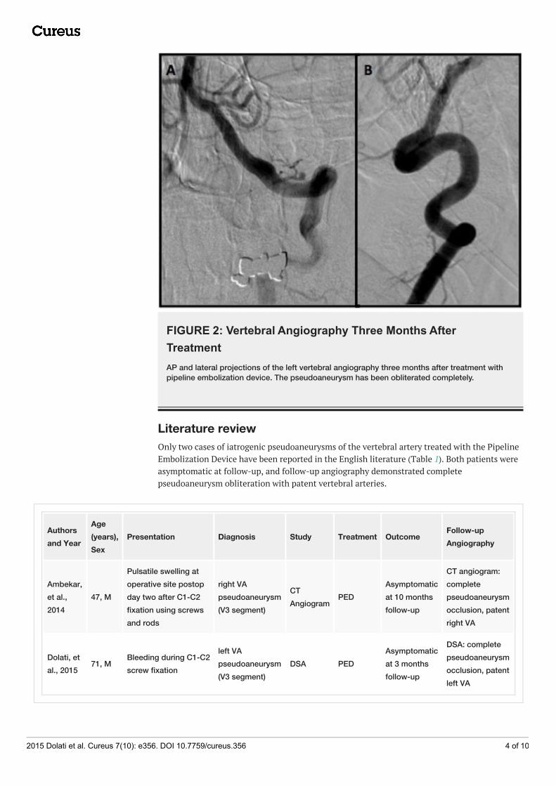
FIGURE 2: Vertebral Angiography Three Months AfterTreatmentAP and lateral projections of the left vertebral angiography three months after treatment withpipeline embolization device. The pseudoaneurysm has been obliterated completely.
Literature reviewOnly two cases of iatrogenic pseudoaneurysms of the vertebral artery treated with the PipelineEmbolization Device have been reported in the English literature (Table 1). Both patients wereasymptomatic at follow-up, and follow-up angiography demonstrated completepseudoaneurysm obliteration with patent vertebral arteries.
Authorsand Year
Age(years),Sex
Presentation Diagnosis Study Treatment OutcomeFollow-upAngiography
Ambekar,et al.,2014
47, M
Pulsatile swelling atoperative site postopday two after C1-C2fixation using screwsand rods
right VApseudoaneurysm(V3 segment)
CTAngiogram
PEDAsymptomaticat 10 monthsfollow-up
CT angiogram:completepseudoaneurysmocclusion, patentright VA
Dolati, etal., 2015
71, MBleeding during C1-C2screw fixation
left VApseudoaneurysm(V3 segment)
DSA PEDAsymptomaticat 3 monthsfollow-up
DSA: completepseudoaneurysmocclusion, patentleft VA
2015 Dolati et al. Cureus 7(10): e356. DOI 10.7759/cureus.356 4 of 10

TABLE 1: Reported Cases of Iatrogenic Pseudoaneurysms of the Vertebral ArteryTreated by Pipeline Embolization Device in English LiteratureAbbreviations: CT: Computed Tomography, M: male, PED: Pipeline Embolization Device, VA: Vertebral Artery, DSA: DigitalSubtraction Angiography
DiscussionPseudoaneurysms are usually encased only by a friable layer of connective tissue rather than atrue wall. Iatrogenic pseudoaneurysms can be either fusiform, which result from arterialdissection resulting in a thinned tunica adventitia layer and resultant vessel dilation, orsaccular, which result from a focal transmural arterial wall lesion [15]. Once identified,pseudoaneurysms require treatment to prevent expansion and rupture.
The third segment of the vertebral artery (V3) is particularly vulnerable to iatrogenic injury dueto its unprotected course adjacent to numerous bony structures. V3 exits the protectivetransverse process of C2, and courses laterally to enter the transverse foramen of C1 [16]. Next,it courses posteriorly around the lateral mass of C1 and inferior to the posterior atlanto-occipital membrane lateral to the cervicomedullary junction [16]. Finally, it coursessuperomedially to enter the dura and arachnoid and then becomes V4 (the final vertebral arterysegment).
Anatomical variation of the vertebrobasilar system is very common, which may contribute tothe associated morbidity of the VA pseudoaneurysm development and treatment [17].Asymmetry due to a unilateral hypoplastic VA, extracranial VA occlusion, or VA ending in theposterior inferior cerebellar artery (PICA) may result in increased reliance on the contralateralVA. Therefore, extreme caution must be taken to identify anatomic variations in thevertebrobasilar system and disrupting the dominant VA during pseudoaneurysm repair. Theanatomic variant, known as duplicated VA, also poses an increased risk for injury as they havemultiple points of attachment, which can result in traction injury [18]. Due to anatomicalvariants of the VA course, the safe placement of transarticular screws (TAS) on at least one sideis not possible in between 10% and 20% of patients [19-20]. For these patients, alternativemethods of fixation and fusion, such as C1 lateral mass and C2 pedicle or pars screws,extension down to C3, halo vest immobilization with wiring and bone grafting, or unilateralTAS fixation should be attempted [21].
The technique used for atlantoaxial fixation may also facilitate the development of a VApseudoaneurysm. For example, one recent study showed that the most common cause ofcomplications associated with this surgery were due to screw malposition [21]. This isconcerning, as screw malposition occurred in 7% of patients [21], and VA injury has beenreported to occur in between 0% and 8% of patients [2-7]. One study found a 1.7% incidence ofVA injury per placed screw [4]. In addition to VA injury, screw malposition may lead topseudarthrosis and damaged screws. The greatest risk factors for screw malposition includeinadequate reduction of C1 on C2 leading to misalignment, lack of surgical experience, andinsufficient knowledge of the patient’s anatomy [5, 7]. For these reasons, three-dimensional(3D) and multiplanar reconstructions of the local anatomy, including VA, C1, and C2, may beindicated for preoperative planning, determination of anatomic suitability [19], and potentiallyfor intraoperative guidance of TAS placement (Figures 3-4). This tri-dimensional reconstructionfrom importing the images and performing the task usually takes 10-15 minutes.
2015 Dolati et al. Cureus 7(10): e356. DOI 10.7759/cureus.356 5 of 10

FIGURE 3: Tri-dimensional and Multiplanar Reconstructions ofthe Occiputo-Cervical Region Including VA and C1-C4 VertebralBodiesTri-dimensional and multiplanar reconstructions of the occiputo-cervical region, including VAand C1-C4 vertebral bodies in lateral, AP, and posterior projections (Panels A-D). By importingthe computed tomography angiography images into any system with the tri-dimensionalreconstruction capability, we will be able to visualize the occiputo-cervical bony structures withthe VA in place. This gives us a 3D image of the position of the vertebral artery before startingscrew fixation.
2015 Dolati et al. Cureus 7(10): e356. DOI 10.7759/cureus.356 6 of 10

FIGURE 4: Three-dimensional Reconstruction of the ComputedTomography Angiography with Testing Different C1-C2 FusionScrew Angle in Relation to the Vertebral ArteryThree-dimensional reconstruction of the computed tomography angiography on theSiemens Leonardo system with testing different C1-C2 fusion screw angles in relation to thevertebral artery. This preoperative assessment of the entrance points and the screw’s anglepenetration will help to realize anatomical variations and prevent VA injury.
Surgical treatment with ligation of pseudoaneurysms is considered the definitive treatment.However, it also is the most invasive of the procedures, so alternative strategies, such asendovascular treatments, are often sought to preserve the parent artery.
The most recently developed endovascular technique for aneurysm treatment is the microporeflow-diverting stent, which induces aneurysm thrombosis and resolution through alteration ofthe aneurysm hemodynamics. The two flow-diverting stents commercially available are the PEDand the SILK (SFD, Balt Extrusion, Montmorency, France) [22]. Recently, PED has been shown
2015 Dolati et al. Cureus 7(10): e356. DOI 10.7759/cureus.356 7 of 10

to have a six-month aneurysm obliteration rate of 82.9% to 87.5%, with a periproceduralcomplication rate of 6.3% and 1.5% mortality rate [23-24]. While the SILK has been reported toachieve a similar aneurysm obliteration rate as the PED (approximately 80%), the SILK appearsto be associated with a greater complication rate (approximately 17%) [25-26]. Insufficientstudies on SILK have been reported to enable a direct comparison with PED. The PED has beenshown to achieve flow diversion while maintaining the integrity of perforating arteries andparent artery branch vessels [19]. This property of the PED may be beneficial in treating VApseudoaneurysms when we need to save important perforator arteries or main branches, suchas the posterior inferior cerebellar or anterior spinal arteries.
While other endovascular techniques are available for aneurysm treatment, they possessdisadvantages that flow-diverting stents were designed to overcome. For example, whenpseudoaneurysms are treated with coil embolization, as there is no real wall for theseaneurysms, the chance for rebleeding and intraoperative rupture and severe bleeding will bevery high, especially in the acute setting of VA perforation by a screw. In contrast, the PED doesnot require intra-aneurysmal manipulation, and adjuvant coiling is not necessary. The PED alsoprovides other theoretical advantages over other endovascular modalities. The PED enables fullcoverage of the aneurysm neck and implant, as it serves as scaffolding for growth of endothelialtissue [23]. Additionally, the PED has greater metal surface area coverage compared to balloon-expendable or self-expanding stents, which promotes neointimal regrowth and aneurysm neckocclusion [23].
Our experience mirrors that of Ambekar, et al. (2014), in that the PED can be used tosuccessfully treat iatrogenic VA pseudoaneurysms [1]. We believe the PED is able to preserveparent vessel patency while excluding the pseudoaneurysm. Because flow-diversion stentsrequire the absence of a large pressure gradient across the pseudoaneurysm wall to inducehemodynamic alteration and thrombosis, the PED is more likely to be effective if thepseudoaneurysm is not actively bleeding during treatment. Thus, the PED may not besuccessful and, in fact, may be contraindicated in treating acutely bleeding pseudoaneurysms.
Regardless, as flow diversion induces thrombosis over the time, rebleeding remains a potentialcomplication in the short-term post-procedural period. Moreover, as a mandatoryperiprocedural requirement for the application of any intra-arterial stent, these patients needto be on a dual anti-platelets regimen to prevent intra-stent clot formation. This may interfereor delay the healing process and even may increase the chance of early postoperative bleeding.These may be the main limitations of the PED. Therefore, careful patient selection, closepostoperative observation, and follow-up images are highly recommended.
ConclusionsIatrogenic pseudoaneurysms of the vertebral artery arising during cervical fusion, particularlyin its V3 segment, are potential vascular lesions that are difficult to treat definitively. To ourknowledge, this is the second case report of a successful application of a flow diversion stentfor treatment of these pseudoaneurysms. PED is a useful tool in the management of suchvascular lesions, leading to pseudoaneurysm isolation from the circulation withoutcompromising parent vessel blood flow. However, pseudoaneurysm rupture remains a potentialcomplication in the early post-procedural period. Therefore, close postoperative patientobservation and interval follow-up images are highly recommended.
Additional InformationDisclosuresHuman subjects: Consent was obtained by all participants in this study. Animal subjects:
2015 Dolati et al. Cureus 7(10): e356. DOI 10.7759/cureus.356 8 of 10

This study did not involve animal subjects or tissue.
References1. Ambekar S, Sharma M, Smith D, Cuellar H: Successful treatment of iatrogenic vertebral
pseudoaneurysm using pipeline embolization device. Case Rep Vasc Med. 2014, 2014:341748.10.1155/2014/341748
2. Neo, M, Fujibayashi S, Miyata M, Takemoto, Nakamura T: Vertebral artery injury duringcervical spine surgery: a survey of more than 5600 operations. Spine (Phila Pa 1976). 2008,33:779–85. 10.1097/BRS.0b013e31816957a7
3. Yeom JS, Buchowski JM, Park KW, Chang BS, Lee CK, Riew KD: Undetected vertebral arterygroove and foramen violations during C1 lateral mass and C2 pedicle screw placement. Spine(Phila Pa 1976). 2008, 33:E942–9. 10.1097/BRS.0b013e3181870441
4. Gluf WM, Schmidt MH, Apfelbaum RI: Atlantoaxial transarticular screw fixation: a review ofsurgical indications, fusion rate, complications, and lessons learned in 191 adult patients. JNeurosurg Spine. 2005, 2:155–63.
5. Grob D, Crisco JJ 3rd, Panjabi MM, Wang P, Dvorak J: Biomechanical evaluation of fourdifferent posterior atlantoaxial fixation techniques. Spine (Phila Pa 1976). 1992, 17:480–90.
6. Haid RW Jr, Subach BR, McLaughlin MR, Rodts GE Jr, Wahlig JB Jr: C1-C2 transarticular screwfixation for atlantoaxial instability: a 6-year experience. Neurosurgery. 2001, 49:65–68.10.1097/00006123-200107000-00010
7. Madawi AA, Casey AT, Solanki GA, Tuite G, Veres R, Crockard HA: Radiological andanatomical evaluation of the atlantoaxial transarticular screw fixation technique. J Neurosurg.1997, 86:961–68. 10.3171/jns.1997.86.6.0961
8. Larson PS, Reisner A, Morassutti DJ, Abdulhadi B, Harpring JE: Traumatic intracranialaneurysms. Neurosurg Focus. 2000, 8:e4. 10.3171/foc.2000.8.1.1829
9. Holmes B, Harbaugh, RE: Traumatic intracranial aneurysms: a contemporary review . JTrauma. 1993, 35:855–60.
10. Lempert TE, Halbach VV, Higashida RT, Dowd CF, Urwin RW, Balousek PA, Hieshima GB:Endovascular treatment of pseudoaneurysms with electrolytically detachable coils . AJNR Am JNeuroradiol. 1998, 19:907–11.
11. Maras D, Lioupis C, Magoufis G, Tsamopoulos N, Moulakakis K, Andrikopoulos V: Coveredstent-graft treatment of traumatic internal carotid artery pseudoaneurysms: a review.Cardiovasc Intervent Radiol. 2006, 29:958–68. 10.1007/s00270-005-0367-7
12. Medel R, Crowley RW, Hamilton DK, Dumont AS: Endovascular obliteration of an intracranialpseudoaneurysm: the utility of Onyx. J Neurosurg Pediatr. 2009, 4:445–48.10.3171/2009.6.PEDS09104
13. Méndez JC, González-Llanos F: Endovascular treatment of a vertebral artery pseudoaneurysmfollowing posterior C1-C2 transarticular screw fixation. Cardiovasc Intervent Radiol. 2005,28:107–9. 10.1007/s00270-004-4068-4
14. Amenta PS, Starke RM, Jabbour PM, Tjoumakaris SI, Gonzalez LF, Rosenwasser RH, PribitkinEA, Dumont AS: Successful treatment of a traumatic carotid pseudoaneurysm with thePipeline stent: Case report and review of the literature. Surg Neurol Int. 2012, 3:160.10.4103/2152-7806.105099
15. Sutton LN: Vascular complications of surgery for craniopharyngioma and hypothalamicglioma. Pediatr Neurosurg. 1994, 21:124–28. 10.1159/000120874
16. Standring S, Gray H: Gray's anatomy: the anatomical basis of clinical practice, 40th edition .Standring S (ed): Churchill Livingstone/Elsevier, Edinburgh; 2008.
17. Haussen DC, Dharmadhikari SS, Snelling B, Lioutas VA, Thomas A, Peterson EC, ElhammadyMS, Aziz-Sultan MA, Yavagdal DR: Posterior communicating and vertebral arteryconfiguration and outcome in endovascular treatment of acute basilar artery occlusion. JNeurointerv Surg. 2014, Epub ahead of print. 10.1136/neurintsurg-2014-011327
18. Satti SR, Cerniglia CA, Koenigsberg RA: Cervical vertebral artery variations: an anatomicstudy. AJNR Am J Neuroradiol. 2007, 28:976–80.
19. Finn MA, Apfelbaum RI: Atlantoaxial transarticular screw fixation: update on technique andoutcomes in 269 patients. Neurosurgery. 2010, 66:A184–92.10.1227/01.NEU.0000365798.53288.A3
20. Paramore CG, Dickman CA, Sonntag VK: The anatomical suitability of the C1-2 complex for
2015 Dolati et al. Cureus 7(10): e356. DOI 10.7759/cureus.356 9 of 10

transarticular screw fixation. J Neurosurg. 1996, 85:221–24. 10.3171/jns.1996.85.2.022121. Elliott RE, Tanweer O, Boah A, Morsi A, Ma T, Frempong-Boadu A, Smith ML: Atlantoaxial
fusion with transarticular screws: meta-analysis and review of the literature. WorldNeurosurg. 2013, 80:627–41. 10.1016/j.wneu.2012.03.012
22. Byrne JV, Beltechi R, Yarnold JA, Birks J, Kamran M: Early experience in the treatment ofintra-cranial aneurysms by endovascular flow diversion: a multicentre prospective study. PLoSOne. 2010, 5:e12492. 10.1371/journal.pone.0012492
23. Leung GK, Tsang AC, Lui WM: Pipeline embolization device for intracranial aneurysm: asystematic review. Clin Neuroradiol. 2012, 22:295–303. 10.1007/s00062-012-0178-6
24. de Barros Faria M, Castro RN, Lundquist J, Scrivano E, Ceratto R, Ferrario A, Lylyk P: The roleof the pipeline embolization device for the treatment of dissecting intracranial aneurysms.AJNR Am J Neuroradiol. 2011, 32:2192–95. 10.3174/ajnr.A2671
25. Pistocchi S, Blanc R, Bartolini B, Piotin M: Flow diverters at and beyond the level of the circleof willis for the treatment of intracranial aneurysms. Stroke. 2012, 43:1032–38.10.1161/STROKEAHA.111.636019
26. Wagner A, Cortsen M, Hauerberg J, Romner B, Wagner MP: Treatment of intracranialaneurysms. Reconstruction of the parent artery with flow-diverting (Silk) stent.Neuroradiology. 2012, 54:709–18. 10.1007/s00234-011-0949-9
2015 Dolati et al. Cureus 7(10): e356. DOI 10.7759/cureus.356 10 of 10