The Classification of Glomerulonephritis in …jasn.asnjournals.org/content/15/2/241.full.pdfSPECIAL...
10
SPECIAL FEATURE The Classification of Glomerulonephritis in Systemic Lupus Erythematosus Revisited JAN J. WEENING,* VIVETTE D. D’AGATI, † MELVIN M. SCHWARTZ, ‡ SURYA V. SESHAN, § CHARLES E. ALPERS, GERALD B. APPEL, ¶ JAMES E. BALOW, # JAN A. BRUIJN,** TERENCE COOK, †† FRANCO FERRARIO, ‡‡ AGNES B. FOGO, §§ ELLEN M. GINZLER, LEE HEBERT, ¶¶ GARY HILL, ## PRUE HILL,*** J. CHARLES JENNETTE, ††† NORELLA C. KONG, ‡‡‡ PHILIPPE LESAVRE, §§§ MICHAEL LOCKSHIN, § LAI-MENG LOOI, HIROFUMI MAKINO, ¶¶¶ LUIZ A. MOURA, ### and MICHIO NAGATA**** ON BEHALF OF THE INTERNATIONAL SOCIETY OF NEPHROLOGY and RENAL PATHOLOGY SOCIETY WORKING GROUP ON THE CLASSIFICATION OF LUPUS NEPHRITIS *Academic Medical Center University of Amsterdam, Amsterdam, The Netherlands; † Columbia University, College of Physicians and Surgeons, New York, New York; ‡ Rush Medical College, Chicago, Illinois; § Weill Medical College, Cornell University, New York, New York; University of Washington, Seattle, Washington; ¶ Columbia Presbyterian Medical Center, New York, New York; # National Institutes of Health, Bethesda, Maryland; **Leiden University Medical Center, Leiden, The Netherlands; †† Imperial College Medical School, London, United Kingdom; ‡‡ San Carlo Borromeo Hospital, Milan, Italy; §§ Vanderbilt University, Nashville, Tennessee; SUNY Health Science Center, Brooklyn, New York; ¶¶ Ohio State University, Columbus, Ohio; ## Georges Pompidou European Hospital, Paris, France; ***St. Vincent’s Hospital, Fitzroy, Victoria, Australia; ††† University of North Carolina School of Medicine, Chapel Hill, North Carolina; ‡‡‡ University Kebangsaan Malaysia, Kuala Lumpur, Malaysia; §§§ Necker Hospital, Paris, France; University of Malaya Medical School, Kuala Lumpur, Malaysia; ¶¶¶ Okayama University Graduate School of Medicine and Dentistry, Okayama, Japan; ### Federal University of Sao Paulo, Sao Paulo, Brazil; and ****University of Tsubuka, Ibaraki, Japan Abstract. The currently used classification reflects our understanding of the pathogenesis of the various forms of lupus nephritis, but clinicopathologic studies have revealed the need for improved cate- gorization and terminology. Based on the 1982 classification pub- lished under the auspices of the World Health Organization (WHO) and subsequent clinicopathologic data, we propose that class I and II be used for purely mesangial involvement (I, mesangial immune deposits without mesangial hypercellularity; II, mesangial immune deposits with mesangial hypercellularity); class III for focal glomer- ulonephritis (involving 50% of total number of glomeruli) with subdivisions for active and sclerotic lesions; class IV for diffuse glomerulonephritis (involving 50% of total number of glomeruli) either with segmental (class IV-S) or global (class IV-G) involve- ment, and also with subdivisions for active and sclerotic lesions; class V for membranous lupus nephritis; and class VI for advanced scle- rosing lesions]. Combinations of membranous and proliferative glo- merulonephritis (i.e., class III and V or class IV and V) should be reported individually in the diagnostic line. The diagnosis should also include entries for any concomitant vascular or tubulointerstitial le- sions. One of the main advantages of the current revised classification is that it provides a clear and unequivocal description of the various lesions and classes of lupus nephritis, allowing a better standardiza- tion and lending a basis for further clinicopathologic studies. We hope that this revision, which evolved under the auspices of the Interna- tional Society of Nephrology and the Renal Pathology Society, will contribute to further advancement of the WHO classification. The morphologic changes in a renal biopsy from a patient with systemic lupus erythematosus (SLE) comprise a spectrum of vascular, glomerular, and tubulointerstitial lesions. The clas- sification of SLE nephritis has evolved over the past 40 years as more lesions were identified and defined. It has been an increasing challenge to apply new pathogenetic insights to the interpretation of the renal biopsy in SLE and to correlate pathologic findings with clinical symptoms, choice of treat- ment, and prognosis. The current classification, which was advanced in 1982 (1) and revised in 1995 (2), reflects our understanding of the pathogenesis of the various forms of renal injury in SLE nephritis. However, subsequent clinicopatho- logic studies have revealed the need for clarification of the different categories and the diagnostic terminology. The clas- Because of the importance of this paper, it is being published simultaneously in Kidney International. Correspondence to Dr. Jan J. Weening, Department of Pathology, Academic Medical Center University of Amsterdam, PO Box 22660, 1006 AZ Amsterdam, The Neth- erlands. Phone: 3120566410; Fax: 31206960389; E-mail: [email protected] 1046-6673/1502-0241 Journal of the American Society of Nephrology Copyright © 2004 by the American Society of Nephrology DOI: 10.1097/01.ASN.0000108969.21691.5D J Am Soc Nephrol 15: 241–250, 2004
Transcript of The Classification of Glomerulonephritis in …jasn.asnjournals.org/content/15/2/241.full.pdfSPECIAL...
SPECIAL FEATURE
The Classification of Glomerulonephritis in Systemic LupusErythematosus Revisited
JAN J. WEENING,* VIVETTE D. D’AGATI,† MELVIN M. SCHWARTZ,‡
SURYA V. SESHAN,§ CHARLES E. ALPERS,� GERALD B. APPEL,¶ JAMES E. BALOW,#
JAN A. BRUIJN,** TERENCE COOK,†† FRANCO FERRARIO,‡‡ AGNES B. FOGO,§§
ELLEN M. GINZLER,�� LEE HEBERT,¶¶ GARY HILL,## PRUE HILL,***J. CHARLES JENNETTE,††† NORELLA C. KONG,‡‡‡ PHILIPPE LESAVRE,§§§
MICHAEL LOCKSHIN,§ LAI-MENG LOOI,��� HIROFUMI MAKINO,¶¶¶ LUIZ A. MOURA,###
and MICHIO NAGATA**** ON BEHALF OF THE INTERNATIONAL SOCIETY OFNEPHROLOGY and RENAL PATHOLOGY SOCIETY WORKING GROUP ON THECLASSIFICATION OF LUPUS NEPHRITIS*Academic Medical Center University of Amsterdam, Amsterdam, The Netherlands; †Columbia University, College ofPhysicians and Surgeons, New York, New York; ‡Rush Medical College, Chicago, Illinois; §Weill Medical College,Cornell University, New York, New York; �University of Washington, Seattle, Washington; ¶Columbia PresbyterianMedical Center, New York, New York; #National Institutes of Health, Bethesda, Maryland; **Leiden University MedicalCenter, Leiden, The Netherlands; ††Imperial College Medical School, London, United Kingdom; ‡‡San Carlo BorromeoHospital, Milan, Italy; §§Vanderbilt University, Nashville, Tennessee; ��SUNY Health Science Center, Brooklyn, NewYork; ¶¶Ohio State University, Columbus, Ohio; ##Georges Pompidou European Hospital, Paris, France; ***St.Vincent’s Hospital, Fitzroy, Victoria, Australia; †††University of North Carolina School of Medicine, Chapel Hill, NorthCarolina; ‡‡‡University Kebangsaan Malaysia, Kuala Lumpur, Malaysia; §§§Necker Hospital, Paris, France; ���Universityof Malaya Medical School, Kuala Lumpur, Malaysia; ¶¶¶Okayama University Graduate School of Medicine andDentistry, Okayama, Japan; ###Federal University of Sao Paulo, Sao Paulo, Brazil; and ****University of Tsubuka,Ibaraki, Japan
Abstract. The currently used classification reflects our understandingof the pathogenesis of the various forms of lupus nephritis, butclinicopathologic studies have revealed the need for improved cate-gorization and terminology. Based on the 1982 classification pub-lished under the auspices of the World Health Organization (WHO)and subsequent clinicopathologic data, we propose that class I and IIbe used for purely mesangial involvement (I, mesangial immunedeposits without mesangial hypercellularity; II, mesangial immunedeposits with mesangial hypercellularity); class III for focal glomer-ulonephritis (involving �50% of total number of glomeruli) withsubdivisions for active and sclerotic lesions; class IV for diffuseglomerulonephritis (involving �50% of total number of glomeruli)either with segmental (class IV-S) or global (class IV-G) involve-
ment, and also with subdivisions for active and sclerotic lesions; classV for membranous lupus nephritis; and class VI for advanced scle-rosing lesions]. Combinations of membranous and proliferative glo-merulonephritis (i.e., class III and V or class IV and V) should bereported individually in the diagnostic line. The diagnosis should alsoinclude entries for any concomitant vascular or tubulointerstitial le-sions. One of the main advantages of the current revised classificationis that it provides a clear and unequivocal description of the variouslesions and classes of lupus nephritis, allowing a better standardiza-tion and lending a basis for further clinicopathologic studies. We hopethat this revision, which evolved under the auspices of the Interna-tional Society of Nephrology and the Renal Pathology Society, willcontribute to further advancement of the WHO classification.
The morphologic changes in a renal biopsy from a patientwith systemic lupus erythematosus (SLE) comprise a spectrum
of vascular, glomerular, and tubulointerstitial lesions. The clas-sification of SLE nephritis has evolved over the past 40 yearsas more lesions were identified and defined. It has been anincreasing challenge to apply new pathogenetic insights to theinterpretation of the renal biopsy in SLE and to correlatepathologic findings with clinical symptoms, choice of treat-ment, and prognosis. The current classification, which wasadvanced in 1982 (1) and revised in 1995 (2), reflects ourunderstanding of the pathogenesis of the various forms of renalinjury in SLE nephritis. However, subsequent clinicopatho-logic studies have revealed the need for clarification of thedifferent categories and the diagnostic terminology. The clas-
Because of the importance of this paper, it is being published simultaneouslyin Kidney International.Correspondence to Dr. Jan J. Weening, Department of Pathology, Academic MedicalCenter University of Amsterdam, PO Box 22660, 1006 AZ Amsterdam, The Neth-erlands. Phone: 3120566410; Fax: 31206960389; E-mail: [email protected]
1046-6673/1502-0241Journal of the American Society of NephrologyCopyright © 2004 by the American Society of Nephrology
DOI: 10.1097/01.ASN.0000108969.21691.5D
J Am Soc Nephrol 15: 241–250, 2004
sification of lupus nephritis is critical to the issue of patientcare and for the comparison of outcome results and therapeutictrials between different clinics. It is imperative that patholo-gists reach a consensus concerning the definition of the differ-ent classes of SLE nephritis and the meaning of the pathologicterminology applied in order to standardize the way biopsiesare interpreted and reported between different centers. Withthese objectives in mind, a group of renal pathologists, neph-rologists, and rheumatologists convened to formulate a revisedclassification of lupus nephritis during a 3-day consensus con-ference held at Columbia University, New York, New York inMay 2002.
Etiology of SLESLE is a multisystem autoimmune disease whose etiology
and pathogenesis are incompletely understood. The develop-ment of autoimmunity in SLE has been attributed to a loss ofself-tolerance due to inadequate central or peripheral deletionor silencing of autoreactive lymphocytes, leading to multipleautoantibody specificities (3). Dysregulated apoptosis and in-adequate removal of apoptotic cells and nuclear remnants maycontribute to autoimmunity by causing prolonged exposure ofthe immune system to nuclear and cell membrane components(4). The characteristic development of autoantibodies to DNAand other nuclear antigens, as well as to membrane phospho-lipids, support the relevance of both mechanisms (5,6). Inaddition to established genetic predisposition, altered immuno-regulatory factors or environmental stimuli may trigger auto-immune phenomena in certain populations. Recent studieshave ascribed specific genetic linkages to the development ofrenal disease in SLE among certain ethnic groups, includingEuropean American and African American populations, someof which may determine the severity of the glomerular disease(7).
Pathogenesis of Tissue Injury in SLEAlthough knowledge of the etiology of SLE is incomplete, it
is clear from the varied forms of tissue injury that a number ofdifferent effector mechanisms may act alone or in concert toproduce the pleomorphic patterns of lupus nephritis. Autoan-tibodies may lead to cell and tissue injury by Fc receptor-mediated inflammation (8) as well as by direct cytotoxicity,which is usually complement-dependent, as has been shownfor antibody-mediated hemolytic anemia or thrombocytopenia.In the kidney, intrinsic antigens such as extracellular matrixcomponents or cell surface glycoproteins may serve as targetsfor autoantibody binding. In addition, renal injury in lupusnephritis may result from autoantibodies that bind to circulat-ing antigens, forming circulating preformed immune com-plexes, or autoantibodies that bind to antigens deposited fromthe circulation in glomerular and vessel walls, causing in situimmune complex formation, as has been shown for nucleo-somes and antidouble-stranded DNA autoantibodies (5). Sub-sequent Fc receptor and complement binding then initiates aninflammatory and cytotoxic reaction. Such cytotoxicity may bedirected toward podocytes in the setting of membranous ne-phropathy, where in situ immune complex formation occurs
along the subepithelial aspect of the glomerular basementmembrane, or toward endocapillary cells in the case of theendocapillary proliferative and exudative inflammatory reac-tion that follows subendothelial immune complex formation.
In addition to direct immune complex-mediated cell andtissue injury, autoantibodies with antiphospholipid or cryo-globulin activity may also promote thrombotic and inflamma-tory vascular lesions in SLE (9). Antineutrophil cytoplasmic-antigen autoantibodies (ANCA) have been described in asubgroup of patients with lupus nephritis and may initiatevasculitis and glomerulonephritis by “pauci-immune” neutro-phil-dependent mechanisms similar to those described for mi-croscopic polyangiitis or Wegener’s granulomatosis (10). Fi-nally, it is also likely that other poorly characterizedautoantibodies of unknown specificity (such as anti-endothelialantibodies) may be operant in the pathogenesis of some formsof lupus nephritis.
Glomerular Patterns of InjuryBased on various experimental models of autoimmune and
immune complex disease in the kidney and on observations inhuman renal biopsies, it is now well established that the glo-merular patterns of immune complex-mediated injury are re-lated to the site of accumulation of immunoglobulins, theirantigen specificity, their capacity to bind and activate comple-ment and other serine proteases, and their ability to evoke acellular inflammatory response (11). These patterns of injurycan be divided into three groups.
Mesangial PatternIn the mesangial pattern, mesangial hypercellularity and
matrix accumulation result from mesangial immune complexaccumulation, as can occur in IgA nephropathy or in mesangialproliferative lupus nephritis.
Endothelial PatternIn the endothelial pattern, an exudative component charac-
terized by leukocyte accumulation, endothelial cell injury, andendocapillary proliferation. This pattern is often associatedwith capillary wall destruction, mild to marked immune com-plex deposition, and varying degrees of mesangial proliferationand crescent formation. This category is exemplified by severepostinfectious glomerulonephritis, antiglomerular basementmembrane (GBM) disease, systemic vasculitis, and endocap-illary proliferative forms of lupus glomerulonephritis, for ex-ample. Within the endothelial pattern of glomerular injury, adiffuse and global form can often be separated from a focalsegmental form (as seen in microscopic polyangiitis), in whichdifferent pathogenetic mechanisms may prevail. The endothe-lial pattern of injury can also be caused by nonimmunologicmechanisms, such as shear-stress in malignant hypertension,bacterial toxins in verocytotoxin-induced thrombotic microan-giopathy, and thrombotic events in SLE-associated lupus anti-coagulant syndrome. Persistent accumulation of immune com-plexes in the subendothelial space may lead to more severeinjury and chronic changes, including cellular interposition andreplication of the GBM. These endocapillary changes usually
242 Journal of the American Society of Nephrology J Am Soc Nephrol 15: 241–250, 2004

Figures 1–6. (1) Lupus nephritis class II. Light micrograph of a glomerulus with mild mesangial hypercellularity [periodic acid-Schiff (PAS)].(2) Lupus nephritis class III (A). Light micrograph showing a glomerulus with segmental endocapillary hypercellularity, mesangial hypercel-lularity, capillary wall thickening, and early segmental capillary necrosis (methenamine silver). (3) Lupus nephritis class III (A). Lightmicrograph showing a glomerulus with segmental capillary necrosis with sparing of the remainder of the capillary tuft—a vasculitis-like lesion(methenamine silver). (4) Lupus nephritis class IV-G (A). Light micrograph showing a glomerulus with global involvement of endocapillaryand mesangial hypercellularity and matrix expansion, influx of leukocytes, and occasional double contours (methenamine silver). (5) Lupusnephritis class IV-S (A). Segment of a glomerulus showing endocapillary hypercellularity, capillary wall double contours, wireloop lesions, andhyaline thrombi (PAS). (6) Lupus nephritis class IV-G (A/C). Light micrograph of a glomerulus showing global severe endo- and extracapillaryproliferation, wireloop lesions, leukocyte influx, apoptotic bodies, capillary necrosis, and mesangial expansion with hypercellularity and matrixexpansion; marked interstitial inflammatory infiltration (PAS).
J Am Soc Nephrol 15: 241–250, 2004 Lupus Nephritis Reclassified 243

Figures 7-12. (7) Lupus nephritis class IV-G (A/C). Glomerulus with global endocapillary proliferation, leukocyte influx and apoptotic bodies, doublecontours, crescent formation with tubular transformation, early sclerosis, and disruption of Bowman’s capsule (PASd). (8) Lupus nephritis class IV-G (A).Glomerulus with widespread subendothelial immune deposits (wireloop lesions) associated with basement membrane new formation along the inner side ofthe capillaries but without endocapillary leukocyte infiltration or hypercellularity (methenamine silver). (9) Lupus nephritis class V. Glomerulus withadvanced-stage lupus membranous nephropathy characterized by massive subepithelial accumulation of immune deposits (immunofluorescence: full house)and interdigitating spike formation (methenamine silver). (10) Lupus nephritis class IV and V (A/C). Glomerulus with lupus membranous nephropathy withsubepithelial spike formation combined with global endocapillary and mesangial hypercellularity, early crescent formation, and beginning mesangial andcapillary sclerosis (methenamine silver). (11) Lupus nephritis class VI. Renal cortex showing almost diffuse, global glomerular sclerosis accompanied byinterstitial fibrosis, mononuclear inflammatory infiltrates, and vascular sclerosis (methenamine silver). (12) Thrombotic microangiopathy in a patient with SLEand circulating anticoagulans. A glomerulus showing severe capillary and arteriolar thrombosis, endothelial cell swelling and necrosis, neutrophil influx, andstasis of erythrocytes. No signs of immune deposits (methenamine silver).
244 Journal of the American Society of Nephrology J Am Soc Nephrol 15: 241–250, 2004
occur in association with mesangial pathology because themesangium is in direct continuity with the subendothelial spaceand is accessible to circulating immune complexes. This com-bined mesangiocapillary or membranoproliferative pattern ofinjury is particularly common in the chronic phase of lupusnephritis.
Epithelial PatternIn the epithelial pattern, antibodies and complement inflict
cytotoxic injury on the podocyte resulting in a nonexudative,nonproliferative capillary wall lesion, as can be seen in idiopathicand SLE-associated forms of membranous glomerulopathy.
The usual clinical manifestations of these three major mor-phologic patterns can be predicted based on the topography andcharacter of the glomerular lesions. Mesangial pathology leadsto a syndrome of microscopic hematuria and subnephroticproteinuria with well-preserved or minimally reduced glomer-ular filtration rate (GFR); the endocapillary pattern is charac-terized by an acute reduction in GFR, hematuria, and mild tomoderate proteinuria; and the membranous pattern is associ-ated with significant proteinuria, often with nephrotic syn-drome, and with preservation or gradual reduction in GFR.These three patterns of injury, which encompass the spectrumof most glomerular diseases regardless of etiology, also applyto the major subtypes of glomerular involvement in SLE. Inlupus glomerulonephritis, as in other glomerular diseases, it isnot uncommon for several different morphologic patterns tocoexist, leading to a more complex clinical expression ofdisease.
Classification of Lupus Nephritis: HistoryThe introduction of renal biopsy in the 1950s, the applica-
tion of immunofluorescence and electron microscopic tech-niques in the 1960s, and increasing knowledge about mecha-nisms of immune-mediated glomerular injury derived fromexperimental studies on serum sickness and other modelsformed the basis of the recognition and classification of thevarious patterns of renal injury in SLE. As early as 1964, focalsegmental glomerulitis, diffuse proliferative glomerulonephri-tis, and membranous glomerulopathy were recognized as sep-arate entities (12,13), followed by the identification of mesan-gial lesions in the 1970s (14).
The first World Health Organization (WHO) classificationwas formulated by Pirani and Pollak in Buffalo, New York in1974 and was first used in publications in 1975 (15) and 1978(16) (Table 1). This classification addressed glomerular lesionsonly. Class I was applied to renal biopsies showing no detect-able glomerular abnormalities by light, fluorescence, or elec-tron microscopy. Class II was defined as purely mesangialimmune deposition and was subdivided into two subclassesdepending on whether mesangial hypercellularity was present.Class III lesions were defined as proliferative glomerulonephri-tis affecting fewer than 50% of the glomeruli (i.e., focal),whereas class IV was defined as proliferative glomerulonephri-tis affecting more than 50% of the glomeruli (i.e., diffuse). Noqualitative differences between class III and class IV lesionswere described. Membranous lupus nephritis was classified as
class V. Tubulointerstitial and vascular lesions were not in-cluded in the classification system.
In 1982, the WHO classification was modified by the Inter-national Study of Kidney Diseases in Children (1) (Table 2).Class I was applied to normocellular glomeruli and was nowdivided into two subclasses based on whether mesangial im-mune deposits were identified. Class II was applied to purelymesangial proliferative glomerulonephritis and was dividedinto two subcategories based on the severity of the mesangialhypercellularity. Class III now denoted focal segmental glo-merulonephritis with necrotizing lesions and class IV was usedfor diffuse glomerulonephritis, without stipulating criteria forthe percentage of affected glomeruli. Within class IV, therewere subdivisions of variants with severe mesangial prolifer-ation, membranoproliferative features, or extensive subendo-thelial immune deposits in the absence of endocapillary pro-liferation. In addition, the 1982 classification introducedsubdivisions for class III and IV based on the presence ofactive, chronic, or mixed types of glomerular injury. Class Vdenoted membranous glomerulonephritis but was subdividedbased on the presence of mesangial hypercellularity and over-laps with focal proliferative (class III) and diffuse proliferative(class IV) lupus nephritis. Class VI was introduced to denoteadvanced sclerosing glomerulonephritis, although the percent-age of glomeruli requiring sclerosis was not stipulated. The useof numerous subcategories and the handling of mixed classesmade this modified classification cumbersome for some pa-thologists to use and impeded effective communication withthe clinicians. These drawbacks prompted many pathologists tocontinue to work with the older 1974 WHO classification.
The concept of active and chronic renal lesions was firstintroduced by Pirani, Pollak, and Schwartz (17) and subse-quently refined by Morel-Maroger et al (18). Austin et al (19)devised a system of applying semiquantitative scores for ac-tivity and chronicity by grading and adding the individual
Table 1. Original World Health Organization (WHO)classification of lupus nephritis (1974)
Class I Normal glomeruli (by light microscopy,immunofluorescence, and electronmicroscopy)
Class II Purely mesangial diseasea. Normocellular mesangium by light
microscopy but mesangial deposits byimmunofluorescence or electronmicroscopy
b. Mesangial hypercellularity withmesangial deposits byimmunofluorescence or electronmicroscopy
Class III Focal proliferative glomerulonephritis(�50%)
Class IV Diffuse proliferative glomerulonephritis(�50%)
Class V Membranous glomerulonephritis
J Am Soc Nephrol 15: 241–250, 2004 Lupus Nephritis Reclassified 245
morphologic components in a given biopsy as a guide totreatment and prognosis. Activity and chronicity scores areused as an adjunct to the WHO classification of lupus nephritisby many practicing pathologists, although the reproducibilityand the predictability of these indices have been questioned bysome (20).
In 1995, attention was again drawn to the significance ofsegmental glomerular capillary wall necrosis (2), a lesion alsocharacteristic of glomerular injury in systemic vasculitis. Someinvestigators consider segmental glomerular necrosis to be thedefining feature of class III lesions, regardless of the percent-age of glomeruli involved. Subsequent studies by Najafi et al(21) revealed the poor outcome of diffuse segmental necrotiz-ing glomerulonephritis involving over 50% of glomeruli,(which these investigators consider a severe form of class III),as compared to other forms of class IV lupus nephritis.
Classification of Lupus Nephritis: New ProposalIn order to accommodate the clinicopathologic and patho-
genetic insights that have accumulated since the 1982 and 1995modifications of the original 1974 WHO classification and toeliminate inconsistencies and ambiguities, we propose a newrevised classification (Tables 3 and 4). This revised classifica-tion preserves the simplicity of the original WHO classifica-tion, incorporates selective refinements concerning activity andchronicity from the 1982 and 1995 revisions, and adds anumber of new modifications. Overall, it bears a strong simi-larity to the 1974 classification, but introduces several impor-
tant modifications concerning quantitative and qualitative dif-ferences between class III and IV lesions. The major objectiveis to standardize definitions, emphasize clinically relevant le-sions, and encourage uniform and reproducible reporting be-tween centers. Like the preceding classifications, this newclassification is based exclusively on glomerular pathology.We strongly recommend that any significant vascular andtubulointerstitial pathology be reported as separate entries inthe diagnostic line.
As a premise, we emphasize that adequacy of the tissuespecimen and histopathologic techniques are mandatory for areliable classification. For accurate pathologic analysis, it isimportant that the tissue should be optimally preserved, pro-cessed by a skilled technician, cut at 3 microns, and sectionedat multiple levels. Proper tissue handling and use of specialstains are essential for accurate and complete assessment ofglomerular number, cellularity, and capillary wall alterations.
In order to reasonably exclude a focal lesion, the biopsyshould contain a minimum of 10 glomeruli for light micro-scopic analysis (22). Immunofluorescence is required for com-plete renal biopsy analysis and should include staining for IgG,IgA, and IgM isotypes, kappa and lambda light chains, andcomplement components C3 and C1q. Glomerular immunedeposits attributable to lupus nephritis as detected by immu-nofluorescence almost always contain dominant polyclonalIgG, as well as C3 and in most instances C1q, with variablecodeposits of IgA and IgM. If glomerular immunoglobulin
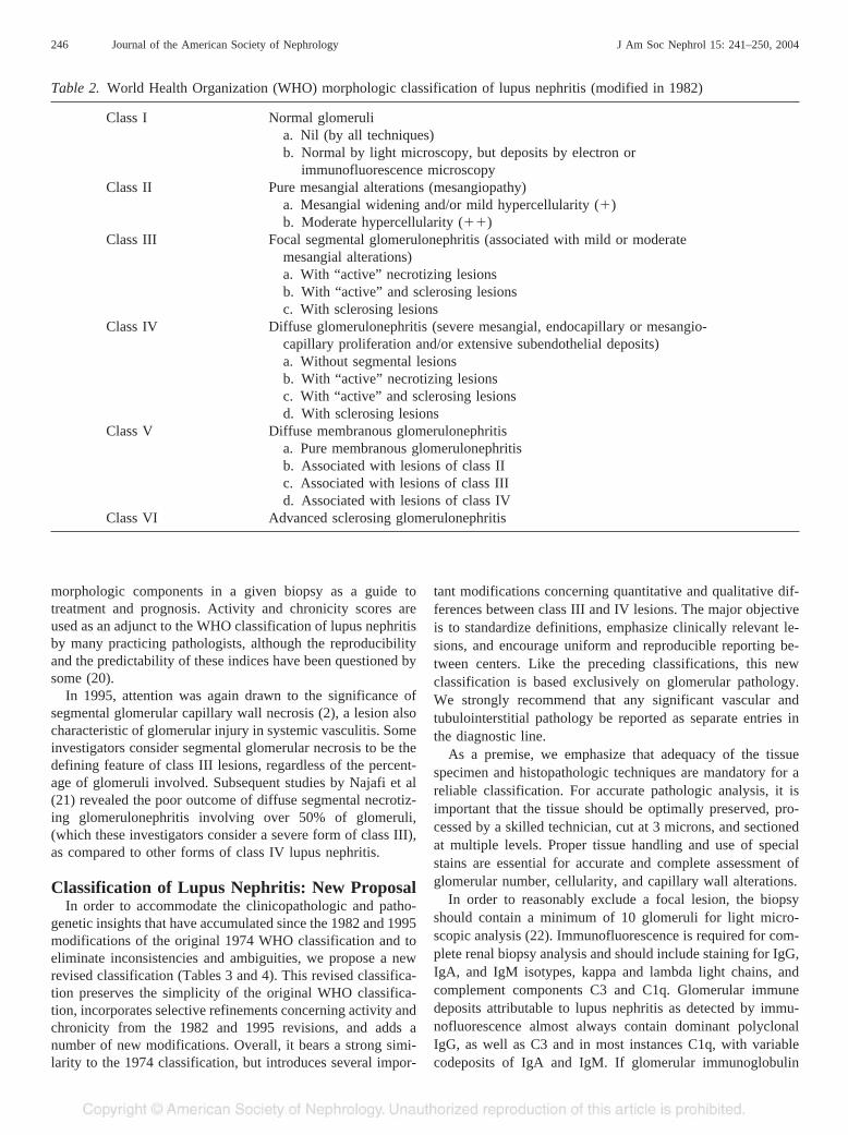
Table 2. World Health Organization (WHO) morphologic classification of lupus nephritis (modified in 1982)
Class I Normal glomerulia. Nil (by all techniques)b. Normal by light microscopy, but deposits by electron or
immunofluorescence microscopyClass II Pure mesangial alterations (mesangiopathy)
a. Mesangial widening and/or mild hypercellularity (�)b. Moderate hypercellularity (��)
Class III Focal segmental glomerulonephritis (associated with mild or moderatemesangial alterations)a. With “active” necrotizing lesionsb. With “active” and sclerosing lesionsc. With sclerosing lesions
Class IV Diffuse glomerulonephritis (severe mesangial, endocapillary or mesangio-capillary proliferation and/or extensive subendothelial deposits)a. Without segmental lesionsb. With “active” necrotizing lesionsc. With “active” and sclerosing lesionsd. With sclerosing lesions
Class V Diffuse membranous glomerulonephritisa. Pure membranous glomerulonephritisb. Associated with lesions of class IIc. Associated with lesions of class IIId. Associated with lesions of class IV
Class VI Advanced sclerosing glomerulonephritis
246 Journal of the American Society of Nephrology J Am Soc Nephrol 15: 241–250, 2004

deposits are restricted to IgA or IgM, diagnostic possibilitiesother than lupus nephritis should be considered in correlationwith serologic and clinical findings.
While the role of electron microscopy in the diagnosis andclassification of lupus glomerulonephritis cannot be underes-timated and may be essential in some cases (23), the lack ofreadily available electron microscopy facilities in many centersthroughout the world should not prevent the skilled pathologistfrom rendering a diagnosis of lupus nephritis using a combi-nation of complete light microscopic and immunofluorescencestudies. We recommend appropriate fixation and storage of a
sample of renal cortical tissue for ultrastructural evaluationwhen needed.
Definitions for diagnostic terms are given in Table 5.
Class IClass I is defined as minimal mesangial lupus nephritis with
mesangial accumulation of immune complexes identified by immu-nofluorescence, or by immunofluorescence and electron microscopy,without concomitant light microscopic alterations. A complete lack ofrenal abnormalities by light microscopy, immunofluorescence, and
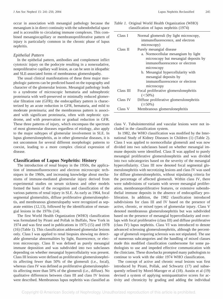
Table 3. International Society of Nephrology/Renal Pathology Society (ISN/RPS) 2003 classification of lupus nephritis
Class I Minimal mesangial lupus nephritisNormal glomeruli by light microscopy, but mesangial immune deposits by immunofluorescence
Class II Mesangial proliferative lupus nephritisPurely mesangial hypercellularity of any degree or mesangial matrix expansion by lightmicroscopy, with mesangial immune depositsMay be a few isolated subepithelial or subendothelial deposits visible by immunofluorescence orelectron microscopy, but not by light microscopy
Class III Focal lupus nephritisa
Active or inactive focal, segmental or global endo- or extracapillary glomerulonephritis involving�50% of all glomeruli, typically with focal subendothelial immune deposits, with or withoutmesangial alterations
Class III (A) Active lesions: focal proliferative lupus nephritisClass III (A/C) Active and chronic lesions: focal proliferative and sclerosing lupus nephritisClass III (C) Chronic inactive lesions with glomerular scars: focal sclerosing lupus nephritis
Class IV Diffuse lupus nephritisb
Active or inactive diffuse, segmental or global endo- or extracapillary glomerulonephritisinvolving �50% of all glomeruli, typically with diffuse subendothelial immune deposits, with orwithout mesangial alterations. This class is divided into diffuse segmental(IV-S) lupus nephritiswhen �50% of the involved glomeruli have segmental lesions, and diffuse global (IV-G) lupusnephritis when �50% of the involved glomeruli have global lesions. Segmental is defined as aglomerular lesion that involves less than half of the glomerular tuft. This class includes cases withdiffuse wire loop deposits but with little or no glomerular proliferation
Class IV-S (A) Active lesions: diffuse segmental proliferative lupus nephritisClass IV-G (A) Active lesions: diffuse global proliferative lupus nephritisClass IV-S(A/C)
Active and chronic lesions: diffuse segmental proliferative and sclerosing lupus nephritis
Active and chronic lesions: diffuse global proliferative and sclerosing lupus nephritisClass IV-S (C) Chronic inactive lesions with scars: diffuse segmental sclerosing lupus nephritisClass IV-G (C) Chronic inactive lesions with scars: diffuse global sclerosing lupus nephritis
Class V Membranous lupus nephritisGlobal or segmental subepithelial immune deposits or their morphologic sequelae by lightmicroscopy and by immunofluorescence or electron microscopy, with or without mesangialalterationsClass V lupus nephritis may occur in combination with class III or IV in which case both will bediagnosedClass V lupus nephritis show advanced sclerosis
Class VI Advanced sclerosis lupus nephritis�90% of glomeruli globally sclerosed without residual activity
a Indicate the proportion of glomeruli with active and with sclerotic lesions.b Indicate the proportion of glomeruli with fibrinoid necrosis and/or cellular crescents.Indicate and grade (mild, moderate, severe) tubular atrophy, interstitial inflammation and fibrosis, severity of arteriosclerosis or other
vascular lesions.
J Am Soc Nephrol 15: 241–250, 2004 Lupus Nephritis Reclassified 247

electron microscopy no longer qualifies as class I, and in this respectis a change from the 1974 WHO classification.
Class IIClass II is defined as mesangial proliferative lupus nephritis
(Figure 1) characterized by any degree of mesangial hypercel-lularity (defined as three or more mesangial cells per mesangialarea in a 3 micron thick section) in association with mesangialimmune deposits. By immunofluorescence or electron micros-copy, there may be rare isolated small immune deposits in-volving the peripheral capillary walls in some examples ofclass II. However, the identification of any subendothelialdeposits by light microscopy would warrant a designation ofclass III or class IV depending on the extent and distribution ofthe subendothelial deposits. Similarly, the presence of anyglobal or segmental glomerular scars that are interpreted as thesequela of previous glomerular endocapillary proliferation, ne-crosis or crescents is incompatible with class II and would beconsistent with either class III or class IV depending on thenumber of scarred glomeruli.
Class IIIClass III is defined as focal lupus nephritis involving less
than 50% of all glomeruli. Affected glomeruli usually displaysegmental endocapillary proliferative lesions (Figure 2) orinactive glomerular scars, with or without capillary wall ne-crosis and crescents, with subendothelial deposits (usually in asegmental distribution). In assessing the extent of the lesions,glomeruli with both active and sclerotic lesions will be takeninto account. Focal or diffuse mesangial alterations (includingmesangial proliferation or mesangial immune deposits) mayaccompany the focal glomerular lesions. In a pilot study ofpathologists from seven different centers on 50 consecutivecases of lupus glomerulonephritis, for a total of 350 specimens,class III lesions were found to be almost invariably segmentaland rarely global. Vasculitis-like lesions characterized by seg-
mental capillary necrosis in the absence of endocapillary pro-liferation were rare (Figure 3).
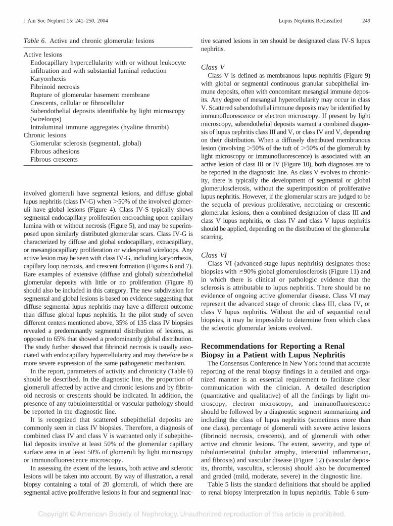
In the body of the report, parameters of activity and chro-nicity (Table 6) should be described. In the diagnostic line, theproportion of glomeruli affected by active and chronic lesionsand by fibrinoid necrosis and crescents should be indicated. Inaddition, the presence of any tubulointerstitial or vascularpathology should be reported in the diagnostic line. This newschema should facilitate correlation of the proportion of glo-meruli affected by active, necrotizing and chronic lesions andclinical outcome. A specific diagnosis of combined class IIIand class V requires membranous involvement of at least 50%of the glomerular capillary surface area of at least 50% ofglomeruli by light microscopy or immunofluorescence.
Class IVClass IV is defined as diffuse lupus nephritis involving 50% or
more of glomeruli in the biopsy. In the affected glomeruli, thelesions as described below may be segmental, defined as sparingat least half of the glomerular tuft, or global, defined as involvingmore than half of the glomerular tuft. This class is subdivided intodiffuse segmental lupus nephritis (class IV-S) when �50% of the
Table 4. Abbreviated International Society of Nephrology/Renal Pathology Society (ISN/RPS) classificationof lupus nephritis (2003)
Class I Minimal mesangial lupus nephritisClass II Mesangial proliferative lupus nephritisClass III Focal lupus nephritisa
Class IV Diffuse segmental (IV-S) or global (IV-G) lupusnephritisb
Class V Membranous lupus nephritisc
Class VI Advanced sclerosing lupus nephritis
a Indicate the proportion of glomeruli with active and withsclerotic lesions.
b Indicate the proportion of glomeruli with fibrinoid necrosis andcellular crescents.
c Class V may occur in combination with class III or IV inwhich case both will be diagnosed.
Indicate and grade (mild, moderate, severe) tubular atrophy,interstitial inflammation and fibrosis, severity of arteriosclerosis orother vascular lesions.
Table 5. Definitions
Diffuse: A lesion involving most (�50%) glomeruliFocal: A lesion involving �50% of glomeruliGlobal: A lesion involving more than half of the glomerular
tuftSegmental: A lesion involving less than half of the
glomerular tuft (i.e., at least half of the glomerular tuft isspared)
Mesangial hypercellularity: At least three mesangial cellsper mesangial region in a 3 micron thick section
Endocapillary proliferation: Endocapillary hypercellularitydue to increased number of mesangial cells, endothelialcells, and infiltrating monocytes, and causing narrowing ofthe glomerular capillary lumina
Extracapillary proliferation or cellular crescent:Extracapillary cell proliferation of more than two celllayers occupying one fourth or more of the glomerularcapsular circumference
Karyorrhexis: Presence of apoptotic, pyknotic, andfragmented nuclei
Necrosis: A lesion characterized by fragmentation of nucleior disruption of the glomerular basement membrane, oftenassociated with the presence of fibrin-rich material
Hyaline thrombi: Intracapillary eosinophilic material of ahomogeneous consistency which by immunofluorescencehas been shown to consist of immune deposits
Proportion of involved glomeruli: Intended to indicate thepercentage of total glomeruli affected by lupus nephritis,including the glomeruli that are sclerosed due to lupusnephritis, but excluding ischemic glomeruli withinadequate perfusion due to vascular pathology separatefrom lupus nephritis
248 Journal of the American Society of Nephrology J Am Soc Nephrol 15: 241–250, 2004
involved glomeruli have segmental lesions, and diffuse globallupus nephritis (class IV-G) when �50% of the involved glomer-uli have global lesions (Figure 4). Class IV-S typically showssegmental endocapillary proliferation encroaching upon capillarylumina with or without necrosis (Figure 5), and may be superim-posed upon similarly distributed glomerular scars. Class IV-G ischaracterized by diffuse and global endocapillary, extracapillary,or mesangiocapillary proliferation or widespread wireloops. Anyactive lesion may be seen with class IV-G, including karyorrhexis,capillary loop necrosis, and crescent formation (Figures 6 and 7).Rare examples of extensive (diffuse and global) subendothelialglomerular deposits with little or no proliferation (Figure 8)should also be included in this category. The new subdivision forsegmental and global lesions is based on evidence suggesting thatdiffuse segmental lupus nephritis may have a different outcomethan diffuse global lupus nephritis. In the pilot study of sevendifferent centers mentioned above, 35% of 135 class IV biopsiesrevealed a predominantly segmental distribution of lesions, asopposed to 65% that showed a predominantly global distribution.The study further showed that fibrinoid necrosis is usually asso-ciated with endocapillary hypercellularity and may therefore be amore severe expression of the same pathogenetic mechanism.
In the report, parameters of activity and chronicity (Table 6)should be described. In the diagnostic line, the proportion ofglomeruli affected by active and chronic lesions and by fibrin-oid necrosis or crescents should be indicated. In addition, thepresence of any tubulointerstitial or vascular pathology shouldbe reported in the diagnostic line.
It is recognized that scattered subepithelial deposits arecommonly seen in class IV biopsies. Therefore, a diagnosis ofcombined class IV and class V is warranted only if subepithe-lial deposits involve at least 50% of the glomerular capillarysurface area in at least 50% of glomeruli by light microscopyor immunofluorescence microscopy.
In assessing the extent of the lesions, both active and scleroticlesions will be taken into account. By way of illustration, a renalbiopsy containing a total of 20 glomeruli, of which there aresegmental active proliferative lesions in four and segmental inac-
tive scarred lesions in ten should be designated class IV-S lupusnephritis.
Class VClass V is defined as membranous lupus nephritis (Figure 9)
with global or segmental continuous granular subepithelial im-mune deposits, often with concomitant mesangial immune depos-its. Any degree of mesangial hypercellularity may occur in classV. Scattered subendothelial immune deposits may be identified byimmunofluorescence or electron microscopy. If present by lightmicroscopy, subendothelial deposits warrant a combined diagno-sis of lupus nephritis class III and V, or class IV and V, dependingon their distribution. When a diffusely distributed membranouslesion (involving �50% of the tuft of �50% of the glomeruli bylight microscopy or immunofluorescence) is associated with anactive lesion of class III or IV (Figure 10), both diagnoses are tobe reported in the diagnostic line. As class V evolves to chronic-ity, there is typically the development of segmental or globalglomerulosclerosis, without the superimposition of proliferativelupus nephritis. However, if the glomerular scars are judged to bethe sequela of previous proliferative, necrotizing or crescenticglomerular lesions, then a combined designation of class III andclass V lupus nephritis, or class IV and class V lupus nephritisshould be applied, depending on the distribution of the glomerularscarring.
Class VIClass VI (advanced-stage lupus nephritis) designates those
biopsies with �90% global glomerulosclerosis (Figure 11) andin which there is clinical or pathologic evidence that thesclerosis is attributable to lupus nephritis. There should be noevidence of ongoing active glomerular disease. Class VI mayrepresent the advanced stage of chronic class III, class IV, orclass V lupus nephritis. Without the aid of sequential renalbiopsies, it may be impossible to determine from which classthe sclerotic glomerular lesions evolved.
Recommendations for Reporting a RenalBiopsy in a Patient with Lupus Nephritis
The Consensus Conference in New York found that accuratereporting of the renal biopsy findings in a detailed and orga-nized manner is an essential requirement to facilitate clearcommunication with the clinician. A detailed description(quantitative and qualitative) of all the findings by light mi-croscopy, electron microscopy, and immunofluorescenceshould be followed by a diagnostic segment summarizing andincluding the class of lupus nephritis (sometimes more thanone class), percentage of glomeruli with severe active lesions(fibrinoid necrosis, crescents), and of glomeruli with otheractive and chronic lesions. The extent, severity, and type oftubulointerstitial (tubular atrophy, interstitial inflammation,and fibrosis) and vascular disease (Figure 12) (vascular depos-its, thrombi, vasculitis, sclerosis) should also be documentedand graded (mild, moderate, severe) in the diagnostic line.
Table 5 lists the standard definitions that should be appliedto renal biopsy interpretation in lupus nephritis. Table 6 sum-
Table 6. Active and chronic glomerular lesions
Active lesionsEndocapillary hypercellularity with or without leukocyteinfiltration and with substantial luminal reductionKaryorrhexisFibrinoid necrosisRupture of glomerular basement membraneCrescents, cellular or fibrocellularSubendothelial deposits identifiable by light microscopy(wireloops)Intraluminal immune aggregates (hyaline thrombi)
Chronic lesionsGlomerular sclerosis (segmental, global)Fibrous adhesionsFibrous crescents
J Am Soc Nephrol 15: 241–250, 2004 Lupus Nephritis Reclassified 249
marizes a number of markers for activity and chronicity oflupus nephritis that we propose should be included in thereport. Tubulointerstitial and vascular markers of activity andchronicity can also be applied. Activity and chronicity can bescored semiquantitatively using the system formulated by Aus-tin et al (19) or as agreed upon in individual medical centersaccording to institutional preference.
Similar guidelines should apply to the reporting of repeatrenal biopsies in an individual patient. In such cases, a com-parison with the previous biopsy should be made and importantchanges in class, activity, and chronicity should be highlighted.
Finally, it is important to realize that the renal biopsy find-ings, per se, cannot be used to establish a diagnosis of SLE.The renal biopsy findings must be interpreted by the referringclinician in the context of the patient’s entire clinical presen-tation, including serologic findings.
AcknowledgmentsThe authors would like to thank Dr. J. Churg, Dr. R. McCluskey,
Dr. C. Pirani, and Dr. V. Pollak for their positive comments, and Dr.T. Nadasdy for critical review of the manuscript. The Lupus NephritisConsensus Meeting at Columbia University, College of Physiciansand Surgeons was supported financially by The International Societyof Nephrology, The Renal Pathology Society, The S.L.E. Foundation,Inc., The Mary Kirkland Center for Lupus Research, and The Kidneyand Urology Foundation of America.
References1. Churg J, Sobin LH: Renal Disease: Classification and Atlas of
Glomerular Disease, Tokyo, Igaku-Shoin, 19822. Churg J, Bernstein J, Glassock RJ: Renal Disease: Classification
and Atlas of Glomerular Diseases, 2nd Ed., New York, Igaky-Shoin, 1995
3. Mecklenbrauker I, Saijo K, Zheng NY, Leitges M, TarakhovskyA: Protein kinase C� controls self-antigen-induced B-cell toler-ance. Nature 416: 860–865, 2002
4. Stuart L, Hughes J: Apoptosis and autoimmunity. Nephrol DialTransplant 17: 697–700, 2002
5. Berden JH: Lupus nephritis. Kidney Int 52: 538–558, 19976. Walport MJ, Davies KA, Botto M: C1q and systemic lupus
erythematosus. Immunobiology 199: 265–285, 19987. Lea J: Lupus nephritis in African Americans. Am J Med Sci 323:
85–89, 20028. Clynes R, Dumitru C, Ravetch JV: Uncoupling of immune
complex formation and kidney damage in autoimmune glomer-ulonephritis. Science 279: 1052–1054, 1998
9. Daugas E, Nochy D, Huong du LT, Duhaut P, Beaufils H,Caudwell V, Bariety J, Piette JC, Hill G: Antiphospholipidsyndrome nephropathy in systemic lupus erythematosus. J AmSoc Nephrol 13: 42–52, 2002
10. Marshall S, Dressler R, D’Agati V: Membranous lupus nephritiswith antineutrophil cytoplasmic antibody-associated segmentalnecrotizing and crescentic glomerulonephritis. Am J Kidney Dis29: 119–124, 1997
11. Fries JW, Mendrick DL, Rennke HG: Determinants of immune com-plex-mediated glomerulonephritis. Kidney Int 34: 333–345, 1988
12. Pollak VE, Pirani C, Schwartz FD: The natural history of therenal manifestations of systemic lupus erythematosus. J Lab ClinMed 63: 537–550, 1964
13. Baldwin DS, Lowenstein J, Rothfield NJ, Gallo GR, McCluskeyRT: The clinical course of proliferative and membranous formsof lupus nephritis. Ann Intern Med 73: 929–942, 1970
14. Baldwin DS, Gluck MC, Lowenstein J, Gallo GR: Lupus nephri-tis: Clinical course as related to morphologic forms and theirtransitions. Am J Med 62: 12–30, 1977
15. McCluskey RT: Lupus nephritis. In: Kidney Pathology Decen-nial 1966–1975, edited by Sommers SC, East Norwalk, CT,Appleton-Century-Crofts, 1975, pp 435–450
16. Appel GB, Silva FG, Pirani CL: Renal involvement in systemiclupus erythematosus (SLE): A study of 56 patients emphasizinghistologic classification. Medicine 75: 371–410, 1978
17. Pirani CL, Pollak VE, Schwartz FD: The reproducibility of semiquan-titative analysis of renal histology. Nephron 1: 230–237, 1964
18. Morel-Maroger L, Mery JP, Droz D, Godin M, Verroust P, KourilskyO, Richet G: The course of lupus nephritis: Contribution of serial renalbiopsies. Adv Nephrol Necker Hosp 6: 79–118, 1976
19. Austin HA 3rd, Muenz LR, Joyce KM, Antonovych TT, Balow JE:Diffuse proliferative lupus nephritis: Identification of specific patho-logic features affecting renal outcome. Kidney Int 25: 689–695, 1984
20. Schwartz MM, Lan SP, Bernstein J, Hill GS, Holley K, Lewis EJ:Irreproducibility of the activity and chronicity indices limits theirutility in the management of lupus nephritis. Lupus Nephritis Col-laborative Study Group. Am J Kidney Dis 21: 374–377, 1993
21. Najafi CC, Korbet SM, Lewis EJ, Schwartz MM, Reichlin M,Evans J: Significance of histologic patterns of glomerular injuryupon long-term prognosis in severe lupus glomerulonephritis.Kidney Int 59: 2156–2163, 2001
22. Corwin HL, Schwartz MM, Lewis E: The importance of sample size inthe interpretation of the renal biopsy. Am J Nephrol 8: 85–93, 1988
23. Herrera GA: The value of electron microscopy in the diagnosisand clinical management of lupus nephritis. Ultrastruct Pathol23: 63–77, 1999
See related editorial, “Reclassification of Lupus Glomerulonephritis: Back to the Future,” on pages 501–503.
Access to UpToDate on-line is available for additional clinical informationat http://www.jasn.org/
250 Journal of the American Society of Nephrology J Am Soc Nephrol 15: 241–250, 2004