


The central role of mitocondria is subscribed principally in the ...
19
1 ATYPICAL CRISTAE MORPHOLOGY OF HUMAN SYNCYTIOTROPHOBLAST MITOCHONDRIA: ROLE FOR COMPLEX V Daniela De Los Rios Castillo 1 , Mariel Zarco-Zavala 2 , Sofia Olvera-Sanchez 1 , Juan Pablo Pardo 1 , Oscar Juarez 3 , Federico Martinez 1 , Guillermo Mendoza-Hernandez 1 , José J. García- Trejo 2 and Oscar Flores-Herrera 1 * 1 National Autonomous University of Mexico, Medicine Faculty, Department of Biochemistry & Molecular Biology, Mexico City, Mexico, 2 National Autonomous University of Mexico, Chemistry Faculty, Department of Biology, Mexico City, Mexico, and 3 Biology Department, Center for Biotechnology and Interdisciplinary Studies, Rensselaer Polytechnic Institute, Troy, NY. Run head: Mitochondrial morphology in human placenta *Corresponding to: PhD. Oscar Flores-Herrera, Departamento de Bioquímica, Facultad de Medicina, Universidad Nacional Autonoma de México, Apdo Postal 70-159, Coyoacan 04510, Mexico, D. F., México, Phone: 01-55-56232510. Fax: 01-55-56162419; E-mail: [email protected] Mitochondrial complexes I, III 2 and IV from human cytotrophoblast and syncytiotrophoblast associate to form supercomplexes or respirosomes, with the following stoichiometries: I 1 :(III 2 ) 1 and I 1 :(III 2 ) 1-2 :IV 1-4 . The content of respirosomes was similar in both cell types after isolating mitochondria. However, syncytiotrophoblast mitochondria possess low levels of dimeric complex V and do not have orthodox cristae morphology. In contrast, cytotrophoblast mitochondria show normal cristae morphology and a higher content of ATP synthase dimer. Consistent with the dimerizing role of the ATPase inhibitory protein (IF 1 ) (García, J. J., Morales-Ríos, E., Cortés-Hernandez, P., and Rodríguez- Zavala, J. S. (2006) Biochemistry 45:12695- 12703) higher relative amounts of IF 1 were observed in cytotrophoblast compared with syncythiotrophloblast mitochondria. Therefore, there is a correlation between dimerization of complex V, IF 1 expression and the morphology of mitochondrial cristae in human placental mitochondria. The possible relationship between cristae architecture and the physiological function of the syncytiotrophoblast mitochondria is discussed. Oxidative phosphorylation, the main source of ATP in aerobic cells, relies on the activity of two components, the oxidative and the phosphorylation systems. The oxidative system (respiratory chain) couples redox reactions to the production of a proton electrochemical gradient, which drives the synthesis of ATP by the phosphorylation system (F 0 F 1 -ATP synthase and the ADP and phosphate carriers). Since the respiratory complexes of the electron transport chain can be isolated, preserving its functional properties, it was generally accepted that these complexes exist as isolated entities that can move laterally and independently in the mitochondrial inner membrane (1-3). However, during the last decade a large number of studies have revealed the strong association between the electron transport complexes into the so-called respiratory supercomplexes. These associations are widespread and can be found in a variety of different mitochondria, including those of vertebrates (4, 5), plants (6), fungi (4) and in the respiratory chains of some bacteria such as Paracoccus denitrificans (7) where by contrast, the complex V or ATP synthase is monomeric (8,9). However, some major differences are evident; for instance, in plants the major supercomplex is the I-III 2 (10), while in mammals other supercomplexes can be found, such as the I-III 2 , I-III 2 -IV, I-III 2 -IV 2 and I-III 2 - IV 4 (4, 5). The evidence indicates that supercomplexes may have significant roles in cell physiology, such as an increase in complex stability, substrate channeling, and reduction of reactive oxygen species (ROS) production. On the other hand, mitochondrial F 1 F 0 ATP synthase self-associates in dimeric and oligomeric structures in bovine (8,11,12), http://www.jbc.org/cgi/doi/10.1074/jbc.M111.252056 The latest version is at JBC Papers in Press. Published on May 13, 2011 as Manuscript M111.252056 Copyright 2011 by The American Society for Biochemistry and Molecular Biology, Inc. by guest on March 29, 2018 http://www.jbc.org/ Downloaded from
Transcript of The central role of mitocondria is subscribed principally in the ...

1
ATYPICAL CRISTAE MORPHOLOGY OF HUMAN SYNCYTIOTROPHOBLAST
MITOCHONDRIA: ROLE FOR COMPLEX V
Daniela De Los Rios Castillo1, Mariel Zarco-Zavala
2, Sofia Olvera-Sanchez
1, Juan Pablo
Pardo1, Oscar Juarez
3, Federico Martinez
1, Guillermo Mendoza-Hernandez
1, José J. García-
Trejo2 and Oscar Flores-Herrera
1*
1National Autonomous University of Mexico, Medicine Faculty, Department of Biochemistry &
Molecular Biology, Mexico City, Mexico, 2National Autonomous University of Mexico, Chemistry
Faculty, Department of Biology, Mexico City, Mexico, and 3Biology Department, Center for
Biotechnology and Interdisciplinary Studies, Rensselaer Polytechnic Institute, Troy, NY.
Run head: Mitochondrial morphology in human placenta
*Corresponding to: PhD. Oscar Flores-Herrera, Departamento de Bioquímica, Facultad de
Medicina, Universidad Nacional Autonoma de México, Apdo Postal 70-159, Coyoacan 04510,
Mexico, D. F., México, Phone: 01-55-56232510. Fax: 01-55-56162419; E-mail:
Mitochondrial complexes I, III2 and
IV from human cytotrophoblast and
syncytiotrophoblast associate to form
supercomplexes or respirosomes, with the
following stoichiometries: I1:(III2)1 and
I1:(III2)1-2:IV1-4. The content of respirosomes
was similar in both cell types after isolating
mitochondria. However, syncytiotrophoblast
mitochondria possess low levels of dimeric
complex V and do not have orthodox cristae
morphology. In contrast, cytotrophoblast
mitochondria show normal cristae
morphology and a higher content of ATP
synthase dimer. Consistent with the
dimerizing role of the ATPase inhibitory
protein (IF1) (García, J. J., Morales-Ríos, E.,
Cortés-Hernandez, P., and Rodríguez-
Zavala, J. S. (2006) Biochemistry 45:12695-
12703) higher relative amounts of IF1 were
observed in cytotrophoblast compared with
syncythiotrophloblast mitochondria.
Therefore, there is a correlation between
dimerization of complex V, IF1 expression
and the morphology of mitochondrial cristae
in human placental mitochondria. The
possible relationship between cristae
architecture and the physiological function
of the syncytiotrophoblast mitochondria is
discussed.
Oxidative phosphorylation, the main
source of ATP in aerobic cells, relies on the
activity of two components, the oxidative and
the phosphorylation systems. The oxidative
system (respiratory chain) couples redox
reactions to the production of a proton
electrochemical gradient, which drives the
synthesis of ATP by the phosphorylation
system (F0F1-ATP synthase and the ADP and
phosphate carriers). Since the respiratory
complexes of the electron transport chain can
be isolated, preserving its functional properties,
it was generally accepted that these complexes
exist as isolated entities that can move laterally
and independently in the mitochondrial inner
membrane (1-3).
However, during the last decade a large
number of studies have revealed the strong
association between the electron transport
complexes into the so-called respiratory
supercomplexes. These associations are
widespread and can be found in a variety of
different mitochondria, including those of
vertebrates (4, 5), plants (6), fungi (4) and in
the respiratory chains of some bacteria such as
Paracoccus denitrificans (7) where by contrast,
the complex V or ATP synthase is monomeric
(8,9). However, some major differences are
evident; for instance, in plants the major
supercomplex is the I-III2 (10), while in
mammals other supercomplexes can be found,
such as the I-III2, I-III2-IV, I-III2-IV2 and I-III2-
IV4 (4, 5). The evidence indicates that
supercomplexes may have significant roles in
cell physiology, such as an increase in complex
stability, substrate channeling, and reduction of
reactive oxygen species (ROS) production.
On the other hand, mitochondrial F1F0
ATP synthase self-associates in dimeric and
oligomeric structures in bovine (8,11,12),
http://www.jbc.org/cgi/doi/10.1074/jbc.M111.252056The latest version is at JBC Papers in Press. Published on May 13, 2011 as Manuscript M111.252056
Copyright 2011 by The American Society for Biochemistry and Molecular Biology, Inc.
by guest on March 29, 2018
http://ww
w.jbc.org/
Dow
nloaded from

2
Saccharomyces cerevisiae (4,13,14),
Polytomella sp. (15,16) and Chlamydomonas
reinhardtii (17,18). The F0F1-ATP synthase
dimer (V2) seems to be closely related with the
mitochondrial architecture. In particular, it has
been suggested that the presence of cristae in
mitochondria depends on the ability of complex
V to form dimers (11,19-21). The factors
involved in the dimerization process appears to
be different in several organisms; in yeast
mitochondria the F1F0 dimer is stabilized by F0
subunits e and g (22-25) while in the
chlorophyceaus algae Chlamydomonas
reinhardii and Polytomella sp., the ASA1-8
subunits are involved in this process (16,17).
Additionally, in bovine and rat mitochondria,
this dimerizing role is also accomplished by the
inhibitor protein (IF1) (12) along with the
dimerizing F0 subunits; this is in contrast with
yeast IF1 which does not seem to be essential
for F1F0 dimerization (26).
Studies on the supramolecular
associations of the oxidative phosphorylation
system have been typically performed in
mitochondria of aerobic cells, whose main role
is the synthesis of ATP. Our group has been
interested in the physiology of mitochondria
from steroidogenic tissues, such as human
placenta. The latter contains two functionally
different layers, the cytotrophoblast and the
syncytiotrophoblast. The cytotrophoblast shows
mitotic activity and is in contact with the
endometrium; by contrast, the
syncytiotrophoblast is facing the uterus, has no
mitotic activity, and displays a high
steroidogenic activity, with progesterone as the
main product. We have described previously
that the morphology of the syncytiotrophoblast
mitochondria is far from typical, showing a
highly condensed matrix and an irregular
folding of the mitochondrial inner membrane
(27). The present study represents the first
report addressing the presence and analysis of
respiratory chain complexes and
supercomplexes of human placental
mitochondria and their role in cristae
morphology. It is shown that the atypical
morphology of the syncytiotrophoblast
mitochondria correlates with low contents of
dimeric F0F1 ATP synthase and of the
inhibitory IF1 subunit confirming the key role
played by these two factors in determining
mitochondrial cristae morphology.
Experimental Procedures
Isolation of human cytotrophoblast and
syncytiotrophoblast mitochondria- Full term
human placenta was collected immediately
after normal delivery. Mitochondria were
prepared as previously described (27). To
purify mitochondria of cytotrophoblast or
syncytiotrophoblast, the enriched mitochondrial
suspension was loaded on a 35% sucrose
solution (25 ml) and centrifuged at 15,000 X g
for 45 min, at 4oC. The two mitochondrial
fractions were collected and centrifuged at
16,000 X g for 15 min at 4oC, and the
mitochondrial pellet resuspended in 250 mM
sucrose, 10 mM Tris (pH 7.4) and stored at -
70oC until use. Protein concentration was
measured as reported (28, 29).
Mitochondrial oxygen consumption- Oxygen
uptake was estimated polarographically using a
Clark type electrode in a mixture containing
250 mM sucrose, 10 mM HEPES pH 7.4, 1
mM EGTA, 1 mM EDTA, 10 mM succinate,
10 mM KH2PO4, 5 mM MgCl2, 0.2% bovine
serum albumin and 1 mg/ml of cytotrophoblast
or syncytiotrophoblast mitochondrial protein
(30). Temperature was set at 37oC and oxygen
consumption was stimulated by the addition of
300-500 nmol ADP.
Mitochondrial enzyme activity determinations-
Activities of complex I (NADH:DCPIP
oxidoreductase) and complex II
(succinate:DCPIP oxidoreductase) were
determined spectrophotometrically at 600 nm
by following the reduction of the artificial
electron acceptor 2,6-dichlorophenol-
indophenol (DCPIP; 50 M; єDCPIP = 21 mM-
1cm
-1). Mitochondria permeabilized with 0.01%
Triton X100 were incubated in 30 mM
KH2PO4, 5 mM MgCl2, 1 mM EGTA, 120 mM
KCl, pH 7.4, and either 500 M NADH
(complex I) or 2 mM succinate (complex II).
The protein concentration of cytotrophoblast or
syncytiotrophoblast mitochondria was 50 g/ml
and the reaction was started by the addition of
NADH or succinate.
by guest on March 29, 2018
http://ww
w.jbc.org/
Dow
nloaded from

3
ATP synthesis by complex V was
measured at 37°C using an assay coupled to the
reduction of NADP+ (є340 nm = 6.2 mM
-1cm
-1).
The reaction mixture contained 0.5 mM
NADP+, 1 mM ADP, 6 units/ml glucose-6-
phosphate dehydrogenase, 16 units/ml
hexokinase, 10 mM succinate, 100μM P1,P
5-
Di(adenosine-5´)pentaphosphate penta-
ammonium, 10 mM glucose, 150 mM sucrose,
5 mM MgCl2, 20 mM Tris/HCl, and 20 mM
KH2PO4, pH 7.5. ATP synthesis was started by
the addition of cytotrophoblast or
syncytiotrophoblast mitochondria (50 g/ml).
The values reported were obtained by
substracting the rate of ATP synthesis in the
presence of oligomycin (5 g/mg mitochondrial
protein) from the amount of ATP synthesis in
the control conditions.
Mitochondrial progesterone synthesis-
Progesterone synthesis was determined at 37oC
as reported previously (30) in 120 mM KCl, 10
mM MOPS, 0.5 mM EGTA, 10 mM isocitrate,
4 g of aprotinin/ml, 1 M leupeptin, 5 mM
KH2PO4 pH 7.4, in a final volume of 500 l
with 1 mg/ml of cytotrophoblast or
syncytiotrophoblast mitochondrial protein.
Where indicated, 25 M 22(R)-hydroxy-
cholesterol was added. After 60 min incubation,
the reaction was stopped with 50- l methanol
and progesterone was determined by
radioimmunoassay kit (Diagnostic Systems
Laboratories, Inc. Webster, Texas, USA)
according to the manufacturer’s instructions.
The concentration of progesterone at time zero
was subtracted from the amount of
progesterone at 60 min and this net value of
progesterone synthesis was reported.
Electron microscopy- Samples for transmission
electron microscopy were fixed in 2%
glutaraldehyde, post-fixed with osmium
tetroxide and sequentially dehydrated with
increasing concentrations of ethanol. Finally,
the samples were embedded in an epoxy resin
(31). Sections were cut and stained with uranyl
acetate and lead citrate, and observed under a
Jeol electron microscope, operated at 60 kV.
Placental tissue was fixed for immunogold
studies with 4% paraformaldehyde, 0.2%
glutaraldehyde and phosphate buffered saline
(31).
Sample preparation for native electrophoresis-
The respiratory complexes from either
cytotrophoblast or syncytiotrophoblast
mitochondria were resolved by native PAGE
following the general procedures reported
previously (32, 33) with minor modifications.
Briefly, cytotrophoblast (2 mg) or
syncytiotrophoblast (2 mg) mitochondria were
suspended in 50 mM Bis-Tris and 500 mM 6-
aminocaproic acid, pH 7.0 and solubilized by
adding dodecyl- -D-maltoside or digitonin, at
detergent/protein ratios ranging from 0.5 – 10
(g/g), in a final volume of 200 l. The mixtures
were incubated for 30 min at 4oC, and
centrifuged at 100 000 X g for 30 min at 4oC.
The supernatants were recovered and
immediately loaded on a linear polyacrylamide
gradient gel (5 – 10% or 3.25 – 7.5%) for Blue
Native PAGE (BN-PAGE) or Clear Native
PAGE (CN-PAGE) (34). The molecular weight
of each respiratory complex or supercomplex
was estimated by using the bovine
mitochondrial complexes as standard.
In-gel catalytic activity assays- The in-gel
activity assays were performed as described by
Wittig (34). Gel strips were assayed for
complex I activity (NADH:
methylthiazolyldiphenyl tetrazolium bromide
(MTT) reductase), complex II activity
(succinate: MTT reductase), and complex IV
activity (cytochrome c: diaminobenzidine
(DAB) reductase). In all cases, the assays were
performed at 20–25oC, and stopped with 50%
methanol, 10% acetic acid, after 10–25 min.
To estimate the in-gel ATP hydrolysis
activity of monomeric and dimeric complex V
and of the F1-ATPase subcomplex, gel strips
were preincubated in 50 mM glycine (adjusted
to pH 8.0 with triethanolamine) for two hours at
37oC in the presence or absence of the complex
V inhibitor oligomycin (5 g/ml). The
equilibration solution was discarded and the
gels strips were then added into the assay buffer
(50 mM glycine, adjusted to pH 8.0 with
triethanolamine, 10 mM MgCl2, 0.2%
Pb(NO3)2, and 8 mM ATP), with or without 5
g/ml oligomycin. ATP hydrolysis correlated
by guest on March 29, 2018
http://ww
w.jbc.org/
Dow
nloaded from

4
with the development of white lead phosphate
precipitates. The reaction was stopped using
50% methanol and subsequently the gel was
transferred to water and scanned against a dark
background as described previously (11, 35).
2D-Tricine-SDS gel electrophoresis and
Western blot analysis- Proteins of a gel lane
excised from native PAGE were separated by
2D-Tricine-SDS-PAGE according to (32) on a
16% polyacrylamide gel under denaturing
conditions. Afterwards, the proteins were
stained with either Coomassie© Brillant Blue
R-125 or silver using a commercial kit (Bio-
Rad). Alternatively, the gels were
electrotransferred to polyvinylidene difluoride
membrane (Immobilon P; Millipore, Bedford,
MA) in a semi-dry electroblotting system (Bio-
Rad) at 25 V for 50 min, or 2 h at 100 mA in a
buffer containing 100 mM CAPS, 10%
methanol (pH = 11). Membranes were blocked
in 500 mM NaCl, 0.05% Tween-20, and 20
mM Tris-Base, pH 7.5 (TTBS buffer),
containing 5% blotting grade blocker non-fat
dry milk (BioRad). The antibodies used were
anti- subunit polyclonal antibody (1:10,000),
anti- subunit antibody (1:250), anti-OSCP
subunit antibody (1:1000), and anti-IF1
monoclonal antibody (1:10,000). Membranes
were incubated for 1 h with the primary
antibody in TTBS buffer, washed thoroughly,
and the immunoreactive bands were visualized
with the Enhanced ChemiLuminescence assay
(Amersham Life Science, Inc) according to the
manufacturer’s instructions, using horseradish
peroxidase-conjugated goat antimouse IgG
(Pierce) at a dilution of 1:35,000, and the
densitometric analyses were performed with the
Alpha-DigiDoc 1000 software (AlphaEaseFCTM
from Alpha Innotech Corporation). The
intensities of subunits were measured by peak
integration after densitometry analyses.
Tandem Mass Spectrometry (LC/ESI-MS/MS)-
The protein spots (indicated by numbers in
figures 2A and 3C, and Table 2) were excised
from the Coomassie stained SDS gels,
distained, reduced, carbamidomethylated and
digested with modified porcine trypsin
(Promega, Madison, WI). Peptide mass
spectrometric analysis was carried out using a
3200 Q TRAP hybrid tandem mass
spectrometer (Applied Biosystems/MDS Sciex,
Concord, ON, Canada), equipped with a
nanoelectrospray ion source (NanoSpray II) and
a MicroIonSpray II head (36). The instrument
was coupled on line to a nanoAcquity Ultra
Performance LC system supplied by Waters
(Waters Corporations, Milford, MA, USA).
Briefly, spectra were acquired in automated
mode using Information Dependent Acquisition
(IDA). Precursor ions were selected in Q1
using the enhanced MS mode (EMS) as survey
scan. The EMS was followed by an enhanced
resolution scan (ER) of the three most intense
ions at the low speed of 250 amu/s to determine
the ion charge states and afterwards by an
enhanced product ion scan (EPI). The precursor
ions were fragmented by collisionally-activated
dissociation (CAD) in the Q2 collision cell. The
fragment ions generated were captured and
mass analyzed in the Q3 linear ion trap.
Database searching and protein
identification were performed with the MS/MS
spectra data sets using the MASCOT search
algorithm (version 1.6b9, Matrix Science,
London, U.K., available at
http://www.matrixscience.com). Mass
tolerances of 0.5 Da for the precursor and 0.3
Da for the fragment ion masses were used.
Carbamidomethyl-cysteine was the fixed
modification and one missed cleavage for
trypsin was allowed. Searches were conducted
using the Human subset of the NCBInr
database (http://www.ncbi.nih.gov). Protein
identifications were accepted when at least two
MS/MS spectra matched at 95% confidence
level (p < 0.05).
Materials- Analytical grade reagents were
purchased from Sigma Chemical Co. (St. Louis,
MO, USA), E. Merck (Darmstadt, Germany),
and BioRad (Hercules, CA, USA); Antibodies
were purchase of Santa Cruz Biotechnology,
and MitoSciences®.
by guest on March 29, 2018
http://ww
w.jbc.org/
Dow
nloaded from

5
RESULTS
Human placental mitochondria
architecture. Transmission electron
micrographs of term placenta sections revealed
two morphological types of mitochondria in the
trophoblast cells (Figure 1A). Large
mitochondria were observed in the
cytotrophoblast cells, whose morphology is
similar to that of typical liver mitochondria,
containing lamellar (and presumably tubular)
cristae in an orthodox configuration. In contrast
the syncytiotrophoblast contained smaller
mitochondria with a condensed matrix and
cristae composed by vesicular regions
connected by narrow tubules. The larger
cytotrophoblast mitochondria had a round
shape, whereas the syncytiotrophoblast
mitochondria display an irregular shape with
protuberances of the outer and inner
membranes. Isolated cytotrophoblast and
syncytiotrophoblast mitochondria retained only
partially these structural characteristics (Figure
1B); in particular, the isolated cytotrophoblast
mitochondria had lost partially the original
cristae configuration seen in situ (Fig 1A).
Physiological state of cytotrophoblast
and syncytiotrophoblast mitochondria. To
determine the functional integrity of isolated
cytotrophoblast and syncytiotrophoblast
mitochondria, respiratory rates and respiratory
controls were calculated from oxygen uptake
traces using succinate as substrate (Table 1).
Values of respiratory control ranged between
2.85 and 12, higher than those previously
reported for this tissue (37); ATP synthesis by
complex V was 151 16 nmol/mg/min and 153
13 nmol/mg/min for cytotrophoblast and
syncytiotrophoblast mitochondria, respectively,
suggesting coupling of mitochondrial
respiration and ATP synthesis. In addition, we
obtained an activity of 113-110 mol/mg/min
for the NADH:DCPIP oxide reductase activity
(complex I), and 12 to 14 mol/mg/min for the
succinate:DCPIP oxide reductase activity
(complex II) (Table 1). These results indicate
the presence of functional mitochondria in both
cell types, retaining the ability to increase the
consumption of oxygen and the synthesis of
ATP upon the addition of ADP.
Human placental mitochondria are
steroidogenic organelles that synthesize
progesterone, due to the presence of the 3- -
hydroxy steroid dehydrogenase in their inner
membrane (27, 38, 39). Since there are two
types of mitochondria, its steroidogenic activity
was determined. Table 1 show that synthesis of
progesterone by syncytiotrophoblast
mitochondria (35.7 0.9 ng
progesterone/mg/min) was tenfold higher than
that of cytotrophoblast mitochondria (3.6 1.34
ng progesterone/mg/min). In both cases, 22(R)-
hydroxy-cholesterol, a soluble substrate used to
assess maximal steroidogenic activity (40)
increase steroidogenic activity to 92.2 ± 3.4 and
10.1 ± 3.95 ng progesterone/mg/h in
syncytiotrophoblast and cytotrophoblast
mitochondria, respectively. These results are in
concordance with the specialized role of each
placental tissue (27) and support that isolated
mitochondria retain their physiological
function.
Identification of respiratory
supercomplexes in mitochondria from
cytotrophoblast and syncytiotrophoblast cells.
To investigate the optimal condition for the
separation of mitochondrial complexes and
supercomplexes, isolated mitochondria from
both cytotrophoblast and syncytiotrophoblast
were solubilized by varying concentrations of
either digitonin or dodecyl- -D-maltoside
(DDM). Results showed that 1-2 g DDM per g
of mitochondrial protein allowed the
solubilization of the respiratory chain
complexes in their monomeric state (Figure
2A), except for complex III, which exists as a
stable dimer. BN-PAGE allowed the resolution
of all the complexes of the oxidative
phosphorylation system (Figure 2A). The
identity and position of complex I, II, IV, and V
on gels were determined using specific
reactions for these complexes (Figure 2A). The
molecular mass estimated for each complex
was: complex I, 1000 kDa; complex V, 750
kDa; complex III2, 500 kDa; complex IV, 200
kDa, and complex II, 130 kDa. In addition, the
identity of each complex was confirmed by
their known subunit composition upon
resolution on 2D-SDS-PAGE and by
identification of subunits by mass spectrometry
by guest on March 29, 2018
http://ww
w.jbc.org/
Dow
nloaded from

6
(Figure 2B and Table 2). Identical results were
obtained for cytotrophoblast and
syncytiotrophoblast mitochondria (Table 2).
With digitonin as the solubilizing agent,
it was possible to isolate the mitochondrial
respiratory chain components as individual
entities or as supercomplexes. The optimal
digitonin/mitochondrial protein ratio was found
between 1.5 and 10. However, the abundance of
some supercomplexes decreased slightly at
higher detergent to protein ratios; therefore, all
further experiments were carried out with a
digitonin:protein ratio of 1.5 (g/g). The identity
and position of the individual complexes I, V,
IV, and II, as well as the composition of
supercomplexes named a – e, were determined
using specific reactions for these complexes
(Figure 3A). The molecular mass of each
individual complex was essentially identical to
that obtained using DDM for the solubilization
process (Table 2). However, the presence of
five different supercomplexes was clearly
evident in the gel. To resolve the composition
of these supercomplexes, BN-PAGE was
carried out in 3.25-7.5% linear polyacrylamide
gradient gels. As shown in figure 3B, about
70% of the NADH:MTT oxidoreductase
activity was located at the position
corresponding to the supercomplexes, while the
cytochrome c:DAB oxidoreductase activity was
detected only in three of them (c – e) (Figure
3B). Occurrence of complex III was determined
by 2D-SDS-PAGE followed by spot
identification by mass spectrometry (Figure
3C). The molecular masses estimated for the
supercomplexes and their possible stoichometry
were: a = 1,300 kDa, (I1:III2); b = 1,500 kDa,
(I1:III2); c = 1,700 kDa, (I1:III2:IV1); d = 2,000
kDa, (I1:III2:IV2-3); and e = 2,300 kDa,
(I1:(III2)1-2:IV1-4).
Monomeric and dimeric F0F1-ATP
synthase in mitochondria from cytotrophoblast
and syncytiotrophoblast. Solubilization of
cytotrophoblast and syncytiotrophoblast
mitochondria with digitonin (1.5 mg/mg
protein), allowed the visualization of the
activity of the monomeric (V) and dimeric (V2)
forms of the F0F1-ATP synthase complex in
BN-PAGE (Figure 3A). It has been reported
that the ATPase activity of the F0F1 complex is
affected by the Coomassie used in the BN-
PAGE (41); therefore, we used the CN-PAGE
to visualize the F0F1 complex activity (Figure
3A, right panel). In accordance with a previous
report (42), the F0F1-ATP synthase is present in
two forms, a monomeric complex of about 750
kDa and a dimeric complex of about 1500 kDa.
In cytotrophoblast mitochondria the in-gel
ATPase activity of V, evidenced as a white
precipitated, was greater than that of V2 (Figure
3A, right panel), while in syncytiotrophoblast
mitochondria this difference in ATPase activity
was much larger. Indeed, in some
syncytiotrophoblast mitochondria preparations,
the ATP synthase activity was represented by
the monomeric form exclusively. Thus, the V2
ATPase activity for the cytotrophoblast
mitochondria was higher than that of
syncytiotrophoblast mitochondria. Since the
differences in intensity of in-gel ATPase bands
may result from different specific activities,
2D-SDS-PAGE was carried out after BN-
PAGE to estimate the complex V
dimer/monomer ratio from Coomassie stained
and subunits. The localization and MS
identification of and in the 2D-SDS-PAGE
(Fig 3C spots 2 and 3), confirmed that the ATP
synthase extracted from cytotrophoblast
mitochondria exist in roughly equal amounts in
dimeric and monomeric forms (Fig 3C left
panel); in contrast, in syncytiotrophoblast
mitochondria only trace amounts of the F1F0
dimer was observed and the monomeric ATP
synthase was therefore highly enriched (Figure
3C and Table 2). These results suggest that in
syncytiotrophoblast mitochondria the ATP
synthase exists preferably in the monomeric
state.
It has been proposed that dimerization
of the F0F1-ATP synthase plays an important
role in mitochondrial cristae formation (11,19-
21). In mammalian mitochondria the dimer of
complex V is stabilized by the inhibitor protein
(IF1) (12). In this sense, the atypical
mitochondrial morphology observed in
syncytiotrophoblast cells could be associated to
a diminished IF1 content. In order to assess this
possibility, Western Blot analyses of IF1, ,
and OSCP subunits were carried out in
cytotrophoblast and syncytiotrophoblast
mitochondria (Figure 4A). Densitometry
by guest on March 29, 2018
http://ww
w.jbc.org/
Dow
nloaded from

7
analysis showed that the relative signal of ,
and OSCP subunits was similar in both
cytotrophoblast and syncytiotrophoblast
mitochondria; however, the relative intensity of
IF1 signal is on average twice as higher as in
cytotrophoblast than in syncytiotrophoblast
mitochondria (Figure 4B). Although the actual
IF1/complex V stoichiometries can not be
estimated by these densitometry analyses, the
results are consistent with the hypothesis that a
higher concentration of IF1 in the
cytotrophoblast cells results in larger amounts
of F0F1-ATP synthase dimers which in turn will
promote the formation of mitochondrial cristae.
DISCUSSION
The present work shows that in
common with mammalian, plant and fungi
mitochondria, human cytotrophoblast and
syncytiotrophoblast mitochondria exhibit
association of individual respiratory complexes
into supercomplexes. When these organelles
were solubilized with digitonin, a low amount
of monomeric complex I was obtained,
suggesting that in vivo most of complex I is
sequestered into supercomplexes. In addition,
complex IV and III2 associates with complex I
into respirosomes; however, more than half of
complex IV is in the monomeric free form,
suggesting the existence an excess of
cytochrome c oxidase. In cytotrophoblast and
syncytiotrophoblast mitochondria there was no
evidence of the interaction of complex II with
other proteins, although associations disrupted
or not resolved by BN-PAGE cannot be ruled.
No significant differences were observed in the
formation of respirosomes of cytotrophoblast
and syncytiotrophoblast mitochondria; however
significant differences were found in complex
V dimerization, as discussed below.
The most significant observation of this
work shows that the ATP synthase dimer is
present in digitonin extracts of cytotrophoblast
mitochondria but it is scarce in
syncytiotrophoblast mitochondria. This result is
consistent with the presence of orthodox
mitochondrial cristae in the former and the lack
of cristae in the latter. It is well documented
that complex V dimerization is a key element in
the formation of tubular mitochondrial cristae
(11, 19-21), and our correlation of a higher
content of ATP synthase dimer in
cytotrophoblast mitochondria is therefore in
concordance with the role of ATP synthase
dimer in cristae biogenesis. Furthermore, a
critical role in the stability of the dimeric
complex V has been proposed for its intrinsic
inhibitor, IF1. Bovine and rat IF1 dimerize in
solution (43). However, yeast IF1 is less prone
to dimerize (44). This different dimerizing
propensity correlate well with the more
prominent role of bovine and rat IF1 in
stabilizing the F1 (45) and F1F0 dimers (12)
compared with the apparent dispensability of
yeast IF1 to dimerize F1F0 (26). Given the
known dimerizing role of animal or human IF1,
we looked for a increase in IF1 expression in
cytotrophoblast over syncytiotrophloblast
mitochondria that will correlate with the higher
cristae content in the former model. In this line,
the relative band intensities as developed by
WB of IF1 compared to F1 subunits ( , and
OSCP) from cytotrophoblast and
syncytiotrophoblast mitochondria were
estimated. The blots showed a similar similar
expression of the F1 moiety in both cell types.
In contrast, a reproducible increase in IF1
expression relative to F1 in cytotrophoblast vs
syncytiotrophloblast was revealed by the
intensity ratios of IF1/F1 subunits ( , or
OSCP). The IF1/ intensity ratios were 3.6, and
1.7 for cytotrophoblast and syncytiotrophoblast,
respectively (Fig. 4). Although it is not possible
to estimate a true IF1/F1 stiochiometry from WB
data, the increase in IF1/F1 intensity ratios
obtained show an average twofold increase in
IF1 expression (relative to F1) in
cytotrophoblast mitochondria compared with
the syncytiotroploblast model. A question
therefore emerges as to the extent to which a
twofold increase in IF1 expression might affect
mitochondrial cristae morphology. Previously,
some of us had shown that a twofold increase in
rat IF1/F1 expression in tumor AS-30D cells led
to a higher association of IF1 with the native
F1F0 complex (46). Accordingly, this work
shows the actual interaction of IF1 with both,
monomeric and dimeric human F1F0 in
cytotrophoblast mitochondria by MS
identification in 2D SDS-PAGE after BN-
PAGE (Fig. 4). Similarly, an average 2.5-fold
by guest on March 29, 2018
http://ww
w.jbc.org/
Dow
nloaded from

8
increase in IF1/ expression as obtained by
transient transfection of HeLa cultured cells
with IF1 led to a major increase in
mitochondrial cristae density in situ (47). These
results indicate that a 2-fold increase in IF1
expression may be able to exert significant and
evident changes in mitochondrial morphology
as those observed here between cytotrophoblast
and syncytiotroploblast mitochondria.
Accordingly, excess IF1 also promotes
oligomerization of the ATP synthase in addition
to stabilizing the F1F0 dimer (12). Furthermore,
recent 3D structure of dimerc yeast ATP
synthase suggests that the oligomerizing effect
of excess IF1 may take place at dimer-dimer
interfaces of a diagonal ATP synthase oligomer
wrapping mitochondrial cristae (35). Ongoing
work (García-Trejo et al.) is assessing the effect
of higher IF1/F1 overexpression ratios on cristae
morphology. Meanwhile, it is important to note
that other factors such as supernumerary F0
subunits or different lipid composition
(including cholesterol or steroid content, see
below) besides IF1 may also contribute to the
differences in cristae morphology.
Although many types of mitochondrial
cristae structure have been described (45), it is
evident, from recent electron microscopic
tomography studies, that there are differences
between typical mitochondria, and those from
steroidogenic tissues. In general, cristae from
typical mitochondria are a mixture of tubular
and lamellar structures (48, 49), while in
steroidogenic cells cristae are tubular, vesicular,
or tubulovesicular (50, 51). It has been
suggested that due to this particular
morphology of the cristae, mitochondria of
Leydig cells should not be able to produce
ATP, because of the narrow gap between
lamellae would not allow the location of the F1
moiety of the ATP synthase (50). However,
recent data indicate that mitochondrial
membrane potential ( m), mitochondrial
ATP synthesis, and mitochondrial respiration
are all required to support Leydig cell
steroidogenesis (52). Furthermore, in
steroidogenic syncytiotrophoblast mitochondria
ATP is essential for progesterone synthesis
(30). Accordingly, this work shows that both
types of isolated mitochondria (steroidogenic
and non-steroidogenic) exhibit similar rates of
oxygen uptake coupled to ATP synthesis (Table
I). However, in order to correlate the
mitochondrial cristae architecture with the rates
of ATP synthesis, further work with intact
mitochondria in situ rather than with isolated
mitochondria will be needed.
Because the human placenta does not
express StAR (40) and TSPO (53) proteins
involved in mitochondrial cholesterol flow, it
has been suggested that the reduction in the size
of syncytiotrophoblast mitochondria and the
change in the structure of the cristae may
improve the steroidogenic activity of the
syncytiotrophoblast cells (27). The
translocation of cholesterol to P450scc has long
been known to be the rate-limiting step in
steroidogenesis; thus, the greater surface to
volume ratio could improve the movement of
cholesterol to the inner membrane where the
P450scc is located. This suggests that the non-
ortodox cristae structure in mitochondria from
steroidogenic tissue allows the cholesterol flow
from the outer to the inner mitochondrial
membranes and improves the hormone
production. Since placental progesterone
synthesis by syncytiotrophoblast mitochondria
is required to suppress maternal uterine
contractions to maintain pregnancy, the non-
orthodox mitochondrial architecture of
syncytiotrophoblast mitochondria may play a
role in supporting sufficient progesterone
synthesis to prevent spontaneous abortion (54).
Syncytiotrophloblast mitochondria may lack
cristae because these organelles are specialized
in progesterone production and may in part
dispense with ATP synthase dimerization and
oligomerization by reducing IF1 expression.
by guest on March 29, 2018
http://ww
w.jbc.org/
Dow
nloaded from

9
REFERENCES
1. Saffman, P. G., and Delbruck, M. (1975) Proc Natl Acad Sci U S A 72, 3111-3113
2. Rich, P. R. (1984) Biochim Biophys Acta 768, 53-79
3. Lenaz, G. (2001) FEBS Lett 509, 151-155
4. Schagger, H., and Pfeiffer, K. (2000) EMBO J 19, 1777-1783
5. Schafer, E., Seelert, H., Reifschneider, N. H., Krause, F., Dencher, N. A., and
Vonck, J. (2006) J Biol Chem 281, 15370-15375
6. Eubel, H., Heinemeyer, J., Sunderhaus, S., and Braun, H. P. (2004) Plant Physiol
Biochem 42, 937-942
7. Stroh, A., Anderka, O., Pfeiffer, K., Yagi, T., Finel, M., Ludwig, B., and Schagger,
H. (2004) J Biol Chem 279, 5000-5007
8. Schagger, H. (2002) Biochim Biophys Acta 1555, 154-159
9. Morales-Rios, E., de la Rosa-Morales, F., Mendoza-Hernandez, G., Rodriguez-
Zavala, J. S., Celis, H., Zarco-Zavala, M., and Garcia-Trejo, J. J. (2010) FASEB J
24, 599-608
10. Boekema, E. J., and Braun, H. P. (2007) J Biol Chem 282, 1-4
11. Minauro-Sanmiguel, F., Wilkens, S., and Garcia, J. J. (2005) Proc Natl Acad Sci U
S A 102, 12356-12358
12. Garcia, J. J., Morales-Rios, E., Cortes-Hernandez, P., and Rodriguez-Zavala, J. S.
(2006) Biochemistry 45, 12695-12703
13. Arnold, I., Pfeiffer, K., Neupert, W., Stuart, R. A., and Schagger, H. (1998) Embo J
17, 7170-7178
14. Thomas, D., Bron, P., Weimann, T., Dautant, A., Giraud, M. F., Paumard, P., Salin,
B., Cavalier, A., Velours, J., and Brethes, D. (2008) Biol Cell 100, 591-601
15. Dudkina, N. V., Heinemeyer, J., Keegstra, W., Boekema, E. J., and Braun, H. P.
(2005) FEBS Lett 579, 5769-5772
16. Cano-Estrada, A., Vazquez-Acevedo, M., Villavicencio-Queijeiro, A., Figueroa-
Martinez, F., Miranda-Astudillo, H., Cordeiro, Y., Mignaco, J. A., Foguel, D.,
Cardol, P., Lapaille, M., Remacle, C., Wilkens, S., and Gonzalez-Halphen, D.
(2010) Biochim Biophys Acta 1797, 1439-1448
17. Vazquez-Acevedo, M., Cardol, P., Cano-Estrada, A., Lapaille, M., Remacle, C., and
Gonzalez-Halphen, D. (2006) J Bioenerg Biomembr 38, 271-282
18. Rexroth, S., Meyer Zu Tittingdorf, J. M., Schwassmann, H. J., Krause, F., Seelert,
H., and Dencher, N. A. (2004) Biochim Biophys Acta 1658, 202-211
19. Allen, R. D. (1995) Protoplasma 189, 1-8
20. Gavin, P. D., Prescott, M., Luff, S. E., and Devenish, R. J. (2004) J Cell Sci 117,
2333-2343
21. Paumard, P., Vaillier, J., Coulary, B., Schaeffer, J., Soubannier, V., Mueller, D. M.,
Brethes, D., di Rago, J. P., and Velours, J. (2002) Embo J 21, 221-230
22. Arselin, G., Giraud, M. F., Dautant, A., Vaillier, J., Brethes, D., Coulary-Salin, B.,
Schaeffer, J., and Velours, J. (2003) Eur J Biochem 270, 1875-1884
23. Arselin, G., Vaillier, J., Salin, B., Schaeffer, J., Giraud, M. F., Dautant, A., Brethes,
D., and Velours, J. (2004) J Biol Chem 279, 40392-40399
24. Brunner, S., Everard-Gigot, V., and Stuart, R. A. (2002) J Biol Chem 277, 48484-
48489
by guest on March 29, 2018
http://ww
w.jbc.org/
Dow
nloaded from

10
25. Everard-Gigot, V., Dunn, C. D., Dolan, B. M., Brunner, S., Jensen, R. E., and
Stuart, R. A. (2005) Eukaryot Cell 4, 346-355
26. Dienhart, M., Pfeiffer, K., Schagger, H., and Stuart, R. A. (2002) J Biol Chem 277,
39289-39295
27. Martinez, F., Kiriakidou, M., and Strauss, J. F., 3rd. (1997) Endocrinology 138,
2172-2183
28. Bensadoun, A., and Weinstein, D. (1976) Anal Biochem 70, 241-250
29. Lowry, O. H., Rosebrough, N. J., Farr, A. L., and Randall, R. J. (1951) J Biol Chem
193, 265-275
30. Flores-Herrera, O., Uribe, A., Garcia-Perez, C., Milan, R., and Martinez, F. (2002)
Biochim Biophys Acta 1585, 11-18
31. Kao, L. C., Caltabiano, S., Wu, S., Strauss, J. F., 3rd, and Kliman, H. J. (1988) Dev
Biol 130, 693-702
32. Schagger, H., Cramer, W. A., and von Jagow, G. (1994) Anal Biochem 217, 220-
230
33. Wittig, I., and Schagger, H. (2007) Methods Cell Biol 80, 723-741
34. Wittig, I., Karas, M., and Schagger, H. (2007) Mol Cell Proteomics 6, 1215-1225
35. Couoh-Cardel, S. J., Uribe-Carvajal, S., Wilkens, S., and Garcia-Trejo, J. J. (2010) J
Biol Chem 285, 36447-36455
36. Gonzalez-Zamorano, M., Mendoza-Hernandez, G., Xolalpa, W., Parada, C.,
Vallecillo, A. J., Bigi, F., and Espitia, C. (2009) J Proteome Res 8, 721-733
37. Olivera, A. A., and Meigs, R. A. (1975) Biochim Biophys Acta 376, 426-435
38. Cherradi, N., Defaye, G., and Chambaz, E. M. (1994) Endocrinology 134, 1358-
1364
39. Brand, C., Cherradi, N., Defaye, G., Chinn, A., Chambaz, E. M., Feige, J. J., and
Bailly, S. (1998) J Biol Chem 273, 6410-6416
40. Tuckey, R. C. (1992) J Steroid Biochem Mol Biol 42, 883-890
41. Wittig, I., Carrozzo, R., Santorelli, F. M., and Schagger, H. (2007) Electrophoresis
28, 3811-3820
42. Wittig, I., and Schagger, H. (2005) Proteomics 5, 4338-4346
43. Gordon-Smith, D. J., Carbajo, R. J., Yang, J. C., Videler, H., Runswick, M. J.,
Walker, J. E., and Neuhaus, D. (2001) J Mol Biol 308, 325-339
44. Cabezon, E., Butler, P. J., Runswick, M. J., Carbajo, R. J., and Walker, J. E. (2002)
J Biol Chem 277, 41334-41341
45. Cabezon, E., Arechaga, I., Jonathan, P., Butler, G., and Walker, J. E. (2000) J Biol
Chem 275, 28353-28355
46. Bravo, C., Minauro-Sanmiguel, F., Morales-Rios, E., Rodriguez-Zavala, J. S., and
Garcia, J. J. (2004) J Bioenerg Biomembr 36, 257-264
47. Campanella, M., Casswell, E., Chong, S., Farah, Z., Wieckowski, M. R., Abramov,
A. Y., Tinker, A., and Duchen, M. R. (2008) Cell Metab 8, 13-25
48. Mannella, C. A., Buttle, K., Rath, B. K., and Marko, M. (1998) Biofactors 8, 225-
228
49. Mannella, C. A., Pfeiffer, D. R., Bradshaw, P. C., Moraru, II, Slepchenko, B.,
Loew, L. M., Hsieh, C. E., Buttle, K., and Marko, M. (2001) IUBMB Life 52, 93-
100
50. Prince, F. P. (2002) Mitochondrion 1, 381-389
51. Reichert, A. S., and Neupert, W. (2002) Biochim Biophys Acta 1592, 41-49
by guest on March 29, 2018
http://ww
w.jbc.org/
Dow
nloaded from
11
52. Allen, J. A., Shankara, T., Janus, P., Buck, S., Diemer, T., Hales, K. H., and Hales,
D. B. (2006) Endocrinology 147, 3924-3935
53. Maldonado-Mercado, M. G., Espinosa-Garcia, M. T., Gomez-Concha, C., Monreal-
Flores, J., and Martinez, F. (2008) Int J Biochem Cell Biol 40, 901-908
54. Miller, W. L. (1998) Clin Perinatol 25, 799-817
ACKNOWLEDGMENTS
We greatly appreciate the gift of heart bovine mitochondria from Professor Marietta Tuena de
Gómez Puyou (Instituto de Fisiología Celular, Universidad Nacional Autónoma de México). We
would like to give our thanks to Dr. Juan Luis Rendón Gómez (Facultad de Medicina, Universidad
Nacional Autónoma de México) and to Professor Andrew J. Rodgers from CSIRO Molecular and
Health Technologies, Australia, for the critical review of the manuscript. This work was supported
by research grant IN238402 (OFH), IN220802 (FM) and IN213809 (JJGT) from Dirección General
de Asuntos del Personal Académico (DGAPA) from Universidad Nacional Autónoma de México;
52211 (OFH) and 59855 (JPP) from Consejo Nacional de Ciencia y Tecnología (CONACyT),
México.
FIGURE LEGENDS
Figure 1. Ultrastructure of human syncytiotrophoblast and cytotrophoblast cells. (A)
Electron micrograph of term placenta villus showing syncytiotrophoblast and underlying
cytotrophoblast. N, nucleus; M, mitochondria. 10,000 X. (B) Isolated mitochondria from human
cytotrophoblast and syncytiotrophoblast. Scale bar, 200 nm.
Figure 2. In-gel activity and identification of DDM-solubilized mitochondrial
OXPHOS complexes from cytotrophoblast and syncytiotrophoblast in native gels. Mitochondria were solubilized using DDM (1-2 g/g protein) and respiratory complex were
separated by BN-PAGE followed by 2D-SDS-PAGE. A) BN-PAGE. Left panel shown the
Coomassie-stained native gel strips; CI, CII, CIV, and CV corresponding to in-gel catalytic activity
assays of complexes I, II, IV, and V. Bovine heart mitochondria were solubilized with DDM as
described in experimental procedures section and used as standard. B) For identification of
respiratory chain complex subunits, proteins were resolved by 2D-SDS-PAGE and its identity was
determined using MALDI-TOF technique (indicated by number shown in Table 2). B, C and S
represent bovine, cytotrophoblast and syncytiotrophoblast mitochondria, respectively.
Figure 3. In-gel activity and identification of digitonin-solubilized mitochondrial
OXPHOS complexes from cytotrophoblast and syncytiotrophoblast in native gels. Mitochondria were solubilized using digitonin (1.5 g/g protein) and respiratory complex were
separated by BN-PAGE and CN-PAGE. Native-PAGE was performed onto linear polyacrylamide
gradient gels from 5 – 10% (A) or from 3.25 – 7.5% (B). Left panel shown the Coomassie-stained
native gel strips. Assignment of complexes and assays were as in figure 2. Right panel shown the
CN-PAGE and the ATPase activity assay (A). Bovine heart mitochondria were solubilized with
digitonin as described in experimental procedures section and used as standard. B, C and S
represent bovine, cytotrophoblast and syncytiotrophoblast mitochondria, respectively. Each
complex subunits from cytotrophoblast and syncytiotrophoblast mitochondrial digitonin-solubilized
supercomplex were resolved after the first BN-PAGE by 2D-SDS-PAGE (C) and its identity was
determined using MALDI-TOF technique (indicated by number shown in Table 2).
by guest on March 29, 2018
http://ww
w.jbc.org/
Dow
nloaded from
12
Figure 4. Western blot analysis of complex V. Protein from cytotrophoblast and
syncytiotrophoblast mitochondria were resolved in a SDS-PAGE and immunodetection of , ,
OSCP subunit and F1F0-ATP synthase inhibitory protein (IF1), were performed (A). The figure
shown one representative experiment from seven human placentas processed. C and S,
cytotrophoblast and syncytiotrophoblast mitochondria, respectively. (B) Densitometric analysis
from WB show in (A). Stadistical analyses showed a significant increase of the IF1 band intensity in
cytotrophoblast mitochondria compared to syncytiotrophoblast, while , and OSCP subunits
intensities were similar in both mitochondria. A Student’s t-test indicates that the differences
between cytotrophoblast and syncytiotrophoblast mitochondria are statistically significant (p =
0.0005, n = 7). Error bars indicates the standard deviation of the data.
by guest on March 29, 2018
http://ww
w.jbc.org/
Dow
nloaded from

13
TABLES
Table 1. Bioenergetics and steroidogenics parameters of cytotrophoblast and syncytiotrophoblast
mitochondria
Mitochondria
Cytotrophoblast Syncytiotrophoblast
Complex activitiesa
Complex I 113 40 M/mg/min 110 46 M/mg/min
Complex II 12 6 M/mg/min 14 4 M/mg/min
Complex V 151 16 nmol/mg/min 153 13 nmol/mg/min
Respiratory controlb
2.93 0.25
2.85 0.15
4.48 1.59
6.00 1.70
3.31 0.44
12.00 5.3
Progesterone synthesisc
Control 3.6 1.34
ng progesterone/mg/min
35.7 0.90
ng progesterone/mg/min
+22(R)-hydroxy-
cholesterol 10.1 3.95
ng progesterone/mg/min
92.2 3.40
ng progesterone/mg/min
aSpecific activities from complexes I and II were measured spectrophotometrically in sonicated
mitochondria: complex I, NADH:DCPIP oxide reductase; complex II, succinate:DCPIP oxide
reductase. Specific complex V activity was determined in intact mitochondria as ATP synthesis. bRespiratory control = oxygen uptake ratio to state 3 (natoms g O)/oxygen uptake ratio to state 4
(natoms g O). cProgesterone synthesis was determined as described in experimental procedure. The
22(R)-hydroxy-cholesterol was used to assess maximal steroidogenic activity. Values shows here
are the mean SD from seven to eight determinations, from eight different placental tissues.
by guest on March 29, 2018
http://ww
w.jbc.org/
Dow
nloaded from
14
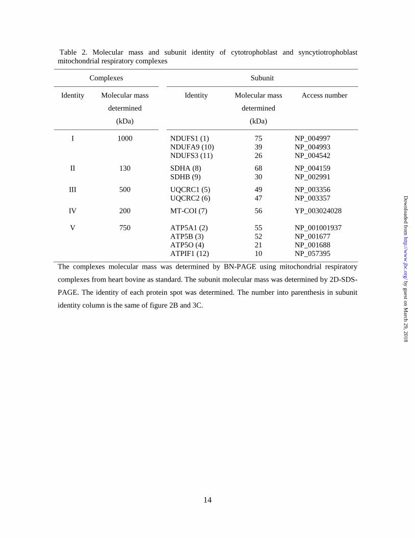
Table 2. Molecular mass and subunit identity of cytotrophoblast and syncytiotrophoblast
mitochondrial respiratory complexes
Complexes Subunit
Identity Molecular mass
determined
(kDa)
Identity Molecular mass
determined
(kDa)
Access number
I 1000 NDUFS1 (1)
NDUFA9 (10)
NDUFS3 (11)
75
39
26
NP_004997
NP_004993
NP_004542
II 130 SDHA (8)
SDHB (9)
68
30
NP_004159
NP_002991
III 500 UQCRC1 (5)
UQCRC2 (6)
49
47
NP_003356
NP_003357
IV 200 MT-COI (7) 56 YP_003024028
V 750 ATP5A1 (2)
ATP5B (3)
ATP5O (4)
ATPIF1 (12)
55
52
21
10
NP_001001937
NP_001677
NP_001688
NP_057395
The complexes molecular mass was determined by BN-PAGE using mitochondrial respiratory
complexes from heart bovine as standard. The subunit molecular mass was determined by 2D-SDS-
PAGE. The identity of each protein spot was determined. The number into parenthesis in subunit
identity column is the same of figure 2B and 3C.
by guest on March 29, 2018
http://ww
w.jbc.org/
Dow
nloaded from

Garcia-Trejo and Oscar Flores-HerreraPardo, Oscar Juarez, Federico Martinez, Guillermo Mendoza-Hernandez, Jose J.
Daniela De Los Rios Castillo, Mariel Zarco-Zavala, Sofia Olvera-Sanchez, Juan Pablocomplex V
Atypical cristae morphology of human syncytiotrophoblast mitochondria: role for
published online May 13, 2011J. Biol. Chem.
10.1074/jbc.M111.252056Access the most updated version of this article at doi:
Alerts:
When a correction for this article is posted•
When this article is cited•
to choose from all of JBC's e-mail alertsClick here
by guest on March 29, 2018
http://ww
w.jbc.org/
Dow
nloaded from