The Cell Biological Basis of Cancer
50
Transcript of The Cell Biological Basis of Cancer


ii
The Cell Biological Basis of Cancer
A thesis submitted to the Miami University Honors Program in partial fulfillment of the
requirements for University Honors with Distinction
By Ankur K. Patel
Miami University Oxford, Ohio
May 2012

iii
ABSTRACT
THE CELL BIOLOGICAL BASIS OF CANCER
By ANKUR K. PATEL
This literature review explores the current knowledge of cancer in humans on the
cellular level. The review will delve into the critical points on the topic of cancer, with a
focus on the biological processes and attributes that characterize cancer cells. These critical
points include the generation of cancer, the effect of cancer on a cell’s function,
proliferation, tumor growth and development, avenues for preventing the spread of cancer
cells as well as treating cancerous conditions, and exploring the use of model organisms and
systems in biomedical research to study the disease. Specifically, the review first discusses the
genetic instability that characterizes cancer. Next, a discussion of oncogenes and tumor
suppressor genes reveals well-known mutational pathways by which cancerous conditions
arise. Disruption of signaling pathways, as well as the mechanisms of angiogenesis and
invasion that characterize malignancies, are also discussed. Finally, the review will look into
cancer therapy and the role that biological and computational models play in elucidating
cellular abnormalities that can be exploited in pharmacological treatment.

iv

v

vi

vii
Acknowledgements
I would like to thank my thesis advisor, Dr. Joyce F. Fernandes, whose patience and
guidance made this project possible. I am also grateful for my readers Dr. David Pennock,
whose passion for science motivated me every step of the way, and Dr. Katia Del Rio-
Tsonis, whose expertise ensured this project would be completed with the utmost quality.
Lastly, I would like to thank my parents for their support in all of my endeavors.
A.K.P.
viii
Table of Contents
Chapter One Introduction 1
Chapter Two Cellular Stability 5
Chapter Three Genetic Instability and Cancer 8
Chapter Four Oncogenes 11
Chapter Five Tumor Suppressor Genes 15
Chapter Six Tumor Growth and Angiogenesis 18
Chapter Seven Metastasis 22
Chapter Eight Model Systems 25
Chapter Nine Treatment 28
Chapter Ten Conclusion 31
Figures 32
References 38

ix
List of Figures
Figure 1. Intracellular pathways involved in the onset of cancer
Figure 2. Cellular acquisition of hallmark cancer traits
Figure 3. Myc expression in mice lung tissue
Figure 4. Angiogenesis stimulated by cancer cells to overcome metabolic stress
Figure 5. Morphology of cancer cells exhibiting mesenchymal and amoeboid movement
Figure 6. Potential cellular targets for cancer drugs

1
CHAPTER ONE
INTRODUCTION
Cancer is a biological disease exemplified by a complex interaction of genetic and
environmental factors that coordinate carcinogenesis (Brennan, Offiah, McSherry, &
Hopkins, 2009). It is one of the most serious diseases that affect the global population
(Zhang, Pan, Cobb, & Anderson, 2007), accounting for 13% of all deaths in 2008 (Cancer,
2012). Cancer is a disease characterized by disruptions in normal cellular functions (Hanahan
& Weinberg, 2000; Kreeger & Lauffenburger, 2010). Mutagenic events that affect a cell’s
genetic material are known to cause deregulation of the pathways that govern the cell’s most
fundamental processes (Kreeger & Lauffenburger, 2010) (see Figure 1). Specifically, the
onset of cancerous conditions is due to an accumulation of multiple genetic mutations which
lead to the deregulation of signaling pathways that control cell growth, apoptosis, and DNA
repair (Bild, et al., 2006; Kreeger & Lauffenburger, 2009). Once these pathways are
transformed to remove the effects of cellular controls, cancer cells are able to proliferate and
grow in the absence of normal restrictions.
There are certain, well-defined means by which normal cells transform into
cancerous ones. Oncological research in the latter half of the century has indicated that
almost all cancerous cells display a relatively few number of acquired molecular, biochemical,
and cellular features that result from alteration of key pathways (Kreeger & Lauffenburger,
2010). This may seem like a strong generalization considering that there are over 100 unique
2
types of cancer, not including further subtypes of malignancies that have been identified
(Hanahan & Weinberg, 2000). However, it is not such a stretch when realizing that the field
of cellular biology emphasizes the similarity between all types of living cells. Mammalian
cells, for example, are all alike in their mechanistic regulation of normal cellular processes,
such as division, differentiation, and programmed cell death (apoptosis). Therefore, it is
unsurprising that there are certain rules that govern the transformation of normal human
cells to cancerous ones (Hanahan & Weinberg, 2000).
The universal nature by which cancer occurs is further evidenced by the ongoing
identification of specific mutation sites on the human genome that are found in many forms
of cancer. Researchers are also attempting to classify the genes crucial to carcinogenesis into
specific classes by studying cancerous phenotypes in experimental models (Hanahan &
Weinberg, 2000). Two specific gene classes are predominantly discussed – oncogenes and
tumor suppressor genes. Cancer is caused by accumulation of activated oncogenes and
inactivated tumor suppressor genes that subsequently confer the abnormal attributes that
characterize cancerous cells (Tran, et al., 2008). Proto-oncogenes, which are precursors to
oncogenes, are altered by dominant mutations, consequently conferring a gain of function
such as proliferation to a normal cell. These specific genes which are known to enhance the
proliferative capability of cells are termed oncogenes in their mutated form. Tumor-
suppressor genes, on the other hand, are altered and inhibited (Tran, et al., 2008).
As a result of mutations in both classes of genes, fundamental cellular processes such
as metabolism, growth, proliferation, and death are altered in cancer cells (Hanahan &
Weinberg, 2000; DeBerardinis & Cheng, 2008). These altered pathways give cancerous cells
3
the ability to grow in number, forming tumors at the local site (Blagosklonny, 2003). Cells
are able grow uncontrollably by avoiding the regulatory effects of the multiple mechanisms
present in a cell that are controlled by key proto-oncogenes and tumor suppressor genes
(Macleod, 2000). Local tumors become carcinomas when they travel and invade foreign
tissues in the body (Blagosklonny, 2003).
Recent attempts to organize cancer progression into distinct, well-defined categories
have been made. Tran, et al. (2008) state that certain pathological features characterize
cancer; specific developmental steps such as independent proliferation, immortalization,
inhibited differentiation, induced growth in the vascular network (angiogenesis), invasive
capability, resistance to apoptosis, and genomic instability are mentioned. Hanahan and
Weinberg (2000) catalog all cancer genotypes as the accumulation of six fundamental
alterations in cellular physiology. They propose that perhaps all types of human cancers
depend on alterations to cellular physiology that confer autonomy from outside growth
signals, insensitivity to antigrowth signals, mechanistic avoidance of apoptosis, boundless
proliferative potential, angiogenesis, and invasion to foreign tissues through capillary walls
and basement membranes (metastasis) (Jayshree, Sreenivas, Tessy, & Krishna, 2009).
Further, Zhang, et al. (2006) outline cancer as a disease state conditioned on five major
developmental steps; initiation, promotion, malignant conversion, progression, and
metastasis. All of these models display some similarities in the features of cancer
development. For example, each model agrees on the ideas of the spread of to foreign
tissues. In general, excessive proliferation is perhaps the most often associated phenotype
with cancer progression (Kreeger & Lauffenburger, 2010).

4
Although the aforementioned cancer models have similarities, the path that is taken
during oncogenesis is highly varied (Hanahan & Weinberg, 2000). Cancer in general is
characterized by a heterogeneous pathology (Brennan, Offiah, McSherry, & Hopkins, 2009;
Kreeger & Lauffenburger, 2009). For example, mutagenesis of specific oncogenes and tumor
suppressor genes may occur early in some tumor progression pathways and late in others
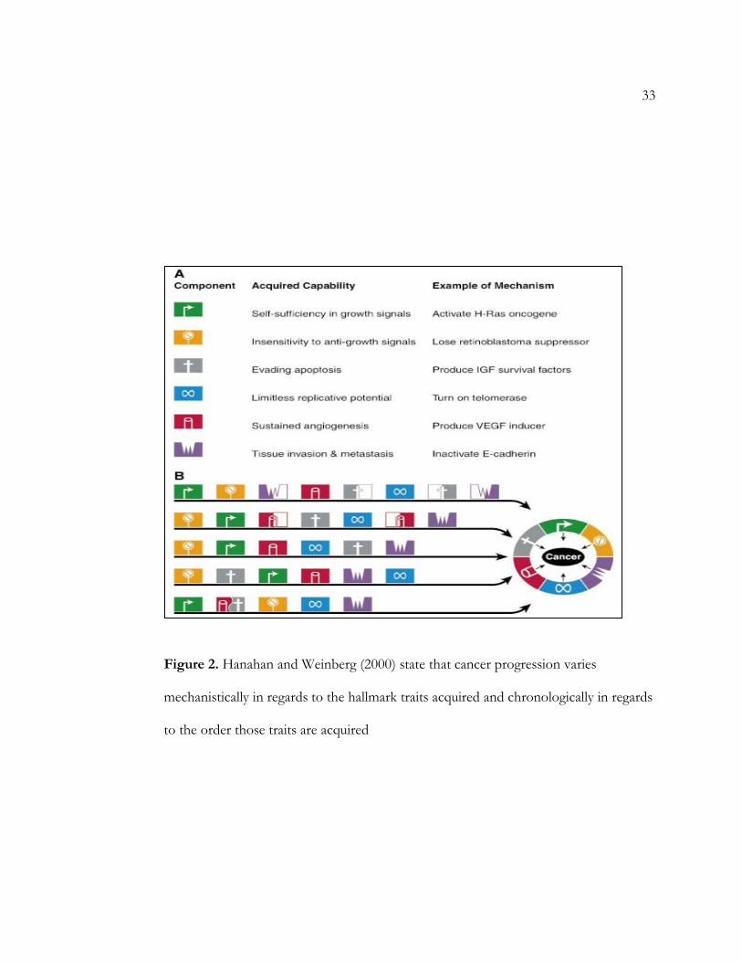
(Hanahan & Weinberg, 2000). Hanahan and Weinberg (2000) state that although all cancers
can be characterized by the acquisition of a set of hallmark capabilities, the means by which
those traits are acquired vary, both mechanistically and even chronologically (see Figure 2).
The order in which cancerous traits are acquired may contrast between the variety of cancer
types and subtypes that have been identified, and there is even wide variation in capability
acquisition between tumors of the same type. Furthermore, some progressions of metastasis
may require a different number of developmental steps, as specific genetic events may
simultaneously confer more than one capability to a cancerous cell (Hanahan & Weinberg,
2000).

5
CHAPTER TWO
CELLULAR STABILITY
It is well known that cancer is caused by changes in the human genome that enhance
cellular proliferation through alteration of normal pathways and mechanisms. However, this
process does not occur overnight. In fact, most solid human tumors are not detected until
20 years after the initial carcinogenic exposure that lead to the cancerous conditions (Loeb,
Loeb, & Anderson, 2003). This is because the genome of normal cells is maintained through
the actions of multiple mechanisms and checkpoints. Normally, random mutations of a cell’s
genetic material are offset by cellular machinery that maintains the normal sequence of
nucleotides (Loeb, Loeb, & Anderson, 2003). Therefore, the progression of cancer is reliant
on an equilibrium that is shifted towards mutagenic changes.
A non-cancerous cell maintains its genomic material in a variety of ways throughout
the cell’s life processes. Cellular DNA is subjected to many processes including replication
and transcription, and cells have an array of DNA repair mechanisms that help to restore the
normal nucleotide sequence in the event of mutations (Loeb, Loeb, & Anderson, 2003). In
non-cancerous cells, DNA replication is exceptionally accurate, with an overall error rate of
one base misalignment for every billion nucleotides that are polymerized (Loeb, Loeb, &
Anderson, 2003). This accuracy can be explained by many factors, most notably by the
functioning of DNA polymerase, the enzyme that constructs the identical strand of DNA.
First, the presence of nucleotide discrimination at the active site of DNA polymerase
6
prohibits mismatching of base pairs. The large free energy difference between matching and
mismatching base pairs further ensures that the replicated strand is constructed in a perfectly
complimentary manner to the original DNA template. DNA polymerase also functions in
correcting mismatches after the new strand is generated; in a phenomenon referred to as
proof-reading, excision of incorrectly placed nucleotides immediately after polymerization
helps to further enhance replication accuracy (Loeb, Loeb, & Anderson, 2003). The
importance of precision in the functioning of DNA polymerase has been studied by
observing the effect of a dysfunctional polymerase in a cell. Loeb, Loeb, and Anderson
(2003) show that swapping wild-type genes that code for DNA polymerase with mutated
alleles leads to the expression of an error-prone polymerase that lacks proofreading activity,
which is shown to induce epithelial cancers in mice. Mutations in DNA polymerase-
expressing genes that decrease base discrimination without affecting catalytic activity are
considered to be the most potent alterations to DNA polymerase activity (Loeb, Loeb, &
Anderson, 2003).
The functioning of DNA polymerase is merely one example of how a cell maintains
the correct nucleotide sequence. When DNA becomes damaged, cells also possess
mechanisms to limit the overall effect of the damaged DNA and to prevent daughter cells
from inheriting dysfunctional genetic material (Kreeger & Lauffenburger, 2009; Loeb, Loeb,
& Anderson, 2003). Cells carry an assortment of repair enzymes and DNA monitors that
function to monitor many cellular processes, most notably mitosis, or cellular division
(Hanahan & Weinberg, 2000). The activation of these checkpoint pathways can result in
arrest of the cell cycle, allowing DNA repair mechanisms time to activate and function.

7
Certain checkpoint pathways can also trigger apoptosis when DNA damage is deemed too
extensive to fix (Loeb, Loeb, & Anderson, 2003). Therefore, through a complex array of
many DNA monitoring and repair enzymes, as well as through mechanisms such as
replication that occur with high accuracy, normal cells’ unceasing maintenance of genomic
integrity ensures that the DNA sequence remains immaculate. With all the cellular
mechanisms that function in sustaining the correct nucleotide sequence, mutations are rare
events when considering the length of a human life (Hanahan & Weinberg, 2000).
8
CHAPTER THREE
GENETIC INSTABILITY AND CANCER
However, although mutation events seem to be unlikely, multiple mutations are
known to occur in tumor cells (Hanahan & Weinberg, 2000). Researchers have found that in
some human cancers, tumor cells average approximately 10 mutations that can occur in each
of several hundred genes (Stites & Ravichandran, 2009). This is in part due to the fact that
damage to cellular DNA can be a continual and process caused by a variety of mutagens
(Loeb, Loeb, & Anderson, 2003). Reactive metabolites and certain environmental species are
known to induce an alteration in the structure of DNA, and these chemical alterations of the
genomic material can lead to mutations and cancer. Carcinogens may directly cause DNA
damage, giving rise to mutations or other adverse chromosomal events, by generating
harmful reactive oxygen species that are known to alter DNA. Furthermore, carcinogens are
also implicated in epigenetic modifications - changes in gene expression that leave the
genetic material unchanged (Marsit, et al., 2006). The process of tissue repair, which involves
inflammation and reparative cellular proliferation to replace damaged cells, generates reactive
oxidative species as well. DNA alteration from oxidative damage can alter 10,000 nucleotide
bases per cell per day (Loeb, Loeb, & Anderson, 2003). The variety of methods by which
genetic damage can occur increases the possibility of mutagenesis.
As previously mentioned, DNA damage can occur through a variety of mutagenic
events. However, cancer progression is usually not possible with just a single mutagenic

9
event (Loeb, Loeb, & Anderson, 2003; Tran, et al., 2008). Rather, cancer development is an
intricate process that hinges on multiple mutageneses (Bild, et al., 2006). This is evidenced by
that fact that there is an exponential increase in the incidence of tumors as a function of age
(Loeb, Loeb, & Anderson, 2003), which implicates the role of continuing mutagenesis in
accumulating damage to DNA and cellular machinery. Further proof of rate-determining
mutagenic events in the formation of cancer has been found in experiments that have
transformed rodent cells in culture to study cancer. These experiments show that at least two
genetic changes are necessary for tumor formation, and studies involving human cells show
that additional changes in the genetic code are required for human cancers (Loeb, Loeb, &
Anderson, 2003).
The idea that cancerous genotypes are characterized by accumulated mutations in the
genetic material has led to several hypotheses. Loeb, Loeb, and Anderson (2003) propose the
mutator phenotype hypothesis, claiming that malignant phenotypes arise from mutations in
genes that, in their normal state, maintain genetic stability. The affected genes normally
function in controlling the accuracy of DNA replication and repair, maintaining the original
nucleotide sequence, and regulating damage checkpoints and cellular responses such as
apoptosis. It is argued that a normal mutation rate cannot account for the large number of
mutations that are often found in tumor cells. The mutator phenotype hypothesis proposes
that an initial mutation that consequently increases the mutation rate may account for the
large volume of genetic alterations seen in cancerous cells. The hypothesis suggests that
mutations in genes coding for proteins involved in maintaining genetic stability would
prevent further mutations from being prevented or fixed, initiating a cascade of dangerous

10
mutagenesis throughout the genome. Cells are turned cancerous after accumulating
mutations, and as cancerous cells proliferate, there are rounds of cellular selection that select
for cells with mutations that confer advantages in growth. (Loeb, Loeb, & Anderson, 2003).
Other propositions to explain the volume of mutagenic events in cancer have been
made. One proposal claims that accumulation of a large number of genetic alterations by
cancerous cells may be detrimental, as most mutations are detrimental in themselves (Loeb,
Loeb, & Anderson, 2003). Another proposal points to aneuploidy, a condition characterized
by cells possessing an incorrect number of chromosomes, as the initiator of malignancy; yet
another model emphasizes the singular role of clonal selection and expansion while
simultaneously claiming that increases in the mutation rate is still unable to mathematically
account for the thousands of mutations in colon cancer cells. While the true underlying
mechanism by which mutations accumulate has yet to be determined, it is agreed that
cancer’s progression depends largely on genetic alterations that confer phenotypic
modifications and advantages to cancerous cells in their biochemical processes (Loeb, Loeb,
& Anderson, 2003).

11
CHAPTER FOUR
ONCOGENES
As mentioned earlier, oncogenes have a significant role in the induction and
maintenance of cancerous conditions in the body (Tran, et al., 2008). Activation of
oncogenes requires dominant mutations of the normal form of the gene, termed a proto-
oncogene. They can be activated with alteration of a single allele because the activity of the
non-mutated allele may be insufficient to sustain normal cellular function (Lu & Bast, 2009).
Mutation of oncogenes, or oncogene activation, can confer many different capabilities to a
cancerous cell since the specific role of an oncogene may vary (Jones & Thompson, 2009).
Oncogenes are so sufficiently involved in tumor formation and maintenance that some
tumors actually require oncogenic activation for prolonged sustenance (Tran, et al., 2008). In
a phenomenon known as oncogene addiction, cancerous cells rely on oncogenes and their
products to play their specified role in order to function and survive (Jones & Thompson,
2009). For example, oncogenic addiction may force cancer cells to rely on a certain
metabolic pathway for growth (Jones & Thompson, 2009).
Ever since the function of c-Src was elucidated (the first proto-oncogene to be
described), the discovery of many more oncogenes has revealed the variety of roles they play
in cancer development and the wide variety of cancer types that rely on oncogenes (Ma &
Adjei, 2009). For example, various oncogenes participate in inhibiting apoptotic pathways
(Macleod, 2000; Jayshree, Sreenivas, Tessy, & Krishna, 2009), while other oncogenes

12
promote tumorigenesis by negatively inhibiting genes that control cellular differentiation
(Zhang, Pan, Cobb, & Anderson, 2007). Oncogenes are able to alter specific mechanisms by
imitating signals for normal growth, and the discovery of specific oncogenes has led to an
appreciation of how cancer cells seize signaling pathways used by growth factors to stimulate
proliferation (Hanahan & Weinberg, 2000). Additionally, oncogenic activation is essential for
cellular immortalization, and conferring the overall malignant phenotype in HPV-caused
cancers for example (Jayshree, Sreenivas, Tessy, & Krishna, 2009). Oncogenes have also
been implicated in altering cellular metabolism to support proliferation (Jones & Thompson,
2009), and studies over the last decade have revealed the intricate connections between
oncogenic activation and altered glucose and glutamine metabolism in tumors (DeBerardinis
& Cheng, 2010). For example, researchers have observed that tumor cells in cancer-induced
mouse models display high rates of glucose consumption and lactate secretion as a result of
oncogenic activation. Scientists have termed this phenomenon as the Warburg Effect
(DeBerardinis & Cheng, 2010).
A particular proto-oncogene called c-Myc is known to code for proteins that regulate
multiple metabolic pathways crucial to growth in non-cancerous cells and cancerous cells,
for example, in lung tumors (Jones & Thompson, 2009; Zhang, Pan, Cobb, & Anderson,
2007). In non-cancerous cells, a transcription factor called Myc regulates many processes
that regulate cellular growth and proliferation (Tran, et al., 2008), as well as entry into the cell
cycle (Zhang, Pan, Cobb, & Anderson, 2007). Myc is normally stimulated by growth factor
binding. In a mutated form, oncogenic c-Myc is overexpressed (Tran, et al., 2008), resulting
in increased rates of glycolysis and expression of enzymes that function in nucleotide and
13
amino acid metabolism (Zhang, Pan, Cobb, & Anderson, 2007), thereby altering specialized
biosynthetic activities in a way that favors cell division and cancer growth (Zhang, Pan,
Cobb, & Anderson, 2006; Tran, et al., 2008). One function of mutated Myc is providing
cancerous cells with a rich supply of NADPH, so that cells can maintain high levels of
anabolic synthesis, which leads to cancerous conditions as cells grow uncontrollably (Jones
& Thompson, 2009).
Tran, et al. (2008) re-examined two classic examples of oncogenes, the
aforementioned Myc and K-Ras. Ras mutations have been found in over 20% of human
cancers (Ma & Adjei, 2009). The Ras family of genes encodes for proteins that function in
transmitting signals from receptors to downstream regulators of survival and growth
(Blagosklonny, 2003; Tran, et al., 2008). Ras genes are extremely crucial in cancer
development (Ma & Adjei, 2009), as mutated forms of Ras genes result in a higher
prevalence of Ras signaling proteins (Bild, et al., 2006; Stites & Ravichandran, 2009; Tran, et
al., 2008). Ras proteins have been found to be downstream of many receptors associated
with cancer and upstream of various signaling pathways also associated with cancer (Kreeger
& Lauffenburger, 2009; Stites & Ravichandran, 2009).
K-Ras is one of three genes that codes for Ras protein signals, and is the most
commonly mutated Ras gene in cancers (Ma & Adjei, 2009; Stites & Ravichandran, 2009). K-
Ras is generally thought to function in stabilizing Myc, and the two cellular molecules in their
mutated forms are thought to cooperate to stimulate tumorigenesis (Tran, et al., 2008). To
study the level of cooperation of and oncogenic addiction to mutated Myc/K-Ras in mice
lung tissue and lymphocytes, Tran, et al. (2008) used dual (Myc and K-Ras) oncogenic

14
activation and individual (Myc or K-Ras) activation to induce cancer in mice, as well as dual
and individual inactivation to induce tumor regression. Lymphomas were induced by
individual Myc mutagenesis, by individual K-Ras mutagenesis, and by dual Myc/K-Ras
mutagenesis; tumor regression upon inactivation of either or both oncogenes (Tran, et al.,
2008) was also observed. However, different results were reported for lung tumor
regression. Inactivation of Myc failed to completely regress lung tumors induced by mutated
Myc, while lung tumors induced by K-Ras mutagenesis and K-Ras/Myc dual-mutagenesis
regressed completely upon inactivation of either or both oncogenes. Only partial regression
was observed with Myc inactivation, suggesting that lung tumors do not depend on Myc for
tumor maintenance (i.e., lung tumors are not oncogenetically addicted to Myc). Subsequent
findings revealed that K-Ras mutagenesis and inactivation is rate-limiting and dominant in
the induction and regression of lung tumors, respectively (Bild, et al., 2006; Tran, et al.,
2008). The results of the study also suggest that K-Ras and Myc have different “cooperation
levels in different tissues (Tran, et al., 2008); researchers observed that Myc and K-Ras fail to
cooperate induce tumorigenesis at a more accelerated pace than either oncogene individually
in lung tissue, but significant cooperation is present during tumorigenesis in lymphocytes.
The results of this study (see Figure 3) show the variation of oncogene function in different
tissues and setting (Tran, et al., 2008), while simultaneously showing how cancer cells seize
normal signal transduction pathways to stimulate proliferation through the function of
oncogenes (Hait, 2009).

15
CHAPTER FIVE
TUMOR SUPPRESSOR GENES
Much like oncogenes, mutations in tumor suppressor genes also lead to the
deregulation of cellular signaling pathways, which eventually leads to changes in gene
expression (Bild, et al., 2006). In non-cancerous cells, tumor suppressor genes function in
inhibiting the attributes that characterize cancer growth. These tumor-inhibiting functions of
specific genes are well-documented, and labels such as ‘gatekeepers, caretakers, and
landscapers’ have been used to describe the role of tumor suppressor genes (Macleod, 2000).
Some tumor suppressor genes code for proteins and enzymes that directly monitor the
integrity of the genetic material by repairing DNA damage; others function in regulating the
extracellular microenvironment around cells (Macleod, 2000). Generally, tumor suppressor
genes have roles in prohibiting cellular growth independent of growth signals, insensitivity to
anti-growth signals, avoidance of apoptosis, acquisition of limitless replication, and
metastasis and angiogenesis (Lu & Bast, 2009). It is easy to see then how tumor suppressor
genes are important in prohibiting the phenotypes of cancer; cells that display abnormal
growth rates and loss of apoptotic regulation have been observed to transform into
cancerous cells (Zhang, Pan, Cobb, & Anderson, 2007). Much like in the case of proto-
oncogenes, tumor suppressor genes play various roles in maintaining normalcy in cells.
Mutations of these genes lead to the classical conditions that characterize cancer.

16
One manner in which most tumor suppressors are different than proto-oncogenes is
the method by which each is altered to promote cancerous phenotypes. While proto-
oncogenes are altered to become active and confer a new function, tumor suppressor genes
are altered to become inactive and confer a loss of function. Further, unlike proto-
oncogenes, both alleles of tumor suppressor genes must be altered to cause the loss of
function. Mutation of the first allele simply serves as a predisposition to tumor formation,
while mutagenesis of the second tumor suppressor allele results in tumor initiation (Macleod,
2000). However, while a single non-mutated tumor suppressor gene allele is able to inhibit
the malignant phenotype (Lu & Bast, 2009), recent studies have shown that simple
epigenetic modification of a single allele of a tumor suppressor gene may be sufficient in
inducing tumorigenesis and deregulating cellular proliferation if the mutated tumor
suppressor gene allele gets deleted (Lu & Bast, Jr., 2009; Ma & Adjei, 2009). This form of
genetic modification, often called epigenetic silencing, is like turning a gene on or off in
regards to whether or not that gene will be expressed in a cell (Ma & Adjei, 2009).
A well-known ‘caretaker’ tumor suppressor gene that is altered in cancer formations
is the p53 tumor suppressor gene (Hanahan & Weinberg, 2000; Zhang, Pan, Cobb, &
Anderson, 2006), which encodes for the nuclear p53 protein (Lu & Bast, 2009). Researchers
have estimated that at least half of all human tumors contain cells that have defective
checkpoint pathways due to mutated p53 (Kreeger & Lauffenburger, 2009; Loeb, Loeb, &
Anderson, 2003). In fact, genetic alteration the p53 gene is the most common genetic
modification in ovarian cancer (Lu & Bast, 2009). Referred to as a ‘guardian of the genome’
(Macleod, 2000), p53 is a transcription factor (Jones & Thompson, 2009) whose main role is

17
to prevent genetic instability by regulating transcription of several genes that function in the
cell cycle (Macleod, 2000). A variety of stimuli, such as DNA damage, hypoxia, and oxidative
stress can lead to p53 activation via post-translational modifications to p53 (Hanahan &
Weinberg, 2000; Jones & Thompson, 2009); the specific gene transcription that is carried out
by activated p53 depends on the context and degree of the stimulus (Jones & Thompson,
2009). Cells respond to p53-induced alterations in genetic expression by arresting progress in
the cell cycle, senescence, differentiation, or activating the apoptotic cascade (Lu & Bast,
2009) if the damage is deemed excessive (Hanahan & Weinberg, 2000). Normal levels of p53
in a cell fluctuate in response to stress, and computational models have shown the feedback
circuits present in networks that monitor DNA damage help to regulate p53 levels (Kreeger
& Lauffenburger, 2010)
The tumor suppressor gene that codes for p53 is often altered by a point mutation
in one allele, and chromosomal deletion of the other allele (Lu & Bast, 2009). In most cases,
this leads to genetic alterations in the DNA-binding domain of p53 (Lu & Bast, 2009). It has
also been shown that via a dominant-negative effect, mutant p53 can form a complex with
wild-type versions of the protein, thereby inhibiting normally-functioning p53 protein by
causing an inactivating conformational change of its DNA-binding domain (Lu & Bast, Jr.,
2009; Macleod, 2000). Cancer progression via dysfunctional p53 is further enhanced, notably
in ovarian cancer, by overexpression of mutated p53 (Lu & Bast, 2009).
18
CHAPTER SIX
TUMOR GROWTH AND ANGIOGENESIS
The aforementioned mutations in oncogenes and tumor suppressor genes confer
crucial alterations in cellular growth. As cells accumulate a succession of genetic mutations,
they are selected for due to advantageous growth attributes (Blagosklonny, 2003; Hanahan &
Weinberg, 2000). There is direct selection for cells that display growth independent of
growth signals and insensitivity to apoptotic signals (Blagosklonny, 2003). It is important to
note that cells in a tumor still undergo apoptosis, albeit at a reduced rate relative to the rate
of proliferation. The number of cells in a tumor significantly under represents the number of
cell generations that were required to reach that number (Hanahan & Weinberg, 2000).
However, tumors are able to grow because cancer cells are able to shift the equilibrium
between apoptosis and proliferation to the side that favors excessive growth (Kreeger &
Lauffenburger, 2010).
Growing tumors must be able to meet the increased energetic demands of cancer
cells as they undergo excessive proliferation. The development of hypermetabolic abilities in
cancer cells, via the mutagenic pathways discussed earlier, allows cancer cells to produce
lipids, proteins, and nucleic acids at a higher rate (DeBerardinis & Cheng, 2010). Specifically,
high rates of glycolysis allow the levels of anabolic growth required by cancer cells (Jones &
Thompson, 2009). Cancer cells may also activate autophagic pathways and use degradation
products to fuel growth (Jones & Thompson, 2009). This reprogramming of cellular

19
metabolic pathways confers a further selective growth advantage, and tumor formation is
initiated as cancer cells are successively selected for.
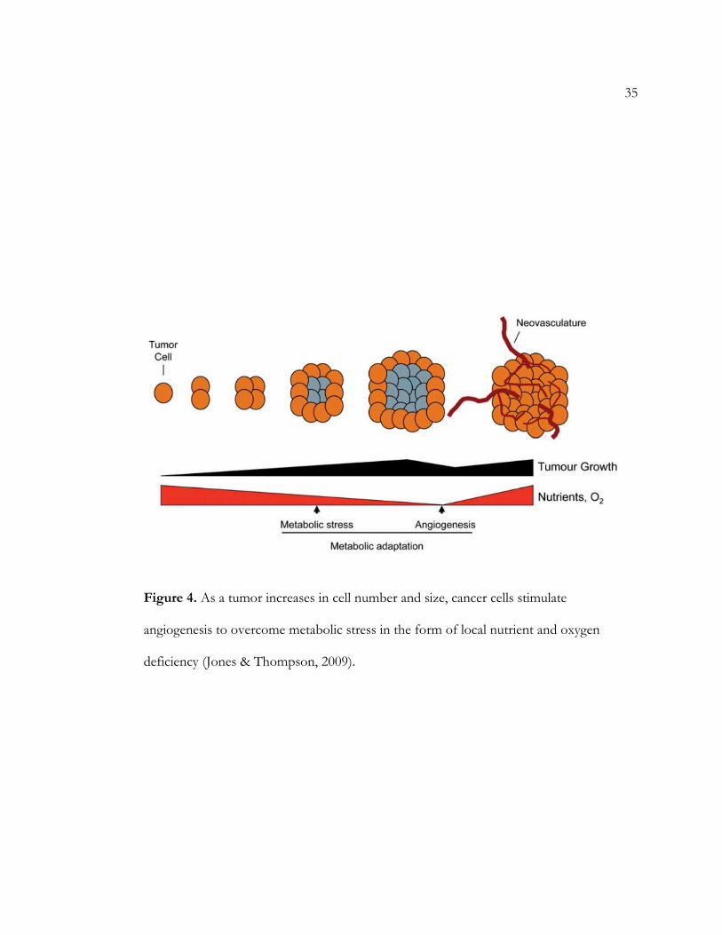
The nutritional requirements of cells, especially cancerous ones, necessitates that
there be nutrient delivery systems present throughout the body – specifically networks of
blood and lymphatic vessels (Nishida, Yano, Nishida, Kamura, & Kojiro, 2006). The pre-
cancerous vasculature present in tissues requires most cells to reside within 100 micrometers
of a capillary blood vessel (Hanahan & Weinberg, 2000). However, as a tumor enlarges at its
ends through excessive mitosis and the central core of parental cell becomes increasingly
further away from vascular systems, the central core becomes necrotic due to lack of
sufficient nutrients. The tumor must then develop ways to recruit nutrients to continue
fueling growth (Kreeger & Lauffenburger, 2010) and remove waste products (Nishida, Yano,
Nishida, Kamura, & Kojiro, 2006), as the pre-cancerous network of blood and lymph vessels
is insufficient for tumor growth due to tumor expansion often outpacing the delivery
capabilities of the accessible vasculature (Jones & Thompson, 2009). In fact, tumors are
unable to continue growing beyond a volume of two cubic millimeters unless they develop
methods to obtain additional nutrients (Ma & Adjei, 2009). In response to tumor growth,
cancerous cells often stimulate angiogenesis and lymphagenesis to relieve nutrient deficiency
(see Figure 4) (Jones & Thompson, 2009). Angiogenesis is the formation of new blood
vessels, while lymphagenesis is the formation of lymphatic vessels; both processes are
examples of neovascularization (Nishida, Yano, Nishida, Kamura, & Kojiro, 2006).
In normal cells that are growing to form tissues, neovascularization is a highly
regulated process (Hanahan & Weinberg, 2000). The ability of cancerous cells to override
20
regulations to induce and maintain angiogenesis is thought to result from activation of an
angiogenic ‘switch’ that may be turned on by increased expression of angiogenic factors and
decreased expression of angiogenic inhibitors (Hanahan & Weinberg, 2000). Turning on this
‘switch’ involves altered gene expression of inhibitors and activators such as vascular
endothelial growth factor (VEGF). In general, cancer cells stimulate altered gene expression
to shift the equilibrium between angiogenic activating and inhibiting factors to induce the
angiogenic phenotype in response to detection of decreased profiles of oxygen and other
nutrients (Kreeger & Lauffenburger, 2009; Nishida, Yano, Nishida, Kamura, & Kojiro,
2006).
Nishida, et al. (2006) outline the basic steps that lead to angiogenesis in cancer. First,
local tissue basement membrane is degraded due to tumor growth, resulting in hypoxia, or
reduced oxygen availability. This may trigger some cancer cells to begin to overexpress
angiogenic factors. Next, angiogenic factors stimulate endothelial cells to produce matrix
metalloproteinases (MMPs) that degrade the extracellular matrix (ECM), allowing the
endothelial cells to begin to migrate. Third, endothelial cells proliferate and stabilize to form
new blood vessels as they embed in surrounding tissues. Finally, sustained angiogenesis is
influenced by the continued expression and activity of angiogenic factors.
Proliferative cells utilize more than a dozen growth factors to initiate angiogenesis
(Hanahan & Weinberg, 2000; Nishida, Yano, Nishida, Kamura, & Kojiro, 2006). VEGF
(vascular endothelial growth factor) is a potent angiogenic factor that is overexpressed in a
variety of tumors (Hanahan & Weinberg, 2000). One study has observed the VEGF family

21
of factors and their specific receptors expressed in about half of the investigated tumors
(Nishida, Yano, Nishida, Kamura, & Kojiro, 2006).
22
CHAPTER SEVEN
METASTASIS
During the development of most cancers, tumor cells are able to leave the primary
growth site and invade adjacent tissues (Hanahan & Weinberg, 2000). In a process known as
metastasis, cancer cells circulate through the vascular system and implant and proliferate at
foreign sites (Nishida, Yano, Nishida, Kamura, & Kojiro, 2006). The process of metastasis is
complex and involves several steps (Hanahan & Weinberg, 2000). In order to be capable of
invasion, cancer cells must be able to first grow and spread locally (via tumor growth and
angiogenesis, as previously discussed), survive in the blood and lymphatic vessels, and
survive in foreign tissue (Kreeger & Lauffenburger, 2009; Paňková, Rösel, Novotný, &
Brábek, 2010). Cancer cells with these properties are able to implant a foreign tissue where
nutrients and space do not immediately limit further proliferation (Hanahan & Weinberg,
2000).
The initiation of metastasis, which involves cancer cells leaving the primary tumor
site to invade local sites, begins with movement through the local extracellular matrix (ECM)
(Paňková, Rösel, Novotný, & Brábek, 2010). Navigation through the ECM is facilitated in
most cases by downregulation of protease inhibitors (Hanahan & Weinberg, 2000) and an
increase in the expression of MMPs that degrade the extracellular matrix (Paňková, Rösel,
Novotný, & Brábek, 2010) and the basement membrane (Kreeger & Lauffenburger, 2010).
Paňková, et al (2010) examine the different forms of migration used by cancer cells to

23
overcome the ECM. Some cancerous cells are able to spread collectively as epithelial tissues
by retaining cell-to-cell junctions. Other cells may migrate individually, and the transition
between the epithelial migration to individual cancer cell migration is called epithelial-
mesenchymal transition (EMT) (Paňková, Rösel, Novotný, & Brábek, 2010). Cancer cells are
able to transition from epithelial sheets in order to invade individually, as evidenced by
studies that have found tight junction proteins and other cell-cell adhesion molecules to be
dysfunctional in various human cancers (Brennan, Offiah, McSherry, & Hopkins, 2009;
Hanahan & Weinberg, 2000). The most widely observed dysfunctional cell adhesion
molecule in cancer is E-cadherin, which fails to function in most epithelial cancers (Hanahan
& Weinberg, 2000).
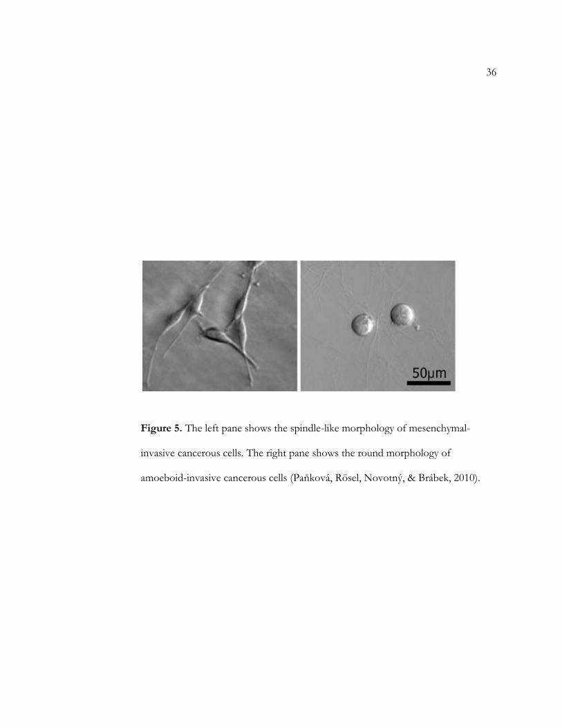
Paňková, et al (2010) specifically examined two distinct modes of individual invasion
and migration. Mesenchymal invasiveness by individual tumor cells is characterized by the
formation of leading pseudopods that facilitate movement at 0.1-0.5 micrometers per
minute. Cancer cells utilizing this form of invasion can be distinguished by a long, spindle-
like shape (see Figure 5). Cancerous cells displaying a second type of invasion, called
amoeboid-like movement, undergo cycles of cell-body expansion and contraction to move
through gaps in the extracellular matrix at speeds ranging from 2 micrometers per minute to
25 micrometers per minute. Tumor cells exhibiting amoeboid-like movement present as
rounded cells in a 3D substrate. The mesenchymal and amoeboid modes of movement are
not mutually exclusive; in response to changes in the microenvironment, cancerous cells are
able to rapidly switch from one mode of invasiveness to the other by regulating specific
pathways. The key difference between the two modes of invasion is that cancerous cells

24
displaying amoeboid movement do not necessarily participate in ECM degradation. Rather,
the mechanical forces that cause the contraction-relaxation cycles typical of amoeboid
movement are strong enough to structurally change the ECM, allowing the cells to migrate
easily. This finding was interesting in that it disagreed with the widely-held notion that tumor
cell invasion involves ECM proteolysis. However, further studies need to be done to test the
viability of amoeboid-like, proteinase-independent invasion in vivo (Paňková, Rösel,
Novotný, & Brábek, 2010).
The second step of metastasis involves a further spread of cancer cells that travel to
secondary sites such as distant organs via the lymphatic system or the bloodstream (Kreeger
& Lauffenburger, 2009; Nishida, Yano, Nishida, Kamura, & Kojiro, 2006; Paňková, Rösel,
Novotný, & Brábek, 2010). By spreading to foreign tissues, cancer cells are able to implant
and proliferate in a new environment that is not limited in nutrient and oxygen availability
(Hanahan & Weinberg, 2000). This spread of cancer cells to foreign sites is the most life-
threatening aspect of cancer (Paňková, Rösel, Novotný, & Brábek, 2010) and has been
implicated in the majority of cancer fatalities (Kreeger & Lauffenburger, 2010) and possibly
up to 90% of all cancer deaths (Hanahan & Weinberg, 2000). Research has shown that
additional mutations may be required to convert local growth and invasion to extravasion to
foreign tissues (Kreeger & Lauffenburger, 2010). Metastasis is hardly a random process, and
scientists also suspect that some genetic mutations may also give cancer cells preferences for
target organs (Kreeger & Lauffenburger, 2010).

25
CHAPTER EIGHT
MODEL SYSTEMS
In all of the studies discussed, certain model systems were employed to study the
genotypes and phenotypes that characterize cancer. In recent oncological research,
transgenic mouse models have been widely used as a valuable means to indentify events
leading to cancer development in humans (Tran, et al., 2008). There are multiple advantages
to studying cancer pathogenesis in mouse models. Mouse models allow temporal control of
oncogene expression and have thus been exploited in elucidating tumorigenic pathways
(Tran, et al., 2008). Furthermore, murine models allow for quick, reproducible tumor
induction.
Carcinogenesis in mice is similar to that in humans in that spontaneously occurring
cancers in rodents is also relatively common (Rosenberg, Giardina, & Tanaka, 2009). Some
of these cancers, and the mechanism by which they arise, show high degrees of similarity in
mice and human systems (Rosenberg, Giardina, & Tanaka, 2009). For example, the
transition between non-invasive adenomas to invasive carcinomas in colorectoral cancer
occurs via a related sequence of events in humans and mice. Furthermore, chemical agents
that induce tumors in rodent systems target genes and genetic pathways that have been
found to be targeted in human cancers (Rosenberg, Giardina, & Tanaka, 2009). With the
availability of detailed genetic information on individual mouse lines, scientists are able to
exploit mouse models to develop experiments that have high degrees of external validity to

26
human systems (Rosenberg, Giardina, & Tanaka, 2009). Certain genetically-defined mice
lines also experience different sensitivities to carcinogens, so cancer studies utilizing mouse
models are able to recreate the variation in cancer development that is known to present
during human carcinogenesis (Rosenberg, Giardina, & Tanaka, 2009). As a result of the all
these similarities in cancer development in mice and humans, mouse models have proven
useful in testing therapeutic agents (Rosenberg, Giardina, & Tanaka, 2009).
Other models to study cancer that have been utilized offer distinct advantages over
mouse models. In the last decade, computational (or mathematical) models have become a
potent tool for elucidating complex biochemical networks that are invariably affected by
cancerous conditions. Stites & Ravichandran (2009) examine the advantages of
computational and mathematical models in elucidating the aforementioned stages of cancer
development. Unlike murine models, computational models can catalog intricate network
properties to better understand the effect of a single mutagenic event on a cell’s function.
Many mathematical models are able to incorporate large volumes of quantitative information
that can describe the complexity of a cell’s biological pathways. These models can be
manipulated in more ways than can traditional models and can simultaneously administer
multiple biochemical reactions. For example, computers have been able to calculate the time
a population of cells requires in accumulating the two mutagenic events that inactivate most
tumor suppressor genes. Furthermore, scientists have recently modeled mechanisms
describing the progressive acquisition of mutations, the role of the microenvironment of the
tumor, and how a tumor is affected by targeted therapy. The latter ability of mathematical
models serves as a large advantage over traditional models; scientists are able to study the

27
effect of a potential drug to evaluate whether its activity in treating cancer is significant
enough to warrant large-scale production. Computational analysis may also identify the most
effective strategy to target specific pathways. Overall, mathematical analysis allows for in-
depth study of the intricate biochemical pathways that are often deregulated in cancer cells,
and these models may be used to develop more effective treatments (Stites & Ravichandran,
2009).

28
CHAPTER NINE
TREATMENT
Understanding of the abnormal intracellular processes that characterize cancer cells,
through the use of model systems previously discussed, has brought to light a variety of
possible mechanisms to treat cancer (Brennan, Offiah, McSherry, & Hopkins, 2009; Ma &
Adjei, 2009). Studies have shown that gene expression profiles of tumors allow scientists to
predict subsequent pathway deregulation (Bild, et al., 2006; Hanahan & Weinberg, 2000).
Elucidation of these deregulated signaling pathways in cancer cells has led to the idea of
targeting key proteins that function in the crucial pathways that give cancer cells their
hallmark capabilities (Hanahan & Weinberg, 2000; Stites & Ravichandran, 2009).
Hait (2009) examines details surrounding targeted and non-targeted therapies. In
general, targeted therapy refers to attacking a specific molecule with a particular drug to
inhibit that molecule’s function. These agents selectively inhibit a target molecule that
functions abnormally in cancer cells. Often, targeted therapies affect events early in
deregulated signaling pathways that confer abnormal growth. In contrast to targeted
therapies, non-targeted therapies involve drugs discovered by phenotypical screening
without prior knowledge of the target molecule. These therapies often affect proteins or
nucleic acids downstream of most signaling pathways.
Each mode of therapy offers advantages and disadvantages. Agents used in targeted
therapies usually have low toxicities, but have proven to possess limitations in treating solid

29
tumors. In some cases, drugs that target proteins involved in the beginning parts of pathways
are subject to downstream mechanisms of resistance that may render targeted therapies less
effective (see Figure 6). Time costs are also associated with targeted therapies as molecular
targets need to be identified prior to drug synthesis. However, previous methods of target
identification, which took 40 years in some cases, have been improved upon. Nontargeted
therapies, on the other hand, include some of the most effective drugs; however, their high
toxicity is a large disadvantage. Both forms of therapy are prevalent in oncological treatment
strategies (Hait, 2009).
Traditional targeted and non-targeted methods of dealing with cancer involve
interfering with mitosis, DNA synthesis, and cellular repair systems (Loeb, Loeb, &
Anderson, 2003; Ma & Adjei, 2009). For example, specific drugs can be synthesized to target
error-prone DNA polymerases without inhibiting normal DNA replication (Loeb, Loeb, &
Anderson, 2003). Other drugs function in stimulating the remains of the apoptotic pathway
in cancer cells, while yet others attempt to inhibit angiogenesis (Hanahan & Weinberg, 2000;
Nishida, Yano, Nishida, Kamura, & Kojiro, 2006). Brennan, et al (2009) suggest tight
junction proteins as optimal targets to prevent metastasis. Other researchers praise the
potential to exploit the metabolic changes that characterize cancer cells to deter cancer
growth (DeBerardinis & Cheng, 2009; Jones & Thompson, 2009).
Cancer treatments that do not rely on the use of drugs are also prevalent. Surgery is
used to removed localized solid tumors, while toxic chemotherapy is used for tumors that
cannot be removed surgically (Nishida, Yano, Nishida, Kamura, & Kojiro, 2006). However,
these methods of direct cancer suppression are often used in combination with drug therapy,

30
and it has been shown the combination therapy increases survival (Nishida, Yano, Nishida,
Kamura, & Kojiro, 2006). Combining chemotherapy and drug treatment is a popular
alternative because it is less toxic than a complete chemotherapy regimen (Loeb, Loeb, &
Anderson, 2003).
31
CHAPTER TEN
CONCLUSION
The content of this literature review summarizes many of the basic concepts dealing
with the cellular biology of cancer. Cancer is generated via mutagenic events often affecting
key oncogenes and tumor suppressors. These genetic alterations, coupled with changes in
gene expression, allow cancer cells to participate in unregulated proliferation. Cancer cells
begin to accumulate mutations and metabolic adaptations which allow them to be selected
for over normal cells. As cancer cells grow into large tumors, they are able to induce
extension of the vasculature which provides them the nutrients required to sustain excessive
proliferation. Cancerous conditions are furthered by metastasis to foreign tissues. Model
systems have been developed to study all of the abnormalities that characterize cancer.
Finally, different modes of cancer treatment are being developed, each with distinct
advantages and disadvantages.
32
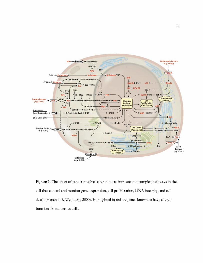
Figure 1. The onset of cancer involves alterations to intricate and complex pathways in the
cell that control and monitor gene expression, cell proliferation, DNA integrity, and cell
death (Hanahan & Weinberg, 2000). Highlighted in red are genes known to have altered
functions in cancerous cells.
33
Figure 2. Hanahan and Weinberg (2000) state that cancer progression varies
mechanistically in regards to the hallmark traits acquired and chronologically in regards
to the order those traits are acquired

34
Figure 3. (A) Tran, et al (2008) show that myc expression in mice lung tissue resulted in
lung adenocarcinomas, shown radiographically (B) and microscopically (C & D). Inactivation
of Myc failed to completely reduce tumors (E-H) .
35
Figure 4. As a tumor increases in cell number and size, cancer cells stimulate
angiogenesis to overcome metabolic stress in the form of local nutrient and oxygen
deficiency (Jones & Thompson, 2009).
36
Figure 5. The left pane shows the spindle-like morphology of mesenchymal-
invasive cancerous cells. The right pane shows the round morphology of
amoeboid-invasive cancerous cells (Paňková, Rösel, Novotný, & Brábek, 2010).

37
Figure 6. Drugs A, B, C, and D target different points of an example pathway that
stimulates DNA synthesis of proteins and assembly of the mitotic symbol for the
increased cell division seen in cancer. Drug A targets a tyrosine kinase pathway but is
susceptible to interference from activated pathways b or c. Drug B targets a nuclear
receptor that regulates gene expression. Drug C targets DNA directly, while Drug D
targets newly-synthesize microtubules of the mitotic apparatus. The targets of Drugs
B, C, and D are further downstream than that targeted by Drug A and are less
susceptible to downstream pathways that may block the drugs’ effects (Hait, 2009).
38
References
Bild, A. H., Yao, G., Chang, J. T., Wang, Q., Potti, A., Chasse, D., . . . Nevins, J. R. (2006).
Oncogenic pathway signatures in human cancers as a guide to targeted therapies.
Nature, 43, 353-357.
Blagosklonny, M. V. (2003). Cell Immortality and Hallmarks of Cancer. Cell Cycle, 2(4), 296-
299.
Brennan, K., Offiah, G., McSherry, E. A., & Hopkins, A. M. (2009). Tight Junctions: A
Barrier to the Initiation and Progression of Breast Cancer? Journal of Biomedicine and
Biotechnology, 2010, 1-16.
Cancer. (2012, February). Retrieved from World Health Organization:
http://www.who.int/mediacentre/factsheets/fs297/en/
DeBerardinis, R. J., & Cheng, T. (2010). Q's next: The diverse functions of glutamine in
metabolism, cell biology and cancer. Oncogene, 29(3), 313-324.
Forster, C. (2008). Tight Junctions and the modulation of barrier function in disease.
Histochemistry and Cell Biology, 130, 55-70.
Hait, W. N. (2009). Targeted Cancer Therapeutics. Cancer Research, 69(4), 1263-1267.
Hanahan, D., & Weinberg, R. A. (2000). The Hallmarks of Cancer. Cell, 100, 57-50.
Jayshree, R. S., Sreenivas, A., Tessy, M., & Krishna, S. (2009). Cell intrinsic and extrinsic
factors in cervical carcinogenesis. Indian Journal of Medical Research, 130, 286-295.
Jones, R. G., & Thompson, C. B. (2009). Tumor suppressors and cell metabolism: a recipe
for cancer growth. Genes & Development, 23, 537-548.

39
Kreeger, P. K., & Lauffenburger, D. A. (2010). Cancer systems biology: a network modeling
perspective. Carcinogenesis, 31(1), 2-8.
Loeb, L. A., Loeb, K. R., & Anderson, J. P. (2003). Multiple mutations and cancer. Proceedings
of the National Academy of Science, 100(3), 776-781.
Lu, Z., & Bast, J. R. (2009). Chapter 5: Tumor Suppressor Genes. Cancer Treatment and
Research, 149, 109-129.
Ma, W. W., & Adjei, A. A. (2009). Novel Agents on the Horizon for Cancer Therapy. CA: A
Cancer Journal for Clinicians, 2009, 111-137.
Macleod, K. (2000). Tumor suppressor genes. Curent Opinion in Genetics & Development, 10, 81-
93.
Marsit, C. J., Karagas, M. R., Danaee, H., Liu, M., Andrew, A., Schned, A., . . . Kelsey, K. T.
(2006). Carcinogen exposure and gene promoter hypermethylation in bladder cancer.
Carcinogenesis, 27(1), 112-116.
Nishida, N., Yano, H., Nishida, T., Kamura, T., & Kojiro, M. (2006). Angiogenesis in cancer.
Vascular Health and Risk Management, 2(3), 213-219.
Paňková, K., Rösel, D., Novotný, M., & Brábek, J. (2010). The molecular mechanisms of
transitoin between mesenchymal and amoeboid invasiveness in tumor cells. Cell and
Molecular Life Sciences, 67, 63-71.
Rosenberg, D. W., Giardina, C., & Tanaka, T. (2009). Mouse models for the study of colon
carcinogenesis. Carcinogenesis, 30(2), 183-196.
Stites, E. C., & Ravichandran, K. S. (2009). A Systems Perspective of Ras Signaling in
Cancer. Clinical Cancer Research, 15(5), 1510-1513.

40
Tran, P. T., Fan, A. C., Bendapudi, P. K., Koh, S., Komatsubara, K., Chen, J., . . . Felsher, D.
W. (2008). Combined Inactivation of MYC and K-Ras Oncogenes Reverses
Tumorigenesis in Lung Adenocarcinomas and Lymphomas. PLOS ONE, 3(5), 1-12.
Zhang, B., Pan, X., Cobb, G. P., & Anderson, T. A. (2007). microRNAs as oncogenes and
tumor suppressors. Developmental Biology, 302, 1-12.