Research grade Metabolic cart for Cardio Pulmonary Cardio ...

THE CARDIO-OESOPHAGEAL SYNDROME IN CHILDHOODBY
ISABELLA FORSHALLFrom Alder Hey Children's Hospital, Liverpool
The term 'cardio-oesophageal syndrome' is usedto cover all infants and children with incompetenceof the cardia. Ninety-three instances of the syn-drome have come to our notice during the lastthree and a half years. In some of these childrenthe cardia was found to be below the diaphragm;in some, above the diaphragm (Table 1).
TABLE ICARDIO-OESOPHAGEAL SYNDROME IN 93 CASES
Sliding HerniaCardia above Diaphragm
(35 Cases -39 *5 .)
35 4- 4 == 39 Cases(42'%)
65%'
Lax OesophagusCardia below Diaphragm
(58 Cases = 69%)
Vomited from birth-- 6%
Vomited before 8 wee;93%' - 96'% -
Vomited projectile25%
Haematemesis51'% 37%
52%
5%
5%
100%Usually
slightly dilat
Highincidence
19%
Cases, Cardia herniatedthrough Diaphragm
- 69'°
ks-_+ 100%
_ 43%
26%
Gross loss of weight36'% -> 23%
Visible gastric peristalsis13%
Rammstedt's operation10%-
Cardia incompetent100'%
--- Oesophagusted gr
- Oesophagitis
Definiteulcers
19'% Stricture
19%
14',
100%Often
rossly dilated
-> LowincidenceNone
None
It is proposed to divide the cases on the basis ofthe position of the cardia. There is a superficialsimilarity between the two groups but the courseof the disease, the prognosis, and perhaps theaetiology, are not the same. We believe that thetreatment is entirely different.
* A paper given at the inaugural meeting of the British Associationof Paediatric Surgeons in London in July, 1954.
Group 1: Lax OesophagusWhen the incompetent cardia is situated below-
the diaphragm, we term the condition 'lax oeso-phagus'. It was first recognized as an entity distinctfrom hiatal hernia by Neuhauser and Berenberg in1947. Although it is quite a common conditionin babies, it has found scant recognition in thesurgical literature (Berenberg and Neuhauser,1950a and b). Neuhauser used the name ' chylasiaof the cardia' because of the obvious incompetenceof the cardiac orifice, but we prefer the name'lax oesophagus', a term coined by our colleague,Dr. Saul Keidan (1953), because we think that it isnot only the cardia which is relaxed but also thewhole lower two-thirds of the oesophagus.
Incidence. It is difficult to form a definite ideaof the incidence of the condition. The 58 casesincluded in this series were not all directly under ourcare; records were made available to us by thepaediatricians working at the three children'shospitals on Merseyside. Mild examples of thesyndrome treated as out-patients are not includedin this survey. We compute that there are about30 to 40 cases amongst the 41,000 babies born eachyear in the Liverpool region.
Sex. Of the 58 cases, 32 were boys and 26 weregirls. There was one pair of fraternal twins, a boyand a girl. Apart from these twins, we have nothad two cases in one family, but mothers havementioned that previous children died or recoveredfrom similar symptoms.
Birth Weight. The birth weights of our casescovered the normal range; few were much belowaverage in weight.
Symptoms and Signs. Vomiting or regurgitation,or both, was the cardinal first symptom in everycase. Vomiting dated from birth in a high propor-tion and almost invariably started before 2 weeks ofage. The vomiting occurred during or betweenfeeds and was projectile in 43% of cases.
46
15% ,-
-
-F
by copyright. on D
ecember 1, 2021 by guest. P
rotectedhttp://adc.bm
j.com/
Arch D
is Child: first published as 10.1136/adc.30.149.46 on 1 F
ebruary 1955. Dow
nloaded from
CARDIO-OESOPHAGEAL SYNDROME
The following points were noticed about thevomitus:
HAEMATEMESIS. Bright red or brown alteredblood was visible to the naked eye in 26 %. It wassignificantly less frequent than in the hiatal herniagroup, due to a lower incidence of reflux oeso-phagitis.
BILE. A few babies vomited bile-stained materialfor a few days after birth.Mucus. Mucus was regurgitated or mixed with
the vomitus from birth in some cases, but less oftenthan in babies with hiatal hernia.
Gross loss of weight occurred in 230. It wasmuch less common than in the other group. Thisis. at least in part, accounted for by a more readyresponse to treatment. Failure to gain for longeror shorter periods was common.
Severe dehydration demanded parenteral fluidsin 15 %.Anaemia due to bleeding or to malnutrition was
found to some degree in about 400%.Pain, discomfort, flatulence and general misery
were noted in various degrees and combinations.Visible gastric peristalsis was noted in 19% of
cases. Visible peristalsis and an association withhypertrophic pyloric stenosis occurred more oftenin this group (Fig. 1). As our numbers are small,this is possibly not significant. It will be noticedthat visible gastric peristalsis occurred more fre-
a . ,.,., Nt%L
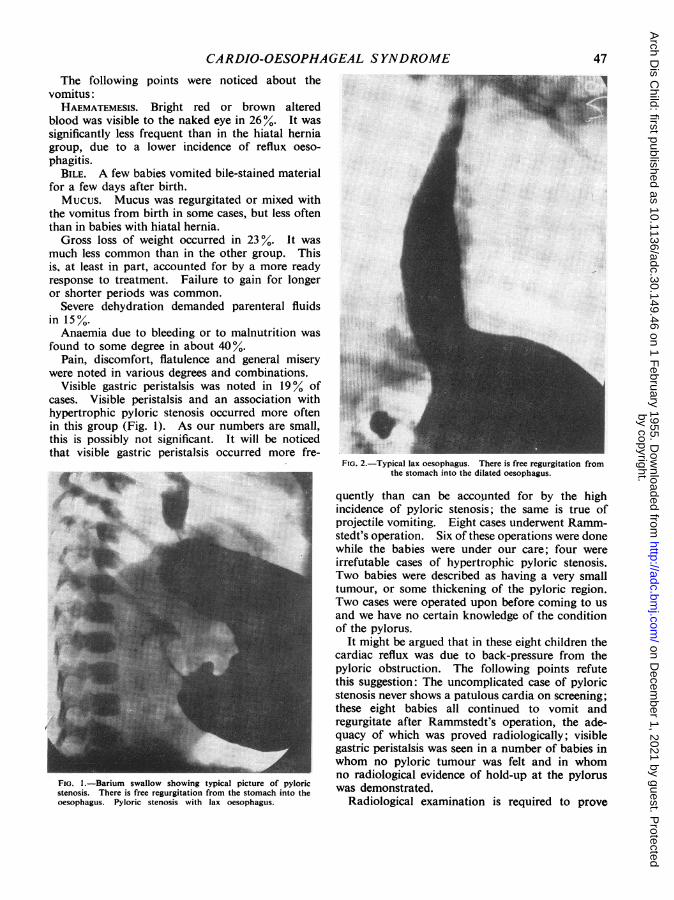
FIG. 1.-Barium swallow showing typical picture of pyloricstenosis. There is free regurgitation from the stomach into theoesophagus. Pyloric stenosis with lax oesophagus.
FIG. 2.-Typical lax oesophagus. There is free regurgitation fromthe stomach into the dilated oesophagus.
quently than can be accounted for by the highincidence of pyloric stenosis; the same is true ofprojectile vomiting. Eight cases underwent Ramm-stedt's operation. Six of these operations were donewhile the babies were under our care; four wereirrefutable cases of hypertrophic pyloric stenosis.Two babies were described as having a very smalltumour, or some thickening of the pyloric region.Two cases were operated upon before coming to usand we have no certain knowledge of the conditionof the pylorus.
It might be argued that in these eight children thecardiac reflux was due to back-pressure from thepyloric obstruction. The following points refutethis suggestion: The uncomplicated case of pyloricstenosis never shows a patulous cardia on screening;these eight babies all continued to vomit andregurgitate after Rammstedt's operation, the ade-quacy of which was proved radiologically; visiblegastric peristalsis was seen in a number of babies inwhom no pyloric tumour was felt and in whomno radiological evidence of hold-up at the pyloruswas demonstrated.
Radiological examination is required to prove
47
by copyright. on D
ecember 1, 2021 by guest. P
rotectedhttp://adc.bm
j.com/
Arch D
is Child: first published as 10.1136/adc.30.149.46 on 1 F
ebruary 1955. Dow
nloaded from

ARCHIVES OF DISEASE IN CHILDHOOD
that an incompetent cardia is present and to decideits situation. The technique of the investigation isnot easy and misleading reports are common unlessthe radiologist is used to dealing with young infantsand is familiar with fluoroscopic appearances of thecardio-oesophageal region during swallowing innormal babies. The points to note are: (1) Freeregurgitation from the stomach of the opaquematerial (Fig. 2); reflux is increased during inspira-tion, crying and coughing, in the Trendelenburgposition, and if pressure is exerted over the abdomen,that is, when intra-thoracic pressure is reduced andintra-abdominal pressure is increased. (2) Thecardia does not close but remains widely patent.(3) The normal acute angle at which the oesophagusjoins the stomach on the left side is obtuse and maybe equal on the right and left. (4) Muscular wavesin the oesophagus are irregular and of small volume.(5) The oesophagus appears wider than normal andis often extremely distensible (Fig. 3).
FIG. 3.-Lax oesophagus showing enormous dilatation of thelower two-thirds of the oesophagus in a baby aged 3 days.
The very gross dilatation of the oesophagus inthese cases is very remarkable; it is often presentwithin a few days of birth, and sufficient to meritthe term 'mega-oesophagus' (Fig. 3.) Only the lowertwo-thirds of the gullet is affected, that is, the plainmuscle portion supplied directly by the vagus andwhich, according to Lendrum (1937), is suppliedby the vagus only.At oesophagoscopy the lumen of the oesophagus
appears voluminous; gastric contents well up fromthe stomach and the instrument falls through the
patulous cardia. The oesophago-gastric junctioncan be recognized without difficulty in most cases.Oesophagitis in various stages of acuteness andaffecting various lengths of the lower third of theoesophagus may be seen. We have not seen achronic peptic ulcer or stricture in a case in whichthe cardia remained below the diaphragm.
It has been suggested that the condition we call'lax oesophagus' is in fact due to a small hiatalhernia which could be shown by careful screening.There is no doubt, however, that apart from fourexceptions to be mentioned later, the cardia in these58 children was below the diaphragm and remainedthere. This was proved by repeated radiographyand oesophagoscopy.Necropsy on three cases gave the following
information:The cardia, taken as the junction of stratified and
columnar epithelium, was below the diaphragm.The cardia could not be pulled up into the chestuntil the membranes attaching the oesophago-gastric tube to the diaphragm had been divided.The hiatus was not considered to be unduly large,but it is difficult to assess the size of the oesophagealhiatus at operation, and probably more difficultat necropsy.
It is easy to understand that these childrenregurgitate and that regurgitation will be encouragedby recumbency and any factor which increases intra-abdominal pressure. It is not so easy to explain whyvomiting, and more especially projectile vomiting,occurs. It is possible that the explanation lies inan associated hypertonicity of the alimentary tractbelow the cardia. This assumption would alsoexplain (a) the high incidence of pyloric stenosis;(b) the occurrence of well-marked gastric peristalsisin cases in which there is no hypertrophy o6f thepylorus; (c) the bile-stained vomit seen in somecases soon after birth; (d) dilatation of the oeso-phagus in the absence of obstruction of the cardia;(e) the very interesting fact that one of our 'laxoesophagus' babies developed obstructive jaundice,which, at operation, was found to be due to spasmof the sphincter of Oddi.
Course of Disease, Prognosis and TreatmentThe treatment of these cases is more or less
standardized. They are nursed in the uprightsitting position by means of pillows, harness, or byplacing them in a padded box, to reduce regur-gitation and make vomiting less easy. The feedsare thickened by the addition of 'nestergel', 'bengers'or cormflour.
Small, frequent feeds may be used so that the
48
by copyright. on D
ecember 1, 2021 by guest. P
rotectedhttp://adc.bm
j.com/
Arch D
is Child: first published as 10.1136/adc.30.149.46 on 1 F
ebruary 1955. Dow
nloaded from

CARDIO-OESOPHAGEAL SYNDROME
stomach is not distended. Aludrox, or some otherantacid, may be used to discourage oesophagitis.Anti-spasmodic drugs have little effect, thoughthere has been an occasional apparent improvementwith pylostropin.The response to these measures is often dramatic;
vomiting ceases, the child gains weight, and as theregime is gradually relaxed, symptoms do not recur.In some cases, improvement is slow or delayed, orsymptoms recur as soon as the child is allowed outof the upright position. Many of these infantshave been critically ill for weeks or months and havesuffered serious complications (Table 2).Four children with a proved lax oesophagus and
a cardia below the diaphragm developed severeoesophagitis, and, over the course of months, thecardia was gradually pulled up into the chest.Three of these children later developed chronic
TABLE 2RESULTS IN 58 CASES OF LAX OESOPHAGUS
Medical TreatmentSatisfactory
Children doing well, vomiting has ceased, weightincreasing .45 cases (79 %)Some had long periods in hospital or repeatedadmissions due to relapse; the followingcomplications were recorded:
Gastro-enteritis .. 6 casesPneumonia .. 4 casesBronchitis .. 1 caseLung abscess .. 1 case
Failed .13 cases (22 4%)(a) Unsatisfactory. Child 1 * years old, still vomiting and
much below expected weight . . 1 case(b) Developed sliding hernia, and included under this
heading .4 cases(c) Operation, aged 5 months, repeated relapses and
failure to gain weight . case(d) Deaths due to the following causes . . 7 cases (12%)
Gastro-enteritisNecrosis of lower end of oesophagusOesophagitis and septicaemiaHaematemesisGastro-enteritis and lung abscessInhalation of vomitInduction of anaesthesia for oesophagos-
copy; poor-risk infant(All cases were initially treated medically).
oesophageal ulcer with stricture (Figs. 4, 5, 6 and 7).One child, still being treated medically at the age
of 18 months, is still vomiting and regurgitating andmuch below weight.
Forty-five of the 58 cases (790%) are consideredto have made, or to be making, a satisfactoryrecovery. Some of them have been radiologicallyexamined after symptoms have ceased for variouslengths of time and in these cases it has been demon-strated that the cardia has contracted, that reflux nolonger occurs, and that the lumen of the oesophagusis narrowing.One child, after repeated relapses, was operated
upon at the age of 5 months and responded well tosimple narrowing of the hiatus by stitches placedthrough the fibres of the right crus behind theoesophagus. At this single operation for 'laxoesophagus' the cardia was below the diaphragm.The hiatus did not appear larger than could beaccounted for by the size of the oesophagus itaccommodated.
Seven children died and the immediate cause ofdeath is shown in Table 2.To summarize, we think, in common with others,
that the condition probably has a nervous or neuro-muscular basis which involves the lower two-thirdsof the oesophagus, the cardia, and perhaps thegastro-intestinal tract below the cardia. We realizethat this is no real explanation. We are in doubtabout the responsibility which the hiatus bears.The condition is often self-limiting and at leastthree-quarters of these children will recover withsimple treatment.Lax oesophagus cannot, however, be regarded
with complacency as an unimportant conditionout of which the child will grow. In our cases,serious complications were common. Many patientswere in hospital for long periods, up to nine months,or had more than one admission. During this timethey occupied badly needed medical beds andabsorbed valuable skilled nursing time. From themother's point of view, it cannot be much satis-faction to have a baby who sits in a box in a hospitalward. There was a mortality of 12%. We haveno evidence that the course of the disease can orshould be shortened by operation. If surgery wereto be seriously considered, it would be difficult todecide its indications; during a bad phase, thesebabies would be poor risks, and when they improve,one cannot judge if the improvement is temporaryor permanent.
Careful follow-up of these children and repeatedradiological examination of those in whom vomitingpersists, or in whom weight gain is unsatisfactory andthose who relapse is indicated, so that the occasionalcase in which the cardia herniates above the dia-phragm may be picked up as early as possible.
Group II: Sliding Hiatal HerniaWhen the incompetent cardia is situated above
the diaphragm the condition is usually called asliding hiatal hernia. Findlay and Kelly (1931) andKelly (1939) recognized that stricture of the oeso-phagus in children was associated with a partialthoracic stomach. Wyllie and Field (1946) des-cribed six children with partial 'short oesophagusand thoracic stomach' but did not mention that the
49
by copyright. on D
ecember 1, 2021 by guest. P
rotectedhttp://adc.bm
j.com/
Arch D
is Child: first published as 10.1136/adc.30.149.46 on 1 F
ebruary 1955. Dow
nloaded from

FIG. 5.-A series of barium meal swallows on the same patienttaken at two-monthly intervals. In this picture the patient has
developed a small hiatus hernia.
FIG. 4.-A series of barium meal swallows on the same patientstaken at two-monthly intervals. This picture shows a typical
lax oesophagus with free regurgitation.
FIG. 7.-A series of barium meal swallows on the same patienttaken at two-monthly intervals. In this picture there is an
enormous hiatus hernia.
~~~~~~~~~~~~~~~~~~~~~~~~~~~............FIG. 6.-A series of barium meal swallows on the same patienttaken at two-monthly intervals. In this picture the hiatus herniahas increased slightly in extent. There is a suggestion of stricture
formation.
by copyright. on D
ecember 1, 2021 by guest. P
rotectedhttp://adc.bm
j.com/
Arch D
is Child: first published as 10.1136/adc.30.149.46 on 1 F
ebruary 1955. Dow
nloaded from

CARDIO-OESOPHAGEAL SYNDROMEsame symptoms are more commonly produced by an
incompetent cardia below the diaphragm. Therehave been many excellent articles in the surgicaljournals on hiatal hernia during the last few years
(Barrett, 1950; Allison, 1946, 1948, 1951; Harrington1942, 1945; Belsey, 1954, etc.), but few surgeons havepublished a series of the abnormality in infants andchildren.
FIG. 8.-Barium swallow on a baby 48 hours old showing definitehiatus hernia.
In 35 of our 93 cases the cardia was above thediaphragm at the initial radiological examination.Some children were radiographed before the age of4 or 5 days (Fig. 8), but most of them were a fewweeks old and some older still.At operation, the fibres of the right crus which
form the hiatus invariably appear poorly developed,especially the part of the crus which forms the leftand posterior aspect of the hiatus. In some cases,
the posterior fibres are virtually absent. We thinkalso that the diaphragm as a whole is often undulythin, almost flaccid to the touch. We do notconsider that the oesophagus is initially short; byand large, the younger the child the more easily canthe cardia be placed below the diaphragm. Sixcases were observed in which, during screening,the cardia was seen to be sometimes above andsometimes below the diaphragm; in all six cases itultimately settled above. The four cases whichstarted as lax oesophagus and went on to becomesliding herniae are also significant. We have failedto find a description of a hernia of this kind in astillborn infant. We do not certainly know theposition of the cardia at birth. We do know thatin at least some cases, the cardia has, at some time,been below the diaphragm and that it is drawn upthrough the hiatus. The migration of the cardiais probably dependent upon the difference in intra-thoracic and intra-abdominal pressures. We are ofthe opinion that a sac is present though it may bevery small.These facts suggest that the condition is probably
a true herniation of the cardia from the abdomeninto the posterior mediastinum. They do not provethat the condition is invariably produced in thisway. Aetiologically, therefore, we consider that apoorly developed hiatus bears a heavy respon-sibility and is the 'congenital' element in this type ofhernia. We consider that shortening of the oeso-phagus is secondary and results from oesophagitisand spasm, as in adults.- In children, failure ofthe oesophagus to grow would ultimately producerelative shortening, and it is at least possible thatthis may be a significant factor in some cases. Itwill be seen that we do not consider the term'congenital short oesophagus' justified.
Radiologically the oesophagus is not so dilatedas in the lax oesophagus group. The diameter ofthe gullet is often about the same as that of the smallsegment of thoracic stomach. The supra-diaphrag-matic stomach can be recognized radiologically bycoarse vertical gastric rugae outlined by barium anda slight constriction at the cardio-oesophagealjunction. On oesophagoscopy the junction can beidentified by the transition from relatively smoothto coarse vertically ridged mucosa. The diaphrag-matic pinch-cock is absent. Reflux is marked andconstant. Oesophagitis is almost invariably seen;the degree of acuteness varies and it has a tendencyto wax and wane.The symptoms of hiatal hernia in infancy are due
to the incompetence of the cardia and not to thecardia being situated above the diaphragm. Wehave, in common with others, had infants with
51
by copyright. on D
ecember 1, 2021 by guest. P
rotectedhttp://adc.bm
j.com/
Arch D
is Child: first published as 10.1136/adc.30.149.46 on 1 F
ebruary 1955. Dow
nloaded from

ARCHIVES OF DISEASE IN CHILDHOOD
hiatal hernia in whom half or more of the stomachis above the diaphragm, and yet the cardia is com-petent, in spite of being divorced from the hiatus.In these cases symptoms may be entirely absent;if present, they conform to an entirely differentpattern from the symptoms of the cases under dis-cussion. The symptoms of these 35 children showedsignificant percentage differences from the laxoesophagus group. Projectile vomiting was lesscommon; it is difficult to account for this. Ifprojectile vomiting is a reflex phenomenon, one wouldexpect it to be more likely to occur in the herniagroup. Haematemesis occurred much more com-monly due to the higher incidence of reflux oeso-phagitis. It is not clear why reflux oesophagitis iscommoner in the hernia group as the same freereflux occurs in both. Gross loss of weight againwas more often noted, at least in part, due to a lessready response to treatment. Visible gastric peri-stalsis was not seen, apart from the two cases ofpyloric stenosis. These cases do not give the sameevidence of being associated with a widespreadneuromuscular imbalance as do children in the laxoeosphagus group.
Course of the Disease. In our experience, theresponse to posture is often unsatisfactory and atendency to relapse marked. Complications fallinto two groups: (1) The same tendency to contractchest infections, gastro-enteritis and to aspiration ofvomit as the lax oesophagus cases; (2) complicationsin the oesophagus itself as follows:SHORTENED OESOPHAGUS. Shortening of the oeso-
phagus is a result of continued severe oesophagitis.It is known that once inflammatory changes broughtabout by the reflux of gastric secretions penetratedeep to the mucosa, fibrosis spreads up and downthe oesophagus and results in shortening. It is alsoknown that the different layers of the oesophagusare more loosely adherent in children, and that inthem inflammatory spread and fibrosis occur morereadily. Shortening of the oesophagus is commonand may occur with astonishing rapidity.
PEPTIC ULCERS. In 300% of our cases there wasradiological evidence of ulcer formation. Theradiological findings were confirmed at oeso-phagoscopy. The ulcer was invariably in theoesophagus and usually within 1 in. of the cardia.We have not seen a chronic peptic ulcer in thethoracic portion of the stomach, either on screeningor on oesophagoscopy. This important and inter-esting difference from sliding hiatal hernia ofadults is probably related to the rarity of chronicpeptic ulceration of the stomach and duodenum inchildren. We have not seen an ulcer at the cardia
or in its immediate neighbourhood, and presumethat the extreme lower end of the oesophagus isimmune to the action of acid pepsin. One ulcerperforated into the mediastinum and at operationgave much the same picture as a chronic duodenalulcer which has perforated into the pancreas.
STRICTURE. Some narrowing of the oesophagusoccurred in 30% of cases. It was more commonin cases with ulcer, but also occurred in cases inwhich a definite ulcer was not seen. The length ofthe narrowing varied from 2 to 21 in.; it is difficultto assess radiologically as the barium is held up atthe upper limit of the stricture and then tricklesthrough in a thin stream, giving the impression of along, narrow area. In the presence of spasm orstricture the proximal oesophagus dilates and hyper-trophies (Figs. 9 and 10). In many cases, oeso-phagoscopy showed less narrowing than the radio-graph had suggested; presumably spasm plays a partin producing the radiological appearances. Onlyone stricture required a gastrostomy.Dysphagia due to a stricture can be temporarily
relieved by dilatation. The amount of dilatationthat can be obtained through an oesophagoscopein a small child is not very great. Dilatationencourages further reflux and perhaps encouragesshortening, but as a temporary measure to improveswallowing and nutrition it has proved valuable.With continued severe oesophagitis and asso-
ciated spasm, or with the development of true
FIG. 9.-Barium swallow in a case of hiatus hernia with strictureshowing marked dilatation of the proximal oesophageal segment.
52
by copyright. on D
ecember 1, 2021 by guest. P
rotectedhttp://adc.bm
j.com/
Arch D
is Child: first published as 10.1136/adc.30.149.46 on 1 F
ebruary 1955. Dow
nloaded from

CARDIO-OESOPHAGEAL SYNDROMEnarrowing, the symptoms change; solids and semi-solids are taken with more difficulty than liquids.Some mothers have stated that their baby is neverhappy until he has vomited. Older children com-plain of epigastric or retrosternal pain, discomfortor a sense of fullness after food. Some childrenbecome resistant to all nourishment.
Regurgitation is more common than true vomit-ing; mucus in large quantities is sometimes broughtup after and between feeds and it may be blood-stained. Sometimes, the chief complaint is of thelong time taken by the child to eat a small meal.Occasionally, children induce vomiting by puttingtheir fingers down their throats. Weight gain isslow, or there is none; the patients are small for theirage and tend to become self-centred and intro-spective. One or two children have given theimpression that their only pleasure in life is to return,as soon as possible, any nourishment forced uponthem.Not all cases follow this severe course. A few
improve with weaning, some have gained weight andappear well and happy with the cardia well above thediaphragm.
FIG. 10.-Same case as Fig. 9 after operation. The stomach isnow well below the diaphragm. There is still slight dilatationof the proximal oesophagus. The level of the diaphragm is indi-
cated by the two Michel's clips.
Operative Findings. The cardia has been foundanywhere from just above the diaphragm to thelevel of D.6. (In one case which we had the oppor-tunity of examining but not of operating upon,it was at the level of D.4-5.)
I consider that there is usually little difficulty inrecognizing the position of the cardia at operation;there is generally a slight constriction at the cardio-oesophageal junction and a slight but definitechange in the arrangement of the muscle fibres, and,I think, in the direction in which the arteries run.The level at which the cardia has been found atoperation has usually conformed to radiological andendoscopic findings. In cases which have hadprolonged oesophagitis, ulcers or strictures, therehas been a varying amount of fixation of theoesophagus and it is firm and rigid to the touch.Enlarged lymph nodes are common, especially withchronic peptic ulcer.
In small children the vagi are usually of sufficientlength not to form an obstacle to replacement of thecardia below the diaphragm. In cases where thecardia is high, and in older children, the vagi havesometimes appeared shorter than the freed oeso-phagus; twice we have divided the branches con-necting the right vagus with the pulmonary plexusin order to get sufficient length.The four cases of lax oesophagus which developed
sliding hernia showed no variation from those inwhich the cardia was above the diaphragm whenthey were first examined.As has been stated in speaking of the aetiology
of the condition, the hiatus has invariably beenfound to be large and lax and to be, to some extent,developmentally deficient. The so-called oeso-phago-phrenic ligament appears to be a verydefinite structure; it is firmly attached to the gastro-oesophageal tube and has to be cut with scissorsbefore the cardia can be brought down below thediaphragm. The small arteries to the oesophagusfrom the aorta appear to me to run transversely fromaorta to oesophagus, not more and more obliquelyupwards as the cardia rises, as one would expect.
Treatment. We are firmly of the opinion thatsliding hernia in children is a surgical condition.It has been suggested that many cases will ultimatelydo well with medical treatment and that symptomstend to subside at the time of weaning or later inchildhood. The result of medical treatment, asexemplified by nine Liverpool cases, does notsuggest that this is so (Table 3).A child who continues to vomit and fails to gain
weight is more likely to fall a victim to infectionthan is his healthy brother. There can surely be
53
by copyright. on D
ecember 1, 2021 by guest. P
rotectedhttp://adc.bm
j.com/
Arch D
is Child: first published as 10.1136/adc.30.149.46 on 1 F
ebruary 1955. Dow
nloaded from

54 ARCHIVES OF DISEASE IN CHILDHOODTABLE 3
RESULTS IN NINE CASES OF SLIDING HIATUS HERNIAMedical Treatment .total number of cases 9(a) Satisfactory; gaining weight, no vomiting . I(b) Unsatisfactory; below weight for age, vomiting, etc. .. 5
Included are a boy of 13 years weighing 65 lb. who cannoteat solids, cardia at level of 5 T. stricture, and a boy of7 years weighing 47 lb., repeated dilatation for stricture.
The ages of the other children are 13 yr., 9 yr., 2 yr., 7 monthsand 6 months.
(c) Still under observation at 10 weeks old(d) Deaths. 2
One child died aged 1 month, pneumonia.3 months, aspiration of vomit and
haematemesis.Examples of cases in which medical treatment failed and operation
was requested:(1) Small hernia at 4 months, large hernia, stricture and perforation
at 4 years.(2) Recurrent pneumonia, repeated haematemesis, failure to gain
at 5 months.(3) Weighed 27 lb. at 3.'- years; vomiting, haematemesis.(4) Increasing hernia; shortening of oesophagus at 3 years.
little reason to allow a child to remain under-nourished, and perhaps in pain, if his abnormalitycan be corrected. There is no guarantee that a childwith a sliding hernia who does well on medicaltreatment will continue to do so, or that symptomswill not develop in later life when they may be moreserious and less easily cured by operation.
There is no tendency for the cardia to becomecompetent; it is permanently separated from thediaphragm and the hiatus is in any case inefficient.The valvular mechanism of the oesophago-gastricangle is obliterated and cannot develop as the gulletgrows shorter. We therefore advise operation inall cases in which the cardia is above the diaphragm.
Time of Operation. Continued vomiting andfailure to gain weight, severe, persistentor recurrent oesophagitis, progressive shorteningof the oesophagus, peptic ulcer of the oesophagusand narrowing of the oesophagus are all absoluteindications for operation.
In a child who is symptom free or in whomsymptoms are mild, there is no urgency and opera-tion can be undertaken at any convenient time,provided the position of the cardia is checked atfrequent intervals.
If operation is advised for all cases it must curesymptoms and prevent complications, give a lowrecurrence rate and be safe (Table 4).The important points in successful operation are
adequate freeing of the oesophagus up to or abovethe aortic arch and complete separation of theoesophago-gastric tube from the diaphragm; firmfixation of the fundus of the stomach to the under-surface of the left dome of the diaphragm; carefulrepair of the hiatus.We have found a thoracic approach to the
oesophagus and a transdiaphragmatic approach tothe stomach satisfactory, and since using thismethod, we have, so far, had no recurrence in27 cases. Previously we operated on seven cases
TABLE 4RESULTS IN OPERATIONS FOR CASES OF SLIDING
HIATUS HERNIASurgical Treatment .. . total number of cases 30(a) Operation above diaphragm .. . 7
(i) Recurrence . . 4(ii) Satisfactory . . 3
(b) Operation above diaphragm and through diaphragm20 -+ 4 reoperated
(c) Operation above diaphragm and through diaphragm anddilatation of stricture through gastrostomy 3
(b) and (c) .. .27Satisfactory; relief of symptoms and no recurrence of hernia
to date ...29Total operations . . .33Mortality .. . . .
without opening the diaphragm and had fourrecurrences. These recurrent cases were later onsuccessfully operated upon by the transpleuraltransdiaphragmatic approach. Severe strictures aredilated with Hagar's dilators through a smallgastrotomy while the chest is open; the three casesso treated have not required post-operative dilata-tion. The children have stood operation well andpost-operative complications have, on the whole,been few and transient. All cases are free of symp-toms, except that two children have been slow togain weight and appetite, in spite of normal or nearnormal appearances on radiography and oeso-phagoscopy. The longest follow-up in this smallseries, is, however, only three and a half years.To conclude, we believe there is some evidence that
lax oesophagus and sliding hiatal hernia areaetiologically distinct conditions. Further studyof the former is required to settle the nature of theresponsible factor. In sliding hiatal hernia we donot know whether the cardia is ever above thediaphragm at birth. Jt is natural to want to knowthe answers to these fascinating problems, but theburden of this discourse is the importance of theposition of the incompetent cardia. I reiteratewords written by Allison in 1948 about slidinghernia in children:
'The condition is of particular interest to paedia-tricians because its early recognition may lead tosurgical reduction of the hernia, and for the patient,escape from a life of dysphagia and malnutrition.'
I wish to acknowledge the assistance of Mr. P. P.Rickham who performed about half the operations,most of the endoscopic examinations and a large propor-tion of the time-consuming radiological examinations.
REFERENCESAllison, P. R. (1946). J. thorac. Surg., 15, 308.
(1948). Thorax, 3, 20.(1951). Surg. Gynec. Obstet., 92, 419.
Barrett, G. M. (1950). Brit. med. J., 1, 245.Belsey, R. (1954). Ann. roy. Coll. Surg. Engl., 14, 303.Berenberg, W. and Neuhauser, E. B. D. (1950a). Amer. J. Dis.
Child., 79, 402.____- (1950b). Pediatrics, 5, 414.Findlay, L. and Kelly, A. B. (1931). J. Laryng., 46, 797.Harrington, S. W. (1942). Ann. Surg., 115, 705.
(1945). Ibid., 122, 546.Keidan, S. E. (1953). Lecture to the Liverpool Paediatric Club.Kelly, A. B. (1939). J. Laryng., 54, 621.Lendrum, F. C. (1937). Arch. intern. Med., 59, 474.Neuhauser, E. B. D. and Berenberg, W. (1947). Radiology, 48, 480.Wyllie, W J. and Field, C. E. (1946). Archives of Disease in Child-
hood, 21, 218.
by copyright. on D
ecember 1, 2021 by guest. P
rotectedhttp://adc.bm
j.com/
Arch D
is Child: first published as 10.1136/adc.30.149.46 on 1 F
ebruary 1955. Dow
nloaded from