Cerebral Lateralization, Cognitive Asymmetry, and Human Consciousness
fnhum-10-00493 October 8, 2016 Time: 16:28 # 1
ORIGINAL RESEARCHpublished: 13 October 2016
doi: 10.3389/fnhum.2016.00493
Edited by:Swathi Kiran,
Boston University, USA
Reviewed by:Veena A. Nair,
University of Wisconsin-Madison,USA
Keith M. McGregor,Emory University, USA
*Correspondence:Aimee Dietz
Received: 01 July 2016Accepted: 16 September 2016
Published: 13 October 2016
Citation:Dietz A, Vannest J, Maloney T,
Altaye M, Szaflarski JP andHolland SK (2016) The Calculationof Language Lateralization Indices
in Post-stroke Aphasia: A Comparisonof a Standard and a Lesion-Adjusted
Formula.Front. Hum. Neurosci. 10:493.
doi: 10.3389/fnhum.2016.00493
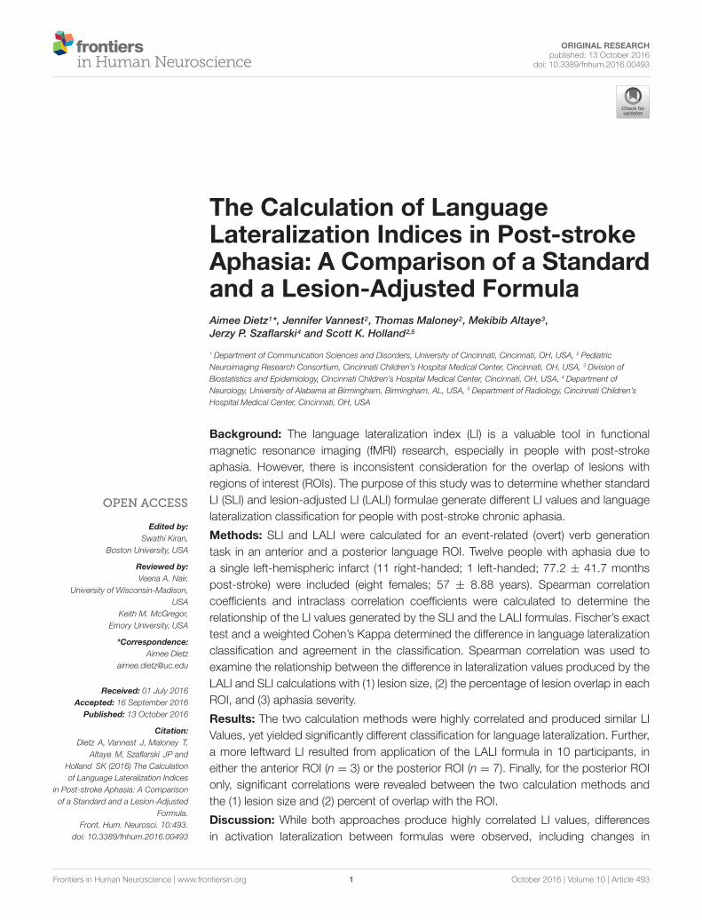
The Calculation of LanguageLateralization Indices in Post-strokeAphasia: A Comparison of a Standardand a Lesion-Adjusted FormulaAimee Dietz1*, Jennifer Vannest2, Thomas Maloney2, Mekibib Altaye3,Jerzy P. Szaflarski4 and Scott K. Holland2,5
1 Department of Communication Sciences and Disorders, University of Cincinnati, Cincinnati, OH, USA, 2 PediatricNeuroimaging Research Consortium, Cincinnati Children’s Hospital Medical Center, Cincinnati, OH, USA, 3 Division ofBiostatistics and Epidemiology, Cincinnati Children’s Hospital Medical Center, Cincinnati, OH, USA, 4 Department ofNeurology, University of Alabama at Birmingham, Birmingham, AL, USA, 5 Department of Radiology, Cincinnati Children’sHospital Medical Center, Cincinnati, OH, USA
Background: The language lateralization index (LI) is a valuable tool in functionalmagnetic resonance imaging (fMRI) research, especially in people with post-strokeaphasia. However, there is inconsistent consideration for the overlap of lesions withregions of interest (ROIs). The purpose of this study was to determine whether standardLI (SLI) and lesion-adjusted LI (LALI) formulae generate different LI values and languagelateralization classification for people with post-stroke chronic aphasia.
Methods: SLI and LALI were calculated for an event-related (overt) verb generationtask in an anterior and a posterior language ROI. Twelve people with aphasia due toa single left-hemispheric infarct (11 right-handed; 1 left-handed; 77.2 ± 41.7 monthspost-stroke) were included (eight females; 57 ± 8.88 years). Spearman correlationcoefficients and intraclass correlation coefficients were calculated to determine therelationship of the LI values generated by the SLI and the LALI formulas. Fischer’s exacttest and a weighted Cohen’s Kappa determined the difference in language lateralizationclassification and agreement in the classification. Spearman correlation was used toexamine the relationship between the difference in lateralization values produced by theLALI and SLI calculations with (1) lesion size, (2) the percentage of lesion overlap in eachROI, and (3) aphasia severity.
Results: The two calculation methods were highly correlated and produced similar LIValues, yet yielded significantly different classification for language lateralization. Further,a more leftward LI resulted from application of the LALI formula in 10 participants, ineither the anterior ROI (n = 3) or the posterior ROI (n = 7). Finally, for the posterior ROIonly, significant correlations were revealed between the two calculation methods andthe (1) lesion size and (2) percent of overlap with the ROI.
Discussion: While both approaches produce highly correlated LI values, differencesin activation lateralization between formulas were observed, including changes in
Frontiers in Human Neuroscience | www.frontiersin.org 1 October 2016 | Volume 10 | Article 493
fnhum-10-00493 October 8, 2016 Time: 16:28 # 2
Dietz et al. LI Calculation in Stroke
lateralization classification. Examination of the issues raised in the current investigationneed to be replicated with a larger sample to determine the utility of a LALI formula inpredicting behavioral performance; the findings may have implications for understandingand interpreting fMRI data of people with post-stroke aphasia.
Keywords: aphasia, fMRI, LI, language lateralization index, ROI, region of interest
INTRODUCTION
Since aphasiologists are interested in determining the influence ofintervention on the neural reorganization of language function,the language lateralization index (LI) is a valuable outcomemeasure in functional magnetic resonance imaging (fMRI)research because it provides a way to quantify the contributionof left hemispheric perilesional tissue assuming responsibility forlanguage functions relative to the right hemispheric homolog(s).Although an exhaustive review of the decades-long “left versusright hemisphere debate” in post-stroke aphasia recovery isbeyond the scope of this paper, a brief summary is warranted[interested readers are referred to Berthier et al. (2011) for a moreextensive review]. A plethora of data suggests that at least initially,the right hemisphere assumes responsibility for performinglanguage tasks formerly managed by the left hemisphere. Then,those who recover best, demonstrate a shift of language functionsback to the left hemispheric, perilesional regions (Saur et al.,2006, 2010; Szaflarski et al., 2013). However, the right hemispheremay play a more critical role in recovery than previouslyacknowledged (Crosson et al., 2009; Fridriksson et al., 2012;Faseyitan et al., 2013); this may be especially true for people withaphasia who have larger lesions and less residual healthy tissuecapable for absorbing the responsibility for linguistic functions(Heiss and Thiel, 2006; Crosson et al., 2009; Berthier et al., 2011).More recently, though, rather than debating the importanceor prominence of the left versus the right hemisphere, manyscientists have adopted the notion that both hemispheres play acritical role in post-stroke language recovery. Thus, the centralproblem now faced by aphasiologists is to determine under whichcircumstances, and for which language functions, left and righthemisphere networks are best suited to take command (Crossonet al., 2007; Turkeltaub et al., 2011; Faseyitan et al., 2013). Further,neuroimaging scientists are beginning to identify treatmentresponders based on neural reorganizational patterns observedduring carefully designed language-based fMRI paradigms thatreflect the skill trained during treatment (Fridriksson et al., 2006;Thompson et al., 2010). For these reasons, it is imperative that thelanguage LI values are reliable, and that researchers and cliniciansalike understand the methodological factors that can influencederivation of the LI value.
A multitude of factors are known to influence thedirectionality of the LI value. According to Seghier (2008),these can be categorized as follows: (1) quantification of therelative contribution of the left and right hemisphere, (2) regionsof interest (ROI) selection, (3) variability and reproducibility ofLI values, (4) reliance on statistical thresholding, (5) thresholdingfor hemispheric dominance, (6) task selection, (7) contrast (orcontrol) conditions, and a catch-all classification, and (8) “other
considerations” (pp. 599). The current study seeks to provideevidence to clarify the importance of how the contribution ofthe left and right hemispheres is quantified specifically, in thepost-stroke population. Presently, there is inconsistent attentionin the literature regarding the influence of lesion size and lesionoverlap with ROIs on the LI values reported in language recoverystudies. A frequently used standard LI (SLI) formula is calculatedby counting the number of voxels that activate above the medianz score of the ROI and dividing by the total number voxelslocated in the ROI (lesioned or not), such that the left and rightROIs are identical in size (Davis et al., 2006; Crosson et al., 2009;Szaflarski et al., 2013). This method seems to naturally bias theLI values rightward since lesioned voxels, which, presumably,cannot be activated, are included in the left-hemispheric ROIs.An alternative to this approach is to apply a lesion-adjusted LI(LALI) formula; one, which accounts for the lesion in somemanner. While some researchers have employed LI formulasthat address this concern (Bonakdarpour et al., 2007; Sebastianand Kiran, 2011) the difference in the derived lateralizationvalues between a standard and a lesion-adjusted method has notbeen examined. As a preliminary step toward understanding theimplications of this tendency in the literature, the purpose ofthis study was to evaluate whether a SLI and an LALI method(1) generate different LI values and (2) different categorizationof language lateralization classification in two language ROIs for12 patients with stroke-induced aphasia on a verb generationtask.
To address the questions posed in this study, we used an event-related verb generation task and a sparse acquisition approach,which allows participants to hear the auditory prompts withoutscanner noise, and provide overt verbal responses while avoidinghead motion during image acquisition (Schmithorst and Holland,2004; Allendorfer et al., 2012a). This approach has been shownto better detect task-related activation patterns than a standardboxcar fMRI (Schmithorst and Holland, 2004; Huettel, 2012). Weexamined the difference in SLI and LALI values in two anatomicalROIs, which encompass broad regions of the language networkand have been shown to capture perilesional activation that mayoccur as a result of post-stroke recovery and language-relatedreorganization (Allendorfer et al., 2012a,b) (see “Materials andMethod” for more details).
Specifically, we asked the following questions:
1. Do the LIs derived by the SLI and LALI formulas:(a) Generate different LI values?(b) Categorize language lateralization in people with post-stroke aphasia differently.
2. What is the relationship between LALI and SLI values and:(a) Left hemisphere lesion size?
Frontiers in Human Neuroscience | www.frontiersin.org 2 October 2016 | Volume 10 | Article 493
fnhum-10-00493 October 8, 2016 Time: 16:28 # 3
Dietz et al. LI Calculation in Stroke
(b) Percent overlap between ROI and left hemispherelesion?
3. What is the relationship between the LALI and SLI valuesand aphasia severity?
We hypothesized that would be a significant difference in theclassification of language lateralization when considering thetwo methods. Further, we hypothesized a positive correlationbetween lesion size and the degree of overlap between the lesionand the ROI on the difference in LI values. That is, the largerthe lesion and the greater the overlap with the ROI, the morelikely the SLI and LALI results would differ. Lastly, since ourprimary goal was to examine the whether the SLI and LALIcalculation methods differed in resultant LI values/languagelateralization classification, the final research question, regardingaphasia severity, was exploratory in nature.
MATERIALS AND METHODS
This study was carried out in accordance with therecommendations of the University of Cincinnati and CincinnatiChildren’s Hospital and Medical Center Institutional ReviewBoard, with written informed consent from all subjects.
ParticipantsThe participants included 12 people with chronic, post-strokeaphasia (single left middle cerebral ischemic infarct) whoparticipated in a larger treatment study. Prior to the stroke,all but one participant were right-handed, according to theEdinburgh Handedness inventory (Oldfield, 1971). Further, allparticipants were native speakers of American English, had atleast a high school education, and reported a no history ofpsychiatric substance abuse. Prior to enrolling in the study, allparticipants reported normal or corrected vision, passed a visualfield cut screening, and a passed a hearing screening in at least oneear at (i.e., 40 db HL 1000, 2000, and 4000 Hz). Table 1 providesa summary of the participants’ demographic data.
FMRI Equipment and MethodsA 3.0 Philips Achieva Whole Body MRI/MRS system allowedacquisition of structural and fMRI scans. The followingparameters were used to capture high-resolution T1-weighted anatomical images: TR/TE = 8.1/3.7 ms, FOV25.6× 25.6× 19.2 cm, matrix 256× 256, slice thickness= 1 mm.A sparse acquisition [see “Event-Related Verb GenerationTask (ER-VGT)” for details] approach was used for eachtrial: MRI silence occurred for the first 6 s to allow stimulipresentation and participant response; followed by 6 s (threeimage volumes) of fMRI data acquisition during the height of thehemodynamic response (Schmithorst and Holland, 2004). ThefMRI scanning was performed with the following parameters:TR/TE= 2000/38 ms, FOV 24.0× 24.0 cm, matrix 64× 64, slicethickness = 4 mm, SENSE factor = 2. This resulted in a voxelsize of 3.75 × 3.75 × 4 mm and 32 axial slices. FMRI task wasdeveloped in DirectRT (version 20121) and presented using anAvotec audio-visual system.
Event-Related Verb Generation Task (ER-VGT)In this study, we employed a well-documented event related verbgeneration task that is recognized for its sensitivity to identifylanguage-related areas in healthy controls and patients with post-stroke aphasia (Allendorfer et al., 2012a,b). Participants viewed aReady screen for 4 s. Then, they completed 15 alternating trialseach of: (1) covert verb generation, (2) overt verb generation,and (3) overt noun repetition, with each of the 45 trials lasting12 s. The first 6 s of each trial began with MRI silence, followedby 6 s of fMRI acquisition. In lieu of written instructions, dueto the known comprehension challenges experienced by peoplewith aphasia, the participants were presented with a pictorialinstructions during the first 1 s of MRI silence. Followingpictorial instruction, the remaining 5 s of MRI silence includedan auditory presentation of a concrete noun (e.g., “cookie”) viaheadphones. The pictorial instruction cued the participant to
1http://www.empirisoft.com/
TABLE 1 | Demographic and linguistic profile of participants.
ID Gender Ethnicity Age Number lesioned voxels MPOa Level of education Aphasia typeb Aphasia quotientb
1 Female Caucasian 57 18727 79 Some college Global 40.8
2 Male Caucasian 61 30570 91 Bachelor’s Broca’sc 68.6
3 Female African amer. 58 1583 48 Bachelor’s Anomic 89.2
4 Female Caucasian 47 14616 124 Bachelor’s Broca’sc 48.9
5d Female Caucasian 60 12950 105 Some college Conduction 74.0
6 Male Caucasian 59 14009 73 Master’s Anomic 68.9
7 Male Caucasian 47 10989 69 High school Wernicke’s 55.5
8 Male Caucasian 71 14066 69 Master’s Broca’sc 62.7
9 Female Caucasian 63 17705 170 Bachelor’s Conduction 71.2
10 Female Caucasian 57 7849 16 High school Wernicke’s 37.6
11 Female Caucasian 39 18807 44 Some college Anomic 82.4
12 Male Caucasian 66 30110 38 Bachelor’s Broca’sc 36.7
aMonths post-onset; bWestern Aphasia Battery-Revised (Kertesz, 2006) used to determine type and severity, Aphasia quotient out of 100; cApraxia of Speech presentbased on clinical judgment; dOnly left-handed participant; all others right-handed; handedness based on the Edinburgh Handedness Inventory (i.e., score ≥ 50 = right-handed) (Oldfield, 1971).
Frontiers in Human Neuroscience | www.frontiersin.org 3 October 2016 | Volume 10 | Article 493
fnhum-10-00493 October 8, 2016 Time: 16:28 # 4
Dietz et al. LI Calculation in Stroke
produce associated verbs (e.g., bake, eat, ice, etc.) either (1) tothemselves (covert generation) or (2) aloud (overt generation),or to (3) repeat the noun aloud (i.e., “cookie, cookie, cookie”; overrepetition). The final 6 s of each trial concluded with fMRI dataacquisition.
Prior to entering the scanner, all participants practiced the ER-VGT and were able to provide at least a least one correct responseto each of the aforementioned three ER-VGT conditions. Thatis they were able to speak an appropriate response for the overtverb generation and overt noun repetition trials; and made noovert responses during the covert verb generation trials. This isin line with other reports of the ER-VGT task performance bypeople who have aphasia (Allendorfer et al., 2012a). People withaphasia can learn the task; however, they typically produce fewerverb productions during the overt verb generation task whencompared to healthy controls. During the scan, participants weremonitored by in-scanner microphone on each trial to ensure thatthe participants responded as instructed. In instances where theparticipant confused the instructions, the sequence was stoppedand the patient was reinstructed (Participants 3, 6, 7, 8, 9, 10,11, and 12). With the exception of Participant 12, all participantswere able to respond, or attempt to respond, appropriately duringeach condition after reinstruction.
The ER-VGT task involves several speech and linguisticfunctions; with auditory processing being required for all tasks.The contrast of overt > covert generation isolates articulatoryability, or motor aspects of speaking, while controlling forrecognition of the auditory noun, and noun-verb semanticassociations needed to generate the verb responses. The contrastof overt verb generation > overt repetition isolates the noun-verbsemantic association process while controlling for recognition ofthe auditory noun and articulatory/motor aspects of generatingan overt response. In this study, we used a general linear modelapproach to identify voxels that were more active in the overt verbgeneration > overt repetition contrast.
Research Design and Data AnalysesA trained neuroanatomist (JPS) manually traced eachparticipant’s lesion on their T1-weighted anatomical image
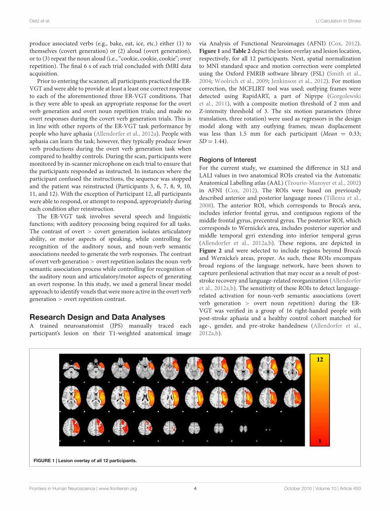
via Analysis of Functional Neuroimages (AFNI) (Cox, 2012).Figure 1 and Table 2 depict the lesion overlay and lesion location,respectively, for all 12 participants. Next, spatial normalizationto MNI standard space and motion correction were completedusing the Oxford FMRIB software library (FSL) (Smith et al.,2004; Woolrich et al., 2009; Jenkinson et al., 2012). For motioncorrection, the MCFLIRT tool was used; outlying frames weredetected using RapidART, a part of Nipype (Gorgolewskiet al., 2011), with a composite motion threshold of 2 mm andZ-intensity threshold of 3. The six motion parameters (threetranslation, three rotation) were used as regressors in the designmodel along with any outlying frames; mean displacementwas less than 1.5 mm for each participant (Mean = 0.33;SD= 1.44).
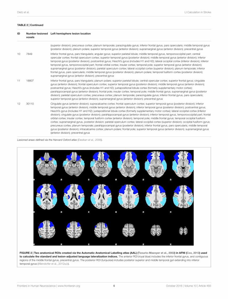
Regions of InterestFor the current study, we examined the difference in SLI andLALI values in two anatomical ROIs created via the AutomaticAnatomical Labelling atlas (AAL) (Tzourio-Mazoyer et al., 2002)in AFNI (Cox, 2012). The ROIs were based on previouslydescribed anterior and posterior language zones (Tillema et al.,2008). The anterior ROI, which corresponds to Broca’s area,includes inferior frontal gyrus, and contiguous regions of themiddle frontal gyrus, precentral gyrus. The posterior ROI, whichcorresponds to Wernicke’s area, includes posterior superior andmiddle temporal gyri extending into inferior temporal gyrus(Allendorfer et al., 2012a,b). These regions, are depicted inFigure 2 and were selected to include regions beyond Broca’sand Wernicke’s areas, proper. As such, these ROIs encompassbroad regions of the language network, have been shown tocapture perilesional activation that may occur as a result of post-stroke recovery and language-related reorganization (Allendorferet al., 2012a,b). The sensitivity of these ROIs to detect language-related activation for noun-verb semantic associations (overtverb generation > overt noun repetition) during the ER-VGT was verified in a group of 16 right-handed people withpost-stroke aphasia and a healthy control cohort matched forage-, gender, and pre-stroke handedness (Allendorfer et al.,2012a,b).
FIGURE 1 | Lesion overlay of all 12 participants.
Frontiers in Human Neuroscience | www.frontiersin.org 4 October 2016 | Volume 10 | Article 493
fnhum-10-00493 October 8, 2016 Time: 16:28 # 5
Dietz et al. LI Calculation in Stroke
TABLE 2 | Description of lesions for the 12 participants.
ID Number lesionedvoxels
Left hemisphere lesion location
1 18727 Inferior frontal gyrus; pars triangularis; subcallosal cortex; angular gyrus; frontal medial cortex; middle temporal gyrus (temporooccipitalregion); central opercular cortex; frontal operculum cortex; superior (posterior), middle (anterior), and inferior gyrus (anterior and posterior);postcentral gyrus; Heschl’s gyrus (includes H1 and H2); lateral occipital cortex; inferior division amygdala; inferior temporal gyrus;temporooccipital part; frontal orbital cortex; insular cortex; temporal fusiform cortex (anterior; posterior); temporal pole; middle frontal gyrus;supramarginal gyrus ( posterior division); parietal operculum cortex; lateral occipital cortex (superior division); planum temporale; inferiorfrontal gyrus; pars opercularis; middle temporal gyrus (posterior division); planum polare; frontal pole; superior temporal gyrus (anteriordivision); supramarginal gyrus (anterior division); precentral gyrus
2 30570 Inferior frontal gyrus, pars triangularis; cuneal cortex; subcallosal cortex; angular gyrus; frontal medial cortex; superior parietal lobule; middletemporal gyrus, temporooccipital part; central opercular cortex; superior frontal gyrus; cingulate gyrus, anterior division; supracalcarinecortex; lingual gyrus; frontal operculum cortex; superior temporal gyrus (posterior division); inferior temporal gyrus (anterior division); middletemporal gyrus (anterior division); inferior temporal gyrus (posterior division); postcentral gyrus; Heschl’s gyrus (includes H1 and H2);juxtapositional lobule cortex (formerly supplementary motor cortex); lateral occipital cortex ( inferior division); cingulate gyrus (posteriordivision); parahippocampal gyrus (anterior division); inferior temporal gyrus, temporooccipital part; frontal orbital cortex; insular cortex;temporal fusiform cortex, anterior division; temporal pole; middle frontal gyrus; temporal fusiform cortex (posterior division); supramarginalgyrus ( posterior division); parietal operculum cortex; lateral occipital cortex (superior division); occipital fusiform gyrus; precuneus cortex;planum temporale; inferior frontal gyrus, pars opercularis; middle temporal gyrus (posterior division); intracalcarine cortex; planum polare;frontal pole; superior temporal gyrus (anterior division); supramarginal gyrus (anterior division); precentral gyrus
3 1583 Frontal operculum cortex; inferior frontal gyrus, pars triangularis; parietal operculum cortex; central opercular cortex; putamen; frontal orbitalcortex; inferior frontal gyrus, pars opercularis; middle frontal gyrus; frontal pole; supramarginal gyrus (anterior division); precentral gyrus;postcentral gyrus
4 14616 Inferior frontal gyrus, pars triangularis; angular gyrus; superior parietal lobule; middle temporal gyrus, temporooccipital part; central opercularcortex; frontal operculum cortex; superior temporal gyrus (posterior division); middle temporal gyrus (anterior division); postcentral gyrus;Heschl’s gyrus (includes h1 and h2); frontal pole; frontal orbital cortex; insular cortex; temporal fusiform cortex, anterior division; temporalpole; middle frontal gyrus; temporal fusiform cortex (posterior division); supramarginal gyrus (posterior division); parietal operculum cortex;lateral occipital cortex (superior division); planum temporale; paracingulate gyrus; inferior frontal gyrus, pars opercularis; middle temporalgyrus (posterior division); planum polare; superior temporal gyrus (anterior division); supramarginal gyrus (anterior division); precentral gyrus
5 12950 Superior parietal lobule; angular gyrus; middle temporal gyrus, temporooccipital part; temporal occipital fusiform cortex; central opercularcortex; amygdala; lingual gyrus; frontal operculum cortex; superior temporal gyrus (posterior division); inferior temporal gyrus (anteriordivision); middle temporal gyrus (anterior division); inferior temporal gyrus (posterior division); postcentral gyrus; Heschl’s gyrus (includes H1and H2); lateral occipital cortex (inferior division); cingulate gyrus (posterior division); inferior temporal gyrus, temporooccipital part; frontalorbital cortex; insular cortex; temporal fusiform cortex (anterior and posterior division); temporal pole; middle frontal gyrus; supramarginalgyrus (posterior division); parietal operculum cortex; lateral occipital cortex (superior division); occipital fusiform gyrus; precuneus cortex;planum temporale; parahippocampal gyrus (posterior division); middle temporal gyrus (posterior division); cuneal cortex; planum polare;frontal pole; superior temporal gyrus (anterior division); supramarginal gyrus (anterior division); precentral gyrus
6 14009 Inferior frontal gyrus, pars triangularis; planum polare; central opercular cortex; superior frontal gyrus; frontal operculum cortex; superiortemporal gyrus (posterior division); inferior temporal gyrus, anterior division; middle temporal gyrus (anterior division); inferior temporal gyrus,posterior division; postcentral gyrus; Heschl’s gyrus (includes H1 and H2); juxtapositional lobule cortex (formerly supplementary motorcortex); parahippocampal gyrus (anterior division); frontal pole; frontal orbital cortex; insular cortex; temporal fusiform cortex (anteriordivision); temporal pole; middle frontal gyrus; temporal fusiform cortex (posterior division); supramarginal gyrus (posterior division); parietaloperculum cortex; planum temporale; paracingulate gyrus; inferior frontal gyrus, pars opercularis; middle temporal gyrus (posterior division);superior temporal gyrus (anterior division); supramarginal gyrus (anterior division); precentral gyrus
7 10989 Angular gyrus; superior parietal lobule; middle temporal gyrus, temporooccipital part; central opercular cortex; supracalcarine cortex;superior temporal gyrus (posterior division); middle temporal gyrus (anterior division); inferior temporal gyrus (posterior division); cunealcortex; Heschl’s gyrus (includes H1 and H2); postcentral gyrus; juxtapositional lobule cortex (formerly supplementary motor cortex);parahippocampal gyrus (anterior division); inferior temporal gyrus, temporooccipital part; hippocampus; insular cortex; temporal pole;temporal occipital fusiform cortex; supramarginal gyrus (posterior division); parietal operculum cortex; lateral occipital cortex (superiordivision); precuneus cortex; planum temporale; parahippocampal gyrus (posterior division); middle temporal gyrus (posterior division);intracalcarine cortex; planum polare; temporal fusiform cortex (posterior division); supramarginal gyrus (anterior division); precentral gyrus
8 14066 Occipital fusiform gyrus; middle temporal gyrus (anterior division); inferior temporal gyrus (posterior division); intracalcarine cortex; Heschl’sgyrus (includes H1 and H2); lateral occipital cortex (inferior division); inferior temporal gyrus, temporooccipital part; frontal orbital cortex;insular cortex; temporal pole; middle frontal gyrus; temporal fusiform cortex (posterior division); supramarginal gyrus (posterior division);parietal operculum cortex; lateral occipital cortex (superior division); planum temporale; inferior frontal gyrus, pars opercularis; middletemporal gyrus (posterior division); postcentral gyrus; planum polare; frontal pole; superior temporal gyrus (anterior division); supramarginalgyrus (anterior division); precentral gyrus
9 17705 Inferior frontal gyrus, pars triangularis; angular gyrus; superior parietal lobule; middle temporal gyrus, temporooccipital part; central opercularcortex; superior frontal gyrus; cingulate gyrus (anterior division); frontal operculum cortex; superior temporal gyrus (posterior division); inferiortemporal gyrus (anterior division); middle temporal gyrus (anterior division); inferior temporal gyrus (posterior division); postcentral gyrus;Heschl’s gyrus (includes H1 and H2); juxtapositional lobule cortex (formerly supplementary motor cortex); lateral occipital cortex (inferiordivision); cingulate gyrus (posterior division); parahippocampal gyrus (anterior division); inferior temporal gyrus, temporooccipital part;parahippocampal gyrus, posterior division; insular cortex; temporal fusiform cortex (anterior division); temporal pole; middle frontal gyrus;temporal fusiform cortex (posterior division); supramarginal gyrus (posterior division); parietal operculum cortex; lateral occipital cortex
(Continued)
Frontiers in Human Neuroscience | www.frontiersin.org 5 October 2016 | Volume 10 | Article 493
fnhum-10-00493 October 8, 2016 Time: 16:28 # 6
Dietz et al. LI Calculation in Stroke
TABLE 2 | Continued
ID Number lesionedvoxels
Left hemisphere lesion location
(superior division); precuneus cortex; planum temporale; paracingulate gyrus; inferior frontal gyrus, pars opercularis; middle temporal gyrus(posterior division); planum polare; superior temporal gyrus (anterior division); supramarginal gyrus (anterior division); precentral gyrus
10 7849 Inferior frontal gyrus, pars triangularis; angular gyrus; superior parietal lobule; middle temporal gyrus, temporooccipital part; centralopercular cortex; frontal operculum cortex; superior temporal gyrus (posterior division); middle temporal gyrus (anterior division); inferiortemporal gyrus (posterior division); postcentral gyrus; Heschl’s gyrus (includes H1 and H2); lateral occipital cortex (inferior division); inferiortemporal gyrus, temporooccipital part; frontal orbital cortex; insular cortex; temporal pole; superior temporal gyrus (anterior division);supramarginal gyrus (posterior division); parietal operculum cortex; lateral occipital cortex (superior division); planum temporale; inferiorfrontal gyrus, pars opercularis; middle temporal gyrus (posterior division); planum polare; temporal fusiform cortex (posterior division);supramarginal gyrus (anterior division); precentral gyrus
11 18807 Inferior frontal gyrus, pars triangularis; planum polare; superior parietal lobule; central opercular cortex; superior frontal gyrus; cingulategyrus (anterior division); frontal operculum cortex; superior temporal gyrus (posterior division); middle temporal gyrus (anterior division);postcentral gyrus; Heschl’s gyrus (includes H1 and H2); juxtapositional lobule cortex (formerly supplementary motor cortex);parahippocampal gyrus (anterior division); frontal pole; insular cortex; temporal pole; middle frontal gyrus; supramarginal gyrus (posteriordivision); parietal operculum cortex; precuneus cortex; planum temporale; paracingulate gyrus; inferior frontal gyrus, pars opercularis;superior temporal gyrus (anterior division); supramarginal gyrus (anterior division); precentral gyrus
12 30110 Cingulate gyrus (anterior division); supracalcarine cortex; frontal operculum cortex; superior temporal gyrus (posterior division); inferiortemporal gyrus (anterior division); middle temporal gyrus (anterior division); inferior temporal gyrus (posterior division); postcentral gyrus;Heschl’s gyrus (includes H1 and H2); juxtapositional lobule cortex (formerly supplementary motor cortex); lateral occipital cortex (inferiordivision); cingulate gyrus (posterior division); parahippocampal gyrus (anterior division); inferior temporal gyrus, temporooccipital part; frontalorbital cortex; insular cortex; temporal fusiform cortex (anterior division); temporal pole; middle frontal gyrus; temporal occipital fusiformcortex; supramarginal gyrus, posterior division; parietal operculum cortex; lateral occipital cortex (superior division); occipital fusiform gyrus;precuneus cortex; planum temporale; parahippocampal gyrus (posterior division); inferior frontal gyrus, pars opercularis; middle temporalgyrus (posterior division); intracalcarine cortex; planum polare; frontal pole; superior temporal gyrus (anterior division); supramarginal gyrus(anterior division); precentral gyrus
Lesioned areas defined via the Harvard Oxford atlas (Desikan et al., 2006).
FIGURE 2 | Two anatomical ROIs created via the Automatic Anatomical Labelling atlas (AAL) (Tzourio-Mazoyer et al., 2002) in AFNI (Cox, 2012) usedto calculate the standard and lesion-adjusted language lateralization indices. The anterior ROI (royal blue) includes the inferior frontal gyrus, and contiguousregions of the middle frontal gyrus, precentral gyrus. The posterior ROI (turquoise) includes posterior superior and middle temporal gyri extending into inferiortemporal gyrus (Allendorfer et al., 2012a,b).
Frontiers in Human Neuroscience | www.frontiersin.org 6 October 2016 | Volume 10 | Article 493

fnhum-10-00493 October 8, 2016 Time: 16:28 # 7
Dietz et al. LI Calculation in Stroke
FIGURE 3 | Percent lesion/ROI overlap for all participants.
Thresholding and Language LateralizationCalculation MethodsThe ROIs were applied to each participant’s functional data andmirrored to the right in MNI space. Figure 3 illustrates thepercent lesion/ROI overlap for each participant in current study.Thresholding for active voxels, for purposes of LI calculation,was performed on a single-participant basis. Specifically, voxelsabove the median z-score (overt verb generation > overtrepetition) in each ROI for each participant were counted asactive (Wilke and Schmithorst, 2006; Wilke and Lidzba, 2007).Next, the language LI was calculated using two methods: aSLI and a lesion-adapted LI formula as described below. Thelanguage lateralization classification schema for this study isbased on the specific criteria used in our previous work withthe verb generation task (Szaflarski et al., 2006, 2008, 2014;Allendorfer et al., 2012a,b), and that of others (Wilke andLidzba, 2007). Specifically, right-lateralization was defined asLI <−0.1, and left-lateralization as >0.1; whereas values between−0.1 < LI ≤ 0.1 indicated bilateral, or symmetric languagedistribution.
Standard lateralization index (SLI)The SLI formula (see below) applied in this study was calculatedby counting the number of active voxels (those above the medianz-score of the ROI). Then, we calculated the difference betweenthe right and left ROIs divided by the total number of activevoxels in the right and left ROIs (lesioned or not). The left andright ROIs are identical in size (Szaflarski et al., 2006, 2014; Wilkeand Lidzba, 2007), and this calculation includes all voxels in leftand right ROIs, including lesioned tissue that overlaps with theROIs.
(#Left Active Voxels− #Right Active Voxels)(#Left Active Voxels+ #Right Active Voxels)
Lesion-adapted lateralization index (LALI)In the LALI formula (see below) active voxels are also determinedby counting those above the median z score within the ROI.
However, in this method, the ROI is limited in each participantto only consider non-lesioned voxels. In other words, the sizeof the left sided ROI is decreased for each participant by thenumber of lesioned voxels that overlap with the ROI. In contrast,the right sided ROI is the same size as the ROI used forthe SLI calculation. Activation in right and left ROIs is thenexpressed as a ratio of the number of active voxels comparedto the total number of voxels in this limited, non-lesionedROI. For example, if the ROI is 15274 voxels (i.e., anteriorROI used in this study), but for a given participant only 1000voxels in the ROI are non-lesioned voxels, active voxels arequantified as the proportion of these 1000 voxels that wereabove the median z-score. This approach allows us to consider,in our population, a left-hemisphere ROI “highly active” if alarge proportion of non-lesioned voxels are active, even if theparticipant has only a small amount of non-lesion tissue in theROI.((
# Left Active Voxels# Left Nonlesioned Voxels
)−
(# Right Active Voxels
# Right Nonlesioned Voxels
))((
# Left Voxels# Left Nonlesioned Voxels
)+
(# Right Voxels
# Right Nonlesioned Voxels
))Statistical TreatmentSpearman correlation coefficients and intraclass correlationcoefficients (ICC) were calculated to determine the relationshipof the LI values generated by the SLI and the LALI formulas.
Fisher’s exact tests were used to examine possible differences inlanguage lateralization classification (left, right, bilateral) betweenSLI and LALI. A weighted Cohen’s Kappa provided a measure ofagreement between SLI and LALI in the classification of languagelateralization.
Lastly, we used Spearman correlation to examine therelationship between the difference in LI values produced by theLALI and SLI calculations (LALI − SLI) with (1) lesion size, (2)the percentage of lesion overlap in each ROI, (3) aphasia severity[i.e., Western Aphasia Battery-Revised Aphasia Quotient; WAB-R AQ (Kertesz, 2006)].
Frontiers in Human Neuroscience | www.frontiersin.org 7 October 2016 | Volume 10 | Article 493
fnhum-10-00493 October 8, 2016 Time: 16:28 # 8
Dietz et al. LI Calculation in Stroke
RESULTS
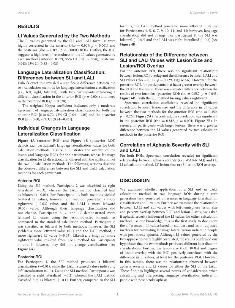
LI Values Generated by the Two MethodsThe LI values generated by the SLI and LALI formulas werehighly correlated in the anterior (rho = 0.909; p < 0.001) andthe posterior (rho = 0.895; p < 0.0001) ROIs. Further, the ICCsuggests a high level of relatedness in the LI values generated byeach method [anterior: 0.939; 95% CI (0.81 – 0.98); posterior:0.943; 95% CI (0.83 – 0.98)].
Language Lateralization Classification:Differences between SLI and LALIFisher’s exact test revealed a significant difference between thetwo calculation methods for language lateralization classification(i.e., left, right, bilateral), with two participants exhibiting adifferent classification in the anterior ROI (p = 0.004) and threein the posterior ROI (p= 0.028).
The weighted Kappa coefficient indicated only a moderateagreement of language lateralization classification for both theanterior ROI [k = 0.72; 95% CI (0.04 – 1.0)] and the posteriorROI [k= 0.60; 95% CI (0.24−0.96)].
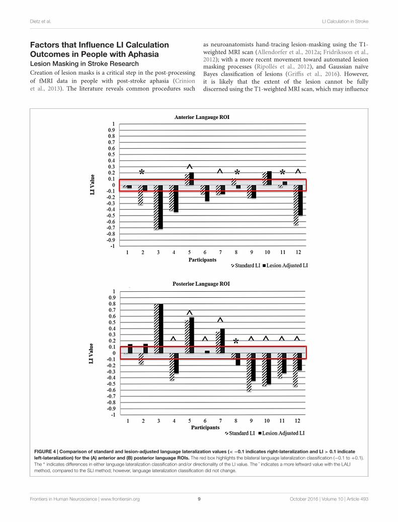
Individual Changes in LanguageLateralization ClassificationFigure 4A (anterior ROI) and Figure 4B (posterior ROI)depicts each participant’s language lateralization values for bothcalculation methods. Figure 5 illustrates the overlap of thelesion and language ROIs for the participants whose languageclassification (or LI directionality) differed with the application ofthe two LI calculation methods. The following sections describethe observed differences between the SLI and LALI calculationmethods for each participant.
Anterior ROIUsing the SLI method, Participant 2 was classified as rightlateralized (−0.3); whereas the LALI method classified himas bilateral (−0.09). For Participant 11, both methods yieldedbilateral LI values; however, SLI method generated a morerightward (−0.03) value, and the LALI a more leftward(0.06) value. Although their language classification didnot change, Participants 5, 7, and 12 demonstrated moreleftward LI values using the lesion-adjusted formula, ascompared to the standard calculation method. Participant 8was classified as bilateral by both methods; however, the SLIyielded a more leftward value (0.1) and the LALI method, amore rightward LI value (−0.05). Likewise, a (slightly) morerightward value resulted from LALI method for Participants4, and 6; however, they did not change classification (seeFigure 4A).
Posterior ROIFor Participant 1, the SLI method produced a bilateralclassification (−0.01), while the LALI returned values indicatingleft lateralization (0.13). Using the SLI method, Participant 2 wasclassified as right lateralized (−0.2); whereas the LALI methodclassified him as bilateral (−0.1). Further, compared to the SLI
formula, the LALI method generated more leftward LI valuesfor Participants 4, 5, 6, 7, 9, 10, 11, and 12; however, languageclassification did not change. For participant 8, the SLI wasbilateral (−0.07) and the LALI was right lateralized (−0.2) (seeFigure 4B).
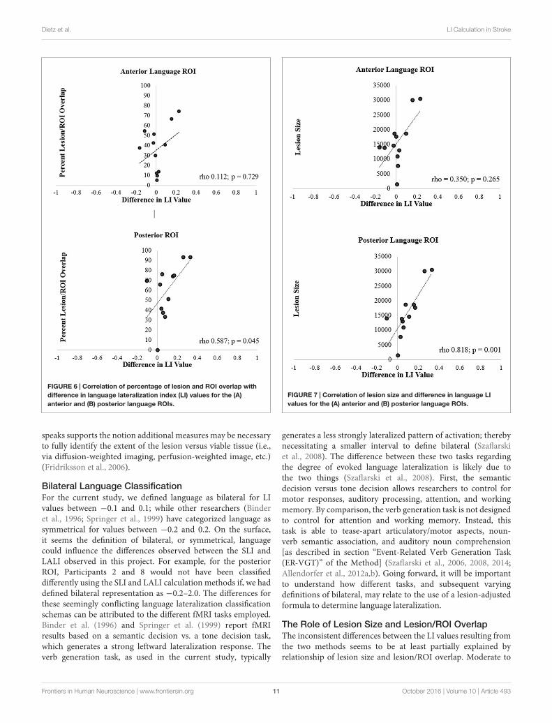
Relationship of the Difference betweenSLI and LALI Values with Lesion Size andLesion/ROI OverlapFor the anterior ROI, there was no significant relationshipbetween lesion/ROI overlap and the difference between LALI andSLI values (rho = 0.112, p = 0.729; Figure 6A). However, for theposterior ROI, for participants that had a greater overlap betweenthe ROI and the lesion, there was a greater difference between theresults of two formulas (posterior ROI: rho = 0.587, p = 0.045;Figure 6B); with the SLI method biasing values rightward.
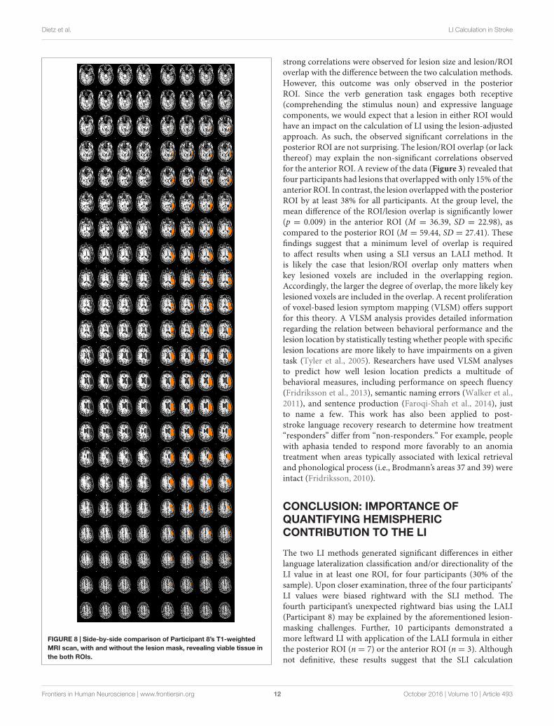
Spearman correlation coefficients revealed no significantcorrelation between lesion size and the difference in LI valuesbetween the two methods for the anterior ROI (rho = 0.350,p= 0.265; Figure 7A). In contrast, the correlation was significantin the posterior ROI (rho = 0.818, p = 0.001; Figure 7B). Inessence, in participants with larger lesions, there was a greaterdifference between the LI values generated by two calculationmethods in the posterior ROI.
Correlation of Aphasia Severity with SLIand LALIFor both ROIs, Spearman correlation revealed no significantrelationship between aphasia severity (i.e., WAB-R AQ) and (1)LI calculation method, (2) lesion size, or (3) lesion/ROI overlap.
DISCUSSION
We examined whether application of a SLI and an LALIcalculation method, in two language ROIs during a verbgeneration task, generated differences in language lateralizationclassification and LI values. Further, we examined the relationshipbetween LALI and SLI values with left-hemisphere lesion sizeand percent overlap between ROI and lesion. Lastly, we askedif aphasia severity influenced the LI values for either calculationmethod. To our knowledge, this is the first study to documentthe differences in LI values based on standard and lesion-adjustedmethods for calculating language lateralization indices in peoplewith post-stroke aphasia. Although LI values generated by thetwo approaches were highly correlated, the results confirmed ourhypothesis that the two methods produced different lateralizationclassifications. Further, the lesion size (both ROIs) and degreeof lesion overlap with the ROI positively correlated with thedifference in LI values, at least for the posterior ROI. However,in this sample, there was no relationship observed betweenaphasia severity and LI values for either the SLI or the LALI.These findings highlight several points of consideration whencalculating and interpreting language lateralization indices inpeople with post-stroke aphasia.
Frontiers in Human Neuroscience | www.frontiersin.org 8 October 2016 | Volume 10 | Article 493
fnhum-10-00493 October 8, 2016 Time: 16:28 # 9
Dietz et al. LI Calculation in Stroke
Factors that Influence LI CalculationOutcomes in People with AphasiaLesion Masking in Stroke ResearchCreation of lesion masks is a critical step in the post-processingof fMRI data in people with post-stroke aphasia (Crinionet al., 2013). The literature reveals common procedures such
as neuroanatomists hand-tracing lesion-masking using the T1-weighted MRI scan (Allendorfer et al., 2012a; Fridriksson et al.,2012); with a more recent movement toward automated lesionmasking processes (Ripollés et al., 2012), and Gaussian naïveBayes classification of lesions (Griffis et al., 2016). However,it is likely that the extent of the lesion cannot be fullydiscerned using the T1-weighted MRI scan, which may influence
FIGURE 4 | Comparison of standard and lesion-adjusted language lateralization values (< −0.1 indicates right-lateralization and LI > 0.1 indicateleft-lateralization) for the (A) anterior and (B) posterior language ROIs. The red box highlights the bilateral language lateralization classification (−0.1 to +0.1).The ∗ indicates differences in either language lateralization classification and/or directionality of the LI value. The ˆ indicates a more leftward value with the LALImethod, compared to the SLI method; however, language lateralization classification did not change.
Frontiers in Human Neuroscience | www.frontiersin.org 9 October 2016 | Volume 10 | Article 493
fnhum-10-00493 October 8, 2016 Time: 16:28 # 10
Dietz et al. LI Calculation in Stroke
FIGURE 5 | Illustration of activation intensity (median z score) and lesion (dark blue outline) overlap with ROIs (turquoise outline) for participants whodemonstrated different language lateralization classifications for the SLI and LALI calculation methods. The anterior ROIs are depicted in the left panelsand the posterior in the right panels.
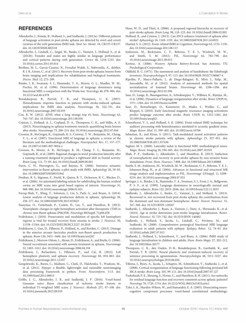
accurate calculation of LI and subsequent language lateralizationclassification. For example, in the current study, Participant 8appears to have healthy tissue included the lesioned mask thatoverlaps the left anterior and posterior ROIs (see Figure 8). His LIvalues revealed a pattern opposite to our hypothesized direction(i.e., the LALI, not the SLI, biased the LI values rightward). Atleast part of the anterior ROI encompasses what appears to be
residual tissue that has been assigned to the lesion. Similarly,in the posterior language area, there are some small islands ofwhat appear to be relatively normal looking brain tissue thatis included in the lesion mask. Inclusion of viable tissue mayhave occurred due to the difficulty in registering the depth ofthe lesion using a T1-weighted scan; in other words, the tissueis likely lesioned, but not as deeply as it was more anteriorly. This
Frontiers in Human Neuroscience | www.frontiersin.org 10 October 2016 | Volume 10 | Article 493
fnhum-10-00493 October 8, 2016 Time: 16:28 # 11
Dietz et al. LI Calculation in Stroke
FIGURE 6 | Correlation of percentage of lesion and ROI overlap withdifference in language lateralization index (LI) values for the (A)anterior and (B) posterior language ROIs.
speaks supports the notion additional measures may be necessaryto fully identify the extent of the lesion versus viable tissue (i.e.,via diffusion-weighted imaging, perfusion-weighted image, etc.)(Fridriksson et al., 2006).
Bilateral Language ClassificationFor the current study, we defined language as bilateral for LIvalues between −0.1 and 0.1; while other researchers (Binderet al., 1996; Springer et al., 1999) have categorized language assymmetrical for values between −0.2 and 0.2. On the surface,it seems the definition of bilateral, or symmetrical, languagecould influence the differences observed between the SLI andLALI observed in this project. For example, for the posteriorROI, Participants 2 and 8 would not have been classifieddifferently using the SLI and LALI calculation methods if, we haddefined bilateral representation as −0.2–2.0. The differences forthese seemingly conflicting language lateralization classificationschemas can be attributed to the different fMRI tasks employed.Binder et al. (1996) and Springer et al. (1999) report fMRIresults based on a semantic decision vs. a tone decision task,which generates a strong leftward lateralization response. Theverb generation task, as used in the current study, typically
FIGURE 7 | Correlation of lesion size and difference in language LIvalues for the (A) anterior and (B) posterior language ROIs.
generates a less strongly lateralized pattern of activation; therebynecessitating a smaller interval to define bilateral (Szaflarskiet al., 2008). The difference between these two tasks regardingthe degree of evoked language lateralization is likely due tothe two things (Szaflarski et al., 2008). First, the semanticdecision versus tone decision allows researchers to control formotor responses, auditory processing, attention, and workingmemory. By comparison, the verb generation task is not designedto control for attention and working memory. Instead, thistask is able to tease-apart articulatory/motor aspects, noun-verb semantic association, and auditory noun comprehension[as described in section “Event-Related Verb Generation Task(ER-VGT)” of the Method] (Szaflarski et al., 2006, 2008, 2014;Allendorfer et al., 2012a,b). Going forward, it will be importantto understand how different tasks, and subsequent varyingdefinitions of bilateral, may relate to the use of a lesion-adjustedformula to determine language lateralization.
The Role of Lesion Size and Lesion/ROI OverlapThe inconsistent differences between the LI values resulting fromthe two methods seems to be at least partially explained byrelationship of lesion size and lesion/ROI overlap. Moderate to
Frontiers in Human Neuroscience | www.frontiersin.org 11 October 2016 | Volume 10 | Article 493
fnhum-10-00493 October 8, 2016 Time: 16:28 # 12
Dietz et al. LI Calculation in Stroke
FIGURE 8 | Side-by-side comparison of Participant 8’s T1-weightedMRI scan, with and without the lesion mask, revealing viable tissue inthe both ROIs.
strong correlations were observed for lesion size and lesion/ROIoverlap with the difference between the two calculation methods.However, this outcome was only observed in the posteriorROI. Since the verb generation task engages both receptive(comprehending the stimulus noun) and expressive languagecomponents, we would expect that a lesion in either ROI wouldhave an impact on the calculation of LI using the lesion-adjustedapproach. As such, the observed significant correlations in theposterior ROI are not surprising. The lesion/ROI overlap (or lackthereof) may explain the non-significant correlations observedfor the anterior ROI. A review of the data (Figure 3) revealed thatfour participants had lesions that overlapped with only 15% of theanterior ROI. In contrast, the lesion overlapped with the posteriorROI by at least 38% for all participants. At the group level, themean difference of the ROI/lesion overlap is significantly lower(p = 0.009) in the anterior ROI (M = 36.39, SD = 22.98), ascompared to the posterior ROI (M = 59.44, SD = 27.41). Thesefindings suggest that a minimum level of overlap is requiredto affect results when using a SLI versus an LALI method. Itis likely the case that lesion/ROI overlap only matters whenkey lesioned voxels are included in the overlapping region.Accordingly, the larger the degree of overlap, the more likely keylesioned voxels are included in the overlap. A recent proliferationof voxel-based lesion symptom mapping (VLSM) offers supportfor this theory. A VLSM analysis provides detailed informationregarding the relation between behavioral performance and thelesion location by statistically testing whether people with specificlesion locations are more likely to have impairments on a giventask (Tyler et al., 2005). Researchers have used VLSM analysesto predict how well lesion location predicts a multitude ofbehavioral measures, including performance on speech fluency(Fridriksson et al., 2013), semantic naming errors (Walker et al.,2011), and sentence production (Faroqi-Shah et al., 2014), justto name a few. This work has also been applied to post-stroke language recovery research to determine how treatment“responders” differ from “non-responders.” For example, peoplewith aphasia tended to respond more favorably to an anomiatreatment when areas typically associated with lexical retrievaland phonological process (i.e., Brodmann’s areas 37 and 39) wereintact (Fridriksson, 2010).
CONCLUSION: IMPORTANCE OFQUANTIFYING HEMISPHERICCONTRIBUTION TO THE LI
The two LI methods generated significant differences in eitherlanguage lateralization classification and/or directionality of theLI value in at least one ROI, for four participants (30% of thesample). Upon closer examination, three of the four participants’LI values were biased rightward with the SLI method. Thefourth participant’s unexpected rightward bias using the LALI(Participant 8) may be explained by the aforementioned lesion-masking challenges. Further, 10 participants demonstrated amore leftward LI with application of the LALI formula in eitherthe posterior ROI (n = 7) or the anterior ROI (n = 3). Althoughnot definitive, these results suggest that the SLI calculation
Frontiers in Human Neuroscience | www.frontiersin.org 12 October 2016 | Volume 10 | Article 493

fnhum-10-00493 October 8, 2016 Time: 16:28 # 13
Dietz et al. LI Calculation in Stroke
method may bias the language lateralization classification, andthus skew interpretation of data if the lesion is not accountedfor in the LI formula (especially in smaller samples). This issuebecomes especially critical as, we seek to refine our understandingof the important, yet distinct contributions of the left and righthemisphere in post-stroke language recovery.
This study represents and initial effort to determine whether alesion-adjusted and a SLI calculation method produce differentlanguage lateralization indices, and determine underlyingrelationships between lesion size and lesion/ROI overlap. Itis premature to suggest that the LALI method has utility inpredicting behavioral performance (and for which language tasksit might be most useful). This point is underscored by the non-significant correlation between aphasia severity and languagelateralization indices (for both calculation methods) observed inthe current study. It is logical to assume that aphasia severitycould impact the outcomes of a LI calculation, irrespective oflesion overlap or lesion size; however, this did not bear out inthe exploratory analyses (likely due to the small sample with arestricted range of severity levels). Nonetheless, the data suggestthat continued examination of the factors described in this paperis warranted. Going forward, it will be important to examinethe relationship of lateralization indices generated by SLI andLALI methods with in-scanner behavioral performance andcomparable behavioral tasks outside of the scanner. Accordingly,it is also essential to examine the differences, if any, acrossvarious fMRI language paradigms. Another factor that must beconsidered is whether differences exists between SLI and LALIcalculation methods in terms of assessing treatment-inducedchanges in language reorganization. A final issue to consideris the various ways LALIs might be derived (Bonakdarpouret al., 2007; Sebastian and Kiran, 2011); the current paper onlyexamined one method. Valid and reliable examination of thesevariables requires larger sample sizes, which may be facilitated bycollaborative efforts.
A better understanding of how, and perhaps moreimportantly, when (perhaps with a certain threshold oflesion/ROI overlap), to apply a SLI or an LALI calculationmethod in the study of aphasia would increase reproducibility ofresults, and perhaps, help to resolve some of the lingering debateregarding the respective role of each hemisphere in languageprocessing and post-stroke recovery. Deeper examination ofthe issues raised in the current study, together with VLSM canalso aid in the development of more efficacious applicationof multimodal interventions. More frequently, researchers arecombining biological treatments such as transcranial magneticstimulation (TMS) or transcranial direct current stimulation(tDCS) with traditional behavioral interventions (Berthier et al.,2011; Allendorfer et al., 2012a; Holland and Crinion, 2012;Faseyitan et al., 2013; Shah et al., 2013). In order to positivelyinfluence clinical practice and language recovery in post-strokeaphasia, clarity is needed regarding the underlying mechanism,we aim to stimulate (or inhibit) with these biological treatments,and thereby boost outcomes of behavioral interventions. Forthese reasons, it is important for researchers to consider theimplications of different LI calculation methods for people withstroke-induced aphasia.
AUTHOR CONTRIBUTIONS
AD made substantial contributions to conception and the designof the work; she was the primary investigator on the grants.She also collected and interpreted the data for the paper anddrafted the paper for critical review by the authors listed below.She agrees to be accountable for all aspects of the work. JVmade substantial contributions to the conception and design ofthe work, contributed to the interpretation of the data, criticallyrevised the manuscript, approved the final version of the paper.She agrees to be accountable for all aspects of the work. Sheserved as AD’s neuroimaging mentor on the grant. TM madesubstantial contributions to the conception and design of thework, contributed to the interpretation of the data, criticallyrevised the manuscript, approved the final version of the paper.He agrees to be accountable for all aspects of the work. MAmade substantial contributions to the conception and design ofthe work, contributed to the interpretation of the data, criticallyrevised the manuscript, approved the final version of the paper.He agrees to be accountable for all aspects of the work. JS madesubstantial contributions to the conception and design of thework, contributed to the interpretation of the data, criticallyrevised the manuscript, approved the final version of the paper.He agrees to be accountable for all aspects of the work. Heserved as AD’s neuroimaging mentor on the grant. SH madesubstantial contributions to the conception and design of thework, contributed to the interpretation of the data, criticallyrevised the manuscript, and approved the final version of thepaper. He agrees to be accountable for all aspects of the work.He served as AD’s neuroimaging mentor on the grant.
FUNDING
(1) Institutional Training Award (KL2) from the University ofCincinnati (UC) and Cincinnati Children’s’ Hospital MedicalCenter for Clinical & Translational Science & Training (CCTST)via the National Center for Research Resources and the NationalCenter for Advancing Translational Sciences, National Institutesof Health, Grants 8 KL2 TR000078-05. (2) Junior TranslationalScience (T1) Award from the University of Cincinnati (UC)and Cincinnati Children’s’ Hospital Medical Center for Clinical& Translational Science & Training (CCTST) via the NationalCenter for Research Resources and the National Center forAdvancing Translational Sciences, National Institutes of Health,Grants8 UL1 TR000077-05. (3) The CCTST also provided datamanagement via Research Electronic Data Capture (REDCap)(UL1-RR026314), statistical resources, and mentoring for thelead author.
ACKNOWLEDGMENTS
The authors would like to thank Joseph Collier, who wasinstrumental in collecting the fMRI data and Dr. Achala Vagal,who reviewed all T1s and helped interpret Participant 8’s results.We are also grateful to the 12 people with aphasia who donatedtheir time to this study.
Frontiers in Human Neuroscience | www.frontiersin.org 13 October 2016 | Volume 10 | Article 493
fnhum-10-00493 October 8, 2016 Time: 16:28 # 14
Dietz et al. LI Calculation in Stroke
REFERENCESAllendorfer, J., Kissela, B., Holland, S., and Szaflarski, J. (2012a). Different patterns
of language activation in post-stroke aphasia are detected by overt and covertversions of the verb generation fMRI task. Med. Sci. Monit. 18, CR135–CR137.doi: 10.12659/MSM.882518
Allendorfer, J., Lindsell, C., Siegel, M., Banks, C., Vannest, J., Holland, S., et al.(2012b). Females and males are highly similar in language performanceand cortical patterns during verb generation. Cortex 48, 1218–1233. doi:10.1016/j.cortex.2011.05.014
Berthier, M. L., Garcia-Casares, N., Froudist Walsh, S., Nabrozidis, A., deMier,R. J. R., Green, C., et al. (2011). Recovery from post-stroke aphasia: lessons frombrain imaging and implications for rehabilitation and biological treatments.Discov. Med. 12, 275–289.
Binder, J. R., Swanson, S. J., Hammeke, T. A., Morris, G. L., Mueller, W. M.,Fischer, M., et al. (1996). Determination of language dominance usingfunctional MRI: a comparison with the Wada test. Neurology 46, 978–984. doi:10.1212/wnl.46.4.978
Bonakdarpour, B., Parrish, T. B., and Thompson, C. K. (2007).Hemodynamic response function in patients with stroke-induced aphasia:implications for fMRI data analysis. Neuroimage 36, 322–331. doi:10.1016/j.neuroimage.2007.02.035
Cox, R. W. (2012). AFNI: what a long strange trip it’s been. Neuroimage 62,743–747. doi: 10.1016/j.neuroimage.2011.08.056
Crinion, J., Holland, A. L., Copland, D. A., Thompson, C. K., and Hillis, A. E.(2013). Neuroimaging in aphasia treatment research: quantifying brain lesionsafter stroke. Neuroimage 73, 208–214. doi: 10.1016/j.neuroimage.2012.07.044
Crosson, B., McGregor, K., Gopinath, K. S., Conway, T. W., Benjamin, M., Chang,Y. L., et al. (2007). Functional MRI of language in aphasia: a review of theliterature and the methodological challenges. Neuropsychol. Rev. 17, 157–177.doi: 10.1007/s11065-007-9024-z
Crosson, B., Moore, A. B., McGregor, K. M., Chang, Y. L., Benjamin, M.,Gopinath, K., et al. (2009). Regional changes in word-production laterality aftera naming treatment designed to produce a rightward shift in frontal activity.Brain Lang. 111, 73–85. doi: 10.1016/j.bandl.2009.08.001
Davis, C. H., Harrington, G., and Baynes, K. (2006). Intensive semanticintervention in fluent aphasia: a pilot study with fMRI. Aphasiology 20, 59–83.doi: 10.1080/02687030500331841
Desikan, R. S., Ségonne, F., Fischl, B., Quinn, B. T., Dickerson, B. C., Blacker, D.,et al. (2006). An automated labeling system for subdividing the human cerebralcortex on MRI scans into gyral based regions of interest. Neuroimage 31,968–980. doi: 10.1016/j.neuroimage.2006.01.021
Faroqi-Shah, Y., Kling, T., Solomon, J., Liu, S., Park, G., and Braun, A. (2014).Lesion analysis of language production deficits in aphasia. Aphasiology 28,258–277. doi: 10.1080/02687038.2013.853023
Faseyitan, O., Turkeltaub, P., Coslett, H., Lee, Y., and Hamilton, R. (2013).Neuroplastic changes in right hemisphere activation after therapeutic rTMS inchronic non-fluent aphasia (P06.058). Neurology 80(Suppl. 7):p06.058.
Fridriksson, J. (2010). Preservation and modulation of specific left hemisphereregions is vital for treated recovery from anomia in stroke. J. Neurosci. 30,11558–11564. doi: 10.1523/JNEUROSCI.2227-10.2010
Fridriksson, J., Guo, D., Fillmore, P., Holland, A., and Rorden, C. (2013). Damageto the anterior arcuate fasciculus predicts non-fluent speech production inaphasia. Brain 136, 3451–3460. doi: 10.1093/brain/awt267
Fridriksson, J., Morrow-Odom, L., Moser, D., Fridriksson, A., and Baylis, G. (2006).Neural recruitment associated with anomia treatment in aphasia. Neuroimage32, 1403–1412. doi: 10.1016/j.neuroimage.2006.04.194
Fridriksson, J., Richardson, J., Fillmore, P., and Cai, B. (2012). Lefthemisphere plasticity and aphasia recovery. Neuroimage 60, 854–863. doi:10.1016/j.neuroimage.2011.12.057
Gorgolewski, K., Burns, C., Madison, C., Clark, D., Halchenko, Y., Waskom, M.,et al. (2011). Nipype: a flexible, lightweight and extensible neuroimagingdata processing framework in python. Front. Neuroinform. 5:13. doi:10.3389/fninf.2011.00013
Griffis, J. C., Allendorfer, J. B., and Szaflarski, J. P. (2016). Voxel-basedGaussian naïve Bayes classification of ischemic stroke lesions inindividual T1-weighted MRI scans. J. Neurosci. Methods 257, 97–108. doi:10.1016/j.jneumeth.2015.09.019
Heiss, W. D., and Thiel, A. (2006). A proposed regional hierarchy in recovery ofpost-stroke aphasia. Brain Lang. 98, 118–123. doi: 10.1016/j.bandl.2006.02.002
Holland, R., and Crinion, J. (2012). Can tDCS enhance treatment of aphasia afterstroke? Aphasiology 26, 1169–1191. doi: 10.1080/02687038.2011.616925
Huettel, S. A. (2012). Event-related fMRI in Cognition. Neuroimage 62, 1152–1156.doi: 10.1016/j.neuroimage.2011.08.113
Jenkinson, M., Beckmann, C. F., Behrens, T. E. J., Woolrich, M. W.,and Smith, S. M. (2012). FSL. Neuroimage 62, 782–790. doi:10.1016/j.neuroimage.2011.09.015
Kertesz, A. (2006). Western Aphasia Battery-Revised. San Antonio, TX:Psychological Corporation.
Oldfield, R. C. (1971). The assessment and analysis of handedness: the Edinburghinventory. Neuropsychologia 9, 97–113. doi: 10.1016/0028-3932(71)90067-4
Ripollés, P., Marco-Pallarés, J., de Diego-Balaguer, R., Miró, J., Falip, M.,Juncadella, M., et al. (2012). Analysis of automated methods for spatialnormalization of lesioned brains. Neuroimage 60, 1296–1306. doi:10.1016/j.neuroimage.2012.01.094
Saur, D., Lange, R., Baumgaertner, A., Schraknepper, V., Willmes, K., Rijntjes, M.,et al. (2006). Dynamics of language reorganization after stroke. Brain 129(Pt 6),1371–1384. doi: 10.1093/brain/awl090
Saur, D., Ronneberger, O., Kummerer, D., Mader, I., Weiller, C., andKloppel, S. (2010). Early functional magnetic resonance imaging activationspredict language outcome after stroke. Brain 133(Pt 4), 1252–1264. doi:10.1093/brain/awq021
Schmithorst, V. J., and Holland, S. K. (2004). Event-related fMRI technique forauditory processing with hemodynamics unrelated to acoustic gradient noise.Magn. Reson. Med. 51, 399–402. doi: 10.1002/mrm.10706
Sebastian, R., and Kiran, S. (2011). Task-modulated neural activation patternsin chronic stroke patients with aphasia. Aphasiology 25, 927–951. doi:10.1080/02687038.2011.557436
Seghier, M. L. (2008). Laterality index in functional MRI: methodological issues.Magn. Reson. Imaging 26, 594–601. doi: 10.1016/j.mri.2007.10.010
Shah, P. P., Szaflarski, J., Allendorfer, J., and Hamilton, R. (2013). Inductionof neuroplasticity and recovery in post-stroke aphasia by non-invasive brainstimulation. Front. Hum. Neurosci. 7:888. doi: 10.3389/fnhum.2013.00888
Smith, S. M., Jenkinson, M., Woolrich, M. W., Beckmann, C. F., Behrens, T. E. J.,Johansen-Berg, H., et al. (2004). Advances in functional and structural MRimage analysis and implementation as FSL. Neuroimage 23(Suppl. 1), S208–S219. doi: 10.1016/j.neuroimage.2004.07.051
Springer, J. A., Binder, J. R., Hammeke, T. A., Swanson, S. J., Frost, J. A., Bellgowan,P. S. F., et al. (1999). Language dominance in neurologically normal andepilepsy subjects. Brain 122, 2033–2046. doi: 10.1093/brain/122.11.2033
Szaflarski, J., Allendorfer, J., Banks, C., Vannest, J., and Holland, S. (2013).Recovered vs. not-recovered from post-stroke aphasia: the contributions fromthe dominant and non-dominant hemispheres. Restor. Neurol. Neurosci. 31,347–360. doi: 10.3233/RNN-120267
Szaflarski, J., Allendorfer, J., Byars, A., Vannest, J., Dietz, A., Hernando, K., et al.(2014). Age at stroke determines post-stroke language lateralization. Restor.Neurol. Neurosci. 32, 733–742. doi: 10.3233/RNN-140402
Szaflarski, J., Holland, S., Jacola, L., Lindsell, C., Privitera, M., andSzaflarski, M. (2008). Comprehensive presurgical functional MRI languageevaluation in adult patients with epilepsy. Epilepsy Behav. 12, 74–83. doi:10.1016/j.yebeh.2007.07.015
Szaflarski, J., Holland, S., Schmithorst, V., and Byars, A. (2006). fMRI study oflanguage lateralization in children and adults. Hum. Brain Mapp. 27, 202–212.doi: 10.1002/hbm.20177
Thompson, C. K., den Ouden, D.-B., Bonakdarpour, B., Garibaldi, K., andParrish, T. B. (2010). Neural plasticity and treatment-induced recovery ofsentence processing in agrammatism. Neuropsychologia 48, 3211–3227. doi:10.1016/j.neuropsychologia.2010.06.036
Tillema, J., Byars, A., Jacola, L., Schapiro, M., Schmithorst, V., Szaflarski, J., et al.(2008). Cortical reorganization of language functioning following perinatal leftMCA stroke. Brain Lang. 105, 99–111. doi: 10.1016/j.bandl.2007.07.127
Turkeltaub, P. E., Messing, S., Norise, C., and Hamilton, R. H. (2011). Are networksfor residual language function and recovery consistent across aphasic patients?Neurology 76, 1726–1734. doi: 10.1212/WNL.0b013e31821a44c1
Tyler, L. K., Marslen-Wilson, W., and Stamatakis, E. A. (2005). Dissociating neuro-cognitive component processes: voxel-based correlational methodology.
Frontiers in Human Neuroscience | www.frontiersin.org 14 October 2016 | Volume 10 | Article 493
fnhum-10-00493 October 8, 2016 Time: 16:28 # 15
Dietz et al. LI Calculation in Stroke
Neuropsychologia 43, 771–778. doi: 10.1016/j.neuropsychologia.2004.07.020
Tzourio-Mazoyer, N., Landeau, B., Papathanassiou, D., Crivello, F., Etard, O.,Delcroix, N., et al. (2002). Automated Anatomical Labeling of Activations inSPM Using a Macroscopic Anatomical Parcellation of the MNI MRI Single-Subject Brain. Neuroimage 15, 273–289. doi: 10.1006/nimg.2001.0978
Walker, G. M., Schwartz, M. F., Kimberg, D. Y., Faseyitan, O., Brecher, A., Dell,G. S., et al. (2011). Support for anterior temporal involvement in semantic errorproduction in aphasia: new evidence from VLSM. Brain Lang. 117, 110–122.doi: 10.1016/j.bandl.2010.09.008
Wilke, M., and Lidzba, K. (2007). LI-tool: a new toolbox to assesslateralization in functional MR-data. J. Neurosci. Methods 163, 128–136.doi: 10.1016/j.jneumeth.2007.01.026
Wilke, M., and Schmithorst, V. J. (2006). A combined bootstrap/histogramanalysis approach for computing a lateralization index from neuroimaging data.Neuroimage 33, 522–530. doi: 10.1016/j.neuroimage.2006.07.010
Woolrich, M. W., Jbabdi, S., Patenaude, B., Chappell, M., Makni, S.,Behrens, T., et al. (2009). Bayesian analysis of neuroimaging data inFSL. Neuroimage 45(Suppl. 1), S173–S186. doi: 10.1016/j.neuroimage.2008.10.055
Conflict of Interest Statement: The authors declare that the research wasconducted in the absence of any commercial or financial relationships that couldbe construed as a potential conflict of interest.
Copyright © 2016 Dietz, Vannest, Maloney, Altaye, Szaflarski and Holland. Thisis an open-access article distributed under the terms of the Creative CommonsAttribution License (CC BY). The use, distribution or reproduction in other forumsis permitted, provided the original author(s) or licensor are credited and that theoriginal publication in this journal is cited, in accordance with accepted academicpractice. No use, distribution or reproduction is permitted which does not complywith these terms.
Frontiers in Human Neuroscience | www.frontiersin.org 15 October 2016 | Volume 10 | Article 493