Copyright © 2010 Pearson Education, Inc. Lateralization of Cortical Function Lateralization...
36
pyright © 2010 Pearson Education, Inc. Lateralization of Cortical Function •Lateralization • Division of labor between hemispheres •Cerebral dominance • Designates the hemisphere dominant for language (left hemisphere in 90% of people)
-
Upload
ronnie-tippetts -
Category
Documents
-
view
224 -
download
1
Transcript of Copyright © 2010 Pearson Education, Inc. Lateralization of Cortical Function Lateralization...

Copyright © 2010 Pearson Education, Inc.
Lateralization of Cortical Function
• Lateralization
• Division of labor between hemispheres
• Cerebral dominance
• Designates the hemisphere dominant for language (left hemisphere in 90% of people)

Copyright © 2010 Pearson Education, Inc.
Lateralization of Cortical Function
• Left hemisphere
• Controls language, math, and logic
• Right hemisphere
• Insight, visual-spatial skills, intuition, and artistic skills
• Left and right hemispheres communicate via fiber tracts in the cerebral white matter

Copyright © 2010 Pearson Education, Inc.
Cerebral White Matter
• Myelinated fibers and their tracts
• Responsible for communication
• Commissures (in corpus callosum)—connect gray matter of the two hemispheres
• Association fibers—connect different parts of the same hemisphere
• Projection fibers—(corona radiata) connect the hemispheres with lower brain or spinal cord

Copyright © 2010 Pearson Education, Inc. Figure 12.10a
Corona radiata
Projectionfibers
Longitudinal fissure
Gray matter
White matter
Associationfibers
Lateralventricle
Fornix
Thirdventricle
Thalamus
Pons
Medulla oblongataDecussationof pyramids
Commissuralfibers (corpus callosum)
Internalcapsule
Superior
Basal nuclei• Caudate• Putamen• Globuspallidus
(a)
White matter tracts in brain

Copyright © 2010 Pearson Education, Inc. Figure 12.11a
Fibers ofcorona radiata
Corpusstriatum
(a)
Projection fibersrun deep to lentiform nucleus
Caudatenucleus Thalamus
Tail ofcaudatenucleus
Lentiformnucleus• Putamen• Globus pallidus (deep to putamen)
Basal Nuclei (=corpus striatum)deep nuclei in cerebral cortexconnect to subs. nigra (in midbrain) & subthalamic nuclei (in diencephalon)
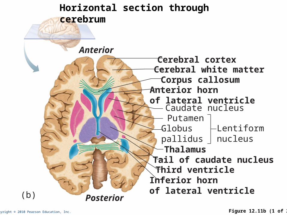
Copyright © 2010 Pearson Education, Inc. Figure 12.11b (1 of 2)
Corpus callosumAnterior hornof lateral ventricleCaudate nucleusPutamen
Lentiformnucleus
(b)
Globuspallidus ThalamusTail of caudate nucleusThird ventricle
Cerebral cortexCerebral white matter
Anterior
Posterior
Inferior hornof lateral ventricle
Horizontal section through cerebrum
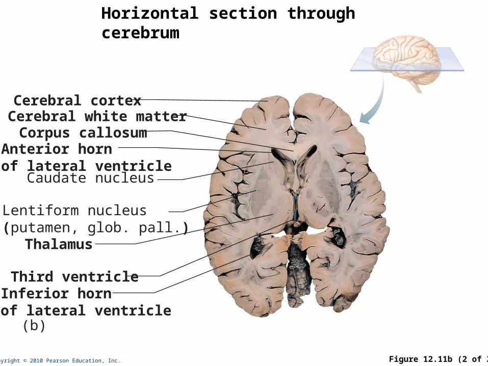
Copyright © 2010 Pearson Education, Inc. Figure 12.11b (2 of 2)
Corpus callosumAnterior hornof lateral ventricleCaudate nucleus
Lentiform nucleus(putamen, glob. pall.)
(b)
Thalamus
Third ventricle
Cerebral cortexCerebral white matter
Inferior hornof lateral ventricle
Horizontal section through cerebrum

Copyright © 2010 Pearson Education, Inc.
Functions of Basal Nuclei
• Though somewhat elusive, the following are thought to be functions of basal nuclei
• Influence muscular control
• Help regulate attention and cognition
• Regulate intensity of slow or stereotyped movements
• Inhibit antagonistic and unnecessary movements

Copyright © 2010 Pearson Education, Inc. Figure 12.12
Corpus callosum
Choroid plexusThalamus(encloses third ventricle)
Pineal gland(part of epithalamus)
Posterior commissure
CorporaquadrigeminaCerebralaqueductArbor vitae (ofcerebellum)Fourth ventricleChoroid plexusCerebellum
Septum pellucidum
Interthalamicadhesion(intermediatemass of thalamus)Interven-tricularforamenAnteriorcommissure
Hypothalamus
Optic chiasma
Pituitary gland
Cerebral hemisphere
Mammillary bodyPonsMedulla oblongata
Spinal cord
Mid-brain
Fornix
Diencephalon: thalamus, hypothalamus, epithalamus

Copyright © 2010 Pearson Education, Inc. Figure 12.13a
Dorsal nuclei
Medial
Anteriornucleargroup
Reticularnucleus
Ventralanterior
Ventrallateral
Ventralpostero-lateral
Lateralgeniculatebody
Medialgeniculatebody
Pulvinar
Lateraldorsal
Lateralposterior
(a) The main thalamic nuclei. (The reticular nuclei that “cap” thethalamus laterally are depicted as curving translucent structures.)
Ventral nuclei
Thalamus

Copyright © 2010 Pearson Education, Inc.
Thalamus
• Contains multiple nuclei, named by location
• Receives ascending sensory information, processes it, and relays it to cerebral cortex
• Afferent impulses from all senses and all parts of the body
• Impulses from the hypothalamus for regulation of emotion and visceral function
• Impulses from the cerebellum and basal nuclei to help direct the motor cortices
• Mediates sensation, motor activities, cortical arousal, learning, and memory

Copyright © 2010 Pearson Education, Inc. Figure 12.13b
Preopticnucleus
SupraopticnucleusSupra-chiasmatic nucleus
Anteriorhypothalamicnucleus
Dorsomedialnucleus
Paraventricularnucleus
FornixAnteriorcommissure
PosteriorhypothalamicnucleusLateralhypothalamicareaVentromedialnucleus
OpticchiasmaInfundibulum(stalk of thepituitary gland)
Pituitarygland
Mammillarybody
(b) The main hypothalamic nuclei.
Arcuatenucleus
Hypothalamus

Copyright © 2010 Pearson Education, Inc.
Hypothalamus
• Below thalamus
• Multiple nuclei
• Autonomic control center for many visceral functions (e.g., blood pressure, rate and force of heartbeat, digestive tract motility)
• Center for emotional response: Involved in perception of pleasure, fear, and rage and in biological rhythms and drives
• Infundibulum: stalk connecting to pituitary

Copyright © 2010 Pearson Education, Inc.
Hypothalamic Function
• Regulates body temperature, food intake, water balance, and thirst
• Regulates sleep and the sleep cycle
• Controls release of hormones by the anterior pituitary
• Produces posterior pituitary hormones

Copyright © 2010 Pearson Education, Inc.
Epithalamus
• Most dorsal portion of the diencephalon; forms roof of the third ventricle
• Pineal gland—extends from the posterior border and secretes melatonin
• Melatonin—helps regulate sleep-wake cycles

Copyright © 2010 Pearson Education, Inc. Figure 12.12
Corpus callosum
Choroid plexusThalamus(encloses third ventricle)
Pineal gland(part of epithalamus)
Posterior commissure
CorporaquadrigeminaCerebralaqueductArbor vitae (ofcerebellum)Fourth ventricleChoroid plexusCerebellum
Septum pellucidum
Interthalamicadhesion(intermediatemass of thalamus)Interven-tricularforamenAnteriorcommissure
Hypothalamus
Optic chiasma
Pituitary gland
Cerebral hemisphere
Mammillary bodyPonsMedulla oblongata
Spinal cord
Mid-brain
Fornix
Mid-sagittal view of brain

Copyright © 2010 Pearson Education, Inc.
Brain Stem
• Three main parts: midbrain, pons, medulla oblongota
• Contains fiber tracts (ascending and descending) and embedded nuclei
• Controls automatic behaviors necessary for survival
• Associated with 10 of the 12 pairs of cranial nerves

Copyright © 2010 Pearson Education, Inc. Figure 12.14
Frontal lobeOlfactory bulb(synapse point ofcranial nerve I)Optic chiasmaOptic nerve (II)Optic tractMammillary body
Pons
MedullaoblongataCerebellum
Temporal lobe
Spinal cord
Midbrain
Inferior view of brain, showing brain stem

Copyright © 2010 Pearson Education, Inc. Figure 12.15a
Optic chiasmaView (a)
Optic nerve (II)
Mammillary body
Oculomotor nerve (III)
Crus cerebri ofcerebral peduncles (midbrain)
Trigeminal nerve (V)
Abducens nerve (VI)Facial nerve (VII)
Vagus nerve (X)
Accessory nerve (XI)
Hypoglossal nerve (XII)
Ventral root of firstcervical nerve
Trochlear nerve (IV)
PonsMiddle cerebellarpeduncle
Pyramid
Decussation of pyramids
(a) Ventral view
Spinal cord
Vestibulocochlearnerve (VIII)
Glossopharyngeal nerve (IX)
Diencephalon• Thalamus• Hypothalamus
Diencephalon
Brainstem
Thalamus
Hypothalamus
Midbrain
Pons
Medullaoblongata
Brainstem, diencephalon, cranial nerve roots

Copyright © 2010 Pearson Education, Inc. Figure 12.15b
View (b)
Crus cerebri ofcerebral peduncles (midbrain)
InfundibulumPituitary gland
Trigeminal nerve (V)
Abducens nerve (VI)
Facial nerve (VII)
Vagus nerve (X)
Accessory nerve (XI)
Hypoglossal nerve (XII)
Pons
(b) Left lateral view
Glossopharyngeal nerve (IX)
Diencephalon
Brainstem
Thalamus
Hypothalamus
Midbrain
Pons
Medullaoblongata
Thalamus
Superior colliculusInferior colliculusTrochlear nerve (IV)
Superior cerebellar peduncle
Middle cerebellar peduncle
Inferior cerebellar peduncle
Vestibulocochlear nerve (VIII)Olive
Brainstem, diencephalon, cranial nerve roots

Copyright © 2010 Pearson Education, Inc. Figure 12.15c
View (c)
Diencephalon
Brainstem
Thalamus
Hypothalamus
Midbrain
Pons
Medullaoblongata
Pineal gland
Diencephalon
Anterior wall offourth ventricle
(c) Dorsal view
Thalamus
Dorsal root offirst cervical nerve
Midbrain• Superior
colliculus• Inferior
colliculus• Trochlear nerve (IV)• Superior cerebellar peduncle
Corporaquadrigeminaof tectum
Medulla oblongata• Inferior cerebellar peduncle• Facial nerve (VII)• Vestibulocochlear nerve (VIII)• Glossopharyngeal nerve (IX)• Vagus nerve (X)• Accessory nerve (XI)
Pons• Middle cerebellar peduncle
Dorsal median sulcus
Choroid plexus(fourth ventricle)
Brainstem, diencephalon, cranial nerve roots

Copyright © 2010 Pearson Education, Inc.
Midbrain
• Located between the diencephalon and the pons
• Cerebral peduncles
• Contain pyramidal motor tracts

Copyright © 2010 Pearson Education, Inc.
Midbrain Nuclei
• Nuclei that control cranial nerves III (oculomotor) and IV (trochlear)
• Corpora quadrigemina—domelike dorsal protrusions
• Superior colliculi—visual reflex centers
• Inferior colliculi—auditory relay centers
• Substantia nigra—functionally linked to basal nuclei
• Red nucleus—relay nuclei for some descending motor pathways and part of reticular formation

Copyright © 2010 Pearson Education, Inc. Figure 12.16a
Dorsal
Cerebral aqueduct
Superiorcolliculus
Reticular formation
Crus cerebri ofcerebral peduncle
Ventral
Fibers ofpyramidal tract
Substantianigra
(a) Midbrain
Rednucleus
Mediallemniscus
Oculomotornucleus (III)
Periaqueductal graymatter
Tectum
Horizontal section through midbrain

Copyright © 2010 Pearson Education, Inc.
Pons (“bridge”)
• Fibers of the pons
• Connect higher brain centers and the spinal cord
• Relay impulses between the motor cortex and the cerebellum
• Origin of some cranial nerves
• Some nuclei of the reticular formation
• Nuclei that help maintain normal rhythm of breathing

Copyright © 2010 Pearson Education, Inc. Figure 12.16b
Reticularformation
Trigeminalnerve (V)
Pontinenuclei
Fibers ofpyramidaltract
Middlecerebellarpeduncle
Trigeminal mainsensory nucleus Trigeminalmotor nucleus
Superior cerebellarpeduncle
Medial lemniscus
Fourthventricle
(b) Pons
Horizontal section through pons

Copyright © 2010 Pearson Education, Inc.
Medulla Oblongata
• Lowest part of brainstem
• Joins (becomes) spinal cord at foramen magnum
• All information passing between spinal cord and brain goes through medulla

Copyright © 2010 Pearson Education, Inc.
Medulla Oblongata
• Pyramids—two ventral longitudinal ridges formed by pyramidal tracts (descending motor fibers, a.k.a. corticospinal tracts (cortex to spinal cord))
• Decussation of the pyramids—crossover of the corticospinal tracts
• Some nuclei for cranial nerves
• Several nuclei (e.g., nucleus cuneatus and nucleus gracilis) which relay ascending sensory information

Copyright © 2010 Pearson Education, Inc.
Medulla Oblongata
Autonomic control areas include
• Solitary nucleus: receives input from pressure and chemical sensors; these inputs are used to regulate cardiovascular and respiratory systems
• Cardiovascular center: Generates motor outflow to regulate heart & blood vessels
• Respiratory: Regulate rate and depth of breathing (connects to resp. areas in pons)

Copyright © 2010 Pearson Education, Inc. Figure 12.16c
Choroidplexus
Fourth ventricle
PyramidMedial lemniscus
Nucleusambiguus
Cochlearnuclei (VIII)
Vestibular nuclearcomplex (VIII)
Solitarynucleus
Dorsal motor nucleusof vagus (X)
Hypoglossal nucleus (XII)
(c) Medulla oblongata
Ret
icu
lar
form
atio
n
Horizontal section through medulla oblongota

Copyright © 2010 Pearson Education, Inc.
The Cerebellum
• 11% of brain mass
• Dorsal to the pons and medulla
• Subconsciously provides precise timing and appropriate patterns of skeletal muscle contraction

Copyright © 2010 Pearson Education, Inc.
Anatomy of the Cerebellum
• Two hemispheres connected by vermis
• Folia—transversely oriented gyri
• Arbor vitae—distinctive treelike pattern of the cerebellar white matter

Copyright © 2010 Pearson Education, Inc. Figure 12.17b
(b)
Medullaoblongata
Choroidplexus offourth ventricle
Arborvitae
Cerebellar cortex

Copyright © 2010 Pearson Education, Inc. Figure 12.17d
(d)
Anteriorlobe
Posteriorlobe
Vermis(d)

Copyright © 2010 Pearson Education, Inc.
Cerebellar Processing for Motor Activity
• Cerebellum receives impulses from the cerebral cortex of the intent to initiate voluntary muscle contraction
• Signals from proprioceptors and visual and equilibrium pathways continuously “inform” the cerebellum of the body’s position and momentum
• Cerebellar cortex calculates the best way to smoothly coordinate a muscle contraction
• A “blueprint” of coordinated movement is sent to the cerebral motor cortex and to brain stem nuclei

Copyright © 2010 Pearson Education, Inc.
Cognitive Function of the Cerebellum
• Recognizes and predicts sequences of events during complex movements
• Plays a role in nonmotor functions such as word association and puzzle solving