The Blueprint of Life, from DNA to Protein - McGraw-Hill Higher
24
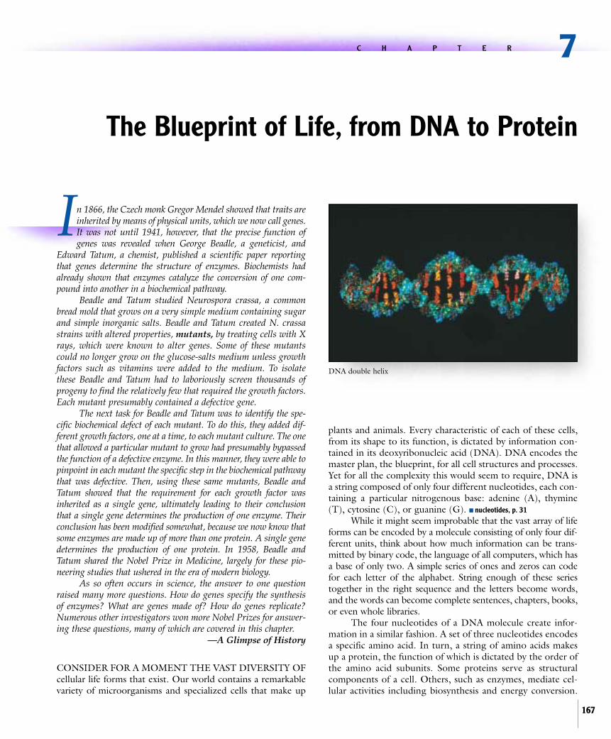
I n 1866, the Czech monk Gregor Mendel showed that traits are inherited by means of physical units, which we now call genes. It was not until 1941, however, that the precise function of genes was revealed when George Beadle, a geneticist, and Edward Tatum, a chemist, published a scientific paper reporting that genes determine the structure of enzymes. Biochemists had already shown that enzymes catalyze the conversion of one com- pound into another in a biochemical pathway. Beadle and Tatum studied Neurospora crassa, a common bread mold that grows on a very simple medium containing sugar and simple inorganic salts. Beadle and Tatum created N. crassa strains with altered properties, mutants, by treating cells with X rays, which were known to alter genes. Some of these mutants could no longer grow on the glucose-salts medium unless growth factors such as vitamins were added to the medium. To isolate these Beadle and Tatum had to laboriously screen thousands of progeny to find the relatively few that required the growth factors. Each mutant presumably contained a defective gene. The next task for Beadle and Tatum was to identify the spe- cific biochemical defect of each mutant. To do this, they added dif- ferent growth factors, one at a time, to each mutant culture. The one that allowed a particular mutant to grow had presumably bypassed the function of a defective enzyme. In this manner, they were able to pinpoint in each mutant the specific step in the biochemical pathway that was defective. Then, using these same mutants, Beadle and Tatum showed that the requirement for each growth factor was inherited as a single gene, ultimately leading to their conclusion that a single gene determines the production of one enzyme. Their conclusion has been modified somewhat, because we now know that some enzymes are made up of more than one protein. A single gene determines the production of one protein. In 1958, Beadle and Tatum shared the Nobel Prize in Medicine, largely for these pio- neering studies that ushered in the era of modern biology. As so often occurs in science, the answer to one question raised many more questions. How do genes specify the synthesis of enzymes? What are genes made of? How do genes replicate? Numerous other investigators won more Nobel Prizes for answer- ing these questions, many of which are covered in this chapter. —A Glimpse of History CONSIDER FOR A MOMENT THE VAST DIVERSITY OF cellular life forms that exist. Our world contains a remarkable variety of microorganisms and specialized cells that make up plants and animals. Every characteristic of each of these cells, from its shape to its function, is dictated by information con- tained in its deoxyribonucleic acid (DNA). DNA encodes the master plan, the blueprint, for all cell structures and processes. Yet for all the complexity this would seem to require, DNA is a string composed of only four different nucleotides, each con- taining a particular nitrogenous base: adenine (A), thymine (T), cytosine (C), or guanine (G). ■ nucleotides, p. 31 While it might seem improbable that the vast array of life forms can be encoded by a molecule consisting of only four dif- ferent units, think about how much information can be trans- mitted by binary code, the language of all computers, which has a base of only two. A simple series of ones and zeros can code for each letter of the alphabet. String enough of these series together in the right sequence and the letters become words, and the words can become complete sentences, chapters, books, or even whole libraries. The four nucleotides of a DNA molecule create infor- mation in a similar fashion. A set of three nucleotides encodes a specific amino acid. In turn, a string of amino acids makes up a protein, the function of which is dictated by the order of the amino acid subunits. Some proteins serve as structural components of a cell. Others, such as enzymes, mediate cel- lular activities including biosynthesis and energy conversion. 7 C H A P T E R The Blueprint of Life, from DNA to Protein 167 DNA double helix
Transcript of The Blueprint of Life, from DNA to Protein - McGraw-Hill Higher
In 1866, the Czech monk Gregor Mendel showed that traits areinherited by means of physical units, which we now call genes.It was not until 1941, however, that the precise function ofgenes was revealed when George Beadle, a geneticist, and
Edward Tatum, a chemist, published a scientific paper reportingthat genes determine the structure of enzymes. Biochemists hadalready shown that enzymes catalyze the conversion of one com-pound into another in a biochemical pathway.
Beadle and Tatum studied Neurospora crassa, a commonbread mold that grows on a very simple medium containing sugarand simple inorganic salts. Beadle and Tatum created N. crassastrains with altered properties, mutants, by treating cells with Xrays, which were known to alter genes. Some of these mutantscould no longer grow on the glucose-salts medium unless growthfactors such as vitamins were added to the medium. To isolatethese Beadle and Tatum had to laboriously screen thousands ofprogeny to find the relatively few that required the growth factors.Each mutant presumably contained a defective gene.
The next task for Beadle and Tatum was to identify the spe-cific biochemical defect of each mutant. To do this, they added dif-ferent growth factors, one at a time, to each mutant culture. The onethat allowed a particular mutant to grow had presumably bypassedthe function of a defective enzyme. In this manner, they were able topinpoint in each mutant the specific step in the biochemical pathwaythat was defective. Then, using these same mutants, Beadle andTatum showed that the requirement for each growth factor wasinherited as a single gene, ultimately leading to their conclusionthat a single gene determines the production of one enzyme. Theirconclusion has been modified somewhat, because we now know thatsome enzymes are made up of more than one protein. A single genedetermines the production of one protein. In 1958, Beadle andTatum shared the Nobel Prize in Medicine, largely for these pio-neering studies that ushered in the era of modern biology.
As so often occurs in science, the answer to one questionraised many more questions. How do genes specify the synthesisof enzymes? What are genes made of? How do genes replicate?Numerous other investigators won more Nobel Prizes for answer-ing these questions, many of which are covered in this chapter.
—A Glimpse of History
CONSIDER FOR A MOMENT THE VAST DIVERSITY OFcellular life forms that exist. Our world contains a remarkablevariety of microorganisms and specialized cells that make up
plants and animals. Every characteristic of each of these cells,from its shape to its function, is dictated by information con-tained in its deoxyribonucleic acid (DNA). DNA encodes themaster plan, the blueprint, for all cell structures and processes.Yet for all the complexity this would seem to require, DNA isa string composed of only four different nucleotides, each con-taining a particular nitrogenous base: adenine (A), thymine(T), cytosine (C), or guanine (G). ■ nucleotides, p. 31
While it might seem improbable that the vast array of lifeforms can be encoded by a molecule consisting of only four dif-ferent units, think about how much information can be trans-mitted by binary code, the language of all computers, which hasa base of only two. A simple series of ones and zeros can codefor each letter of the alphabet. String enough of these seriestogether in the right sequence and the letters become words,and the words can become complete sentences, chapters, books,or even whole libraries.
The four nucleotides of a DNA molecule create infor-mation in a similar fashion. A set of three nucleotides encodesa specific amino acid. In turn, a string of amino acids makesup a protein, the function of which is dictated by the order ofthe amino acid subunits. Some proteins serve as structuralcomponents of a cell. Others, such as enzymes, mediate cel-lular activities including biosynthesis and energy conversion.
7C H A P T E R
The Blueprint of Life, from DNA to Protein
167
DNA double helix

168 Chapter 7 The Blueprint of Life, from DNA to Protein
Replication
Protein
RNA
DNA DNA Duplicate of original molecule
Transcription
Translation
Figure 7.1 Overview of Replication, Transcription, and Translation DNAreplication is the process that duplicates DNA so that its encoded information can bepassed on to future generations.Transcription is the process that copies the geneticinformation into a transitional form, RNA.Translation is the process that deciphers theencoded information to synthesize a specific protein.
Together, proteins synthesized by a cell are responsible forevery aspect of that cell. Thus, the sequential order ofnucleotide bases in a cell’s DNA ultimately dictates the charac-teristics of that cell. ■ amino acids, p. 25 ■ protein structure, p. 27 ■ flagella,p. 63 ■ enzymes, pp. 131, 138
This chapter will focus on the cellular process of convert-ing the information encoded within DNA into proteins, concen-trating primarily on the mechanisms used by prokaryotic cells.The eukaryotic processes have many similarities, but they are con-siderably more complicated and will only be discussed briefly.
7.1 OverviewThe complete set of genetic information for a cell is referred toas its genome. Technically, this includes plasmids as well as thechromosome; however, the term genome is often used inter-changeably with chromosome. The genome of all cells is com-posed of DNA, but some viruses have an RNA genome. Thefunctional unit of the genome is a gene. A gene encodes a prod-uct, the gene product, most commonly a protein. The study ofthe function and transfer of genes is called genetics, whereas thestudy and analysis of the nucleotide sequence of DNA is calledgenomics. ■ chromosome, p. 66 ■ plasmid, p. 66
All living cells must accomplish two general tasks in orderto multiply. The double-stranded DNA must be duplicatedbefore cell division so that its encoded information can bepassed on to future generations. This is the process of DNAreplication. In addition, the information encoded by the DNAmust be deciphered, or expressed, so that the cell can synthe-size the necessary gene products at the appropriate time. Geneexpression involves two interrelated processes, transcription andtranslation. Transcription copies the information encoded inDNA into a slightly different molecule, RNA. The RNA servesas a transitional, temporary form of the genetic information andis the one that is actually deciphered. Translation interpretsinformation carried by RNA to synthesize the encoded protein.The chemistry and structure of DNA and RNA ensure that eachof these processes can occur with great accuracy.
The flow of information from DNA to RNA to protein isoften referred to as the central dogma of molecular biology(figure 7.1). It was once believed that information flow pro-ceeded only in this direction. Although this direction is by farthe most common, certain viruses, such as the one that causesAIDS, have an RNA genome but copy that information into theform of DNA.
Characteristics of DNAA single strand of DNA is composed of a series of deoxyri-bonucleotide subunits, more commonly called nucleotides.These are joined in a chain by a covalent bond between the 5„PO4
(5 prime phosphate) group of one nucleotide and the 3„OH (3prime hydroxyl) group of the next. Note that the designations 5„and 3„ refer to the numbered carbon atoms of the pentose sugarof the nucleotide (see figure 2.22). Joining of the nucleotides inthis manner creates a series of alternating sugar and phosphatemoieties, called the sugar-phosphate backbone. Connected to
each sugar is one of the nitrogenous bases, an adenine (A),thymine (T), guanine (G), or cytosine (C). Because of the chem-ical structure of the nucleotides and how they are joined, a singlestrand of DNA will always have a 5„PO4 group at one end and a3„OH group at the other. These ends are often referred to as the5„ end and the 3„ end and have important implications in DNAand RNA synthesis that will be discussed later. ■ deoxyribonucleic acid(DNA), p. 31 ■ nucleotides, p. 31
The DNA in a cell usually occurs as a double-stranded,helical structure (figure 7.2). The two strands are held togeth-er by weak hydrogen bonds between the nitrogenous bases ofthe opposing strands. While individual hydrogen bonds arereadily broken, the duplex structure of double-stranded DNA isquite stable because of the sheer number of bonds that occursalong its length. Because short fragments of DNA have corre-spondingly fewer hydrogen bonds, they are readily separatedinto single-stranded pieces. Separating the two strands is calleddenaturing. ■ hydrogen bonds, p. 21
The two strands of double-stranded DNA are comple-mentary (figure 7.3). Wherever an adenine is in one strand, athymine is in the other; these two opposing nucleotides are heldtogether by two hydrogen bonds between them. Similarly,wherever a cytosine is in one strand, a guanine is in the other.These are held together by the formation of three hydrogenbonds, a slightly stronger attraction than that of an A:T pair.The characteristic bonding of A to T and G to C is called base-pairing and is fundamental to the remarkable functionality ofDNA. Because of the rules of base-pairing, one strand canalways be used as a template for the synthesis of the comple-mentary opposing strand. ■ complementary, p. 33
While the two strands of DNA in the double helix arecomplementary, they are also antiparallel. That is, they are ori-ented in opposite directions. One strand is oriented in the 5„ to

7.1 Overview 169
A highly coiled line isused to depict genomic DNA.
Base-pairing
Red and blue lines placed in a helical arrangement depict thetwo complementary strands andhighlight the three-dimensionalstructure of DNA.
A circular arrangement of thered and blue lines is used as the simplified form of prokaryotic DNA.
Red and blue lines separatedby a thin black line are used asa simple representation of thedouble-stranded DNA molecule.
Two parallel lines are used toemphasize the base-pairing interactionsand nucleotide sequence characteristicsof the two complementary strands. The"tracks" between the lines are not intendedto depict a specific number of base pairs,only the general interaction betweencomplementary strands.
Either a red or a blue line canbe depicted as the "top" strand,since DNA is a three-dimensionalstructure.
Denatured DNA is depictedas separate red and blue linesto emphasize its single-stranded nature.
Figure 7.2 Diagrammatic Representations of the Structure of DNA Although DNA is a double-stranded helical structure,explanatory diagrams may depict it in a number of different ways. In this chapter we will use these color-coded representations.
3„ direction and its complement is oriented in the 3„ to 5„ direc-tion. This also has important implications in the function andsynthesis of nucleic acids.
Characteristics of RNARNA is in many ways comparable to DNA, but with someimportant exceptions. One difference is that RNA is made upof ribonucleotides rather than deoxynu-cleotides, although in both cases these areusually referred to simply as nucleotides.Another distinction is that RNA contains thenitrogenous base uracil in place of the thyminefound in DNA. Like DNA, RNA consists of asequence of nucleotides, but RNA usuallyexists as a single-stranded linear molecule thatis much shorter than DNA. ■ ribonucleic acid (RNA),p. 33 ■ nucleotide, p. 31
A fragment of RNA, a transcript, is syn-thesized using a region of one of the two strandsof DNA as a template. In making the RNA tran-script, the same base-pairing rules of DNA applyexcept uracil, rather than thymine, base-pairswith adenine. This base-pairing is only transient,however, and the molecule quickly leaves theDNA template. Numerous different RNA tran-scripts can be generated from a single chromo-some using specific regions as templates. Eitherstrand may serve as the template. In a region thesize of a single gene, however, only one of thetwo strands is generally transcribed. As a result,two complementary strands of RNA are notnormally generated.
There are three different functional groupsof RNA molecules, each one transcribed fromdifferent genes. Most genes encode proteins and
are transcribed into messenger RNA (mRNA). These moleculesare translated during protein synthesis. Encrypted information inmRNA is deciphered according to the genetic code, which corre-lates each set of three nucleotides, called a codon, to a particularamino acid. Some genes are never translated into proteins;instead the RNAs themselves are the ultimate products. These
P
Sugar
P
Sugar
P
Sugar
P
Sugar
P
Sugar
5'
3'
Sugar–phosphatebackbone
Sugar–phosphatebackbone
A
G C
C G
C G
T A
HO
P
Sugar
P
Sugar
P
Sugar
P
Sugar
P
Sugar
TO
O
O
O
O O
O
O
O
O
OH 3'
5'
Basepairs
Hydrogenbonds
Nucleotide
Figure 7.3 The Double Helix of DNA The two strands of DNA are antiparallel; one strand is oriented in the5„ to 3„ direction, and its complement is oriented in the 3„ to 5„ direction. Hydrogen bonding occurs between thecomplementary base pairs; three bonds form between a G-C base pair, and two bonds form between an A-T base pair.
170 Chapter 7 The Blueprint of Life, from DNA to Protein
genes encode either ribosomal RNA (rRNA) or transferRNA (tRNA), each of which plays a different but critical rolein protein synthesis.
Regulating the Expression of GenesWhile the basic structure of DNA and RNA is relatively simple, theinformation the molecules encode is extensive and complex. Thenucleotide sequence contains genes that encode the amino acidsequence of proteins, and it also encodes mechanisms to regulateexpression of those genes. Not all proteins are required by a cell inthe same quantity and at all times; therefore, mechanisms thatdetermine the extent and duration of their synthesis are needed.
One of the key mechanisms a cell uses to control proteinsynthesis is to regulate the synthesis of mRNA molecules.Unless a gene is transcribed into mRNA, the encoded proteincannot be synthesized. The number of mRNA copies of thegene also influences the level of expression. If transcription of agene ceases, the level of gene expression rapidly declines. This isbecause mRNA is short-lived, often only a few minutes, due tothe activity of enzymes called RNases that rapidly degrade it.
M I C R O C H E C K 7 . 1
Replication is the process of duplicating double-stranded DNA. Transcription is the process of copying
the information encoded in DNA into RNA.Translation is the process of interpreting theinformation carried by messenger RNA in order tosynthesize the encoded protein.
■ How does the 5„ end of DNA differ from the 3„ end?■ If the nucleotide sequence of one strand of DNA is
5„ ACGTTGCA 3„, what is the sequence of thecomplementary strand?
■ Why is a short-lived RNA important in cell controlmechanisms?
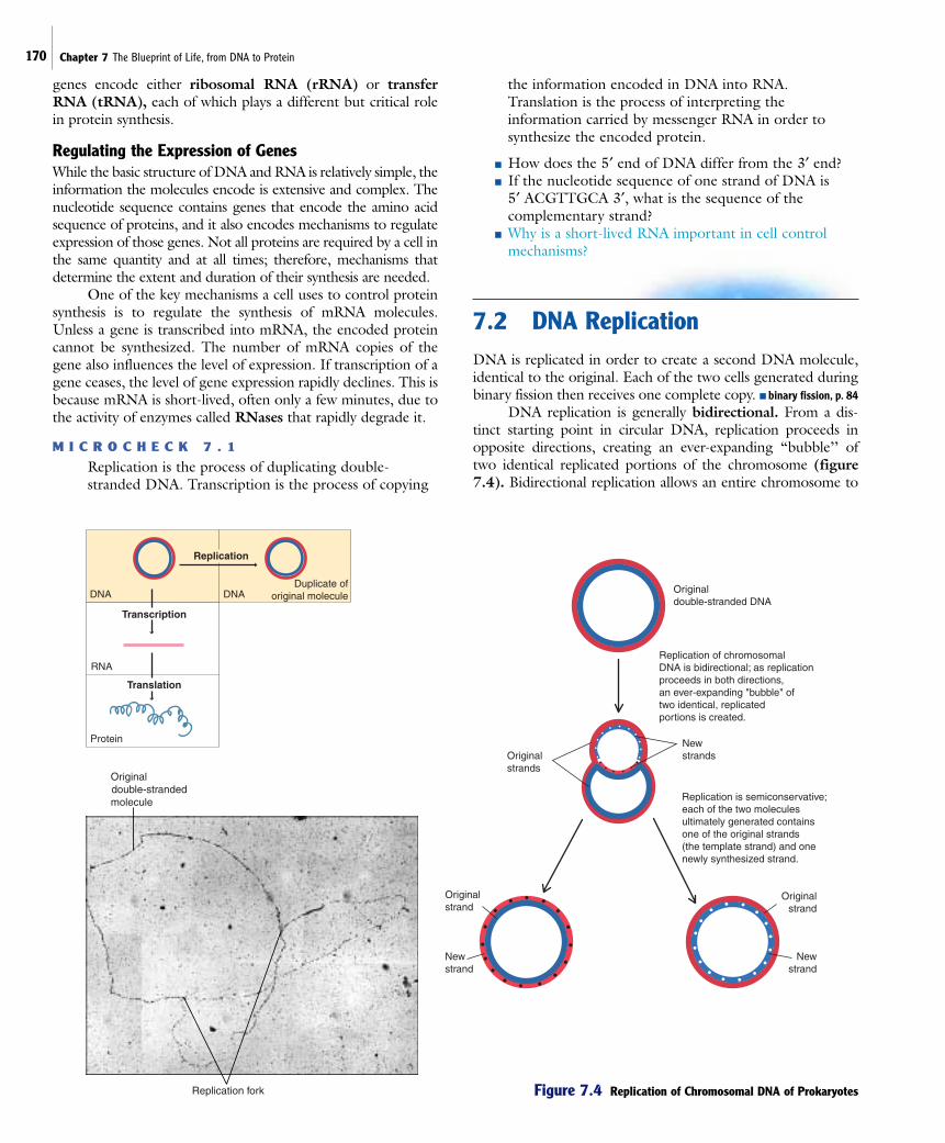
7.2 DNA ReplicationDNA is replicated in order to create a second DNA molecule,identical to the original. Each of the two cells generated duringbinary fission then receives one complete copy. ■ binary fission, p. 84
DNA replication is generally bidirectional. From a dis-tinct starting point in circular DNA, replication proceeds inopposite directions, creating an ever-expanding “bubble’’ oftwo identical replicated portions of the chromosome (figure7.4). Bidirectional replication allows an entire chromosome to
Replication
Protein
RNA
DNA DNADuplicate of
original moleculeOriginaldouble-stranded DNA
Replication of chromosomalDNA is bidirectional; as replicationproceeds in both directions,an ever-expanding "bubble" of two identical, replicated portions is created.
Replication is semiconservative;each of the two molecules ultimately generated contains one of the original strands (the template strand) and onenewly synthesized strand.
Originalstrands
Originalstrand
Originalstrand
Newstrands
Newstrand
Newstrand
Replication fork
Original double-stranded
molecule
Transcription
Translation
Figure 7.4 Replication of Chromosomal DNA of Prokaryotes

7.2 DNA Replication 171
Component Comments
Primer Fragment of nucleic acid to which DNA polymerase can add nucleotides (the enzyme can only add nucleotides to apreexisting fragment).
DNA gyrase Enzyme that temporarily breaks the strands of DNA, relieving the tension caused by unwinding the two strands of theDNA helix; it is the target of a class of antibacterial medications called fluoroquinolones.
DNA ligase Enzyme that joins two DNA fragments by forming a covalent bond between the sugar-phosphate residues of adjacentnucleotides.
DNA polymerases Enzymes that synthesize DNA; they use one strand of DNA as a template to generate the complementary strand.Synthesis always occurs in the 5„ to 3„ direction.
Helicases Enzymes that unwind the DNA helix ahead of the replication fork.
Okazaki fragment Nucleic acid fragment generated during discontinuous replication of the lagging strand of DNA.
Origin of replication Distinct region of a DNA molecule at which replication is initiated.
Primase Enzyme that synthesizes small fragments of RNA to serve as primers for DNA synthesis during discontinuousreplication of the lagging strand.
Table 7.1 Components of DNA Replication in Prokaryotes
be replicated in half the time itwould take if replication were uni-directional.
Replication of double-stranded DNA is semiconserva-tive. Each of the two moleculesgenerated contains one of theoriginal strands (the templatestrand) and one newly synthe-sized strand. Thus, the two cellsproduced as a result of divisioneach have one of the originalstrands of DNA paired with a newcomplementary strand.
The process of DNA repli-cation requires the coordinatedaction of many different enzymesand other proteins (table 7.1).The most critical of these existtogether as a complex called areplisome. The replisome appearsto act as a fixed DNA-synthesiz-ing factory, reeling in the DNA tobe replicated. DNA polymerasesare enzymes in the replisome thatsynthesize DNA, using one strand as a template to generate thecomplementary strand. These enzymes can only add nucleotidesonto a preexisting fragment of nucleic acid, either DNA orRNA. Thus, the fragment serves as a primer from which syn-thesis can continue.
DNA is synthesized one nucleotide at a time as thedeoxynucleoside triphosphates (dATP, dGTP, dCTP, and dTTP)are covalently joined to the nucleotide at the 3„ end of the grow-ing strand. Hydrolysis of a phosphate bond in the incoming mol-ecule provides energy for the reaction. DNA polymerase always
elongates the chain in the 5„ to 3„ direction. Because the twoDNA strands are antiparallel, however, the enzyme must “read’’the template strand in the 3„ to 5„ direction (figure 7.5). Thebase-pairing rules determine the specific nucleotides added.
The replication process is very accurate, resulting in onlyone mistake approximately every billion nucleotides. Part of thereason for this remarkable precision is the proofreading abilityof some DNA polymerases. If an incorrect nucleotide is incor-porated into the growing chain, the enzyme can edit the mis-take by replacing that nucleotide before moving on.
Figure 7.5 The Process of DNA Synthesis DNA polymerase synthesizes a new strand by adding one nucleotide at a timeto the 3„ end of the elongating strand. Because DNA is synthesized in the 5„ to 3„ direction, the enzyme must “read’’ the templatestrand in the 3„ to 5„ direction.The base-pairing rules determine the specific nucleotides that are added.
C C C C
C C C
A A A A A A
AAAAA
T T T T T T T T T
TTTT
G G G
C
A
A
T
T
G
C
A
T
G G G
GG
5'
3'
Direction
of synthesis
3'
5'
New strand
Template strand
A
DNApolymerase

Several other proteins are also involved in DNA replica-tion. Among them is DNA gyrase, an enzyme that temporari-ly breaks the strands of DNA, relieving the tension caused bythe unwinding of the two strands of the DNA helix. Thisenzyme is one of the targets of ciprofloxacin and other membersof a class of antibacterial drugs called fluoroquinolones. Byinhibiting the function of gyrase, the fluoroquinolones interferewith bacterial DNA replication and prevent the growth of bac-teria. ■ fluoroquinolones, p. 517
M I C R O C H E C K 7 . 2
DNA polymerases synthesize DNA in the 5„ to 3„direction, using one strand as a template to generate the complementary strand. Replication of DNA begins at a specific sequence, called the origin ofreplication, and then proceeds bidirectionally, creating two replication forks.
■ Why is a primer required for DNA synthesis?■ How does synthesis of the lagging strand differ from
that of the leading strand?■ If DNA replication were shown to be “conservative,’’
what would this mean?
172 Chapter 7 The Blueprint of Life, from DNA to Protein
It takes approximately 40 minutes for the chromosomeof E. coli to be replicated, regardless of the environmentalconditions. How, then, can E. coli sometimes multiply with ageneration time of only 20 minutes? Under favorable grow-ing conditions, a cell initiates replication before the precedinground of replication is completed. In this way, the two prog-eny resulting from cell division each will get one completechromosome that has already started another round of repli-cation. ■ generation time, p. 85
Initiation of DNA ReplicationTo begin the process of DNA replication, specific proteins mustrecognize and bind to a distinct region of the DNA, called anorigin of replication. All molecules of DNA, including chro-mosomes and plasmids, must have this region of approximately250 nucleotides for replication to be initiated. The binding ofthe proteins causes localized denaturation, or melting, of a spe-cific region within the origin. Using the exposed single strandsas templates, small fragments of RNA are synthesized to serve asprimers for DNA synthesis. The enzymes that synthesize RNAdo not require a primer.
The Replication ForkThe bidirectional progression of replicationaround a circular DNA molecule creates twoadvancing Y-shaped regions where active repli-cation is occurring. Each of these is called areplication fork. The template strands contin-ue to “unzip’’ at each fork due to the activityof enzymes called helicases. Synthesis of onenew strand proceeds continuously in the 5„ to3„ direction, as fresh single-stranded templateDNA is exposed (figure 7.6). This strand iscalled the leading strand. Synthesis of theopposing strand, the lagging strand, is con-siderably more complicated because the DNApolymerase cannot add nucleotides to the 5„end of DNA. Instead, synthesis must be reini-tiated periodically as advancement of the repli-cation fork exposes more of the templateDNA. Each initiation event must be precededby the synthesis of an RNA primer by theenzyme primase. The result is the synthesis ofa series of fragments, called Okazaki frag-ments, each of which begins with a shortstretch of RNA. As DNA polymerase addsnucleotides to the 3„ end of an Okazaki frag-ment, it eventually reaches the initiating pointof the previous fragment. A different type ofDNA polymerase then removes those RNAprimer nucleotides and simultaneously replacesthem with deoxynucleotides. The enzymeDNA ligase seals the gaps between fragmentsby catalyzing the formation of a covalent bondbetween the adjacent nucleotides.
5'3'
3'5'
3'5'
1. Primase synthesizes an RNA primer
RNA primer
Helicase separates thedouble-stranded molecule
Synthesis of the leading strand proceeds continuously.
Synthesis of the lagging strand is discontinuous; synthesis is reinitiated periodically, generating a series of fragments that are later joined.
4. DNA ligase seals the gaps between adjacent fragments
2. DNA polymerase adds nucleotides onto the 3' end of the fragment
3. DNA polymerase replaces the RNA primer with deoxynucleotides
Figure 7.6 The Replication Fork This simplified diagram of the replication fork highlights the key steps inthe synthesis of the lagging strand.

7.3 Gene Expression 173
Replication
Transcription
Translation
Protein
RNA
DNA DNADuplicate of
original molecule
A T G C T G A T T A T C C G C G T A G G T G C T A G
T A C G A C T A A T A G G C G C A T C C A C G A T C
A U G C U G A U U A U C C G C G U A G G U G C U A G
5'
5'
3'
3' Plus (+) strand of DNA
5' Minus (–) strand of DNA
3' mRNA
Figure 7.7 RNA Is Transcribed from a DNA Template The DNA strand that serves as a template for RNAsynthesis is called the (:) strand of DNA.The nucleotide sequence of the transcript is analogous to that of the (;) strand,with uracil (U) occurring in place of thymine (T ) in the mRNA.
7.3 Gene ExpressionGene expression involves two separate but interrelated process-es, transcription and translation. Transcription is the process ofsynthesizing RNA from a DNA template. During translation,information encoded on an mRNA transcript is deciphered tosynthesize a protein.
TranscriptionThe enzyme RNA polymerase catalyzes the process of tran-scription, producing a single-stranded RNA molecule that iscomplementary and antiparallel to the DNA template (figure7.7). To describe the two strands of DNA in a region that istranscribed into RNA, the terms minus (–) strand and plus(+) strand are sometimes used (table 7.2). The strand that
serves as the template for RNA synthesis is called the minus(:) strand, whereas its complement is called the plus (;)strand. Recall that the base-pairing rules of DNA and RNAare the same, except that RNA contains uracil in place ofthymine. Therefore, because the RNA is complementary tothe (:) strand, its nucleotide sequence is the same as the (;)strand, except it has uracil in place of thymine. Likewise, theRNA transcript has the same 5„ to 3„ direction, or polarity, asthe (;) strand.
In prokaryotes, an mRNA molecule can carry the infor-mation for one or multiple genes. A transcript that carries onegene is called monocistronic (a cistron is synonymous with agene). Those that carry multiple genes are called polycistronic.Generally, the proteins encoded on a polycistronic message areall involved in a single biochemical pathway. This enables thecell to express related genes in a coordinated manner.
Component Comments
: strand Strand of DNA that serves as the template for RNA synthesis; the resulting RNA molecule is complementary to this strand.
; strand Strand of DNA complementary to the one that serves as the template for RNA synthesis; the sequence of theresulting RNA molecule is analogous to this strand.
Promoter Nucleotide sequence to which RNA polymerase binds to initiate transcription.
RNA polymerase Enzyme that synthesizes RNA using single-stranded DNA as a template; synthesis always occurs in the 5„ to 3„ direction.
Sigma (s) factor Component of RNA polymerase that recognizes the promoter regions. A cell may have different types of s factorsthat recognize different promoters.These may be expressed at different stages of cell growth, enabling the cell to transcribe specialized sets of genes as needed.
Terminator Sequence at which RNA synthesis stops; the RNA polymerase falls off the DNA template and releases the newlysynthesized RNA.
Table 7.2 Components of Transcription in Prokaryotes

174 Chapter 7 The Blueprint of Life, from DNA to Protein
Replication
Transcription
Translation
Protein
RNA
DNA DNADuplicate of
original molecule
3'
5'DNA
5'
3'
Promoter 1 Promoter 2
Gene 1 Gene 2
3'
5'DNA
5'
3'
A sequence of nucleotides, called a promoter, identifies the region of DNA that will be transcribed into RNA.
Template strand
Template strand
The promoter orients RNA polymerase,determining the direction of transcription.
RNA polymerase
The direction of transcription dictates which strand of DNA is used as the template.
3' 5' 5' 3' RNARNA
Figure 7.8 Promoters Direct Transcription A promoter not only identifies the region of DNA that will betranscribed into RNA, its orientation determines which strand will be used as the template.
Transcription begins when RNA polymerase recognizes asequence of nucleotides on the DNA called a promoter. Thepromoter identifies the region of the DNA molecule that will betranscribed into RNA. In addition, the promoter orients theRNA polymerase in one of the two possible directions. This dic-tates which of the two DNA strands is used as a template (fig-ure 7.8). Like DNA polymerase, RNA polymerase can onlysynthesize nucleic acid in the 5„ to 3„ direction and must “read’’the template in the 3„ to 5„ direction. Unlike DNA polymerase,however, RNA polymerase can begin to synthesize a new chainwithout a primer.
The transcribed RNA molecule can be used as a referencepoint to describe direction on the analogous DNA. Upstreamimplies the direction toward the 5„ end of the transcribedregion, whereas downstream implies the direction toward the3„ end. Thus, a promoter is upstream of a gene.
Initiation of RNA SynthesisTranscription begins when RNA polymerase recognizes andbinds to a promoter region on the double-stranded DNAmolecule. The binding melts a short stretch of DNA, creatinga region of exposed nucleotides that serves as a template forRNA synthesis.
A particular subunit of RNA polymerase recognizes thepromoter region prior to the initiation of transcription. This sub-unit, sigma (s) factor, is only a loose component of the enzyme.After transcription is initiated, the s factor dissociates from theenzyme, leaving the remaining portion of RNA polymerase,called the core enzyme, to complete transcription. A cell mayhave different types of s factors that recognize different promot-
ers. These may be expressed at different stages of cell growth,enabling the cell to transcribe specialized sets of genes as needed.
ElongationIn the elongation phase, the RNA polymerase moves along thetemplate strand of the DNA, synthesizing the complementarysingle-stranded RNA molecule. The RNA molecule is synthe-sized in the 5„ to 3„ direction as the enzyme adds nucleotides tothe 3„OH group at the end of the growing chain. The coreRNA polymerase advances along the DNA, melting a newstretch and allowing the previous stretch to close (figure 7.9).This exposes a new region of the template, permitting the elon-gation process to continue. The rate of polymerization is about30 nucleotides per second.
Once elongation has proceeded far enough for RNA poly-merase to clear the promoter, another molecule of RNA poly-merase can bind to that promoter, initiating a new round oftranscription. Thus, a single gene can be transcribed multipletimes in a very short time interval.
TerminationJust as an initiation of transcription occurs at a distinct site onthe DNA, so does termination. When RNA polymerase encoun-ters a terminator, it falls off the DNA template and releases thenewly synthesized RNA. The terminator is a sequence ofnucleotides in the DNA that, when transcribed, permits twocomplementary regions of the resulting RNA to base-pair,forming a hairpin loop structure. For reasons that are not yetunderstood, this causes the RNA polymerase to stall, resultingin its dissociation from the DNA template and release of the

7.3 Gene Expression 175
RNA. The termination of transcription should not be confusedwith the termination of translation, which occurs by a totallydifferent mechanism and will be discussed shortly.
TranslationTranslation is the process of decoding the information carriedon the mRNA to synthesize the specified protein. Proteins aresynthesized by adding amino acid subunits sequentially to thecarboxyl group at the end of an elongating polypeptide chain.Each amino acid added is specified by one codon of the mRNA,as directed by the genetic code. The process of translationrequires three major components—mRNA, ribosomes, andtRNAs—in addition to various accessory proteins (table 7.3).■ carboxyl group, p. 26
The Role of mRNAThe mRNA is a temporary copy of genetic information; it car-ries encoded instructions for synthesis of a specific polypeptide,or in the case of a polycistronic message, a specific group ofpolypeptides. That information is deciphered using the geneticcode (figure 7.10). This is a universal code, used by all livingthings, and correlates each series of three nucleotides, a codon,
with one amino acid. Because a codon is a sequence of any com-bination of the four nucleotides, there are 64 different codons(43). Three of these are stop codons; these will be discussedlater. The remaining 61 translate to the 20 different aminoacids. This means that more than one codon can encode a spe-cific amino acid. For example, both ACA and ACG encode theamino acid threonine. Because of this redundancy, the geneticcode is said to be degenerate. Note, however, that two differ-ent amino acids are never coded for by the same codon.
An equally important aspect of mRNA is that it carries theinformation that indicates where the coding region actuallybegins. This is critical because the genetic code is read as groupsof three nucleotides. Thus, any given sequence has three possi-ble reading frames, or ways in which triplets can be grouped(figure 7.11). If translation occurs in the wrong reading frame,a very different, and generally non-functional, polypeptidewould be synthesized.
The Role of RibosomesRibosomes serve as the sites of translation, and their structurefacilitates the joining of one amino acid to another. Ribosomesbring each amino acid into a favorable position so that an
5'
3'
3'
5'
Promoter
5'
3'
3'
5'
5'
3'
3'
5'
Promoter
5'
3'
3'
5'
Promoter
Transcription terminator
INITIATION
ELONGATION
TERMINATION
RNA polymerase binds tothe promoter and melts a shortstretch of DNA.
Sigma
RNA polymerase
Templatestrand Sigma factor then dissociates from RNA
polymerase, leaving the core enzyme tocomplete transcription. The RNA transcript issynthesized in the 5' to 3' direction as the enzymeadds nucleotides to the 3' OH of the growing chain.
5'G A C U GC T G A C
mRNA
When RNA polymeraseencounters a terminator, it falls off the template and releases the newly synthesized RNA.
5'
Hairpinloop
RNA polymerasedissociatesfrom templateFigure 7.9 The Process of RNA Synthesis

PERSPECTIVE 7.1 Making Sense of Antisense RNA
Figure 1 Formation of a Double-Stranded RNA Molecule The two copies of Gene A are oriented inopposite directions with respect to their promoters. When these genes are both transcribed, two complementarycopies of mRNA (sense and antisense) are generated.These two molecules base-pair, forming a double-strandedmolecule that cannot be translated into protein.
5'
3'
3'
5'DNA
5'
3'
3'
5'DNA
gene ACopy ofgene A
Promoter Promoter
5' 3' 3' 5'
5' 3'
3' 5'
Normal"sense"mRNA
of gene A
AntisensemRNA
of gene A
mRNA doublehelix of gene A
The knowledge gained through basic science research isfundamental to commercially valuable applications. Forexample, by understanding how genes are transcribed andtranslated, scientists can develop methods to suppressexpression of certain genes. We know that only one strandof DNA is transcribed into a single strand of mRNA.ThismRNA, the sense strand or plus (+) strand, is translatedinto a sequence of amino acids. An RNA molecule that isthe complement of the sense strand is called an antisensestrand or a minus (–) strand. Antisense RNA, which is nottypically made by a cell, can base-pair with the sensestrand to form a double-stranded RNA molecule, whichcannot be translated.
Short fragments of antisense RNA can be chemicallysynthesized and used to interfere with gene expression.Recently, the first therapeutic drug based on antisensetechnology, fomivirsen, was approved for treating eye infections by cytomegalovirus (CMV) in AIDS patients. Fomivirsen is antisense RNA that iscomplementary to the mRNA of CMV; it preventsexpression of two proteins required for viral replication.■ cytomegalovirus, p. 757
Cells can also be genetically engineered to produceantisense RNA by introducing a copy of the gene with thepromoter upstream from the antisense strand rather thanfrom the sense strand (figure 1). Exploiting this principle, aplant biotechnology company genetically engineered tomatoplants to synthesize antisense RNA of the gene that codesfor the enzyme polygalacturonase.This plant enzyme breaksdown plant cell walls and is responsible for the mushinessof ripe tomatoes a few days after they are picked. As aresult of the genetic engineering, the tomatoes with
antisense RNA to polygalacturonase do not get mushy forseveral weeks after they are picked, since the antisenseRNA prevents polygalacturonase from being synthesized.Such technological achievement, however, does not
guarantee economic success; commercially, thebioengineered tomatoes were a failure. ■ geneticengineering, pp. 220, 230
176 Chapter 7 The Blueprint of Life, from DNA to Protein
enzyme can catalyze the formation of a peptidebond between them. The ribosome also helps toidentify key punctuation sequences on themRNA molecule, such as the point at which pro-tein synthesis should be initiated. The ribosomemoves along the mRNA in the 5„ to 3„ direction,“presenting’’ each codon in a sequential orderfor deciphering, while maintaining the correctreading frame.
A prokaryotic ribosome is composed of a30S subunit and a 50S subunit, each of which ismade up of protein and rRNA; the “S’’ standsfor Svedberg unit, which is a unit of size (figure7.12). Some of the ribosomal components areimportant in other aspects of microbiology aswell. For example, comparison of the nucleo-tide sequences of rRNA molecules is playing anincreasingly prominent role in the establish-ment of the genetic relatedness of variousorganisms. Medically, ribosomal proteins andrRNA are significant because they are the tar-gets of several groups of antimicrobial drugs.■ ribosomal subunits, p. 67 ■ rRNA sequencing, p. 255 ■ anti-microbial drugs, p. 511
FirstLetter
U
C
A
G
UUUUUC
UUAUUG
CUUCUC
CUACUG
AUUAUC
AUAAUG
GUUGUC
GUAGUG
PhenylalaninePhenylalanine
LeucineLeucine
LeucineLeucine
LeucineLeucine
IsoleucineIsoleucine
IsoleucineMethionine
ValineValine
ValineValine
U
5' 3'
Middle Letter
UGUUGC
UGAUGG
CGUCGC
CGACGG
AGUAGC
AGAAGG
GGUGGC
GGAGGG
CysteineCysteine
(Stop) Tryptophan
ArginineArginine
ArginineArginine
SerineSerine
ArginineArginine
GlycineGlycine
GlycineGlycine
G
5' 3'
UAUUAC
UAAUAG
CAUCAC
CAACAG
AAUAAC
AAAAAG
GAUGAC
GAAGAG
TyrosineTyrosine
(Stop)(Stop)
HistidineHistidine
GlutamineGlutamine
AsparagineAsparagine
LysineLysine
AspartateAspartate
GlutamateGlutamate
A
5' 3'
UCUUCC
UCAUCG
CCUCCC
CCACCG
ACUACC
ACAACG
GCUGCC
GCAGCG
SerineSerine
SerineSerine
ProlineProline
ProlineProline
ThreonineThreonine
ThreonineThreonine
AlanineAlanine
AlanineAlanine
C
5' 3'
(Start)
Reading frame Reading frame Reading frame Reading frameLast
Letter
UC
AG
UC
AG
UC
AG
UC
AG
Figure 7.10 The Genetic Code The genetic code correlates each series of three nucleotides, a codon,with one amino acid.Three of the codons do not code for an amino acid and instead serve as a stop codon,terminating translation. AUG functions as a start codon.
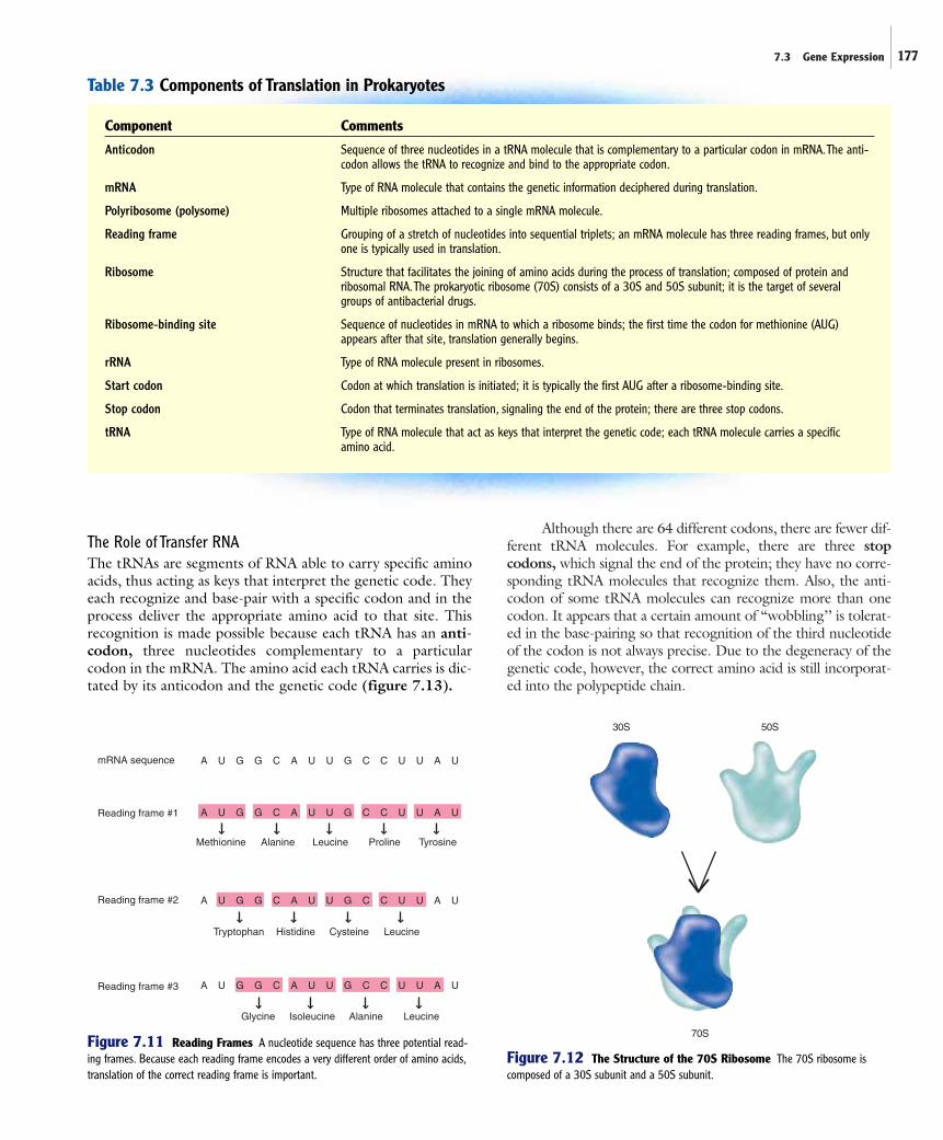
Although there are 64 different codons, there are fewer dif-ferent tRNA molecules. For example, there are three stopcodons, which signal the end of the protein; they have no corre-sponding tRNA molecules that recognize them. Also, the anti-codon of some tRNA molecules can recognize more than onecodon. It appears that a certain amount of “wobbling’’ is tolerat-ed in the base-pairing so that recognition of the third nucleotideof the codon is not always precise. Due to the degeneracy of thegenetic code, however, the correct amino acid is still incorporat-ed into the polypeptide chain.
7.3 Gene Expression 177
Table 7.3 Components of Translation in Prokaryotes
Component Comments
Anticodon Sequence of three nucleotides in a tRNA molecule that is complementary to a particular codon in mRNA.The anti-codon allows the tRNA to recognize and bind to the appropriate codon.
mRNA Type of RNA molecule that contains the genetic information deciphered during translation.
Polyribosome (polysome) Multiple ribosomes attached to a single mRNA molecule.
Reading frame Grouping of a stretch of nucleotides into sequential triplets; an mRNA molecule has three reading frames, but onlyone is typically used in translation.
Ribosome Structure that facilitates the joining of amino acids during the process of translation; composed of protein andribosomal RNA.The prokaryotic ribosome (70S) consists of a 30S and 50S subunit; it is the target of severalgroups of antibacterial drugs.
Ribosome-binding site Sequence of nucleotides in mRNA to which a ribosome binds; the first time the codon for methionine (AUG)appears after that site, translation generally begins.
rRNA Type of RNA molecule present in ribosomes.
Start codon Codon at which translation is initiated; it is typically the first AUG after a ribosome-binding site.
Stop codon Codon that terminates translation, signaling the end of the protein; there are three stop codons.
tRNA Type of RNA molecule that act as keys that interpret the genetic code; each tRNA molecule carries a specific amino acid.
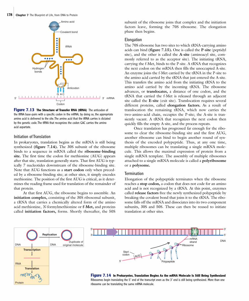
The Role of Transfer RNAThe tRNAs are segments of RNA able to carry specific aminoacids, thus acting as keys that interpret the genetic code. Theyeach recognize and base-pair with a specific codon and in theprocess deliver the appropriate amino acid to that site. Thisrecognition is made possible because each tRNA has an anti-codon, three nucleotides complementary to a particularcodon in the mRNA. The amino acid each tRNA carries is dic-tated by its anticodon and the genetic code (figure 7.13).
A U G G C A U U G C C U U A U
A U G G C A U U G C C U U A U
A U G G C A U U G C C U U A U
A U G G C A U U G C C U U A U
mRNA sequence
Reading frame #1
Reading frame #2
Reading frame #3
Methionine Alanine Leucine Proline Tyrosine
Tryptophan Histidine Cysteine Leucine
Glycine Isoleucine Alanine Leucine
Figure 7.11 Reading Frames A nucleotide sequence has three potential read-ing frames. Because each reading frame encodes a very different order of amino acids,translation of the correct reading frame is important.
30S
70S
50S
Figure 7.12 The Structure of the 70S Ribosome The 70S ribosome iscomposed of a 30S subunit and a 50S subunit.
178 Chapter 7 The Blueprint of Life, from DNA to Protein
C U G Anticodon
Hydrogenbonds
5' 3' mRNA
G A C
Codon
tRNA
Aspartate
Amino acid
Covalent bond
Figure 7.13 The Structure of Transfer RNA (tRNA) The anticodon of the tRNA base-pairs with a specific codon in the mRNA; by doing so, the appropriateamino acid is delivered to the site.The amino acid that the tRNA carries is dictated by the genetic code.The tRNA that recognizes the codon GAC carries the amino acid aspartate.
Initiation of TranslationIn prokaryotes, translation begins as the mRNA is still beingsynthesized (figure 7.14). The 30S subunit of the ribosomebinds to a sequence in mRNA called the ribosome-bindingsite. The first time the codon for methionine (AUG) appearsafter that site, translation generally starts. That first AUG is typ-ically 7 nucleotides downstream of the ribosome-binding site.Note that AUG functions as a start codon only when preced-ed by a ribosome-binding site; at other sites, it simply encodesmethionine. The position of the first AUG is critical, as it deter-mines the reading frame used for translation of the remainder ofthat protein.
At that first AUG, the ribosome begins to assemble. Aninitiation complex, consisting of the 30S ribosomal subunit,a tRNA that carries a chemically altered form of the aminoacid methionine, N-formylmethionine or f-Met, and proteinscalled initiation factors, forms. Shortly thereafter, the 50S
subunit of the ribosome joins that complex and the initiationfactors leave, forming the 70S ribosome. The elongationphase then begins.
ElongationThe 70S ribosome has two sites to which tRNA-carrying aminoacids can bind (figure 7.15). One is called the P-site (peptidylsite), and the other is called the A-site (aminoacyl site, com-monly referred to as the acceptor site). The initiating tRNA,carrying the f-Met, binds to the P-site. A tRNA that recognizesthe next codon on the mRNA then fills the unoccupied A-site.An enzyme joins the f-Met carried by the tRNA in the P-site tothe amino acid carried by the tRNA that just entered the A-site.This transfers the amino acid from the initiating tRNA to theamino acid carried by the incoming tRNA. The ribosomeadvances, or translocates, a distance of one codon, and thetRNA that carried the f-Met is released through an adjacent site called the E-site (exit site). Translocation requires severaldifferent proteins, called elongation factors. As a result oftranslocation the remaining tRNA, which now carries thetwo-amino-acid chain, occupies the P-site; the A-site is tran-siently vacant. A tRNA that recognizes the next codon thenquickly fills the empty A-site, and the process repeats.
Once translation has progressed far enough for the ribo-some to clear the ribosome-binding site and the first AUG,another ribosome can bind to begin another round of syn-thesis of the encoded polypeptide. Thus, at any one time,multiple ribosomes can be translating a single mRNA mole-cule. This allows the maximal expression of protein from asingle mRNA template. The assembly of multiple ribosomesattached to a single mRNA molecule is called a polyribosomeor a polysome.
TerminationElongation of the polypeptide terminates when the ribosomereaches a stop codon, a codon that does not code for an aminoacid and is not recognized by a tRNA. At this point, enzymescalled release factors free the newly synthesized polypeptide bybreaking the covalent bond that joins it to the tRNA. The ribo-some falls off the mRNA and dissociates into its two componentsubunits, 30S and 50S. These can then be reused to initiatetranslation at other sites.
Replication
Transcription
Translation
Protein
RNA
DNA DNADuplicate of
original molecule
5'
3'
3'
5'
5'
DNA
Startcodon
AUG
Ribosome
Polypeptide
mRNAstrand
Figure 7.14 In Prokaryotes, Translation Begins As the mRNA Molecule Is Still Being SynthesizedRibosomes begin translating the 5„ end of the transcript even as the 3„ end is still being synthesized. More than oneribosome can be translating the same mRNA molecule.

7.4 Differences Between Eukaryotic and Prokaryotic Gene Expression 179
Post-Translational ModificationProteins must often be modified after they are synthesized. Forexample, some proteins require the assistance of another pro-tein, a chaperone, to fold into the final functional shape. Those
proteins destined for transport outside of thecytoplasmic membrane also must be modi-fied. Such proteins have a characteristic seriesof hydrophobic amino acids, a signalsequence, at their amino terminal end,which “tags’’ them for transport through themembrane. The signal sequence is removedwhen the protein leaves the cytoplasm. ■ chap-erones, p. 29 ■ hydrophobic amino acids, p. 26
M I C R O C H E C K 7 . 3
RNA polymerase recognizessequences in DNA called promotersand at those sites initiates synthesisof RNA using one strand of DNA asa template. The RNA is synthesizedin the 5„ to 3„ direction and synthesisstops when RNA polymeraseencounters a terminator. Translationoccurs as ribosomes move alongmRNA in the 5„ to 3„ direction, withthe ribosomes serving as thestructure that facilitates the joiningof one amino acid to another. tRNAscarry specific amino acids, thusacting to decode the genetic code.
■ How does the orientation of thepromoter dictate which strand isused as a template for RNAsynthesis?
■ Explain why it is important for thetranslation machinery to recognizethe correct reading frame.
■ Could two mRNAs have differentnucleotide sequences and yet codefor the same protein?
7.4 Differences BetweenEukaryotic andProkaryotic GeneExpression
Eukaryotes differ significantly from prokary-otes in several aspects of transcription andtranslation (table 7.4). For example, ineukaryotic cells, most mRNA molecules areextensively modified, or processed, in thenucleus during and after transcription.Shortly after transcription begins, the 5„ end
of the transcript is modified, or capped, by the addition of amethylated guanine derivative, creating what is called a cap. Thecap likely stabilizes the transcript and enhances translation. The3„ end of the molecule is also modified, even before transcription
AA AU U AG G GC C C
AA AU U AG G GC C C
AG AA U UU A GU A C
AC AC G UGA U G C GU A A
A
AA AU U AG G GC C CU A C
mRNA
5' 3'
E-site A-site
U A C5' 3'
f-Met
f-Met
G G C
ProE-site
U A C5' 3'
G G C
E-site
A AG
U A C A U G
C U A
5' 3'
f-Met
G G C
Tyr
C U U
5' 3'C U A
f-Met
Ribosome moves along mRNA
Stopcodon
mRNA
E-siteP-site
A-site
f-Met
f-Met
InitiationThe initiating tRNA,carrying the amino acid f-Met, base-pairs with the start codon and occupies the P-site.
A tRNA that recognizesthe next codon then fillsthe unoccupied A-site.
The amino acid carried bythe tRNA in the P-site iscovalently joined to theamino acid carried by the tRNA in the A-site.
ElongationTranslocation results in the advancement of the ribosome a distance of one codon. The tRNA that occupied the P-site exits through the E-site and the tRNA that was in the A-site, which now carries thetwo amino acid chain, occupies the P-site. A tRNA that recognizes the next codon quickly fills the empty A-site.
TerminationThe process continues until astop codon terminates the process. No tRNA molecule recognizes a stop codon.
The components dissemble,releasing the newly formed polypeptide.
Pro
Pro
ProTyr
GluAsp
Pro
TyrGlu Asp
Figure 7.15 The Process of Translation

180 Chapter 7 The Blueprint of Life, from DNA to Protein
Table 7.4 Major Differences Between Prokaryotic and Eukaryotic Transcription and Translation
Prokaryotes Eukaryotes
mRNA is not processed. A cap is added to the 5´ end ofmRNA, and a poly A tail is addedto the 3´ end.
mRNA does not contain introns. mRNA contains introns, which areremoved by splicing.
Translation of mRNA begins as The mRNA transcript is transported it is being transcribed. out of the nucleus so that it can be
translated in the cytoplasm.
mRNA is often polycistronic; mRNA is monocistronic; translation translation usually begins at the begins at the first AUG.first AUG that follows a ribosome-binding site.
Transcription generatesprecursor mRNA thatcontains introns.
Splicing removes introns to form mRNA that istransported to the cytoplasm, where it will be translated.
Eukaryotic DNAcontains introns, whichinterrupt coding regions.
IntronIntron
Eukaryotic DNA
PrecursormRNA
mRNA
Figure 7.16 Splicing of Eukaryotic RNA
has been terminated. This process, called polyadenylation,involves cleaving the transcript at a specific sequence ofnucleotides and then adding approximately 200 adenine deriv-atives to the newly exposed 3„ end. This creates what is called apoly A tail, which is thought to stabilize the transcript as wellas enhance translation. Another important modification is splic-ing, a process that removes specific segments of the transcript(figure 7.16). Splicing is necessary because eukaryotic genesare not always contiguous; they are often interrupted by non-coding nucleotide sequences. These intervening sequences, orintrons, are transcribed along with the expressed regions, orexons, generating what is called precursor mRNA. The intronsmust be removed from precursor mRNA to form the maturemRNA that is then translated.
The mRNA in eukaryotic cells must be transported out ofthe nucleus before it can be translated in the cytoplasm. Thus,the same mRNA molecule cannot be transcribed and translatedat the same time or even in the same cellular location. Unlike inprokaryotes, the mRNA of eukaryotes is generally mono-cistronic. Translation of the message generally begins at the firstoccurrence of AUG in the molecule.
The ribosomes of eukaryotes are different from those ofprokaryotes. Whereas the prokaryotic ribosome is 70S, madeup of 30S and 50S subunits, the eukaryotic ribosome is 80S, made up of 40S and 60S subunits. The differences in ribo-some structure account for the ability of certain types of antibi-otics to kill bacteria without causing significant harm to mam-malian cells.
Some of the proteins that play essential roles in translationdiffer between eukaryotic and prokaryotic cells. Diphtheriatoxin, which selectively kills eukaryotic but not prokaryoticcells, illustrates this difference. This toxin is produced byCorynebacterium diphtheriae; it binds to and inactivates one ofthe elongation factors of eukaryotes. Since this protein isrequired for translocation of the ribosome, translation ceasesand the eukaryotic cell dies, resulting in the typical symptoms ofdiphtheria. ■ diphtheria toxin, p. 570
M I C R O C H E C K 7 . 4
Eukaryotic mRNA must be processed, which involvescapping, polyadenylation, and splicing. In eukaryoticcells, the mRNA must be transported out of thenucleus before it can be translated in the cytoplasm.Eukaryotic mRNA is monocistronic.
■ What is an intron?■ Explain the mechanism of action of diphtheria toxin.■ Would a deletion of two base pairs have a greater
consequence if it occurred in an intron or in an exon?
7.5 GenomicsIncreasingly rapid methods of determining the nucleotidesequence of DNA have led to exciting advancements ingenomics. Fueled by the commitment to sequence the entirehuman genome, scientists honed the methodologies by firstsequencing the genomes of select microorganisms. In 1995, thesequence of the chromosome of Haemophilus influenzae waspublished, marking the first complete genomic sequence everdetermined. The genome sequences of more than 75 otherorganisms have now been determined (table 7.5). A draft ofthe human genome is also complete.
Although sequencing methodologies are becomingmore rapid, analyzing the resulting data and extracting thepertinent information is far more complex than it might ini-tially seem. One of the most difficult steps is to locate andcharacterize the potential protein-encoding regions. Imaginetrying to determine the amino acid sequence of a proteinencoded by a 1,000-base-pair (bp) stretch of DNA without

PERSPECTIVE 7.2 RNA: The First Macromolecule?
The 1989 Nobel Prize in Chemistry was awarded to twoAmericans, Sidney Altman of Yale University and Thomas Cechof the University of Colorado, who independently made thesurprising and completely unexpected observation that RNAmolecules can act as enzymes. Before their studies, it wasbelieved that only proteins had enzymatic activity.The keyobservation was made by Cech in 1982 when he was trying tounderstand how introns were removed from mRNA that codedfor ribosomal RNA in the eukaryotic protozoan Tetrahymena.Since he was convinced that proteins were responsible forcutting out these introns, he added all of the protein in thecells’ nuclei to the mRNA that still contained the introns.
As expected, the introns were cut out. As a control,Cech looked at the ribosomal RNA to which no nuclearproteins had been added, fully expecting that nothing
would happen. Much to his surprise, the introns were alsoremoved. It did not make any difference whether theprotein was present—the introns were removed regardless.Thus, Cech could only conclude that the RNA acted on itselfto cut out pieces of RNA.
The question remained of how widespread thisphenomenon was. Did RNA have catalytic properties otherthan that of cutting out introns from rRNA? The studies ofAltman and his colleagues, carried out simultaneously toand independently of Cech’s, provided answers to thesefurther questions. Altman’s group found that RNA couldconvert a tRNA molecule from a precursor form to its finalfunctional state. Additional studies have shown thatenzymatic reactions in which catalytic RNAs, termedribozymes, play a role are very widespread. Ribozymes
have been shown to occur in the mitochondria of eukaryoticcells and to catalyze other reactions that resemble thepolymerization of RNA. Whether catalytic RNA cuts outintrons from mRNA in the nucleus is not known.
These observations have profound implications forevolution: Which came first, proteins or nucleic acids? Theanswer seems to be that nucleic acids came first, specificallyRNA, which acted both as a carrier of genetic informationas well as an enzyme. Billions of years ago, before thepresent universe in which DNA, RNA, and protein are found,probably the only macromolecule that existed was RNA.Once tRNA became available, these adapters could carryamino acids present in the environment to specificnucleotide sequences on a strand of RNA. In this scenario,the RNA functions as the genes as well as the mRNA.
7.5 Genomics 181
knowing anything about the orientation of the promoter orthe reading frame of the transcribed mRNA. Since eitherstrand of the double-stranded DNA molecule could be thetemplate strand, two entirely different mRNA moleculescould potentially code for the protein. In turn, each of those
two molecules has three reading frames, for a total of six read-ing frames. Yet only one of these actually codes for the protein.Understandably, computers are an invaluable aid and are usedextensively in deciphering the meaning of the raw sequencedata. In turn, this has resulted in the emergence of a new field,
Table 7.5 Representative Microorganisms Whose Genome Sequences Have Been Determined
Genome Size Name of Organism (106 base pairs) Important Characteristics
Agrobacterium tumefaciens 5.67 Plant pathogen; causes crown gall. Scientists use its plasmid to introduce desired genesinto plant cells.
Bacillus subtilis 4.20 Endospore-former; has served as a model for studies of Gram-positive bacteria.
Borrelia burgdorferi 1.44 Important human pathogen; causative agent of Lyme disease.
Deinococcus radiodurans 3.28 Radiation-resistant bacterium. Genome consists of two chromosomes, a large plasmid anda small plasmid.
Escherichia coli K12 4.64 Has served as model for studies of Gram-negative bacteria. Common inhabitant of theintestinal tract.
Escherichia coli O157:H7 5.53 Important human pathogen; causes hemorrhagic colitis (bloody diarrhea).
Haemophilus influenzae 1.83 First bacterial genome sequenced; important human pathogen; causes ear and respiratoryinfections and meningitis, mostly in children.
Helicobacter pylori 1.66 Important human pathogen; causes gastric diseases, including stomach ulcers.
Lactococcus (Streptococcus) lactis 2.35 Important bacterium to the dairy industry; used to make cheeses and other fermented milkproducts.
Methanococcus jannaschii 1.75 First archaeal genome sequenced; also the first autotrophic organism sequenced; hyperthermophile isolated from a hydrothermal vent; strict anaerobe; methane producer.
Mycobacterium tuberculosis 4.40 Important human pathogen; causes tuberculosis.
Mycoplasma genitalium 0.58 Smallest known bacterial genome; represents what might be the minimal genome; humanpathogen.
Pseudomonas aeruginosa 6.3 Important cause of the infection in burn victims and people who have cystic fibrosis.
Saccharomyces cerevisiae 13 Yeast; first eukaryotic genome completed.
Sinorhizobium (Rhizobium) meliloti 6.7 Fixes nitrogen; forms a symbiotic relationship with legumes.
Synechocystis species 3.57 Cyanobacterium; has served as a model for studies of photosynthesis.
Treponema pallidum 1.14 Important human pathogen; causes syphilis; has not been cultured in vitro.

182 Chapter 7 The Blueprint of Life, from DNA to Protein
bioinformatics, which has created the computer technology tostore, retrieve, and analyze nucleotide sequence data.
Analyzing a Prokaryotic DNA SequenceWhen analyzing a DNA sequence, the nucleotide sequence ofthe (;) strand is used to infer information contained in the cor-responding RNA transcript. Because of this, terms like startcodon, which actually refers to a sequence in mRNA, are usedto describe sequences in DNA. For example, to locate the startcodon AUG, which would be found in mRNA, one would lookfor the analogous sequence, ATG, in the (;) strand of the DNAmolecule. In most cases it is not initially known which of thetwo strands is actually used as a template for RNA synthesis, sothat both strands are potentially a (;) strand. Only after a pro-moter is located is it known which strand in a given region isactually the (;) strand.
To locate protein-encoding regions, computers are usedto search for open reading frames (ORFs), stretches of DNA,generally longer than 300 bp, that begin with a start codon andend with a stop codon. An ORF potentially encodes a protein.
Other characteristics, such as the presence of an upstreamsequence that can serve as a ribosome-binding site, also indicatethat an ORF encodes a protein.
The nucleotide sequence of the ORF or deduced aminoacid sequence of the encoded protein can be compared withother known sequences by searching computerized databases ofpublished sequences. Not surprisingly, as genomes of moreorganisms are being sequenced, information contained in thesedatabases is growing at a remarkable rate. If the encoded pro-tein shows certain amino acid similarities, or homology, tocharacterized proteins, a putative function can sometimes beassigned. For example, proteins that bind DNA share aminoacid sequences in certain regions. Likewise, regulatory regionsin DNA such as promoters can sometimes be identified basedon the nucleotide homologies to known sequences. ■ Regulatoryproteins that bind DNA, p. 183
The 580,070-base-pair genome of Mycoplasma genitali-um is the smallest bacterial genome known. The predicted cod-ing regions and the functional role of the genes are shown infigure 7.17.
Figure 7.17 Map of the Mycoplasma genitalium genome The wide arrows indicate the predicted protein-encoding regions.The orientation of the arrows indicates the direction of transcription; the color indicates the functional role.

7.6 Regulating Gene Expression 183
M I C R O C H E C K 7 . 5
The first genomic sequence of a microorganism wascompleted in 1995. The sequencing methodologies arequickly becoming more rapid, but analyzing the dataand extracting the pertinent information is difficult.
■ What is an open reading frame?■ Describe two things that can be learned by searching a
computerized database for sequences that havehomologies to a newly sequenced gene.
■ There are some characteristic differences in thenucleotide sequences of the leading and laggingstrands. Why might this be so?
7.6 Regulating Gene ExpressionTo cope with changing conditions in their environment,microorganisms have evolved elaborate control mechanisms tosynthesize the maximum amount of cell material from a limitedsupply of energy. This is critical, because generally a microor-ganism must reproduce more rapidly than its competitors inorder to be successful.
Consider the situation of Escherichia coli. For over 100 mil-lion years, it has successfully inhabited the gut of mammals, whereit reaches concentrations of 106 cells per milliliter. In this habitat,it must cope with alternating periods of feast and famine. For alimited time after a mammal eats, E. coli in the large intestineprosper, wallowing in the milieu of amino acids, vitamins, andother nutrients. The cells actively take up these compounds theywould otherwise synthesize, expending minimal energy.Simultaneously, the cells shut down their biosynthetic pathways,channeling the conserved energy into the rapid synthesis ofmacromolecules, including DNA, RNA, and protein. Underthese conditions, the cells divide at their most rapid rate. Famine,however, follows the feast. Between meals, which may be manydays in the case of some mammals, the rich source of nutrients isdepleted. Now the cells’ biosynthetic pathways must be activated,using energy and markedly slowing cell division. Cells dividingseveral times an hour in a nutrient-rich environment may divideonly once every 24 hours in a famished mammalian gut.
A cell controls its metabolic pathways by two generalmechanisms. The most immediate of these is the allosteric inhi-bition of enzymes. The most energy-efficient strategy, however,is to control the actual synthesis of the enzymes, making onlywhat is required. To do this, cells have the ability to controlexpression of certain genes. ■ allosteric regulation, p. 140
Principles of RegulationNot all genes are subjected to the same type of regulation. Manyare routinely expressed, whereas others are either turned on or offby certain conditions. Enzymes are often described according tocharacteristics of the regulation that governs their synthesis:
■ Constitutive enzymes are constantly synthesized; thegenes that encode these enzymes are always active.
Constitutive enzymes usually play indispensable roles inthe central metabolic pathways. For example, theenzymes of glycolysis are constitutive. ■ central metabolicpathways, p. 142
■ Inducible enzymes are not regularly produced; instead, their synthesis is turned on by certainconditions. Inducible enzymes are often involved in the utilization of specific energy sources. A cell would waste precious resources if it synthesized theenzyme when the energy source is not present. Anexample of an inducible enzyme is b-galactosidase,whose sole function is to break down the disaccharidelactose into its two component monosaccharides,glucose and galactose. The mechanisms by which the cell controls b-galactosidase synthesis serve as an important model for regulation and will be described shortly.
■ Repressible enzymes are routinely synthesized, but theycan be turned off by certain conditions. Repressibleenzymes are generally involved in biosynthetic pathways,such as those that produce amino acids. Cells require asufficient amount of a given amino acid to multiply;thus, the amino acid must be either synthesized oravailable as a component of the growth medium. If acertain amino acid is not present in the medium, thenthe cell must synthesize the enzymes involved in itsmanufacture. When the amino acid is supplied, however,synthesis of the enzymes would waste energy.
Mechanisms to Control TranscriptionThe mechanisms that a cell uses to prevent or facilitate tran-scription must be readily reversible, allowing cells to effective-ly control the relative number of transcripts made. In somecases, the control mechanisms may affect the transcription ofonly a limited number of genes; in other cases, a wide array ofgenes is coordinately controlled. For example, in E. coli theexpression of more than 300 different genes is affected by theavailability of glucose as an energy source. The simultaneousregulation of numerous genes unrelated in function is calledglobal control.
Transcription of genes is often controlled by means of aregulatory region near the promoter to which a specific proteincan bind, acting as a sophisticated on/off switch. When a regu-latory protein binds to DNA, it can either act as a repressor,which blocks transcription, or an activator, which facilitatestranscription. A set of adjacent genes coordinately controlled bya regulatory protein and transcribed as a single polycistronicmessage is called an operon.
RepressorsA repressor is a regulatory protein that blocks transcription. Itdoes this by binding to DNA at a region, called the operator,located immediately downstream of a promoter. This effective-ly prevents RNA polymerase from progressing past that regionto initiate transcription. Regulation involving a repressor iscalled negative control.

184 Chapter 7 The Blueprint of Life, from DNA to Protein
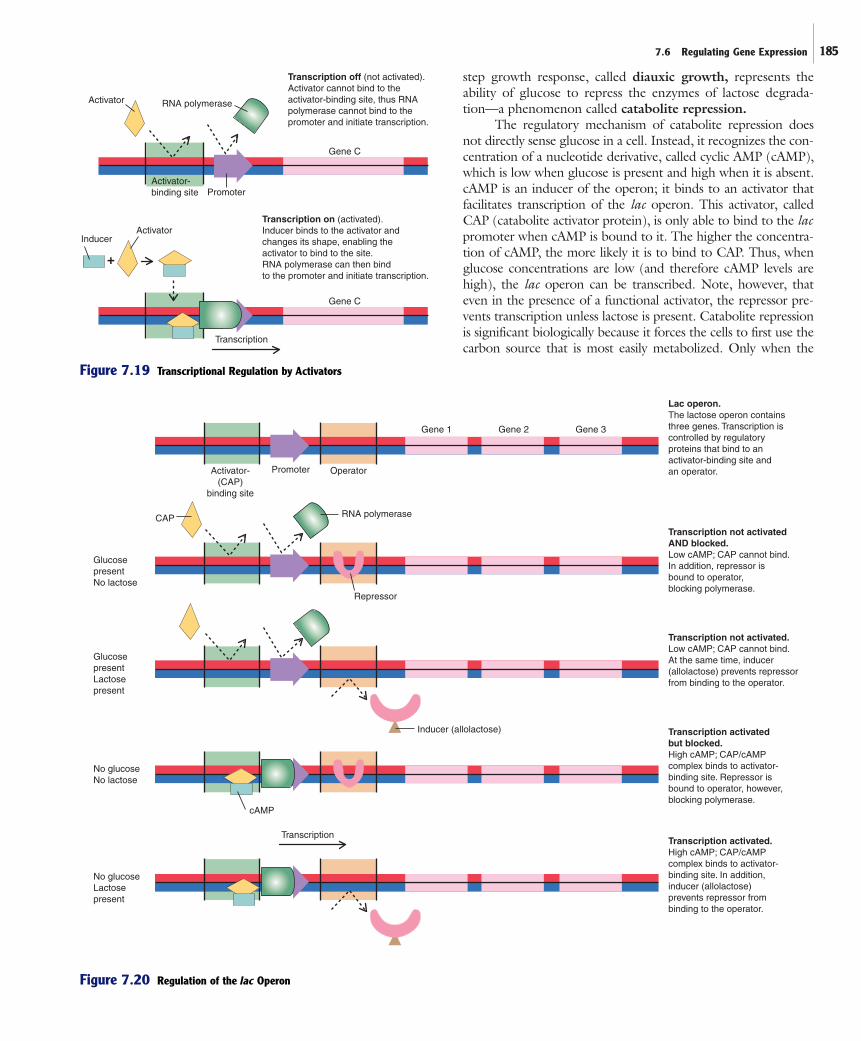
ActivatorsAn activator is a regulatory protein that facilitates transcription.Genes that are controlled by an activator have an ineffective pro-moter that is preceded by an activator-binding site. The bind-ing of the activator to the DNA enhances the ability of RNApolymerase to initiate transcription at that promoter. Regulationinvolving an activator is sometimes called positive control.
Like repressors, activators are allosteric proteins whosefunction can be modulated by the binding of other molecules. Amolecule that binds to an activator and alters its shape so it caneffectively bind to the activator-binding site functions as an induc-er (figure 7.19). Thus, the term inducer applies to a moleculethat turns on transcription, either by stimulating the function ofan activator or interfering with the function of a repressor.
The lac Operon As a Model for Control of Metabolic PathwaysOriginally elucidated in the early 1960s by Francois Jacob andJacques Monod, the lac operon has served as an importantmodel for understanding the control of gene expression in bacte-
ria. The operon, which consists of three genesinvolved with lactose degradation, along withregulatory components, is subject to dualcontrol by both a repressor and an activator(figure 7.20). The net effect is that the genesare expressed only when lactose is present butglucose is absent.
The Effect of Lactose on the Control of the Lactose OperonThe lac operon employs a repressor that pre-vents transcription of the genes when lactose isunavailable. When lactose is not present, therepressor binds to the operator, effectivelyblocking transcription. When lactose is presentin the cell, however, some of the molecules areconverted into a compound called allolactose.This compound binds to the repressor, alter-ing its shape so that it can no longer bind tothe operator. Thus, when lactose is present, therepressor no longer prevents RNA polymerasefrom transcribing the operon. Note, however,that the activator described in the next sectionis needed for successful transcription.
The Effect of Glucose on the Control of the Lactose OperonEscherichia coli preferentially uses glucoseover other sugars such as lactose. This canreadily be demonstrated by observinggrowth and sugar utilization of E. coli in amedium containing glucose and lactose.Cells actively grow, metabolizing only glu-cose until its supply is exhausted (figure7.21). Growth then ceases for a short perioduntil the cells begin utilizing lactose. At thispoint, the cells start multiplying. This two-
OperatorPromoter(with RNApolymerase)
Transcription
Mechanism 1
Mechanism 2
Transcription on.Repressor alone cannotbind to the operator.
Gene A
Gene A
Gene B
Transcription
Gene B
+
+
Corepressor
Repressor
Repressor
Repressor
Repressor
Transcription off (blocked).Corepressor binds to the repressorand alters its shape, enabling therepressor to bind to the operator.
Transcription off (blocked).Repressor binds to operator,blocking transcription.
Operator
Inducer
Transcription on.Inducer binds to the repressorand alters its shape, preventingthe repressor from bindingto the operator.
Figure 7.18 Transcriptional Regulation by Repressors
Specific molecules may bind to the repressor and, bydoing so, alter the ability of the repressor to bind to DNA. Thiscan occur because repressors are allosteric proteins, having a dis-tinct site to which another molecule can bind. When that mol-ecule binds, the shape of the repressor is altered. In turn, thisaffects the ability of the repressor to bind to DNA. As shown infigure 7.18, there are two general mechanisms by which differ-ent repressors can function:
1. The repressor is synthesized as a form that alone cannotbind to the operator. When a molecule, termed a corepressor binds to it, however, its shape is altered so that it can bind to the operator, blocking transcription initiation.
2. The repressor is synthesized as a form that effectivelybinds to the operator, blocking transcription initiation.The binding to it of a molecule that functions as aninducer, alters the shape of the repressor so that it nolonger binds to the operator. Consequently, the genecan be transcribed.
step growth response, called diauxic growth, represents theability of glucose to repress the enzymes of lactose degrada-tion—a phenomenon called catabolite repression.
The regulatory mechanism of catabolite repression doesnot directly sense glucose in a cell. Instead, it recognizes the con-centration of a nucleotide derivative, called cyclic AMP (cAMP),which is low when glucose is present and high when it is absent.cAMP is an inducer of the operon; it binds to an activator thatfacilitates transcription of the lac operon. This activator, calledCAP (catabolite activator protein), is only able to bind to the lacpromoter when cAMP is bound to it. The higher the concentra-tion of cAMP, the more likely it is to bind to CAP. Thus, whenglucose concentrations are low (and therefore cAMP levels arehigh), the lac operon can be transcribed. Note, however, thateven in the presence of a functional activator, the repressor pre-vents transcription unless lactose is present. Catabolite repressionis significant biologically because it forces the cells to first use thecarbon source that is most easily metabolized. Only when the
7.6 Regulating Gene Expression 185
Activator
ActivatorInducer
RNA polymerase
PromoterActivator-binding site
Gene C
Gene C
Transcription off (not activated).Activator cannot bind to theactivator-binding site, thus RNA polymerase cannot bind to the promoter and initiate transcription.
+
Transcription on (activated).Inducer binds to the activator andchanges its shape, enabling the activator to bind to the site.RNA polymerase can then bind to the promoter and initiate transcription.
Transcription
Figure 7.19 Transcriptional Regulation by Activators
Operator
Gene 1 Gene 3Gene 2
Activator-(CAP)
binding site
Promoter
Lac operon.The lactose operon containsthree genes. Transcription iscontrolled by regulatory proteins that bind to an activator-binding site and an operator.
CAP RNA polymerase
Glucose presentNo lactose
Glucose presentLactosepresent
No glucose No lactose
No glucose Lactose present
Transcription not activated AND blocked.Low cAMP; CAP cannot bind. In addition, repressor is bound to operator, blocking polymerase.
Inducer (allolactose)
Transcription not activated.Low cAMP; CAP cannot bind.At the same time, inducer(allolactose) prevents repressorfrom binding to the operator.
Transcription activated but blocked.High cAMP; CAP/cAMP complex binds to activator-binding site. Repressor is bound to operator, however, blocking polymerase.
Transcription
cAMP
Repressor
Transcription activated.High cAMP; CAP/cAMPcomplex binds to activator-binding site. In addition, inducer (allolactose) prevents repressor from binding to the operator.
Figure 7.20 Regulation of the lac Operon
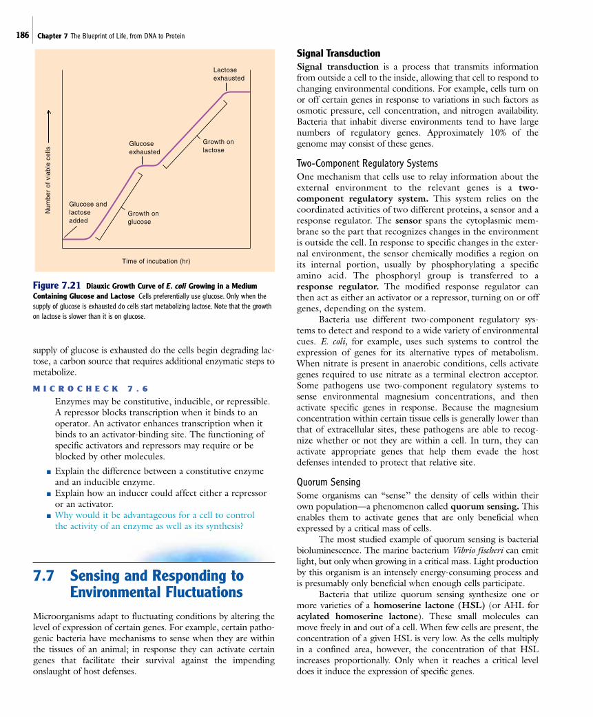
186 Chapter 7 The Blueprint of Life, from DNA to Protein
supply of glucose is exhausted do the cells begin degrading lac-tose, a carbon source that requires additional enzymatic steps tometabolize.
M I C R O C H E C K 7 . 6
Enzymes may be constitutive, inducible, or repressible.A repressor blocks transcription when it binds to anoperator. An activator enhances transcription when itbinds to an activator-binding site. The functioning ofspecific activators and repressors may require or beblocked by other molecules.
■ Explain the difference between a constitutive enzymeand an inducible enzyme.
■ Explain how an inducer could affect either a repressoror an activator.
■ Why would it be advantageous for a cell to control the activity of an enzyme as well as its synthesis?
7.7 Sensing and Responding toEnvironmental Fluctuations
Microorganisms adapt to fluctuating conditions by altering thelevel of expression of certain genes. For example, certain patho-genic bacteria have mechanisms to sense when they are withinthe tissues of an animal; in response they can activate certaingenes that facilitate their survival against the impendingonslaught of host defenses.
Signal TransductionSignal transduction is a process that transmits informationfrom outside a cell to the inside, allowing that cell to respond tochanging environmental conditions. For example, cells turn onor off certain genes in response to variations in such factors asosmotic pressure, cell concentration, and nitrogen availability.Bacteria that inhabit diverse environments tend to have largenumbers of regulatory genes. Approximately 10% of thegenome may consist of these genes.
Two-Component Regulatory SystemsOne mechanism that cells use to relay information about theexternal environment to the relevant genes is a two-component regulatory system. This system relies on thecoordinated activities of two different proteins, a sensor and aresponse regulator. The sensor spans the cytoplasmic mem-brane so the part that recognizes changes in the environmentis outside the cell. In response to specific changes in the exter-nal environment, the sensor chemically modifies a region onits internal portion, usually by phosphorylating a specificamino acid. The phosphoryl group is transferred to aresponse regulator. The modified response regulator canthen act as either an activator or a repressor, turning on or offgenes, depending on the system.
Bacteria use different two-component regulatory sys-tems to detect and respond to a wide variety of environmentalcues. E. coli, for example, uses such systems to control theexpression of genes for its alternative types of metabolism.When nitrate is present in anaerobic conditions, cells activategenes required to use nitrate as a terminal electron acceptor.Some pathogens use two-component regulatory systems tosense environmental magnesium concentrations, and thenactivate specific genes in response. Because the magnesiumconcentration within certain tissue cells is generally lower thanthat of extracellular sites, these pathogens are able to recog-nize whether or not they are within a cell. In turn, they canactivate appropriate genes that help them evade the hostdefenses intended to protect that relative site.
Quorum SensingSome organisms can “sense’’ the density of cells within theirown population—a phenomenon called quorum sensing. Thisenables them to activate genes that are only beneficial whenexpressed by a critical mass of cells.
The most studied example of quorum sensing is bacterialbioluminescence. The marine bacterium Vibrio fischeri can emitlight, but only when growing in a critical mass. Light productionby this organism is an intensely energy-consuming process andis presumably only beneficial when enough cells participate.
Bacteria that utilize quorum sensing synthesize one ormore varieties of a homoserine lactone (HSL) (or AHL foracylated homoserine lactone). These small molecules canmove freely in and out of a cell. When few cells are present, theconcentration of a given HSL is very low. As the cells multiplyin a confined area, however, the concentration of that HSLincreases proportionally. Only when it reaches a critical leveldoes it induce the expression of specific genes.
Time of incubation (hr)
Num
ber
of v
iabl
e ce
lls
Lactoseexhausted
Glucose andlactoseadded
Glucoseexhausted
Growth onglucose
Growth onlactose
Figure 7.21 Diauxic Growth Curve of E. coli Growing in a MediumContaining Glucose and Lactose Cells preferentially use glucose. Only when thesupply of glucose is exhausted do cells start metabolizing lactose. Note that the growthon lactose is slower than it is on glucose.
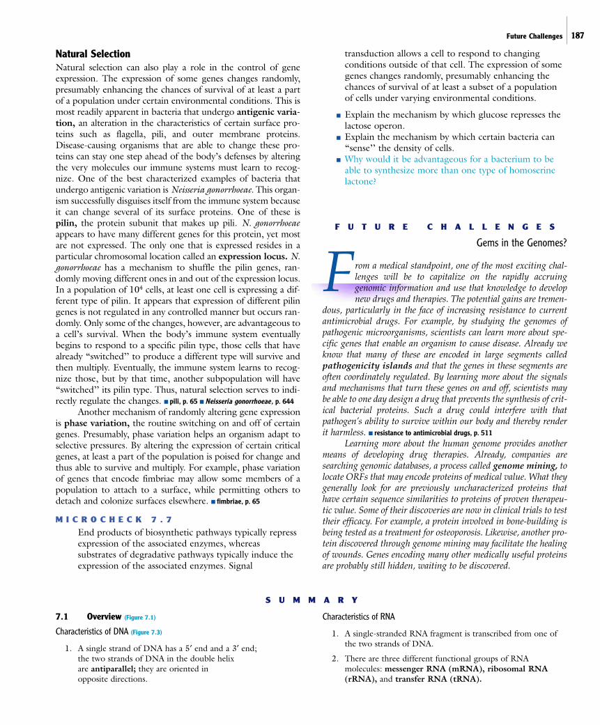
Future Challenges 187
Natural SelectionNatural selection can also play a role in the control of geneexpression. The expression of some genes changes randomly,presumably enhancing the chances of survival of at least a partof a population under certain environmental conditions. This ismost readily apparent in bacteria that undergo antigenic varia-tion, an alteration in the characteristics of certain surface pro-teins such as flagella, pili, and outer membrane proteins.Disease-causing organisms that are able to change these pro-teins can stay one step ahead of the body’s defenses by alteringthe very molecules our immune systems must learn to recog-nize. One of the best characterized examples of bacteria thatundergo antigenic variation is Neisseria gonorrhoeae. This organ-ism successfully disguises itself from the immune system becauseit can change several of its surface proteins. One of these ispilin, the protein subunit that makes up pili. N. gonorrhoeaeappears to have many different genes for this protein, yet mostare not expressed. The only one that is expressed resides in aparticular chromosomal location called an expression locus. N.gonorrhoeae has a mechanism to shuffle the pilin genes, ran-domly moving different ones in and out of the expression locus.In a population of 104 cells, at least one cell is expressing a dif-ferent type of pilin. It appears that expression of different pilingenes is not regulated in any controlled manner but occurs ran-domly. Only some of the changes, however, are advantageous toa cell’s survival. When the body’s immune system eventuallybegins to respond to a specific pilin type, those cells that havealready “switched’’ to produce a different type will survive andthen multiply. Eventually, the immune system learns to recog-nize those, but by that time, another subpopulation will have“switched’’ its pilin type. Thus, natural selection serves to indi-rectly regulate the changes. ■ pili, p. 65 ■ Neisseria gonorrhoeae, p. 644
Another mechanism of randomly altering gene expressionis phase variation, the routine switching on and off of certaingenes. Presumably, phase variation helps an organism adapt toselective pressures. By altering the expression of certain criticalgenes, at least a part of the population is poised for change andthus able to survive and multiply. For example, phase variationof genes that encode fimbriae may allow some members of apopulation to attach to a surface, while permitting others todetach and colonize surfaces elsewhere. ■ fimbriae, p. 65
M I C R O C H E C K 7 . 7
End products of biosynthetic pathways typically repressexpression of the associated enzymes, whereassubstrates of degradative pathways typically induce theexpression of the associated enzymes. Signal
transduction allows a cell to respond to changingconditions outside of that cell. The expression of somegenes changes randomly, presumably enhancing thechances of survival of at least a subset of a populationof cells under varying environmental conditions.
■ Explain the mechanism by which glucose represses thelactose operon.
■ Explain the mechanism by which certain bacteria can“sense’’ the density of cells.
■ Why would it be advantageous for a bacterium to beable to synthesize more than one type of homoserinelactone?
F U T U R E C H A L L E N G E S
Gems in the Genomes?
From a medical standpoint, one of the most exciting chal-lenges will be to capitalize on the rapidly accruinggenomic information and use that knowledge to developnew drugs and therapies. The potential gains are tremen-
dous, particularly in the face of increasing resistance to currentantimicrobial drugs. For example, by studying the genomes ofpathogenic microorganisms, scientists can learn more about spe-cific genes that enable an organism to cause disease. Already weknow that many of these are encoded in large segments calledpathogenicity islands and that the genes in these segments areoften coordinately regulated. By learning more about the signalsand mechanisms that turn these genes on and off, scientists maybe able to one day design a drug that prevents the synthesis of crit-ical bacterial proteins. Such a drug could interfere with thatpathogen’s ability to survive within our body and thereby renderit harmless. ■ resistance to antimicrobial drugs, p. 511
Learning more about the human genome provides anothermeans of developing drug therapies. Already, companies aresearching genomic databases, a process called genome mining, tolocate ORFs that may encode proteins of medical value. What theygenerally look for are previously uncharacterized proteins thathave certain sequence similarities to proteins of proven therapeu-tic value. Some of their discoveries are now in clinical trials to testtheir efficacy. For example, a protein involved in bone-building isbeing tested as a treatment for osteoporosis. Likewise, another pro-tein discovered through genome mining may facilitate the healingof wounds. Genes encoding many other medically useful proteinsare probably still hidden, waiting to be discovered.
S U M M A R Y
7.1 Overview (Figure 7.1)
Characteristics of DNA (Figure 7.3)
1. A single strand of DNA has a 5„ end and a 3„ end; the two strands of DNA in the double helix are antiparallel; they are oriented in opposite directions.
Characteristics of RNA
1. A single-stranded RNA fragment is transcribed from one ofthe two strands of DNA.
2. There are three different functional groups of RNAmolecules: messenger RNA (mRNA), ribosomal RNA(rRNA), and transfer RNA (tRNA).

188 Chapter 7 The Blueprint of Life, from DNA to Protein
Regulating the Expression of Genes
1. Protein synthesis is generally controlled by regulating thesynthesis of mRNA molecules; mRNA is short-lived becauseRNases degrade it within minutes.
7.2 DNA Replication
1. DNA replication is generally bidirectional andsemiconservative. (Figure 7.4)
2. The DNA chain always elongates in the 5„ to 3„ direction; thebase-pairing rules determine the specific nucleotides that areadded. (Figure 7.5)
Initiation of DNA Replication
1. DNA replication begins at the origin of replication. DNApolymerase synthesizes DNA in the 5„ to 3„ direction, using one strand as a template to generate thecomplementary strand.
The Replication Fork (Figure 7.6)
1. The bidirectional progression of replication around a circularDNA molecule creates two replication forks; numerousenzymes and other proteins are involved.
7.3 Gene Expression
Transcription
1. The enzyme RNA polymerase catalyzes the process oftranscription, producing a single-stranded RNA molecule that is complementary and antiparallel to the DNA template.(Figure 7.7)
2. Transcription begins when RNA polymerase recognizes andbinds to a promoter. (Figure 7.8)
3. RNA is synthesized in the 5„ to 3„ direction. (Figure 7.9)
4. When RNA polymerase encounters a terminator, it falls offthe DNA template and releases the newly synthesized RNA.
Translation
1. The information encoded by mRNA is deciphered using thegenetic code. (Figure 7.10)
2. A nucleotide sequence has three potential reading frames.(Figure 7.11)
3. Ribosomes function as the site of translation. (Figure 7.12)
4. tRNAs carry specific amino acids and act as keys thatinterpret the genetic code. (Figure 7.13)
5. In prokaryotes, initiation of translation begins when theribosome binds to the ribosome-binding site of the mRNAmolecule. Translation starts at the first AUG downstream ofthat site. (Figure 7.14)
6. The ribosome moves along mRNA in the 5„ to 3„ direction;translation terminates when the ribosome reaches a stopcodon. (Figure 7.15)
7. Proteins are often modified after they are synthesized; thosethat contain a signal sequence are transported to the outsideof the cell.
7.4 Differences Between Eukaryotic and Prokaryotic Gene Expression (Table 7.4)
1. Eukaryotic mRNA is processed; a cap and a poly A tail areadded.
2. Eukaryotic genes often contain introns which are removedfrom precursor mRNA by a process called splicing. (Figure 7.16)
3. In eukaryotic cells, the mRNA must be transported out of thenucleus before it can be translated in the cytoplasm.
7.5 Genomics
Analyzing a Prokaryotic DNA Sequence (Figure 7.17)
1. When analyzing a DNA sequence, the nucleotide sequence ofthe (;) strand is used to infer information carried by thecorresponding RNA transcript; computers are used to searchfor open reading frames (ORFs).
7.6 Regulating Gene Expression
Principles of Regulation
1. Constitutive enzymes are constantly synthesized.
2. The synthesis of inducible enzymes can be turned on bycertain conditions.
3. The synthesis of repressible enzymes can be turned off bycertain conditions.
Mechanisms to Control Transcription
1. A repressor is a regulatory protein that blocks transcription.(Figure 7.18)
2. An activator is a regulatory protein that enhancestranscription. (Figure 7.19)
The lac Operon As a Model for Control of Metabolic Pathways (Figure 7.20)
1. The lac operon employs a repressor that preventstranscription of the genes when lactose is not available; aderivative of lactose functions as an inducer.
2. Catabolite repression prevents transcription of the lac operonwhen glucose is available.
7.7 Sensing and Responding to Environmental Fluctuations
Signal Transduction
1. Two-component regulatory systems utilize a sensor thatrecognizes changes outside the cell and then transmits thatinformation to a response regulator.
2. Bacteria that utilize quorum sensing synthesize a solublecompound, a homoserine lactone, which can move freely inand out of a cell. Only when that compound reaches a criticalconcentration does it activate specific genes.

Review Questions 189
Natural Selection
1. The expression of some genes changes randomly, presumably enhancing the chances of survival of at least a subset of a population under varying environmental conditions.
2. Antigenic variation is a routine change in the expression ofsurface proteins such as flagella, pili, and outer membraneproteins.
3. Phase variation is the routine switching on and off of certaingenes.
R E V I E W Q U E S T I O N S
Short Answer
1. Explain what the term semiconservative means with respect toDNA replication.
2. How can E. coli have a generation time of only 20 minutes whenit takes 40 minutes to replicate its chromosome?
3. What is the function of primase in DNA replication? Why is thisenzyme necessary?
4. What is polycistronic mRNA?
5. Explain why knowing the orientation of a promoter is criticalwhen determining the amino acid sequence of an encodedprotein.
6. What is characteristic about the nucleotide sequence of atranscription terminator?
7. What happens to a polypeptide that has a signal sequence?
8. Compare and contrast regulation by a repressor and an activator.
9. Explain how bacteria sense the density of cells in their ownpopulation.
10. Explain why it is sometimes difficult to locate genomic regionsthat encode a protein.
Multiple Choice Questions
1. All of the following are involved in transcription, exceptA. polymerase. B. primer. C. promoter.D. sigma factor. E. uracil.
2. All of the following are involved in DNA replication, exceptA. elongation factors. B. gyrase. C. polymerase.D. primase. E. primer.
3. All of the following are directly involved in translation, exceptA. promoter. B. ribosome. C. start codon.D. stop codon. E. tRNA.
4. Using the DNA strand depicted here as a template, what will bethe sequence of the RNA transcript?
A. 5„ GCGUUAACGUAGGC 3„B. 5„ CGGAUGCAAUUGCG 3„C. 5„ CGCAAUUGCAUCCG 3„D. 5„ GCCUACGUUAACGC 3„
5′ GCGTTAACGTAGGC 3′—Dpromoter
3′ CGCAATTGCATCCG 5′
5. A ribosome binds to the following mRNA at the site indicatedby the dark box. What are the first three amino acids that will beincorporated into the resulting polypeptide?
5′ GCCGGAAUGCUGCUGGC
A. alanine aspartic acid alanineB. methionine threonine cysteineC. methionine leucine leucineD. alanine glycine methionine
6. Allolactose induces the lac regulon by binding to a(n)…A. operator. B. repressor.C. activator. D. CAP protein.
7. Under which of the following conditions will transcription of thelac operon occur?A. Lactose present/glucose presentB. Lactose present/glucose absentC. Lactose absent/glucose presentD. Lactose absent/glucose absentE. A and B
8. Which of the following statements about gene expression is false?A. More than one RNA polymerase can be transcribing a specif-
ic gene at a given time.B. More than one ribosome can be translating a specific tran-
script at a given time.C. Translation begins at a site called a promoter.D. Transcription stops at a site called a terminator.E. Some amino acids are coded for by more than one codon.
9. Which of the following is not characteristic of eukaryotic geneexpression?A. 5„ cap is added to the mRNA.B. A poly A tail is added to the 3„ end of mRNA.C. Introns must be removed to create the mRNA that
is translated.D. The mRNA is often polycistronic.E. Translation begins at the first AUG.
10. Which of the following statements is false?A. A derivative of lactose serves as an inducer of the lac operon.B. Signal transduction provides a mechanism for a cell to sense
the conditions of its external environment.C. The function of homoserine lactone is to enable a cell to
sense the density of like cells.D. An example of a two-component regulatory system is the
lactose operon, which is controlled by a repressor and anactivator.
E. An ORF is a stretch of DNA that may encode a protein.
190 Chapter 7 The Blueprint of Life, from DNA to Protein
Applications
1. A graduate student is trying to isolate the gene coding for anenzyme found in a species of Pseudomonas that degradestrinitrotoluene (TNT). The student is frustrated to find that theorganism does not produce the enzyme when grown in nutrientbroth, making it is difficult to collect the mRNA needed to helpidentify the gene. What could the student do to potentiallyincrease the amount of the desired enzyme?
2. A student wants to remove the introns from a segment of DNAcoding for protein X. Devise a strategy for how this could beaccomplished.
Critical Thinking
1. The study of protein synthesis often uses a cell-free system wherecells are ground with an abrasive to release the cell contents andthen filtered to remove the abrasive. These materials are addedto the system, generating the indicated results:
Materials Added ResultsRadioactive amino acids Radioactive protein produced
Radioactive amino acids No radioactive protein producedand RNase (an RNA-digesting enzyme)
What is the best interpretation of these observations?
2. In a variation of the experiment in the previous question, thefollowing materials were added to three separate cell-freesystems, generating the indicated results:
Materials Added ResultsRadioactive amino acids Radioactive protein produced
Radioactive amino acids Radioactive protein producedand DNase (a DNA-digesting enzyme)
Several hours after grinding: No radioactive protein producedRadioactive amino acids and DNase
What is the best interpretation of these observations?