The Nervous System. Overview of the Nervous System Overview of the Nervous System.
Upload
winifred-simpsonCategory
view
217download
0
The Biological Perspective
Chapter 2

Overview of Nervous System• Nervous System - an extensive network of
specialized cells that carry information to and from all parts of the body.
• Neuroscience – deals with the structure and function of the brain, neurons, nerves, and nervous tissue.• Relationship to behavior and learning.
LO 2.1 What are the nervous system, neurons and nerves
Menu
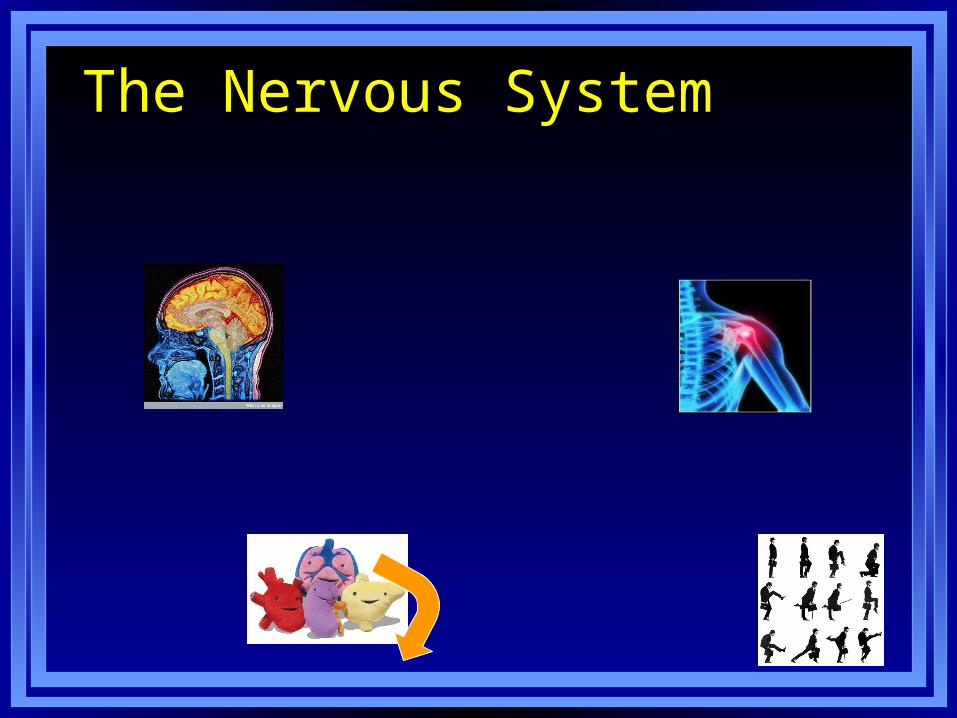
The Nervous System

The Nervous System
Peripheral Nervous System
Central Nervous System

Central Nervous System• Central nervous system (CNS) - part of
the nervous system consisting of the brain and spinal cord.
• Spinal cord - a long bundle of neurons that • carries messages • is responsible for very fast,
lifesaving reflexes.
LO 2.3 Brain and spinal cord
Menu

Peripheral Nervous System
• Peripheral nervous system (PNS) - all nerves and neurons that are not contained in the brain and spinal cord but that run through the body itself
• Divided into the:• Somatic nervous system• Autonomic nervous system
Menu
LO 2.4 Somatic and autonomic nervous systems

Autonomic Nervous System• Autonomic nervous system (ANS) -
division of the PNS consisting of nerves that control all of the involuntary muscles, organs, and glands
• Composed of:• Sympathetic division Reacts to
stressful events and bodily arousal.
• Parasympathetic division – restores body to normal functioning after arousal
LO 2.4 Somatic and Autonomic nervous systems
Menu
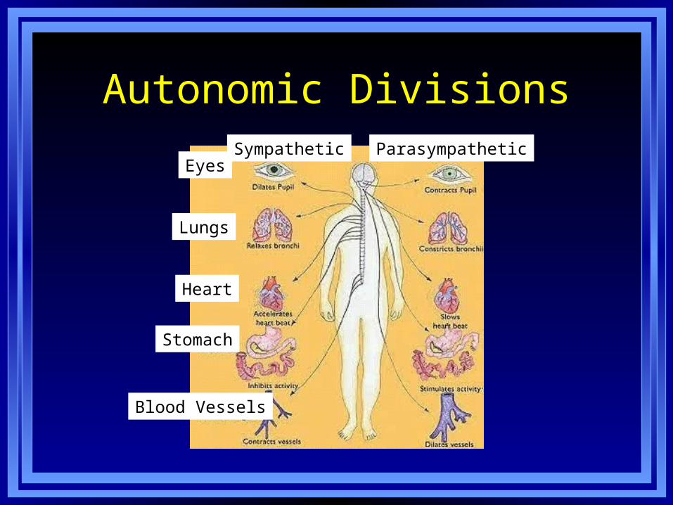
Autonomic Divisions
Eyes
Lungs
Heart
Stomach
Blood Vessels
Sympathetic Parasympathetic
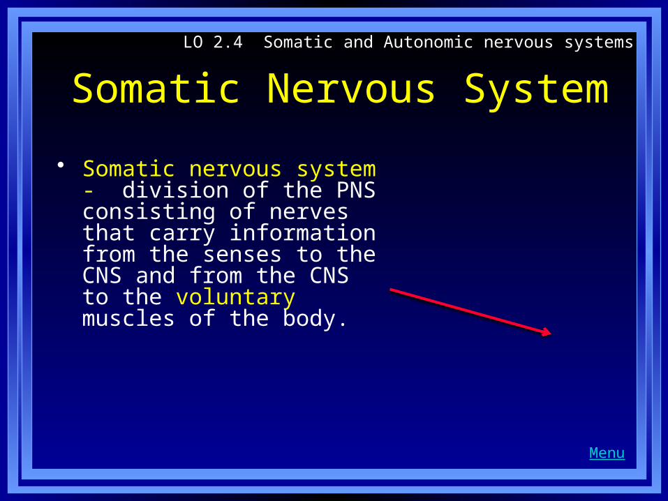
Somatic Nervous System
• Somatic nervous system - division of the PNS consisting of nerves that carry information from the senses to the CNS and from the CNS to the voluntary muscles of the body.
LO 2.4 Somatic and Autonomic nervous systems
Menu

Structure of the Neuron• Neuron – cell in the nervous
system that receives and sends messages.
• Parts of a Neuron• Dendrites - branch-like
structures that receive messages.
• Soma – contains the nucleus and keeps the cell alive and functioning
• Axon - long tube-like structure that carries messages out to other cells
LO 2.1 What are the nervous system, neurons and nerves
Menu
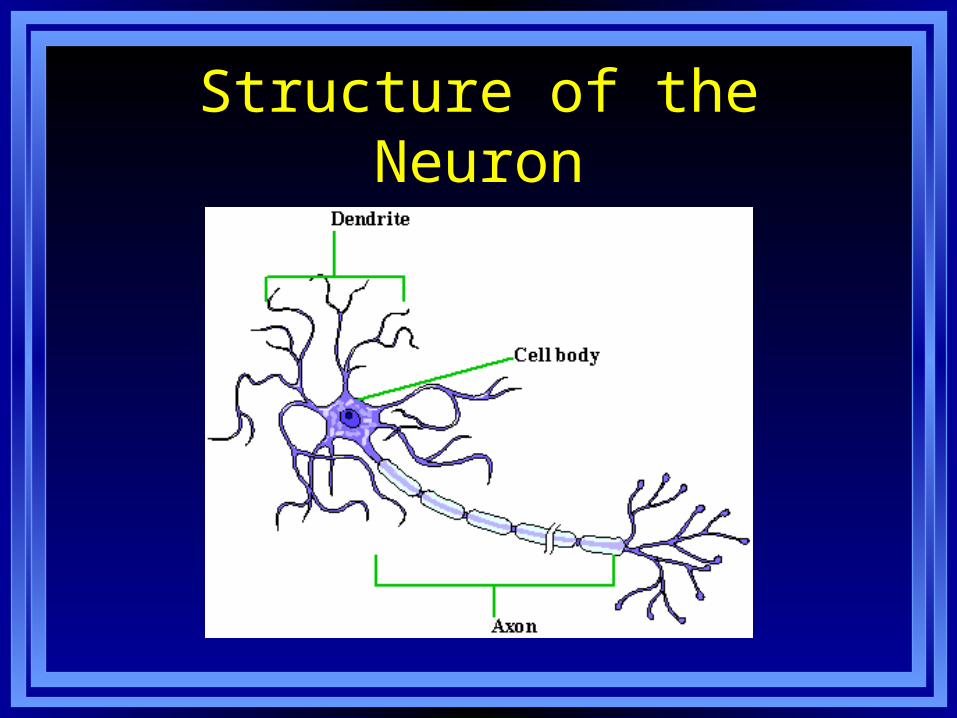
Structure of the Neuron
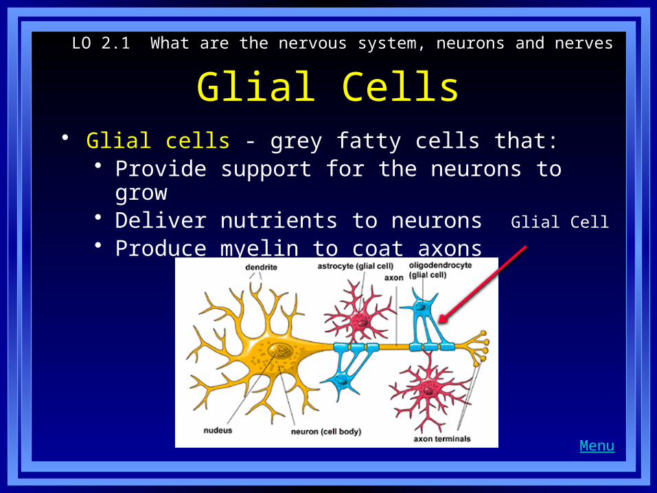
Glial Cells• Glial cells - grey fatty cells that:
• Provide support for the neurons to grow• Deliver nutrients to neurons• Produce myelin to coat axons
LO 2.1 What are the nervous system, neurons and nerves
Menu
Glial Cell

Myelin Sheath
Myelin - fatty substances that coat the axons of neurons to insulate, protect, and speed up the neural impulse.
• Clean up waste products and dead neurons.
Myelin

The Synapse • Space between Axon and Dendrite of a receiving cell
• The location where Neurotransmitters are released
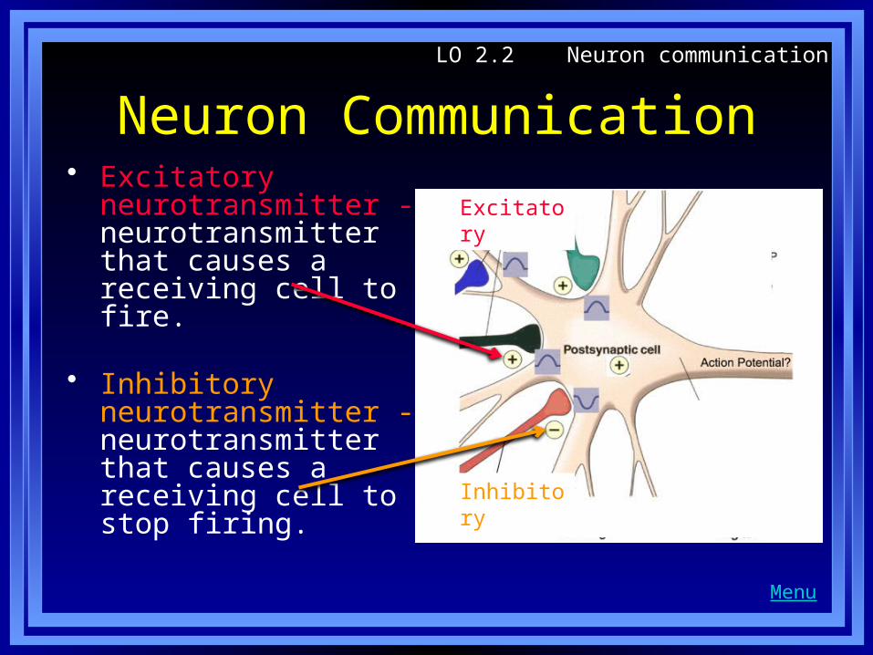
Neuron Communication• Excitatory
neurotransmitter - neurotransmitter that causes a receiving cell to fire.
• Inhibitory neurotransmitter - neurotransmitter that causes a receiving cell to stop firing.
LO 2.2 Neuron communication
Menu
Excitatory
Inhibitory
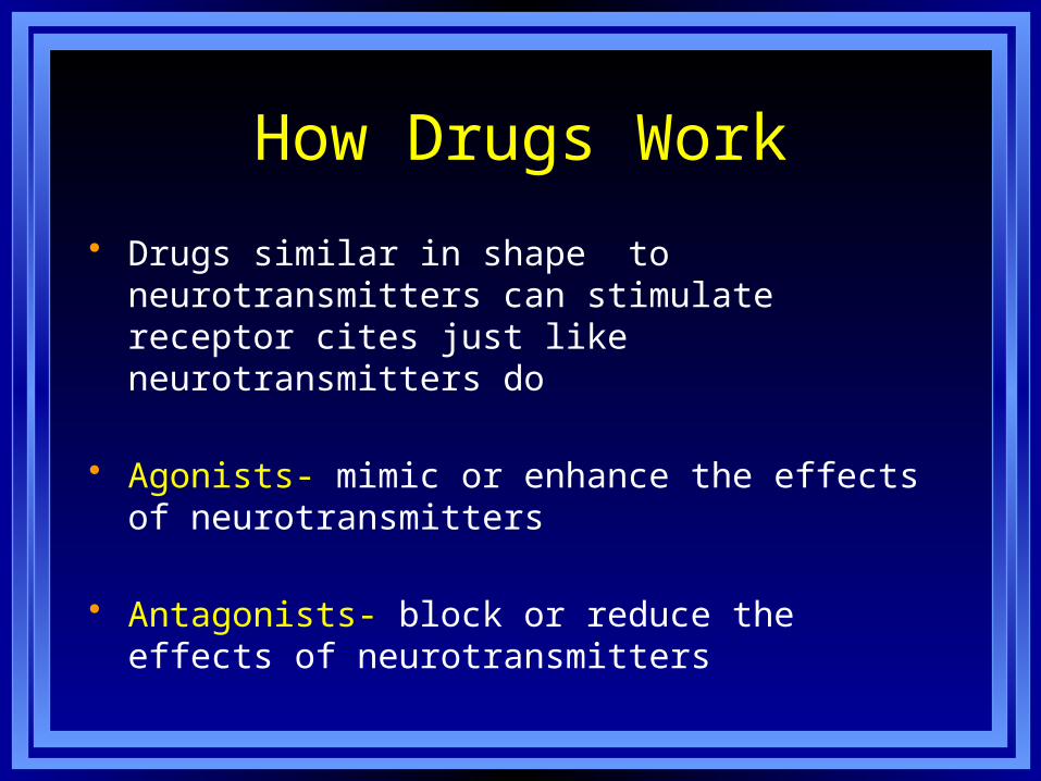
How Drugs Work
• Drugs similar in shape to neurotransmitters can stimulate receptor cites just like neurotransmitters do
• Agonists- mimic or enhance the effects of neurotransmitters
• Antagonists- block or reduce the effects of neurotransmitters
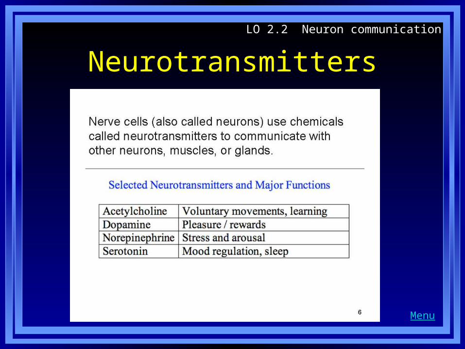
NeurotransmittersLO 2.2 Neuron communication
Menu
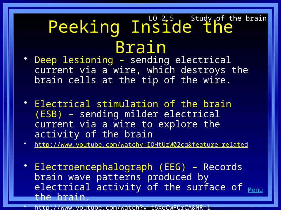
Peeking Inside the Brain• Deep lesioning – sending electrical current via a
wire, which destroys the brain cells at the tip of the wire.
• Electrical stimulation of the brain (ESB) – sending milder electrical current via a wire to explore the activity of the brain
• http://www.youtube.com/watchv=IOHtUzW02cg&feature=related
• Electroencephalograph (EEG) – Records brain wave patterns produced by electrical activity of the surface of the brain.
• http://www.youtube.com/watch?v=t6XeCwFQrCA&NR=1
LO 2.5 Study of the brain
Menu
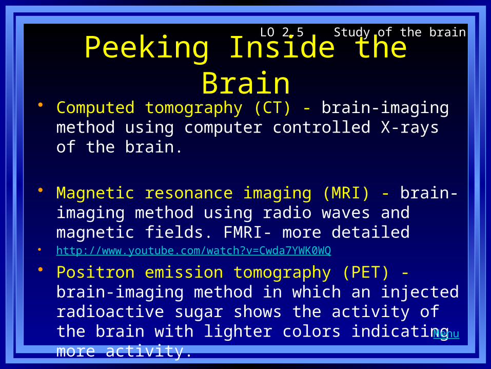
Peeking Inside the Brain• Computed tomography (CT) - brain-imaging method
using computer controlled X-rays of the brain.
• Magnetic resonance imaging (MRI) - brain-imaging method using radio waves and magnetic fields. FMRI- more detailed
• http://www.youtube.com/watch?v=Cwda7YWK0WQ
• Positron emission tomography (PET) - brain-imaging method in which an injected radioactive sugar shows the activity of the brain with lighter colors indicating more activity.
LO 2.5 Study of the brain
Menu
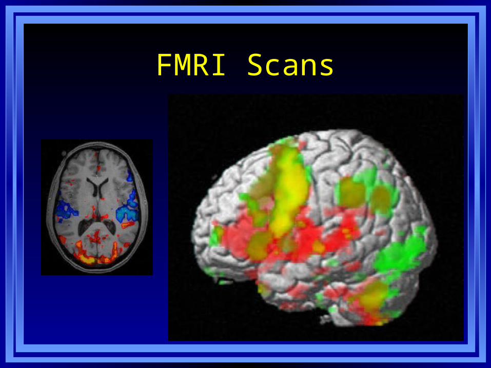
FMRI Scans

The Brain Stem• Medulla – Forms
the lowest part of the brain • Responsible
for life-sustaining functions such as breathing, swallowing, and heart rate.
LO 2.6 Structures of the bottom part of brain
Menu
Medulla

The Brain Stem
• Pons – Structure above the medulla
• Connects the top of the brain to the bottom
• Plays a part in sleep, dreaming, left–right body coordination, and arousal.
Pons
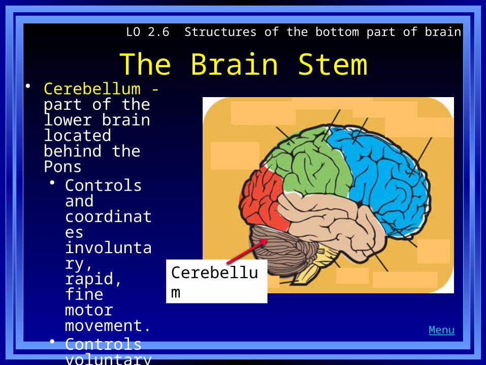
The Brain Stem• Cerebellum -
part of the lower brain located behind the Pons • Controls and
coordinates involuntary, rapid, fine motor movement.
• Controls voluntary movements that happen in rapid succession
LO 2.6 Structures of the bottom part of brain
Menu
Cerebellum
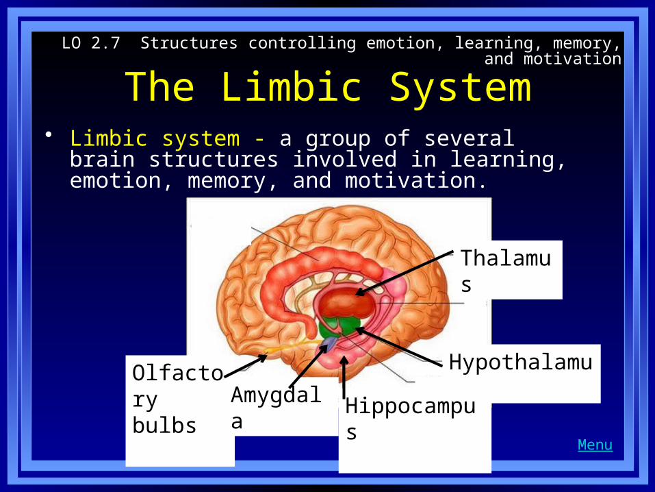
The Limbic System• Limbic system - a group of several brain structures
involved in learning, emotion, memory, and motivation.
LO 2.7 Structures controlling emotion, learning, memory, and motivation
Menu
HypothalamusOlfactory bulbs Amygdala Hippocampus
Thalamus

The Limbic System• Hypothalamus-
located below the thalamus and directly above the pituitary gland
• Regulates body temperature, thirst, hunger, sleeping, waking,
• Responsible for motivational behavior
LO 2.7 Structures controlling emotion, learning, memory, and motivation
Menu
Hypothalamus
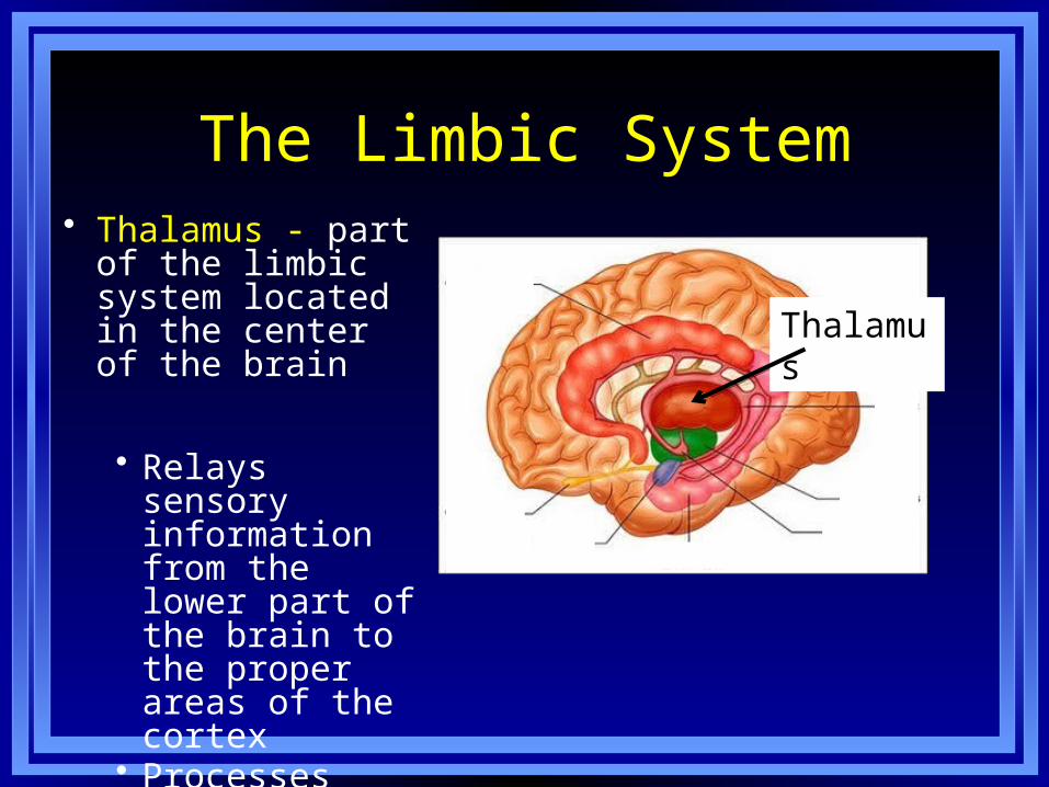
The Limbic System• Thalamus - part of
the limbic system located in the center of the brain
• Relays sensory information from the lower part of the brain to the proper areas of the cortex
• Processes some info prior to relaying it
Thalamus
The Limbic System
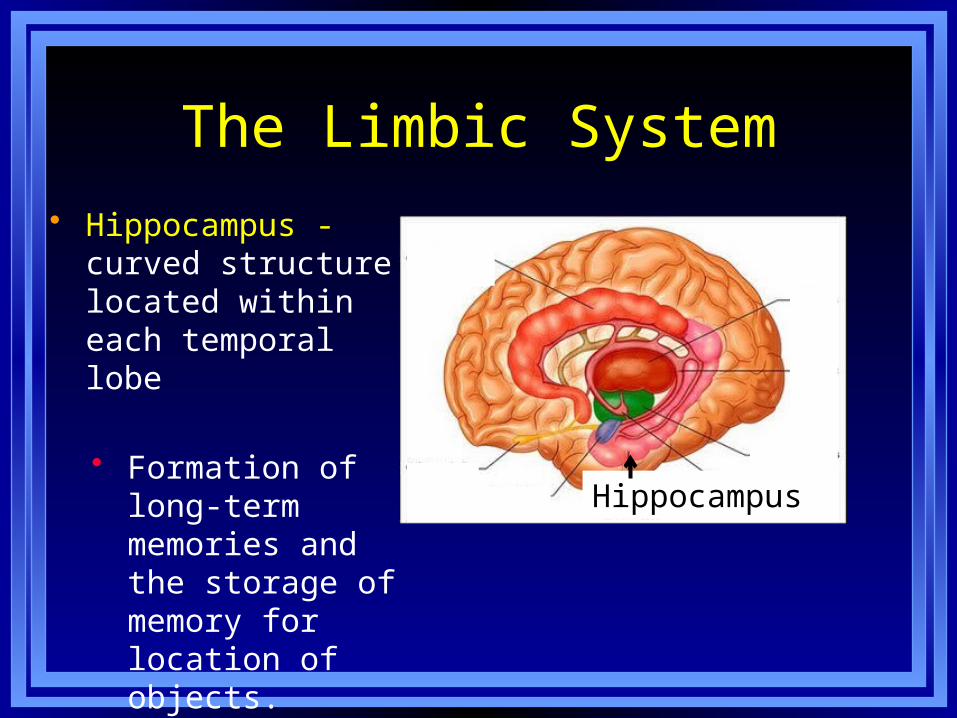
• Hippocampus - curved structure located within each temporal lobe
• Formation of long-term memories and the storage of memory for location of objects.
Hippocampus

The Limbic System
• Amygdala - brain structure located near the hippocampus
• Responsible for fear responses and memory of fear.
Amygdala

The Limbic System• Olfactory bulbs -
two projections just under the front of the brain
• Receive information from the receptors in the nose located just below.
Olfactory bulbs

The Cortex• Cortex - outermost
covering of the brain consisting of densely packed neurons • responsible for
higher thought processes and interpretation of sensory input.
LO 2.7 Structures controlling emotion, learning, memory, and motivation
Menu
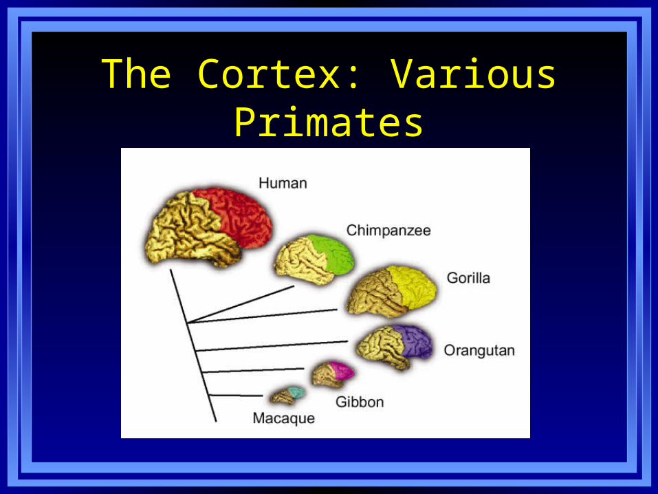
The Cortex: Various Primates

Cerebral Hemispheres• Cerebral hemispheres - the two sections of the cortex
on the left and right sides of the brain.
• Corpus callosum - thick band of neurons that connects the right and left cerebral hemispheres.
LO 2.8 Parts of cortex controlling senses and movement
Menu
Corpus callosumCorpus callosum
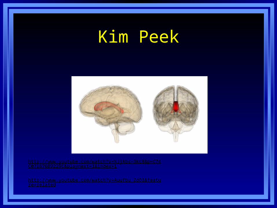
Kim Peek
http://www.youtube.com/watch?v=NJjAbs-3kc8&p=C74C071676B9229C&playnext=1&index=1
http://www.youtube.com/watch?v=Auufbu_ZdDI&feature=related
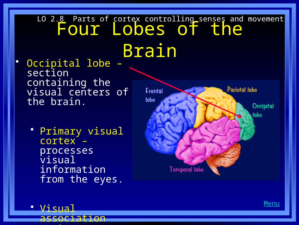
Four Lobes of the Brain• Occipital lobe – section
containing the visual centers of the brain.
• Primary visual cortex – processes visual information from the eyes.
• Visual association cortex – identifies and makes sense of visual information.
LO 2.8 Parts of cortex controlling senses and movement
Menu

Four Lobes of the Brain
• Parietal lobes – sections containing centers for touch, taste, and temperature sensations.
• Somatosensory cortex – responsible for processing information from the skin and receptors for touch, temperature, body position, and possibly taste.

Four Lobes of the Brain• Temporal lobes – areas
containing the neurons responsible for the sense of hearing and meaningful speech.
• Primary auditory cortex – processes auditory information from the ears.
• Auditory association cortex – identifies/ makes sense of auditory information.
LO 2.8 Parts of cortex controlling senses and movement
Menu
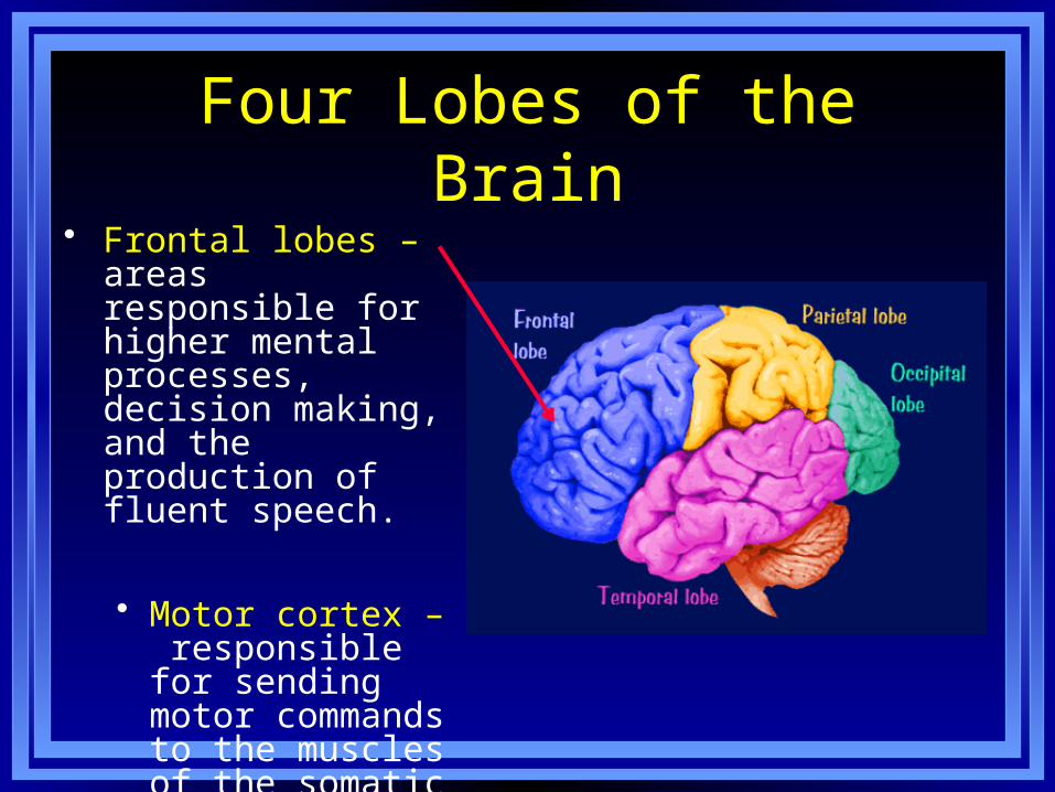
Four Lobes of the Brain• Frontal lobes – areas
responsible for higher mental processes, decision making, and the production of fluent speech.
• Motor cortex – responsible for sending motor commands to the muscles of the somatic nervous system.
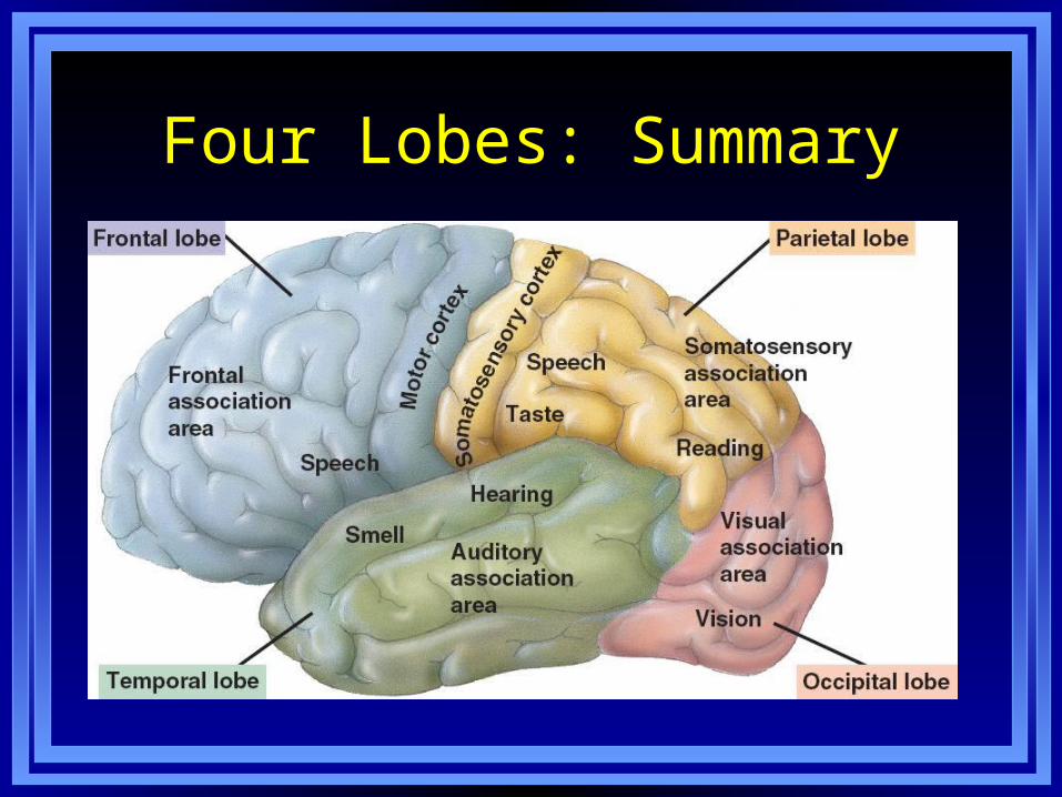
Four Lobes: Summary

Association Areas of Cortex• Association areas- areas responsible
for interpreting information
• Broca’s aphasia – result of damage to Broca’s area-Inability to speak fluently, to mispronounce words, and to speak haltingly.
• http://www.youtube.com/watch?v=Fw6d54gjuvA
• Wernicke’s aphasia – result of damage to Wernicke’s area-Inability to understand or produce meaningful language.
• http://www.youtube.com/watch?v=aVhYN7NTIKU&feature=related
LO 2.9 Parts of cortex responsible for higher thought
Menu
Broca’s aphasia
Wernicke’s aphasia
Association Areas of the Brain
• Spatial neglect - condition produced by damage to the association areas of the right hemisphere
• Inability to recognize objects or body parts in the left visual field.
• http://www.youtube.com/watch?v=ADchGO-0kGo&feature=related
Split Brain Research
• Split brain research
• Study of patients with severed corpus callosum.
• Involves sending messages to only one side of the brain.
• Demonstrates right and left brain specialization.
• http://www.youtube.com/watch?v=aCv4K5aStdU
LO 2.10 Left side and right side of brain
Menu
Results of Split Brain Research• Left side of the brain:
• seems to control language, writing, logical thought, analysis, and mathematical abilities,
• processes information sequentially,
• Right side of the brain• controls emotional expression, spatial perception,
recognition of faces, patterns, melodies, and emotions,
• processes information globally
LO 2.10 Left side and right side of brain
Menu