The association between chemical-induced porphyria and ...
17
Toxicology Research REVIEW Cite this: Toxicol. Res., 2018, 7, 647 Received 12th January 2018, Accepted 4th April 2018 DOI: 10.1039/c8tx00019k rsc.li/toxicology-research The association between chemical-induced porphyria and hepatic cancer Andrew G. Smith * a and John R. Foster b The haem biosynthetic pathway is of fundamental importance for cellular metabolism both for the ery- throid and nonerythroid tissues. There are several genetic variants of the pathway in the human popu- lation that cause dysfunction of one or other of the enzymes resulting in porphyrias of varying severity. Serious chronic hepatic and systemic diseases may result. Some of these can be precipitated byexposure to drugs including hormones, barbiturates and antibiotics, as well as alcohol and particular chlorinated aromatic chemicals. In experimental animals some of the steps of this pathway can also be severely disrupted by a variety of environmental chemicals, potential drugs and pesticides, especially in the liver, leading to the accumulation of uroporphyrins derived from the intermediate uroporphyrinogens or proto- porphyrin IX, the immediate precursor of haem. With some of these chemicals this also leads to cholesta- sis and liver cell injury and eventually hepatic tumours. The review evaluates the available evidence linking hepatic porphyria with carcinogenesis in naturally occurring human genetic conditions and in chemically- induced porphyrias in laboratory animals. The existing data showing gender, strain, and species differences in sensitivity to the chemical-induced porphyrias, liver injury and liver tumours are discussed and the role that transgenically altered mouse models have played in defining the varying mechanisms. Finally, the review proposes a novel, unifying hypothesis linking the hepatotoxicity induced by the accumulation of various porphyrins, with the increased risk of developing hepatic cancer as a long term consequence. 1. Introduction The hepatic toxicities of a variety of drugs, pesticides and environmental chlorinated chemicals in animals include marked disturbances of the haem biosynthetic pathway. 1–4 The formation of haem in mammals is an essential pathway accounting for much of the use of iron either for transport and storage of oxygen, or for its use in cytochromes of res- piration, oxygenases and signalling. Most of the haem syn- thesized in the body is used in the bone marrow for haemo- globin and about 10% for intracellular metabolism. However, although virtually all cells require haem, the liver accounts for the greatest amount of non-erythrocyte haem formation, not only for mitochondrial function and steroid, bile acid and prostaglandin synthesis, but particularly in the metabolism of drugs, xenobiotics and plant constituents. The hepatic pathway is very responsive to the demand for haem as in circadian rhythm and in induction of cyto- chrome P450, whilst turnover can be quickly stimulated by the action of inducible haem oxygenase 1 (HMOX1). In humans, dominant or recessive genetic variants of the pathway can lead to porphyrias, malfunctions and accumu- lation of intermediate precursors of haem some of which may be toxic (including oxidized product porphyrins), and have serious clinical outcomes. 5 The genetic penetrance can be very low and the clinical disorder may be precipitated by exogenous factors such as alcohol or by changes in physio- logical demand for haem controlled by hormones or nutri- tion. 4 Some chemicals cause porphyria in rodents similar to types of human porphyrias without the requirement of pre- disposing gene variants of haem synthesis. 3,4 The incidences of liver cancer are higher than expected in surveys of patients with some types of clinical porphyria where the numbers are sufficient for valid studies. Many of the chemicals that cause porphyria in rodents cause eventually hepatocellular tumours. This review focuses on some drugs, herbicides and chlorinated aromatic chemicals that cause both porphyria and liver tumours or precancerous changes in rodents and compares with types of human porphyrias in which associations with increased incidences of liver cancer have been described. a MRC Toxicology Unit, Hodgkin Building, University of Leicester, Lancaster Road, Leicester LE2 4UA, UK. E-mail: [email protected] b ToxPath Sciences Ltd, 1 Troutbeck Avenue, Congleton, Cheshire, CW12 4JA, UK This journal is © The Royal Society of Chemistry 2018 Toxicol. Res. , 2018, 7, 647–663 | 647 Open Access Article. Published on 01 June 2018. Downloaded on 12/7/2021 8:05:47 PM. This article is licensed under a Creative Commons Attribution-NonCommercial 3.0 Unported Licence. View Article Online View Journal | View Issue
Transcript of The association between chemical-induced porphyria and ...
Toxicology Research
REVIEW
Cite this: Toxicol. Res., 2018, 7, 647
Received 12th January 2018,Accepted 4th April 2018
DOI: 10.1039/c8tx00019k
rsc.li/toxicology-research
The association between chemical-inducedporphyria and hepatic cancer
Andrew G. Smith *a and John R. Fosterb
The haem biosynthetic pathway is of fundamental importance for cellular metabolism both for the ery-
throid and nonerythroid tissues. There are several genetic variants of the pathway in the human popu-
lation that cause dysfunction of one or other of the enzymes resulting in porphyrias of varying severity.
Serious chronic hepatic and systemic diseases may result. Some of these can be precipitated by exposure
to drugs including hormones, barbiturates and antibiotics, as well as alcohol and particular chlorinated
aromatic chemicals. In experimental animals some of the steps of this pathway can also be severely
disrupted by a variety of environmental chemicals, potential drugs and pesticides, especially in the liver,
leading to the accumulation of uroporphyrins derived from the intermediate uroporphyrinogens or proto-
porphyrin IX, the immediate precursor of haem. With some of these chemicals this also leads to cholesta-
sis and liver cell injury and eventually hepatic tumours. The review evaluates the available evidence linking
hepatic porphyria with carcinogenesis in naturally occurring human genetic conditions and in chemically-
induced porphyrias in laboratory animals. The existing data showing gender, strain, and species
differences in sensitivity to the chemical-induced porphyrias, liver injury and liver tumours are discussed
and the role that transgenically altered mouse models have played in defining the varying mechanisms.
Finally, the review proposes a novel, unifying hypothesis linking the hepatotoxicity induced by the
accumulation of various porphyrins, with the increased risk of developing hepatic cancer as a long term
consequence.
1. Introduction
The hepatic toxicities of a variety of drugs, pesticides andenvironmental chlorinated chemicals in animals includemarked disturbances of the haem biosynthetic pathway.1–4
The formation of haem in mammals is an essential pathwayaccounting for much of the use of iron either for transportand storage of oxygen, or for its use in cytochromes of res-piration, oxygenases and signalling. Most of the haem syn-thesized in the body is used in the bone marrow for haemo-globin and about 10% for intracellular metabolism.However, although virtually all cells require haem, the liveraccounts for the greatest amount of non-erythrocyte haemformation, not only for mitochondrial function and steroid,bile acid and prostaglandin synthesis, but particularly in themetabolism of drugs, xenobiotics and plant constituents.The hepatic pathway is very responsive to the demand forhaem as in circadian rhythm and in induction of cyto-
chrome P450, whilst turnover can be quickly stimulated bythe action of inducible haem oxygenase 1 (HMOX1). Inhumans, dominant or recessive genetic variants of thepathway can lead to porphyrias, malfunctions and accumu-lation of intermediate precursors of haem some of whichmay be toxic (including oxidized product porphyrins), andhave serious clinical outcomes.5 The genetic penetrance canbe very low and the clinical disorder may be precipitated byexogenous factors such as alcohol or by changes in physio-logical demand for haem controlled by hormones or nutri-tion.4 Some chemicals cause porphyria in rodents similar totypes of human porphyrias without the requirement of pre-disposing gene variants of haem synthesis.3,4
The incidences of liver cancer are higher than expected insurveys of patients with some types of clinical porphyria wherethe numbers are sufficient for valid studies. Many of thechemicals that cause porphyria in rodents cause eventuallyhepatocellular tumours. This review focuses on some drugs,herbicides and chlorinated aromatic chemicals that causeboth porphyria and liver tumours or precancerous changes inrodents and compares with types of human porphyrias inwhich associations with increased incidences of liver cancerhave been described.
aMRC Toxicology Unit, Hodgkin Building, University of Leicester, Lancaster Road,
Leicester LE2 4UA, UK. E-mail: [email protected] Sciences Ltd, 1 Troutbeck Avenue, Congleton, Cheshire, CW12 4JA, UK
This journal is © The Royal Society of Chemistry 2018 Toxicol. Res., 2018, 7, 647–663 | 647
Ope
n A
cces
s A
rtic
le. P
ublis
hed
on 0
1 Ju
ne 2
018.
Dow
nloa
ded
on 1
2/7/
2021
8:0
5:47
PM
. T
his
artic
le is
lice
nsed
und
er a
Cre
ativ
e C
omm
ons
Attr
ibut
ion-
Non
Com
mer
cial
3.0
Unp
orte
d L
icen
ce.
View Article OnlineView Journal | View Issue

2. Interference of haem synthesis bychemicals
The biosynthesis of haem involves eight steps in which simpleprecursors from the tricarboxylic acid (TCA) cycle are built intothe complex macrocyclic molecule, protoporphyrin IX, intowhich iron is then incorporated (Fig. 1). Both in the liver andred blood cells, the first step is the condensation betweenglycine and succinyl CoA to give 5-aminolaevulinate (5-ALA).Condensation differs between non-erythroid and erythroidtissue being catalyzed by different 5-aminolaevulinate synthe-tase enzymes (ALAS1 and ALAS2) regulated by available haemor iron pools respectively.6 Particular enzymes of the haempathway in the liver are affected by a variety of chemicals bothdirectly and indirectly (Fig. 1 and Table 1). Some are suscep-tible to direct inhibition by metal ions, drugs, herbicides ortheir metabolites. Other enzymes are inhibited by xenobioticmetabolites generated endogenously from haem precursorsafter stimulation of xenobiotic responsive pathways, some ofwhich still need a more complete understanding. Inhibition ofa particular enzyme can lead to accumulation of intermediateporphyrinogens, and their oxidized product porphyrins, andcan be compounded by feedback stimulation of the rate con-trolling enzyme ALAS1. When inhibition of the haem pathwayoccurs, it is the pathological accumulation of hepatic porphyr-ins in rodent liver, by some of these drugs and other chemi-cals, that is causal in inducing hepatic toxicity. The toxicitymay not be due entirely to the porphyrins per se; porphyriaand liver injury may also be the outcome of common mecha-nisms, probably of an oxidative nature. While carcinogenicitystudies have been carried out for some of these porphyrogenic
chemicals, many may not have been tested in cancer studies.Most of the porphyrogenic chemicals that have been shown tocause liver tumours in rodents are associated with hepatic tox-icity and are thought to be carcinogenic by non-genotoxicmechanisms, perhaps as a consequence of their cellular tox-icity, of which the key initiating events (KIE) may have beenhypothesized but not yet elucidated.
3. Hepatocellular cancer in porphyriapatients
All but one of the enzyme steps of non-erythrocyte haem bio-synthesis are associated in humans with rare, but clinicallyestablished, phenotypes of porphyria or related disorderswhich are caused by recessive variants, or autosomal dominantvariants, triggered by endogenous or exogenous factors.4,5 Adisorder associated with genetic variants of ALAS1 is notknown, possibly because either it would be lethal, or becauseof the potential for the gene to be upregulated to compensatefor any deficiency in enzyme activity (Fig. 1). For some types ofporphyria, particularly acute intermittent porphyria (AIP), andporphyria cutanea tarda (PCT), higher incidences of primaryliver cancer in patients than a reference population have longbeen reported from different countries.7–10
In patients with acute hepatic porphyrias, AIP, variegateporphyria (VP) and hereditary coproporphyria (HCP) (due tovariants in the HMBS, PPOX, and CPOX, genes respectively, seeFig. 1), acute neurovisceral attacks are triggered by theaccumulation of porphyrin precursors in the circulation. Manyindividuals carrying these mutations will remain asympto-matic but can be triggered into disease after physiologicalstimuli such as changes in hormones or stress, or followingthe administration of porphyrogenic drugs such as sex hor-mones, barbiturates, and sulfonamide antibiotics. The acuteattacks are probably associated with the neurotoxicity of highlyelevated levels of 5-ALA produced in the liver.5,11–13 Howeverporphobilinogen, uroporphyrin III, coproporphyrin III andprotoporphyrin IX, derived from the malfunctioning haempathway, also accumulate in the liver depending on the type ofacute porphyria. In VP and HCP these haem precursors maycontribute to the liver injury, but they also provoke dermalphotosensitivity due to deposition in the skin.
Over the last few decades it has become clear that the acutehepatic porphyrias are associated with a markedly increasedrisk of patients developing primary liver cancer (HCC), at leastfor Northern European countries and without underlying cir-rhosis. This was first described for AIP from northern Sweden9
and now confirmed in a number of other independent studiesof not only AIP but also VP and HCP in countries such asSweden, Norway, France and Switzerland. Investigations onFinnish and Swedish cohorts with AIP reported increased riskratios for HCC compared to reference populations of up toabout 100 times. It is unclear why reports are only fromEuropean countries but this may simply reflect health dataorganizational arrangements and clinical interest.14–20 Study
Fig. 1 Nonerythrocyte haem synthesis showing enzymic steps that areaffected by chemicals and drugs in vivo leading to malfunctions of haemproduction. ALAS1, 5-aminolevulinic acid synthase; ALAD, aminolevulinicacid dehydratase; HMBS, 1-hydroxymethybilane synthase; UROS, uro-porphyrinogen III synthase; UROD, uroporphyrinogen III decarboxylase;CPOX, coproporphyrinogen III oxidase; PPOX protoporphyrinogen IXoxidase; FECH, ferrochelatase; HMOX1, haem oxygenase 1. AIP, acuteintermittent porphyria; HCP, hereditary coproporphyria; PCT, porphyriacutanea tarda; VP, variegate porphyria.
Review Toxicology Research
648 | Toxicol. Res., 2018, 7, 647–663 This journal is © The Royal Society of Chemistry 2018
Ope
n A
cces
s A
rtic
le. P
ublis
hed
on 0
1 Ju
ne 2
018.
Dow
nloa
ded
on 1
2/7/
2021
8:0
5:47
PM
. T
his
artic
le is
lice
nsed
und
er a
Cre
ativ
e C
omm
ons
Attr
ibut
ion-
Non
Com
mer
cial
3.0
Unp
orte
d L
icen
ce.
View Article Online

Tab
le1
Summaryofch
emicalsan
dhuman
geneticco
nditionsasso
ciatedwiththeproductionofhepatic
porphyria
andtheirlin
kto
liverca
nce
r
Chem
ical
Mainpo
rphyrin
involved
aNuc
lear
receptor?
Molecular
MOA
Hep
atotoxicity
Species
Carcinog
enic
resp
onse
(incide
nce)
Ref.
Rod
entch
emical
porphyrog
en3-[2-(2,4,6-Trim
ethylph
enyl)-
thioethyl]-4
-methylsydn
one
(TTMS)
Protop
orph
yrin
IXCAR/PXR
Ferrochelataseinhibition
Wt↑;inflam
mation;n
ecrosis;
hep
atocellularhyp
ertrop
hy
Mou
seUnkn
own
87an
d88
Tralko
xydim
Protop
orph
yrin
IXCAR/PXR
Ferrochelataseinhibition
Wt↑;inflam
mation;n
ecrosis;
hep
atocellularhyp
ertrop
hy;bile
duct
hyp
erplasia;p
orph
yrin
Mou
se≫
rat=
ham
ster
≫hum
an
(−ve
rat,ham
ster)
Not
condu
cted
inmou
se!
87,9
4an
d10
0
Griseofulvin
Protop
orph
yrin
IXCAR/PXR
Ferrochelataseinhibition
Wt↑;inflam
mation;n
ecrosis;
hep
atocellularhyp
ertrop
hy;
porphyrin
Mou
seM
>F⋙
hum
an∼90
%mou
se(−ve
inrat,ham
ster)
85,8
6,98
,10
4an
d10
53,5-Diethoxycarbo
nyl-1,4-
dihyd
ro-2,4,6-trimethylpyridine
(DDC)
Protop
orph
yrin
IXCAR/PXR
Ferrochelataseinhibition
Wt↑;inflam
mation;n
ecrosis;
hep
atocellularhyp
ertrop
hyin
mou
se,rat
andch
ick,
porphyrin
Rat
=mou
se=
chick
Unkn
own
2,80
and
81
Fomesafen
Protop
orph
yrin
IXN/A
Inhibitionof
PPOX
ALT
↑liverwt↑
inmou
se;p
orph
yrin
Mou
seICR
Yes;mou
se(16%
HCC;1
00%
AHF)
77
Human
&roden
tch
emical
porphyrog
enPo
lych
lorinated
biph
enyls
(PCBs)
Uropo
rphyrin
AHR
agon
ist
Inhibitionof
UROD
Wt↑;inflam
mation;n
ecrosis,
hyp
ertrop
hy;po
rphyrin
Rat
=mou
se≫
hum
anYe
s;mou
se>rat
36an
d56
2,3,7,8-Te
trachorod
iben
zo-p-
dioxin
(TCDD)
Uropo
rphyrin
AHR
agon
ist
Inhibitionof
UROD
Wt↑;inflam
mation;n
ecrosis,
hyp
ertrop
hy;po
rphyrin
Rat
=mou
se≫
hum
anYe
s;mou
se>rat
3,34
,35,
54an
d12
1Hexachlorobe
nzene(H
CB)
Uropo
rphyrin
AHR
agon
ist?
Inhibitionof
UROD
Wt↑;inflam
mation,n
ecrosis;
hyp
ertrop
hy;po
rphyrin
Rat
=mou
se≫
Hum
anYe
s;mou
se>rat
37–39,
54an
d12
2Human
genetic
disorders
Acu
teinterm
ittentpo
rphyria
(AIP)
Porphob
ilinog
enN/A
Hyd
roxymethylbilane
synthase(H
MBS)
deficien
cyDrug-indu
cedsevere
hep
atitis
Hum
anYe
s9an
d14
–21
Porphyria
cutanea
tarda(PCT)
Uropo
rphyrin
N/A
Uropo
rphyrinog
ende
carboxylase(U
ROD)
deficien
cy
Mild-mod
eratehep
atitis
Hum
anYe
s(5–12%
ofpa
tien
ts)
10an
d25
–31
Variegatepo
rphyria
(VP)
Porphob
ilinog
enN/A
Protop
orph
yrinog
enoxidase
(PPO
X)d
eficiency
Mod
eratehep
atitis
Hum
anYe
s21
Erythropo
ieticpo
rphyria
(EPP
)Protop
orph
yrin
IXN/A
Ferrochelatase(FECH);less
common
ly5-am
inolevulinic
acid
synthase-2(ALA
S2)
Drug-indu
cedhep
atitis
Hum
anYe
s5,
78an
d11
7
Hered
itarycoprop
orph
yria
(HCP)
Porphob
ilinog
en;
coprop
orph
yrin
N/A
Deficiency
incoprop
orph
yrinog
enoxidase
(CPO
X)
Drug-indu
cedhep
atitis
Hum
anYe
s5,
11an
d13
aDoe
snot
includ
eelevationof
plasmaan
dhep
atic
5-ALA
whichmay
besu
bstantial
insomeexpe
rimen
tala
ndhum
anacutepo
rphyrias.N/A
Not
applicab
le.
Toxicology Research Review
This journal is © The Royal Society of Chemistry 2018 Toxicol. Res., 2018, 7, 647–663 | 649
Ope
n A
cces
s A
rtic
le. P
ublis
hed
on 0
1 Ju
ne 2
018.
Dow
nloa
ded
on 1
2/7/
2021
8:0
5:47
PM
. T
his
artic
le is
lice
nsed
und
er a
Cre
ativ
e C
omm
ons
Attr
ibut
ion-
Non
Com
mer
cial
3.0
Unp
orte
d L
icen
ce.
View Article Online

of an AIP and of a VP patient has suggested that as well as theunderlying HMBS and PPOX germline mutations, the hepatictumour tissue also had second-hit somatic mutations in therespective genes.21 Whether these second mutations occurredincidentally or were instrumental in the subsequent develop-ment of the hepatic cancer is unknown and their role in thechronic elevation of heme precursors such as 5-ALA, porphobi-linogen and porphyrins, remains unclear. It is possible that5-ALA is pro-oxidant and genotoxic in hepatocytes22 butperhaps other mechanisms may operate such as dysfunctionof the mitochondria if haem synthesis is severely disturbed.What does seem clear is that, like clinical AIP, the develop-ment of HCC is more likely to occur in women than in men. Inaddition, unlike most cases of HCC, the incidence of cirrhosisis low and without other major findings of underlying liverdisease in these porphyric patients.23
Type 1 tyrosinemia is a genetic disease of tyrosine metab-olism in which fumarylacetoacetate hydrolase (FAH) is ineffi-cient and some of the neurological and other symptomsresemble those of AIP. Side reactions of accumulating fumaryl-acetoacetate and maleylacetoacetate form succinylacetone, apotent inhibitor of aminolevulinate dehydratase (ALAD)(Fig. 1). Patients can be treated with nitrisinone (2-(2-nitro-tri-fluoro-methylbenzoyl-1,3-cyclohexadione) originally developedas a pesticide) restricting precursors for FAH.136 If untreatedthere is a high incidence of liver damage and HCC in thesepatients.137
PCT patients show marked inhibition of hepatic uropor-phyrinogen decarboxylase activity (UROD) with mild or moder-ate liver damage and an increased susceptibility to developingliver cancer (Fig. 1). Under uv light the uroporphyrins andother porphyrins arising from disruption of the haem pathwayfluoresce red in the liver due to their deposition. Porphyrinsreleased into circulation are deposited elsewhere in the bodyand can cause marked cutaneous photosensitivity. Unlike theacute porphyrias there is no marked rise in plasma or urinary5-ALA and no neurotoxicity. Autosomal dominant inheritancewith low penetrance of a UROD gene variant is a contributingfactor for familial form of PCT. However, most patients have asporadic form with no apparent association with adverse var-iants of the gene.5 Consumption of alcohol, oestrogenic drugs,other chemicals and hepatitis viruses have all been reported asprecipitating agents for PCT, even for the familial form whichmay occur at a younger age.3 Most of the PCT patients are diag-nosed at middle age or older. This is the most common type ofclinical porphyria in many populations. Iron metabolism isimplicated in the pathogenesis of PCT and depletion of bodyiron stores is one treatment approach. For some patientcohorts inheritance of the haemochromatosis HFE variant hasbeen a strong risk factor in the development of the por-phyria.24 The role of iron is not really understood, other thanpossibly promoting reactive oxygen species, but must clearlybe the consequence of genetic variants in iron mobilizationperhaps releasing Fe2+. This has been discussed in more detailelsewhere.3 A disorder very similar to PCT occurred in manypeople, particularly children, poisoned by hexachlorobenzene
(HCB) and there is evidence that other highly chlorinated aro-matic chemicals such as the 2,3,7,8-tetrachorodibenzo-p-dioxin(TCDD) and polychlorinated biphenyls (PCBs) have a similarpotential.3
As with the acute porphyrias, there is clear evidence of anassociation between PCT and the risk of developingHCC.10,25–30 In one of these studies, 342 patients with PCTwere observed over a 15-year period, 5% of them died fromprimary liver cancer.31 In another study, 6 out of 78 patientswith HCC had PCT. Most studies have confirmed the earlyfindings while others have been less positive, probablybecause of the complex nature of PCT development, and thepossible confounding role of concurrent cirrhosis. Recognitionof PCT and subsequent treatment may be resulting in adecrease in the likelihood of patients progressing to HCC.However, the subject is of interest in consideration of the riskassessment of cancer associated with exposure to environ-mental levels of polychlorinated aromatic chemicals.
4. Hepatic tumours in rodentscaused by porphyria-inducingchemicals
Although metals such as lead and mercury are known to inter-fere in haem synthesis, both in liver and in erythrocytes, theresulting clinical chemical signs from human and experi-mental studies are usually porphyrinurias, frequentlyincreased coproporphyrin I or III, or excretion in stool of proto-porphyrin rather than overt porphyrias.1 Sometimes thesereflect relatively mild disturbances of haem synthesis. Whatappears to be especially important in determining the onset,or otherwise, of hepatic porphyrias is the particular enzyme inthe pathway that is affected and permits the accumulation oftoxic haem precursors. In lead poisoning there is inhibition ofhaem biosynthesis but the metal has been shown to inhibitALA dehydratase resulting in increased urinary 5-ALA withoutaffecting porphobilinogen or resulting in the hepatic build-upof toxic porphyrins. Hence while lead is known to cause por-phyrinuria with an increased excretion of coproporphyrins, itsinability to inhibit markedly later enzymes prevents the onsetof hepatic porphyrias and their dire consequences.135 Drugsand chemicals that trigger AIP in humans do not do so inanimals without genetic models particularly the disruptedHMBDS gene.32,33
Some of the chemicals and drugs known to cause disturb-ance of hepatic haem synthesis in animals, and sometimes inhumans, induce not only overt porphyrias in rodents but alsoinduce hepatocellular cancer, or forms of chronic liver injurywhich might predispose to cancer.
4a. Chemicals causing hepatic uroporphyria
HCB, dioxins and PCBs have been known for many years tocause hepatic symptoms in rodents similar to human PCTwith inhibition of UROD (probably through an oxidative
Review Toxicology Research
650 | Toxicol. Res., 2018, 7, 647–663 This journal is © The Royal Society of Chemistry 2018
Ope
n A
cces
s A
rtic
le. P
ublis
hed
on 0
1 Ju
ne 2
018.
Dow
nloa
ded
on 1
2/7/
2021
8:0
5:47
PM
. T
his
artic
le is
lice
nsed
und
er a
Cre
ativ
e C
omm
ons
Attr
ibut
ion-
Non
Com
mer
cial
3.0
Unp
orte
d L
icen
ce.
View Article Online

sequence involving oxidation of uroporphyrinogen),34,35 por-phyria and liver injury.3 As with human patients there isstrong evidence for a synergistic role of iron metabolism, andgenetic predispositions. One key mechanistic aspect to thesechemicals for porphyria seems to be through their binding to,and activation of, the aryl hydrocarbon receptor (AHR) withassociated induction of hepatic CYP1A enzymes.3 For chemi-cals, such as chlorinated dioxins and dioxin-like PCB constitu-ents of commercial PCB mixtures such as Aroclor 1254 and1260, porphyria potency can be ascribed partly to the toxicityequivalence compared to the prototype AHR ligand, TCDD.36
However, for HCB the evidence is less clear.37 Hahn et al. con-cluded that porphyria induced by HCB is caused by weak AHRbinding activity of the chemical38,39 and a genetic locus forsusceptibility in mice corresponds to variants of the Ahrgene.40 World Health Organization views did not regard HCBas an AHR ligand and noted that the chemical could be con-taminated by dioxins.36 This has been a four decade debate. Infact, the particular commercial HCB source used by manyresearch laboratories for porphyria studies was chosen orig-inally for its known high purity as a chemical standard41,42
and shown to have CYP1A inducing properties in rodents.43,44
Up to now the levels of contaminants present in this sourcehave not been fully reported. The levels of TCDD equivalentsdue to PCDDs and PCDFs present in this HCB are now shownin Table 2. Comparison with previous porphyria experimentssuggest the levels are too low to account significantly forin vivo hepatic porphyrogenic activity of HCB in rodents andthat probably it can act as a weak AHR ligand.
In subchronic studies in rats HCB, TCDD, PCB and PBBmixtures have all shown a marked preponderance of femalescompared to males in inhibition of hepatic UROD activity anddeveloping porphyria.45,46 Interestingly, this marked sexualdimorphism in porphyria also occurs in chronic studies ofthe development of liver tumours with these chemicals(Fig. 2).47–53 This is in contrast to a range of other genotoxicand nongenotoxic hepatocarcinogens. With HCB, the hepato-toxicity gives rise to peliosis and necrosis with haemosiderosiswhich may increase tumourgenic potential.54 The developmentof hepatocellular cancer in rats has been of some interest forthe risk assessment of polyhalogenated aromatic chemicalssuch as TCDD and PCBs. The promotion of rodent liverhepatic carcinogenesis by these chemicals has been investi-gated and discussed in detail55–57 but the reason for initiationof tumourgenesis has not been explored fully although anumber of earlier hypotheses sought to explain the predomi-nance of tumours in females. Initiation with diethylnitrosamine(DEN) followed by TCDD or other polyhalogenated chemicalshas often been used to model the complete neoplasticprocess.57 Comparison of hepatic tumours in the rat, caused byHCB, with hepatic tumours from rats first given an initiatingdose of DEN followed by a promoting regimen of HCB, showedmarkedly different results. There was no consistent sex differ-ence in timing or severity with the initiation-promotionregimen.58 When tissue from the two regimens was comparedunder uv light for uroporphyrin fluorescence as a consequence
of hepatic porphyria the distribution of porphyrins betweentumour and non tumour areas were very different (Fig. 3).
Given the evidence for a role of iron metabolism in PCT itwas of interest that HCB porphyria in female rats seemed to beassociated with iron status,47,59 and also CYP1A2 inducibil-ity.46 Iron overload moderately enhanced hepatic carcinogen-esis induced by HCB alone.60 In other studies with aflatoxinB1 and DEN, iron overload had no, or a reducing, effect on theincidence of liver carcinogenesis in rats (A. G. Smith, unpub-lished data). Thus rat studies might suggest a link betweenporphyria, perhaps inducing cytotoxicity, and liver cancercaused by polyhalogenated aromatic chemicals, and whichmay have a parallel in human PCT.
In mice, the development of porphyria by TCDD, PCBs andHCB was found to be influenced profoundly by hepatic ironstatus during experiments designed to explore mechanisticmodels of human PCT.3 Administration of iron not onlyenhanced porphyria, and hepatic toxicity, in mice with an AHRshowing high affinity for TCDD, but overcoming resistance insome mouse strains with low affinity AHR.61,62 Subsequentstudies demonstrated that there are genetic loci in mice inde-pendent of the AHR gene that contribute to the development
Table 2 Concentrations of polychlorinated dibenzo-p-dioxins (PCDDs)and polychlorinated dibenzofurans (PCDFs) in Organic Standard gradehexachlorobenzene (HCB) a source used in many published experi-mental studiesa
PCDDs or PCDFs in HCB
ConcentrationWHO TEF(2005)
TEQconcentration
ng g−1 ofsolid
ng g−1 ofsolid
2,3,7,8-TCDD 88.00 1 88.01,2,3,7,8-PeCDD <0.59 1 0.61,2,3,4,7,8-HxCDD <0.59 0.1 0.61,2,3,6,7,8-HxCDD <0.94 0.1 0.11,2,3,7,8,9-HxCDD <0.71 0.1 0.11,2,3,4,6,7,8-HpCDD 18.94 0.01 0.2OCDD 16 011 0.0003 4.8
2,3,7,8-TCDF <1.03 0.1 0.11,2,3,7,8-PeCDF <0.47 0.03 0.012,3,4,7,8-PeCDF <4.35 0.3 0.11,2,3,4,7,8-HxCDF 2.82 0.1 0.31,2,3,6,7,8-HxCDF 1.76 0.1 0.21,2,3,7,8,9-HxCDF <1.06 0.1 0.12,3,4,6,7,8-HxCDF <1.41 0.1 0.11,2,3,4,6,7,8-HpCDF 1060 0.01 10.61,2,3,4,7,8,9-HpCDF 30.6 0.01 0.3OCDF 5978 0.0003 1.8
Total 108
aOrganic Standard grade HCB for elemental analyses was from B.D.H.Ltd, UK. and analysed for A.G.S. (2005) at the Central ScienceLaboratory, DEFRA, York, UK by high resolution gas chromatographywith high resolution mass spectrometry for PCDDs and PCDFs using13C-labelled analogues for standardisation. Results were UKAS accre-dited. WHO Toxic Equivalent Factors (TEFs)36 were used to calculateToxic equivalents to TCDD (TEQs) with upper values of ranges.Comparison of TEQs present in HCB with doses of TCDD that causeporphyria in rats and mice showed levels were insufficient to accountfor the responses.46,59,133,134
Toxicology Research Review
This journal is © The Royal Society of Chemistry 2018 Toxicol. Res., 2018, 7, 647–663 | 651
Ope
n A
cces
s A
rtic
le. P
ublis
hed
on 0
1 Ju
ne 2
018.
Dow
nloa
ded
on 1
2/7/
2021
8:0
5:47
PM
. T
his
artic
le is
lice
nsed
und
er a
Cre
ativ
e C
omm
ons
Attr
ibut
ion-
Non
Com
mer
cial
3.0
Unp
orte
d L
icen
ce.
View Article Online

of porphyria and hepatic toxicity.63 The genes responsible havenot yet been identified but we would suggest some of them areassociated with iron mobilization and use. Importantly, inchronic experiments with the AHR responsive strain C57BL/10ScSn, not only were the porphyria and hepatic toxicity indu-cing properties of HCB and PCBs markedly potentiated byprior iron treatment but so too was the subsequent incidenceof liver adenomas and cancer to a marked degree.64,65 In con-trast, the low affinity AHR DBA/2 strain also possessing otherresistant loci was highly refractory to both porphyria and
hepatocarcinogenesis following iron and PCBs. CYP1A2 genenull C57BL/6J mice with no CYP1A2 expression (mostly undercontrol of the AHR) are highly resistant to porphyria caused byTCDD and HCB, even after iron loading pretreatment.66,67 Theexact role of CYP1A2 in the porphyria mechanisms is stillunclear although postulated to be in uroporphyrinogenoxidation. CYP1A2 knockout mice did not develop hepato-cellular foci or adenomas following treatment with PCBs68
Octachlorostyrene, which only causes mild porphyria in iron-loaded C57BL10ScSn mice, was only weakly tumourgenic incontrast to HCB in the same mouse strain.69 Other nongeno-toxic carcinogens in mice such as phenobarbital and nafeno-pin, were unaffected by iron pretreatment. Hepatocellulartumors resected from mice following treatment with HCB andPCBs did not show increased incidences of Ha-ras mutationsbut did show the emergence of a mononucleated diploid popu-lation consistent with precancerous changes.70,71 Tumourexplants derived from the induced hepatocellular cancerscould be passaged subcutaneously many times in syngeneicmice and showed many similarities whether of HCB or PCBorigins. Homozygous knockout of the P53 gene in mice hadlittle influence on the liver tumour incidence. Uroporphyria inrodents and PCT are likely due to an oxidative mechanism.Increased oxidative DNA damage was detected in C57BL/10ScSn mice after treatment with PCBs.3,72 Point mutationscould not be detected using an in vivo mutation systemsuggesting that more complex chromosomal deletions orrearrangement are involved in the oxidative damage or in themutagenicity of the porphyrins.73 However, other mechanismsassociated with disturbed production of haem could beinvolved but are yet to be understood.
Fig. 2 Incidences of liver tumours in toxicological studies of male andfemale rats. (a). Selection of some genotoxic and nongenotoxiccarcinogens.126–131 (b). Polyhalogenated aromatic chemicals that alsocause hepatic porphyria.47–53 Additional references of hepatocarcino-gens can be found in Smith (2003).139 DEN, diethylnitrosamine, FA orAAF, N-2-fluorenylacetamide or 2 acetylaminofluorene; 3-Me-DAB,3’-methyl-4-dimethylaminoazobenzene, tamoxifen and aflatoxin maybe regarded as genotoxic carcinogens; DDT and phenobarbital may beregarded as nongenotoxic.
Fig. 3 Difference between liver tumours in female rats caused by HCBalone and in those initiated with diethylnitrosamine (DEN) and promotedwith HCB. (a) Female rats were fed HCB in diet (0.02%) for 90 weeks.47
(b) Female rats received DEN in the drinking water (0.015%) for 3 weeksthen after 2 weeks’ rest were administered HCB in the diet (0.02%) for30 weeks.58 Sections of liver were examined under uv light to demon-strate red fluorescence of uroporphyrin. Studies were conducted orig-inally under the authorizations of UK Home Office regulations in theMRC toxicology unit.
Review Toxicology Research
652 | Toxicol. Res., 2018, 7, 647–663 This journal is © The Royal Society of Chemistry 2018
Ope
n A
cces
s A
rtic
le. P
ublis
hed
on 0
1 Ju
ne 2
018.
Dow
nloa
ded
on 1
2/7/
2021
8:0
5:47
PM
. T
his
artic
le is
lice
nsed
und
er a
Cre
ativ
e C
omm
ons
Attr
ibut
ion-
Non
Com
mer
cial
3.0
Unp
orte
d L
icen
ce.
View Article Online

4b. Protoporphyrinogen oxidase inhibitors
Some chemicals that are inhibitors of protoporphyrinogen IXoxidase (PPOX) in green plants have been formulated as herbi-cides for post emergence weeds.74 Early products were diphe-nyl ethers, although later generation herbicides are oftenbased on non-oxygen bridge compounds. The protoporphyri-nogen that accumulates in an unregulated way is light sensi-tive and is oxidized chemically, rather than enzymically, to pro-toporphyrin IX which may catalyse oxygen free radical-induceddamage in the photosynthetic plant cell. These herbicides canhave similar inhibitory properties in mammals and althoughthe protoporphyrin IX formed in the liver is not exposed tolight, it still has hepatotoxic potential. The toxicity of thesePPOX inhibitors in animals might be considered as havingsome similarities with variegate porphyria (Fig. 1) but the riskto humans from herbicide use has been considered to be verylow due to their low exposure, rapid metabolism and excretion.However, chronic administration to rodents of a diphenylether PPOX inhibitor, fomesafen, has been studied particularlywith respect to its possible mammalian porphyria inducingproperties and toxicity.75,76 Interestingly, in iron treatedC57BL/6 mice fomesafen enhanced preneoplastic changes inthe liver such as frequency of altered foci and nodules.77
4c. Drugs and pesticides that inhibit ferrochelatase
In humans with erythropoietic porphyria (EPP) the enzymeferrochelatase that catalyzes the incorporation of iron intoprotoporphyrin IX is compromised and some patients havesevere hepatic disease probably reflecting mostly the depo-sition of toxic erythrocytic protoporphyrin from the blood intothe liver (Table 1).5,78 There are several chemicals that havebeen shown to induce a similar porphyria in rodents andother animals through inhibition of hepatic ferrochelatase(Fig. 1). These chemicals are of a variety of types but arecommon in their ability to convert the haem in some liver cyto-chrome P450s (CYPs) into N-alkylated porphyrins which canbe strong inhibitors of ferrochelatase.2,79 As well as porphyriathese drugs and pesticides often cause cholestasis and hepatictoxicity in animals, which may lead to neoplastic lesions, butthis can be species specific and has been the subject of riskassessment concerns.
The generalized molecular mechanism of induced proto-porphyria with these types of chemicals involves suicidal inac-tivation of cytochrome P450 with loss of associated enzymeactivities (Fig. 4).2,80,81,94 Reactive metabolites from the parentmolecules bind covalently to the prosthetic haem of the cyto-chrome through N-alkylation of one of the pyrrole moieties.The alkylated haem then dissociates from the apoprotein andthe iron atom is liberated to yield N-alkylprotoporphyrin IXcompounds (Fig. 5). The subsequent loss of haem results in afeedback upregulation of hepatic ALAS1.82,83 Stimulation ofthis enzyme drives increased haem synthesis compoundingthe inhibition of ferrochelatase and adding to the subsequenthepatic accumulation of porphyrin. In porphyria induced by3,5-diethoxycarbonyl-1,4-dihydro-2,4,6-trimethylpyridine (DDC),
cytochrome P450 mediated metabolism generates a metabolitefrom which a 4-methyl substituent is transferred onto thepyrrole nitrogen atom of the haem molecule and the alkylatedproduct, N-methyl protoporphyrin, is a powerful inhibitor offerrochelatase.79,84 In mice fed (1% of diet) the antifungal
Fig. 4 Effect of a porphyrinogenic agent, an anti-epileptic synapticvesicle 2a (SV2a) ligand, 2-[4-(2-chloro-2,2-difluoroethyl)-2-oxopyrroli-din-1-yl]butanamide, in causing depressed activity of hepatic pheno-barbital-type CYP2B family enzyme in the dog. Data are means from4 male and 4 female dogs following 4 weeks’ administration. Redrawnfrom Nicolas et al. (2014).94 CYP2B11 was induced by the prodrug but atthe highest dose given reduced enzyme activity was observed (but notthe apoprotein by immunoblot). At the highest dose ferrochelatase wasinhibited, hepatic protoporphyrin IX levels were highly elevated,N-alkylated porphyrins were detected, and hepatic transaminases in theplasma were markedly elevated indicating liver injury. Rats dosed witheven higher amounts of the ligand (up to 1000 mg kg−1 day−1) showedmuch higher induction of CYP2B enzyme activities but no decrease atthe highest dose and only 2-fold increase in porphyrins and very smallelevations of plasma transaminases.
Fig. 5 Effect of the ferrochelatase inhibiting porphyrinogenic agents,3,5-diethoxycarbonyl-1,4-dihydro-2,4,6-trimethylpyridine (DDC) andallylisopropyl acetamide (AIA) on the N-alkylated porphyrin content ofisolated rat hepatocytes. PB = the effect of pre-dosing in vivo withphenobarbital on the production of N-alkylated porphyrins by increasingcytochrome P4502B content. PB + Haem = the effect of pre-exposurewith phenobarbital and haematin (to provide additional source of haem)on the production of N-alkylated porphyrins. Drawn from data in DeMatteis et al. (1982).132
Toxicology Research Review
This journal is © The Royal Society of Chemistry 2018 Toxicol. Res., 2018, 7, 647–663 | 653
Ope
n A
cces
s A
rtic
le. P
ublis
hed
on 0
1 Ju
ne 2
018.
Dow
nloa
ded
on 1
2/7/
2021
8:0
5:47
PM
. T
his
artic
le is
lice
nsed
und
er a
Cre
ativ
e C
omm
ons
Attr
ibut
ion-
Non
Com
mer
cial
3.0
Unp
orte
d L
icen
ce.
View Article Online

agent griseofulvin, hepatic ferrochelatase activity wasdecreased to 24% after 3 days. Concomitantly, the ALAS1activity was increased by 6.6 fold.85,86
Other chemicals inducing protoporphyria or causingN-alkylated porphyrins in rodents include the anti-arthriticdrug, 3-[2-(2,4,6-trimethylphenyl)-thioethyl]-4-methylsydnone(TTMS), ethylene, diethylnitrosamine, norethindrone, and theherbicides, tralkoxydim, ATMP, and ETC.87–92
The chemical-induced protoporphyria is associated withsignificant liver weight increases in susceptible animal speciesand although the actual cause of the weight increases has notbeen definitively ascribed, it is most probably multifactorialinvolving a combination of microsomal CYP induction,hepatic inflammation and necrosis, and hepatocellular hyper-trophy.93,94 Dependent upon the degree of hepatopathy (liverpathology), the weight increases are frequently accompaniedby increases in plasma alanine aminotransferase (ALT), alka-line phosphatase, aspartate aminotransferase and, mostlydependent upon the animal species involved, γ-glutamyl trans-ferase.87,94 Although none of these enzyme biomarkers arediagnostic of the porphyria they reflect the degree of liverdamage that may be associated with accumulation of proto-porphyrin. In some studies, elevations in bilirubin can alsobe seen indicating a degree of cholestasis associated withthe porphyrin-induced liver damage.94,95 In mice fed grise-ofulvin characteristic gene expression profiles were associatedwith disruption of lipid metabolism, and the pathologicstates of liver toxicity, inflammation, early fibrosis, andcholestasis.86
Histopathologically the initial accumulation of the proto-porphyrin IX occurs as red “Maltese cross” accumulationswithin the hepatocytes and Kupffer cells. Prolonged dosing ofthe porphyrogenic chemicals leads to progressive accumu-lation within the bile canaliculi between hepatocytes, theappearance of crystalline forms of the protoporphyrin IXwithin distended and hypertrophic bile ducts(Fig. 6a),87,94,96,97 and hepatocyte degeneration and inflam-mation, all of which are most severe in the periportal regionsof the liver lobules (Fig. 6c).87,98 The protoporphyrin IX depos-its appear birefringent under polarized light and display a redfluorescence under ultraviolet light (Fig. 6b and d). Of particu-lar importance, with regard to the subsequent development ofneoplasia by some of these porphyrogenic chemicals, is thatinduction of mitotic activity in the hepatocytes occurs between2 to 5 days following administration of griseofulvin in thefood97 and, while not recorded with other porphyrogenicdrugs, would be expected to have occurred from the increasedliver weight that almost invariably occurs following exposure tohigh doses of these porphyrogenic chemicals. The increase ofmitotic activity in the mouse liver correlates with the increasein liver weight over time reaching 4.2-fold concurrent controlliver weight after 4 to 6 months of feeding of griseofulvin at aconcentration of 2.5% in the diet. Although few in number,where histological investigations are available of humans withporphyria, in PCT the pattern of pathology is similar to thatdescribed in the livers of mice given griseofulvin with the pro-
minent presence of needle-like porphyrin crystals (althoughuroporphyrin not protoporphyrin), hepatocellular necrosis andregenerative hyperplasia.25
While some of these chemicals appear to induce porphyriairrespective of animal species, others show distinct strain,species and sex differences in sensitivity, with griseofulvinbeing more toxic to male than female mouse liver, and tralkox-idim being a potent porphyrogen in the mouse but not in therat or hamster.83,95,99,100 Similarly an antiepileptic, synapticvesicle 2a (SV2a) ligand, drug candidate was found to form aCYP mediated N-alkylprotoporphyrin derivative that inhibitedferrochelatase in the dog but not in the rat nor in humanhepatocytes in vitro (Fig. 4).94 The species difference operatingin this instance is the metabolic ability to generate the reactivemetabolite needed to alkylate protoporphyrin IX in the dog asopposed to the insensitive species. In contrast, the porphyrino-genic drug, DDC, induces porphyria as a result of ferrochela-tase inhibition in rats, mice and chick livers,2 while TTMSinduces porphyria in rats, mice and dogs.87 For those chemi-cals that induce protoporphyria in one or more animal speciesthe outcome appears to be largely, if not solely, determined bythe rate of generation of the appropriate CYP dependenthepatic metabolite.101 Those species that fail to generateappropriate amounts of the alkylated protoporphyrin will notinhibit ferrochelatase leading to protoporphyria. This hasbeen adequately demonstrated for tralkoxydim which inducesprotoporphria in the mouse in vivo, and in mouse hepatocytesin vitro, but not in rat in vivo nor in vitro, nor human hepato-cytes.102 Clearly these species differences are important in con-sideration of extrapolation to human risks.
Fig. 6 Periportal area in liver from a mouse given tralkoxydim for 28days showing distended bile duct with hypertrophic lining mucosa(×20 microscope magnification) (a and b) and bile duct and phagocyticcells with brown pigment accumulation in hepatocytes and phagocyticcells (×40 magnification) (c and d). Viewed by conventional light (a andc) and polarized light demonstrating birefringent red porphyrin crystals(b and d). Liver sections were from studies conducted under the author-izations of UK Home Office regulations.
Review Toxicology Research
654 | Toxicol. Res., 2018, 7, 647–663 This journal is © The Royal Society of Chemistry 2018
Ope
n A
cces
s A
rtic
le. P
ublis
hed
on 0
1 Ju
ne 2
018.
Dow
nloa
ded
on 1
2/7/
2021
8:0
5:47
PM
. T
his
artic
le is
lice
nsed
und
er a
Cre
ativ
e C
omm
ons
Attr
ibut
ion-
Non
Com
mer
cial
3.0
Unp
orte
d L
icen
ce.
View Article Online

In a survey of drug-induced liver injury in humans inTaiwan,103 of a total of 90 847 patients taking one or more fun-gicides, including griseofulvin, eight were reported to havesuffered hepatic injury, necessitating termination, followingtreatment with high doses of griseofulvin. Increased porphyrinhas been reported in humans given high doses of griseofulvinbut it is normally mild in comparison with that seen in themouse model.93 In rare cases where the drug was taken at highdoses over prolonged periods of time liver damage and seriousskin reactions were reported accompanying the protopor-phyria.93 Evidence supporting the drug-induced production ofporphyria in man includes a survey of patients receivinggriseofulvin and compared to a further group who had beenoff treatment for between 2–80 weeks.104 Hence griseofulvinis not recommended for patients suffering pre-existingporphyrias.105
Porphyrins are reported to be generators of reactive oxygenspecies, they affect hepatic redox balance and lead to inductionof the antioxidant defense enzyme systems such that micetreated with griseofulvin show increased activity of glutathionereductase, superoxide dismutase, glutathione-S-transferase, andincreased levels of reduced glutathione and malondialdehyde.In contrast the hepatic activities of glutathione peroxidase andcatalase were reduced following griseofulvin administration.106
Griseofulvin has also been shown to reduce bile flow as a resultof direct liver damage and effects on the biliary system, leadingto the hepatocytes accumulating toxic endogenous bile acidssuch as taurocholic and taurodeoxycholic acid.107
Hepatocellular carcinomas, at incidences up to 90%, havebeen observed in mice fed griseofulvin at maximum concen-trations of 3% in the diet for periods of up to 120 weeks.95,108,109
As well as causing accumulation of porphyrin within the liver,griseofulvin also induces necrosis and inflammation in a dosedependent fashion.106,110 Lifetime studies with griseofulvin, ofsimilar duration to those in mice, in the rat and hamster havenot shown the development of liver cancer and neither do thesetwo species show the protoporphyria following shorter durationexposure.93 It is of note that the NOEL (approximately 0.1% inthe diet) for griseofulvin induced liver enlargement and por-phyria in mice111,112 is similar to the NOEL for tumor inductionin mice (0.3% in the diet).109 To complicate factors further, hepa-tocellular carcinomas have been reported to develop in malemice offspring suggesting that irreversible nuclear changes hadoccurred after as little as 21 days of exposure.
Lifetime carcinogenicity studies on tralkoxydim have notbeen carried out in mice, which show protoporphyria aftersubchronic studies. It is not known if mice would have devel-oped liver cancer after chronic exposure. Carcinogenicitystudies have been conducted in rats and hamsters eventhough porphyria is not seen in sub-chronic studies in eitherthe rat or the hamster. In neither species were hepatocellularneoplasms observed following lifetime exposure to tralkoxy-dim. The prediction from these studies is that tralkoxydimwould be a liver carcinogen in mice if lifetime studies hadbeen carried out, and that the absence of hepatocarcinogeni-city in the non-porphyrinogenic rat and hamster is a conse-
quence of the lack of porphyria induced by tralkoxydim inthese species.100
5. Evidence for pathogenesis frommutated mice
Direct evidence that disruption of haem biosynthesis and theresulting porphyrin-induced hepatopathy can cause hepatictumours in rodents, has come from genetically modified mice.BALB/c Fech(m1/Pas1) mice that have a radiation-induced mis-sense mutation in the ferrochelatase gene (resulting in ∼6%activity of wild type), have markedly increased hepatic andserum protoporphyrin IX levels. They also have severe liver tox-icity as evidenced by elevated plasma transaminases and bilir-ubin, cholestasis and pathological evidence of hepaticdegeneration and inflammation (Fig. 7).113,114 In less than ayear, these mice develop hepatocellular tumors in the absenceof chemical treatment, demonstrating that porphyria alonecan be carcinogenic to the liver.114,115 These mice also showdepressed expression of total cytochrome P450 and many of itsisoforms in comparison to their expression in wild-type mice.A related mouse model, more pertinent of human EPP, andcarrying a targeted mutated ferrochelatase gene, showed sig-nificantly enlarged livers and spleens, and a high grade hepa-tobiliary pathology with a prominent accumulation of proto-porphyrin IX in the biliary canaliculi and portal macro-phages116 very similar to that shown in human porphyricpatients with a pronounced liver pathology.117
Mitoferrin 1 protein is a mitochondrial iron transportercritical for haem synthesis. In a mouse model with a deletedmitoferrin 1 gene, animals fed 5-ALA to stimulate haem syn-thesis showed increased liver weight in comparison to wildtype mice, with homozygotes having appreciably higher liverweights than the heterozygotes.118 Livers from heterozygousmice showed bile duct proliferation and chronic inflammationin the periportal area with a marked accumulation of proto-porphyrin IX pigment in the bile ducts of the 5-ALA-adminis-tered mutated mice that was not present in wildtype micetreated similarly. The heterozygous mice had a cholestaticpathology and early fibrosis which was more severe in thehomozygous mice. Fibrosis was bridged between the portaland centrilobular areas with the development of hyperplasticliver nodules. Hence in this model, in the presence ofincreased porphyrin synthesis, deletion of the mitoferrin genein mouse liver resulted in a decreased ability to convert proto-porphyrin IX into haem, leading to protoporphyria, chronichepatic damage, cholestasis and bridging fibrosis showingsimilar characteristics to that seen in human EPP.
It is important to consider that mechanisms other thanporphyrin-induced liver injury may occur associated with dis-ruption of enzymes of the haem and related pathway, perhapsas a consequence of mitochondria malfunction. Althoughmitochondrial failure is observed in mice with partial knock-out of the hydroxymethylbilane synthase (HMBS) gene, nochronic studies appear to have been conducted that might
Toxicology Research Review
This journal is © The Royal Society of Chemistry 2018 Toxicol. Res., 2018, 7, 647–663 | 655
Ope
n A
cces
s A
rtic
le. P
ublis
hed
on 0
1 Ju
ne 2
018.
Dow
nloa
ded
on 1
2/7/
2021
8:0
5:47
PM
. T
his
artic
le is
lice
nsed
und
er a
Cre
ativ
e C
omm
ons
Attr
ibut
ion-
Non
Com
mer
cial
3.0
Unp
orte
d L
icen
ce.
View Article Online

explain the incidence of liver cancer in AIP patients in whichhigh levels of non-porphyrin precursors of haem are impli-cated in toxicity.119 That oxidative processes alone can betumorigenic is illustrated by the mitochondrial disruption, oxi-dative stress and subsequent liver tumors that are observed inmice with mutation of the frataxin gene which is involved inhepatic iron and heme metabolism.120 In a fumarylcetoacetatehydrolase (FAH) knockout mouse model of type 1 tyrosinemia(section 3) producing succinylacetone that inhibits ALAD, sus-tained activation of stress pathways were associated with hepa-tocellular damage, steatosis, oval cell proliferation and devel-opment of liver adenomas when the protective effect of nitrisi-none was withdrawn.138 One possibility is that this is due toalkylating activity of tyrosine metabolites and related products.
6. An adverse outcome pathway forporphyria-induced liver cancer inrodents
An adverse outcome pathway (AOP) has been proposed for livercancer induced by dioxin-like compounds through promotionfollowing sustained activation of the AHR.56 Fig. 8a summar-izes this AOP developed by Becker et al.56 In this case the mole-
cular initiating event (MIE) is binding and activation of theAHR but while the MIE is a necessary effect, it is not, on itsown sufficient to produce the adverse outcome (AO). Thedownstream biological effects, termed key events (KE), withinthe proposed mode of action (MOA) for the development ofliver cancers are similarly necessary but not in isolation,sufficient to provoke the final AO.121,122 This proposal suggeststhat following activation of the AHR, downstream KEs involvechanges in the dynamics of cellular growth as a response toaltered gene expression following AHR activation, observed asthe development of hepatic foci, and decreases in the apopto-tic rate both generally and especially within the foci, extensiveliver toxicity described in the publication as “toxic hepatopa-thy”, and subsequent cellular proliferation and hyperplasia inseveral hepatic cell types. This progression of KEs then leadsto the development of hepatocellular and cholangiocellularneoplasms. There is experimental evidence to support muchof the proposed AOP but a critical review of the dose responseand time relationships between the various key events andtheir subsequent KE would suggest that the sequence,although not necessarily the roles, of several of the KE mayneed to be evolved. Even with potent AHR ligands hepatic neo-plasia still appears to only occur at relatively high dose levelswhen saturation of the AHR binding and activation would beexpected to occur. Indeed in experimental situations in
Fig. 7 Effect of severely depressed hepatic ferrochelatase activity causing protoporphyria and hepatic toxicity in homozygote BALB/c Fech(m1/Pas1) mice carrying a mutated ferrochelatase gene. (a) Hepatic protoporphyrin levels, (b) plasma transaminase (ALT) levels, (c) plasma total bilirubinlevels, (d) liver weight relative to body weight. Data are 8 weeks old male mice from Davies et al. (2005) descended from those described by Tutoiset al. (1991) and produced as an F2 cross from wild type BALB/C and mutant mice (6 WT, 14 heterozygotes, 5 homozygotes).113,114 Studies were con-ducted originally in the MRC toxicology Unit under the authorization of UK Home office regulations. Homozygous mice eventually developed eosi-nophilic and clear cell foci, adenomas and adenocarcinomas.
Review Toxicology Research
656 | Toxicol. Res., 2018, 7, 647–663 This journal is © The Royal Society of Chemistry 2018
Ope
n A
cces
s A
rtic
le. P
ublis
hed
on 0
1 Ju
ne 2
018.
Dow
nloa
ded
on 1
2/7/
2021
8:0
5:47
PM
. T
his
artic
le is
lice
nsed
und
er a
Cre
ativ
e C
omm
ons
Attr
ibut
ion-
Non
Com
mer
cial
3.0
Unp
orte
d L
icen
ce.
View Article Online
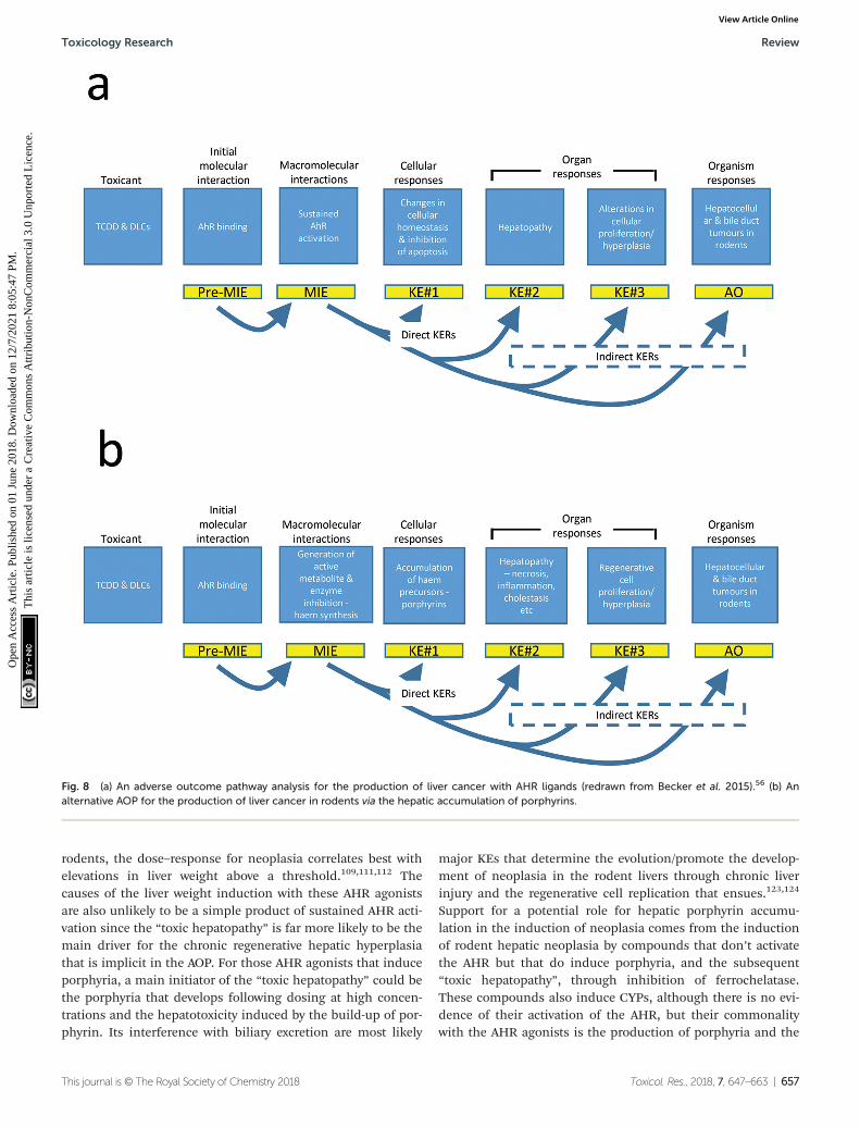
rodents, the dose–response for neoplasia correlates best withelevations in liver weight above a threshold.109,111,112 Thecauses of the liver weight induction with these AHR agonistsare also unlikely to be a simple product of sustained AHR acti-vation since the “toxic hepatopathy” is far more likely to be themain driver for the chronic regenerative hepatic hyperplasiathat is implicit in the AOP. For those AHR agonists that induceporphyria, a main initiator of the “toxic hepatopathy” could bethe porphyria that develops following dosing at high concen-trations and the hepatotoxicity induced by the build-up of por-phyrin. Its interference with biliary excretion are most likely
major KEs that determine the evolution/promote the develop-ment of neoplasia in the rodent livers through chronic liverinjury and the regenerative cell replication that ensues.123,124
Support for a potential role for hepatic porphyrin accumu-lation in the induction of neoplasia comes from the inductionof rodent hepatic neoplasia by compounds that don’t activatethe AHR but that do induce porphyria, and the subsequent“toxic hepatopathy”, through inhibition of ferrochelatase.These compounds also induce CYPs, although there is no evi-dence of their activation of the AHR, but their commonalitywith the AHR agonists is the production of porphyria and the
Fig. 8 (a) An adverse outcome pathway analysis for the production of liver cancer with AHR ligands (redrawn from Becker et al. 2015).56 (b) Analternative AOP for the production of liver cancer in rodents via the hepatic accumulation of porphyrins.
Toxicology Research Review
This journal is © The Royal Society of Chemistry 2018 Toxicol. Res., 2018, 7, 647–663 | 657
Ope
n A
cces
s A
rtic
le. P
ublis
hed
on 0
1 Ju
ne 2
018.
Dow
nloa
ded
on 1
2/7/
2021
8:0
5:47
PM
. T
his
artic
le is
lice
nsed
und
er a
Cre
ativ
e C
omm
ons
Attr
ibut
ion-
Non
Com
mer
cial
3.0
Unp
orte
d L
icen
ce.
View Article Online

development of toxic hepatopathy. Therefore a revision of theBecker et al. AOP56 is presented below (Fig. 8b) which changesthe order of some of the KEs but also introduces the key roleof porphyrin in the subsequent induction of the hepatopathyand consequentially of all of the downstream events leading tohepatic neoplasia.
7. Summary
Some chemicals, drugs and pesticides are known to interferein experimental animals with hepatic haem synthesis causingporphyria. With some clinical human porphyrias there is aknown association with increased incidences of hepatocellularcancer. Hepatic porphyrin accumulation caused in rodentsmay occur either by mechanisms that produce oxidized haemprecursors which act as inhibitors of enzymes in the pathwayor by metabolism of the chemical that by a side reaction pro-duces an inhibitor of ferrochelatase, from the haem of cyto-chrome P450. In both mechanisms there is evidence thatunder chronic conditions the accumulation of porphyrins,mostly either uroporphyrins or protoporphyrin IX may causeliver injury and aid subsequent liver tumour formation.
Conflicts of interest
There are no conflicts to declare.
Acknowledgements
Preliminary presentation on this topic took place to a HESIWorkshop U.S. EPA, 2004.125
References
1 W. E. Daniell, H. L. Stockbridge, R. F. Labbe, J. S. Woods,K. E. Anderson, D. M. Bissell, J. R. Bloomer,R. D. Ellefson, M. R. Moore, C. A. Pierach, W. E. Schreiber,A. Tefferi and G. M. Franklin, Environmental chemicalexposures and disturbances of heme synthesis, Environ.Health Perspect., 1997, 105, 37–53.
2 F. De Matteis and G. S. Marks, Cytochrome P450 and itsinteractions with the heme biosynthetic pathway,Can. J. Physiol. Pharmacol., 1996, 74, 1–8.
3 A. G. Smith and G. H. Elder, Complex gene-chemical inter-actions: hepatic uroporphyria as a paradigm, Chem.Research Toxicol., 2010, 23, 712–723.
4 V. Vagany and A. G. Smith, Complicity of haem in someadverse drug-reactions, Toxicol. Res., 2015, 1128–1142.
5 Z. Karim, S. Lyoumi, G. Nicolas, J. C. Deybach, L. Gouyaand H. Puy, Porphyrias: A 2015 update, Clin. Res. Hepatol.Gastroenterol., 2015, 39, 412–425.
6 H. Puy, L. Gouya and J. C. Deybach, Porphyrias, Lancet,2010, 375, 924–937.
7 J. Berman and A. Braun, Incidence of hepatoma in por-phyria cutanea tarda, Rev. Czech. Med., 1962, 8, 290–295.
8 N. O. Bengtsson and L. Hardell, Porphyrias, porphyrinsand hepatocellular cancer, Br. J. Cancer, 1986, 54, 115–117.
9 F. Lithner and L. Wetterberg, Hepatocellular carcinoma inpatients with acute intermittent porphyria, Acta Med.Scand., 1984, 21, 271–274.
10 V. Kordac, Frequency of occurrence of hepatocellular car-cinoma in patients with porphyria cutanea tarda in long-term follow-up, Neoplasia, 1972, 19, 135–139.
11 Z. F. Soonawalla, T. Orug, M. N. Badminton, G. H. Elder,J. M. Rhodes, S. R. Bramhall and E. Elias, Liver transplan-tation as a cure for acute intermittent porphyria, Lancet,2004, 363, 705–706.
12 D. M. Bissell, J. C. Lai, R. K. Meister and P. D. Blanc, Roleof delta-aminolevulinic acid in the symptoms of acute por-phyria, Am. J. Med., 2015, 128, 313–317.
13 D. M. Bissell and B. Wang, Acute hepatic porphyria,J. Clin. Transl. Hepatol., 2015, 3, 17–26.
14 R. Kauppinen and P. Mustajoki, Acute hepatic porphyriaand hepatocellular carcinoma, Br. J. Cancer, 1988, 57,117–120.
15 M. J. Tidman, E. M. Higgins, G. H. Elder andD. M. MacDonald, Variegate porphyria associated withhepatocellular carcinoma, Br. J. Dermatol., 1989, 121, 503–505.
16 C. Andant, H. Puy, C. Bogard, J. Faivre, J. C. Soule,Y. Nordmann and J. C. Deybach, Hepatocellular carci-noma in patients with acute hepatic porphyria: frequencyof occurrence and related factors, J. Hepatol., 2000, 32,933–939.
17 X. Schneider-Yin, A. M. Van Tuyll Van Serooskerken,P. Went, W. Tyblewski, P. Poblete-Gutiérrez, E. I. Minderand J. Frank, Hepatocellular carcinoma in variegate por-phyria: A serious complication, Acta Derm.-Venereol., 2010,90, 512–515.
18 E. Innala and C. Andersson, Screening for hepatocellularcarcinoma in acute intermittent porphyria: a 15-yearfollow-up in northern Sweden, J. Intern. Med., 2011, 26,538–545.
19 E. Sardh, S. Wahlin, M. Bjornstedt, P. Harper andD. E. Andersson, High risk of primary liver cancer in acohort of 179 patients with acute hepatic porphyria,J. Inherited Metab. Dis., 2013, 1063–1071.
20 C. M. Baravelli, S. Sandberg, A. K. Aarsand, R. M. Nilsenand M. C. Tollanes, Acute hepatic porphyria and cancerrisk: a nationwide cohort study, J. Intern. Med., 2017, 282,229–240.
21 X. Schneider-Yin, A. M. van Tuyll van Serooskerken,M. Siegesmund, P. Went, J. Barman-Aksozen,R. S. Bladergroen, P. Komminoth, R. H. Cloots,V. J. Winnepenninckx, A. zur Hausen, M. Weber,A. Driessen, P. Poblete-Gutierrez, P. Bauer, C. Schroeder,M. van Geel, E. I. Minder and J. Frank, Biallelic inacti-vation of protoporphyrinogen oxidase and hydroxymethyl-
Review Toxicology Research
658 | Toxicol. Res., 2018, 7, 647–663 This journal is © The Royal Society of Chemistry 2018
Ope
n A
cces
s A
rtic
le. P
ublis
hed
on 0
1 Ju
ne 2
018.
Dow
nloa
ded
on 1
2/7/
2021
8:0
5:47
PM
. T
his
artic
le is
lice
nsed
und
er a
Cre
ativ
e C
omm
ons
Attr
ibut
ion-
Non
Com
mer
cial
3.0
Unp
orte
d L
icen
ce.
View Article Online
bilane synthase is associated with liver cancer in acuteporphyrias, J. Hepatol., 2015, 62, 734–738.
22 J. Onuki, P. C. Teixeira, M. H. Medeiros, D. Dornemann,T. Douki, J. Cadet and P. Di Mascio, Is 5-aminolevulinicacid involved in the hepatocellular carcinogenesis of acuteintermittent porphyria?, Cell. Mol. Biol., 2002, 48, 17–26.
23 J. C. Deybach and H. Puy, Hepatocellular carcinomawithout cirrhosis: think acute hepatic porphyrias and viceversa, J. Intern. Med., 2011, 269, 521–524.
24 A. G. Roberts, S. D. Whatley, R. R. Morgan, M. Worwoodand G. H. Elder, Increased frequency of the haemochro-matosis Cys282Tyr mutation in sporadic porphyriacutanea tarda, Lancet, 1997, 349, 321–323.
25 J. M. Cortes, H. Oliva, F. J. Paradinas and C. Hernandez-Guio, The pathology of the liver in porphyria cutaneatarda, Histopathology, 1980, 4, 471–485.
26 H. Salata, J. M. Cortes, R. Enriquez de Salamanca,H. Oliva, A. Castro, E. Kusak, V. Carreno andC. Hernandez Guio, Porphyria cutanea tarda and hepato-cellular carcinoma. Frequency of occurrence and relatedfactors, J. Hepatol., 1985, 1, 477–487.
27 P. D. Siersema, F. J. ten Kate, P. G. Mulder andJ. H. Wilson, Hepatocellular carcinoma in porphyriacutanea tarda: frequency and factors related to its occur-rence, Liver, 1992, 12, 56–61.
28 G. C. Topi, L. D’Alessandro Gandolfo, D. Griso andS. Morini, Porphyria cutanea tarda and hepatocellular car-cinoma, Int. J. Biochem., 1980, 12, 883–885.
29 J. P. Gisbert, L. Garcia-Buey, A. Alonso, S. Rubio,A. Hernández, J. M. Pajares, A. García-Díez andR. Moreno-Otero, Hepatocellular carcinoma risk inpatients with porphyria cutanea tarda,Eur. J. Gastroenterol. Hepatol., 2004, 16, 689–692.
30 A. L. Fracanzani, E. Taioli, M. Sampietro, E. Fatta,C. Bertelli, G. Fiorelli and S. Fargion, Liver cancer risk isincreased in patients with porphyria cutanea tarda incomparison to matched control patients with chronic liverdisease, J. Hepatol., 2001, 35, 498–503.
31 V. Kordac, P. Martasek, V. Zoubek and M. Kalab,Porphyria cutanea-tarda - manifestation and therapy,Ann. N. Y. Acad. Sci., 1987, 514, 335–336.
32 R. L. Lindberg, C. Porcher, B. Grandchamp,B. Ledermann, K. Burki, S. Brandner, A. Aguzzi andU. A. Meyer, Porphobilinogen deaminase deficiency inmice causes a neuropathy resembling that of humanhepatic porphyria, Nat. Genet., 1996, 12, 195–199.
33 C. Handschin, J. Lin, J. Rhee, A. K. Peyer, S. Chin,P. H. Wu, U. A. Meyer and B. M. Spiegelman, Nutritionalregulation of hepatic heme biosynthesis and porphyriathrough PGC-1alpha, Cell, 2005, 122, 505–515.
34 J. E. Francis and A. G. Smith, Oxidation of uroporphyrino-gens by hydroxyl radicals. Evidence for nonporphyrin pro-ducts as potential inhibitors of uroporphyrinogen decar-boxylase, FEBS Lett., 1988, 233, 311–314.
35 J. D. Phillips, H. A. Bergonia, C. A. Reilly, M. R. Franklinand J. P. Kushner, A porphomethene inhibitor of uropor-
phyrinogen decarboxylase causes porphyria cutanea tarda,Proc. Natl. Acad. Sci. U. S. A., 2007, 104, 5079–5084.
36 M. Van den Berg, L. S. Birnbaum, M. Denison, M. De Vito,W. Farland, M. Feeley, H. Fiedler, H. Hakansson,A. Hanberg, L. Haws, M. Rose, S. Safe, D. Schrenk,C. Tohyama, A. Tritscher, J. Tuomisto, M. Tysklind,N. Walker and R. E. Peterson, The 2005 World HealthOrganization reevaluation of human and mammaliantoxic equivalency factors for dioxins and dioxin-like com-pounds, Toxicol. Sci., 2006, 93, 223–241.
37 A. P. van Birgelen, Hexachlorobenzene as a possible majorcontributor to the dioxin activity of human milk, Environ.Health Perspect., 1998, 106, 683–688.
38 M. E. Hahn, T. A. Gasiewicz, P. Linko and J. A. Goldstein,The role of the Ah locus in hexachlorobenzene-inducedporphyria, Studies in congenic C57BL/6J mice, Biochem. J.,1988, 254, 245–254.
39 M. E. Hahn, J. A. Goldstein, P. Linko and T. A. Gasiewicz,Interaction of hexachlorobenzene with the receptor for2,3,7,8-tetrachlorodibenzo-p-dioxin in vitro and in vivo.Evidence that hexachlorobenzene is a weak Ah receptoragonist, Arch. Biochem. Biophys., 1989, 270, 344–355.
40 R. A. Akhtar and A. G. Smith, Chromosomal linkage ana-lysis of porphyria in mice induced by hexachlorobenzene-iron synergism: a model of sporadic porphyria cutaneatarda, Pharmacogenetics, 1998, 8, 485–494.
41 M. D. Stonard, Mixed type hepatic microsomal enzymeinduction by hexachlorobenzene, Biochem. Pharmacol.,1975, 24, 1959–1963.
42 M. D. Stonard and J. B. Greig, Different patterns ofhepatic microsomal-enzyme activity produced by adminis-tration of pure hexachlorobiphenyl isomers and hexa-chlorobenzene, Chem.-Biol. Interact., 1976, 15, 365–379.
43 A. G. Smith, J. E. Francis and I. Bird, Distinction betweenoctachlorostyrene and hexachlorobenzene in their poten-tials to induce ethoxyphenoxazone deethylase and causeporphyria in rats and mice, J. Biochem. Toxicol., 1986, 1,105–117.
44 L. Cantoni, M. Rizzardini, M. T. Tacconi and A. Graziani,Comparison of hexachlorobenzene-induced alterations ofmicrosomal membrane composition and monooxygenaseactivity in male and female rats, Toxicology, 1987, 45,291–305.
45 D. L. Grant, F. Iverson, G. V. Hatina and D. C. Villeneuve,Effects of hexachlorobenzene on liver porphyrin levelsand microsomal enzymes in the rat, Environ. Physiol.Biochem., 1974, 4, 159–165.
46 A. G. Smith, J. E. Francis, J. A. Green, J. B. Greig,C. R. Wolf and M. M. Manson, Sex-linked hepatic uropor-phyria and the induction of cytochromes P450IA in ratscaused by hexachlorobenzene and polyhalogenated biphe-nyls, Biochem. Pharmacol., 1990, 40, 2059–2068.
47 A. G. Smith, J. E. Francis, D. Dinsdale, M. M. Manson andJ. R. Cabral, Hepatocarcinogenicity of hexachlorobenzene inrats and the sex difference in hepatic iron status and devel-opment of porphyria, Carcinogenesis, 1985, 6, 631–636.
Toxicology Research Review
This journal is © The Royal Society of Chemistry 2018 Toxicol. Res., 2018, 7, 647–663 | 659
Ope
n A
cces
s A
rtic
le. P
ublis
hed
on 0
1 Ju
ne 2
018.
Dow
nloa
ded
on 1
2/7/
2021
8:0
5:47
PM
. T
his
artic
le is
lice
nsed
und
er a
Cre
ativ
e C
omm
ons
Attr
ibut
ion-
Non
Com
mer
cial
3.0
Unp
orte
d L
icen
ce.
View Article Online

48 D. L. Arnold, C. A. Moodie, S. M. Charbonneau,H. C. Grice, P. F. McGuire, F. R. Bryce, B. T. Collins,Z. Z. Zawidzka, D. R. Krewski, E. A. Nera, et al., Long-termtoxicity of hexachlorobenzene in the rat and the effect ofdietary vitamin A, Food Chem. Toxicol., 1985, 23, 779–793.
49 E. Erturk, R. W. Lambrecht, H. A. Peters, D. J. Cripps,A. Gocmen, C. R. Morris and G. T. Bryan, Oncogenicity ofhexachlorobenzene, IARC Sci. Publ., 1986, 77, 417–423.
50 D. H. Norback and R. H. Weltman, Polychlorinated biphe-nyl induction of hepatocellular carcinoma in the Sprague-Dawley rat, Environ. Health Perspect., 1985, 60, 97–105.
51 R. D. Kimbrough, V. W. Burse and J. A. Liddle, Persistentliver lesions in rats after a single oral dose of polybromi-nated biphenyls (firemaster FF-1) and concomitant PBBtissue levels, Environ. Health Perspect., 1978, 23, 265–273.
52 R. D. Kimbrough, D. F. Groce, M. P. Korver andV. W. Burse, Induction of liver tumors in female Shermanstrain rats by polybrominated biphenyls, J. Natl. CancerInst., 1981, 66, 535–542.
53 R. J. Kociba, D. G. Keyes, J. E. Beyer, R. M. Carreon,C. E. Wade, D. A. Dittenber, R. P. Kalnins, L. E. Frauson,C. N. Park, S. D. Barnard, R. A. Hummel andC. G. Humiston, Results of a two-year chronic toxicity andoncogenicity study of 2,3,7,8-tetrachlorodibenzo-p-dioxinin rats, Toxicol. Appl. Pharmacol., 1978, 46, 279–303.
54 P. Carthew and A. G. Smith, Pathological mechanisms ofhepatic tumour formation in rats exposed chronically todietary hexachlorobenzene, J. Appl. Toxicol., 1994, 14, 447–452.
55 R. A. Budinsky, D. Schrenk, T. Simon, M. Van den Berg,J. F. Reichard, J. B. Silkworth, L. L. Aylward, A. Brix,T. Gasiewicz, N. Kaminski, G. Perdew, T. B. Starr,N. J. Walker and J. C. Rowlands, Mode of action and dose-response framework analysis for receptor-mediated tox-icity: The aryl hydrocarbon receptor as a case study, Crit.Rev. Toxicol., 2014, 44, 83–119.
56 R. A. Becker, G. Patlewicz, T. W. Simon, J. C. Rowlandsand R. A. Budinsky, The adverse outcome pathway forrodent liver tumor promotion by sustained activation ofthe aryl hydrocarbon receptor, Regul. Toxicol. Pharmacol.,2015, 73, 172–190.
57 G. D. Kennedy, M. Nukaya, S. M. Moran, E. Glover,S. Weinberg, S. Balbo, S. S. Hecht, H. C. Pitot,N. R. Drinkwater and C. A. Bradfield, Liver tumor pro-motion by 2,3,7,8-tetrachlorodibenzo-p-dioxin is depen-dent on the aryl hydrocarbon receptor and TNF/IL-1 recep-tors, Toxicol. Sci., 2014, 140, 135–143.
58 F. P. Stewart, M. M. Manson, J. R. Cabral and A. G. Smith,Hexachlorobenzene as a promoter of diethylnitrosamine-initiated hepatocarcinogenesis in rats and comparisonwith induction of porphyria, Carcinogenesis, 1989, 10,1225–1230.
59 A. G. Smith, J. R. Cabral and F. De Matteis, A differencebetween two strains of rats in their liver non-haem ironcontent and in their response to the porphyrogenic effect
of hexachlorobenzene, Chem.-Biol. Interact., 1979, 27,353–363.
60 A. G. Smith, P. Carthew, J. E. Francis, J. R. Cabral andM. M. Manson, Enhancement by iron of hepatic neoplasiain rats caused by hexachlorobenzene, Carcinogenesis,1993, 14, 1381–1387.
61 A. G. Smith and J. E. Francis, Synergism of iron and hexa-chlorobenzene inhibits hepatic uroporphyrinogen decar-boxylase in inbred mice, Biochem. J., 1983, 214, 909–913.
62 J. B. Greig, J. E. Francis, S. J. Kay, D. P. Lovell andA. G. Smith, Incomplete correlation of 2,3,7,8-tetrachloro-dibenzo-p-dioxin hepatotoxicity with Ah phenotype inmice, Toxicol. Appl. Pharmacol., 1984, 74, 17–25.
63 S. W. Robinson, B. Clothier, R. A. Akhtar, A. L. Yang,I. Latour, C. Van Ijperen, M. F. Festing and A. G. Smith,Non-ahr gene susceptibility loci for porphyria and liverinjury induced by the interaction of ‘dioxin’ with ironoverload in mice, Mol. Pharmacol., 2002, 61, 674–681.
64 A. G. Smith, J. R. Cabral, P. Carthew, J. E. Francis andM. M. Manson, Carcinogenicity of iron in conjunctionwith a chlorinated environmental chemical, hexachloro-benzene, in C57BL/10ScSn mice, Int. J. Cancer, 1989, 43,492–496.
65 A. G. Smith, J. E. Francis and P. Carthew, Iron as a syner-gist for hepatocellular carcinoma induced by polychlori-nated biphenyls in Ah-responsive C57BL/10ScSn mice,Carcinogenesis, 1990, 11, 437–444.
66 A. G. Smith, B. Clothier, P. Carthew, N. L. Childs,P. R. Sinclair, D. W. Nebert and T. P. Dalton, Protection ofthe Cyp1a2(-/-) null mouse against uroporphyria andhepatic injury following exposure to 2,3,7,8-tetrachlorodi-benzo-p-dioxin, Toxicol. Appl. Pharmacol., 2001, 173, 89–98.
67 P. R. Sinclair, N. Gorman, H. S. Walton, W. J. Bement,T. P. Dalton, J. F. Sinclair, A. G. Smith and D. W. Nebert,CYP1A2 is essential in murine uroporphyria caused byhexachlorobenzene and iron, Toxicol. Appl. Pharmacol.,2000, 162, 60–67.
68 P. Greaves, B. Clothier, R. Davies, F. M. Higginson,R. E. Edwards, T. P. Dalton, D. W. Nebert and A. G. Smith,Uroporphyria and hepatic carcinogenesis induced by poly-chlorinated biphenyls-iron interaction: absence in theCyp1a2(-/-) knockout mouse, Biochem. Biophys. Res.Commun., 2005, 331, 147–152.
69 A. G. Smith, P. Carthew, J. E. Francis and K. Ingebrigtsen,Influence of iron on the induction of hepatic tumors andporphyria by octachlorostyrene in C57BL/10ScSn mice,Cancer Lett., 1994, 81, 145–150.
70 S. Madra, J. Styles and A. G. Smith, Perturbation of hep-atocyte nuclear populations induced by iron and poly-chlorinated biphenyls in C57BL/10ScSn mice during car-cinogenesis, Carcinogenesis, 1995, 16, 719–727.
71 P. C. Rumsby, J. G. Evans, H. E. Phillimore, P. Carthewand A. G. Smith, Search for Ha-ras codon 61 mutations inliver tumours caused by hexachlorobenzene and Aroclor
Review Toxicology Research
660 | Toxicol. Res., 2018, 7, 647–663 This journal is © The Royal Society of Chemistry 2018
Ope
n A
cces
s A
rtic
le. P
ublis
hed
on 0
1 Ju
ne 2
018.
Dow
nloa
ded
on 1
2/7/
2021
8:0
5:47
PM
. T
his
artic
le is
lice
nsed
und
er a
Cre
ativ
e C
omm
ons
Attr
ibut
ion-
Non
Com
mer
cial
3.0
Unp
orte
d L
icen
ce.
View Article Online

1254 in C57BL/10ScSn mice with iron overload,Carcinogenesis, 1992, 13, 1917–1920.
72 S. P. Faux, J. E. Francis, A. G. Smith and J. K. Chipman,Induction of 8-hydroxydeoxyguanosine in Ah-responsivemouse liver by iron and Aroclor 1254, Carcinogenesis,1992, 13, 247–250.
73 R. Davies, B. Clothier and A. G. Smith, Mutation frequencyin the lacI gene of liver DNA from lambda/lacI transgenicmice following the interaction of PCBs with iron causinghepatic cancer and porphyria, Mutagenesis, 2000, 15, 379–383.
74 F. G. Dayan and S. O. Duke, Protoporphyrinogen oxidase-inhibiting herbicides, in Hayes Handbook of PesticideToxicology, ed. R. Krieger, Academic Press, 2010, ch. 81,vol. 2, pp. 1733–1751.
75 J. Krijt, M. Vokurka, J. Sanitrak, V. Janousek, L. vanHolsteijn and B. J. Blaauboer, Effect of the protoporphyri-nogen oxidase-inhibiting herbicide fomesafen on liveruroporphyrin and heptacarboxylic porphyrin in twomouse strains, Food Chem. Toxicol., 1994, 32, 641–650.
76 J. Krijt, O. Pšenák, M. Vokurka, A. Chlumská andF. Fakan, Experimental hepatic uroporphyria induced bythe diphenyl-ether herbicide fomesafen in male DBA/2 mice, Toxicol. Appl. Pharmacol., 2003, 189, 28–38.
77 J. Krijt, P. Stranska, J. Sanitrak, A. Chlumska andF. Fakan, Liver preneoplastic changes in mice treated withthe herbicide fomesafen, Hum. Exp. Toxicol., 1999, 18,338–344.
78 M. Sachar, K. E. Anderson and X. Ma, Protoporphyrin IX:the good, the bad, and the ugly, J. Pharmacol. Exp. Ther.,2016, 356, 267–275.
79 F. De Matteis, A. H. Gibbs, A. H. Jackson andS. Weerasinghe, Conversion of liver haem intoN-substituted porphyrins or green pigments. Nature ofthe substituent at the pyrrole nitrogen atom, FEBS Lett.,1980, 119, 109–112.
80 J. T. Gamble, K. Nakatsu and G. S. Marks, Comparison ofrat and human cytochrome P450 (CYP) sources ofN-alkylprotoporphyrin IX. Formation after interaction withporphyrinogenic xenobiotics: studies with cDNA-expressedsingle CYP enzymes, Xenobiotica, 2002, 32, 997–1006.
81 J. T. Gamble, S. G. Wong, H. A. Dailey and G. S. Marks,Use of recombinant human ferrochelatase as a sensitivebioassay for N-alkylprotoporphyrin IX formed after inter-action of porphyrinogenic xenobiotics with rat liver micro-somes, Can. J. Physiol. Pharmacol., 2000, 78, 578–581.
82 K. Nakao, O. Wada, F. Takaku, S. Sassa, Y. Yano andG. Urata, The origin of the increased protoporphyrin inerythrocytes of mice with experimentally induced por-phyria, J. Lab. Clin. Med., 1967, 70, 923–932.
83 A. Holley, L. J. King, A. H. Gibbs and F. De Matteis, Strainand sex differences in the response of mice to drugs thatinduce protoporphyria: role of porphyrin biosynthesis andremoval, J. Biochem. Toxicol., 1990, 5, 175–182.
84 F. De Matteis, A. H. Gibbs, P. B. Farmer and J. H. Lamb,Liver production of N-alkylated porphyrins caused in mice
by treatment with substituted dihydropyridines. Evidencethat the alkyl group on the pyrrole nitrogen atom orig-inates from the drug, FEBS Lett., 1981, 129, 328–331.
85 F. De Matteis and A. H. Gibbs, Stimulation of the pathwayof porphyrin synthesis in the liver of rats and mice by gri-seofulvin, 3,5-diethoxycarbonyl-1,4-dihydrocollidine andrelated drugs: evidence for two basically different mecha-nisms, Biochem. J., 1975, 146, 285–287.
86 T. W. Gant, P. R. Baus, B. Clothier, J. Riley, R. Davies,D. J. Judah, R. E. Edwards, E. George, P. Greaves andA. G. Smith, Gene expression profiles associated withinflammation, fibrosis, and cholestasis in mouse liverafter griseofulvin, Environ. Health Perspect., 2003, 111,847–853.
87 R. Stejskal, M. Itabashi, J. Stanek and Z. Hruban,Experimental porphyria induced by (3-(2,4,6-trimethyl-phenyl)-thioethyl)-4 methylsydnone, Virchows Arch. B,1975, 18, 83–100.
88 A. M. Brady, R. L. Hasmall, B. M. Elcombe, E. A. Lock andF. De Matteis, Effects of 2-[1-(ethoxyimino)propyl]-3-hydroxy-5-(2,4,6-trimethylphenyl) cyclohex-2-enone onhepatic haem biosynthesis: species differences in hepaticporphyria, Toxicology, 1993, 84, 199–215.
89 Y. Frater, A. Brady, E. A. Lock and F. De Matteis,Formation of N-methyl protoporphyrin in chemically-induced protoporphyria. Studies with a novel porphyro-genic agent, Arch. Toxicol., 1993, 67, 179–185.
90 F. De Matteis, A. H. Gibbs and A. G. Smith, Inhibition ofprotohaem ferro-lyase by N-substituted porphyrins.Structural requirements for the inhibitory effect, Biochem.J., 1980, 189, 645–648.
91 F. De Matteis, A. H. Gibbs, L. Cantoni and J. Francis,Substrate-dependent irreversible inactivation of cyto-chrome P-450: conversion of its haem moiety into modi-fied porphyrins, Ciba Found. Symp., 1980, 76, 119–139.
92 I. N. White, Destruction of liver haem by norethindrone.Conversion into green pigments, Biochem. J., 1981, 196,575–583.
93 S. Knasmuller, W. Parzefall, C. Helma, F. Kassie, S. Eckerand R. Schulte-Hermann, Toxic effects of griseofulvin:disease models, mechanisms, and risk assessment, Crit.Rev. Toxicol., 1997, 27, 495–537.
94 J. M. Nicolas, H. Chanteux, V. Mancel, G. M. Dubin,B. Gerin, L. Staelens, O. Depelchin and S. Kervyn, N-alkyl-protoporphyrin formation and hepatic porphyria in dogsafter administration of a new antiepileptic drug candidate:Mechanism and species specificity, Toxicol. Sci., 2014,141, 353–364.
95 F. De Matteis, A. J. Donnelly and W. J. Runge, The effectsof prolonged administration of griseofulvin in mice withreference to sex differences, Cancer Res., 1966, 26, 721–726.
96 A. Matilla and E. A. Molland, A light and electron micro-scopic study of the liver in case of erythrohepatic proto-porphyria and in griseofulvin-induced porphyria in mice,J. Clin. Pathol., 1974, 27, 698–709.
Toxicology Research Review
This journal is © The Royal Society of Chemistry 2018 Toxicol. Res., 2018, 7, 647–663 | 661
Ope
n A
cces
s A
rtic
le. P
ublis
hed
on 0
1 Ju
ne 2
018.
Dow
nloa
ded
on 1
2/7/
2021
8:0
5:47
PM
. T
his
artic
le is
lice
nsed
und
er a
Cre
ativ
e C
omm
ons
Attr
ibut
ion-
Non
Com
mer
cial
3.0
Unp
orte
d L
icen
ce.
View Article Online

97 F. Gschnait, K. Konrad, H. Honigsmann, H. Denk andK. Wolff, Mouse model for protoporphyria. I. The liverand hepatic protoporphyrin crystals, J. Invest. Dermatol.,1975, 65, 290–299.
98 S. W. Choi, J. H. Han, K. T. Lim, H. M. Cho, K. W. Chung,H. S. Sun, D. H. Park, B. S. Kim and E. J. Seo, Effect of urso-deoxycholic acid on experimental hepatic porphyria inducedby griseofulvin, J. Korean Med. Sci., 1991, 6, 146–156.
99 B. D. Pauli and S. W. Kennedy, Hepatic porphyria inducedby the herbicide tralkoxydim in small mammals isspecies-specific, Environ. Toxicol. Chem., 2005, 24, 450–456.
100 EFSA Report, Conclusion on the peer review of the pesti-cide risk assessment of the active substance Tralkoxydim,EFSA J., 2008, 139, 1–78.
101 F. de Matteis, C. Hollands, A. H. Gibbs, N. de Sa andM. Rizzardini, Inactivation of cytochrome P-450 and pro-duction of N-alkylated porphyrins caused in isolatedhepatocytes by substituted dihydropyridines. Structuralrequirements for loss of haem and alkylation of thepyrrole nitrogen atom, FEBS Lett., 1982, 145, 87–92.
102 A. M. Brady and E. A. Lock, Inhibition of ferrochelataseand accumulation of porphyrins in mouse hepatocyte cul-tures exposed to porphyrinogenic chemicals, Arch.Toxicol., 1992, 66, 175–181.
103 W. Y. Kao, C. W. Su, Y. S. Huang, Y. C. Chou, Y. C. Chen,W. H. Chung, M. C. Hou, H. C. Lin, F. Y. Lee and J. C. Wu,Risk of oral antifungal agent-induced liver injury inTaiwanese, Br. J. Clin. Pharmacol., 2014, 77, 180–189.
104 C. Rimington, P. N. Morgan, K. Nicholls, J. D. Everall andR. R. Davies, Griseofulvin administration and porphyrinmetabolism. A survey, Lancet, 1963, 2, 318–322.
105 A. Berman and R. L. Franklin, Precipitation of acute inter-mittent porphyria by griseofulvin therapy, J. Am. Med.Assoc., 1965, 192, 1005–1007.
106 C. Martinez Mdel, S. G. Afonso, R. P. Meiss, A. M. Buzalehand A. Batlle, Hepatic damage and oxidative stressinduced by griseofulvin in mice, Cell Mol. Biol., 2009, 55,127–139.
107 D. E. Hatoff and W. G. Hardison, Bile acids modify alka-line phosphatase induction and bile secretion pressureafter bile duct obstruction in the rat, Gastroenterology,1981, 80, 666–672.
108 E. Weston-Hurst and G. E. Paget, Protoporphyrin, cirrho-sis and hepatomata in the livers of mice given griseo-fulvin, Br. J. Dermatol., 1963, 75, 105–112.
109 M. Rustia and P. Shubik, Thyroid tumours in rats andhepatomas in mice after griseofulvin treatment,Br. J. Cancer, 1978, 38, 237–249.
110 C. F. Polo, A. M. Buzaleh, E. S. Vazquez, S. G. Afonso,N. M. Navone and A. M. Batlle, Griseofulvin-inducedhepatopathy due to abnormalities in heme pathway, Gen.Pharmacol., 1997, 29, 207–210.
111 T. Shimoyama and S. Nonaka, Biochemical studies on gri-seofulvin-induced protoporphyria, Ann. N. Y. Acad. Sci.,1987, 514, 160–169.
112 T. Shimoyama, S. Nonaka, T. Honda, F. Murayama,T. Ohgami and H. Yoshida, A biochemical study of experi-mental porphyria. I. The influence of griseofulvin atvarious concentrations on porphyrin metabolism,J. Dermatol., 1984, 11, 49–55.
113 S. Tutois, X. Montagutelli, V. Da Silva, H. Jouault,P. Rouyer-Fessard, K. Leroy-Viard, J. L. Guenet,Y. Nordmann, Y. Beuzard and J. C. Deybach, Erythropoieticprotoporphyria in the house mouse. A recessive inheritedferrochelatase deficiency with anemia, photosensitivity,and liver disease, J. Clin. Invest., 1991, 88, 1730–1736.
114 R. Davies, A. Schuurman, C. R. Barker, B. Clothier,T. Chernova, F. M. Higginson, D. J. Judah, D. Dinsdale,R. E. Edwards, P. Greaves, T. W. Gant and A. G. Smith,Hepatic gene expression in protoporphyic Fech mice isassociated with cholestatic injury but not a markeddepletion of the heme regulatory pool, Am. J. Pathol.,2005, 166, 1041–1053.
115 L. Libbrecht, L. Meerman, F. Kuipers, T. Roskams,V. Desmet and P. Jansen, Liver pathology and hepatocarci-nogenesis in a long-term mouse model of erythropoieticprotoporphyria, J. Pathol., 2003, 199, 191–200.
116 J. Barman-Aksozen, C. W. P. V. B. Bansode, F. Koentgen,J. Trub, P. Pelczar, P. Cinelli, X. Schneider-Yin,D. Schumperli and E. I. Minder, Modeling the ferrochela-tase c.315–48C modifier mutation for erythropoietic pro-toporphyria (EPP) in mice, Dis. Models & Mech., 2017, 10,225–233.
117 M. Lecha, H. Puy and J. C. Deybach, Erythropoietic proto-porphyria, Orphanet. J. Rare Dis., 2009, 4, 19.
118 M. B. Troadec, D. Warner, J. Wallace, K. Thomas,G. J. Spangrude, J. Phillips, O. Khalimonchuk, B. H. Paw,D. M. Ward and J. Kaplan, Targeted deletion of the mouseMitoferrin1 gene: from anemia to protoporphyria, Blood,2011, 117, 5494–5502.
119 C. Homedan, J. Laafi, C. Schmitt, N. Gueguen,T. Lefebvre, Z. Karim, V. Desquiret-Dumas, C. Wetterwald,J. C. Deybach, L. Gouya, H. Puy, P. Reynier andY. Malthiery, Acute intermittent porphyria causes hepaticmitochondrial energetic failure in a mouse model,Int. J. Biochem. Cell Biol., 2014, 51, 93–101.
120 R. Thierbach, T. J. Schulz, F. Isken, A. Voigt, B. Mietzner,G. Drewes, J. C. von Kleist-Retzow, R. J. Wiesner,M. A. Magnuson, H. Puccio, A. F. Pfeiffer, P. Steinbergand M. Ristow, Targeted disruption of hepatic frataxinexpression causes impaired mitochondrial function,decreased life span and tumor growth in mice, Hum. Mol.Genet., 2005, 14, 3857–3864.
121 E. Julien, A. R. Boobis and S. S. Olin, The Key EventsDose-Response Framework: a cross-disciplinary mode-of-action based approach to examining dose-response andthresholds, Crit. Rev. Food Sci. Nutr., 2009, 49, 682–689.
122 T. W. Simon, S. S. Simons Jr., R. J. Preston, A. R. Boobis,S. M. Cohen, N. G. Doerrer, P. A. Fenner-Crisp,T. S. McMullin, C. A. McQueen and J. C. Rowlands, Theuse of mode of action information in risk assessment:
Review Toxicology Research
662 | Toxicol. Res., 2018, 7, 647–663 This journal is © The Royal Society of Chemistry 2018
Ope
n A
cces
s A
rtic
le. P
ublis
hed
on 0
1 Ju
ne 2
018.
Dow
nloa
ded
on 1
2/7/
2021
8:0
5:47
PM
. T
his
artic
le is
lice
nsed
und
er a
Cre
ativ
e C
omm
ons
Attr
ibut
ion-
Non
Com
mer
cial
3.0
Unp
orte
d L
icen
ce.
View Article Online
quantitative key events/dose-response framework for mod-eling the dose-response for key events, Crit. Rev. Toxicol.,2014, 44(Suppl. 3), 17–43.
123 S. M. Cohen and L. B. Ellwein, Cell proliferation in car-cinogenesis, Science, 1990, 249, 1007–1011.
124 J. R. Foster, The role of cell proliferation in chemicallyinduced carcinogenesis, J. Comp. Pathol., 1997, 116, 113–144.
125 M. P. Holsapple, H. C. Pitot, S. M. Cohen, A. R. Boobis,J. E. Klaunig, T. Pastoor, V. L. Dellarco and Y. P. Dragan,Mode of action in relevance of rodent liver tumors tohuman cancer risk, Toxicol. Sci., 2006, 89, 51–56.
126 L. Rossi, M. Ravera, G. Repetti and L. Santi, Long-termadministration of DDT or phenobarbital-Na in Wistar rats,Int. J. Cancer, 1977, 19, 179–185.
127 P. Greaves, R. Goonetilleke, G. Nunn, J. Topham andT. Orton, Two-year carcinogenicity study of tamoxifen inAlderley Park Wistar-derived rats, Cancer Res., 1993, 53,3919–3924.
128 M. D. Reuber and C. W. Lee, Effect of age and sex onhepatic lesions in Buffalo strain rats ingesting diethyl-nitrosamine, J. Natl. Cancer Inst., 1968, 41, 1133–1140.
129 H. W. Rumsfeld Jr., W. L. Miller Jr. and C. A. Baumann, Asex difference in the development of liver tumors in ratsfed 3′-methyl-4-dimethylaminoazobenzene or 4′-fluoro-4-dimethylaminoazobenzene, Cancer Res., 1951, 11, 814–819.
130 G. N. Wogan and P. M. Newberne, Dose-response charac-teristics of aflatoxin B1 carcinogenesis in the rat, CancerRes., 1967, 27, 2370–2376.
131 H. Sidransky, B. P. Wagner and H. P. Morris, Sex differ-ence in liver tumorgenesis in rats ingesting N-2-fluorenyl-acetamide, J. Natl. Cancer Inst., 1961, 26, 151–187.
132 F. De Matteis, C. Hollands, A. H. Gibbs, N. de Sa andM. Rizzardini, Inactivation of cytochrome P-450 and pro-
duction of porphyrins caused in isolated hepatocytes bysubstituted dihydropyridines. Structural requirements forthe loss of haem and alkylation of the pyrrole nitrogenatom, FEBS Lett., 1982, 145, 87–92.
133 A. G. Smith and J. E. Francis, Chemically-induced for-mation of an inhibitor of hepatic uroporphyrinogen de-carboxylase in inbred mice with iron overload, Biochem. J.,1987, 246, 221–226.
134 A. G. Smith, J. E. Francis, S. J. Kay and J. B. Greig, Hepatictoxicity and uroporphyrinogen decarboxylase activity fol-lowing a single dose of 2,3,7,8-tetrachlorodibenzo-p-dioxin to mice, Biochem. Pharmacol., 1981, 30, 2825–2830.
135 M.-T. Tsai, S.-Y. Huang and S.-Y. Cheng, Lead poisoningcan be easily misdiagnosed as acute porphyria and non-specific abdominal pain, Case Rep. Emerg. Med., 2017,9050713.
136 S. Lindstedt, E. Holme, E. A. Lock, O. Hjalmarson andB. Strandvik, Treatment of hereditary tyrosinaemia type 1by inhibition of 4-hydroxyphenylpyruvate dioxygenase,Lancet, 1992, 340, 813–817.
137 A. G. Weinberg, C. E. Mize and H. G. Worthen, The occur-rence of hepatoma in the chronic form of hereditary tyro-sinemia, J. Pediatr., 1976, 88, 434–438.
138 F. Angileri, V. Roy, G. Morrow, J. Y. Scoazec, N. Gadot,D. Grejuela and R. M. Tanguay, Molecular changes associ-ated with chronic liver damage and neoplatic lesions in amurine model of hereditary tyrosinemia type1, Biochim.Biophys. Acta, 2015, 1852, 2603–2617.
139 A. G. Smith, Porphyrias caused by chlorinated AH receptorligands and associated mechanisms of liver injury andcancer, in The Porphyrin Handbook, Vol 14, Medical aspectsof porphyrins, ed. K. I. Kadish, K. M. Smith & andR. D. Guilard, Academic Press, Amsterdam, 2003, ch. 92,pp. 169–210.
Toxicology Research Review
This journal is © The Royal Society of Chemistry 2018 Toxicol. Res., 2018, 7, 647–663 | 663
Ope
n A
cces
s A
rtic
le. P
ublis
hed
on 0
1 Ju
ne 2
018.
Dow
nloa
ded
on 1
2/7/
2021
8:0
5:47
PM
. T
his
artic
le is
lice
nsed
und
er a
Cre
ativ
e C
omm
ons
Attr
ibut
ion-
Non
Com
mer
cial
3.0
Unp
orte
d L
icen
ce.
View Article Online