The Art of Abscess I&D - Texas Children's
43
The Art of Abscess I&D Susannah Ferguson, PA-C Department of Surgery, Advanced Practice Providers Clinical Lead, Surgical Hospitalist APP Head of Surgical Simulation Texas Children’s Hospital Assistant Professor, Baylor College of Medicine
Transcript of The Art of Abscess I&D - Texas Children's

The Art of Abscess I&D
Susannah Ferguson, PA-CDepartment of Surgery, Advanced Practice ProvidersClinical Lead, Surgical Hospitalist APPHead of Surgical SimulationTexas Children’s HospitalAssistant Professor, Baylor College of Medicine
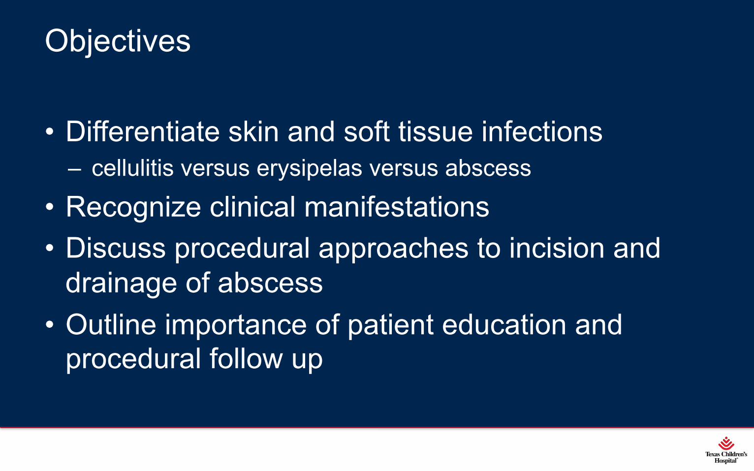
Objectives
• Differentiate skin and soft tissue infections– cellulitis versus erysipelas versus abscess
• Recognize clinical manifestations• Discuss procedural approaches to incision and
drainage of abscess• Outline importance of patient education and
procedural follow up

Skin and Soft Tissue Infections (SSTI)
• Epidemiology– Skin barrier disruption due to trauma
• Bite, abrasion, penetrating wound, ulcer, injection site, etc– Skin inflammation
• Eczema, radiation therapy– Edema due to venous insufficiency and/or impaired
lymphatic drainage– Immunosuppression
• Diabetes, HIV

SSTI
• Microbiology—most common causes– Staphylococcus Aureus
• MSSA• MRSA
– Beta-hemolytic Streptococcus• Group A Streptococcus• Streptococcus pyogenes

SSTI Standard Treatment
• Cellulitis and Erysipelas – Warm Compress– Antibiotic treatment
• Abscess – Incision and Drainage– +/- Antibiotic treatment

Soft Tissue Skin Infections

Cellulitis
• Involves deeper dermis and subcutaneous fat• Skin erythema, edema, warmth• No fluid collections• Warm, indurated, erythematous• +/- Red streaking?• Mark area of erythema to evaluate improvement• Responsive to antibiotic treatment

Cellulitis

Erysipelas
• Involves upper dermis• Clear demarcation between involved and
uninvolved tissue• Erythema, warmth, edema• Mark area of erythema to evaluate improvement• Responsive to antibiotic treatment

Erysipelas

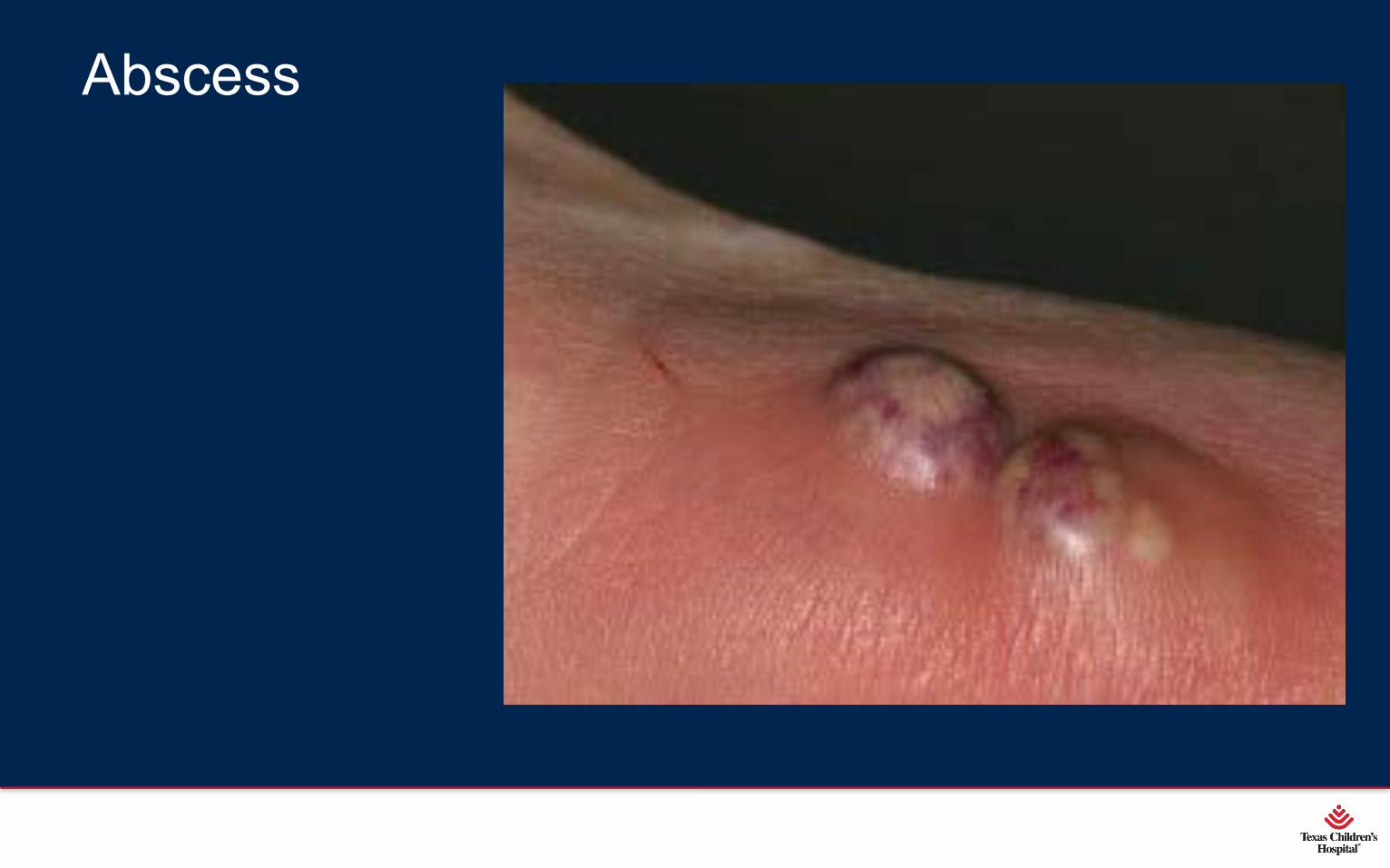
Abscess
• Collection of pus within dermis and/or subcutaneous space
• Painful, fluctuant, erythematous nodule• +/- surrounding cellulitis• +/- spontaneous drainage
Abscess

Abscess
• Incision & Drainage is curative most abscess• Wound cultures not routinely performed on
otherwise healthy patient
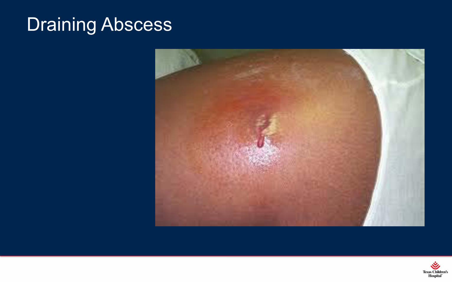
Draining Abscess

Point-of-care Ultrasound
• Useful when examination is equivocal– Cellulitis
• No fluid collection, increased thickness of subcutaneous tissue– Abscess
• Fluid collection identified • Estimate size and depth of abscess cavity
• Painful during procedure
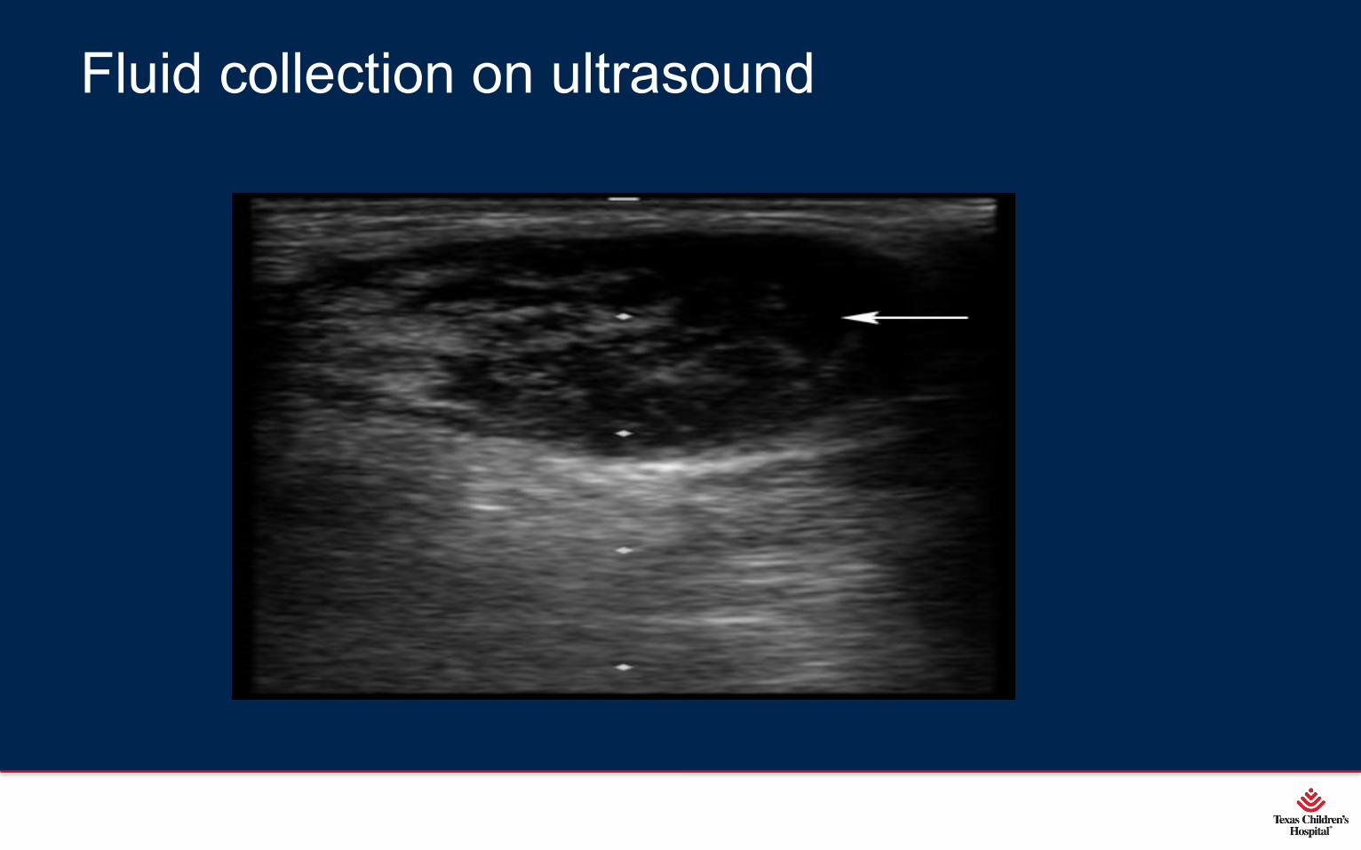
Fluid collection on ultrasound

Obtain Procedural Consent
• Review and document diagnosis– Specifically document location of abscess
• Example: Right anterolateral mid thigh abscess
• Explain and document procedure– Document procedure and location
• Example: Incision and drainage right anterolateral mid thigh abscess
– Do not use abbreviations

Obtain Procedural Consent
• Possible risks and complications– Pain – Infection– Bleeding– Scarring– Recurrence– Need for additional procedures

Procedural Location
• Bedside Incision and Drainage– Patient can tolerate sedation with local anesthetic
• Intraoperative Incision and Drainage– Difficult airway– Aspiration risk– Proximity of abscess location to neurovascular structures

Procedural Sedation Precautions
• Patient Factors– Difficult airway– Aspiration risk– Extremes of age– Bleeding dyscrasia / coagulopathy– Lidocaine allergy

Bedside Incision and Drainage Contraindications
• Abscess location– Proximity to neurovascular structures– Breast– Perirectal
• Abscess type– Recurrent– Multiple interconnected abscesses

Local Injection and Block
• Plain Lidocaine 1%– Max dose = 4mg/kg of 1% plain lidocaine
• Lidocaine 1% with Epinephrine– Max Dose = 7mg/kg of 1% lidocaine with epinephrine– Additional benefits
• Decrease local bleeding
• Reduces systemic lidocaine absorption
• Extends duration of anesthesia

Equipment needed– Sterile gloves, drapes, and 4x4 gauze– Prep solution – chlorhexidine or povidone iodine– Scalpel – 11 blade– Laceration set – curved hemostat, scissors, forceps– Basin with sterile normal saline for irrigation– +/- penrose drain, vessel loop, packing material – iodoform
or plain packing gauze– Dressing – 4x4 gauze / tape / coban / ace wrap / spandage

Incision and Drainage Procedure
• Incision to conform to natural folds of skin, when possible
• Express and evacuate all purulent material• Probe for purulent loculations• Copious irrigation with sterile saline until no
purulence remains

Conventional Incision and Drainage
• Linear incision whole length of abscess• Probe cavity
– Break up loculations– Identify foreign bodies– Ensure proper drainage
• Care given not to undermine healthy tissue or create new tunnels / tracks

Conventional Incision and Drainage (I&D)

Conventional I&D Closure Options
• Leave open for secondary intention
• Close loosely with non-absorbable suture– Vertical mattress– Penrose drain placement
• Abscess cavity packing

Secondary Intention after I&D

Secondary Intention Discharge Instructions
• Follow up 1 week with Primary Care and/or Surgeon
• Change dressing bid to qid prn to keep dry– 4x4 gauze, tape / coban / ace wrap / spandage

Penrose Drain Closure

Penrose Drain Discharge Instructions
• Follow up 1 week with Primary Care and/or Surgeon
• Remove drain 7 -10 days when drainage resolved
• Change outer dressing daily or prn to keep dry– 4x4 gauze, tape / coban / ace wrap / spandage

Abscess Cavity Packing after I&D Options
• Loosely pack cavity– > 5cm in diameter
– Pilonidal abscess
– Immunocompromised or diabetic
• Packing Options– Sterile noniodoform / iodoform packing gauze
– Silver-containing hydrofiber packing strips (Aquacel Ag)

Abscess Cavity Packing – Iodoform

Abscess Cavity Packing Discharge Instructions
• Follow up 1 week with Primary Care and/or Surgeon
• Change packing 1 – 2 times daily
• Change outer dressing daily or prn to keep dry– 4x4 gauze, tape / coban / ace wrap / spandage

Loop Drainage I&D Benefits
• Decrease– Pain – negates need for repetitive packing– Scarring– Frequency of follow up appointments– Lower healthcare cost
• Wound care supplies• Postoperative home health visits
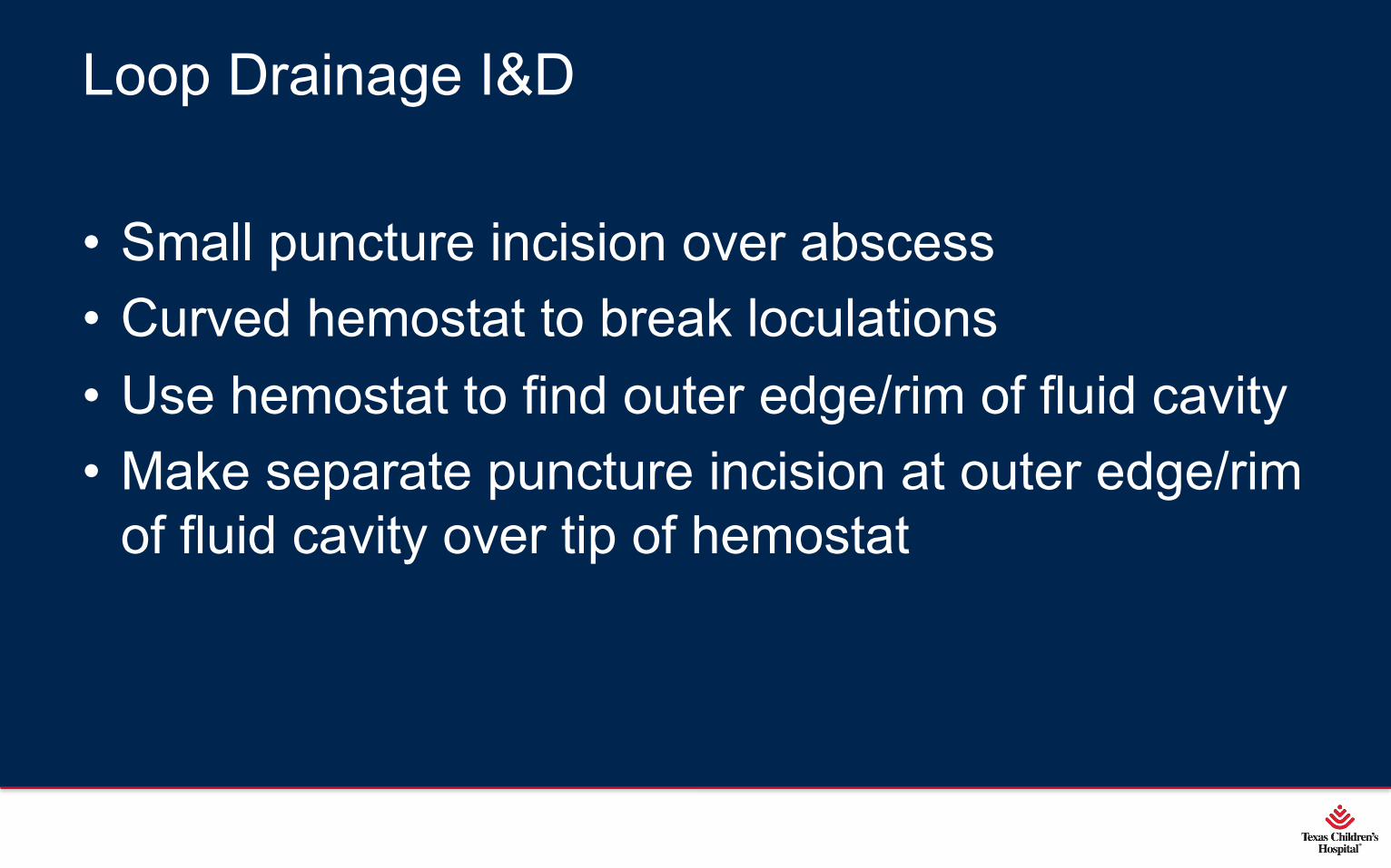
Loop Drainage I&D
• Small puncture incision over abscess• Curved hemostat to break loculations• Use hemostat to find outer edge/rim of fluid cavity• Make separate puncture incision at outer edge/rim
of fluid cavity over tip of hemostat
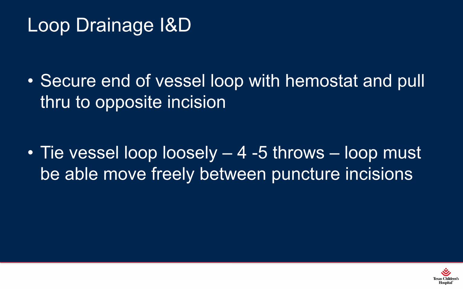
Loop Drainage I&D
• Secure end of vessel loop with hemostat and pull thru to opposite incision
• Tie vessel loop loosely – 4 -5 throws – loop must be able move freely between puncture incisions

Loop Drainage I&D

Loop Drainage I&D

Loop Drainage Discharge Instructions
• Follow up 1 week with Primary Care and/or Surgeon
• Wiggle vessel loop back and forth daily– Encourages drainage
• Removal of vessel loop in 7 -10 days when drainage resolved
• Change outer dressing daily or prn to keep dry– 4x4 gauze, tape / coban / ace wrap / spandage
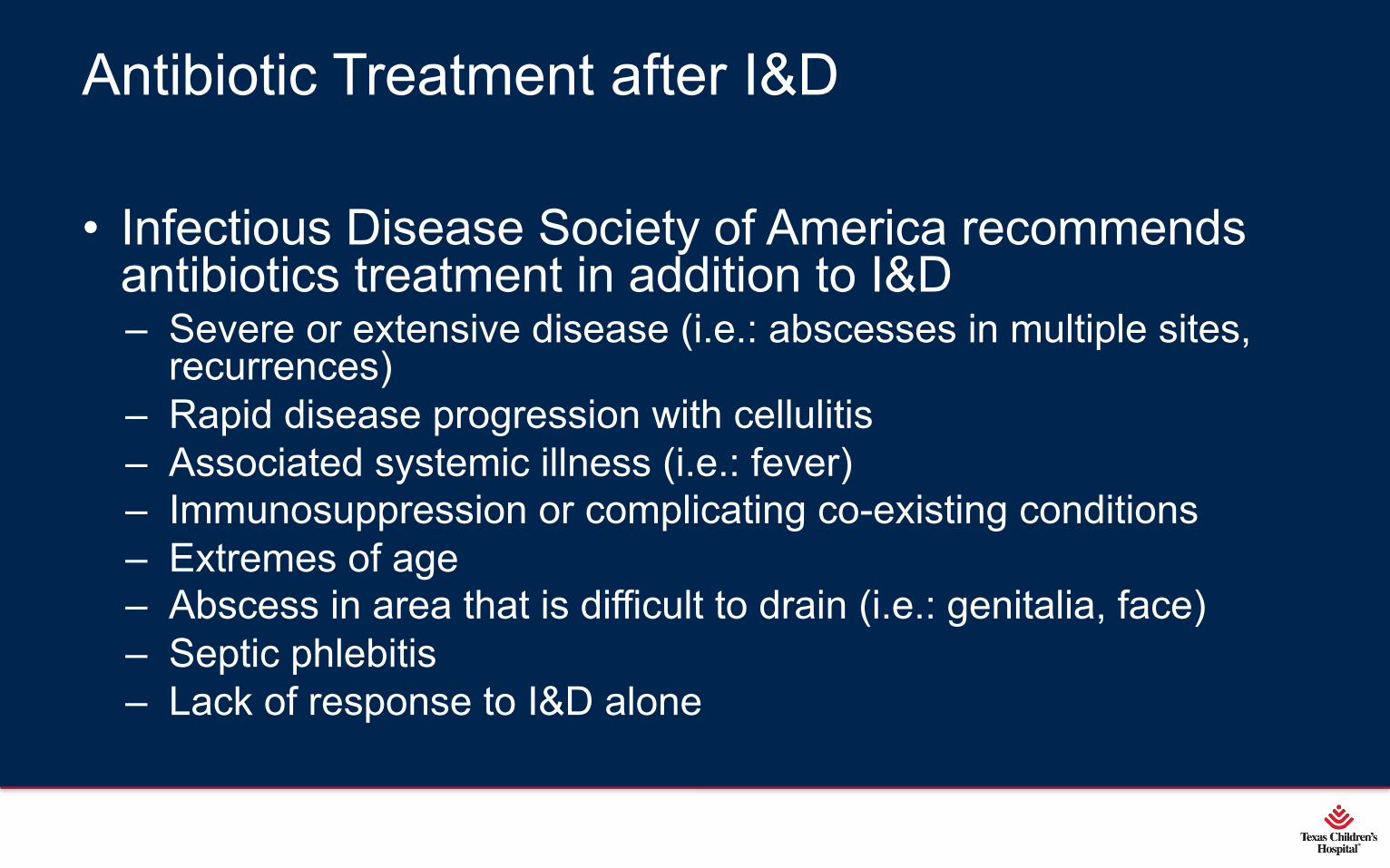
Antibiotic Treatment after I&D
• Infectious Disease Society of America recommends antibiotics treatment in addition to I&D – Severe or extensive disease (i.e.: abscesses in multiple sites,
recurrences)– Rapid disease progression with cellulitis– Associated systemic illness (i.e.: fever)– Immunosuppression or complicating co-existing conditions– Extremes of age– Abscess in area that is difficult to drain (i.e.: genitalia, face)– Septic phlebitis– Lack of response to I&D alone

Thank you!