The antimicrobial protein, CAP37, is upregulated in ... · The antimicrobial protein, CAP37, is...
16
1 3 Histochem Cell Biol (2015) 144:293–308 DOI 10.1007/s00418-015-1347-x ORIGINAL PAPER The antimicrobial protein, CAP37, is upregulated in pyramidal neurons during Alzheimer’s disease Amanda J. Brock 1 · Anne Kasus‑Jacobi 1,2 · Megan Lerner 3 · Sreemathi Logan 4 · Adekunle M. Adesina 7 · H. Anne Pereira 1,2,5,6 Accepted: 29 June 2015 / Published online: 14 July 2015 © The Author(s) 2015. This article is published with open access at Springerlink.com source(s) of CAP37 in brains of AD patients. Brain tissues from patients and age-matched controls were analyzed for CAP37 expression using immunohistochemistry (IHC). To determine factors that induce CAP37 in AD, HCN-1A pri- mary human neurons were treated with tumor necrosis fac- tor-alpha (TNF-α) or amyloid β 1−40 (Aβ) and analyzed by IHC. Western blotting and quantitative reverse transcription polymerase chain reaction (qRT-PCR) were used to con- firm CAP37 expression in neurons and brain tissues. IHC revealed CAP37 in cortical neurons in temporal and pari- etal lobes as well as CA3 and CA4 hippocampal neurons in patients with AD. CAP37 was found in more neurons in AD patients compared with age-matched controls. qRT- PCR and Western blotting showed an increase in CAP37 transcript and protein in the AD temporal lobe, a brain region that is highly impacted in AD. qRT-PCR observa- tions confirmed CAP37 expression in neurons. TNF-α and Aβ increased neuronal expression of CAP37. These find- ings support our hypothesis that neuronal CAP37 may modulate the neuroinflammatory response in AD. Keywords Neuroinflammation · CAP37 · Alzheimer’s disease · Neurons · Amyloid-beta · Microglia Abbreviations AD Alzheimer’s disease CAP37 Cationic antimicrobial protein of 37 kDa PMN Polymorphonuclear leukocyte IHC Immunohistochemistry TNF-α Tumor necrosis factor-alpha Aβ Amyloid-beta qRT-PCR Quantitative reverse transcription polymerase chain reaction PKC Protein kinase C LPS Lipopolysaccharide Abstract Inflammation is a well-defined factor in Alz- heimer’s disease (AD). There is a strong need to identify the molecules contributing to neuroinflammation so that therapies can be designed to prevent immune-mediated neurotoxicity. The cationic antimicrobial protein of 37 kDa (CAP37) is an inflammatory mediator constitutively expressed in neutrophils (PMNs). In addition to antibi- otic activity, CAP37 exerts immunomodulatory effects on microglia. We hypothesize that CAP37 mediates the neu- roinflammation associated with AD. However, PMNs are not customarily associated with the pathology of AD. This study was therefore designed to identify non-neutrophilic * H. Anne Pereira [email protected] 1 Oklahoma Center for Neuroscience, University of Oklahoma Health Sciences Center, 1110 N. Stonewall Ave., CPB 255, Oklahoma City, OK 73117, USA 2 Department of Pharmaceutical Sciences, University of Oklahoma Health Sciences Center, 1110 N. Stonewall Ave., CPB 255, Oklahoma City, OK 73117, USA 3 Department of Surgery, University of Oklahoma Health Sciences Center, 1122 NE 13th St., ORB 350, Oklahoma City, OK 73117, USA 4 Department of Geriatrics, University of Oklahoma Health Sciences Center, 975 NE 10th St., BRC 1303, Oklahoma City, OK 73104, USA 5 Department of Cell Biology, University of Oklahoma Health Sciences Center, 1110 N. Stonewall Ave., CPB 329, Oklahoma City, OK, USA 6 Department of Pathology, University of Oklahoma Health Sciences Center, 1110 N. Stonewall Ave., CPB 329, Oklahoma City, OK 73117, USA 7 Department of Pathology, Baylor College of Medicine, One Baylor Plaza, Rm 286A, Houston, TX 77030, USA
Transcript of The antimicrobial protein, CAP37, is upregulated in ... · The antimicrobial protein, CAP37, is...

1 3
Histochem Cell Biol (2015) 144:293–308DOI 10.1007/s00418-015-1347-x
ORIGINAL PAPER
The antimicrobial protein, CAP37, is upregulated in pyramidal neurons during Alzheimer’s disease
Amanda J. Brock1 · Anne Kasus‑Jacobi1,2 · Megan Lerner3 · Sreemathi Logan4 · Adekunle M. Adesina7 · H. Anne Pereira1,2,5,6
Accepted: 29 June 2015 / Published online: 14 July 2015 © The Author(s) 2015. This article is published with open access at Springerlink.com
source(s) of CAP37 in brains of AD patients. Brain tissues from patients and age-matched controls were analyzed for CAP37 expression using immunohistochemistry (IHC). To determine factors that induce CAP37 in AD, HCN-1A pri-mary human neurons were treated with tumor necrosis fac-tor-alpha (TNF-α) or amyloid β1−40 (Aβ) and analyzed by IHC. Western blotting and quantitative reverse transcription polymerase chain reaction (qRT-PCR) were used to con-firm CAP37 expression in neurons and brain tissues. IHC revealed CAP37 in cortical neurons in temporal and pari-etal lobes as well as CA3 and CA4 hippocampal neurons in patients with AD. CAP37 was found in more neurons in AD patients compared with age-matched controls. qRT-PCR and Western blotting showed an increase in CAP37 transcript and protein in the AD temporal lobe, a brain region that is highly impacted in AD. qRT-PCR observa-tions confirmed CAP37 expression in neurons. TNF-α and Aβ increased neuronal expression of CAP37. These find-ings support our hypothesis that neuronal CAP37 may modulate the neuroinflammatory response in AD.
Keywords Neuroinflammation · CAP37 · Alzheimer’s disease · Neurons · Amyloid-beta · Microglia
AbbreviationsAD Alzheimer’s diseaseCAP37 Cationic antimicrobial protein of 37 kDaPMN Polymorphonuclear leukocyteIHC ImmunohistochemistryTNF-α Tumor necrosis factor-alphaAβ Amyloid-betaqRT-PCR Quantitative reverse transcription polymerase
chain reactionPKC Protein kinase CLPS Lipopolysaccharide
Abstract Inflammation is a well-defined factor in Alz-heimer’s disease (AD). There is a strong need to identify the molecules contributing to neuroinflammation so that therapies can be designed to prevent immune-mediated neurotoxicity. The cationic antimicrobial protein of 37 kDa (CAP37) is an inflammatory mediator constitutively expressed in neutrophils (PMNs). In addition to antibi-otic activity, CAP37 exerts immunomodulatory effects on microglia. We hypothesize that CAP37 mediates the neu-roinflammation associated with AD. However, PMNs are not customarily associated with the pathology of AD. This study was therefore designed to identify non-neutrophilic
* H. Anne Pereira [email protected]
1 Oklahoma Center for Neuroscience, University of Oklahoma Health Sciences Center, 1110 N. Stonewall Ave., CPB 255, Oklahoma City, OK 73117, USA
2 Department of Pharmaceutical Sciences, University of Oklahoma Health Sciences Center, 1110 N. Stonewall Ave., CPB 255, Oklahoma City, OK 73117, USA
3 Department of Surgery, University of Oklahoma Health Sciences Center, 1122 NE 13th St., ORB 350, Oklahoma City, OK 73117, USA
4 Department of Geriatrics, University of Oklahoma Health Sciences Center, 975 NE 10th St., BRC 1303, Oklahoma City, OK 73104, USA
5 Department of Cell Biology, University of Oklahoma Health Sciences Center, 1110 N. Stonewall Ave., CPB 329, Oklahoma City, OK, USA
6 Department of Pathology, University of Oklahoma Health Sciences Center, 1110 N. Stonewall Ave., CPB 329, Oklahoma City, OK 73117, USA
7 Department of Pathology, Baylor College of Medicine, One Baylor Plaza, Rm 286A, Houston, TX 77030, USA
294 Histochem Cell Biol (2015) 144:293–308
1 3
CNS Central nervous systemCERAD Consortium to establish a registry for Alzhei-
mer’s diseaseDMEM Dulbecco’s modified Eagle’s mediumATCC American type culture collectionHRP Horseradish peroxidaseGAPDH Glyceraldehyde dehydrogenaseBCA Bicinchoninic acidTBST Tris-buffered saline with TweenECL Enhanced chemiluminescenceCA Cornu AmmonisCSF Cerebrospinal fluidAMP Antimicrobial peptideNO Nitric oxideROS Reactive oxygen species
Introduction
The cationic antimicrobial protein of molecular weight 37 kDa (CAP37) is an inflammatory mediator expressed constitutively in the azurophil granules of polymorpho-nuclear neutrophils (PMNs), and is considered an impor-tant component of the innate immune system (Pereira et al. 1990a; Griffith et al. 2013). Previous findings have shown increased CAP37 levels during inflammatory conditions such as sepsis and atherosclerosis (Lee et al. 2002; Pereira et al. 2003; Linder et al. 2009). CAP37 has potent antimicrobial activity (Pereira et al. 1993, 1995, 2006) and also exerts various regulatory functions in mammalian cells. Some of these functions include activa-tion of protein kinase C (PKC) and upregulation of adhe-sion proteins on endothelial cells and corneal epithelial cells; contraction of endothelial cells; proliferation of smooth muscle cells; corneal epithelial wound healing; and chemotaxis of monocytes, microglia, smooth muscle cells, and corneal epithelial cells (Pereira et al. 1990a, 2003, 2004; Gautam et al. 2001; Gonzalez et al. 2004; Griffith et al. 2014).
CAP37 is induced in corneal epithelial cells, endothe-lial cells, and smooth muscle cells in response to cytokines, lipopolysaccharide (LPS), and infection (Pereira et al. 1996b; Lee et al. 2002; Ruan et al. 2002; Gonzalez et al. 2004). In addition, CAP37 expression has been found in endothelial cells of hippocampal vasculature in patients with Alzheimer’s disease (AD), while this expression was absent in age-matched controls (Pereira et al. 1996a). This finding is important, as the hippocampus is responsible for memory formation and is one of the main regions where AD pathol-ogy first manifests (Pereira et al. 1996a). Although CAP37 shares ~45, ~42, and ~32 % sequence homology with the serine proteases elastase, proteinase 3, and cathepsin G,
respectively, CAP37 itself lacks serine protease activity due to the loss of 2 of 3 conserved residues of the catalytic triad (Pereira et al. 1990b). Elastase and proteinase-3 have been observed in various regions of the brain, including the hip-pocampus, cerebellum, and cerebral cortex (Davies et al. 1998), and elastase has been detected in murine microglial cells (Nakajima et al. 1992). However, the exact cell expres-sion profile of these other proteases in the human brain is unknown. Expression of human β-defensin-1, another cati-onic antimicrobial peptide, has been reported within brain hippocampal astrocytes, neurons, and the choroid plexus and was increased in these regions in patients with AD (Williams et al. 2013). The role of these proteins in AD is unknown. However, since all are involved in innate immu-nity and defense, their expression in brain cells raises the question of whether any of these proteins might have a role in the low-grade chronic inflammatory response that occurs in neurodegenerative diseases such as AD (Wilson et al. 2002; Heneka et al. 2010; Hensley 2010; Grammas 2011; Eikelenboom et al. 2012).
A primary research focus of our laboratory is defin-ing the mechanisms whereby CAP37 modulates neuroin-flammation. As CAP37 is a potent activator of microglia, we hypothesized that CAP37 expressed within the brain parenchyma was one of the mediators of neuroinflamma-tion in Alzheimer’s disease. Thus, in this study, we aimed to determine the CAP37 cellular expression and localiza-tion in brains from patients with Alzheimer’s disease. Our results indicate that CAP37 is expressed in neutrophils, the vascular endothelium, and neurons in specific brain regions from patients with AD. CAP37 transcript and protein levels are increased in patients with AD, and primary neurons can be induced to express CAP37 in response to tumor necrosis factor-alpha (TNF-α) and amyloid-beta (Aβ).
Materials and methods
Tissue specimens
Tissues from nine patients diagnosed with AD and nine respective age-matched controls were kindly provided by Dr. Eileen Bigio of the Department of Pathology, North-western University Feinberg School of Medicine, Alzhei-mer’s Disease Center, Neuropathology Core. All patients and age-matched controls were characterized for AD pathology using the Consortium to Establish a Registry for Alzheimer’s Disease (CERAD) plaque grades (A–C) and Braak tangles stages (I–VI) to determine the approxi-mate disease stage. All AD tissue specimens used for IHC were given CERAD scores of C and Braak & Braak stage VI for tangles. Age-matched controls were given CERAD
295Histochem Cell Biol (2015) 144:293–308
1 3
scores of A, B, or 0, and were Braak & Braak stages I–III or 0.
Cell culture and treatment of HCN‑1A neurons
We purchased HCN-1A, primary human cortical neuronal cells, from American Type Culture Collection (ATCC, Manassas, VA). Dulbecco’s modified Eagle’s medium (DMEM, ATCC) supplemented with 20 % bovine calf serum, 1 % antibiotic–antimycotic, and 1 % l-glutamine (Life Technologies, Grand Island, NY) was used to cul-ture HCN-1A cells according to the ATCC recommen-dations. The HCN-1A cells were used in immunohis-tochemistry. During the course of our studies, ATCC discontinued the distribution of HCN-1A cells due to quick senescence of the cells and insufficient inventory. The HCN-1A cells were thus unavailable for use in other assays. HCN-1A cells were treated overnight (17 h) with either the 40 amino acid Aβ peptide (Aβ1−40, Bachem, Torrance, CA, stock solution in ultrapure water) at a con-centration of 125 µg/ml or human recombinant tumor necrosis factor-alpha (TNF-α, Roche, Indianapolis, IN) at a concentration of 25 ng/ml. Control treatments included HCN-1A incubation with either inactive (reverse peptide order) Aβ peptide (Aβ40−1) (Bachem, stock solution in 10 % acetic acid) or vehicle alone (basal medium or basal medium containing an equivalent volume of 10 % acetic acid). Aβ was diluted in basal medium and incubated for 2 h at room temperature before administration, to allow the formation of toxic oligomers and fibrillar aggregates that are found in AD.
Antisera
We used 7 μg/ml rabbit anti-CAP37 antiserum as described previously (Pereira et al. 1996a), a control of 7 μg/ml nor-mal rabbit antiserum (Jackson ImmunoResearch Labora-tories, West Grove, PA), an in-house made mouse mono-clonal antibody to CAP37 (D5F10), and a mouse IgG1 isotype antibody was used as a control (Sigma-Aldrich, St. Louis, MO). D5F10 and the isotype control were diluted in Emerald antibody diluent (Cell Marque, Rocklin, CA) to a concentration of 4 μg/ml for IHC and were diluted in 2 % bovine serum albumin (BSA, Calbiochem, Billerica, MA) in Tris-buffered saline with 0.05 % Tween (TBST) block-ing solution to a concentration of 0.2 μg/ml for Western blots. Rabbit polyclonal anti-Aβ (Cell Signaling, Danvers, MA) and rabbit polyclonal anti-phospho-tau (Santa Cruz, Dallas, TX) were diluted in Emerald antibody diluent to a concentration of 10 μg/ml to detect Aβ plaques and tau tangles, respectively. A rabbit IgG antibody was employed as an isotype control (Cell Signaling, Danvers, MA). We also used the following secondary antibodies: horseradish
peroxidase (HRP)-conjugated donkey anti-mouse and don-key anti-rabbit IgGs diluted in TBST to concentrations of 0.04 μg/ml (Jackson ImmunoResearch Laboratories, West Grove, PA).
Immunocytochemistry
HCN-1A cells were evenly seeded into 4-well LAB-TEK tissue culture chambers (NUNC, Inc., Naperville, IL) and incubated until confluency. Cells were then treated with either Aβ1−40, TNF-α, or serum-free DMEM as described above. Following treatment, cells were fixed with formol acetone and stained using the Vectastain ABC peroxidase system (Vector Laboratories, Burlingame, CA) as previ-ously described (Pereira et al. 1996a). The rabbit anti-CAP37 and normal rabbit serum were added for 17 h. Images of stained neurons were taken at 400X magnifica-tion (Nikon TE2000, Nikon Instruments Inc., Melville, NY). Figures were created using Microsoft PowerPoint 2010.
Immunohistochemistry
Formalin-fixed, paraffin-embedded brain tissues from patients with Alzheimer’s disease and age-matched con-trols were sectioned at a thickness of 5 μm. Sections were incubated in antigen retrieval solution (Tris buffer, pH 9) for 20 min in a rice steamer, followed by a 20-min cool down in distilled water. Staining was performed using reagents from the HiDef HRP kit (Cell Marque) according to the manufacturer’s instructions. Tissues were incubated with mouse anti-CAP37 (4 μg/ml) or the equivalent amount of mouse IgG1 isotype control for 60 min. Color was developed with 3,3′-diaminoben-zidine (Cell Marque), and sections were counterstained with hematoxylin (American Master Tech, Lodi, CA). Images were examined, and photographs were taken using bright-field microscopy at 400X and 1000X mag-nifications (Nikon eclipse E200, Nikon Instruments Inc., Melville, NY). Figures were created using Microsoft PowerPoint 2010.
Quantitative RT‑PCR
Total RNA from primary human neurons and primary human astrocytes (Sciencell, Carlsbad, CA) and total RNA from temporal, frontal, and occipital lobe tissues of patients with AD and five donor pool normal controls (Bio-chain, Newark, CA) were purchased from their respective commercial vendors. AD patients included a 73-year-old male (frontal lobe RNA), 77-year-old male (occipital lobe RNA), 80-year-old male (temporal and frontal lobe RNA), 83-year-old male (temporal lobe RNA), 85-year-old female
296 Histochem Cell Biol (2015) 144:293–308
1 3
(occipital lobe RNA), and an 87-year-old male (temporal, frontal, and occipital lobe RNA) for a total of six patients analyzed with three utilized for each brain region. Pooled controls were all from males with ages ranging from 20 to 44 years. PCR-ready first-strand cDNA from peripheral blood leukocytes was obtained from Biochain. PCR-ready first-strand cDNA from human microglia was obtained from Sciencell. Primary neuron RNA (2 μg), primary astrocyte RNA (2 μg), AD patient RNA (3 μg), and nor-mal control pooled RNA (3 μg) were converted to cDNA using the Qiagen RT2 First-Strand Kit (Qiagen Inc., Valen-cia, CA) in a final volume of 111 μL according to the manufacturer’s instructions. Amplification of cDNA was performed using RT2 SYBR Green mastermix (containing HotStart DNA Taq Polymerase), Qiagen RT2qPCR primers (GAPDH: #PPH00150F, PRTN3: #PPH07029A, ELANE: #PPH01057A, AZU1: #PPH01031A), and Solaris prim-ers (CTSG #AX-005838-00-0100, GAPDH# AX-005838-00-0100), following the manufacturer’s instructions. For each 25-μL reaction, 1 μL of the prepared cDNA was mixed with 1 μL (0.4 μM) of respective primer, 12.5 μL of RT2 SYBR Green mastermix, and 10.5 μL of RNase-free water. All reactions were performed in triplicate. PCRs were performed using the MyiQ Single Color Real-Time PCR Detection System (Bio-Rad, Hercules, CA) begin-ning with a denaturation step (10 min at 95 °C) followed by 40 repeated cycles of annealing/extension: 15 s at 95 °C and 1 min at 60 °C. qRT-PCR was performed on primary neurons, primary microglia, primary astrocytes, and nor-mal control pools twice and on AD patients and leuko-cytes once. The ΔCt values were calculated to normal-ize each gene expression to glyceraldehyde 3-phosphate dehydrogenase (GAPDH). To calculate fold difference in mRNA expression relative to GAPDH, the equation [fold change = 2(−ΔCt)] was used. Figures were created using Prism software (GraphPad Software Inc., version 6, La Jolla, CA.)
Western blot analysis
Total protein lysates from the temporal lobe of one patient with AD (75-year-old male), from the frontal lobe of one patient with AD (83-year-old male), and from the tempo-ral (26-year-old male) and frontal (71-year-old male) lobes of normal controls were obtained from Biochain. Accord-ing to the vendor, all tissues were obtained 4–6 h post-mortem and were stored in liquid nitrogen before distribu-tion. Human astrocyte and microglia lysates were derived from single healthy donors and obtained from Sciencell. The protein concentration of each sample was provided by the respective vendors. The protein concentration of total temporal and frontal lobe lysates was verified using the
bicinchoninic acid (BCA) assay (Pierce, Rockford, IL). Purified CAP37 derived from human neutrophils (Athens Research Technology, Athens, Georgia) was also run as a marker for CAP37 migration. Total lysates from human astrocytes, human microglia, temporal lobes, frontal lobes (40 μg), PMNs (50 ng), and purified CAP37 (5 ng) were separated by a 12.5 % SDS-PAGE gel. Proteins were trans-ferred to a nitrocellulose membrane (Whatman, Pittsburgh, PA) overnight, and the membranes were blocked in 2 % BSA in TBST. Membranes were probed with either mouse monoclonal anti-CAP37 (0.2 μg/ml) or the equivalent amount of mouse isotype control overnight at 4 °C. Blots were incubated with HRP-conjugated donkey anti-mouse secondary antibody (0.04 μg/ml; Jackson Laboratories) for 1 h at room temperature. Enhanced chemiluminescence (ECL) substrates (Pierce, Rockford, IL) were used to develop all blots. ImageJ software (National Institutes of Health [NIH], Bethesda, MD) was used to quantify mean band density. Figures were created using Microsoft Pow-erPoint 2010.
Statistical analysis
Statistical analysis was performed using Prism software. Student’s unpaired t tests were used to analyze the AZU1, ELANE, and PRTN3 mRNA expressions of individual patients with AD relative to the normal control pools. The mRNA expression values were calculated as described in the above methods and are represented as mean ± SEM, and p < 0.05 was considered statistically significant.
Results
Both parietal and temporal lobes from AD patients express CAP37
CAP37 expression in the endothelial cells lining vessels of the hippocampus in patients with AD has been pre-viously demonstrated (Pereira et al. 1996a). However, whether CAP37 was also expressed in additional brain regions from AD patients has not been studied. There-fore, parietal and temporal lobe tissues from patients with AD (n = 9) or age-matched normal controls (n = 9) were evaluated for CAP37 expression. Performing IHC using a novel monoclonal antibody specific for CAP37 detected expression of this immune modulator in the polymorphonuclear leukocytes (PMNs) and endothelial cells in both the parietal (Fig. 1a) and temporal lobes (Fig. 1b) of all AD patients and age-matched controls (Fig. 1e, f). Staining with isotype control antibody failed to detect PMNs and endothelial cells, indicating the

297Histochem Cell Biol (2015) 144:293–308
1 3
specificity of staining for CAP37 in these two cell types (Fig. 1c, d, g, h). These assays confirmed the applicabil-ity of this monoclonal antibody for use in IHC on brain
tissues since CAP37 is constitutively expressed in neu-trophils and is also expressed in endothelial cells from other tissues.
Fig. 1 CAP37 is expressed in neutrophils and endothelial cells of parietal and temporal cortices. a Parietal lobe and b temporal lobe cortical tissues from an Alzheimer’s disease (AD) patient stained with anti-CAP37. c Parietal lobe and d temporal lobe cortical tissues from an AD patient stained with isotype control. e Parietal lobe and f temporal lobe cortical tissues from an age-matched control stained with anti-CAP37. g Parietal lobe and h temporal lobe cortical tissues from an age-matched control stained with isotype control. Neutrophils are indicated by arrowheads, and endothelial cells are represented by arrows. Scale bars 20 μm
a b
c d
Alzheimer’s disease
Age-matched controls
e f
g h
parietal temporal
anti-
CA
P37
Isot
ype
cont
rol
anti-
CA
P37
Isot
ype
cont
rol

298 Histochem Cell Biol (2015) 144:293–308
1 3
Cortical pyramidal neurons in temporal and parietal lobes are a novel cellular source of CAP37 expression and are a site of CAP37 upregulation in AD patients
The temporal and parietal lobes of AD patients were then evaluated to identify additional CAP37 cellular sources. CAP37 was detected in the cytoplasm of cortical neurons in the temporal lobes of patients with AD (Fig. 2ai, aii). The pattern of CAP37 staining varied from localized in pyramidal layers 3 and 5 to a diffuse cortical distribution.
We observed a predominant limitation to pyramidal layers 3 and 5 more often than a diffuse cortical pattern. How-ever, a diffuse pattern was often associated with a more intense cytoplasmic staining on a per-patient basis. No temporal lobe staining was observed with the isotype con-trol (Fig. 2bi, bii), indicating the CAP37 staining specific-ity in PMNs, endothelial cells, and neurons. CAP37 was observed in temporal lobe pyramidal layers 3 and 5 in some control subjects as well. However, fewer neurons were CAP37-positive in control tissues (Fig. 2ci, cii) than in
Fig. 2 Temporal cortical neurons have increased CAP37 expression during AD. ai AD temporal cortical lobe tissue stained with monoclonal anti-CAP37 showing strong staining in the AD neuron cell bodies. CAP37 staining varied from localized staining in pyramidal layers 3 and 5 to a diffuse corti-cal distribution. bi AD temporal cortical lobe tissue stained with isotype control. ci Age-matched control brain stained with monoclonal anti-CAP37. Note the reduced staining in the neuron cell bodies. aii, bii, cii Lower magnification images of respective sections. Aster-isks (*) with pointing arrows indicate microglial cells that lack CAP37. Scale bars ai, bi, and ci, 20 μm; aii, bii, and cii, 50 μm
aiiai
*
bi bii
ci cii
Ant
i-CA
P37
Isot
ype
cont
rol
Ant
i-CA
P37
*
*
Age-matched controls
Alzheimer's disease
299Histochem Cell Biol (2015) 144:293–308
1 3
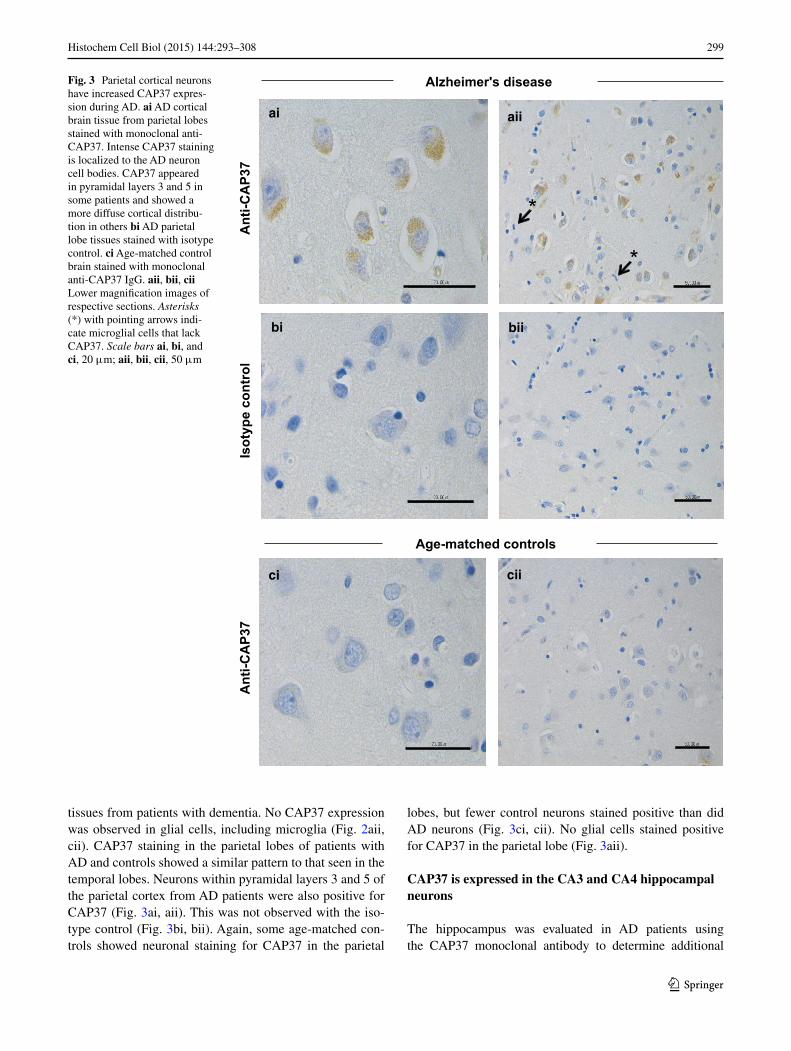
tissues from patients with dementia. No CAP37 expression was observed in glial cells, including microglia (Fig. 2aii, cii). CAP37 staining in the parietal lobes of patients with AD and controls showed a similar pattern to that seen in the temporal lobes. Neurons within pyramidal layers 3 and 5 of the parietal cortex from AD patients were also positive for CAP37 (Fig. 3ai, aii). This was not observed with the iso-type control (Fig. 3bi, bii). Again, some age-matched con-trols showed neuronal staining for CAP37 in the parietal
lobes, but fewer control neurons stained positive than did AD neurons (Fig. 3ci, cii). No glial cells stained positive for CAP37 in the parietal lobe (Fig. 3aii).
CAP37 is expressed in the CA3 and CA4 hippocampal neurons
The hippocampus was evaluated in AD patients using the CAP37 monoclonal antibody to determine additional
Fig. 3 Parietal cortical neurons have increased CAP37 expres-sion during AD. ai AD cortical brain tissue from parietal lobes stained with monoclonal anti-CAP37. Intense CAP37 staining is localized to the AD neuron cell bodies. CAP37 appeared in pyramidal layers 3 and 5 in some patients and showed a more diffuse cortical distribu-tion in others bi AD parietal lobe tissues stained with isotype control. ci Age-matched control brain stained with monoclonal anti-CAP37 IgG. aii, bii, cii Lower magnification images of respective sections. Asterisks (*) with pointing arrows indi-cate microglial cells that lack CAP37. Scale bars ai, bi, and ci, 20 μm; aii, bii, cii, 50 μm
ai aii
bi bii
ci cii
*
*
Alzheimer's disease
Age-matched controls
Ant
i-CA
P37
Isot
ype
cont
rol
Ant
i-CA
P37
300 Histochem Cell Biol (2015) 144:293–308
1 3
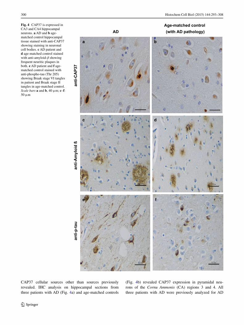
CAP37 cellular sources other than sources previously revealed. IHC analysis on hippocampal sections from three patients with AD (Fig. 4a) and age-matched controls
(Fig. 4b) revealed CAP37 expression in pyramidal neu-rons of the Cornu Ammonis (CA) regions 3 and 4. All three patients with AD were previously analyzed for AD
Fig. 4 CAP37 is expressed in CA3 and CA4 hippocampal neurons. a AD and b age-matched control hippocampal tissue stained with anti-CAP37 showing staining in neuronal cell bodies. c AD patient and d age-matched control stained with anti-amyloid-β showing frequent neuritic plaques in both. e AD patient and f age-matched control stained with anti-phospho-tau (Thr 205) showing Braak stage VI tangles in patient and Braak stage II tangles in age-matched control. Scale bars a and b, 40 μm; c–f: 50 μm
ADAge-matched control (with AD pathology)
anti-
CA
P37
anti-
Am
yloi
d ß
anti-
p-ta
u
a b
c d
e f
301Histochem Cell Biol (2015) 144:293–308
1 3
pathology and characterized to have CERAD plaque scores of C and Braak and Braak stage VI for tangles. Our IHC staining also confirmed the high frequency of plaques (Fig. 4c) and tangles (Fig. 4e) in these patients. Although one of the age-matched controls showed no signs of neuritic plaques or tangles, the other two contained both plaques and tangles, with one also having frequent neuritic plaques (Fig. 4d) and Braak stage II for tangles (Fig. 4f). One age-matched control was, therefore, characterized as having low AD neuropathological change, while another was character-ized as displaying intermediate AD pathological change.
Next, CAP37 association with either Aβ plaques or tau tangles was determined by IHC to analyze the localiza-tion patterns of CAP37, Aβ, and tau. Serial sections of the temporal cortex and hippocampus stained for CAP37, Aβ, and tau revealed that both Aβ and tau were more heavily distributed than the sporadic neuron staining observed for CAP37. Although CAP37, Aβ, and tau overlapped in some regions, there did not appear to be a strong correlation in localization as some regions with CAP37 did not show tan-gles or plaques, and many regions with plaques and tangles did not also contain CAP37 (data not shown).
TNF‑α and Aβ induced expression of CAP37 in human primary cortical neurons
The ability of two AD mediators, TNF-α, a pro-inflam-matory cytokine, and Aβ1−40 to induce the expression of CAP37 was tested using primary human cortical neurons (HCN-1A) as a model for induced expression. These cells are non-malignant and retain their native neuronal phe-notype. They have the ability to divide more rapidly due to their derivation from a patient with unilateral megalen-cephaly, a low-grade proliferation and migration disorder affecting neurons. We initially examined these cells for constitutive expression of CAP37 and detected faint CAP37 expression (data not shown). Faint immunopositivity was also seen in vehicle-treated controls (Fig. 5a). HCN-1A neu-rons were treated with TNF-α and Aβ1−40. Then, CAP37 expression was determined using IHC. Both TNF-α and Aβ1−40 induced CAP37 expression in the neuronal cyto-plasm (Fig. 5c, d). Cells treated with the inactive form of Aβ (Aβ40−1) were not induced to express CAP37 (Fig. 5b). Minimal staining was observed with Aβ- and TNF-α-treated cells probed with rabbit control serum (Fig. 5e, f), confirm-ing the specificity of the CAP37 staining.
CAP37 mRNA is expressed in neurons, astrocytes, and microglia
The expression of CAP37 mRNA in primary human neu-rons, astrocytes, and microglia was analyzed using qRT-PCR. AZU1 mRNA, which encodes for CAP37 protein,
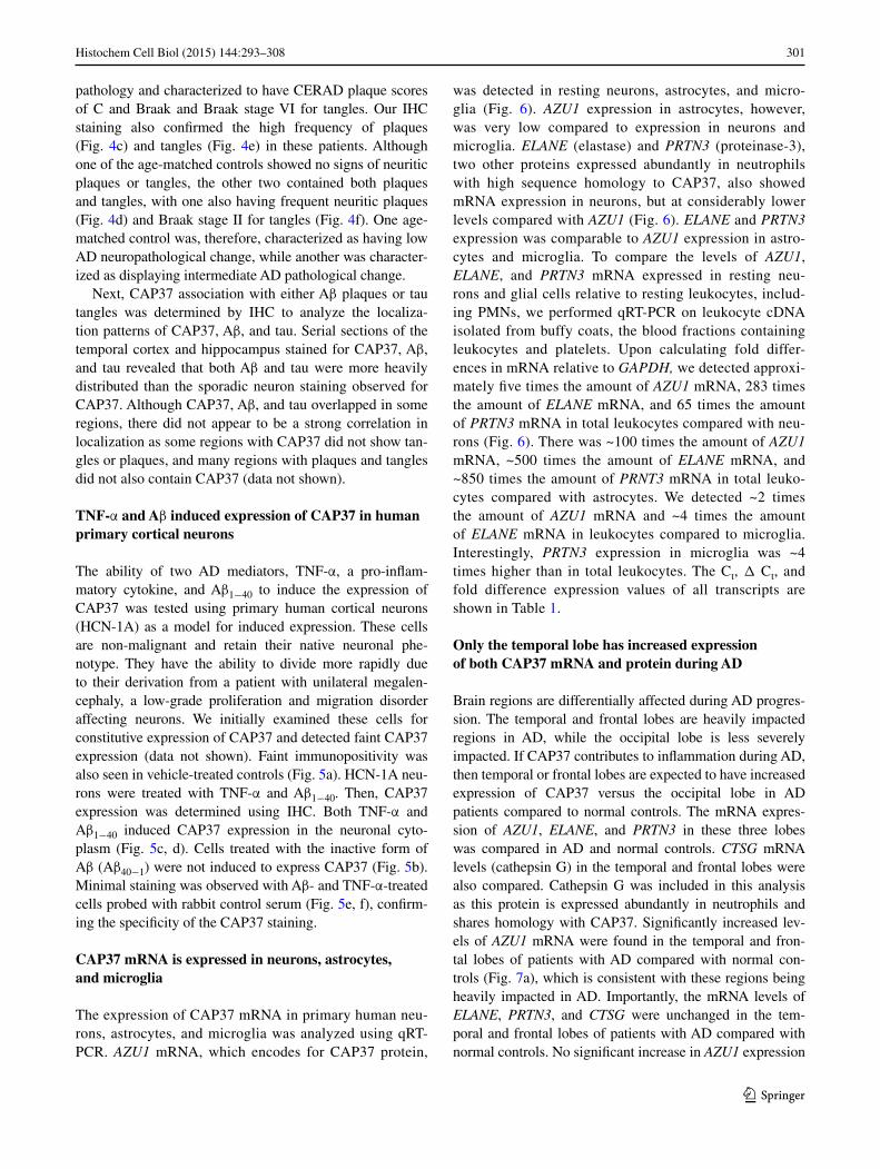
was detected in resting neurons, astrocytes, and micro-glia (Fig. 6). AZU1 expression in astrocytes, however, was very low compared to expression in neurons and microglia. ELANE (elastase) and PRTN3 (proteinase-3), two other proteins expressed abundantly in neutrophils with high sequence homology to CAP37, also showed mRNA expression in neurons, but at considerably lower levels compared with AZU1 (Fig. 6). ELANE and PRTN3 expression was comparable to AZU1 expression in astro-cytes and microglia. To compare the levels of AZU1, ELANE, and PRTN3 mRNA expressed in resting neu-rons and glial cells relative to resting leukocytes, includ-ing PMNs, we performed qRT-PCR on leukocyte cDNA isolated from buffy coats, the blood fractions containing leukocytes and platelets. Upon calculating fold differ-ences in mRNA relative to GAPDH, we detected approxi-mately five times the amount of AZU1 mRNA, 283 times the amount of ELANE mRNA, and 65 times the amount of PRTN3 mRNA in total leukocytes compared with neu-rons (Fig. 6). There was ~100 times the amount of AZU1 mRNA, ~500 times the amount of ELANE mRNA, and ~850 times the amount of PRNT3 mRNA in total leuko-cytes compared with astrocytes. We detected ~2 times the amount of AZU1 mRNA and ~4 times the amount of ELANE mRNA in leukocytes compared to microglia. Interestingly, PRTN3 expression in microglia was ~4 times higher than in total leukocytes. The Ct, Δ Ct, and fold difference expression values of all transcripts are shown in Table 1.
Only the temporal lobe has increased expression of both CAP37 mRNA and protein during AD
Brain regions are differentially affected during AD progres-sion. The temporal and frontal lobes are heavily impacted regions in AD, while the occipital lobe is less severely impacted. If CAP37 contributes to inflammation during AD, then temporal or frontal lobes are expected to have increased expression of CAP37 versus the occipital lobe in AD patients compared to normal controls. The mRNA expres-sion of AZU1, ELANE, and PRTN3 in these three lobes was compared in AD and normal controls. CTSG mRNA levels (cathepsin G) in the temporal and frontal lobes were also compared. Cathepsin G was included in this analysis as this protein is expressed abundantly in neutrophils and shares homology with CAP37. Significantly increased lev-els of AZU1 mRNA were found in the temporal and fron-tal lobes of patients with AD compared with normal con-trols (Fig. 7a), which is consistent with these regions being heavily impacted in AD. Importantly, the mRNA levels of ELANE, PRTN3, and CTSG were unchanged in the tem-poral and frontal lobes of patients with AD compared with normal controls. No significant increase in AZU1 expression

302 Histochem Cell Biol (2015) 144:293–308
1 3
was detected in the occipital lobe, which is a region of the brain that is less severely impacted in AD. Interestingly, PRTN3 mRNA expression was significantly decreased in the occipital lobe of the brain with AD (Fig. 7a). Neither the cause nor the consequence of this decrease is known. Com-paring the mRNA levels of AZU1, ELANE, PRTN3, and CTSG in five donor pools of normal controls, we found that
levels of AZU1 and CTSG were ~5–35 times lower than the levels of ELANE and PRTN3 in all brain regions analyzed (Fig. 7b). As noted in the methods section, all mRNA values were normalized to GAPDH expression.
CAP37 protein expression in the temporal and frontal lobes of patients with AD and normal controls was analyzed by Western blot to confirm transcript expression analysis.
Fig. 5 CAP37 is induced in HCN-1A primary human neurons. a HCN-1A neurons incubated with vehicle only (basal medium ± equivalent volume of 10 % acetic acid used as solvent for peptide). b HCN-1A neurons treated with Aβ40−1 (reverse/inactive peptide) stained with rabbit anti-CAP37 serum. c HCN-1A cortical neu-rons treated with TNF-α (25 ng/ml) and stained with rabbit anti-CAP37 serum. d HCN-1A cells treated with Aβ1−40 (125 μg/ml, pre-aggregated) and stained with rabbit anti-CAP37 serum. e HCN-1A cortical neurons treated with TNF-α (25 ng/ml) and stained with normal rabbit control serum. f HCN-1A neurons treated with Aβ1−40 (125 μg/ml, pre-aggregated) and stained with normal rabbit control serum. Treatments were performed overnight (17 h), and cells were stained using the Vectastain ABC peroxidase system. Scale bars 20 μm
Ant
i-CA
P37
Vehicle
TNF-
TNF-
Con
trol
ant
i-ser
um
ba
dc
e f

303Histochem Cell Biol (2015) 144:293–308
1 3
Total lysates from the temporal and frontal lobes showed low levels of CAP37 expression in normal controls (Fig. 8ai, bi, lane 1) as determined by the detection of an approximately
29-kDa band when blots were probed with monoclonal anti-CAP37. This is the same molecular weight to which the iso-lated native CAP37 migrated (Fig. 8ai, bi, lane 4). The tissues from the patient with AD demonstrated a band of increased intensity in the temporal lobe lysate that was absent in the normal control (Fig. 8ai, lane 1, 2). This result confirms qRT-PCR and IHC results showing an increase in CAP37 expres-sion in the temporal lobe of AD patients. We observed no difference in the intensity of the 29-kDa CAP37 band in the frontal lobe normal control or AD lysates (Fig. 8bi, lanes 1, 2). However, the extract from the frontal lobe of the patient with AD demonstrated a band of high intensity migrating at a molecular weight of ~15 kDa (Fig. 8bi, lane 2). This was not observed in the normal control (Fig. 8bi, lane 1). Blots were also probed with mouse isotype control which showed no corresponding bands at 29 or 15 kDa for the total lysates or purified CAP37 (Fig. 8aii, bii, lanes 1, 2, 4).
CAP37 protein is not detected in astrocytes or microglia by Western blotting
Since we observed CAP37 transcript in astrocytes and microglia, but did not observe CAP37 protein in these cells by IHC analysis of brain sections, we performed Western blotting on proteins extracted from astrocytes and micro-glial cells. No band corresponding to the CAP37 molecular weight was detected in astrocytes or microglia (Fig. 9, lanes 1,2). Purified native CAP37, PMN lysate, and total lysates
Fold
diff
eren
ce in
mR
NA
rela
tive
to G
APD
H
AZU1
ELANE
PRTN3
AZU1
ELANE
PRTN3
AZU1
ELANE
PRTN3
AZU1
ELANE
PRTN3
0.000
0.005
0.010
0.015
0.020Human Neurons
Human Leukocytes
Human Astrocytes
Human Microglia
Fig. 6 CAP37 mRNA is expressed in human primary neurons, astro-cytes, microglia, and leukocytes. AZU1, ELANE, and PRTN3 (encode for CAP37, elastase, and proteinase-3 proteins, respectively) mRNA expression in neurons (black bars), astrocytes (gray bars), microglia (light gray bars), and leukocytes (open bars) was determined using qRT-PCR. All values were normalized to GAPDH (internal control), and results are expressed as fold differences in mRNAs relative to GAPDH (2(−ΔCT)). Analysis of AZU1, ELANE, and PRTN3 neuronal and glial expression was performed in triplicate, and leukocyte expres-sion was performed in duplicate. Data are mean ± SEM of results
Table 1 Expression of AZU1, ELANE, and PRTN3 transcripts in primary human neurons, astrocytes, microglia, and leukocytes
HN Human neurons; HA human astrocytes; HM human microglia; HL human leukocytes
GAPDH AZU1 ELANE PRTN3
Mean Ct Ct Δ Ct 2(−Δ Ct) Ct Δ Ct 2(−Δ Ct) Ct Δ Ct 2(−Δ Ct)
HN 17.95 27.8327.1927.05
9.889.249.10
0.001060.001650.00182
31.1932.0332.04
13.2414.0814.09
0.000105.7742 × 10−5
5.7344 × 10⁻⁵
33.6432.5233.55
15.6914.5715.60
1.8916 × 10⁻⁵
4.1114 × 10⁻⁵
2.0134 × 10⁻⁵
18.04 28.3027.2127.32
10.269.179.28
0.008170.001740.00161
33.0131.7031.52
14.9713.6613.48
3.1231 × 10⁻⁵
7.7434 × 10⁻⁵
8.7724 × 10⁻⁵
HA 15.76 30.0730.2829.71
14.3114.5213.95
4.9119 × 10⁻⁵
4.2466 × 10⁻⁵
6.3042 × 10⁻⁵
31.4131.2830.60
15.6515.5214.84
1.9403 × 10⁻⁵
2.1233 × 10⁻⁵
3.4018 × 10⁻⁵
34.2735.6735.27
18.5119.9119.51
2.6726 × 10⁻⁶
1.0127 × 10⁻⁶
1.3363 × 10⁻⁶
15.91 30.8529.79
14.9413.88
3.1814 × 10⁻⁵
6.6329 × 10⁻⁵
HM 14.74 23.5923.6323.57
8.858.898.83
0.002170.002110.00220
23.0022.8623.00
8.268.128.26
0.003270.003600.00327
22.1122.0922.16
7.377.357.42
0.006060.006140.00585
15.37 24.0123.95
8.658.59
0.002500.00260
HL 13.60 21.2321.16
7.647.57
0.005030.00528
19.5019.39
5.915.80
0.016690.01801
22.8022.65
9.219.06
0.001690.00188
304 Histochem Cell Biol (2015) 144:293–308
1 3
from the temporal and frontal lobes of normal controls were loaded as controls for CAP37 migration and expression. Bands of high intensity can be seen for isolated CAP37 (Fig. 9, lane 4) and PMN lysates (Fig. 9, lane 5) between 25 and 30 kDa. Low levels of CAP37 were observed in tempo-ral and frontal lobes of normal controls (Fig. 9, lanes 7, 8).
Discussion
The common phenotype among many neurodegenerative diseases is the death of neurons, which leads to cognitive
decline in patients. Therapeutic routes that clinicians and researchers focus on include preventing neuron death, iden-tifying methods to reverse or treat the neuronal damage, or using stem cell therapy to replace the defunct neurons with new healthy neurons. Some researchers focus on a prophy-lactic approach, which involves identifying early disease biomarkers. Such biomarkers could then be used to deter-mine treatment options before clinical symptoms arise. Further research identifying and characterizing these bio-markers could potentially lead to therapeutics that prevent disease progression.
Unexpectedly, hippocampal staining with IHC herein demonstrated CAP37 expression in brain tissue from two controls that contained mild-to-moderate AD pathological changes, without showing clinical symptoms of demen-tia. This surprising finding indicates the potential use of this early increase in CAP37 expression as an early AD biomarker. Changes in protein levels in cerebrospinal fluid (CSF) have recently been demonstrated in forms of dementia, such as AD, and CSF is now considered a key source for identifying biomarkers that predict the onset of dementia (Sun et al. 2003; Kaerst et al. 2013; Kapaki et al. 2013; Scherling et al. 2014). Interestingly, CAP37 has been reported in the CSF at significantly increased levels in patients with bacterial meningitis (Linder et al. 2011). Whether there is an increase or decrease in CAP37 expres-sion in the CSF of AD patients is currently unknown, but it is worth investigating in future studies. We must be able to identify more of these biomarkers that can be detected at early stages of these progressive diseases to determine which are toxic and can be targeted, and which are protec-tive and can potentially be developed into therapeutics.
Within the past 5 years, researchers have postulated that chronic bacterial and viral infections may be respon-sible for initiating the formation of Aβ plaques and thus the subsequent pathological events that occur in AD (Balin et al. 2008; Miklossy 2008; Urosevic and Martins 2008; Bu et al. 2014; Piacentini et al. 2014; Welling et al. 2014). The role of antimicrobial peptides (AMPs) which increase in response to these infections has also been ques-tioned. Aβ itself has been determined to be an AMP and has also been found to inhibit both the H3N2 and H1N1 influenza A viruses (Soscia et al. 2010; White et al. 2014). As previously mentioned, β-defensin-1 is another AMP that is upregulated in AD (Williams et al. 2013). CAP37 can now also be added to this list. Many pathogens that cause chronic infections have been found to compromise the blood–brain barrier which is disrupted in AD (Dick-stein et al. 2006; van Sorge and Doran 2012; Erickson and Banks 2013; Marques et al. 2013; White et al. 2014). Notably, CAP37 promotes vascular permeability by induc-ing rearrangement of the cytoskeleton in endothelial cells and increasing endothelial cell permeability (Gautam et al.
Fold
diff
eren
ce o
f mR
NA
sre
lativ
e to
GA
PDH
*
*
*
AZU1
ELANE
PRTN3CTSG
0.0000
0.0002
0.0004
0.0006
0.0008
0.0010Normal temporalNormal frontalNormal occipital
Fold
diff
eren
ce o
f AD
mR
NA
sre
lativ
e to
nor
mal
con
trol
s
AZU1
ELANE
PRTN3CTSG
0
5
10
15
20AD temporalAD frontalAD occipital
a
b
Fig. 7 CAP37 mRNA is upregulated in AD temporal and fron-tal lobes. AZU1, ELANE, PRTN3, and CTSG mRNA (encode for CAP37, elastase, proteinase-3, and cathepsin G-proteins, respec-tively) expression was determined by performing qRT-PCR. a Total RNA from tissues from the temporal (circles, n = 3), frontal (squares, n = 3), and occipital lobes (triangles, n = 3) of individual AD patients (n = 6) was used. Values are relative to the calculated values of the normal controls for each respective tissue that were each set to 1 (indicated by the dashed line). b Total RNA from the temporal (black bars), frontal (gray bars), or occipital lobes (open bars) of normal adult human controls (five donor pool) expressed as fold differences in mRNAs relative to GAPDH (2(−ΔCT)). All values in a and b were normalized to GAPDH (internal control). Data are mean ± SEM of results. *p < 0.05 (Student’s unpaired t test)

305Histochem Cell Biol (2015) 144:293–308
1 3
2001; Ley 2001). One study has suggested that CAP37 may be involved in the breakdown of the blood–retinal bar-rier (Skondra et al. 2008), but direct effects of CAP37 on the blood–brain barrier are currently unknown.
In the present study, we showed that CAP37 is the only neutrophil-derived mRNA of the four analyzed homologs that demonstrates high expression in neurons and increased levels in the AD brain. Although IHC analysis and Western blotting did not reveal CAP37 expression in astrocytes or microglia, we did detect CAP37 transcript in these cells. CAP37, there-fore, could potentially be translated and expressed in micro-glia and astrocytes in other conditions or perhaps pathologies with more acute inflammation. Further studies must be con-ducted to determine this. The lack of an increase in the neu-trophil markers ELANE, PRTN3, and CTSG (Korkmaz et al. 2010) in patients with AD indicates that the increase in AZU1 (CAP37) expression was not due to an increase in neutrophil influx into the brains of these patients. The low expression of ELANE, PRTN3, and CTSG in neurons and the high expres-sion of these transcripts in brain tissues of normal controls indicate the source of these mRNAs is non-neuronal brain cells such as neutrophils or glial cells.
During AD, the entorhinal cortex is the first region of the brain that is affected, and it acts as a gateway for dam-age into the hippocampus, where atrophy has been dem-onstrated in many presymptomatic individuals (Fox et al. 1996; Scahill et al. 2002). As the disease progresses, atro-phy begins to occur in the inferior temporal cortex, cingu-late cortex, and the precuneus, a region of the superior pari-etal lobe. In advanced stages of the disease, atrophy of the frontal lobe occurs before eventual spread to the entire neo-cortex (Scahill et al. 2002; Serrano-Pozo et al. 2011; Khan et al. 2014). Regions of the neocortex that suffer the most severe atrophy include the inferior temporal cortex and the prefrontal cortex, while the occipital cortex is one of the last and least severely impacted (Serrano-Pozo et al. 2011). Herein, we detected CAP37 within pyramidal neuron cell bodies in cortical layers 3 and 5 of the temporal and parietal lobes, which are the specific layers with the most neurofibrillary degeneration in AD (Serrano-Pozo et al. 2011). The levels of CAP37 in neurons of the hippocampus were already elevated in age-matched controls. We did not observe an increase in CAP37 in neurons from this loca-tion in patients with AD. Our observation that tissues from
Fig. 8 CAP37 protein expres-sion is increased in the temporal lobes of an AD patient. Protein lysates (40 μg) from an AD patient and normal control (NC) were electrophoresed with 12.5 % SDS-PAGE gels and transferred to nitrocellulose membranes. Lane order is as follows: Lane 1, NC; lane 2, AD sample; lane 3, empty; lane 4, purified CAP37. Temporal lobe blots were probed with ai monoclonal anti-CAP37 (D5F10) or aii mouse isotype control. CAP37 migrates at 29 kDa in the AD sample on SDS-PAGE gels. CAP37 varies between 28 and 39 kDa depending on its glycosyla-tion state. The mouse isotype control shows no corresponding 29-kDa band. Frontal lobe blots were probed with bi D5F10 or bii mouse isotype control. A 29-kDa band corresponding to the CAP37 molecular weight was observed in both the normal control and the AD patient with equivalent intensity. A dense band at ~15 kDa was observed in the AD patient lysate, but not in the normal control. Mouse isotype control shows no corre-sponding 29- or 15-kDa bands
3025
kDa
Lane 1 2 3 4Anti-CAP37
Lane 1 2 3 4Isotype control
ai aii
3025
15
Lane1 2 3 4Anti-CAP37
1 2Lane
kDa
bi bii
Isotype control

306 Histochem Cell Biol (2015) 144:293–308
1 3
the age-matched controls contained a substantial number of plaques and some neurofibrillary tangles indicates that CAP37 may be an early factor in the disease process. These age-matched control tissues demonstrated pathological changes, but the patients did not show clinical symptoms of dementia. However, based on the progressive nature of AD, there is substantial potential for such clinical symptoms to have developed with increased longevity of the patients. The high increase in CAP37 mRNA and the lack of an increase in CAP37 protein in the frontal lobe suggest that CAP37 may also precede the pattern of atrophy that occurs in AD. We cannot, however, rule out the possibility that the 15-kDa band observed in the frontal lobe of the patient with AD is a proteolytic degradation product of CAP37 that is only present in the tissues from the patient with AD and not the control. Many Aβ degradation products cleaved by enzymes, such as neprilysin and insulin-degrading enzyme, have recently been identified; the role of several altered proteolytic pathways in AD is under investigation (De Strooper 2010; Saido and Leissring 2012). Whether CAP37 is also degraded or cleaved in particular regions of the brain
in AD is currently unknown. The increase in CAP37 in regions severely impacted in AD suggests that CAP37 may play a role in regulating the toxic events that occur in the specific areas of the brain that suffer the greatest atrophy in AD.
Although we are uncertain whether CAP37 expression induces molecular events that cause AD progression or is a result of molecular events that arise in AD, our observa-tions indicate that CAP37 does not directly overlap with Aβ or tau. The plaques and tangles could spread into CAP37-positive regions (or vice versa) with disease progression. Our results using HCN-1A neurons showed that Aβ1−40 could induce CAP37 expression, while the inactive peptide Aβ40−1 could not. This finding indicates that CAP37 induc-tion is specific to the Aβ structure associated with AD and not to a similar protein sequence found within the inactive peptide. These results also suggest that CAP37 expression may occur after Aβ accumulation. If so, CAP37 would also fall into the microglial activation and inflammatory response stage of the amyloid cascade hypothesis. The amyloid cascade hypothesis posits that AD pathological events are initiated by Aβ, which augments an inflamma-tory response that leads to oxidative stress, and is followed by tangles and widespread neuron degeneration (Citron 2004, 2010; Harrington 2012; McGeer and McGeer 2013). Most ongoing clinical trials are understandably aimed at targeting Aβ in an attempt to stop cascade initiation. Aβ, which may be responsible for many toxic AD events, has a plethora of receptors it can bind and activate (Doens and Fernandez 2014). Blocking Aβ, therefore, may block a large number of cellular events that are needed to main-tain homeostasis and thus may produce unintended effects. Although anti-inflammatory approaches may not be the central focus for AD therapeutics, it is plausible that tar-geting specific molecules responsible for initiating fewer signaling pathways in this component of the cascade may decrease nonspecific events and adverse side effects.
Inflammation in the brain is predominantly driven by resident macrophages known as microglia. These cells function primarily to survey the microenvironment for pathogens, maintain neuronal synaptic integrity, and kill exogenous pathogens. Alterations or disruptions in the homeostasis of the brain are known to activate the micro-glia, causing them to change from a ramified to ameboid morphology, upregulate specific cell surface receptors, and secrete toxic molecules, including pro-inflammatory cytokines, nitric oxide (NO), and reactive oxygen species (ROS) (Perry et al. 2010). Importantly, CAP37 activates microglial cells by various mechanisms including the abil-ity to change the morphology of microglia from ramified to ameboid, induce the release of pro-inflammatory cytokines, increase the expression of class II major histocompatibil-ity antigens and chemokines, and induce phagocytic and
3025
kDa
Lane 1 2 3 4 5 6 7 8Anti-CAP37
Fig. 9 CAP37 protein is not detected in astrocytes or microglia by Western blotting. Protein lysates in the following order were loaded: lane 1, human astrocytes (HA, 40 μg); lane 2, human microglia (HM, 40 μg); lane 3, empty; lane 4, purified CAP37 (5 ng); lane 5, PMNs (50 ng); lane 6, empty; lane 7, normal control brain lysates (40 μg) from the temporal lobe (NC-T); lane 8, normal control brain lysates from the frontal lobe (NC-F) and were electrophoresed with 12.5 % SDS-PAGE gels and transferred to nitrocellulose membranes. Blot was probed with anti-CAP37 (D5F10). No band corresponding to the CAP37 molecular weight was observed in HA or HM lysates

307Histochem Cell Biol (2015) 144:293–308
1 3
chemotactic activities (Pereira et al. 2003). The receptor for CAP37 on microglial cells is currently unknown. However, previous studies from our laboratory suggest that signal-ing may occur through a G-protein-coupled receptor since CAP37 induced chemotaxis of human corneal epithelial cells is inhibited with pertussis toxin, a known disruptor of GPCR activation (Griffith et al. 2013).
Conclusions
Based on the results of the present study, we infer that CAP37, an established inflammatory mediator previously shown to activate microglial cells, may mediate the chronic neuroinflammation associated with AD from within the brain parenchyma. Whether CAP37 could be a potential target or therapeutic is uncertain. However, our data sug-gest that CAP37 is involved in the AD process, and it should be a strong candidate for further investigation.
Acknowledgments We would like to thank Dr. Eileen Bigio of the Department of Pathology, Northwestern University Feinberg School of Medicine, Alzheimer’s Disease Center, Neuropathology Core, for kindly providing us with the brain tissue specimens from patients and age-matched controls that were used for IHC. We are grateful for the funding that we received from the National Eye Institute (NEI) -5R01EY015534 and the Oklahoma Center for the Advancement of Science and Technology (OCAST)-HR12-068 to support this study.
Conflict of interests H. Anne Pereira is the inventor on US Patent 8,450,071, entitled “Biomarker and method for detecting a chronic inflammatory associated disease.” All other authors declare they have no competing interests.
Open Access This article is distributed under the terms of the Creative Commons Attribution 4.0 International License (http://crea-tivecommons.org/licenses/by/4.0/), which permits unrestricted use, distribution, and reproduction in any medium, provided you give appropriate credit to the original author(s) and the source, provide a link to the Creative Commons license, and indicate if changes were made.
References
Balin BJ, Little CS, Hammond CJ, Appelt DM, Whittum-Hudson JA, Gerard HC, Hudson AP (2008) Chlamydophila pneumoniae and the etiology of late-onset Alzheimer’s disease. J Alzheimers Dis 13(4):371–380
Bu XL, Yao XQ, Jiao SS, Zeng F, Liu YH, Xiang Y, Liang CR, Wang QH, Wang X, Cao HY, Yi X, Deng B, Liu CH, Xu J, Zhang LL, Gao CY, Xu ZQ, Zhang M, Wang L, Tan XL, Xu X, Zhou HD Wang YJ (2014) A study on the association between infectious burden and Alzheimer’s disease. Eur J Neurol. doi:10.1111/ene.12477
Citron M (2004) Strategies for disease modification in Alzheimer’s disease. Nat Rev Neurosci 5(9):677–685
Citron M (2010) Alzheimer’s disease: strategies for disease modifica-tion. Nat Rev Drug Discov 9(5):387–398
Davies BJ, Pickard BS, Steel M, Morris RGM, Lathe R (1998) Serine proteases in rodent hippocampus. J Biol Chem 273(36):23004–23011
De Strooper B (2010) Proteases and proteolysis in Alzheimer dis-ease: a multifactorial view on the disease process. Physiol Rev 90(2):465–494
Dickstein DL, Biron KE, Ujiie M, Pfeifer CG, Jeffries AR, Jefferies WA (2006) Abeta peptide immunization restores blood-brain barrier integrity in Alzheimer disease. FASEB J 20(3):426–433
Doens D, Fernandez PL (2014) Microglia receptors and their impli-cations in the response to amyloid beta for Alzheimer’s disease pathogenesis. J Neuroinflammation 11:48
Eikelenboom P, Hoozemans JJ, Veerhuis R, van Exel E, Rozemul-ler AJ, van Gool WA (2012) Whether, when and how chronic inflammation increases the risk of developing late-onset Alzhei-mer’s disease. Alzheimers Res Ther 4(3):15
Erickson MA, Banks WA (2013) Blood-brain barrier dysfunction as a cause and consequence of Alzheimer’s disease. J Cereb Blood Flow Metab 33(10):1500–1513
Fox NC, Warrington EK, Freeborough PA, Hartikainen P, Kennedy AM, Stevens JM, Rossor MN (1996) Presymptomatic hippocam-pal atrophy in Alzheimer’s disease. A longitudinal MRI study. Brain 119(Pt 6):2001–2007
Gautam N, Olofsson AM, Herwald H, Iversen LF, Lundgren-Aker-lund E, Hedqvist P, Arfors KE, Flodgaard H, Lindbom L (2001) Heparin-binding protein (HBP/CAP37): a missing link in neu-trophil-evoked alteration of vascular permeability. Nat Med 7(10):1123–1127
Gonzalez ML, Ruan X, Kumar P, Grammas P, Pereira HA (2004) Functional modulation of smooth muscle cells by the inflamma-tory mediator CAP37. Microvasc Res 67(2):168–181
Grammas P (2011) Neurovascular dysfunction, inflammation and endothelial activation: implications for the pathogenesis of Alz-heimer’s disease. J Neuroinflammation 8:26
Griffith GL, Russell RA, Kasus-Jacobi A, Thavathiru E, Gonzalez ML, Logan S, Pereira HA (2013) CAP37 activation of PKC pro-motes human corneal epithelial cell chemotaxis. Invest Ophthal-mol Vis Sci 54(10):6712–6723
Griffith GL, Kasus-Jacobi A, Lerner MR, Pereira HA (2014) Corneal wound healing, a newly identified function of CAP37, is medi-ated by protein kinase C delta (PKCdelta). Invest Ophthalmol Vis Sci 55(8):4886–4895
Harrington CR (2012) The molecular pathology of Alzheimer’s dis-ease. Neuroimaging Clin N Am 22(1):11–22
Heneka MT, O’Banion MK, Terwel D, Kummer MP (2010) Neuro-inflammatory processes in Alzheimer’s disease. J Neural Transm 117(8):919–947
Hensley K (2010) Neuroinflammation in Alzheimer’s disease: mecha-nisms, pathologic consequences, and potential for therapeutic manipulation. J Alzheimers Dis 21(1):1–14
Kaerst L, Kuhlmann A, Wedekind D, Stoeck K, Lange P, Zerr I (2013) Cerebrospinal fluid biomarkers in Alzheimer’s disease, vascular dementia and ischemic stroke patients: a critical analysis. J Neu-rol 260(11):2722–2727
Kapaki E, Paraskevas GP, Emmanouilidou E, Vekrellis K (2013) The diagnostic value of CSF alpha- synuclein in the differential diag-nosis of dementia with Lewy bodies versus normal subjects and patients with Alzheimer’s disease. PLoS One 8(11):e81654
Khan UA, Liu L, Provenzano FA, Berman DE, Profaci CP, Sloan R, Mayeux R, Duff KE, Small SA (2014) Molecular drivers and cortical spread of lateral entorhinal cortex dysfunction in preclin-ical Alzheimer’s disease. Nat Neurosci 17(2):304–311
Korkmaz B, Horwitz MS, Jenne DE, Gauthier F (2010) Neutrophil elastase, proteinase 3 and cathepsin G as therapeutic targets in human diseases. Pharmacol Rev 62(4):726–759
308 Histochem Cell Biol (2015) 144:293–308
1 3
Lee TD, Gonzalez ML, Kumar P, Chary-Reddy S, Grammas P, Pereira HA (2002) CAP37, a novel inflammatory mediator: its expression in endothelial cells and localization to atherosclerotic lesions. Am J Pathol 160(3):841–848
Ley K (2001) Plugging the leaks. Nat Med 7(10):1105–1106Linder A, Christensson B, Herwald H, Bjorck L, Akesson P (2009)
Heparin-binding protein: an early marker of circulatory failure in sepsis. Clin Infect Dis 49(7):1044–1050
Linder A, Akesson P, Brink M, Studahl M, Bjorck L, Christensson B (2011) Heparin-binding protein: a diagnostic marker of acute bacterial meningitis. Crit Care Med 39(4):812–817
Marques F, Sousa JC, Sousa N, Palha JA (2013) Blood-brain-barriers in aging and in Alzheimer’s disease. Mol Neurodegener 8:38
McGeer PL, McGeer EG (2013) The amyloid cascade-inflammatory hypothesis of Alzheimer disease: implications for therapy. Acta Neuropathol 126(4):479–497
Miklossy J (2008) Chronic inflammation and amyloidogenesis in Alzheimer’s disease—role of Spirochetes. J Alzheimers Dis 13(4):381–391
Nakajima K, Shimojo M, Hamanoue M, Ishiura S, Sugita H, Kohsaka S (1992) Identification of elastase as a secretory protease from cultured rat microglia. J Neurochem 58(4):1401–1408
Pereira HA (1995) CAP37, a neutrophil-derived multifunctional inflammatory mediator. J Leukoc Biol 57(6):805–812
Pereira HA (2006) Novel therapies based on cationic antimicrobial peptides. Curr Pharm Biotechnol 7(4):229–234
Pereira HA, Shafer WM, Pohl J, Martin LE, Spitznagel JK (1990a) CAP37, a human neutrophil-derived chemotactic factor with monocyte specific activity. J Clin Invest 85(5):1468–1476
Pereira HA, Spitznagel JK, Pohl J, Wilson DE, Morgan J, Palings I, Larrick JW (1990b) CAP 37, a 37 kD human neutrophil granule cationic protein shares homology with inflammatory proteinases. Life Sci 46(3):189–196
Pereira HA, Erdem I, Pohl J, Spitznagel JK (1993) Synthetic bacte-ricidal peptide based on CAP37: a 37-kDa human neutrophil granule-associated cationic antimicrobial protein chemotactic for monocytes. Proc Natl Acad Sci USA 90(10):4733–4737
Pereira HA, Kumar P, Grammas P (1996a) Expression of CAP37, a novel inflammatory mediator, in Alzheimer’s disease. Neurobiol Aging 17(5):753–759
Pereira HA, Moore P, Grammas P (1996b) CAP37, a neutrophil granule-derived protein stimulates protein kinase C activity in endothelial cells. J Leukoc Biol 60(3):415–422
Pereira HA, Ruan X, Kumar P (2003) Activation of microglia: a neu-roinflammatory role for CAP37. Glia 41(1):64–72
Pereira HA, Ruan X, Gonzalez ML, Tsyshevskaya-Hoover I, Cho-dosh J (2004) Modulation of corneal epithelial cell functions by the neutrophil-derived inflammatory mediator CAP37. Invest Ophthalmol Vis Sci 45(12):4284–4292
Perry VH, Nicoll JA, Holmes C (2010) Microglia in neurodegenera-tive disease. Nat Rev Neurol 6(4):193–201
Piacentini R, De Chiara G, Li Puma DD, Ripoli C, Marcocci ME, Garaci E, Palamara AT, Grassi C (2014) HSV-1 and Alzheimer’s disease: more than a hypothesis. Front Pharmacol 5:97
Ruan X, Chodosh J, Callegan MC, Booth MC, Lee TD, Kumar P, Gilmore MS, Pereira HA (2002) Corneal expression of the inflammatory mediator CAP37. Invest Ophthalmol Vis Sci 43(5):1414–1421
Saido T, Leissring MA (2012) Proteolytic degradation of amyloid beta-protein. Cold Spring Harb Perspect Med 2(6):a006379
Scahill RI, Schott JM, Stevens JM, Rossor MN, Fox NC (2002) Map-ping the evolution of regional atrophy in Alzheimer’s disease: unbiased analysis of fluid-registered serial MRI. Proc Natl Acad Sci USA 99(7):4703–4707
Scherling CS, Hall T, Berisha F, Klepac K, Karydas A, Coppola G, Kramer JH, Rabinovici G, Ahlijanian M, Miller BL, Seeley W, Grinberg LT, Rosen H, Meredith J Jr, Boxer AL (2014) Cerebro-spinal fluid neurofilament concentration reflects disease severity in frontotemporal degeneration. Ann Neurol 75(1):116–126
Serrano-Pozo A, Frosch MP, Masliah E, Hyman BT (2011) Neuro-pathological alterations in Alzheimer disease. Cold Spring Harb Perspect Med 1(1):a006189
Skondra D, Noda K, Almulki L, Tayyari F, Frimmel S, Nakazawa T, Kim IK, Zandi S, Thomas KL, Miller JW, Gragoudas ES, Hafezi-Moghadam A (2008) Characterization of azurocidin as a permeability factor in the retina: involvement in VEGF-induced and early diabetic blood-retinal barrier breakdown. Invest Oph-thalmol Vis Sci 49(2):726–731
Soscia SJ, Kirby JE, Washicosky KJ, Tucker SM, Ingelsson M, Hyman B, Burton MA, Goldstein LE, Duong S, Tanzi RE, Moir RD (2010) The Alzheimer’s disease-associated amyloid beta-protein is an antimicrobial peptide. PLoS One 5(3):e9505
Sun YX, Minthon L, Wallmark A, Warkentin S, Blennow K, Jan-ciauskiene S (2003) Inflammatory markers in matched plasma and cerebrospinal fluid from patients with Alzheimer’s disease. Dement Geriatr Cogn Disord 16(3):136–144
Urosevic N, Martins RN (2008) Infection and Alzheimer’s disease: the APOE epsilon4 connection and lipid metabolism. J Alzhei-mers Dis 13(4):421–435
van Sorge NM, Doran KS (2012) Defense at the border: the blood-brain barrier versus bacterial foreigners. Future Microbiol 7(3):383–394
Welling MM, Nabuurs RJ, van der Weerd L (2015) Potential role of antimicrobial peptides in the early onset of Alzheimer’s disease. Alzheimers Dement 11(1):51–57
White MR, Kandel R, Tripathi S, Condon D, Qi L, Taubenberger J, Hartshorn KL (2014) Alzheimer’s associated beta-amyloid pro-tein inhibits influenza A virus and modulates viral interactions with phagocytes. PLoS One 9(7):e101364
Williams WM, Torres S, Siedlak SL, Castellani RJ, Perry G, Smith MA, Zhu X (2013) Antimicrobial peptide beta-defensin-1 expression is upregulated in Alzheimer’s brain. J Neuroinflam-mation 10:127
Wilson CJ, Finch CE, Cohen HJ (2002) Cytokines and cognition–the case for a head-to-toe inflammatory paradigm. J Am Geriatr Soc 50(12):2041–2056