The 8th International Symposium on Ocular Pharmacology and ... · Ocular Pharmacology and...
18
Dr. Fabio De Gregorio Optic Disk Haemorrhages: a Clinical Model for Optic Disk Haemorrhages: a Clinical Model for Testing Neuroprotective Agents. Effects of Testing Neuroprotective Agents. Effects of Intramuscular Cytidine-5’ diphosphocholine Intramuscular Cytidine-5’ diphosphocholine (1) Deptartment of Ophthalmology, University “La Sapienza”, Rome, Italy (2) “Pio Albergo Trivulzio” Hospital, Milan, Italy (3) Research & Development, Tubilux Pharma, Pomezia, Rome, Italy The 8 The 8 th th International Symposium on International Symposium on Ocular Pharmacology and Therapeutics Ocular Pharmacology and Therapeutics December 3-6, 2009 - Rome, Italy December 3-6, 2009 - Rome, Italy Fabio De Gregorio (1)(3) , Gaetano Stecchi (2) , Chiara Barozzi (3) , Roberto Falchetti (3)
Transcript of The 8th International Symposium on Ocular Pharmacology and ... · Ocular Pharmacology and...
Dr. Fabio De Gregorio
Optic Disk Haemorrhages: a Clinical Model for Optic Disk Haemorrhages: a Clinical Model for Testing Neuroprotective Agents. Effects of Testing Neuroprotective Agents. Effects of
Intramuscular Cytidine-5’ diphosphocholineIntramuscular Cytidine-5’ diphosphocholine
(1) Deptartment of Ophthalmology, University “La Sapienza”, Rome, Italy(2) “Pio Albergo Trivulzio” Hospital, Milan, Italy(3) Research & Development, Tubilux Pharma, Pomezia, Rome, Italy
The 8The 8thth International Symposium on International Symposium on Ocular Pharmacology and TherapeuticsOcular Pharmacology and Therapeutics
December 3-6, 2009 - Rome, ItalyDecember 3-6, 2009 - Rome, Italy
Fabio De Gregorio(1)(3) , Gaetano Stecchi(2) , Chiara Barozzi(3) , Roberto Falchetti(3)

Dr. Fabio De Gregorio
BACKGROUND
Optic disk haemorrhages (DH) are a negative prognostic sign in glaucoma and are considered a risk factor for optic neuropathy progression (Rasker, Arch Ophthalm 1997; Shihab,
Ophthalmology 1982)
Ocular hypotensive therapy does not reduce progression after DH (Bengtsson, 2008).
The onset or progression of VF defects is not immediate, as mean time to observe it is approximately 1.5 years (Siegner et al. Ophthalmology 1996)

Dr. Fabio De Gregorio
May 2000
Splinter haemorrhages Splinter haemorrhages are a signal of are a signal of
progressive optic progressive optic neuropathyneuropathy

Dr. Fabio De Gregorio
PATHOGENESIS
The pathogenetic mechanism of neuropathy following DH is not clear.
Among pathogenetic ipothesis, there is the apoptosis activation due apoptosis activation due to phopsholipid synthesis depressionto phopsholipid synthesis depression, typical event in course of ischemic diseases and/or disturbances to axoplasmic flow that might be induced by microfibrotic lesion (microscar)induced by microfibrotic lesion (microscar) produced by DH, with a mechanism similar to that occurring after cerebral infarction.
The latency and the great likelihood to observe progression offer the opportunity to interfere pharmacologically with this process.
Therefore, DH might represent a good model for testing clinically neuroprotectants

Dr. Fabio De Gregorio
PURPOSE
Since patients presenting DH are at high risk to develop visual field (VF) defects in a short/medium period, the purpose of this study was to investigate on the clinical efficacy of CDP-choline (citicoline) as a neuroprotective agent in glaucoma, evaluating whether it is capable to prevent VF from the deterioration that typically follows the occurrence of a DH.

Dr. Fabio De Gregorio
INCLUSION CRITERIA Patients suffering from primary open angle glaucoma (POAG)
who presented at least one DH during their clinical history IOP pharmacologically normalized (never >19 mmHg) At least 6-month follow-up Complete clinical documentation including ophthalmoscopy,
VF test, laser polarimetry (GDx) and tonometry.
EXPERIMENTAL DESIGN:
Retrospective case-control study
METHODS

Dr. Fabio De Gregorio
METHODS
accurate description and localisation of DH
accurate medical history, including detailed description of concomitant treatments
accurate description of ophthalmoscopy during time performed at least every 6 months
minimum of 3 Humphrey (30-2) VF test during observation time
accurate description and localisation of DH
accurate medical history, including detailed description of concomitant treatments
accurate description of ophthalmoscopy during time performed at least every 6 months
minimum of 3 Humphrey (30-2) VF test during observation time
Clinical documentation was considered complete when were available for the analysis:
(Patients with incomplete polarimetry (GDx) documentation were included in the study)

Dr. Fabio De Gregorio
EXCLUSION CRITERIA IOP > 20mmHg
therapy with miotics
concomitant treatment with any neuroprotective substance, apart from CDP-choline
patients who modified previous systemic treatment (antiaggregant, hypotensive, cardiovascular) throughout the observation period
secondary glaucoma
narrow angle
cataract (any opacity superior to N2-Ctr-P0 according to LOCS II)
corneal opacifications
non glaucomatous neuropathies and/or optic disk malformations
myopia (>5 diopters)
hyperopia (> 3.5 diopters)
astigmatisms (> 2.5 diopters)
age related macular degeneration
diabetes

Dr. Fabio De Gregorio
METHODS
The patients recruited (n=32) were divided into two groups: TREATED: patient (n=10) who received i.m. CDP-choline (1 g daily per
15 days every 3-4 months) CONTROLS: patients (n=22) who never received any neuroprotectants
(*) Definition of “progressor”: Patient presenting a sensitivity loss >5 dB in at least 2 adjacent points in the visual field area corresponding to the DH quadrant
(*) Definition of “progressor”: Patient presenting a sensitivity loss >5 dB in at least 2 adjacent points in the visual field area corresponding to the DH quadrant
PRIMARY OUTCOME: frequency of progressors (*)
SECONDARY OUTCOMES: changes in MD, PSD, retinal nerve fiber layer thickness, and “the number” (GDx).
Dr. Fabio De Gregorio
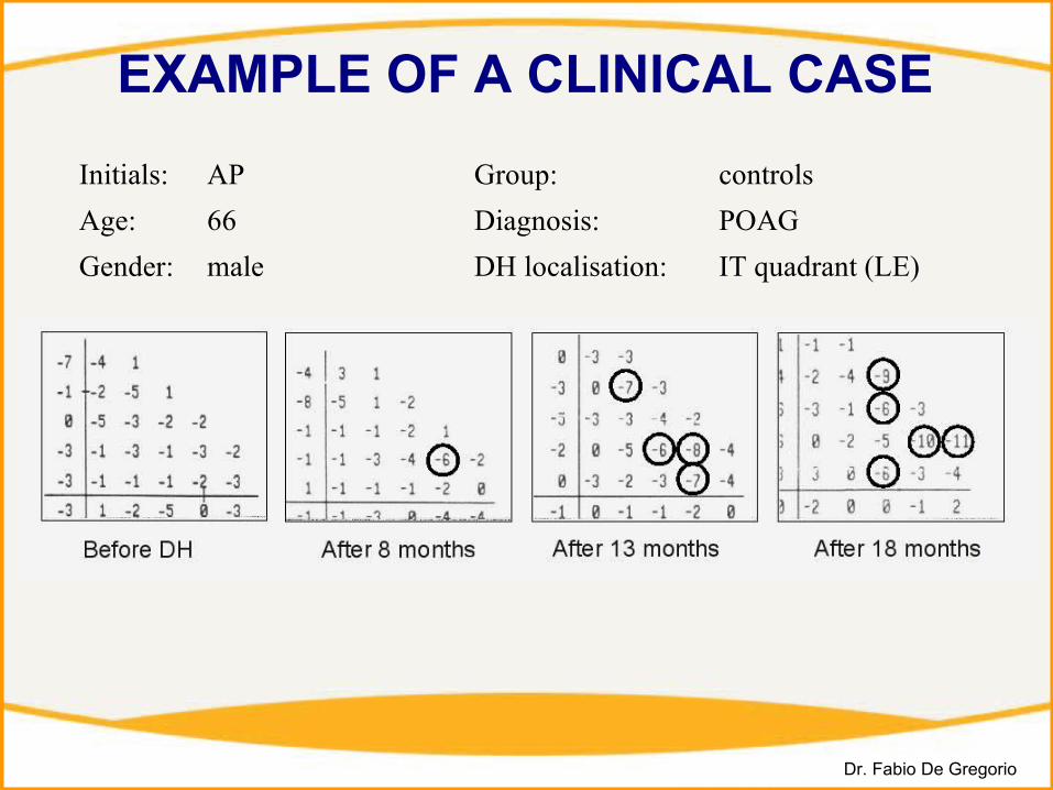
EXAMPLE OF A CLINICAL CASE
Initials: AP Group: controls
Age: 66 Diagnosis: POAG
Gender: male DH localisation: IT quadrant (LE)

Dr. Fabio De Gregorio
STATISTICS
Inferential statistics:
Contingency tables with chi-square test and survival analysis were applied to evaluate the frequency of progressors.
ANOVA was used to test parametric data.
Power of the study (1-Power of the study (1-ββ ) = 0.8) = 0.8
Able to detect as significant Able to detect as significant (P<0.05) a reduction (P<0.05) a reduction >>50% in 50% in frequency of progressorsfrequency of progressors

Dr. Fabio De Gregorio
TREATED CONTROLS P
n. 10 22 -
Age (years) 75 (sd 9.2) 68 (sd 10.9) 0.1
Gender (M/F) 5/5 7/15 0.4
IOP (mmHg) 16.5 (sd 3.3) 15.7 (sd 2.9) 0.49
MD (dB) -2.28 (sd 3.60) -3.36 (sd 3.62) 0.43
GDX (the n°) 23 (sd 21) 34 (sd 22) 0.19
NFL thickness (µn) 61.5 (sd 7.2) 66.9 (sd 88) 0.1
Recurrent DH 5 (50%) 9 (41%) 0.7
Bilateral DH 2 (20%) 5 (23%) 0.8
Diagnosis (POAG/NTG) 8/2 19/3 0.63
Follow-up (months) 14 (sd 6.7)range: 6-24
18 (sd 7.1)range: 6-24
0.14
DEMOGRAPHICS

Dr. Fabio De Gregorio
PROGRESSORS NON-PROGRESSORS
CONTROLS 77% (17/22) (*) 23% (5/22) (*) recurrent DH
100% (9/9)w/o recurrent DH
62% (8/13)recurrent DH
0% (0/9)w/o recurrent DH
38% (5/13)
TREATED 30% (3/10) (*) 70% (7/10) (*)
recurrent DH
60% (3/5)w/o recurrent DH
0% (0/5)recurrent DH
40% (2/5)w/o recurrent DH
100% (5/5)
RESULTS: Frequency of progressors
χ2 = 13.7 P = 0.004
(*) χ2 = 4.7 P = 0.03

Dr. Fabio De Gregorio
recurrent DH
all patients
w/o recurrent DH
77%
23%
CONTROLS TREATED
30%
70%
100%
0%
60%
40%
62%
38%
0%
100%
= progressors= non-progressors
P=0.036
P=0.11
P=0.030
RESULTS: Frequency of progressors

Dr. Fabio De Gregorio
RESULTS: Kaplan-Meyer curveLog rank test P=0.08Log rank test P=0.08

Dr. Fabio De Gregorio
RESULTS: secondary outcomes
controls
treated

Dr. Fabio De Gregorio
CONCLUSIONS As VF defects follow DH in a very high percentage of
subjects (77%), DH may represent a good clinical model for testing efficacy of neuroprotective agents in glaucoma
The present study represents an additional support to the clinical benefits of citicoline when administered as a complement to ocular hypotensive agents.
It also showed that pulses of treatment protect a significantly high percentage of glaucomatous patients from optic neuropathy progression consequent to DH.

Dr. Fabio De Gregorio
CONCLUSIONS: mode of actionAmong putative neuroprotectants, citicoline is, perhaps, the drug with the widest scientific documentation.
Massive degradation of phospholipids following various insults is detrimental to the cells. Phospatidylcholine catabolism is enhanced by ischemia and/or traumas. That is attributed mainly to the activation of phospholipase-A2 (Farooqui, 1997). Accumulation of arachidonic acid and other free fatty acids, inner mithochondrial membrane dysfunction, release of proapotpotic factors like ceramide, and calcium overload are possible mechanisms that lead to cell death (Kristan, 1998). Similar events can occur in retinal ganglion cells in course of glaucoma (Tatton, 2001). Inhibition of phosphatidylcholine breakdown and/or stimulation of its synthesis may spare cells from apoptosis (Bladergroen, 1999).
Citicoline stimulates the formation of phosphatidylcholine in brain (Lopez Coviella, 1995; Wang, 2000), inhibits activation of phospholipase A2 and (Rao, 2001) and prevents accumulation of free fatty acids. Citicoline also attenuates glutamate exitotoxicity in vitro (Mir, 2003)
Moreover, citicoline is also know to increase the levels of acetylcholine, norepinephrine, dopamine and serotonin in some brain areas. In particular, the dopamine stimulating effect, whose mechanism remains unclear, seems responsible for the beneficial neurological responses observed in patients with Parkinson's disease (Agnoli, 1982; Eberhardt, 1990) and for the improvement in visual acuity observed in amblyopic patients (Fresina, 2008).
Hence, citicoline seems to protect retinal ganglion cells from citicoline seems to protect retinal ganglion cells from degeneration and might represent a valuable option for attempting a degeneration and might represent a valuable option for attempting a neuroprotective therapy in glaucoma. neuroprotective therapy in glaucoma.