The 89,000-Mr murine cytomegalovirus immediate-early ... · The89,000-Mr Murine Cytomegalovirus...
7
JOURNAL OF VIROLOGY, Sept. 1988, p. 3341-3347 0022-538X/881093341-07$02.00/0 Copyright © 1988, American Society for Microbiology The 89,000-Mr Murine Cytomegalovirus Immediate-Early Protein Stimulates c-fos Expression and Cellular DNA Synthesis JORG SCHICKEDANZ,lt LENNART PHILIPSON,2 WILHELM ANSORGE,2 RAINER PEPPERKOK,2 RUDIGER KLEIN,' AND ULRICH H. KOSZINOWSKIlt* Federal Research Centre for Virus Diseases of Animals, D-7400 Tubingen,' and European Molecular Biology Laboratory, D-6900 Heidelberg,2 Federal Republic of Germany Received 4 March 1988/Accepted 26 May 1988 The immediate-early (IE) genes of murine cytomegalovirus (MCMV) are expressed in the absence of prior viral protein synthesis and regulate the transcription of MCMV early genes. The effect of MCMV IE genes on growth induction was studied. Different plasmids containing MCMV IE genes were microinjected into arrested NIH 3T3 mouse fibroblasts. Plasmids containing the iel gene coding for the 89,000-M, major IE protein pp89 were found to stimulate the expression of the c-fos protooncogene. Synthesis of pp89 and its transport to the nucleus appeared to be required for c-fos expression. DNA synthesis occurred in cells that were injected with MCMV IE genes and in neighboring cells that were not injected. The results suggest that the phosphoprotein pp89 stimulates cells to enter the cell cycle. Cytomegaloviruses (CMVs) can establish productive cy- tolytic as well as nonproductive latent infections in cells, and reactivation from latency to productive infection can occur. Certain observations indicate an interdependence between CMV replication and phase transitions of the cell cycle. Murine CMV (MCMV) replication is dependent on the physiological state of the cell, and the virus requires events associated with the host S phase for initiation of viral DNA synthesis (23). The virus itself, however, can promote cel- lular conditions favorable for viral replication by stimulating cells to synthesize cellular DNA (31) and to release a growth factor (10). These results suggest that at least one CMV gene should have an effect on the cellular genes that control the cell cycle. Given that the expression of this gene is subject to the control of cellular factors, this gene could play a role in the regulation of productive and nonproductive infection of cells. Gene expression in MCMV is regulated in a cascade fashion and can be subdivided into three phases, immediate- early (IE), early (E), and late. Because stimulation of cellular DNA synthesis and release of cellular growth factors do not require CMV DNA synthesis (10, 31), viral functions ex- pressed IE or E after infection should be involved. We studied the IE genes of MCMV as candidates. The IE genes of MCMV are clustered in a region of about 10 kilobase pairs (13). Three IE transcription units are contained in this region, iel, ie2, and ie3 (14). The gene ieI in transcription unit iel and genes transcribed from ie3 use the same pro- moter which is controlled by a very long and complex enhancer located upstream (5, 15). The promoter of the transcription unit ie2 at a position downstream of the en- hancer is also regulated by this sequence. MCMV and the human CMV have similar enhancer sequences (2), and * Corresponding author. t Present address: Institut fur Molekularbiologie und Biochemie, Freie Universitat, Arnimallee 22, D-1000 Berlin 33, Federal Repub- lic of Germany. t Present address: Institut fur Mikrobiologie, Abteilung Virolo- gie, Universitat Ulm, P.O. Box 4066, D-7900 Ulm, Federal Republic of Germany. several cellular factors bind to different sequence motifs and thus regulate the expression of the IE genes (9). CMV IE gene products play a role in the transcriptional activation of CMV E genes. Among the MCMV IE genes, ieI is most abundantly expressed and gives rise to the nonvirion phosphoprotein pp89, the major IE protein (16). pp89 acti- vates gene in trans without apparent promoter specificity (18). To exert its function in gene regulation, pp89 should directly or indirectly interact with DNA. Direct binding to DNA has not yet been demonstrated, but binding to histones has been reported (24a). As with viral proteins of other viruses that influence cell cycle regulation (11, 27, 29), an association of pp89 with kinase activity has recently been shown (A. Barnekow, J. Schickedanz, and U. H. Koszi- nowski, manuscript in preparation). These multifunctional properties of pp89 have led us to assume that it plays a role in cell cycle regulation. To investigate this possibility, we looked for stimulation of cellular DNA synthesis by pp89. To monitor the effect of the ieI gene product on cell cycle regulation, we studied the expression of the protooncogene c-fos. The characteristics of c-fos expression in various cell types have led to the suggestion that it has a role in the control of cellular growth and differentiation. Appropriate expression of c-fos appears to be a prerequisite for the reentry of quiescent cells into the cell cycle (25). The expression of c-fos is rapidly induced within minutes during the transition from the Go to the G1 phase (3, 34), and both protein and mRNA have very short half-lives (19, 24). The effect of the iel gene product on cell cycle regulation was studied by microinjection of IE genes. The results suggest that the nonstructural regulatory protein pp89 of MCMV stimulates c-fos expression and triggers cell cycle activation. MATERIALS AND METHODS Cell culture. NIH 3T3 mouse fibroblasts, clone 7 (orig- inally from D. Lowy, National Institutes of Health, Be- thesda, Md.), were seeded onto cover slips (10 by 10 mm) with prescratched squares and grown in Dulbecco modified Eagle medium supplemented with 10% fetal bovine serum in 7% CO2. To arrest the cells in the subconfluent state, medium was changed after 24 h to Dulbecco modified Eagle 3341 Vol. 62, No. 9
Transcript of The 89,000-Mr murine cytomegalovirus immediate-early ... · The89,000-Mr Murine Cytomegalovirus...
JOURNAL OF VIROLOGY, Sept. 1988, p. 3341-33470022-538X/881093341-07$02.00/0Copyright © 1988, American Society for Microbiology
The 89,000-Mr Murine Cytomegalovirus Immediate-Early ProteinStimulates c-fos Expression and Cellular DNA Synthesis
JORG SCHICKEDANZ,lt LENNART PHILIPSON,2 WILHELM ANSORGE,2 RAINER PEPPERKOK,2RUDIGER KLEIN,' AND ULRICH H. KOSZINOWSKIlt*
Federal Research Centre for Virus Diseases ofAnimals, D-7400 Tubingen,' and European Molecular Biology Laboratory,D-6900 Heidelberg,2 Federal Republic of Germany
Received 4 March 1988/Accepted 26 May 1988
The immediate-early (IE) genes of murine cytomegalovirus (MCMV) are expressed in the absence of priorviral protein synthesis and regulate the transcription ofMCMV early genes. The effect ofMCMV IE genes ongrowth induction was studied. Different plasmids containing MCMV IE genes were microinjected into arrestedNIH 3T3 mouse fibroblasts. Plasmids containing the iel gene coding for the 89,000-M, major IE protein pp89were found to stimulate the expression of the c-fos protooncogene. Synthesis of pp89 and its transport to thenucleus appeared to be required for c-fos expression. DNA synthesis occurred in cells that were injected withMCMV IE genes and in neighboring cells that were not injected. The results suggest that the phosphoproteinpp89 stimulates cells to enter the cell cycle.
Cytomegaloviruses (CMVs) can establish productive cy-tolytic as well as nonproductive latent infections in cells, andreactivation from latency to productive infection can occur.Certain observations indicate an interdependence betweenCMV replication and phase transitions of the cell cycle.Murine CMV (MCMV) replication is dependent on thephysiological state of the cell, and the virus requires eventsassociated with the host S phase for initiation of viral DNAsynthesis (23). The virus itself, however, can promote cel-lular conditions favorable for viral replication by stimulatingcells to synthesize cellular DNA (31) and to release a growthfactor (10). These results suggest that at least one CMV geneshould have an effect on the cellular genes that control thecell cycle. Given that the expression of this gene is subject tothe control of cellular factors, this gene could play a role inthe regulation of productive and nonproductive infection ofcells.Gene expression in MCMV is regulated in a cascade
fashion and can be subdivided into three phases, immediate-early (IE), early (E), and late. Because stimulation of cellularDNA synthesis and release of cellular growth factors do notrequire CMV DNA synthesis (10, 31), viral functions ex-pressed IE or E after infection should be involved. Westudied the IE genes of MCMV as candidates. The IE genesofMCMV are clustered in a region of about 10 kilobase pairs(13). Three IE transcription units are contained in thisregion, iel, ie2, and ie3 (14). The gene ieI in transcriptionunit iel and genes transcribed from ie3 use the same pro-moter which is controlled by a very long and complexenhancer located upstream (5, 15). The promoter of thetranscription unit ie2 at a position downstream of the en-hancer is also regulated by this sequence. MCMV and thehuman CMV have similar enhancer sequences (2), and
* Corresponding author.t Present address: Institut fur Molekularbiologie und Biochemie,
Freie Universitat, Arnimallee 22, D-1000 Berlin 33, Federal Repub-lic of Germany.
t Present address: Institut fur Mikrobiologie, Abteilung Virolo-gie, Universitat Ulm, P.O. Box 4066, D-7900 Ulm, Federal Republicof Germany.
several cellular factors bind to different sequence motifs andthus regulate the expression of the IE genes (9).CMV IE gene products play a role in the transcriptional
activation ofCMV E genes. Among the MCMV IE genes, ieIis most abundantly expressed and gives rise to the nonvirionphosphoprotein pp89, the major IE protein (16). pp89 acti-vates gene in trans without apparent promoter specificity(18). To exert its function in gene regulation, pp89 shoulddirectly or indirectly interact with DNA. Direct binding toDNA has not yet been demonstrated, but binding to histoneshas been reported (24a). As with viral proteins of otherviruses that influence cell cycle regulation (11, 27, 29), anassociation of pp89 with kinase activity has recently beenshown (A. Barnekow, J. Schickedanz, and U. H. Koszi-nowski, manuscript in preparation). These multifunctionalproperties of pp89 have led us to assume that it plays a rolein cell cycle regulation. To investigate this possibility, welooked for stimulation of cellular DNA synthesis by pp89.To monitor the effect of the ieI gene product on cell cycle
regulation, we studied the expression of the protooncogenec-fos. The characteristics of c-fos expression in various celltypes have led to the suggestion that it has a role in thecontrol of cellular growth and differentiation. Appropriateexpression of c-fos appears to be a prerequisite for thereentry of quiescent cells into the cell cycle (25). Theexpression of c-fos is rapidly induced within minutes duringthe transition from the Go to the G1 phase (3, 34), and bothprotein and mRNA have very short half-lives (19, 24). Theeffect of the iel gene product on cell cycle regulation wasstudied by microinjection of IE genes. The results suggestthat the nonstructural regulatory protein pp89 of MCMVstimulates c-fos expression and triggers cell cycle activation.
MATERIALS AND METHODS
Cell culture. NIH 3T3 mouse fibroblasts, clone 7 (orig-inally from D. Lowy, National Institutes of Health, Be-thesda, Md.), were seeded onto cover slips (10 by 10 mm)with prescratched squares and grown in Dulbecco modifiedEagle medium supplemented with 10% fetal bovine serum in7% CO2. To arrest the cells in the subconfluent state,medium was changed after 24 h to Dulbecco modified Eagle
3341
Vol. 62, No. 9
at Universitaetsbibliothek M
uenchen on October 22, 2008
jvi.asm.org
Dow
nloaded from
3342 SCHICKEDANZ ET AL.
medium containing 2.5% bovine platelet-poor plasma. Cellswere injected after an arrest of 3 days.
Plasmid DNA. Cloning of MCMV DNA fragments andplasmids pAMB25, pIE111, and pIE100 have been describedbefore (7, 13, 17, 18). Plasmid DNA was prepared by themethod of Holmes and Quigley (12). Restriction enzymedigestion, agarose gel electrophoresis, and ethanol precipi-tation of DNA were performed by standard methods (21).
Microinjection. Linearized plasmid DNA was suspendedin sterile water at concentrations of 50 ng/ml. In selectedexperiments sterile filtered fluorescein isothiocyanate(FITC)-labeled dextran (FD-150; Sigma Chemical Co.,Munich, Federal Republic of Germany) was coinjected atconcentrations of 1.5%. The probes were centrifuged for 15min at 20,000 x g. To ensure physiological pH conditions,the cell medium was adjusted with HEPES (N-2-hydrox-yethylpiperazine-N'-2-ethanesulfonic acid) to 1 mM justbefore injection. Microinjection was performed with aninjection system (developed by Ansorge [1]). Injection cap-illaries were pulled with an automatic puller (Mechanex,Geneva, Switzerland). In individual squares of the coverslip, the nuclei of about 15 cells were injected with .10-11ml of solution. In each experiment 200 to 300 cells wereusually injected during 20 to 30 min. After injection the cellswere incubated in 7% CO2 for different periods of time.
Immunofluorescence. (i) Detection of MCMV IE proteins.Cells were fixed with 3% paraformaldehyde in phosphate-buffered saline and treated with 0.2% Triton X-100 in phos-phate-buffered saline at 20°C. The cells were then incubatedat 37°C with the murine monoclonal antibody 6/56/6 specificfor MCMV IE protein pp89 (16). In the second step cellswere incubated at 37°C with rhodamine-labeled rabbit anti-mouse immunoglobulin G (Dakopatts, Hamburg, FederalRepublic of Germany). For a control, nuclei of cells werestained by incubation for 20 min with 5 jig of bisbenzimide(Hoechst 33342; Hoechst-Roussel Pharmaceuticals Inc.,Somerville, N.J.) per ml. Cover slips were mounted on slidesand fluorescence was visualized on an inverted light micro-scope (Zeiss) at 495 nm (FITC), 554 nm (rhodamine), or 365nm (bisbenzimide).
(ii) Detection of c-fos antigen. Cells were fixed and permea-bilized as described above. The c-fos antigen was detectedby polyclonal rabbit anti-fos serum, and rhodamine-labeledswine anti-rabbit immunoglobulin served as a second anti-body.DNA synthesis assay. To test the stimulation of cellular
DNA synthesis, cells were arrested at the Go phase asdescribed above. After plasmid DNA injection, 1 ,uCi of [3H]thymidine per ml was added, and cells were incubated at37°C in 7% CO2 for different periods of time. After fixation ofthe cells, indirect immunofluorescence, and bisbenzimidestaining, the mounted cover slips were covered with autora-diographic stripping film (AR 10; Eastman Kodak Co.,Rochester, N.Y.). After exposure for 2 to 3 days at roomtemperature, the films were developed and fixed. [3H]thymi-dine incorporation was visualized by black silver grains andphotographed under a microscope.
RESULTS
Expression of MCMV IE genes in mouse NIH 3T3 fibro-blasts after microinjection. For the analysis of regulatoryeffects of isolated viral genes and their products on cellularfunctions, microinjection offers some advantages. First, thecells injected with DNA can be identified by coinjection of afluorescence marker. This allowed us to find again each
individual injected cell. Second, the temporal relation be-tween gene uptake and expression can be determined, as canthe number of injected cells that express the gene. Third, theregulatory effects on cellular genes by injected viral genescan be studied, and discrimination between direct effects oninjected cells and indirect effects on surrounding cells ispossible.The correct expression of injected MCMV IE genes was
tested in growing subconfluent mouse NIH 3T3 fibroblastsby using linearized plasmids containing various regions ofthe MCMV IE gene region (Fig. 1). FITC-labeled dextranwas coinjected to locate the injected cells. Expression of theIE protein pp89 was monitored for 20 h after injection byusing a monoclonal antibody specific for pp89 and a rho-damine-labeled second antibody for detection by immuno-fluorescence. As indicated by the number of FITC-positivecells and the ratio between FITC- and rhodamine-positivecells, about 70% of injected cells survived the injectionprocedure, and approximately 80% of survivors expressedpp89. Microinjection of all constructs containing the tran-scription until iel resulted in expression of pp89. Controlinjections with plasmids linearized by HindIII, whichcleaves within the fourth exon of the pp89-coding sequence,or injection with the vector pSP62 did not reveal anyrhodamine fluorescence.
Subceliular distribution of pp89 after microinjection of theiel gene in arrested mouse NIH 3T3 fibroblasts. Expression ofthe c-fos gene as well as DNA synthesis was low or absent inresting cells. To study the stimulation of the cell cyclefollowing microinjection of MCMV IE genes, NIH 3T3 cellswere synchronized in the Go phase of the cell cycle bypreincubation for 3 days with medium containing 2.5%platelet-poor plasma. Platelet-poor plasma is depleted ofplatelet-derived growth factor, which is necessary for NIH3T3 cell proliferation, but possesses all other componentsthat are required (35). This protocol resulted in about 95% ofcells being arrested in the Go phase, as demonstrated by alack of [3H]thymidine incorporation into the nucleus duringa 24-h incubation period. After the addition of 20% fetalbovine serum for 24 h, incorporation of [3H]thymidine inabout 90% of cell nuclei confirmed that the cells could bereleased from growth arrest.
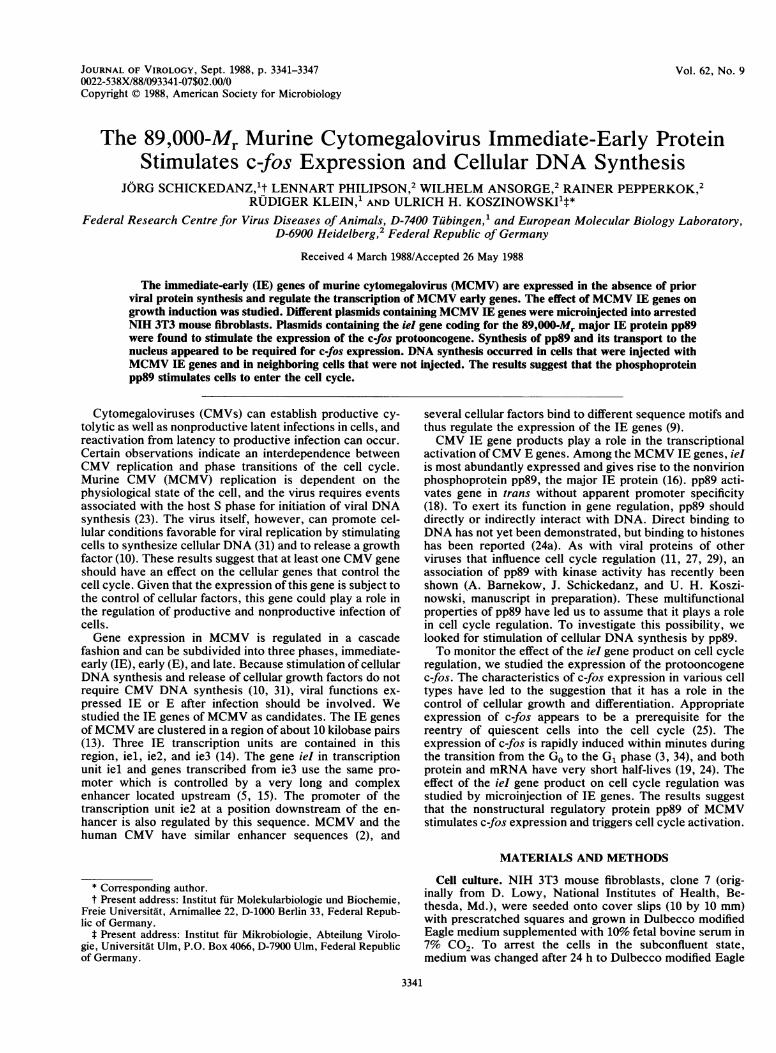
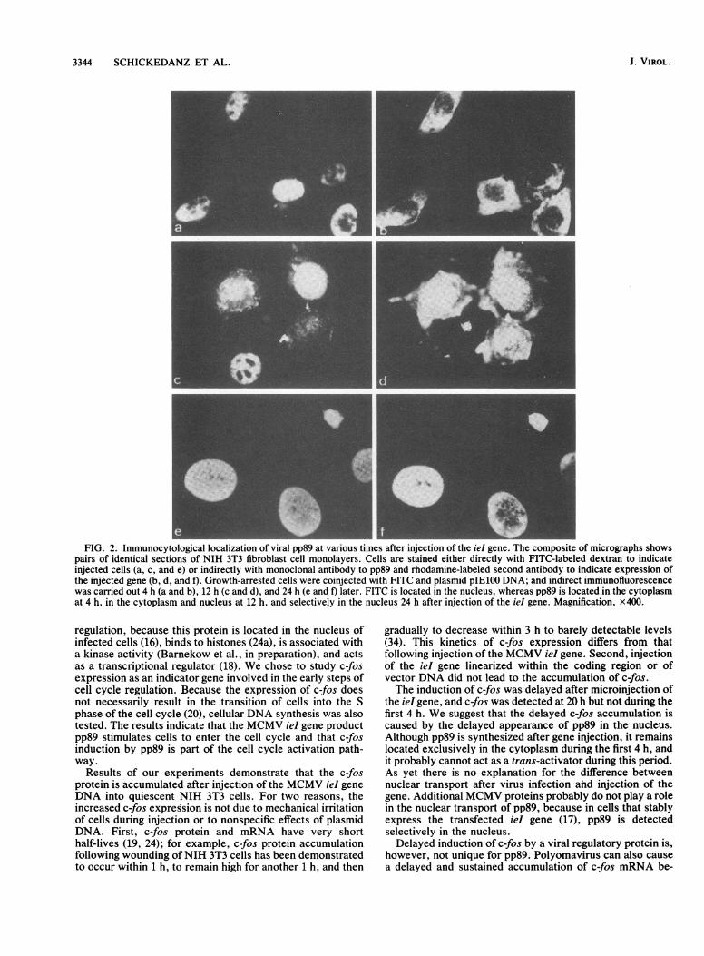
In contrast to growing cells, only approximately 20% ofarrested cells survived after injection, but similar to growingcells, 80% of these expressed pp89 20 h later. It waspostulated that prior to any effect on cellular gene functions,the ieI gene product had to arrive at the nucleus. Duringpermissive infection with MCMV, the ieI gene is the firstviral gene that is expressed. The protein is already synthe-sized in the first 2 h of infection, and the antigen can bedetected by immunofluorescence at about the same time inthe nucleus but not in the cytoplasm of infected cells. Theprotein is stable and resides in the nucleus throughout thereplication period (13, 16). We examined the kinetics of pp89antigen expression and the nuclear transport of the antigenfollowing microinjection of the ieI gene into quiescent cells.Surprisingly, the kinetics of subcellular distribution of pp89antigen was different between infection by virus and micro-injection of the gene (Fig. 2). Four hours following injectionof ieI (Fig. 2b) and also 8 h postinjection (data not shown),pp89 antigen was mainly located in the cytoplasm, whereasonly as late as 12 h after microinjection the majority of theantigen appeared to be located in the nucleus (Fig. 2d). Anexclusive nuclear localization of pp89 antigen was seen at 24h (Fig. 2f) and 30 h after microinjection (data not shown).MCMV pp89 stimulates c-fos antigen expression. The c-fos
J. VIROL.
at Universitaetsbibliothek M
uenchen on October 22, 2008
jvi.asm.org
Dow
nloaded from
CELL CYCLE INDUCTION BY CMV PROTEIN 3343
a N A
Ea
B M H 0 C G F K L J I OP E
U10
a a a__0x 4c
c
0.769 map units 0.782
CL
a aO z
0.803
--4
ie 2
s E
co
0.818
pAMB 25
p IE 111
p IE 100
FIG. 1. Physical map ofMCMV IE genes. (a) HindIII cleavage sites. (b) Organization of the IE region. The locations of the transcriptionunits iel, ie2, and ie3; the direction of transcription; and the structure of the major RNA transcribed from the ieI gene are indicated. The openreading frame is indicated as a segmented solid bar, and the promoter regulatory enhancer region is indicated as a hatched box. The structuralorganizations of ie2 and ie3 are not precisely known; therefore, they are symbolized by open arrows. (c) Plasmid-cloned DNA fragments ofthe MCMV IE region used for microinjection. kbp, Kilobase pairs.
protooncogene product is present in low amounts in normalcells, and its expression is induced rapidly during cell growthand proliferation (3). Expression of c-fos was monitored atdifferent times after injection of the iel gene by immunoflu-orescence (Fig. 3). We reasoned that different than otherstimuli which rapidly turn on c-fos expression, any effect ofieI gene injection that was associated with the function ofpp89 should be delayed. Because pp89 remains in thecytoplasm during the first 4 h postinjection of DNA, c-fosexpression should be turned on later. No c-fos expressionwas detectable in the nuclei of injected cells after 1 h (Fig.3b) or 2 and 4 h postinjection (data not shown), whereas at 20h postinjection, at times when pp89 was found predomi-nantly in the nucleus, a distinct c-fos expression was alsoobserved in the nuclei of injected cells (Fig. 3d). On the otherhand, c-fos expression was already seen after 1 h followingstimulation of the cells with 20% fetal bovine serum (Fig.3e).
Linearization of plasmids containing the ieI gene byHindlIl cleavage with cuts within the fourth exon encodingpp89 (Fig. 1) abolished trans-activation (17). Similarly, noeffect on c-fos expression was seen after injection of plas-mids that were linearized with HindlIl (data not shown).After microinjection with the intact ieI gene, expression ofc-fos was seen in 90% of FITC-positive cells; this wascomparable to the number of cells that expressed pp89. Noc-fos expression was detected in noninjected neighboringcells. After microinjection of the vector plasmid pSP62, c-fosexpression was not detectable during the 20-h incubationperiod (data not shown), excluding nonspecific effects ofirrelevant DNA on c-fos expression. These data suggest thatstimulation of c-fos expression by the MCMV ieI generequires synthesis of pp89 and its transport to the nucleus.
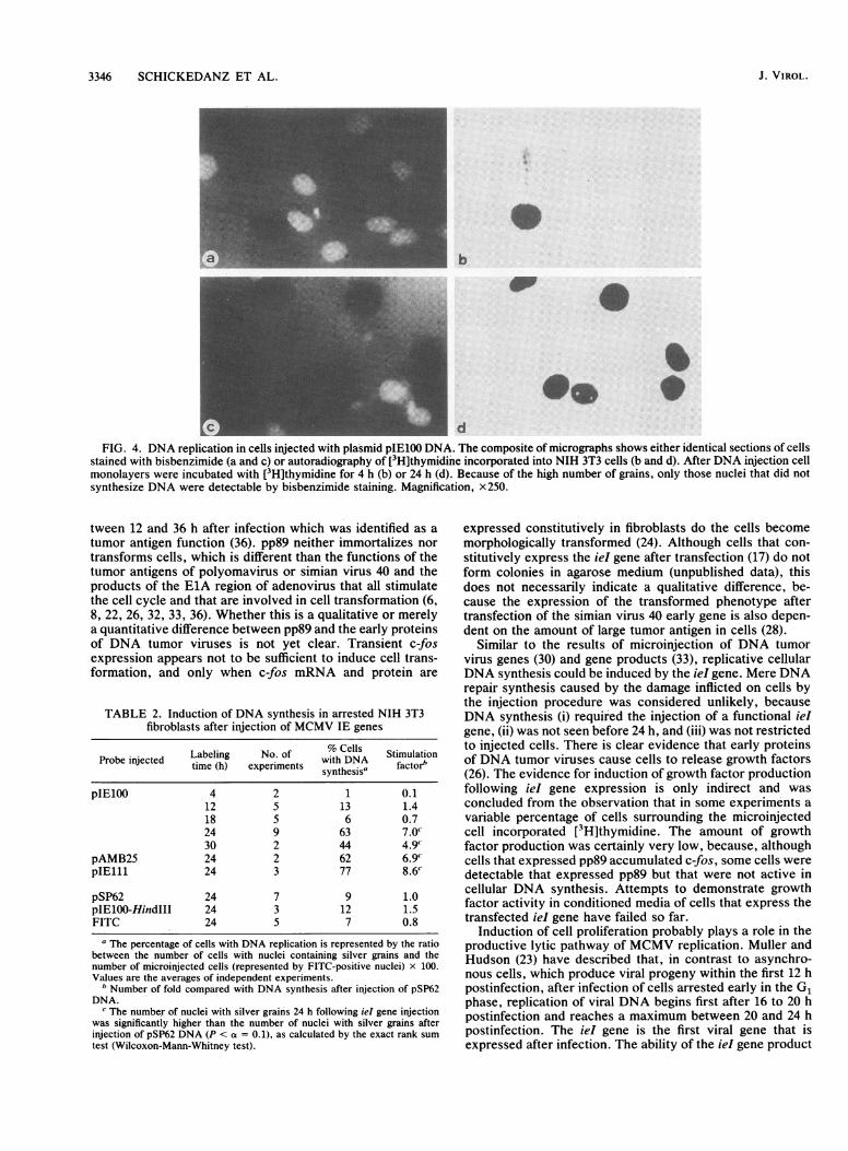
Stimulation of DNA synthesis in quiescent cells after micro-injection of the ieI gene. To study whether the growth arrestin quiescent cells is released by pp89, cells were labeled with[3H]thymidine after injection of the ieI gene. At differenttimes postinjection, incorporation of the radioactive nucleo-tide was examined by autoradiography. As described above,
about 80% of cells that survived the injection expressedpp89. After 4 to 12 h no enhanced DNA synthesis wasobserved (Table 1 and Fig. 4b), whereas 24 h after injectiona significant stimulation of DNA synthesis was seen (Table 1and Fig. 4d). Note that different than c-fos expression, therewas no stringent correlation between expression of pp89 andDNA synthesis. In some experiments, [3H]thymidine incor-poration also occurred in noninjected surrounding cells. Thiscould result in a higher number of cells with labeled nucleithan of cells that express pp89. Results of a typical experi-ment in which such conditions were observed are repre-sented in Table 1. Conversely, few individual cells were seenthat expressed pp89 but that were not active in DNAsynthesis (data not shown). Nevertheless, stimulation ofDNA synthesis was dependent on the injection and expres-sion of the iel gene, since after injection with plasmidlinearized by Hindlll cleavage or injection with the vectorpSP62 DNA for control, there was no induction of DNAsynthesis (Table 1), and DNA synthesis in the noninjectedcontrol areas remained as low as expected for arrested cells.
Further experiments were carried out to detect a putativeadditional effect of the other two IE transcription units ofMCMV, ie2 and ie3. The data are summarized in Table 2.The ieI gene was again sufficient for the induction of DNAsynthesis, and there was no evidence that the presence of ie2or ie3 sequences improved the degree of stimulation.
DISCUSSION
Stimulation of cellular DNA synthesis by CMV (31)caused by the release of a growth factor by permissive andnonpermissive cells infected with CMV (10) demonstratedthe effect of CMV infection on cell cycle regulation. Sinceviral DNA synthesis is not required for induction of host cellDNA synthesis (4), the effect is either due to a viralstructural protein transferred by infection or viral genefunctions expressed during the IE or E phase of infection.The MCMV ieI gene product pp89 appeared to be a likelycandidate for a CMV protein that is involved in cell cycle
VOL. 62, 1988
IHin dM
b ...................'-
ie3iel -1MMAWGI 1--
at Universitaetsbibliothek M
uenchen on October 22, 2008
jvi.asm.org
Dow
nloaded from
3344 SCHICKEDANZ ET AL.
I
._ . . .__--A m..............................................
FIG. 2. Immunocytological localization of viral pp89 at various times after injection of the iel gene. The composite of micrographs showspairs of identical sections of NIH 3T3 fibroblast cell monolayers. Cells are stained either directly with FITC-labeled dextran to indicateinjected cells (a, c, and e) or indirectly with monoclonal antibody to pp89 and rhodamine-labeled second antibody to indicate expression ofthe injected gene (b, d, and f). Growth-arrested cells were coinjected with FITC and plasmid pIE100 DNA; and indirect immunofluorescencewas carried out 4 h (a and b), 12 h (c and d), and 24 h (e and f) later. FITC is located in the nucleus, whereas pp89 is located in the cytoplasmat 4 h, in the cytoplasm and nucleus at 12 h, and selectively in the nucleus 24 h after injection of the ieI gene. Magnification, X400.
regulation, because this protein is located in the nucleus ofinfected cells (16), binds to histones (24a), is associated witha kinase activity (Barnekow et al., in preparation), and actsas a transcriptional regulator (18). We chose to study c-fosexpression as an indicator gene involved in the early steps ofcell cycle regulation. Because the expression of c-fos doesnot necessarily result in the transition of cells into the Sphase of the cell cycle (20), cellular DNA synthesis was alsotested. The results indicate that the MCMV ieI gene productpp89 stimulates cells to enter the cell cycle and that c-fosinduction by pp89 is part of the cell cycle activation path-way.
Results of our experiments demonstrate that the c-fosprotein is accumulated after injection of the MCMV ieI geneDNA into quiescent NIH 3T3 cells. For two reasons, theincreased c-fos expression is not due to mechanical irritationof cells during injection or to nonspecific effects of plasmidDNA. First, c-fos protein and mRNA have very shorthalf-lives (19, 24); for example, c-fos protein accumulationfollowing wounding of NIH 3T3 cells has been demonstratedto occur within 1 h, to remain high for another 1 h, and then
gradually to decrease within 3 h to barely detectable levels(34). This kinetics of c-fos expression differs from thatfollowing injection of the MCMV ieI gene. Second, injectionof the ieI gene linearized within the coding region or ofvector DNA did not lead to the accumulation of c-fos.The induction of c-fos was delayed after microinjection of
the iel gene, and c-fos was detected at 20 h but not during thefirst 4 h. We suggest that the delayed c-fos accumulation iscaused by the delayed appearance of pp89 in the nucleus.Although pp89 is synthesized after gene injection, it remainslocated exclusively in the cytoplasm during the first 4 h, andit probably cannot act as a trans-activator during this period.As yet there is no explanation for the difference betweennuclear transport after virus infection and injection of thegene. Additional MCMV proteins probably do not play a rolein the nuclear transport of pp89, because in cells that stablyexpress the transfected ieI gene (17), pp89 is detectedselectively in the nucleus.Delayed induction of c-fos by a viral regulatory protein is,
however, not unique for pp89. Polyomavirus can also causea delayed and sustained accumulation of c-fos mRNA be-
J. VIROL.
at Universitaetsbibliothek M
uenchen on October 22, 2008
jvi.asm.org
Dow
nloaded from
CELL CYCLE INDUCTION BY CMV PROTEIN 3345
_FIG. 3. Induction of the c-fos protein by viral pp89 at various times after ieI gene injection. The composite of micrographs shows c-fos
antigen expression in NIH 3T3 fibroblast cell monolayers. Growth-arrested cells were coinjected with FITC and plasmid pIE100 DNA, andindirect immunofluorescence with antiserum to c-fos and rhodamine-labeled second antibody was carried out 1 h (a and b) and 20 h (c andd) later. FITC fluorescence is seen as an injection marker in panels a and c, and immunofluorescence to detect c-fos antigen expression isshown in panels b and d. The micrographs in panels e and f show c-fos antigen 1 h after stimulation with 20% fetal bovine serum (e) and noc-fos in cells without stimulation (f). Magnification, x400.
TABLE 1. Relationship between pp89 expression and DNA synthesis in arrested NIH 3T3 fibroblasts after microinjection of DNA
Labeling No. of No. of No. of No. of cellsProbe injecteda time (h) injected FITC-positive pp89-positive with DNA
cells cells cells synthesisbpIE100 4 342 80 61 0
8 310 61 54 012 304 123 74 024 321 40 35 50
pSP62 24 350 70 0 5pIE100-HindIIIc 24 157 NDd 0 3
a Results are of one representative experiment.b The total number of cells with silver grains was determined in areas containing injected cells. From this value, the basal DNA synthesis activity was subtracted
and evaluated by counting the number of positive cells in a noninjected area with a comparable number of cells.c The plasmid was linearized by HindIII cleavage to interrupt the coding region of the iel gene.d ND, No coinjection by FITC.
VOL. 62, 1988
at Universitaetsbibliothek M
uenchen on October 22, 2008
jvi.asm.org
Dow
nloaded from
3346 SCHICKEDANZ ET AL.
IS
b
0
9d
FIG. 4. DNA replication in cells injected with plasmid pIE100 DNA. The composite of micrographs shows either identical sections of cellsstained with bisbenzimide (a and c) or autoradiography of [3H]thymidine incorporated into NIH 3T3 cells (b and d). After DNA injection cellmonolayers were incubated with [3H]thymidine for 4 h (b) or 24 h (d). Because of the high number of grains, only those nuclei that did notsynthesize DNA were detectable by bisbenzimide staining. Magnification, x250.
tween 12 and 36 h after infection which was identified as atumor antigen function (36). pp89 neither immortalizes nortransforms cells, which is different than the functions of thetumor antigens of polyomavirus or simian virus 40 and theproducts of the ElA region of adenovirus that all stimulatethe cell cycle and that are involved in cell transformation (6,8, 22, 26, 32, 33, 36). Whether this is a qualitative or merelya quantitative difference between pp89 and the early proteinsof DNA tumor viruses is not yet clear. Transient c-fosexpression appears not to be sufficient to induce cell trans-formation, and only when c-fos mRNA and protein are
TABLE 2. Induction of DNA synthesis in arrested NIH 3T3fibroblasts after injection of MCMV IE genes
Probe injected Labeling No. of w%th DllsA StimulationProbinjcted time (h) experiments wynthesiNA factorb'
pIE100 4 2 1 0.112 5 13 1.418 5 6 0.724 9 63 7.0'30 2 44 4.9'
pAMB25 24 2 62 6.9CpIE111 24 3 77 8.6'
pSP62 24 7 9 1.0pIElOO-HindIII 24 3 12 1.5FITC 24 5 7 0.8
The percentage of cells with DNA replication is represented by the ratiobetween the number of cells with nuclei containing silver grains and thenumber of microinjected cells (represented by FITC-positive nuclei) x 100.Values are the averages of independent experiments.
b Number of fold compared with DNA synthesis after injection of pSP62DNA.
c The number of nuclei with silver grains 24 h following iel gene injectionwas significantly higher than the number of nuclei with silver grains afterinjection of pSP62 DNA (P < a = 0.1), as calculated by the exact rank sumtest (Wilcoxon-Mann-Whitney test).
expressed constitutively in fibroblasts do the cells becomemorphologically transformed (24). Although cells that con-stitutively express the ieI gene after transfection (17) do notform colonies in agarose medium (unpublished data), thisdoes not necessarily indicate a qualitative difference, be-cause the expression of the transformed phenotype aftertransfection of the simian virus 40 early gene is also depen-dent on the amount of large tumor antigen in cells (28).
Similar to the results of microinjection of DNA tumorvirus genes (30) and gene products (33), replicative cellularDNA synthesis could be induced by the ieI gene. Mere DNArepair synthesis caused by the damage inflicted on cells bythe injection procedure was considered unlikely, becauseDNA synthesis (i) required the injection of a functional ieIgene, (ii) was not seen before 24 h, and (iii) was not restrictedto injected cells. There is clear evidence that early proteinsof DNA tumor viruses cause cells to release growth factors(26). The evidence for induction of growth factor productionfollowing ieI gene expression is only indirect and wasconcluded from the observation that in some experiments avariable percentage of cells surrounding the microinjectedcell incorporated [3H]thymidine. The amount of growthfactor production was certainly very low, because, althoughcells that expressed pp89 accumulated c-fos, some cells weredetectable that expressed pp89 but that were not active incellular DNA synthesis. Attempts to demonstrate growthfactor activity in conditioned media of cells that express thetransfected ieI gene have failed so far.
Induction of cell proliferation probably plays a role in theproductive lytic pathway of MCMV replication. Muller andHudson (23) have described that, in contrast to asynchro-nous cells, which produce viral progeny within the first 12 hpostinfection, after infection of cells arrested early in the G1phase, replication of viral DNA begins first after 16 to 20 hpostinfection and reaches a maximum between 20 and 24 hpostinfection. The iel gene is the first viral gene that isexpressed after infection. The ability of the ieI gene product
J. VIROL.
at Universitaetsbibliothek M
uenchen on October 22, 2008
jvi.asm.org
Dow
nloaded from
CELL CYCLE INDUCTION BY CMV PROTEIN 3347
to stimulate cells to proliferate supports viral replication. Therelease of growth factors would stimulate surrounding cells tobecome target cells for secondary infection. Whether thesuccess or the failure to induce the cell cycle could also playa role in the maintenance of viral latency and the reactivationto productive infection is a matter of speculation.
ACKNOWLEDGMENTS
This study was supported by grant Ko 571/8 from the DeutscheForschungsgemeinschaft. J.S. was supported by a short-term fel-lowship from the European Molecular Biology Organization.We thank R. Muller, EMBL, Heidelberg, Federal Republic of
Germany, for c-fos antisera and A. Habenicht, University of Hei-delberg, for platelet-poor plasma. S. Grau provided skillful typing ofthe manuscript.
LITERATURE CITED1. Ansorge, W. 1982. Improved system for capillary microinjection
into living cells. Exp. Cell Res. 140:31-37.2. Boshart, M., F. Weber, G. Jahn, K. Dorsch-Hasler, B. Flecken-
stein, and W. Schaffner. 1985. A very strong enhancer is locatedupstream of an immediate early gene of human cytomegalovi-rus. Cell 41:521-530.
3. Bravo, R., J. Burckhardt, T. Curran, and R. Muller. 1986.Expression of c-fos in NIH 3T3 cells is very low but induciblethroughout the cell cycle. EMBO J. 5:695-700.
4. Demarchi, J. M., and A. S. Kaplan. 1977. The role of defectivecytomegalovirus particles in the induction of host cell DNAsynthesis. Virology 82:93-99.
5. Dorsch-Hasler, K., G. M. Keil, F. Weber, M. Jasin, W.Schaffner, and U. H. Koszinowski. 1985. A long and complexenhancer activates transcription of the gene coding for thehighly abundant immediate early mRNA in murine cytomegalo-virus. Proc. Natl. Acad. Sci. USA 82:8325-8329.
6. Dulbecco, R., L. H. Hartwell, and M. Vogt. 1965. Induction ofcellular DNA synthesis by polyoma virus. Proc. Natl. Acad.Sci. USA 53:403-410.
7. Ebeling, A., G. M. Keil, E. Knust, and U. H. Koszinowski. 1983.Molecular cloning and physical mapping of murine cytomegalo-virus DNA. J. Virol. 47:421-433.
8. Gershon, D., L. Sachs, and E. Winocour. 1966. The induction ofcellular DNA synthesis by simian virus 40 in contact-inhibitedand in X-irradiated cells. Proc. Natl. Acad. Sci. USA 56:918-925.
9. Ghazal, P., H. Lubon, B. Fleckenstein, and L. Hennighausen.1987. Binding of transcription factors and creation of a largenucleoprotein complex on the human cytomegalovirus en-hancer. Proc. Natl. Acad. Sci. USA 84:3658-3662.
10. Gonczol, E., and S. A. Plotkin. 1984. Cells infected with humancytomegalovirus release a factor(s) that stimulates cell DNAsynthesis. J. Gen. Virol. 65:1833-1837.
11. Griffin, J. D., G. Spangler, and D. M. Livingston. 1979. Proteinkinase activity associated with simian virus 40 T antigen. Proc.Natl. Acad. Sci. USA 76:2610-2614.
12. Holmes, D. S., and M. Quigley. 1981. A rapid boiling method forthe preparation of bacterial plasmids. Anal. Biochem. 114:193-197.
13. Keil, G. M., A. Ebeling-Keil, and U. H. Koszinowski. 1984.Temporal regulation of murine cytomegalovirus transcriptionand mapping of viral RNA synthesized at immediate early timesafter infection. J. Virol. 50:784-795.
14. Keil, G. M., A. Ebeling-Keil, and U. H. Koszinowski. 1987.Immediate-early genes of murine cytomegalovirus: location,transcripts, and translation products. J. Virol. 61:526-533.
15. Keil, G. M., A. Ebeling-Keil, and U. H. Koszinowski. 1987.Sequence and structural organization of murine cytomegalovi-rus immediate-early gene 1. J. Virol. 61:1901-1908.
16. Keil, G. M., M. R. Fibi, and U. H. Koszinowski. 1985. Charac-terization of the major immediate-early polypeptides encodedby murine cytomegalovirus. J. Virol. 54:422-428.
17. Koszinowski, U. H., G. M. Keil, H. Schwarz, J. Schickedanz, andM. J. Reddehase. 1987. A nonstructural polypeptide encoded by
immediate-early transcription unit 1 of murine cytomegalovirusis recognized by cytolytic T lymphocytes. J. Exp. Med. 166:289-294.
18. Koszinowski, U. H., G. M. Keil, H. Volkmer, M. R. Fibi, A.Ebeling-Keil, and K. Munch. 1986. The 89,000-Mr murine cyto-megalovirus immediate-early protein activates gene transcrip-tion. J. Virol. 58:59-66.
19. Kruijer, W., J. A. Cooper, T. Hunter, and I. M. Verma. 1984.Platelet-derived growth factor induces rapid but transientexpression of the c-fos gene and protein. Nature (London) 312:711-716.
20. Larsson, L.-G., H. E. Gray, T. Totterman, U. Pettersson, and K.Nilsson. 1987. Drastically increased expression of myc and fosprotooncogenes during in vitro differentiation of chronic lym-phocytic leukemia cells. Proc. Natl. Acad. Sci. USA 84:223-227.
21. Maniatis, T., E. F. Fritsch, and J. Sambrook. 1982. Molecularcloning: a laboratory manual. Cold Spring Harbor Laboratory,Cold Spring Harbor, N.Y.
22. Mueller, C., A. Graessmann, and M. Graessmann. 1978. Map-ping of early SV40-specific functions by microinjection of dif-ferent early viral DNA fragments. Cell 15:579-585.
23. Muller, M. T., and J. B. Hudson. 1977. Cell cycle dependency ofmurine cytomegalovirus replication in synchronized 3T3 cells.J. Virol. 22:267-272.
24. Muller, R., R. Bravo, and J. Burckhardt. 1984. Induction ofc-fos gene and protein by growth factor precedes activation ofc-myc. Nature (London) 312:716-720.
24a.Munch, K., G. M. Keil, M. Messerle, and U. H. Koszinowski.1988. Interaction of the 89K murine cytomegalovirus immedi-ate-early protein with core histones. Virology 163:405-412.
25. Nishikura, K., and J. M. Murray. 1987. Antisense RNA ofprotooncogene c-fos blocks renewed growth of quiescent 3T3cells. Mol. Cell. Biol. 7:639-649.
26. Quinlan, M. P., N. Sullivan, and T. Grodzicker. 1987. Growthfactor(s) produced during infection with an adenovirus variantstimulates proliferation of nonestablished epithelial cells. Proc.Natl. Acad. Sci. USA 84:3283-3287.
27. Schaffhausen, B., and T. L. Benjamin. 1981. Protein kinaseactivity associated with polyoma virus middle T antigen. Proteinphosphorylation (book B). Cold Spring Harbor Conferences onCell Proliferation, vol. 8. Cold Spring Harbor Laboratory, ColdSpring Harbor, N.Y.
28. Segawa, K., and N. Yamnaguchi. 1987. Induction of c-Ha-rastranscription in rat cells by simian virus 40 large T antigen. Mol.Cell. Biol. 7:556-559.
29. Smith, A. E., R. Smith. B. Griffin, and M. Fried. 1979. Proteinkinase activity associated with polyoma virus middle T antigenin vitro. Cell 18:915-924.
30. Stabel, S., P. Argos, and L. Philipson. 1985. The release ofgrowth arrest by microinjection adenovirus ElA DNA. EMBOJ. 4:2329-2336.
31. St. Jeor, S. C., T. B. Albrecht, F. D. Funk, and F. Rapp. 1974.Stimulation of cellular DNA synthesis by human cytomegalovi-rus. J. Virol. 13:353-362.
32. Takahashi, M., T. Ogino, K. Baba, and M. Onaka. 1969.Synthesis of deoxyribonucleic acid in human and hamsterkidney cells infected with human adenovirus types 5 and 12.Virology 37:513-520.
33. Tjian, R., G. Fey, and A. Graessmann. 1978. Biological activityof purified simian virus 40 T antigen proteins. Proc. Natl. Acad.Sci. USA 75:1279-1283.
34. Verrier, B., D. Muller, R. Bravo, and R. Muller. 1986. Wound-ing a fibroblast monolayer results in the rapid induction of thec-fos protooncogene. EMBO J. 5:913-917.
35. Vogel, A., E. Raines, B. Kariya, M.-J. Rivest, and R. Ross. 1978.Coordinate control of 3T3 cell proliferation by platelet-derivedgrowth factor and plasma components. Proc. Natl. Acad. Sci.USA 75:2810-2814.
36. Zullo, J., C. D. Stiles, and R. L. Garcea. 1987. Regulation ofc-myc and c-fos mRNA levels by polyomavirus: distinct rolesfor the capsid protein VP1 and the viral early protein. Proc.Natl. Acad. Sci. USA 84:1210-1214.
VOL. 62, 1988
at Universitaetsbibliothek M
uenchen on October 22, 2008
jvi.asm.org
Dow
nloaded from