
Th2 Dominance of T Helper Cell Response to Preproinsulin ...
5
209 Ann. N.Y. Acad. Sci. 958: 209–213 (2002). © 2002 New York Academy of Sciences. Th2 Dominance of T Helper Cell Response to Preproinsulin in Individuals with Preclinical Type 1 Diabetes IVANA DURINOVIC-BELLÓ, a MARTINA RIEDL, a SILKE ROSINGER, a NICOLA MAISEL, a HUBERT KALBACHER, b MARTIN DEEG, c HANS-JÜRGEN SCHRECKLING, d MICHAEL SCHLOSSER, e MANFRED ZIEGLER, e PETER KUEHNL, f AND BERNHARD O. BOEHM a a Department of Internal Medicine I, University of Ulm, Ulm, Germany b Medical Scientific Center, University of Tübingen, Tübingen, Germany c Section of Transplantation and Immunology, University of Tübingen Medical Clinic, Tübingen, Germany d Diabetes Clinic, Bad Mergentheim, Germany e Institute of Pathophysiology Karlsburg, University of Greifswald, Greifswald, Germany f University Hospital Eppendorf, University of Hamburg, Hamburg, Germany ABSTRACT: In human type 1 diabetes (T1D) autoantibodies to insulin precede clinical disease, while little is known about the contribution of insulin-specific T lymphocytes—in particular, T helper (Th) subsets. Here we have studied the in vivo primed cytokine response to preproinsulin in peripheral blood mononu- clear cells (PBMCs) and two major Th cell subsets—CD45RO + memory cells and CD45RA + naive/resting cells—in 35 individuals with HLA-DRB1*04, DQB1*0302 diabetes risk marker: 12 patients with T1D, 12 autoantibody- positive (Ab + ) individuals, and 11 healthy controls. Cytokine secretion (TNF- , IFN-, IL-2, IL-4, IL-5, and IL-10) was measured in the supernatants of the cultures stimulated with 21 overlapping preproinsulin peptides as well as pro- insulin and insulin. In Ab + individuals our results reveal higher IL-4 levels in CD45RO + memory cells and higher IL-5 levels in CD45RA + naive/resting cells, while higher IL-2 production was found in PBMCs. In contrast, in PBMCs of T1D patients higher IFN- and IL-10 secretion was found. Our data delineate characteristic cytokine patterns in peripheral T lymphocytes from patients at different stages of the T1D development. KEYWORDS: preproinsulin; PBMC; CD45RO + memory and CD45RA + naive/ resting Th cells; cytokines Address for correspondence: Dr. Ivana Durinovic-Belló, Department of Internal Medicine I, University of Ulm, Robert-Koch Str. 8, 89081 Ulm, Germany. Voice: +49 731 5002-4732; fax: +49 731 5002-4302. [email protected]
Transcript of Th2 Dominance of T Helper Cell Response to Preproinsulin ...
209
Ann. N.Y. Acad. Sci. 958: 209–213 (2002). © 2002 New York Academy of Sciences.
Th2 Dominance of T Helper Cell Response to Preproinsulin in Individuals with Preclinical Type 1 Diabetes
IVANA DURINOVIC-BELLÓ,a MARTINA RIEDL,a SILKE ROSINGER,a
NICOLA MAISEL,a HUBERT KALBACHER,b MARTIN DEEG,c
HANS-JÜRGEN SCHRECKLING,d MICHAEL SCHLOSSER,e MANFRED ZIEGLER,e
PETER KUEHNL,f AND BERNHARD O. BOEHMa
aDepartment of Internal Medicine I, University of Ulm, Ulm, GermanybMedical Scientific Center, University of Tübingen, Tübingen, GermanycSection of Transplantation and Immunology, University of Tübingen Medical Clinic, Tübingen, GermanydDiabetes Clinic, Bad Mergentheim, GermanyeInstitute of Pathophysiology Karlsburg, University of Greifswald, Greifswald, GermanyfUniversity Hospital Eppendorf, University of Hamburg, Hamburg, Germany
ABSTRACT: In human type 1 diabetes (T1D) autoantibodies to insulin precedeclinical disease, while little is known about the contribution of insulin-specificT lymphocytes—in particular, T helper (Th) subsets. Here we have studied thein vivo primed cytokine response to preproinsulin in peripheral blood mononu-clear cells (PBMCs) and two major Th cell subsets—CD45RO+ memory cellsand CD45RA+ naive/resting cells—in 35 individuals with HLA-DRB1*04,DQB1*0302 diabetes risk marker: 12 patients with T1D, 12 autoantibody-positive (Ab+) individuals, and 11 healthy controls. Cytokine secretion (TNF-�, IFN-�, IL-2, IL-4, IL-5, and IL-10) was measured in the supernatants of thecultures stimulated with 21 overlapping preproinsulin peptides as well as pro-insulin and insulin. In Ab+ individuals our results reveal higher IL-4 levels inCD45RO+ memory cells and higher IL-5 levels in CD45RA+ naive/resting cells,while higher IL-2 production was found in PBMCs. In contrast, in PBMCs ofT1D patients higher IFN-� and IL-10 secretion was found. Our data delineatecharacteristic cytokine patterns in peripheral T lymphocytes from patients atdifferent stages of the T1D development.
KEYWORDS: preproinsulin; PBMC; CD45RO+ memory and CD45RA+ naive/resting Th cells; cytokines
Address for correspondence: Dr. Ivana Durinovic-Belló, Department of Internal Medicine I,University of Ulm, Robert-Koch Str. 8, 89081 Ulm, Germany. Voice: +49 731 5002-4732; fax:+49 731 5002-4302.

210 ANNALS NEW YORK ACADEMY OF SCIENCES
INTRODUCTION
In type 1 diabetes (T1D), pancreatic beta-cells expressing preproinsulin and otherautoantigens are targeted by T cell–mediated autoimmune destruction. However, lit-tle is known about the role of distinct T cell subsets and their cytokine secretion inT1D development. On the basis of their activation status, T helper (Th) cells couldbe divided in naive/resting cells expressing high molecular weight CD45RA markerand memory cells expressing low molecular weight CD45RO marker. Recently ac-tivated cells represent a transient stage and coexpress both markers CD45RA+RO+
(Peterson et al.1 and references therein). Furthermore, Th cells exert their regulatoryfunctions via the release of cytokines and are classified according to their cytokinepatterns: Th1 cells predominantly secrete IFN-γ, TNF-α, and IL-2 and direct im-mune responses towards cell-mediated immunity, and Th2 cells preferentially pro-duce IL-4, IL-5, and IL-10 and provide efficient help for B lymphocyte activationand induction of humoral immune responses. It has been proposed that Th1 cellscontribute to the pathogenesis of organ-specific autoimmune diseases, while Th2cells may prevent the disease.2
In order to evaluate the role of single Th cell subsets in T1D development, westimulated peripheral blood mononuclear cells (PBMCs) and two Th subsets—mem-ory CD45RO+ and naive/resting CD45RA+—with 21 overlapping peptides of au-toantigen preproinsulin, and with proinsulin and insulin molecules. Cytokinesecretion was quantified in the supernatants of the cultures and relative cytokine se-cretion per positive reaction calculated.
METHODS
A total of 35 HLA-DRB1*04, DQB1*0302–positive individuals were analyzed:12 patients with T1D (median age 26 years, range 2–56 years; median duration ofinsulin treatment 6 months, range 1–12 months); 12 Ab+ school children withoutfamily history of T1D from the the Karlsburg type 1 diabetes risk study3 (median age20 years, range 8–24 years); and 11 healthy control subjects without family historyof T1D (median 22 years, range 2–43 years).
Enrichment of CD45RO+ memory and CD45RA+ naive/resting Th cells fromPBMC was performed in two steps using CD4+ T cell isolation kit (Miltenyi Biotec,CA) and CD45RA+ MicroBeads. Human proinsulin (kind gift of Lilly, Indianapolis,IN) and insulin (kind gift of Aventis, Frankfurt, Germany) were tested simultaneous-ly in a modified proliferation assay described previously.4,5 Proliferation assay hasbeen modified for enriched Th cell subsets and is currently being evaluated in ourlaboratory as a part of the ongoing Second International Workshop for Standardiza-tion of T cell Assays.6 Cells were stimulated with 21 overlapping preproinsulin pep-tides (16 amino acids long and 12 amino acids overlapping) and antigens (proinsulin,insulin) in 96-well microtiter plates for 5 days. IFN-γ, TNF-α, IL-2, IL-4, IL-5, andIL-10 were quantified in the supernatants of the cultures.
Cytokine measurements were performed using an antigen-capture ELISA fromPharMingen (San Diego, CA). Spontaneous cytokine release—cells incubated underthe same conditions but without the antigen—were subtracted from experimentalvalues before they were included in the analysis. Relative cytokine release per posi-
211DURINOVIC-BELLÓ et al.: TH2 DOMINANCE OF TH CELL RESPONSE
tive reaction was calculated by dividing cumulative response to 21 preproinsulin pep-tides and antigens for each cytokine by the number of positive reactions (≥1 pg/mL).
Statistical analyses were performed using the SPSS software package (SPSSGmbH Software, Munich, Germany). Differences in cytokine secretion were com-pared by the nonparametric Mann-Whitney U test for unpaired observations. Differ-ences were considered significant at P < 0.05.
RESULTS AND DISCUSSION
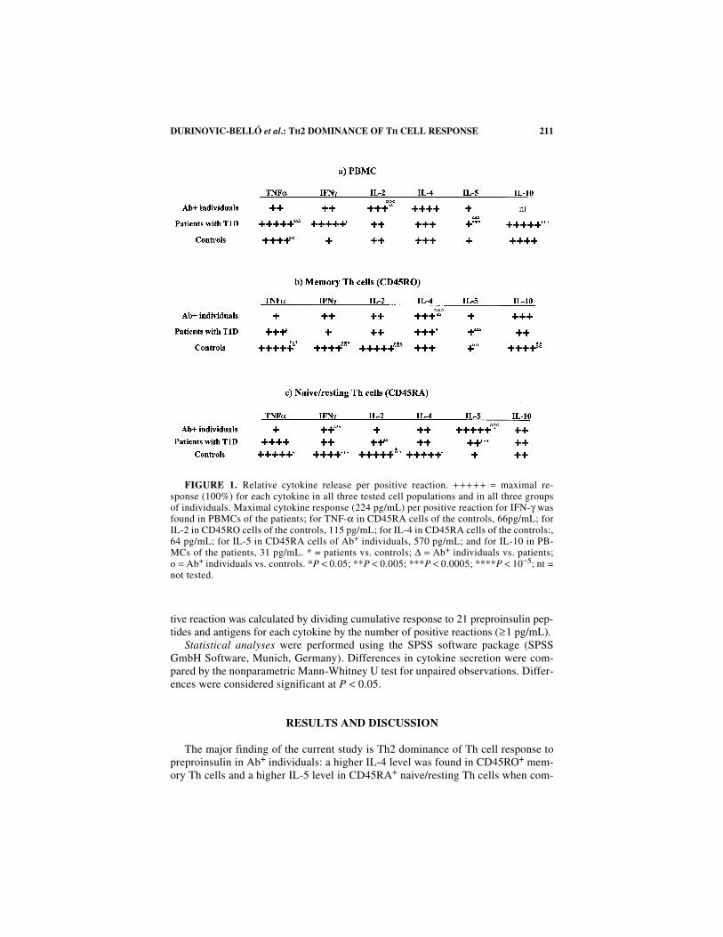
The major finding of the current study is Th2 dominance of Th cell response topreproinsulin in Ab+ individuals: a higher IL-4 level was found in CD45RO+ mem-ory Th cells and a higher IL-5 level in CD45RA+ naive/resting Th cells when com-
FIGURE 1. Relative cytokine release per positive reaction. + ++++ = maximal re-sponse (100%) for each cytokine in all three tested cell populations and in all three groupsof individuals. Maximal cytokine response (224 pg/mL) per positive reaction for IFN-γ wasfound in PBMCs of the patients; for TNF-α in CD45RA cells of the controls, 66pg/mL; forIL-2 in CD45RO cells of the controls, 115 pg/mL; for IL-4 in CD45RA cells of the controls:,64 pg/mL; for IL-5 in CD45RA cells of Ab+ individuals, 570 pg/mL; and for IL-10 in PB-MCs of the patients, 31 pg/mL. * = patients vs. controls; ∆ = Ab+ individuals vs. patients;o = Ab+ individuals vs. controls. *P < 0.05; **P < 0.005; ***P < 0.0005; ****P < 10−5; nt =not tested.
212 ANNALS NEW YORK ACADEMY OF SCIENCES
pared to both the patients and controls (FIG. 1). It is conceivable that IL-4- and IL-5-dominated responses of Th cells—in early prediabetes—are critical for the suste-nance of antibody production. Interestingly, in PBMCs of Ab+ individuals an in-crease in IL-2 response was present that was not found in memory and naive/restingcells of the same individuals. Dominance of the IL-2 response in PBMCs may ag-gravate proliferation of the autoreactive T cell pool in the prediabetic stage. More-over, in the PBMCs of recently diagnosed T1D patients we found an increase in IFN-γ and IL-10 secretion. Taken together our results implicate specific roles for Th cellsubsets in islet cell autoimmunity. They also implicate involvement of cell types oth-er than Th cells (present in PBMCs) in T1D development. Indeed, in the NOD mousemodel of T1D different types of immune cells, such as CD8+ cells, NK cells, mac-rophages and B lymphocytes, are involved in the disease process.7,8 Moreover, it hasbeen shown that under certain conditions, “protective” Th2 responses may becomedestructive by activating typical Th1 effector mechanisms.9 In conclusion, the cytok-ine patterns observed in peripheral circulation and in Th cell subpopulations of indi-viduals with islet cell autoimmunity may prove useful in monitoring diseaseprogression and in prevention trials.
ACKNOWLEDGMENTS
This work was supported by grants from the Deutsche Forschungsgemeinschaft,Sonderforschungsbereich, SFB 518 (to I.D.B. and B.O.B.), the Eli Lilly FoundationInternational (to I.D.B.), Deutsche Diabetes Stiftung (to I.D.B.), and Deutsche Dia-betes Gesellschaft (to I.D.B.). We acknowledge B. Feldman and M. Kuhn-Halder forclinical care of the patients and J. Neckermann for excellent technical assistance.
REFERENCES
1. DOUGLAS-PETERSEN, L., M. VAN DER KEUR, R.R. DE VRIES & B.O. ROEP. 1999. Autore-active and immunoregulatory T-cell subsets in insulin-dependent diabetes mellitus.Diabetologia 42: 443–449.
2. LIBLAU, R.S., S.M. SINGER & H.O. MCDEVITT. 1995. Th1 and Th2 CD4+ T cells in thepathogenesis of organ-specific autoimmune diseases. Immunol. Today 16: 34–38.
3. STREBELOW, M., M. SCHLOSSER, B. ZIEGLER, et al.. 1999. Karlsburg Type I diabetes riskstudy of a general population: frequencies and interactions of the four major type Idiabetes-associated autoantibodies studied in 9419 schoolchildren. Diabetologia 42:661–670.
4. DURINOVIC-BELLO, I, A. STEINLE, A-G. ZIEGLER & D.J. SCHENDEL. 1994. HLA-DQ-restricted, islet-specific T-cell clones of a type I diabetic patient: T-cell receptorsequence similarities to insulitis-inducing T-cells of nonobese diabetic mice. Diabe-tes 43: 1318–1325.
5. DURINOVIC-BELLO, I., M. HUMMEL & A-G. ZIEGLER. 1996. Cellular immune responseto diverse islet cell antigens in IDDM. Diabetes 45: 795–800.
6. PEAKMAN, M., T. TREE, J. ENDL, et al. 2001 Characterization of Preparations ofGAD65, Proinsulin, and the Islet Tyrosine Phosphatase IA-2 for use in Detection ofAutoreactive T-Cells in Type 1 Diabetes. Report of Phase II of the Second Interna-tional Immunology of Diabetes Society Workshop for Standardization of T-cellAssays in Type 1 Diabetes. Diabetes 50: 1749–1754.
213DURINOVIC-BELLÓ et al.: TH2 DOMINANCE OF TH CELL RESPONSE
7. WONG, F.S., J. KARTTUNEN, C. DUMONT, et al. 1999. Identification of an MHC class I-restricted autoantigen in type 1 diabetes by screening an organ-specific cDNAlibrary. Nat. Med. 5: 1026–1031.
8. KOLB, H. 1999. On the aetiopathogenesis of type 1 diabetes: key roles for innateimmunity and dietary antigens? Exp. Clin. Endocrinol. Diabetes 107: S13–16.
9. KOLB, H. 1997. Benign versus destructive insulitis. Diabetes Metab. Rev. 13: 139–146.


![Circulating and Tumor-Infiltrating Foxp3 Regulatory T Cell ... · traditional Th1, Th2 helper T cell subsets, Foxp3+ reg-ulatory T cell (Tregs) and IL-17-producing Th17 cells[9].](https://static.fdocuments.in/doc/165x107/5e4b79c0f61ac961cb5bf5de/circulating-and-tumor-infiltrating-foxp3-regulatory-t-cell-traditional-th1.jpg)


