TECO® - Diapharma · PDF fileTECOmedical TECO® Human Alpha GST Urine ELISA Kit. CONT ....
17
TE1056 | 02/2017 © TECOmedical Group | 03/2017 DIAPHARMA Group TE1056REV01 TECO® Human Alpha GST ELISA Serum & Urine Instructions for use English For Research Use Only Catalogue No. TE1056 © 02/2017 | TECOmedical Group, Switzerland
Transcript of TECO® - Diapharma · PDF fileTECOmedical TECO® Human Alpha GST Urine ELISA Kit. CONT ....
TE10
56 |
02/2
017
© TEC
Omedical
Gro
up |
03/2
017 DIAPH
ARM
A G
roup
TE1
056R
EV01
TECO® Human Alpha GST ELISA Serum & Urine
Instructions for use English
For Research Use Only Catalogue No. TE1056
© 02/2017 | TECOmedical Group, Switzerland
TECOmedical
2
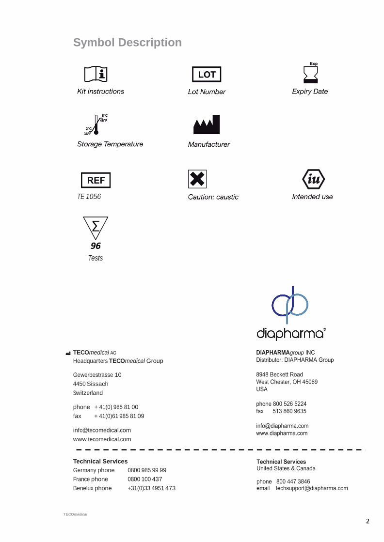
Symbol Description
TE 1056
Tests
TECOmedical AG
Headquarters TECOmedical Group
Gewerbestrasse 10 4450 Sissach Switzerland
phone + 41(0) 985 81 00 fax + 41(0)61 985 81 09
[email protected] www.tecomedical.com
Technical Services Germany phone 0800 985 99 99 France phone 0800 100 437 Benelux phone +31(0)33 4951 473
DIAPHARMAgroup INCDistributor: DIAPHARMA Group
8948 Beckett RoadWest Chester, OH 45069USA
phone 800 526 5224fax 513 860 9635
Technical Services United States & Canada
phone 800 447 3846email [email protected]

TECOmedical
TECO® Human Alpha GST Urine ELISA Kit CONT Reagents and Materials Supplied:
SYMBOL DESCRIPTION FORMAT
1 αGST Antibody Coated Microtiter Plate 12 strips of 8 wells (96 break apart wells in total), in a frame with cover plate. Ready to use.
1 plate
S Standard Stock 2 mg/L
1 x 0.2 mL
C1 Control C1 Ready to use.
1 x 1.0 mL
C2 Control C2 Ready to use.
1 x 1.0 mL
2 Wash Buffer 50x 1 x 30 mL
3 Sample Diluent Ready to use.
1 x 30 mL
6 Enzyme Conjugate Ready to use.
1 x 12 mL
7 TMB Substrate Ready to use.
1 x 12 mL
8 Stop Solution – 1 M HCI 1 M hydrochloric acid. Ready to use.
1 x 12 mL
Kit instructions 1 x
TECOmedical
Storage Store kit at 2–8 °C. Do not freeze. Store unused reagents at 2–8 °C.
Intended Use The Alpha GST ELISA provides a method for the quantitative determination of alpha glutathione S-transferase (αGST) in human urine, serum and plasma. To assay αGST in other media or assay other GST subclasses, contact us for further information. The Alpha GST ELISA is for research use only and not for use in diagnostic procedures.
Background URINE In kidney, alpha glutathione S-transferase (αGST) is found in the proximal tubule region whereas pi glutathione S-transferase (πGST) is confined mainly to the distal tubules1. Low levels of αGST are released into the urine in normal individuals, as confirmed by immunoassay and Western blot anlaysis2. Any event which precipitates proximal tubular damage may cause increased release of αGST into urine and elevations of urinary αGST levels have been shown to be indicative of proximal tubule damage in nephrotoxicity3-5, environmental toxicity6, surgery7, acute renal failure8 and transplantation9-12. The release of αGST has been shown to be associated with distal tubular damage6, thus simultaneous measurement of αGST and αGST may allow discrimination between proximal and tubular damage5,
9-11.
SERUM In liver, alpha glutathione S-transferase is located in the hepatocytes whereas pi GST (πGST) is confined to the intrahepatic bile duct cells1, 13-14. This heterogeneous GST subclass distribution suggests that the isoenzymes have unique in vivo functions in different hepatic regions and that the detection of GST subclass levels in biological fluids would be of significant use in monitoring the integrity of specific hepatic regions. Currently, liver injury is studied by the measurement of liver enzymes such as alanine aminotransferase (ALT) and aspartate aminotransferase (AST). A disadvantage of these markers is that they are not distributed uniformly throughout the liver, the periportal concentration being greater than the centrilobular15. In contrast, αGST has been found to be equally distributed in both the centrilobular and periportal regions13-14. Since the centrilobular hepatocytes are very susceptible to damage in a variety of conditions including Allograft Rejection16-18, Viral Hepatitis19, and Hepatotoxicity20, αGST is a more sensitive indicator of hepatic status.
Alpha GST ELISA is a specific, precise immunoassay for αGST21,22 and, being a quantitative test, is unaffected by modulators of enzyme activity (e.g. bile salts and bilirubin)21. Thus, it is now possible to use αGST quantitation to study the hepatocellular status of individuals at risk of hepatic damage.
TECOmedical
Assay Principle Alpha GST ELISA is a quantitative enzyme immunoassay. The test procedure is based on the sequential addition of sample, antibody-enzyme conjugate and substrate to microassay wells coated with anti-αGST IgG. The resultant color intensity is proportional to the amount of αGST present in the sample.
Materials Required and not Supplied
• Pipettes 10 µL – 1000 µL• Multichannel pipettes for 100 µL• Graduated cylinders for reconstituting or diluting reagents• Manual Aspiration System or Automatic washer for ELISA plates• Aqua dest• Vortex mixer• ELlSA plate reader suitable for 96 well formats and capable of measuring at 450
and 405 nm (Reference: 590-650 nm)• ELISA plate shaker (500 rpm) (orbital shaker)• Software package for data generation and analysis
TECOmedical
Warnings and Precautions This kit is intended for research use by professional persons only. Follow the instructions carefully. Observe expiration dates stated on the labels and the specified stability for reconstituted reagents. Refer to ”Materials Safety Data Sheet” for more detailed safety information.
Material of animal origin used in the preparation of this kit has been obtained from animals certified as healthy but these materials should be handled as potentially infectious.
Material of human origin used in the preparation of this kit has been tested and found non reactive for HIV-1 and HIV-2 as well as for HCV antibodies and HbsAg but should, nonetheless, be handled as potentially infectious.
TECOmedical AG is not liable for loss or harm caused by non-observance of the Kit instructions.
1 For research use only. 2 Treat all specimen samples as potentially biohazardous material.
Follow General Precautions when handling contents of this kit and any patient samples.
3 Disposal of containers and unused contents should be done in accordance with federal and local regulatory requirements.
4 Use the supplied reagents as an integral unit prior to the expiration date indicated on the package label.
5 Store assay reagents as indicated. 6 Do not use coated strips if pouch is punctured. 7 Test each sample in duplicate. 8 Use of multichannel pipettes is recommended to ensure the timely
delivery of liquids. 9 a. 1 M hydrochloric acid is caustic and can be harmfull for skin, eyes and
mucosae. b. Handle TMB with care. Do not ingest. Avoid contact with skin, eyes, or
clothing. Should there be any contact, wash with water. If ingested, call a physician.
10 A mercury-free preservative is used. Incidental contact with or ingestion of buffer solutions may cause irritation of skin, eyes or mouth. Should there be any contact, wash with water. If ingested, call a physician.
TECOmedical
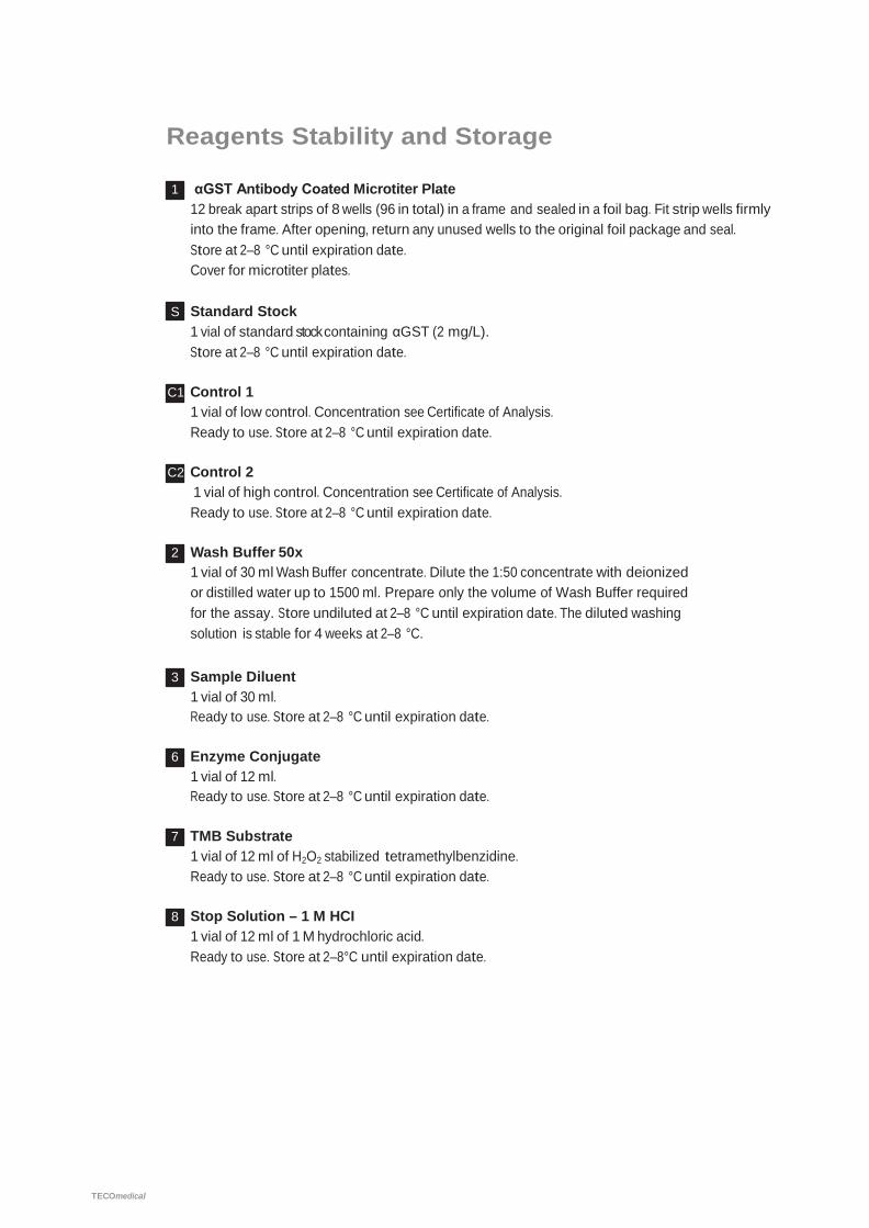
Reagents Stability and Storage
1 αGST Antibody Coated Microtiter Plate 12 break apart strips of 8 wells (96 in total) in a frame and sealed in a foil bag. Fit strip wells firmly into the frame. After opening, return any unused wells to the original foil package and seal. Store at 2–8 °C until expiration date. Cover for microtiter plates.
S Standard Stock F 1 vial of standard stock containing αGST (2 mg/L).
Store at 2–8 °C until expiration date.
C1 Control 1 1 vial of low control. Concentration see Certificate of Analysis. Ready to use. Store at 2–8 °C until expiration date.
C2 Control 2
1 vial of high control. Concentration see Certificate of Analysis. Ready to use. Store at 2–8 °C until expiration date.
2 Wash Buffer 50x
1 vial of 30 ml Wash Buffer concentrate. Dilute the 1:50 concentrate with deionized or distilled water up to 1500 ml. Prepare only the volume of Wash Buffer required for the assay. Store undiluted at 2–8 °C until expiration date. The diluted washing solution is stable for 4 weeks at 2–8 °C.
3 Sample Diluent
1 vial of 30 ml. Ready to use. Store at 2–8 °C until expiration date.
6 Enzyme Conjugate
1 vial of 12 ml. Ready to use. Store at 2–8 °C until expiration date.
7 TMB Substrate
1 vial of 12 ml of H2O2 stabilized tetramethylbenzidine. Ready to use. Store at 2–8 °C until expiration date.
8 Stop Solution – 1 M HCI
1 vial of 12 ml of 1 M hydrochloric acid. Ready to use. Store at 2–8°C until expiration date.
TECOmedical
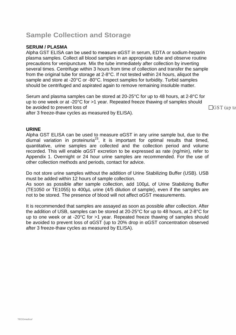
Sample Collection and Storage SERUM / PLASMA Alpha GST ELISA can be used to measure αGST in serum, EDTA or sodium-heparin plasma samples. Collect all blood samples in an appropriate tube and observe routine precautions for venipuncture. Mix the tube immediately after collection by inverting several times. Centrifuge within 3 hours from time of collection and transfer the sample from the original tube for storage at 2-8°C. If not tested within 24 hours, aliquot the sample and store at -20°C or -80°C. Inspect samples for turbidity. Turbid samples should be centrifuged and aspirated again to remove remaining insoluble matter. Serum and plasma samples can be stored at 20-25°C for up to 48 hours, at 2-8°C for up to one week or at -20°C for >1 year. Repeated freeze thawing of samples should be avoided to prevent loss of GS T (up to after 3 freeze-thaw cycles as measured by ELISA). URINE Alpha GST ELISA can be used to measure αGST in any urine sample but, due to the diurnal variation in proteinuria23, it is important for optimal results that timed, quantitative, urine samples are collected and the collection period and volume recorded. This will enable αGST excretion to be expressed as rate (ng/min), refer to Appendix 1. Overnight or 24 hour urine samples are recommended. For the use of other collection methods and periods, contact for advice. Do not store urine samples without the addition of Urine Stabilizing Buffer (USB). USB must be added within 12 hours of sample collection. As soon as possible after sample collection, add 100µL of Urine Stabilizing Buffer (TE1050 or TE1055) to 400µL urine (4/5 dilution of sample), even if the samples are not to be stored. The presence of blood will not affect αGST measurements. It is recommended that samples are assayed as soon as possible after collection. After the addition of USB, samples can be stored at 20-25°C for up to 48 hours, at 2-8°C for up to one week or at -20°C for >1 year. Repeated freeze thawing of samples should be avoided to prevent loss of αGST (up to 20% drop in αGST concentration observed after 3 freeze-thaw cycles as measured by ELISA).
TECOmedical
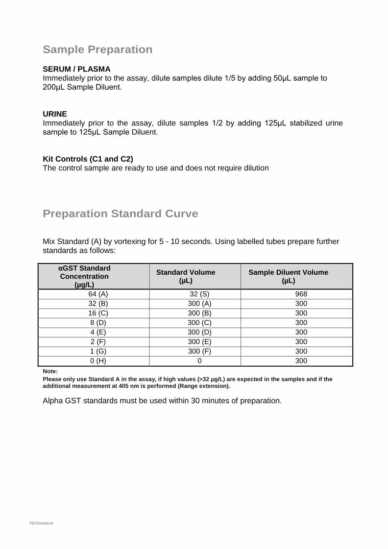
Sample Preparation SERUM / PLASMA Immediately prior to the assay, dilute samples dilute 1/5 by adding 50μL sample to 200μL Sample Diluent. URINE Immediately prior to the assay, dilute samples 1/2 by adding 125μL stabilized urine sample to 125μL Sample Diluent. Kit Controls (C1 and C2) The control sample are ready to use and does not require dilution
Preparation Standard Curve Mix Standard (A) by vortexing for 5 - 10 seconds. Using labelled tubes prepare further standards as follows:
αGST Standard Concentration
(µg/L) Standard Volume
(µL) Sample Diluent Volume
(µL) 64 (A) 32 (S) 968 32 (B) 300 (A) 300 16 (C) 300 (B) 300 8 (D) 300 (C) 300 4 (E) 300 (D) 300 2 (F) 300 (E) 300 1 (G) 300 (F) 300 0 (H) 0 300
Note: Please only use Standard A in the assay, if high values (>32 µg/L) are expected in the samples and if the additional measurement at 405 nm is performed (Range extension).
Alpha GST standards must be used within 30 minutes of preparation.
TECOmedical
Assay Procedure
All determinations (standards, controls and samples) should be assayed in duplicate. When performing the assay, the standards, controls and samples should be pipetted as fast as possible (<15 minutes). To avoid distortions due to differences in incubation times, Enzyme Conjugate, Substrate Solution and Stop Solution should be added to the plate in the same order and with the same time interval as the samples. A multichannel pipette is essential.
Allow all reagents to stand at 20–25°C for at least 30 minutes. During all incubation steps, plates should be sealed with the adhesive foil or a plastic cover. For light protection, incubate in a dark chamber or cover plate with aluminium foil.
1 Allocate the wells of the Microtiter plate 1 for standards, controls and samples.
2 Pipette 100 µl of each standards (A until H), controls (C1 and C2) and diluted samples into the corresponding wells.
3 Cover the wells with a plastic cover and incubate the plate for 1h ± 5 min at (20–25°C) on a shaker (500 rpm).
4 After incubation, aspirate the wells by using a plate washer or manually decant by inverting the plate. Wash the wells 4 times with 350 µl diluted Wash Buffer per well. After the last wash cycle tap the inverted wells on a dry absorbent surface to remove excess Wash Solution. The use of an automatic plate washer is recommended.
5 Following the last washing step, pipette 100 µl of the Enzyme Conjugate 6 in each well (multichannel pipette).
6 Cover the wells with a plastic cover and incubate the plate for 1h ± 5 min at 20–25°C on a shaker (500 rpm).
7 After incubation wash the wells 4 times with Wash Buffer as described in step 4.
8 Pipette 100 µl of the TMB Substrate 7 in each well (multichannel pipette).
9 Incubate the plate for 15-30 min, in the dark, at 20–25°C on a shaker (500 rpm).
10 Stop the reaction by adding 100 µl of Stop Solution 8 (multichannel pipette). 11 Read the absorbance of the wells (450 and/or 405 nm). Reference filter at 590–650 nm.
TECOmedical
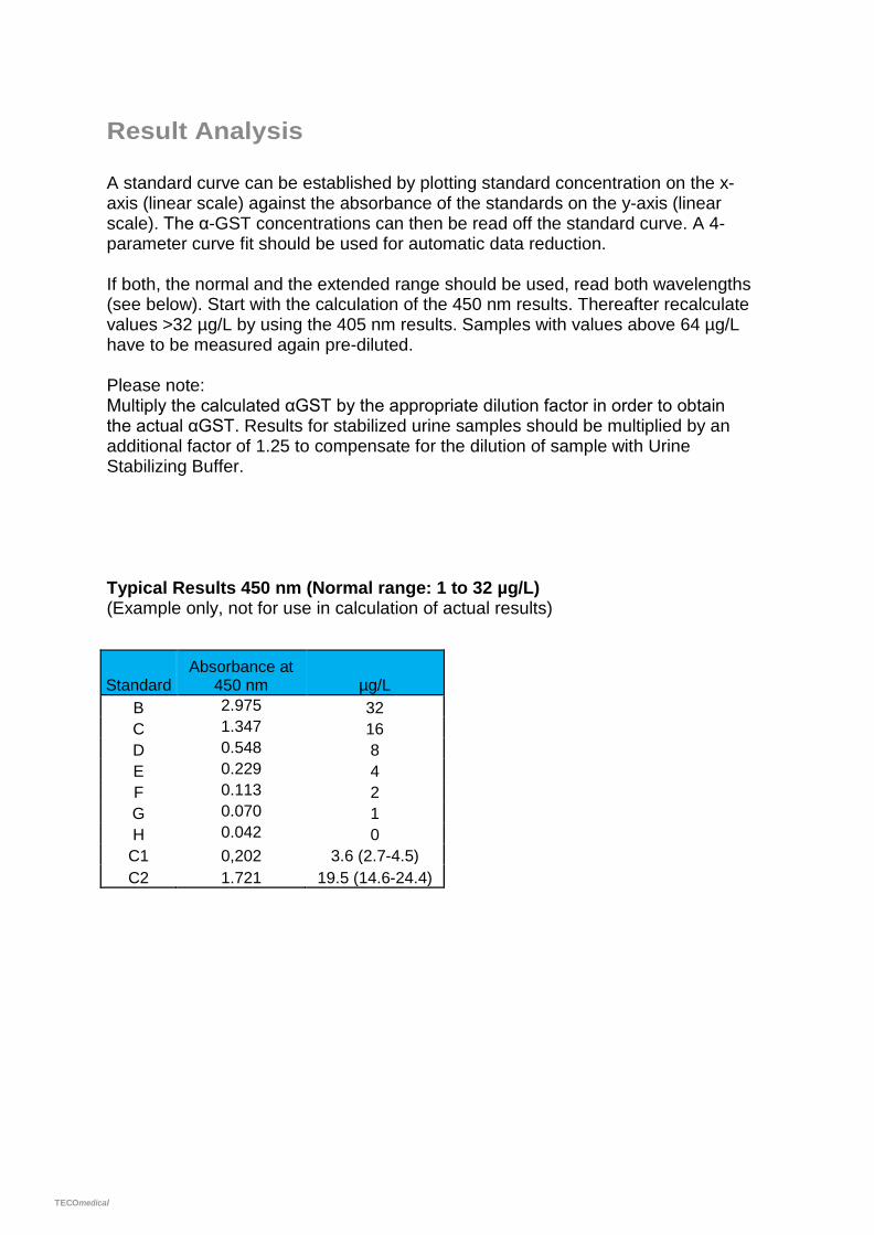
Result Analysis A standard curve can be established by plotting standard concentration on the x-axis (linear scale) against the absorbance of the standards on the y-axis (linear scale). The α-GST concentrations can then be read off the standard curve. A 4-parameter curve fit should be used for automatic data reduction.
If both, the normal and the extended range should be used, read both wavelengths (see below). Start with the calculation of the 450 nm results. Thereafter recalculate values >32 µg/L by using the 405 nm results. Samples with values above 64 µg/L have to be measured again pre-diluted. Please note: Multiply the calculated αGST by the appropriate dilution factor in order to obtain the actual αGST. Results for stabilized urine samples should be multiplied by an additional factor of 1.25 to compensate for the dilution of sample with Urine Stabilizing Buffer. Typical Results 450 nm (Normal range: 1 to 32 µg/L) (Example only, not for use in calculation of actual results)
Standard Absorbance at
450 nm µg/L B 2.975 32 C 1.347 16 D 0.548 8 E 0.229 4 F 0.113 2 G 0.070 1 H 0.042 0
C1 0,202 3.6 (2.7-4.5) C2 1.721 19.5 (14.6-24.4)
TECOmedical
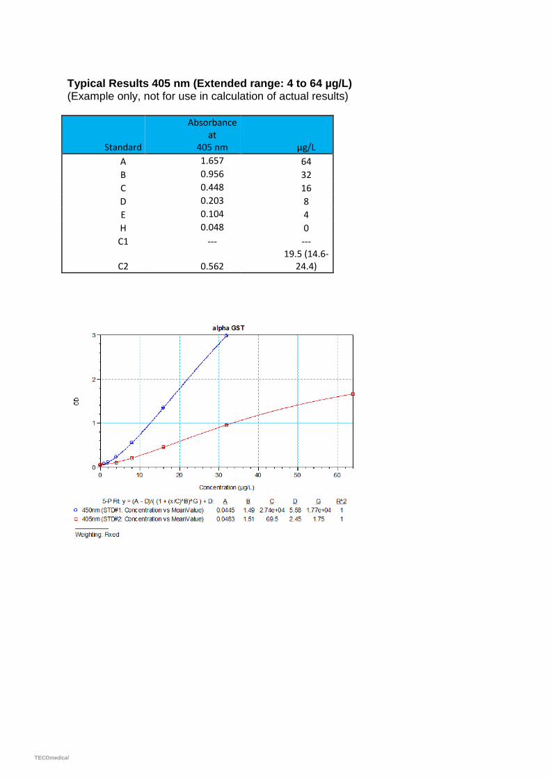
Typical Results 405 nm (Extended range: 4 to 64 µg/L) (Example only, not for use in calculation of actual results)
Standard
Absorbance at
405 nm µg/L A 1.657 64 B 0.956 32 C 0.448 16 D 0.203 8 E 0.104 4 H 0.048 0 C1 --- ---
C2 0.562 19.5 (14.6-
24.4)
TECOmedical
Reference Ranges SERUM / PLASMA Samples were obtained from apparently healthy donors without any clinical abnormal indications. GS T le ve ls we re de te rmine d us ing the Alp ELISA in order to establish the GS T conce ntra tion in the norma l popula tion. The reference interval (5th to 95th percentiles) for Alpha GST ELISA is 0-12.0µg/L in serum (n=120). The reference intervals reflect the donor population of this study group. It is recommended that each laboratory determine their own reference range appropriate for their study group. URINE Samples were obtained from apparently healthy donors without any clinical abnormal indications. αGST levels were determined using the Alpha GST ELISA in order to establish the αGST concentration in the normal population. The reference interval (5th to 95th percentiles) for Alpha GST ELISA is 0-29.0µg/L in urine (n=120). The reference intervals reflect the donor population of this study group. It is recommended that each laboratory determine their own reference range appropriate for their study group. Performance Characteristics SPECIFICITY Alpha GST ELISA is highly specific for αGST. No cross-reactivity was observed with µGST at 500µg/L, or πGST at 500µg/L. MEASURING RANGE AND SENSITIVITY The limit of detection (LoD) of Alpha GST ELISA was found to be <0.5 μg/L αGST, which corresponds to 1.25 μg/L in a stabilized urine sample diluted 1/2. For serum/plasma, the LoD is 2.5 μg/L for a sample diluted 1/5. The measurement Range for serum/plasma is 5 – 320 µg/L and for urine 2.5 to 160 µg/L.
TECOmedical
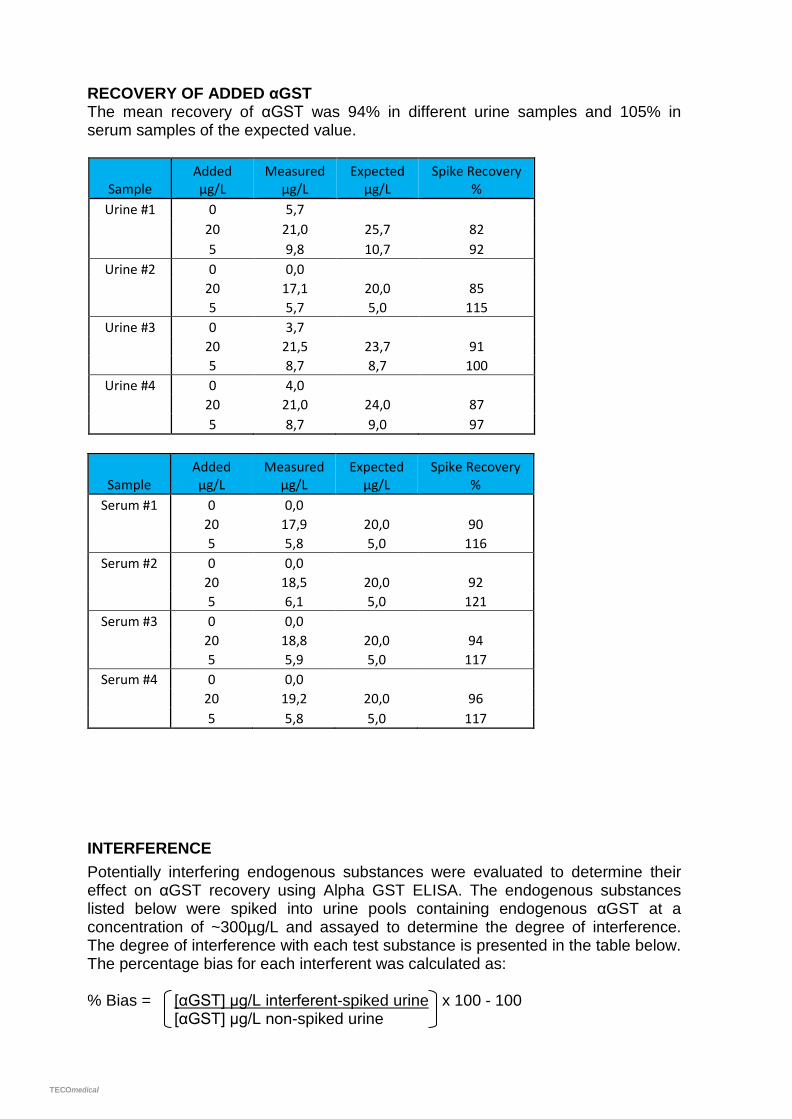
RECOVERY OF ADDED αGST The mean recovery of αGST was 94% in different urine samples and 105% in serum samples of the expected value.
Sample Added µg/L
Measured µg/L
Expected µg/L
Spike Recovery %
Urine #1 0 5,7 20 21,0 25,7 82 5 9,8 10,7 92
Urine #2 0 0,0
20 17,1 20,0 85 5 5,7 5,0 115
Urine #3 0 3,7 20 21,5 23,7 91 5 8,7 8,7 100
Urine #4 0 4,0
20 21,0 24,0 87 5 8,7 9,0 97
Sample Added µg/L
Measured µg/L
Expected µg/L
Spike Recovery %
Serum #1 0 0,0 20 17,9 20,0 90 5 5,8 5,0 116
Serum #2 0 0,0
20 18,5 20,0 92 5 6,1 5,0 121
Serum #3 0 0,0 20 18,8 20,0 94 5 5,9 5,0 117
Serum #4 0 0,0
20 19,2 20,0 96 5 5,8 5,0 117
INTERFERENCE Potentially interfering endogenous substances were evaluated to determine their effect on αGST recovery using Alpha GST ELISA. The endogenous substances listed below were spiked into urine pools containing endogenous αGST at a concentration of ~300µg/L and assayed to determine the degree of interference. The degree of interference with each test substance is presented in the table below. The percentage bias for each interferent was calculated as: % Bias = [αGST] µg/L interferent-spiked urine x 100 - 100 [αGST] µg/L non-spiked urine
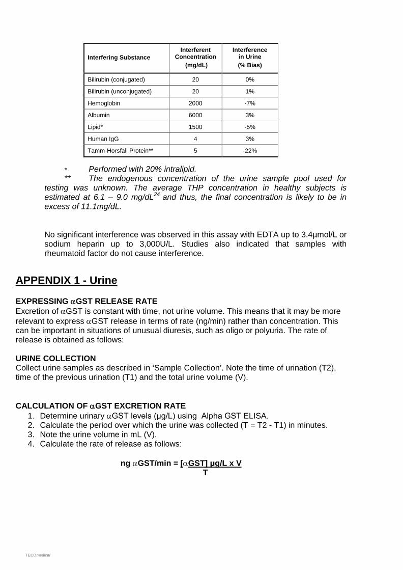
TECOmedical
Interfering Substance Interferent
Concentration (mg/dL)
Interference in Urine (% Bias)
Bilirubin (conjugated) 20 0%
Bilirubin (unconjugated) 20 1%
Hemoglobin 2000 -7%
Albumin 6000 3%
Lipid* 1500 -5%
Human IgG 4 3%
Tamm-Horsfall Protein** 5 -22%
* Performed with 20% intralipid. ** The endogenous concentration of the urine sample pool used for testing was unknown. The average THP concentration in healthy subjects is estimated at 6.1 – 9.0 mg/dL24 and thus, the final concentration is likely to be in excess of 11.1mg/dL. No significant interference was observed in this assay with EDTA up to 3.4µmol/L or sodium heparin up to 3,000U/L. Studies also indicated that samples with rheumatoid factor do not cause interference.
APPENDIX 1 - Urine EXPRESSING αGST RELEASE RATE Excretion of αGST is constant with time, not urine volume. This means that it may be more relevant to express αGST release in terms of rate (ng/min) rather than concentration. This can be important in situations of unusual diuresis, such as oligo or polyuria. The rate of release is obtained as follows: URINE COLLECTION Collect urine samples as described in ‘Sample Collection’. Note the time of urination (T2), time of the previous urination (T1) and the total urine volume (V). CALCULATION OF αGST EXCRETION RATE
1. Determine urinary αGST levels (μg/L) using Alpha GST ELISA. 2. Calculate the period over which the urine was collected (T = T2 - T1) in minutes. 3. Note the urine volume in mL (V). 4. Calculate the rate of release as follows:
ng αGST/min = [αGST] μg/L x V
T
TECOmedical
REFERENCES 1. Campbell, J.A.H. et. al. (1991). Immunohistologic localization of alpha, mu and pi class glutathione S-transferase in
human tissues. Cancer (Philadelphia) 67 (6), 1608-1613. 2. Hassett, B. and Doyle, S. (1995). Biotrin International internal research. 2 Goldberg, M.E. et. al. (1999). Dose of compound A, not sevoflurane, determines changes in the biochemical
markers of renal injury in healthy volunteers. Anesthesia and Analgesia 88(2), 437-445. 3 Eger II, E.I. et. al. (1997). Nephrotoxicity of sevoflurane versus desflurane anesthesia in volunteers. Anesthesia and
Analgesia 84, 160-168. 5. Kirby K.B. et. al. (1997). Urinary glutathione transferase as an early marker of renal impairmentin psoriasis patients
treated with Cyclosporin A (CsA). Paper presented at the XIVth International Congress of Nephrology 25-29 May 1997, Sydney, Australia.
1. Sundberg, A.G.M. et. al. (1994). Glutathione transferases in the urine: sensitive methods for detection of kidney damage induced by nephrotoxic agents in humans. Environmental Health Perspectives 102 (Suppl 3), 293-296.
2. Cressey G et. al. (2002). Renal tubular injury after infrarenal aortic aneurysm repair. Journal Cardiothoracic and Vascular Anesthesia 16(3), 290-3.
8. Cakalaroski, K. et. al. (1999). α-glutathione S transferases as markers of tubular cell dysfunction in acute renal failure patients. Abstract from the Third Congress of the Balkan Cities Association of Nephrology, Dialysis and Artificial Organs (BANTAO) Belgrade, Yugoslavia, 1998. Nephrol. Dial. Transplant 14, 2978.
9. Sundberg, A.G.M. et. al. (1994). Quantitation of glutathione transferase-pi in the urine by radioimmunoassay. Nephron 66(2), 162-169.
10. Stegeman, C.A. et. al. (1996). Differential diagnosis of early graft dysfunction by urinary excretion of αGST 7(9), 1986.
11. Kievit, J.K. et. al. (1997). Release of alpha-glutathione S-transferase (αGST) and pi-glutathione S-transferase (πGST) from ischemic damaged kidneys into the machine perfusate - relevance to viability assessment Transplantation Proceedings 29(8), 3591-3593.
12. Daeman, J.W.H.C. et. al. (1997). Glutathione S-transferase as predictor of functional outcome in transplantation of machine-preserved non-heart beating donor kidneys. Transplantation 63(1), 89-93.
13. Sundberg, A.G. et al. (1993). Immunohistochemical localisation of alpha and pi class glutathione transferases in normal human tissues. Pharmacology and Toxicology 72(4-5), 321- 331.
14. Manning, F. et al. (1995). Argutus Medical International Internal Research. 15. Beckett, G. J. and Hayes, J.D. (1993) Glutathione S-transferases: biomedical applications. Advances in Clinical
Chemistry 30, 281-380. 16. Trull. A.K. et al. (1994). Serum alpha-glutathione S-transferase: a sensitive marker of hepatocellular damage
associated with acute liver allograft rejection. Transplantation 58(12), 1345-51. 17. Platz K.P. et al. (1997). Determination of alpha- and Pi-glutathione-S-transferase will improve monitoring after liver
transplantation. Transplant Proc. 29 (7), 2827-2829. 18. Hughes, V.F. et al. (1997) Randomized trial to evaluate the clinical benefits of serum α-glutathione S-transferase
concentration monitoring after liver transplantation. Transplantation 64(10), 1446-1452. 19. Nelson D.R. et al. (1995). alpha-glutathione S-transferase as a marker of hepatocellular damage in chronic hepatitis
C virus infection. Am. J. Clin. Pathol. 104(2), 193-198. 20. Murray, J. M. et al. (1992). Indocyanine green clearance and hepatic function during and after prolonged
anaesthesia: comparison of halothane with isoflurane Br. J. Anaesth. 68(2), 168-171. 21. Rees, G.W. et al. (1995). Evaluation of an enzyme-immunometric assay for serum alpha-gultathione S-transferase.
Ann. Clin. Biochem. 32, 575-583. 22. Doyle, S. et al. (1994). Detection of serum α glutathione S-transferase by enzyme immunoassay. International
Symposium on Liver and Drugs, Bratislava, Slovakia. 23. Jung, K. (1994). Urinary enzymes and low molecular weight proteins as markers of tubular dysfunction. Kidney
International Suppl. 47, S29-33. 24. Lau, W-H. et al. (2008). Qualification and application of an ELISA for the determination of Tamm Horsfall protein
(THP) in human urine and its use for screening of kidney stone disease. Journal of Biological Sciences 4(4), 215-22.
TECOmedical
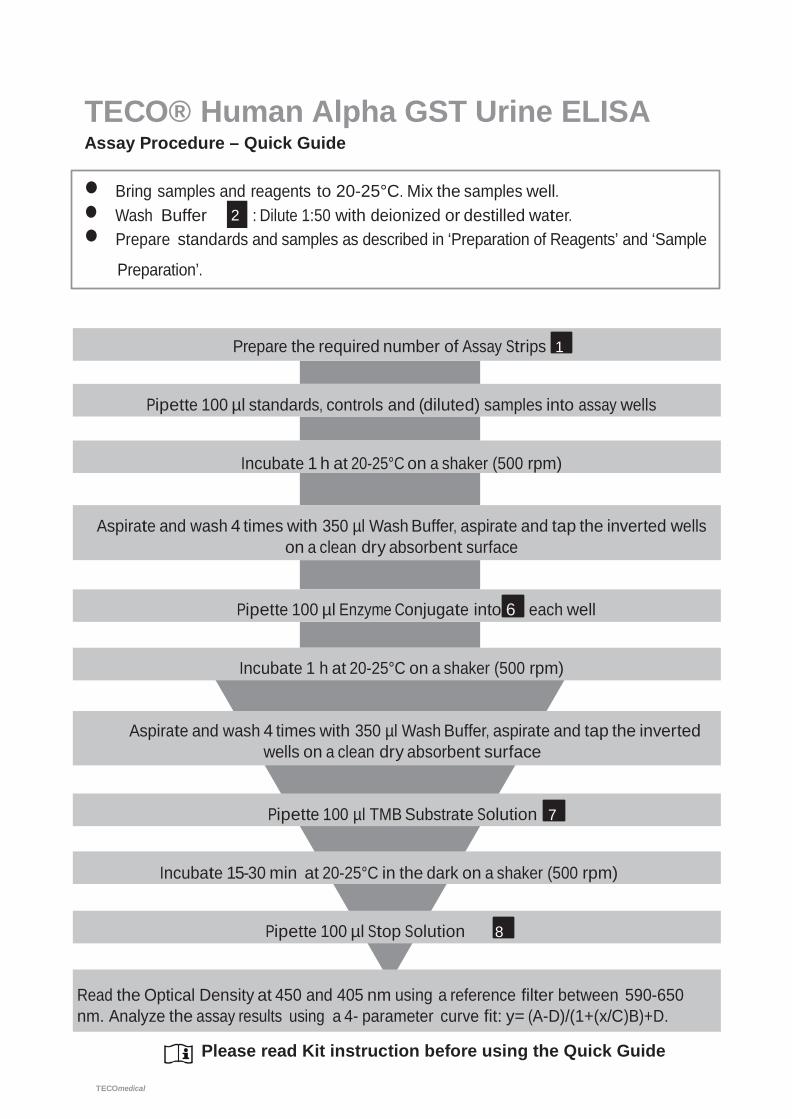
TECO® Human Alpha GST Urine ELISA Assay Procedure – Quick Guide
• Bring samples and reagents to 20-25°C. Mix the samples well.
• Wash Buffer 2 : Dilute 1:50 with deionized or destilled water. • Prepare standards and samples as described in ‘Preparation of Reagents’ and ‘Sample
Preparation’.
Prepare the required number of Assay Strips 1
Pipette 100 µl standards, controls and (diluted) samples into assay wells
Incubate 1 h at 20-25°C on a shaker (500 rpm)
Aspirate and wash 4 times with 350 µl Wash Buffer, aspirate and tap the inverted wells on a clean dry absorbent surface
Pipette 100 µl Enzyme Conjugate into 6 each well
Incubate 1 h at 20-25°C on a shaker (500 rpm)
Aspirate and wash 4 times with 350 µl Wash Buffer, aspirate and tap the inverted wells on a clean dry absorbent surface
Pipette 100 µl TMB Substrate Solution 7
Incubate 15-30 min at 20-25°C in the dark on a shaker (500 rpm)
Pipette 100 µl Stop Solution 8
Read the Optical Density at 450 and 405 nm using a reference filter between 590-650 nm. Analyze the assay results using a 4- parameter curve fit: y= (A-D)/(1+(x/C)B)+D.
Please read Kit instruction before using the Quick Guide