Technical Guide Technical Guide A KNOW-HOW GUIDE TO … KNOW-HOW GUIDE TO USING LARVAL THERAPY FOR...
4
A KNOW-HOW GUIDE TO USING LARVAL THERAPY FOR WOUND DEBRIDEMENT Maggot therapy, also known as larval therapy or biosurgery, has been used for centuries and has been reintroduced more recently into wound care. It is an effective and rapid method of debriding all types of wounds and combating infection. Although there are many different types of wounds, there are fundamental aspects of wound management which should be followed to enable every wound to progress though the healing process. It has been suggested that debridement is a vital part of this process (Falanga, 2000). Aside from surgical debridement none of the many methods of debridement work as quickly (or as cost-effectively) as larval therapy (Thomas, 2006). There are still concerns among healthcare staff with regards to the use of larval therapy. This is largely due to the ‘yuck’ factor. However, the advantages of the use of this therapy far outweigh Claire Acton is Tissue Viability and Vascular Nurse Specialist, Queen Elizabeth Hospital NHS Trust, London The use of maggots in wound care may still have the ‘yuck’ factor but its effectiveness at debriding infected and necrotic tissue for a range of different wound types makes it an essential part of the tissue viability nurse’s toolkit. Here the theory behind maggot therapy is described and a step-by- step guide to the two different ways of applying maggot therapy is given. any negative aspects and it is worth helping the nervous patient to overcome any reservations they may have about this therapy. This article will outline the different applications of larval therapy, and will provide a step-by-step guide to its use. It will also dispel any myths surrounding this form of treatment and highlight its benefits. Description The sterile maggots supplied for use in wound management are the larvae of the common greenbottle (Lucilia sericata). Upon application to the wound, they are 2–3 mm long, but they can reach 8–10 mm in size when fully grown. The larvae work by producing a proteolytic enzyme that degrades and liquefies the necrotic tissue present in the wound, which they then ingest as a nutrient (Casu et al, 1996). The maggots also combat odour and infection by ingesting any bacteria present in the wound. There have been reports of a reduction in wound pain (Evans,1997) associated with their use and they can stimulate the formation of granulation tissue (Prete, 1997).The maggots of the Lucilia sericata will not attack or burrow into healthy tissue. It takes 10–14 days for a newly hatched maggot to complete a lifecycle and turn into a fly. Dressings should be changed every 3–4 days, removing fully grown larvae before they are ready to pupate. However, this will not happen as the larvae like a dry and warm area to do this so a moist wound environment is not appropriate. Indications Larval therapy can be used to treat many types of wound including leg ulcers, pressure ulcers, diabetic foot ulcers, surgical wounds and burns. It has also been proven to be effective on infected, necrotic and sloughy tissue (Thomas and Jones, 1999). Technical Guide 156 Wound Essentials • Volume 2 • 2007
-
Upload
nguyennguyet -
Category
Documents
-
view
247 -
download
0
Transcript of Technical Guide Technical Guide A KNOW-HOW GUIDE TO … KNOW-HOW GUIDE TO USING LARVAL THERAPY FOR...

A KNOW-HOW GUIDE TO USING LARVAL THERAPY FOR WOUND DEBRIDEMENT
Maggot therapy, also known as larval therapy or biosurgery, has been used for centuries and has been reintroduced more recently into wound care. It is an effective and rapid method of debriding all types of wounds and combating infection.
Although there are many different types of wounds, there are fundamental aspects of wound management which should be followed to enable every wound to progress though the healing process. It has been suggested that debridement is a vital part of this process (Falanga, 2000).Aside from surgical debridement none of the many methods of debridement work as quickly (or as cost-effectively) as larval therapy (Thomas, 2006).
There are still concerns among healthcare staff with regards to the use of larval therapy. This is largely due to the ‘yuck’ factor. However, the advantages of the use of this therapy far outweigh
Claire Acton is Tissue Viability and Vascular Nurse Specialist, Queen Elizabeth Hospital NHS Trust, London
The use of maggots in wound care may still have the ‘yuck’ factor but its effectiveness at debriding infected and necrotic tissue for a range of different wound types makes it an essential part of the tissue viability nurse’s toolkit. Here the theory behind maggot therapy is described and a step-by-step guide to the two different ways of applying maggot therapy is given.
any negative aspects and it is worth helping the nervous patient to overcome any reservations they may have about this therapy.
This article will outline the different applications of larval therapy, and will provide a step-by-step guide to its use. It will also dispel any myths surrounding this form of treatment and highlight its benefi ts.
DescriptionThe sterile maggots supplied for use in wound management are the larvae of the common greenbottle (Lucilia sericata). Upon application to the wound, they are 2–3 mm long, but they can reach 8–10 mm in size when fully grown. The larvae work by producing a proteolytic enzyme that degrades and liquefi es the necrotic tissue present in the wound, which they then ingest as a nutrient (Casu et al, 1996). The maggots also combat odour and infection by ingesting any bacteria present in the wound.
There have been reports of a reduction in wound pain (Evans,1997) associated with their use and they can stimulate the formation of granulation tissue (Prete, 1997).The maggots of the Lucilia sericata will not attack or burrow into healthy tissue. It takes 10–14 days for a newly hatched maggot to complete a lifecycle and turn into a fl y. Dressings should be changed every 3–4 days, removing fully grown larvae before they are ready to pupate. However, this will not happen as the larvae like a dry and warm area to do this so a moist wound environment is not appropriate.
IndicationsLarval therapy can be used to treat many types of wound including leg ulcers, pressure ulcers, diabetic foot ulcers, surgical wounds and burns. It has also been proven to be effective on infected, necrotic and sloughy tissue (Thomas and Jones, 1999).
Technical GuideTechnical Guide
156 Wound Essentials • Volume 2 • 2007 Wound Essentials • Volume 2 • 2007 157
156-59larvae.indd 2 3/6/07 11:31:16 pm
ContraindicationsLarvae should not be applied to wounds that have a tendency to bleed easily, or be introduced into wounds that communicate with a body cavity or internal organ. They should also not be applied to any large blood vessels.
How to use maggot therapyThere are two ways to deliver maggot therapy. The ‘free range’ technique involves applying the maggots directly to the wound (LarvE, ZooBiotic, Bridgend). Alternatively, they are applied using a pouch containing maggots and foam chips which are designed to manage the wound exudate and provide an ideal environment for the maggots (LarvE BioFOAM, ZooBiotic, Bridgend). When using the free-range technique there is no need to count the maggots in and out of the wound as there will always be a proportion of larvae that fail to survive.
Why use one type of application over the other? LarvE BioFOAM is generally the more practical and aesthetically acceptable method, however, free-range maggots are more suitable for use in cavity or undermining wounds and larger leg ulcers.
AssessmentBefore the application of larval therapy the patient’s consent should be gained and all aspects of the treatment fully explained. The Zoobiotic website has a downloadable patient leaflet that can help with this process (www.zoobiotic.com).
A full patient assessment should be undertaken to ensure that
Technical GuideTechnical Guide
156 Wound Essentials • Volume 2 • 2007 Wound Essentials • Volume 2 • 2007 157
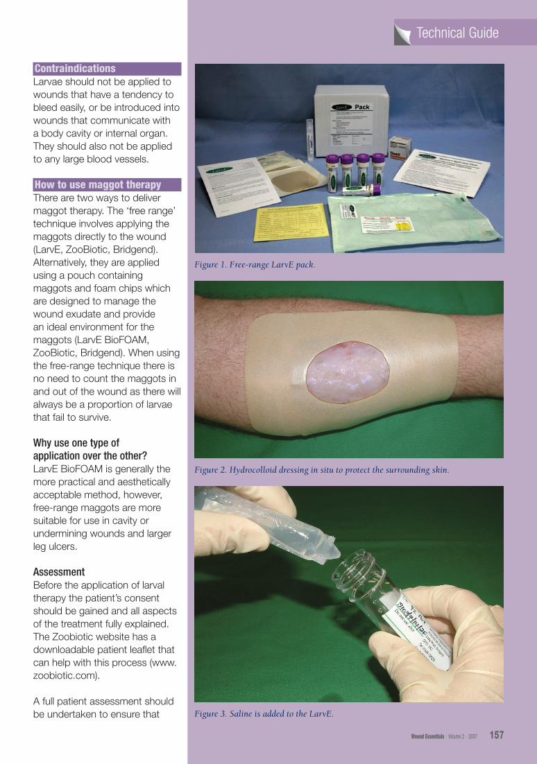
Figure 3. Saline is added to the LarvE.
Figure 1. Free-range LarvE pack.
Figure 2. Hydrocolloid dressing in situ to protect the surrounding skin.
156-59larvae.indd 3 3/6/07 11:31:39 pm

the use of this treatment is appropriate. Both free range LarvE and BioFOAM LarvE are available on prescription as well as within the hospital environment. This allows for continuity of care should a patient be discharged while receiving the therapy.
Before application, pain levels should be considered and analgesia prescribed before the dressing change, as well as on a regular basis if required.
StorageThe maggots need to be used within eight hours and stored at a temperature of 8–10 degrees. They should be removed from the courier delivery bag as soon as possible after they arrive, before being placed in the fridge as recommended by the manufacturer.
Application
Application of free-range larvE Before application, use the LarvE calculator to assess the number of maggots to be used for the individual wound. The calculator can be found on the Zoobiotic website (www.zoobiotic.org/larve-calc/larve-calculator-2005.pdf).The amount used is based on the size of the wound that is being treated.
Remove the existing dressing and cleanse any dressing residue. Components of a standard maggot dressing are shown in Figure 1.
Cut a hole the size and shape of the wound in the Granuflex hydrocolloid sheet which is supplied with the free-range maggots (Figure 2). For larger
Technical GuideTechnical Guide
Figure 5. LarvE applied to the wound, covered with net and secured with sleek tape at the edges.
Figure 6. Perforated film dressing is applied, along with an absorbant pad, and is secured with a cotton bandage.
158 Wound Essentials • Volume 2 • 2007 Wound Essentials • Volume 2 • 2007 159
Figure 4. Maggots on sterile net.
156-59larvae.indd 4 3/6/07 11:31:43 pm

wounds it may be better to cut strips of hydrocolloid and place around the margin of the wound. Where there are pressure areas, such as on the heel, build up the surrounding area to create a trench using a hydrocolloid. Skin barriers that can be used to protect the surrounding skin are Sudocrem®, paste bandages impregnated with zinc (Viscopaste PB7™, Smith and Nephew; Steripaste, Mölnlycke Healthcare) or Cavilon™ barrier cream (3M Health Care).
Add 5ml of sterile saline to the LarvE (Figure 3) and then pour the maggots onto a sterile net (Figure 4).
Place the net on the wound and tape it into place (Figure 5). A swab moistened with water should be placed over the net. The perforated film dressing should then be applied along with an absorbent pad and cotton bandage (Figure 6).
Application of BioFOAM Remove any residue from the old dressing and apply barrier cream (Sudocrem is provided with the BioFOAM) to the surrounding skin.
Components of the LarvE BioFOAM dressing are pictured in Figure 7. Apply BioFOAM to wound and cover with a permeable dressing (NA or Release).
Outer dressings should be checked or changed on a daily basis. Exudate will be a red/brown colour and this should not be confused with bleeding when the maggots are removed (Figure 8).
RemovalThe therapy should be removed after 3–5 days. If further debridement is required, re-order the larvae and reapply. All used larvae dressings should be disposed of as clinical waste.
ConclusionThe use of larval therapy has proved to be very effective in the treatment of infected wounds with dead tissue, which needs to be removed. It should be considered an essential part of any nurse’s toolkit for treating all types of wounds due to its speedy effectiveness.
Casu RE, Eisemann CH, Vuocolo T, Tellman RL (1996) The major exceretory/secretory protease from
Technical GuideTechnical Guide
Lucilia cuprina larvae is also a gut digestive protease. Int J Parasitology 26(6): 623–8
Evans H (1997) A treatment of last resort. Nursing Times 93(23): 62–5
Falanga V (2000) Classifications for wound bed preparation and stimulation of chronic wounds. Wound Repair Regen 8(5): 347–52
Prete, P (1997) Growth effects of Phaenicia sericata larval extracts on fibroblasts: mechanism for wound healing by maggot therapy. Life Sciences 60(8): 505–10
Thomas, S (2006) Cost of managing chronic wounds in the UK, with particular emphasis on maggot debridement therapy. J Wound Care 15(10): 465–9
Thomas S, Jones M (1999) The Use of Sterile Maggots in Wound Management. Wound Care Society, Huntingdon
158 Wound Essentials • Volume 2 • 2007 Wound Essentials • Volume 2 • 2007 159
Figure 7. The LarvE BioFOAM dressing.
Figure 8. BioFOAM in-situ on a heel ulcer.
WE
156-59larvae.indd 5 3/6/07 11:31:46 pm