Targeting Sirtuin 1 to Improve Metabolism: All You Need Is NAD
22
ASSOCIATE EDITOR: BEATRICE DESVERGNE Targeting Sirtuin 1 to Improve Metabolism: All You Need Is NAD ? Carles Canto ´ and Johan Auwerx Ecole Polytechnique Fe ´de ´rale de Lausanne, Lausanne, Switzerland Abstract ................................................................................ 166 I. Introduction............................................................................. 166 II. Sirtuin 1 in a nutshell ................................................................... 168 A. Sirtuin 1: what and where is it? ....................................................... 168 B. Sirtuin 1 as an NAD sensor .......................................................... 169 C. Sirtuin 1 actions ..................................................................... 169 1. Nuclear targets.................................................................... 169 2. Cytosolic targets................................................................... 173 III. Sirtuin 1 and metabolic disease: evidence from mice models ................................. 173 IV. Physiological and pharmacological modulation of sirt1 activity ............................... 175 A. The modulation of sirtuin 1 expression ................................................. 175 B. Post-translational modifications ....................................................... 177 C. Protein interactions .................................................................. 178 D. Sirtuin 1-activating compounds ........................................................ 178 E. Indirect modulation through affecting NAD metabolism................................. 179 1. Modulation by increasing NAD synthesis ........................................... 180 2. Modulation by decreasing NAD consumption ........................................... 181 a. Poly(ADP-ribose) polymerase .................................................... 181 b. CD38 .......................................................................... 182 V. Conclusions and future directions ......................................................... 183 Acknowledgments ....................................................................... 183 References .............................................................................. 183 Abstract——Sirtuin 1 (SIRT1) is an evolutionarily con- served NAD -dependent deacetylase that is at the pinna- cle of metabolic control, all the way from yeast to humans. SIRT1 senses changes in intracellular NAD levels, which reflect energy level, and uses this information to adapt the cellular energy output such that it matches cellular energy requirements. The changes induced by SIRT1 activation are generally (but not exclusively) transcriptional in na- ture and are related to an increase in mitochondrial me- tabolism and antioxidant protection. These attractive fea- tures have validated SIRT1 as a therapeutic target in the management of metabolic disease and prompted an inten- sive search to identify pharmacological SIRT1 activators. In this review, we first give an overview of the SIRT1 biol- ogy with a particular focus on its role in metabolic control. We then analyze the pros and cons of the current strategies used to activate SIRT1 and explore the emerging evidence indicating that modulation of NAD levels could provide an effective way to achieve such goals. I. Introduction During the last decade the mammalian sirtuin (SIRT 1 ) family (formed by paralogs SIRT1–SIRT7) has emerged as Address correspondence to: Dr. Johan Auwerx, Laboratory of In- tegrative Systems Physiology (LISP) and Nestle Chair in Energy Metabolism (NCEM), Institute of Bioengineering, Ecole Polytech- nique Fe ´de ´ rale de Lausanne (EPFL), Station 15, CH-1015 Lausanne, Switzerland. E-mail: [email protected] This article is available online at http://pharmrev.aspetjournals.org. http://dx.doi.org/10.1124/pr.110.003905. 1 Abbreviations: AceCS, acetyl-CoA synthetase; AMPK, AMP-activated protein kinase; AROS, active regulator of SIRT1; ChREBP, carbohydrate response element-binding protein; CREB, cAMP response element-binding protein; CRTC, CREB-regulated transcriptional coactivator; CtBP, C-ter- minal binding protein; DBC, deleted in breast cancer; eNOS, endothelial nitric-oxide synthase; ERC, extrachromosomal rDNA circle; FOXO, Fork- head-O-box; HIC1, hypermethylated in cancer 1; JNK, c-Jun NH 2 - terminal kinase; LXR, liver X receptor; miRNA, microRNA; NA, nicotinic acid; NAM, nicotinamide; Nampt, nicotinamide phosphoribosyltrans- ferase; NCoR, nuclear receptor corepressor; NMN, nicotinamide mononu- cleotide; NR, nicotinamide riboside; PARP, poly(ADP-ribose) polymerase; PGC, PPAR coactivator; PPAR, peroxisome proliferator-activated recep- 1521-0081/12/6401-166 –187$25.00 PHARMACOLOGICAL REVIEWS Vol. 64, No. 1 Copyright © 2012 by The American Society for Pharmacology and Experimental Therapeutics 3905/3740172 Pharmacol Rev 64:166 –187, 2012 166 by guest on November 18, 2018 Downloaded from
Transcript of Targeting Sirtuin 1 to Improve Metabolism: All You Need Is NAD

ASSOCIATE EDITOR: BEATRICE DESVERGNE
Targeting Sirtuin 1 to Improve Metabolism: All YouNeed Is NAD�?
Carles Canto and Johan Auwerx
Ecole Polytechnique Federale de Lausanne, Lausanne, Switzerland
Abstract . . . . . . . . . . . . . . . . . . . . . . . . . . . . . . . . . . . . . . . . . . . . . . . . . . . . . . . . . . . . . . . . . . . . . . . . . . . . . . . . 166I. Introduction. . . . . . . . . . . . . . . . . . . . . . . . . . . . . . . . . . . . . . . . . . . . . . . . . . . . . . . . . . . . . . . . . . . . . . . . . . . . . 166
II. Sirtuin 1 in a nutshell . . . . . . . . . . . . . . . . . . . . . . . . . . . . . . . . . . . . . . . . . . . . . . . . . . . . . . . . . . . . . . . . . . . 168A. Sirtuin 1: what and where is it? . . . . . . . . . . . . . . . . . . . . . . . . . . . . . . . . . . . . . . . . . . . . . . . . . . . . . . . 168B. Sirtuin 1 as an NAD� sensor . . . . . . . . . . . . . . . . . . . . . . . . . . . . . . . . . . . . . . . . . . . . . . . . . . . . . . . . . . 169C. Sirtuin 1 actions . . . . . . . . . . . . . . . . . . . . . . . . . . . . . . . . . . . . . . . . . . . . . . . . . . . . . . . . . . . . . . . . . . . . . 169
1. Nuclear targets. . . . . . . . . . . . . . . . . . . . . . . . . . . . . . . . . . . . . . . . . . . . . . . . . . . . . . . . . . . . . . . . . . . . 1692. Cytosolic targets. . . . . . . . . . . . . . . . . . . . . . . . . . . . . . . . . . . . . . . . . . . . . . . . . . . . . . . . . . . . . . . . . . . 173
III. Sirtuin 1 and metabolic disease: evidence from mice models . . . . . . . . . . . . . . . . . . . . . . . . . . . . . . . . . 173IV. Physiological and pharmacological modulation of sirt1 activity . . . . . . . . . . . . . . . . . . . . . . . . . . . . . . . 175
A. The modulation of sirtuin 1 expression . . . . . . . . . . . . . . . . . . . . . . . . . . . . . . . . . . . . . . . . . . . . . . . . . 175B. Post-translational modifications . . . . . . . . . . . . . . . . . . . . . . . . . . . . . . . . . . . . . . . . . . . . . . . . . . . . . . . 177C. Protein interactions . . . . . . . . . . . . . . . . . . . . . . . . . . . . . . . . . . . . . . . . . . . . . . . . . . . . . . . . . . . . . . . . . . 178D. Sirtuin 1-activating compounds . . . . . . . . . . . . . . . . . . . . . . . . . . . . . . . . . . . . . . . . . . . . . . . . . . . . . . . . 178E. Indirect modulation through affecting NAD� metabolism. . . . . . . . . . . . . . . . . . . . . . . . . . . . . . . . . 179
1. Modulation by increasing NAD� synthesis. . . . . . . . . . . . . . . . . . . . . . . . . . . . . . . . . . . . . . . . . . . 1802. Modulation by decreasing NAD� consumption . . . . . . . . . . . . . . . . . . . . . . . . . . . . . . . . . . . . . . . . . . . 181
a. Poly(ADP-ribose) polymerase . . . . . . . . . . . . . . . . . . . . . . . . . . . . . . . . . . . . . . . . . . . . . . . . . . . . 181b. CD38 . . . . . . . . . . . . . . . . . . . . . . . . . . . . . . . . . . . . . . . . . . . . . . . . . . . . . . . . . . . . . . . . . . . . . . . . . . 182
V. Conclusions and future directions . . . . . . . . . . . . . . . . . . . . . . . . . . . . . . . . . . . . . . . . . . . . . . . . . . . . . . . . . 183Acknowledgments . . . . . . . . . . . . . . . . . . . . . . . . . . . . . . . . . . . . . . . . . . . . . . . . . . . . . . . . . . . . . . . . . . . . . . . 183References . . . . . . . . . . . . . . . . . . . . . . . . . . . . . . . . . . . . . . . . . . . . . . . . . . . . . . . . . . . . . . . . . . . . . . . . . . . . . . 183
Abstract——Sirtuin 1 (SIRT1) is an evolutionarily con-served NAD�-dependent deacetylase that is at the pinna-cle of metabolic control, all the way from yeast to humans.SIRT1 senses changes in intracellular NAD� levels, whichreflect energy level, and uses this information to adapt thecellular energy output such that it matches cellular energyrequirements. The changes induced by SIRT1 activationare generally (but not exclusively) transcriptional in na-ture and are related to an increase in mitochondrial me-tabolism and antioxidant protection. These attractive fea-
tures have validated SIRT1 as a therapeutic target in themanagement of metabolic disease and prompted an inten-sive search to identify pharmacological SIRT1 activators.In this review, we first give an overview of the SIRT1 biol-ogy with a particular focus on its role in metabolic control.We then analyze the pros and cons of the current strategiesused to activate SIRT1 and explore the emerging evidenceindicating that modulation of NAD� levels could providean effective way to achieve such goals.
I. Introduction
During the last decade the mammalian sirtuin (SIRT1)family (formed by paralogs SIRT1–SIRT7) has emerged as
Address correspondence to: Dr. Johan Auwerx, Laboratory of In-tegrative Systems Physiology (LISP) and Nestle Chair in EnergyMetabolism (NCEM), Institute of Bioengineering, Ecole Polytech-nique Federale de Lausanne (EPFL), Station 15, CH-1015 Lausanne,Switzerland. E-mail: [email protected]
This article is available online at http://pharmrev.aspetjournals.org.http://dx.doi.org/10.1124/pr.110.003905.
1Abbreviations: AceCS, acetyl-CoA synthetase; AMPK, AMP-activatedprotein kinase; AROS, active regulator of SIRT1; ChREBP, carbohydrateresponse element-binding protein; CREB, cAMP response element-bindingprotein; CRTC, CREB-regulated transcriptional coactivator; CtBP, C-ter-minal binding protein; DBC, deleted in breast cancer; eNOS, endothelialnitric-oxide synthase; ERC, extrachromosomal rDNA circle; FOXO, Fork-head-O-box; HIC1, hypermethylated in cancer 1; JNK, c-Jun NH2-terminal kinase; LXR, liver X receptor; miRNA, microRNA; NA, nicotinicacid; NAM, nicotinamide; Nampt, nicotinamide phosphoribosyltrans-ferase; NCoR, nuclear receptor corepressor; NMN, nicotinamide mononu-cleotide; NR, nicotinamide riboside; PARP, poly(ADP-ribose) polymerase;PGC, PPAR� coactivator; PPAR, peroxisome proliferator-activated recep-
1521-0081/12/6401-166–187$25.00PHARMACOLOGICAL REVIEWS Vol. 64, No. 1Copyright © 2012 by The American Society for Pharmacology and Experimental Therapeutics 3905/3740172Pharmacol Rev 64:166–187, 2012
166
by guest on Novem
ber 18, 2018D
ownloaded from
a constellation of enzymes with key roles in whole-bodymetabolic homeostasis and an interesting therapeutic po-tential applicable to multiple pathophysiological states.
The history of sirtuins began almost 3 decades ago, withthe identification of Sir2 (silent information regulator 2), aprotein-forming part of a complex that enabled gene silenc-ing at selected regions of the yeast genome (Shore et al.,1984; Ivy et al., 1986). A major turning point in the historyof Sir2 came from the discovery that Sir2 was involved inthe yeast replicative aging process (Kaeberlein et al.,1999). The accumulation of extrachromosomal rDNA cir-cles (ERCs) as the organism ages is believed to be a majordeterminant of yeast replicative lifespan (Sinclair andGuarente, 1997). Although the mechanism by which theaccumulation of ERCs influences lifespan is not fully un-derstood, different genetic manipulations promote reason-able, despite correlational, evidence that the accumulationof ERCs is negatively correlated with yeast replicativeaging (Sinclair and Guarente, 1997; Defossez et al., 1999;Kaeberlein et al., 1999). It was originally thought that theimpact of Sir2 on replicative lifespan of yeast was conse-quent to its silencing activity on ERCs. However, the ef-fects of Sir2 on aging extend further than ERC silencing,because genetic manipulations of Sir2 orthologs can alsoaffect lifespan of higher eukaryotes, such as the nematodeCaenorhabditis elegans (Tissenbaum and Guarente, 2001;Viswanathan et al., 2005; Berdichevsky et al., 2006; Rizkiet al., 2011) and insects such as Drosophila melanogaster
(Rogina and Helfand, 2004; Bauer et al., 2009), whereERCs are not thought to cause aging. However, there aresome caveats on the consistency, amplitude, and mamma-lian translation of the lifespan-extension effects of Sir2orthologs (Kaeberlein and Powers, 2007; Burnett et al.,2011; Lombard et al., 2011; Viswanathan and Guarente,2011), which suggest that the effects of Sir2 on organismallifespan might be indirect and/or largely depend on a spe-cific repertoire of third-party modulators.
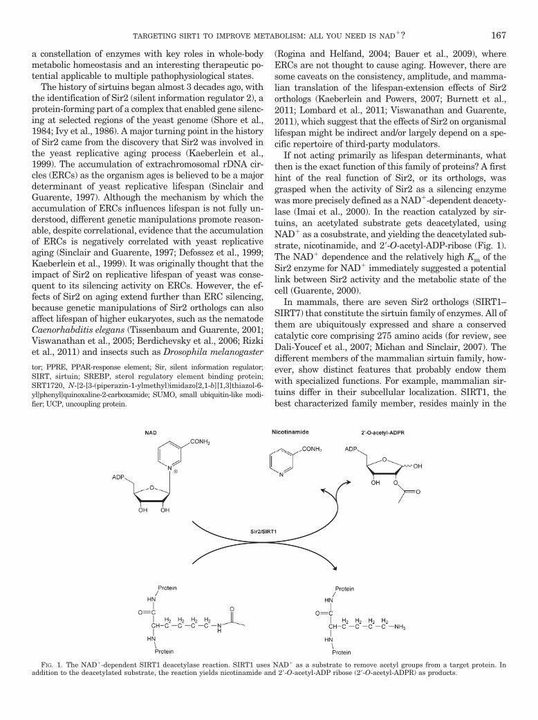
If not acting primarily as lifespan determinants, whatthen is the exact function of this family of proteins? A firsthint of the real function of Sir2, or its orthologs, wasgrasped when the activity of Sir2 as a silencing enzymewas more precisely defined as a NAD�-dependent deacety-lase (Imai et al., 2000). In the reaction catalyzed by sir-tuins, an acetylated substrate gets deacetylated, usingNAD� as a cosubstrate, and yielding the deacetylated sub-strate, nicotinamide, and 2�-O-acetyl-ADP-ribose (Fig. 1).The NAD� dependence and the relatively high Km of theSir2 enzyme for NAD� immediately suggested a potentiallink between Sir2 activity and the metabolic state of thecell (Guarente, 2000).
In mammals, there are seven Sir2 orthologs (SIRT1–SIRT7) that constitute the sirtuin family of enzymes. All ofthem are ubiquitously expressed and share a conservedcatalytic core comprising 275 amino acids (for review, seeDali-Youcef et al., 2007; Michan and Sinclair, 2007). Thedifferent members of the mammalian sirtuin family, how-ever, show distinct features that probably endow themwith specialized functions. For example, mammalian sir-tuins differ in their subcellular localization. SIRT1, thebest characterized family member, resides mainly in the
tor; PPRE, PPAR-response element; Sir, silent information regulator;SIRT, sirtuin; SREBP, sterol regulatory element binding protein;SRT1720, N-[2-[3-(piperazin-1-ylmethyl)imidazo[2,1-b][1,3]thiazol-6-yl]phenyl]quinoxaline-2-carboxamide; SUMO, small ubiquitin-like modi-fier; UCP, uncoupling protein.
FIG. 1. The NAD�-dependent SIRT1 deacetylase reaction. SIRT1 uses NAD� as a substrate to remove acetyl groups from a target protein. Inaddition to the deacetylated substrate, the reaction yields nicotinamide and 2�-O-acetyl-ADP ribose (2�-O-acetyl-ADPR) as products.
TARGETING SIRT1 TO IMPROVE METABOLISM: ALL YOU NEED IS NAD�? 167

nucleus (Michishita et al., 2005) but can shuttle from thenucleus to the cytosol (Tanno et al., 2007), where several ofits targets are found. SIRT2 is localized mainly in thecytoplasm, although it can also regulate gene expressionby deacetylation of transcription factors that shuttle fromthe cytoplasm to the nucleus (Jing et al., 2007), and itcontributes to chromatin compaction upon disassembly ofthe cell nucleus during mitosis (Vaquero et al., 2006).SIRT3, SIRT4, and SIRT5 are generally considered mito-chondrial proteins (Onyango et al., 2002; Schwer et al.,2002; Michishita et al., 2005), whereas SIRT6 and SIRT7are nuclear proteins. However, although SIRT6 is locatedpredominantly in the heterochromatin, SIRT7 is thoughtto be mainly enriched in the nucleoli (Michishita et al.,2005).
In addition to their differential cellular locations, thesirtuin family members can also be distinguished by theirdifferent enzymatic activities. SIRT1 and SIRT5 act asdeacetylases (Imai et al., 2000; Vaziri et al., 2001), whereasSIRT4 seems to be a mono-ADP-ribosyl transferase (Hai-gis et al., 2006). SIRT2, SIRT3, and SIRT6 can display bothactivities (North et al., 2003; Liszt et al., 2005; Shi et al.,2005; Michishita et al., 2008). The activity of SIRT7 hasnot been clearly established, even though it has been hy-pothesized to act as a deacetylase (Vakhrusheva et al.,2008). It is noteworthy that SIRT5 was recently describedto demalonylate and desuccinylate proteins (Du et al.,2011; Peng et al., 2011). It is tempting to speculate that thespectrum of action of sirtuin is not limited to deacetylation
but would cover a much wider range of acylation-basedpost-translational modifications. The identification of sir-tuin substrates during the last few decades has clearlypointed out a prominent role of sirtuins as metabolic reg-ulators. For the purpose of this review, we mostly focus onthe actions of SIRT1. For extensive discussion of the ac-tions of other sirtuin members, we refer the reader toreviews elsewhere (Dali-Youcef et al., 2007; Michan andSinclair, 2007; Yamamoto et al., 2007; Schwer and Verdin,2008; Finkel et al., 2009; Guarente, 2011).
II. Sirtuin 1 in a Nutshell
A. Sirtuin 1: What and Where Is It?
Among all sirtuins, SIRT1 is the best characterized.Human SIRT1 contains the conserved catalytic core ofsirtuins and both N- and C-terminal extensions that allspan �240 amino acids (Fig. 2). These extensions serve asplatforms for interaction with regulatory proteins and sub-strates. In total, the human SIRT1 spans 747 amino acids.SIRT1 contains two nuclear localization signals as well astwo nuclear exportation signals (Tanno et al., 2007). Thebalanced functionality of these signals determines thepresence of SIRT1 in either the nuclear or the cytoplasmiccompartment and explains why SIRT1 location may differdepending on the cell type or tissue evaluated. For in-stance, although SIRT1 is found mainly in the nuclearcompartment in COS-7 cells (McBurney et al., 2003; Saka-moto et al., 2004), it is abundantly found in the cytosol of
NLS1 NES1 NES2NLS2
Sirtuin homology domain
236 490 748
NLS1(35-45)
NES1(138-145)
NES2(425-431)
NLS2(223-230)
236 490 748P P P P S
Region / aminoacids Func�on Reference
Sirtuin homology domain
Nuclear Localiza�on Signal
Nuclear Exporta�on Signal
aa 244-498 enzyma�c aci�vity
NLS1 (aas 34-44)NLS2 (aas 232-239) Nuclear importa�on
NES1 (aas 146-?) Nuclear exporta�on Tanno et
Tanno et al., 2007
Frye, 1999
Nuclear Exporta�on Signal NES1 (aas 146 ?)NES2 (aas 435-443)
Nuclear exporta�on
(by JNK1)Ser27, Ser47, Thr530
(by Cyclin/cdk1)
Specific ac�va�on towards H3, but not p53
Phosphoryla�onIncreases deacetylase Sasaki et al.,
Nasrin et al., 2009
Tanno et al., 2007
Thr530, Ser540
Sumoyla�on
yac�vity
,2008
Lys734 Increases deacetylase ac�vity
Yang et al., 2007c
FIG. 2. Relevant domains in the human form of the SIRT1 protein. The figure schematizes the span of the conserved sirtuin homology domain aswell as the nuclear localization signal (NLS) and nuclear exportation signals (NES). The residues subject to phosphorylation by JNK1 and Cyclin/cdk1and by SUMOylation are also indicated.
168 CANTÓ AND AUWERX

rodent � cells, myotubes, and cardiomyocytes (Moynihanet al., 2005; Tanno et al., 2007). Although the implicationsand regulation of SIRT1 shuttling are still largely un-known, some experiments indicate that SIRT1 shuttlesfrom the nuclei to the cytosol upon inhibition of insulinsignaling (Tanno et al., 2007). The latter observationssuggested a link between SIRT1 activity and the sens-ing of the metabolic status of the cell, as discussed inthe next chapter.
B. Sirtuin 1 as an NAD� Sensor
SIRT1 activity is generally increased in situations ofenergy/nutrient stress. The fact that SIRT1 activity is reg-ulated by NAD� raised the hypothesis that NAD� couldact as a metabolic sensor in situations of energy stress,where NAD� levels are generally affected. Some aspects ofthis hypothesis are still controversial (for review, seeCanto and Auwerx, 2009). First among them is whethersirtuin activity can really respond to changes in intracel-lular NAD� levels. One premise, at least, must be met inthat context: that the Km of the sirtuins for NAD� falls intothe physiological range of NAD� bioavailability. Directexperimental evidence supporting this point is, in mostcases, preliminary or absent. In great part, this is becausethe true bioavailable NAD� levels remain difficult to eval-uate. The estimated total intracellular content of NAD� isin the 0.2 to 0.5 mM range (for review, see (Sauve et al.,2006; Houtkooper et al., 2010), which lies within the esti-mated Km values of SIRT1 for NAD�. This would indicatethat NAD� might actually be rate-limiting in certain cir-cumstances to propel SIRT1 to its maximal activity. Theselevels, however, do not differentiate free and protein-boundNAD�. Likewise, this approximation does not take intoaccount the existence of cellular compartmentalization ofNAD�.
Changes in intracellular NAD� rarely fluctuate morethan 2-fold (Rodgers et al., 2005; Chen et al., 2008; Fulco etal., 2008; Canto et al., 2009), which is a likely range toaffect sirtuin activity. In general, NAD� levels increase inmammalian tissues in response to energy/nutrientstresses such as exercise (Canto et al., 2009, 2010; Costfordet al., 2010), fasting (Rodgers et al., 2005; Canto et al.,2010), or calorie restriction (Chen et al., 2008). Accord-ingly, SIRT1 activity is enhanced by all these conditions. Itis noteworthy that NAD� levels have been reported tofluctuate in a circadian fashion (Nakahata et al., 2009;Ramsey et al., 2009). The influence of SIRT1 on the controlof clock-related gene expression (Asher et al., 2008; Naka-hata et al., 2008) makes it very attractive to conceive thisrelation as a way by which feeding/fasting cycles influencethe circadian clock. In general, high glycolytic rates in thefed state would bring about higher NAD� reduction rates,whereas the reduced glycolytic rate in the fasted statewould enhance mitochondrial oxidative metabolism, de-rived from fatty acid oxidation, which is generally pairedwith higher NAD� levels. This scenario constitutes abeautiful mechanism by which metabolism would be
directly coupled to the enzymatic activity of SIRT1 anddownstream pathways.
SIRT1 activity is also controlled by other NAD�-derivedmetabolites. It was proposed that NADH would competewith NAD� binding to SIRT1 and inhibit SIRT1 activity(Lin et al., 2004). However, NADH can competitively in-hibit NAD� binding only in the millimolar range, which iswell above its physiological levels (Schmidt et al., 2004). Amore prominent and consistent inhibitory effect isachieved with nicotinamide (NAM), which exerts a potentend-product inhibition on SIRT1 activity in a noncompet-itive fashion with NAD� (Bitterman et al., 2002; Andersonet al., 2003). Kinetic studies demonstrate that NAM acts ata Km between 30 and 200 �M (Bitterman et al., 2002). Thereference values for the intracellular concentration andsubcellular compartmentalization of NAM are still farfrom determined, an issue that is further complicated bythe diffusive nature of NAM (van Roermund et al., 1995).Indirect evidence of the large influence of NAM on SIRT1activity is derived from experiments that manipulate NAMmetabolism through changing the activity of nicotinamidephosphorybosyltransferase (Nampt). In the cell, NAM isused as a substrate for NAD� resynthesis through theaction of Nampt (Revollo et al., 2004). Inhibition or down-regulation of Nampt leads to NAM accumulation andNAD� depletion, ultimately decreasing SIRT1 activity(Revollo et al., 2004). This highlights how NAM can influ-ence SIRT1 activity through different means: first, as anoncompetitive SIRT1 inhibitor, and, second, as an NAD�
precursor. Low levels of NAM might therefore be beneficialfor SIRT1 activity, because NAM can act as an NAD�
precursor, but, more importantly, accumulation of NAMcould be deleterious through the inhibition of SIRT1 (Yangand Sauve, 2006).
C. Sirtuin 1 Actions
1. Nuclear Targets. In agreement with its dual cel-lular localization, SIRT1 targets can be found in both thenuclear and cytosolic compartments. SIRT1 activity inthe nucleus articulates dynamic and varied transcrip-tional responses through the deacetylation of a largespectrum of transcriptional regulators. Therefore, thedeacetylation by SIRT1 can lead to the direct activationor inhibition of transcriptional regulators and modifytheir interaction profiles, depending on the cellular con-text. From a metabolic perspective, it is exciting to seethat many SIRT1 deacetylation targets are key meta-bolic regulators, further enhancing the notion thatSIRT1 is a metabolic stress effector governing transcrip-tional adaptations aimed to synchronize energy metab-olism with nutrient availability. SIRT1 activity and tar-gets, however, expand beyond the realm of metabolism.For example, SIRT1 has marked anti-inflammatory ef-fects in diverse tissues and cell models (Pfluger et al.,2008; Purushotham et al., 2009; Yoshizaki et al., 2009,2010), probably through the negative regulation of thenuclear factor-�B pathway (Yeung et al., 2004). In addi-
TARGETING SIRT1 TO IMPROVE METABOLISM: ALL YOU NEED IS NAD�? 169

tion, SIRT1 activity has a strong influence on cell pro-liferation, apoptosis, and cancer. Although the data invitro is controversial, the work on genetically engineeredmouse models indicates that enhanced SIRT1 activitywould be protective against the development of sometypes of cancer (Herranz et al., 2010). SIRT1 may also beof interest in the central nervous system. It was recentlyproven that SIRT1 has key roles modulating cognitivefunction and synaptic plasticity (Gao et al., 2010;Michan et al., 2010). In addition, there is evidence thatenhanced SIRT1 activity could be protective in condi-tions such as neurodegeneration, Alzheimer’s disease,and amyotrophic lateral sclerosis (Araki et al., 2004;Chen et al., 2005a; Kim et al., 2007a; Donmez et al.,2010). Because we focus mainly on the metabolic impactof SIRT1, we refer the reader to other reviews for dis-cussion of these other fields of action for SIRT1 (Finkelet al., 2009; Herranz and Serrano, 2010; Guarente,2011).
The identification of p53 as a SIRT1 substrate enlight-ened the scientific community on the versatility of SIRT1,which was until then largely considered a histone deacety-lase. Two different laboratories simultaneously reportedhow SIRT1 interacts with and deacetylates p53 (Luo et al.,2001; Vaziri et al., 2001). Although p53 can be acetylatedin up to six residues, SIRT1 seems to preferentiallydeacetylate Lys379 (human Lys382). The deacetylation ofp53 by SIRT1 attenuated its activity on the p21 promoterand inhibited p53-dependent apoptosis (Luo et al., 2001;Vaziri et al., 2001). This link between p53 and SIRT1activities led to the premature hypothesis that SIRT1 in-hibition could lead to tumor suppression and, vice versa,that SIRT1 activation would promote tumor formation.However, SIRT1 transgenic models challenge this hypoth-esis and point out that SIRT1 activation actually sup-presses tumor formation (for review, see (Herranz andSerrano, 2010). This discrepancy might stem from differ-ent, yet unresolved, issues. For example, it is not clearwhether physiological deacetylation of p53 in situations ofhigher SIRT1 activity are modulated via direct deacetyla-tion or take place indirectly through changes in other cel-lular processes, such as affecting its interaction with p300(Bouras et al., 2005). In addition, as described later in thissection for other transcriptional regulators, p53 activitydepends not only on the modulation of its acetylation levelsbut also on many other post-translational modifications,which create a “bar-code”-like situation determining spe-cific activity (Murray-Zmijewski et al., 2008). A very niceexample about why p53 “activation” should be reworded as“specification” is provided by the actions of p53 on mito-chondrial metabolism. In general, p53 activation has beenlinked to enhanced mitochondrial oxidation, while p53 de-letion is associated with defective mitochondrial respira-tory rates (Zhou et al., 2003; Matoba et al., 2006; Saleem etal., 2009). However, activation of p53 in the context of DNAdamage can paradoxically lead to decreased mitochondrialbiogenesis (Sahin et al., 2011). This highlights how the
activity of transcription factors can be channelled in differ-ent ways depending on the biological context of their acti-vation and, probably, on a differential post-translationalmodification “bar-code.”
The Forkhead-O-box (FOXO) family of transcription fac-tors constitute another example of how SIRT1 channelsthe activity of transcriptional regulators toward specifictargets FOXOs are key regulators of lipid metabolism,stress resistance, and apoptosis (Gross et al., 2008), andSIRT1 has been shown to interact with and deacetylate theFOXO family of transcription factors (Brunet et al., 2004;Motta et al., 2004). It is noteworthy that deacetylation ofFOXO3 by SIRT1 inhibited its activity on apoptosis-related gene expression but drove its actions toward theinduction of oxidative stress resistance genes (Brunet etal., 2004). In addition, SIRT1-mediated FOXO deacetyla-tion also enhances autophagy (Hariharan et al., 2010).This is in line with the hypothesis that activation of SIRT1allows the cell to adapt to situations of energy stress. It isnoteworthy that SIRT1 activity, as well as FOXO1 andFOXO3 deacetylation, is prompted by situations of oxida-tive stress, energy stress, and fasting (Brunet et al., 2004;Canto et al., 2009, 2010). Also remarkable is that FOXOand SIRT1 orthologs in lower eukaryotes have both beenlinked to lifespan extension (Greer and Brunet, 2008;Canto and Auwerx, 2009). All together, the correlativeactivation and effects on lifespan, metabolism, and adap-tation to energy stress suggest that SIRT1 and FOXOactivities might be linked mechanistically through thisSIRT1-mediated deacetylation. When the acetylatable res-idues in FOXO are mutated to mimic a constant acetylatedstate (Lys3Gln), FOXO becomes more sensitive to Akt-mediated phosphorylation and nuclear exclusion (Qiang etal., 2010). Conversely, when the mutations mimic thedeacetylated state (Lys3Arg), FOXO is retained in thenucleus (Qiang et al., 2010). These mutants further con-firmed that FOXO deacetylation is required for the effectsof oxidative stress, FOXO nuclear trapping, and the induc-tion of stress resistance gene expression (Qiang et al.,2010). The mere coexistence of FOXOs and SIRT1 in thenucleus is insufficient to promote their interaction in theabsence of energy or oxidative stresses (Brunet et al.,2004), underscoring the necessity of an additional stress-derived signal to trigger their functional interaction.
A similar case can be made for the transcriptional co-activator peroxisome proliferator-activated receptor(PPAR)� coactivator 1� (PGC-1�). PGC-1� acts as a mas-ter regulator of mitochondrial biogenesis in vertebrates(Puigserver et al., 1998; Rodgers et al., 2005) and orches-trates a constellation of transcription factors (such as theestrogen-related receptors, the nuclear respiratory factors1 and 2, or PPARs) to induce mitochondrial gene expres-sion (Wu et al., 1999; Rodgers et al., 2005). Seminal workby the Puigserver laboratory illustrated how PGC-1� isacetylated and how deacetylation of PGC-1� by SIRT1 is akey event required for its activation (Rodgers et al., 2005;Lerin et al., 2006). In situations of energy stress or SIRT1
170 CANTÓ AND AUWERX

activation, PGC-1� is prominently deacetylated and acti-vated (Rodgers et al., 2005; Gerhart-Hines et al., 2007;Canto et al., 2009). PGC-1� can be acetylated in up to 13lysine residues (Rodgers et al., 2005), and although there isstill not a clear idea on the differential contribution of eachresidue, mutation of all 13 lysines into arginine (mimick-ing constant deacetylation), constitutively activatesPGC-1� (Rodgers et al., 2005). PGC-1� is a nuclear pro-tein, and therefore may coexist with SIRT1 in the nucleus.However, as happened with the FOXOs, only upon energystress is physiological activation of PGC-1� by SIRT1prominent, indicating that additional signals are requiredto prompt SIRT1-mediated PGC-1� deacetylation. Themechanism by which this specification happens has re-cently been unveiled. Energy or nutrient stress is generallytranslated in imbalanced AMP/ATP ratios (Hardie, 2007).Whenever there is an increase in the AMP/ATP ratio orADP/ATP ratio, be it by enhanced ATP consumption ordefective ATP synthesis, the enzymatic activity of theAMP-activated protein kinase (AMPK) is enhanced (for amechanistic review, see Hardie, 2007). PGC-1� is a sub-strate for AMPK phosphorylation, leading to its activation(Jager et al., 2007), even though the manner by which thisphosphorylation activates PGC-1� remains elusive. Cantoet al. (2009) described how the phosphorylation of PGC-1�by AMPK in situations of energy stress is required toprime it for subsequent deacetylation and activation bySIRT1. This AMPK/SIRT1/PGC-1� signaling pathway is,furthermore, the mechanism by which several hormonesenhance mitochondrial metabolism, as is the case for adi-ponectin (Iwabu et al., 2010), leptin (Li et al., 2011), orfibroblast growth factor 21 (Chau et al., 2010). It is note-worthy that SIRT1 can deacetylate nonphosphorylatedPGC-1� in vitro (Nemoto et al., 2005), indicating that thecellular context poses some constraints for this reaction/interaction to happen. It is likely that AMPK-mediatedphosphorylation of PGC-1� modifies the nuclear localiza-tion of PGC-1� and/or allows the interaction with third-party proteins that reinforce the stability of the SIRT1 andPGC-1� interaction. It is also tempting to hypothesize thata similar mechanism explains why, despite their coexis-tence in the nucleus, FOXOs are only interacting withSIRT1 in situations of energy stress. In fact, FOXOs arealso phosphorylated in response to energy stress by AMPK(Greer et al., 2007). Further experiments will have to ver-ify whether, as is the case for PGC-1�, this phosphoryla-tion by AMPK converges with SIRT1 deacetylation to tar-get FOXO toward specific gene sets, such as those relatedto protection against oxidative stress. In all, the AMPKand SIRT1 signaling pathway highlights the interactivenature of different post-translational modifications ontranscriptional activators, such as PGC-1� and FOXO,that allow them to select specific downstream pathways.
Both PGC-1� and FOXOs are transcription factors that,upon deacetylation by SIRT1, will enhance lipid catabo-lism and mitochondrial respiration. However, SIRT1 canalso directly block lipid anabolism by interfering with
PPAR� and liver X receptor (LXR) signaling. PPAR� is anuclear receptor that is mainly expressed in white adiposetissue and plays key roles in adipocyte differentiation, lipidsynthesis, and storage (Heikkinen et al., 2007). SIRT1represses PPAR� activity, even though it is not clearwhether this is mediated by acetylation-related events inthe PPAR� protein (Picard et al., 2004). A recent reportindicates that PPAR� can be directly deacetylated bySIRT1, although the relevance of this acetylation forPPAR� activity is not yet known (Han et al., 2010). It isknown, however, that the repressive effect of SIRT1 re-quires the formation of a corepressor complex that alsoinvolves the nuclear receptor corepressor 1 (NCoR1)(Picard et al., 2004). Hence, during fasting, SIRT1 associ-ates with NCoR1 and represses PPAR� function, favoringfat mobilization instead of storage. This work also high-lights how many actions of SIRT1 are determined by itsinteraction with specific protein complexes. Therefore, un-derstanding the dynamics of how SIRT1 merges into dif-ferent protein complexes will be essential to understandinghow to drive SIRT1 toward specific sets of actions.
Although PPAR� is a major controller of lipid anabolismin adipose tissue, other nuclear receptors can also performsimilar functions in other tissues. For example, LXR� and-� are well known for their ability to sense oxysterols andregulate genes that decrease total body cholesterol levels(Kalaany and Mangelsdorf, 2006). LXRs, however, are alsopotent stimulators of lipid anabolism through the induc-tion of the sterol regulatory element binding protein-1c(SREBP-1c) and its downstream targets, stearoyl-CoA de-saturase 1, acyl-CoA carboxylase, and fatty acid synthase(Kalaany and Mangelsdorf, 2006). LXRs are acetylated atLys432 in LXR� and Lys433 in LXR� (Li et al., 2007). UponLXR activation, LXR interacts with SIRT1, which thenremoves their acetyl groups (Li et al., 2007). Deacetylationof LXR increases its transcriptional activity, even thoughthe deacetylated lysine residue of LXR makes it also moreprone to ubiquitination and degradation (Li et al., 2007).The key role of SIRT1 in the modulation of LXR activityfits with the impaired cholesterol homeostasis and hepaticcholesterol accumulation observed in SIRT1-null mice. Theimpact of SIRT1 on cholesterol homeostasis was furthersupported by other studies showing how the absence ofSIRT1 reduced the expression of CYP7A1, the rate-limiting enzyme in the bile acid synthesis (Rodgers andPuigserver, 2007), even though whether this phenomenonis strictly LXR-dependent is currently unclear. Given thatSIRT1 stimulates both macrophage cholesterol efflux tothe liver (Li et al., 2007) and the hepatic conversion ofcholesterol into bile acids potentially through LXR (Rodg-ers and Puigserver, 2007), the SIRT1-LXR pathway seemstherefore to be important for reverse cholesterol transport.The effects of LXR and SIRT1 on lipid homeostasis, how-ever, are more conflictive. In theory, LXR activation bySIRT1 should increase liver triglyceride accumulation.Liver-specific deletion of SIRT1, however, induces hepaticsteatosis, whereas gain of SIRT1 function is protective (see
TARGETING SIRT1 TO IMPROVE METABOLISM: ALL YOU NEED IS NAD�? 171

section III). Although the latter observation fits with thehigher oxidative metabolism expected after SIRT1 activa-tion, it seems incompatible with LXR activation. A likelyexplanation is that SIRT1 activation not only deacetylatesLXR but also its major mediator in the induction of triglyc-eride synthesis, SREBP-1c (Ponugoti et al., 2010; Walkeret al., 2010). In fact, SREBP-1c is stabilized by p300-mediated acetylation (Giandomenico et al., 2003), and thedeacetylation of SREBP-1c by SIRT1 at Lys289 and Lys309
makes the protein prone to ubiquitin-mediated degrada-tion (Giandomenico et al., 2003; Ponugoti et al., 2010). Theabrogation of SREBP-1c activity would hence allow SIRT1to promote beneficial effects on cholesterol metabolismthrough activation of LXR in the absence of detrimentaleffects on liver lipid accumulation.
Although the modulation of all the above transcriptionfactors mainly influences lipid metabolism in white adi-pose tissue and liver, SIRT1 also modulates carbohydratemetabolism via deacetylation of other transcription fac-tors. The liver maintains blood glucose levels during fast-ing through gluconeogenesis. A key transcriptional regu-lator of gluconeogenic gene expression is the cAMPresponse element-binding (CREB) protein, whose activityis largely controlled by the binding of its coactivator CREB-regulated transcriptional coactivators (CRTCs) (Altarejosand Montminy, 2011). Among the three members of theCRTC family, CRTC-2 has been reported to be key toproperly induce gluconeogenic gene expression (Altarejosand Montminy, 2011). In the fed state, CRTC-2 is hyper-phosphorylated, probably by the salt-inducible kinase 2,
which sequesters it in the cytosol by avid binding to 14-3-3platform proteins (Screaton et al., 2004; Koo et al., 2005).Upon exposure to cAMP or calcium signals during fasting,CRTC2 is dephosphorylated by calcineurin, released from14-3-3 and therefore able to translocate to the nucleus,where it binds and activates CREB on relevant gluconeo-genic gene promoters, such as those of the phosphoenolpy-ruvate carboxykinase and glucose 6-phosphatase genes(Screaton et al., 2004). The coactivation of CREB byCRTC2, however, is only transient during the early stagesof fasting (Liu et al., 2008). Upon prolonged fasting, andcoinciding with the hepatic increase in NAD� and SIRT1activation, CRTC2 activity decreases (Rodgers et al., 2005;Liu et al., 2008). SIRT1 activation in fact deacetylatesCRTC2 at Lys628, leading to the COP-1-mediated ubiqui-tylation and proteasome-dependent degradation of CRTC2(Liu et al., 2008). Because gluconeogenesis consumes ATP,this action of SIRT1 may attenuate gluconeogenesis in aneffort to prevent premature energy depletion upon pro-tracted fasting. It is noteworthy that restraining the activ-ity of orthologs of CRTC and CREB in C. elegans prolongslifespan (Mair et al., 2011), which suggest that decreasedlevels of CRTCs after deacetylation may be one way bywhich SIRT1 orthologs may affect lifespan in worms.
All together, the ensemble of metabolic transcriptionalregulators directly affected by SIRT1 enables it to orches-trate cellular and whole-body metabolism to extract energyfrom noncarbohydrate sources, especially by mitochondrialrespiration-based routes (Fig. 3). This is in line with the
Genera�ng Acetyl-coA from acetate
Vasodilata�on, nutrient use and delivery
Autophagy
AceCS-1 eNOS Atgsg
Cyto
sol
SIRT1
sN
ucle
us
PGC-1α FOXOs CRTC2 SREBP-1 LXRp53
Mitochondrial respira�on
GluconeogenesisAdapta�on to nutrient scarcity and
oxida�ve stress
Lipid anabolism
FIG. 3. SIRT1 metabolic targets. SIRT1 deacetylates a large array of protein targets involved in metabolic regulation. The bottom part of the figurehighlights nuclear targets implicated in transcriptional metabolic adaptations. SIRT1’s cytosolic targets are illustrated in the top part. The full namesfor the abbreviations can be found in the main text. Atgs, autophagy-related proteins.
172 CANTÓ AND AUWERX

observation that SIRT1 is activated in situations of nutri-ent deprivation and energy stress.
2. Cytosolic Targets. As mentioned before, SIRT1 isalso present in the cytosol in many cell types, especiallywhen insulin signals are lacking (Tanno et al., 2007).This suggests that SIRT1 also modifies the activity ofcytosolic enzymes through direct deacetylation (Fig. 3).
Initial evidence for the existence cytosolic SIRT1 targetscame from the discovery that the cytosolic acetyl-CoA syn-thetase 1 (AceCS-1) enzyme is deacetylated by SIRT1, butnot by other sirtuins (Hallows et al., 2006). AceCS-1 cangenerate acetyl-CoA from acetate. Although this enzymehas a key role in bacterial energy metabolism, the impactof acetate metabolism and AceCS-1 on mammalian whole-body metabolism is not yet clear. AceCS-1 is acetylated onLys661 in the catalytic domain (Hallows et al., 2006). Theactivity of AceCS-1 is almost 50 times lower in itsacetylated state (Hallows et al., 2006), and SIRT1deacetylation, therefore, serves as an activationswitch. The dynamic regulation of AceCS-1 acetyla-tion in response to physiological events, however, hasnot yet been explored.
Another cytoplasmic enzyme deacetylated by SIRT1 isthe endothelial nitric-oxide synthase (eNOS) (Mattagajas-ingh et al., 2007). SIRT1 deacetylates Lys496 and Lys506 inthe calmodulin-binding domain of eNOS, thereby activat-ing it to boost endothelial nitric oxide levels (Mattagajas-ingh et al., 2007). Endothelial inhibition of SIRT1 leads toinefficient endothelium-dependent vasodilation (Mattaga-jasingh et al., 2007), a process key for proper nutrientsupply to tissues. The activation of eNOS by SIRT1 couldhence be a mechanism by which nutrient scarcity in-creases energy delivery into tissues. It is noteworthy thatimpaired eNOS function has major consequences onwhole-body metabolism, since it affects peripheral glucoseuptake (Kapur et al., 1997; Li et al., 2004) and mitochon-drial biogenesis (Nisoli et al., 2003; Nisoli et al., 2005; LeGouill et al., 2007).
The impact of SIRT1 in metabolic cytosolic pro-cesses was further underscored by the discovery thatSIRT1 forms molecular complexes with critical com-ponents of the autophagy machinery, including Atg5,Atg7, and Atg8 (Lee et al., 2008). SIRT1 deacetylatesthese proteins in an NAD�-dependent manner, eventhough the substrate residues and the consequences ofthis deacetylation have not yet been fully elucidated(Lee et al., 2008). Autophagy during starvation ishence impeded in embryonic fibroblasts of SIRT1(�/�) miceand leads to the accumulation of damaged organelles,especially mitochondria (Lee et al., 2008). This phenom-enon contributes to the fact that impaired SIRT1 activ-ity systematically correlates with deficiencies in energymetabolism and fits with the hypothesis of SIRT1 beinga master metabolic switch driving the cell to obtainenergy from noncarbohydrate energy sources.
III. Sirtuin 1 and Metabolic Disease: Evidencefrom Mice Models
The attractive effects of SIRT1 orthologs in lower organ-isms, as well as at the cellular and molecular level inmammalian cells, prompted the generation of mouse mod-els to evaluate the impact of SIRT1 on whole body metab-olism. This goal proved more difficult to achieve than ex-pected, because inbred germline SIRT1-deficient micehave high prenatal death rates (McBurney et al., 2003).The very few pups that were born presented severe neu-rological and cardiac defects, resulting in early postnataldeath (McBurney et al., 2003). Outbred mice with theSIRT1 mutation, however, were viable (McBurney et al.,2003). From a metabolic perspective, SIRT1 knockout micewere metabolically inefficient and showed impaired calorierestriction-induced effects on metabolism and longevity(Boily et al., 2008). The outbred mouse line, however, is notideal for metabolic studies, and inducible models will berequired to evaluate how whole-body deletion of the SIRT1gene will affect global metabolism.
In the meantime, however, several tissue-specific so-matic SIRT1-deficient mouse models already provided am-ple evidence that most of the in vitro biology of SIRT1translates into an in vivo context. Most of the metabolicwork on SIRT1 has been focused on muscle cells and hepa-tocytes. Although most evidence in cultured muscle cellsindicates a key role for SIRT1 in the modulation of mito-chondrial metabolism (Gerhart-Hines et al., 2007; Canto etal., 2009), initial studies in the muscle-specific SIRT1knockout mice indicate that SIRT1 is not required forexercise-induced deacetylation of PGC-1� or mitochondrialbiogenesis in skeletal muscle (Philp et al., 2011). Comple-mentary mice models and physiological challenges will berequired to help clarify the role of SIRT1 in skeletal mus-cle. In contrast to the situation in muscle, many differentstudies have focused on liver-specific SIRT1 gene deletion(Chen et al., 2008; Purushotham et al., 2009; Wang et al.,2010). The lack of SIRT1 in liver does not induce an overtphenotype on chow diet, and these mice respond normallyto calorie restriction (Chen et al., 2008). However, diamet-rically opposite results became apparent when the physi-ological impact of high-fat diet in two independent liver-specific SIRT1-deficient mice lines was evaluated.Whereas Chen et al. (2008) found that the liver-specificSIRT1-null mice gained less weight upon high-fat feeding,maintained better glucose tolerance, and were protectedagainst hepatic steatosis, Purushotham et al. (2009) re-ported that liver SIRT1 deletion increased susceptibility tohepatic steatosis and body weight gain upon high-fat feed-ing. Furthermore, Purushotham et al. (2009) also reportedthat the lack of SIRT1 in liver also enhanced hepatic tri-glyceride accumulation upon fasting. A subsequent study,using yet another hepatocyte SIRT1 knockout mouse line,reported prominent liver steatosis in chow-fed mice atyoung ages that worsened with age (Wang et al., 2010). Itmust be noted that germline heterozygous SIRT1-deficient
TARGETING SIRT1 TO IMPROVE METABOLISM: ALL YOU NEED IS NAD�? 173

mice also show a marked tendency toward liver lipid accu-mulation (Xu et al., 2010). This conclusion would also betotally in line with the in vitro observations suggestingthat SIRT1 enhances fat oxidation and that SIRT1 activitydown-regulates SREBP-1c, a master controller of fatty acidsynthesis (Rodgers and Puigserver, 2007; Ponugoti et al.,2010; Walker et al., 2010).
Other investigators have analyzed the role of SIRT1 inthe liver by knocking its expression down through tail veininjection of adenoviruses carrying a SIRT1 short hairpinRNAs. Strikingly, no alterations in triglyceride accumula-tion were observed in livers acutely depleted of SIRT1(Rodgers and Puigserver, 2007). Instead, glucose homeo-stasis was severely impaired and gluconeogenic capacitywas defective upon SIRT1 reduction (Rodgers and Puig-server, 2007). This role of SIRT1 in glucose homeostasis,however, was not observed in any of the above-mentionedhepatic SIRT1-deficient mouse lines. This might indicateeither that the adenoviral short hairpin RNA delivery iscausing additional effects or that the defects in glucosehomeostasis upon acute decrease in SIRT1 levels are some-how compensated in the liver-specific SIRT1 knockout andthe germline heterozygotic SIRT1-deficient mice, whichhave chronic reductions in hepatic SIRT1 expression. Arole of SIRT1 in gluconeogenesis, furthermore, is highlydebated. From one side, it is speculated that SIRT1 en-hances gluconeogenesis via the deacetylation and activa-tion of PGC-1�, which, in turn, would coactivate CREB onthe promoters of gluconeogenic genes (Herzig et al., 2001;Yoon et al., 2001). However, although there is no doubtthat artifactual overexpression of PGC-1� enhances gluco-neogenic gene expression in liver, it must be pointed outthat the evidence indicating that physiological modulationof PGC-1� activity is participating in gluconeogenesis isweak (Herzog et al., 2004). On the other hand, a plethora ofscenarios have illustrated how SIRT1 activation in liver isnot per se associated with enhanced glucose production butrather to attenuated gluconeogenic rates (summarized inCanto and Auwerx, 2010).
The role of SIRT1 in pancreatic function has also beencharacterized in genetically engineered mouse models.Studies in outbred SIRT1 knockout mice indicated thatSIRT1 deficiency blunts pancreatic insulin secretion (Bor-done et al., 2006). The etiology of this defect is not entirelyclear, even though it was proposed to have come from thenegative regulation that SIRT1 exerts on uncoupling pro-tein 2 (UCP2) expression (Bordone et al., 2006). The lack ofSIRT1 leads to higher UCP2 levels, which alter the abilityof glucose to modulate ADP/ATP ratios in pancreatic �cells and trigger insulin release (Zhang et al., 2001). Theinfluence of SIRT1 on insulin release was confirmed inmice that specifically overexpressed SIRT1 in pancreatic �cells, which manifested enhanced glucose-induced insulinsecretion (Moynihan et al., 2005). This study further cer-tified how UCP2 is negatively regulated by SIRT1, there-fore allowing better coupling and ATP production in re-sponse to high glucose in the SIRT1-overexpressing mice
(Moynihan et al., 2005). However, SIRT1 also enhancedinsulin secretion upon artificial depolarization with KCl,indicating that SIRT1 alters insulin release by additionalmechanisms downstream of depolarization and indepen-dent of UCP2 (Moynihan et al., 2005). It is noteworthy thatthe beneficial effects of �-cell SIRT1 overexpression wererestricted to young mice and lost upon aging (Ramsey etal., 2008). Although the explanation for this phenomenonis not clear yet, it might originate in the NAD�-dependenceof SIRT1, because aging is known to decrease NAD� levelsin rodent tissues (Braidy et al., 2011). Therefore, it is likelythat the reduction in NAD� limits SIRT1 activity duringaging, attenuating its beneficial effects on insulin secretionand glucose homeostasis.
The functions of SIRT1 in central nervous system con-trol of metabolism have also not escaped attention. Theneuronal deletion of SIRT1 does not affect brain develop-ment but reduces body size as a consequence of a specificdeficiency in pituitary growth hormone production (Cohenet al., 2009). Although mice with the neuronal deletion ofSIRT1 showed no major differences in glucose tolerancecompared with wild-type littermates on both chow andhigh-fat diets at young age, defects in glucose homeostasiswere exacerbated upon aging (Cohen et al., 2009). Thereasons for these particular phenotypes have not beenelucidated. Conversely, brain-specific overexpression ofSIRT1 did not result in a major phenotypic change in thebasal state (Satoh et al., 2010). Based on these reports, noclear picture of how central nervous system SIRT1 activityinfluences global metabolism has emerged as yet.
Although other tissue-specific mouse models are beinggenerated at present, the role of SIRT1 in metabolism hasalso been studied using whole-body gain-of-function SIRT1mice. The first SIRT1 gain-of-function mouse model dis-played several phenotypes that resembled calorie-restricted mice; the animals were leaner, metabolicallymore active, and had increased glucose tolerance sugges-tive of insulin sensitization (Bordone et al., 2007). Theother two mouse lines that overexpress SIRT1 further ex-plored the impact of diet- and genetics-induced obesity,both concluding that mild SIRT1 overexpression protectsagainst the development of hyperglycemia, metabolic dis-ease, and fatty liver (Banks et al., 2008; Pfluger et al.,2008). The above results would be in line with the obser-vations in liver SIRT1 knockout mice reporting higherhepatic fat accumulation (Purushotham et al., 2009; Wanget al., 2010). It is noteworthy that although SIRT1 trans-genic mice were protected against the onset of age-relateddiseases, such as cancer and metabolic diseases, they didnot live longer (Herranz et al., 2010). These data furtheremphasize that no firm evidence has yet been found indi-cating that SIRT1 influences lifespan in mammals.
All together, the information provided by geneticallyengineered mouse models supports the notion that SIRT1activation has metabolic benefits. In the next section, wedescribe the efficiency, specificity, and results obtained
174 CANTÓ AND AUWERX

from different strategies aimed at artificially activatingSIRT1.
IV. Physiological and PharmacologicalModulation of Sirt1 Activity
A. The Modulation of Sirtuin 1 Expression
A first line of action when aiming to enhance the biolog-ical action of a protein is to increase its expression levels.Indeed, the simple overexpression of SIRT1 in cells andtissues is enough to increase SIRT1 activity (Rodgers et al.,2005; Rodgers and Puigserver, 2007; Banks et al., 2008),indicating that NAD� might not be limiting for SIRT1activity in basal conditions. Strikingly, the elucidation ofthe transcriptional mechanisms controlling SIRT1 expres-sion has only recently begun. We focus here on the mech-anisms related to the metabolic and redox control of SIRT1expression (Fig. 4).
SIRT1 expression is generally higher in situations of lownutrient availability and endurance exercise (Nemoto etal., 2004). In mice and humans, SIRT1 expression wasshown to correlate with higher expression of nuclear en-coded mitochondrial genes and energy expenditure (La-gouge et al., 2006; Rutanen et al., 2010). The earliest stud-ies on SIRT1 gene expression aimed to understand howSIRT1 mRNA levels increase in response to nutrient de-privation. In these studies, FOXO3a, a member of theFOXO family of transcription factors, was shown to indi-rectly increase rodent SIRT1 transcription through itsproximal promoter. It is noteworthy that FOXO3a modu-lates SIRT1 activity via its interaction with p53, and thisinteraction was shown to be nutrient-dependent (Nemotoet al., 2004). In the absence of this interaction, p53 acts as
a repressor of the SIRT1 promoter (Nemoto et al., 2004).Therefore, a model was build in which, under normal con-ditions, p53 represses SIRT1, and, upon nutrient starva-tion, activated FOXO3a interacts with p53 and relieves theinhibition of SIRT1 transcription, probably by changingthe balance of coactivators/corepressors on the SIRT1 pro-moter. It is noteworthy that this inter-relation highlightshow SIRT1, p53, and FOXO3a activities are intercon-nected with feedback loops: SIRT1 and p53 negativelyregulate each other, via deacetylation (see section II.C) andtranscriptional events, respectively, generating a homeo-static loop balancing both activities. Conversely, SIRT1-mediated deacetylation of FOXO3a enhances FOXO3a ac-tivity, and this is further amplified by the FOXO3a-mediated induction of SIRT1 expression. It is noteworthythat the rat SIRT1 promoter contains multiple FOXO1core binding motifs and a forkhead-like consensus bindingsite, which enable FOXO1 to directly activate SIRT1 tran-scription, which is in contrast to the indirect effects ofFOXO3a (Xiong et al., 2011). As happened with FOXO3a,FOXO1 and SIRT1 would create a feed-forward loop inwhich FOXO1 activation by SIRT1-mediated deacetylationamplifies SIRT1 expression and activity. It will be inter-esting to elucidate whether these FOXO1 binding sites areevolutionarily conserved and how this feed-forward loop isintegrated with other regulatory mechanisms of SIRT1transcription. Likewise, it will be crucial to understandthe mechanisms by which FOXOs are driven to theSIRT1 promoter upon glucose deprivation. It is of notethat FOXOs are activated by AMPK (Greer et al.,2007), which is known to increase SIRT1 expression(Suwa et al., 2011).
CREB
FOXO3a
FOXO1
PPAR /PPAR /p21 +
SIRT1
FOXO3a
SIRT1
PPAR /PPAR /p21
ChREBP
++
- HIC1/CtBP
PPAR�P53
PARP-2
--
SIRT1 gene
…
Proximal ( < -200 bp)Distal (up to 1.5 kb)
SIRT1 gene
( p)( p )
α β
FIG. 4. Transcriptional regulation of the SIRT1 gene. Many transcription factors influence the transcriptional activity by acting on both theproximal and distal regions of the SIRT1 promotor. Transcriptional regulators in the green part of the boxes positively regulate SIRT1 gene expression,whereas those in the red part of the boxes act as negative regulators. It is noteworthy that the SIRT1 protein can create many feed-forward loops bydeacetylating and enhancing the activity of some positive regulators (FOXOs) while deacetylating and/or inactivating repressor complexes (p53,PPAR�, HIC1/CtBP). Full names for the abbreviations can be found in the text.
TARGETING SIRT1 TO IMPROVE METABOLISM: ALL YOU NEED IS NAD�? 175
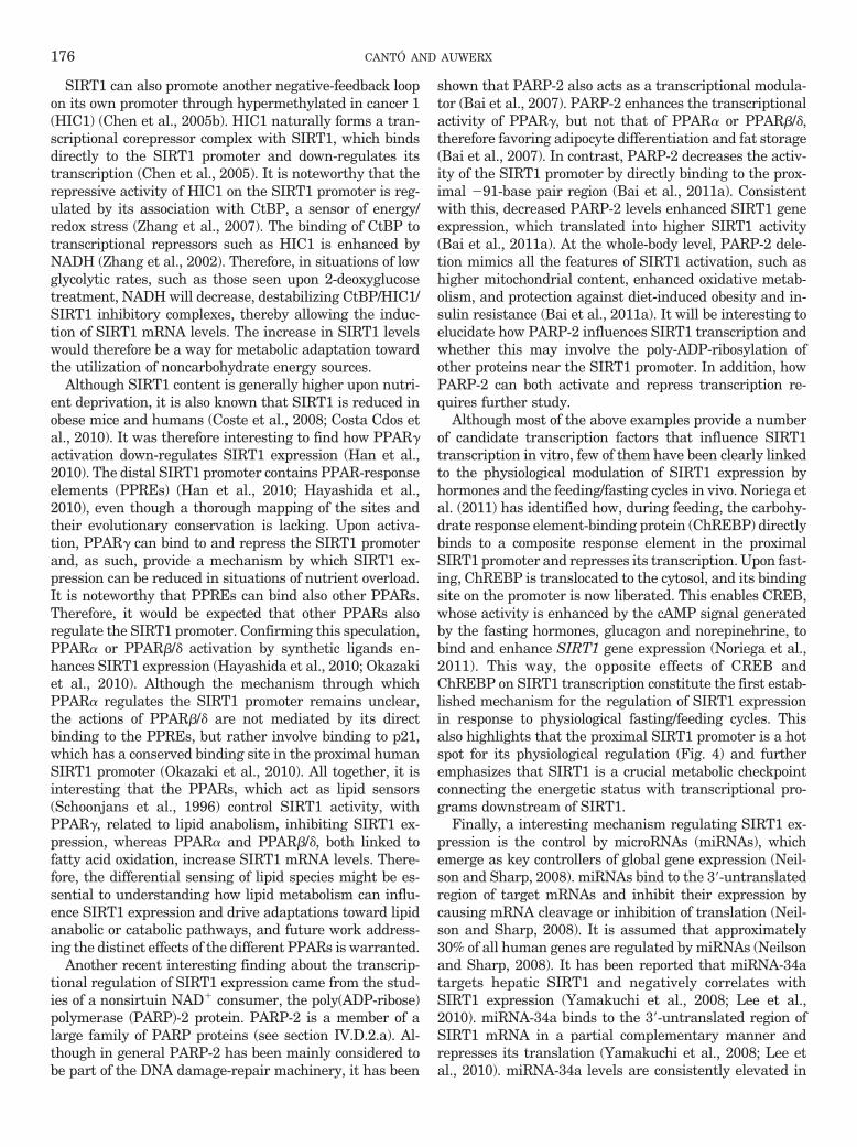
SIRT1 can also promote another negative-feedback loopon its own promoter through hypermethylated in cancer 1(HIC1) (Chen et al., 2005b). HIC1 naturally forms a tran-scriptional corepressor complex with SIRT1, which bindsdirectly to the SIRT1 promoter and down-regulates itstranscription (Chen et al., 2005). It is noteworthy that therepressive activity of HIC1 on the SIRT1 promoter is reg-ulated by its association with CtBP, a sensor of energy/redox stress (Zhang et al., 2007). The binding of CtBP totranscriptional repressors such as HIC1 is enhanced byNADH (Zhang et al., 2002). Therefore, in situations of lowglycolytic rates, such as those seen upon 2-deoxyglucosetreatment, NADH will decrease, destabilizing CtBP/HIC1/SIRT1 inhibitory complexes, thereby allowing the induc-tion of SIRT1 mRNA levels. The increase in SIRT1 levelswould therefore be a way for metabolic adaptation towardthe utilization of noncarbohydrate energy sources.
Although SIRT1 content is generally higher upon nutri-ent deprivation, it is also known that SIRT1 is reduced inobese mice and humans (Coste et al., 2008; Costa Cdos etal., 2010). It was therefore interesting to find how PPAR�activation down-regulates SIRT1 expression (Han et al.,2010). The distal SIRT1 promoter contains PPAR-responseelements (PPREs) (Han et al., 2010; Hayashida et al.,2010), even though a thorough mapping of the sites andtheir evolutionary conservation is lacking. Upon activa-tion, PPAR� can bind to and repress the SIRT1 promoterand, as such, provide a mechanism by which SIRT1 ex-pression can be reduced in situations of nutrient overload.It is noteworthy that PPREs can bind also other PPARs.Therefore, it would be expected that other PPARs alsoregulate the SIRT1 promoter. Confirming this speculation,PPAR� or PPAR�/� activation by synthetic ligands en-hances SIRT1 expression (Hayashida et al., 2010; Okazakiet al., 2010). Although the mechanism through whichPPAR� regulates the SIRT1 promoter remains unclear,the actions of PPAR�/� are not mediated by its directbinding to the PPREs, but rather involve binding to p21,which has a conserved binding site in the proximal humanSIRT1 promoter (Okazaki et al., 2010). All together, it isinteresting that the PPARs, which act as lipid sensors(Schoonjans et al., 1996) control SIRT1 activity, withPPAR�, related to lipid anabolism, inhibiting SIRT1 ex-pression, whereas PPAR� and PPAR�/�, both linked tofatty acid oxidation, increase SIRT1 mRNA levels. There-fore, the differential sensing of lipid species might be es-sential to understanding how lipid metabolism can influ-ence SIRT1 expression and drive adaptations toward lipidanabolic or catabolic pathways, and future work address-ing the distinct effects of the different PPARs is warranted.
Another recent interesting finding about the transcrip-tional regulation of SIRT1 expression came from the stud-ies of a nonsirtuin NAD� consumer, the poly(ADP-ribose)polymerase (PARP)-2 protein. PARP-2 is a member of alarge family of PARP proteins (see section IV.D.2.a). Al-though in general PARP-2 has been mainly considered tobe part of the DNA damage-repair machinery, it has been
shown that PARP-2 also acts as a transcriptional modula-tor (Bai et al., 2007). PARP-2 enhances the transcriptionalactivity of PPAR�, but not that of PPAR� or PPAR�/�,therefore favoring adipocyte differentiation and fat storage(Bai et al., 2007). In contrast, PARP-2 decreases the activ-ity of the SIRT1 promoter by directly binding to the prox-imal �91-base pair region (Bai et al., 2011a). Consistentwith this, decreased PARP-2 levels enhanced SIRT1 geneexpression, which translated into higher SIRT1 activity(Bai et al., 2011a). At the whole-body level, PARP-2 dele-tion mimics all the features of SIRT1 activation, such ashigher mitochondrial content, enhanced oxidative metab-olism, and protection against diet-induced obesity and in-sulin resistance (Bai et al., 2011a). It will be interesting toelucidate how PARP-2 influences SIRT1 transcription andwhether this may involve the poly-ADP-ribosylation ofother proteins near the SIRT1 promoter. In addition, howPARP-2 can both activate and repress transcription re-quires further study.
Although most of the above examples provide a numberof candidate transcription factors that influence SIRT1transcription in vitro, few of them have been clearly linkedto the physiological modulation of SIRT1 expression byhormones and the feeding/fasting cycles in vivo. Noriega etal. (2011) has identified how, during feeding, the carbohy-drate response element-binding protein (ChREBP) directlybinds to a composite response element in the proximalSIRT1 promoter and represses its transcription. Upon fast-ing, ChREBP is translocated to the cytosol, and its bindingsite on the promoter is now liberated. This enables CREB,whose activity is enhanced by the cAMP signal generatedby the fasting hormones, glucagon and norepinehrine, tobind and enhance SIRT1 gene expression (Noriega et al.,2011). This way, the opposite effects of CREB andChREBP on SIRT1 transcription constitute the first estab-lished mechanism for the regulation of SIRT1 expressionin response to physiological fasting/feeding cycles. Thisalso highlights that the proximal SIRT1 promoter is a hotspot for its physiological regulation (Fig. 4) and furtheremphasizes that SIRT1 is a crucial metabolic checkpointconnecting the energetic status with transcriptional pro-grams downstream of SIRT1.
Finally, a interesting mechanism regulating SIRT1 ex-pression is the control by microRNAs (miRNAs), whichemerge as key controllers of global gene expression (Neil-son and Sharp, 2008). miRNAs bind to the 3�-untranslatedregion of target mRNAs and inhibit their expression bycausing mRNA cleavage or inhibition of translation (Neil-son and Sharp, 2008). It is assumed that approximately30% of all human genes are regulated by miRNAs (Neilsonand Sharp, 2008). It has been reported that miRNA-34atargets hepatic SIRT1 and negatively correlates withSIRT1 expression (Yamakuchi et al., 2008; Lee et al.,2010). miRNA-34a binds to the 3�-untranslated region ofSIRT1 mRNA in a partial complementary manner andrepresses its translation (Yamakuchi et al., 2008; Lee etal., 2010). miRNA-34a levels are consistently elevated in
176 CANTÓ AND AUWERX

the livers from diet-induced and genetically obese mice(Lee et al., 2010). Remarkably, p53, which negatively reg-ulates SIRT1 expression directly, also induces miRNA-34a, providing an additional mechanism to ensure SIRT1repression upon p53 activation (Yamakuchi and Lowen-stein, 2009). Other miRNAs have been reported to alsoaffect SIRT1 expression in different tissues, such asmiRNA-132, which down-regulates SIRT1 expression inadipose tissue, prompting inflammatory responses (Strumet al., 2009). Therefore, miRNAs are providing a com-pletely new level for the regulation of SIRT1 levels that weare only beginning to grasp.
Together, these mechanisms illustrate the complexity ofthe regulation of SIRT1 at the level of its expression. It isnoteworthy that most of the transcriptional regulators de-scribed are also substrates for SIRT1, which illustrates theintricate nature of SIRT1 regulation and how it is drivenby multiple regulatory loops. However, multiple interac-tions between proteins and the multifunctionality of theindividual proteins involved make it difficult to predicthow pharmacological targeting of one of the players willaffect the others. Although the identification of novel play-ers will certainly contribute to our understanding of SIRT1transcriptional regulation, it is understandable that, atthis point, alternative strategies to enhance SIRT1 activityare also of interest. Such strategies will be described in thenext sections.
B. Post-Translational Modifications
The activity of SIRT1, like that of most enzymes, is alsomodulated by a number of post-translational modifica-tions. The first report indicating this possibility was theidentification of SIRT1 as a nuclear phosphoprotein in alarge screening using mass spectrometry (Beausoleil et al.,2004). Although two phosphoresidues, Ser27 and Ser47,were identified, their function has not yet been explored.Subsequent efforts identified up to 13 phosphorylatableresidues, including the two found previously (Sasaki et al.,2008). Dephosphorylated SIRT1 was less active than thephosphorylated form (Sasaki et al., 2008). Among theseresidues, Thr530 and Ser540 were phosphorylated by cy-clinB/cdk1, and their mutation resulted in aberrant cellproliferation and cell cycle profiles that could not be ex-plained by changes in SIRT1 stability but rather resultedfrom lower SIRT1 activity (Sasaki et al., 2008). Anotherreport indicated that JNK1 can also phosphorylate SIRT1in three residues, Ser27, Ser47, and Thr530, in response tooxidative stress promoted by H2O2 or anisomycin (Nasrinet al., 2009). In agreement with the previous report, phos-phorylation of these sites by JNK1 increased nuclear local-ization of SIRT1 and its activity (Nasrin et al., 2009).Surprisingly, it also seemed that JNK1 phosphorylationoriented SIRT1 activity toward specific substrates, be-cause it triggered the deacetylation of histone H3 but notp53 (Nasrin et al., 2009). Paradoxically, JNK1 activity isinduced in situations of obesity and metabolic disease (Hi-rosumi et al., 2002), where SIRT1 activity is decreased
(Coste et al., 2008). A constellation of other kinases, includ-ing casein kinase II (Kang et al., 2009; Zschoernig andMahlknecht, 2009), the dual-specificity tyrosine phospho-rylation-regulated kinases (Guo et al., 2010), and themammalian sterile 20-like kinase 1 (Yuan et al., 2011a),have also been suggested to phosphorylate SIRT1. Al-though these data indicate that the activity of SIRT1might be modulated through the phosphorylation of theabove-mentioned residues by multiple kinases, it is dis-couraging that most of the phosphorylatable residues iden-tified, or their flanking sequences, are very poorly con-served across species, making it difficult to argue thatthese residues have been essential throughout evolutionfor the most conserved metabolic functions of SIRT1 or-thologs across species. Furthermore, the metabolic roles ofthese phosphorylation events, as well as their interactionwith other post-translational modifications, needs to beaddressed in vivo to fully understand their true biologicalfunction.
A second type of post-translational modification that canaffect SIRT1 activity is SUMOylation. SIRT1 is SUMOy-lated at Lys734 upon UV irradiation or H2O2 treatment(Yang et al., 2007c). The SUMOylation of SIRT1 increasedits intrinsic deacetylase activity (Yang et al., 2007c). Con-versely, mutation of the residue or forced de-SUMOylationby the small ubiquitin-like modifier (SUMO) 1/sentrin spe-cific peptidase 1 enzyme, rendered SIRT1 less enzymati-cally active and cells more prone to apoptosis (Yang et al.,2007c). Although it was speculated that SIRT1 SUMOyla-tion acts as a switch between cell survival and cell death,further work is required to define the mechanisms bywhich cellular stress enhances the interaction of SIRT1with SUMOylation enzymes and/or decreases the associa-tion with SENP. As with phosphorylation, an additionalcaveat is the poor conservation of the SUMOylation resi-due and its flanking regions. For example, mouse SIRT1cannot be SUMOylated because the human Lys734 is anArg residue in mice. Although this does not rule out apotential effect of SUMOylation on SIRT1 activity in manyspecies, including human, it is unlikely that this will con-tribute to the activity of SIRT1 in other species, unlessSUMOylation takes place at other residues.
It is often overlooked that SIRT1 activity can affectmany different targets. It is unlikely then, that, upon itsactivation, SIRT1 unselectively deacetylates all of its tar-gets, which could often lead to opposite physiological ef-fects. Therefore, some specification must exist. A clear hintthat this is the case was already mentioned by the work onJNK1 (Nasrin et al., 2009), which illustrated how, despitebeing intrinsically more active, SIRT1 deacetylated onlyspecific substrates. Consequently, post-translational mod-ifications might not only affect SIRT1 activity at the levelof its intrinsic activity but also channel SIRT1 towardspecific subsets of targets. It is noteworthy that we haveillustrated how phosphorylation of PGC-1� by AMPK isessential for SIRT1-mediated deacetylation (Canto et al.,2009). Given the large number of SIRT1 substrates, it is
TARGETING SIRT1 TO IMPROVE METABOLISM: ALL YOU NEED IS NAD�? 177

very likely that substrate accessibility is also controlled bypost-translational modifications. Understanding thesespecification mechanisms will be essential to designingfuture strategies aimed to selectively affect certain func-tions of SIRT1 but not others.
C. Protein Interactions
SIRT1 activity is controlled not only intrinsically at thelevel of the SIRT1 protein but also through its associationwith different protein complexes, which may affect its ac-tivation or inhibition as well as its target specificity. Insection II.C, we specified how the presence of SIRT1 in acomplex with NCoR1 actually inhibits PPAR� action inadipose tissue. In this section, we discuss two additionaland physiologically relevant mammalian SIRT1 “nonsub-strate” interactors.
Two simultaneous reports in 2007 indicated that thenuclear protein deleted in breast cancer-1 (DBC1) forms astable complex with the catalytic domain of SIRT1 andinhibits SIRT1 activity both in vivo and in vitro (Kim et al.,2008; Zhao et al., 2008). As a consequence, the artificialreduction of DBC1 in cell-based experiments stimulatedSIRT1 activity, diminishing the acetylation levels of p53and inhibiting p53-dependent apoptosis (Kim et al., 2008;Zhao et al., 2008). The physiological pathways influencingthe dynamic interaction of SIRT1 and DBC1, however,remain unexplored. The in vitro evidence indicating thatDBC1 is a SIRT1 inhibitor was further supported by ob-servations in mouse liver indicating that DBC1 and SIRT1colocalize within the nucleus and coimmunoprecipitate innuclear extracts (Escande et al., 2010). Mice with a germ-line deletion of the DBC1 gene showed a 2- to 4-fold in-crease in endogenous SIRT1 activity in a wide range oftissues, rendering p53 hypoacetylated (Escande et al.,2010). The dynamics of the DBC1/SIRT1 interaction havebeen evaluated in vivo, demonstrating that, under normalfeeding conditions, at least 50% of total liver SIRT1 isassociated with DBC1, and that this interaction wasnearly absent after starvation, contributing to the increasein SIRT1 activity observed in fasting livers (Escande et al.,2010). It is noteworthy that the increase in SIRT1 activityduring fasting was blunted in DBC1 knockout mice (Es-cande et al., 2010). In contrast to the effects observedduring fasting, high-fat feeding stabilized the associationof DBC1 with SIRT1 (Escande et al., 2010). Understandingthe molecular determinants influencing the association be-tween DBC1 and SIRT1 will be an interesting field forfuture investigation. The overall phenotype of the DBC1knockout mice did not differ from wild-type littermates onchow diet. These mice, however, were protected against thedevelopment of hepatic steatosis and liver damage inducedby high-fat feeding (Escande et al., 2010), in line with thedata obtained in the liver-specific SIRT1 knockout models(Purushotham et al., 2009; Wang et al., 2010).
Around the same time that DBC1 was reported as asirtuin inhibitor, another report identified a possible acti-vator of SIRT1 activity, the active regulator of SIRT1
(AROS) (Kim et al., 2007b). The association of AROS withSIRT1 in, presumably, its catalytic domain enhancesSIRT1 activity 2-fold, resulting in p53 inhibition throughdeacetylation (Kim et al., 2007). Conversely, the artificialreduction in AROS levels sensitized cells to p53-inducedapoptosis (Kim et al., 2007). It is relevant that AROSinteracts specifically with SIRT1 but not with other sir-tuins (Kim et al., 2007). The exact nature of the actions ofAROS on SIRT1 and how it affects metabolism will requirefuture study.
These data on NCoR1, DBC1, and AROS illustrate howSIRT1 activity is influenced by its association with specificprotein partners. It will be important to elucidate whetherthese partnerships also influence SIRT1 substrate selec-tivity. It is reasonable to think that the known and yet-to-be identified SIRT1 interactors also have an impacton the deacetylation level of SIRT1 substrates other thanp53, such as PGC-1� and FOXO1, which could have far-reaching implications for metabolic regulation. It will alsobe interesting for future studies to understand to whatextent the deacetylase activity of SIRT1 influences theactivity of corepressor or coactivator complexes and toidentify the possible roles of SIRT1 as an adaptor protein.It goes without saying that modifying the interaction ofSIRT1 with such interactors could constitute a promisingavenue to modulate SIRT1 activity.
D. Sirtuin 1-Activating Compounds
An obvious strategy to artificially enhance SIRT1 wouldbe through chemical compounds that directly bind andactivate SIRT1. Howitz et al. (2003), using a screeningstrategy with a fluorescently labeled substrate, identifiedresveratrol and a few other polyphenols, including querce-tin and piceatannol, as natural compounds that could en-hance the deacetylating activity of SIRT1. A number ofsubsequent studies showed activated SIRT1 in diverse spe-cies (for review, see Baur, 2010). Resveratrol treatmentmimics numerous aspects of calorie restriction in all eu-karyotes tested to date (Howitz et al., 2003; Wood et al.,2004; Baur et al., 2006; Lagouge et al., 2006; Valenzano etal., 2006; Barger et al., 2008; Pearson et al., 2008); in mostof them, this effect seems to depend on SIRT1 (Howitzet al., 2003; Wood et al., 2004; Lagouge et al., 2006). Notsurprisingly, in several models tested (Howitz et al., 2003;Wood et al., 2004; Baur et al., 2006; Valenzano et al., 2006),albeit not all (Pearson et al., 2008), resveratrol increasedlifespan. In mice, resveratrol promoted SIRT1 activationand energy expenditure (Baur et al., 2006; Lagouge et al.,2006). Upon high-fat feeding, resveratrol prominently pre-vented the onset of diet-induced obesity and metabolicdisease, which ended up protecting the treated miceagainst the lifespan curbing associated with high caloricdiets (Baur et al., 2006; Lagouge et al., 2006). At the mo-lecular level, resveratrol boosted mitochondrial content, asa result of the activation of the SIRT1/PGC-1� axis (La-gouge et al., 2006). Resveratrol also improved mitochon-drial function and fatty acid oxidation in humans, as dem-
178 CANTÓ AND AUWERX

onstrated in a recent study, but at much lowerconcentrations that those used in mice (Timmers et al.,2011). All together, resveratrol proved to be an effectiveway to activate SIRT1 in vivo and promote beneficialhealth effects, most of which resemble the effects observedupon the overexpression of SIRT1.
However, the ability of resveratrol to directly activateSIRT1 was seriously questioned by results demonstratingthat the nonphysiological fluorescent “Fluor de Lys” sub-strate used for SIRT1 activity assays can lead to artifac-tual results (Borra et al., 2005; Kaeberlein et al., 2005).Blinded by the beneficial effects in line with SIRT1 activa-tion, resveratrol actions on other possible molecules/pathways have been largely neglected. In fact, resveratrolwas much earlier reported as a polyphenol that interferedwith the mitochondrial respiratory chain (Zini et al., 1999).It is relevant, furthermore, that many reports have re-cently demonstrated that resveratrol can also activateAMPK (Baur et al., 2006; Zang et al., 2006; Dasgupta andMilbrandt, 2007; Park et al., 2007; Feige et al., 2008),which is consistent with its possible effect on the mitochon-drial respiratory chain. Elegant studies, using isogenic celllines stably expressing AMPK complexes containing AMP-insensitive �2 subunit variants (R531G), convincinglydemonstrated that AMPK activation in response to res-veratrol derives from an AMP/ATP imbalance as a conse-quence of interference with mitochondrial respiration(Hawley et al., 2010). AMPK activation by resveratrol isvery fast and already prominent after a few minutes,whereas the activation of SIRT1 becomes detectable onlyafter a few hours (C. Canto and J. Auwerx, unpublishedobservations), clearly indicating that AMPK activation isan earlier event upon resveratrol treatment. Although ithas been argued that resveratrol action on AMPK isSIRT1-dependent (Suchankova et al., 2009), the use ofmouse embryonic fibroblast cells from SIRT1 knock-outmice unequivocally demonstrated that SIRT1 is dispens-able for resveratrol-induced AMPK activation (Dasguptaand Milbrandt, 2007; Um et al., 2010). Conversely, severalapproaches have convincingly proven that resveratrol can-not activate SIRT1 in the absence of functional AMPK(Canto et al., 2010; Um et al., 2010). A final picture of theactions of resveratrol on AMPK and SIRT1 activation wasfinally drawn by the demonstration that SIRT1 is thedownstream mediator of AMPK actions. AMPK activationinitially leads to a gradual increase in NAD� levels, as aconsequence to the activation of fatty acid oxidation, sub-sequently activating SIRT1 (Canto et al., 2009). This ini-tial raise in NAD� is sustained by enhanced Nampt ex-pression, which favors the synthesis of NAD� from NAMthrough the NAD� salvage pathway (Fulco et al., 2008).The requirement for NAD� accumulation also explainswhy the activation of SIRT1 is not immediate upon res-veratrol treatment. Therefore, SIRT1 may be essential forresveratrol action but as a downstream consequence ofAMPK activation, rather than as a direct molecular targetof resveratrol (Fig. 5).
A more recent screening for other possible small molecularSIRT1 activators provided a second batch of compounds,among which N-[2-[3-(piperazin-1-ylmethyl)imidazo[2,1-b][1,3]thiazol-6-yl]phenyl]quinoxaline-2-carboxamide(SRT1720) has been best characterized (Milne et al., 2007).SRT1720 was in vitro a much more potent and efficientactivator of SIRT1 than resveratrol (Milne et al., 2007).Treatment of rodents with SRT1720 prevented diet-induced obesity and ameliorated the diabetic phenotype ofgenetically obese mice (Milne et al., 2007; Feige et al.,2008). Similar to what was observed with resveratrol,SRT1720 enhanced SIRT1 activity, oxidative metabolism,and mitochondrial biogenesis in mouse tissues (Feige etal., 2008). Pacholec et al. (2010) also indicated that directactivation of SIRT1 by SRT1720 observed in the in vitrosetting suffered from the same artifacts derived from theuse of the “Fluor de Lys” moiety and that were alreadydescribed for resveratrol. Therefore, one needs to considerthat SIRT1 activation in tissues from mice fed withSRT1720 might be an indirect event, potentially againinvolving activation of AMPK, which was evident uponlong-term treatment of SRT1720 in vivo (Feige et al.,2008). In addition to that, although the in vitro assaysindicated that SRT1720 was a more potent activator ofSIRT1 than resveratrol, this was not translated in vivo,indicating that the actions of these compounds in vivoinvolve indirect activation of SIRT1 (Feige et al., 2008)and/or rather poor bioavailability.
E. Indirect Modulation through AffectingNAD� Metabolism
We have described in section II.B how sirtuins in gen-eral, and SIRT1 in particular, meet all the requirements toact as intracellular NAD� sensors. Therefore, enhancingNAD� availability could be used to activate SIRT1 and
Resveratrol
AMPKAMP/ATPMetabolism?
SIRT1
NAD+
C I C IIC III C IV C V
Mitochondal/lipid oxidation
C II
Mitochondal/lipid oxidation gene expression
FIG. 5. Resveratrol promotes mitochondrial biogenesis and lipid oxi-dation gene expression through indirect AMPK and SIRT1 activation.Although still a matter of debate, most data currently indicate that themetabolic actions of resveratrol or its metabolites stem from its ability toact as a mild mitochondrial poison, impairing ATP synthesis. The energystress induced by resveratrol activates AMPK, subsequently stimulatingSIRT1 by enhancing NAD� levels. Then, SIRT1 activates key down-stream targets through deacetylation (see section II.C), ultimately lead-ing to an adaptive potentiation of mitochondrial biogenesis and lipidoxidation pathways. CI-V represent mitochondrial respiratory complexesI to V.
TARGETING SIRT1 TO IMPROVE METABOLISM: ALL YOU NEED IS NAD�? 179

promote beneficial health effects. We will now review dif-ferent strategies used to achieve such goal and discusswhether the experimental results support that modulatingNAD� metabolism might be a useful tool for health bene-fits (Fig. 6).
1. Modulation by Increasing NAD� Synthesis. Themost obvious strategy for testing how enhancing NAD�
levels affects sirtuin activity consists in boosting NAD�
synthesis by supplementation with NAD� precursors.Different precursors can be used to promote NAD� syn-thesis. The primary de novo synthesis of NAD� gener-ally initiates from tryptophan (Houtkooper et al., 2010).Nicotinic acid (NA) is another NAD� precursor that istransformed into NAD� through the Preiss-Handlerpathway, therefore converging with the same NAD�
synthesis pathway used by tryptophan. It is assumed,however, that perhaps the principle source of NAD�
comes from salvage pathways from other adenine nucle-otide metabolites (Houtkooper et al., 2010). The mainNAD� precursors that funnel through the salvage path-ways are NAM and the more recently described nicotin-amide riboside (NR). NAM generates NAD� through anindependent pathway, in which the rate-limiting en-zyme Nampt transforms NAM into NMN, which on itsturn is converted into NAD� by NMN adenylyltrans-ferase (Revollo et al., 2004). NR is phosphorylated uponits entry in the cell by the NR kinases, generating NMN,which is then converted to NAD� by NMN adenylyl-transferase (Bieganowski and Brenner, 2004).
Few studies to date have described how supplementa-tion with these precursors influence NAD� levels and inparticular affect sirtuin activity. Both NA and NAM canlead to higher NAD� levels, even though the effects mightbe tissue-specific. NAM seems to be a more stable NAD�
precursor in the liver (Collins and Chaykin, 1972), but NAseems to be a more efficient in the kidney (Hara et al.,2007). The metabolism of these NAD� precursors by thegut flora and intestinal enzymes can also contribute totheir absorption and their efficacy to increase NAD�
(Gross and Henderson, 1983). Cell-based experiments alsosupport the existence of cell-specific differences. For exam-ple, in NIH3T3 cells, concentrations of up to 5 mM NAMwere unable to increase intracellular NAD� levels (Revolloet al., 2004). Although NAM was also a relatively ineffi-cient NAD� precursor in human embryonic kidney 293cells, requiring 5 mM concentrations to increase NAD�,NA was more efficient, and concentrations as low as 20 �Mdoubled the NAD� content (Hara et al., 2007). The cell/tissue-specific efficiencies of distinct NAD� precursorsmight be consequent to the differential expression of therate-limiting enzymes in their respective metabolic path-ways. NA metabolism into NAD� is rate-limited by the NAphosphoribosyltransferase, which is highly enriched insome tissues, such as liver or kidney (Hara et al., 2007). Areduction in NA phosphoribosyltransferase activity in hu-man embryonic kidney 293 cells does not affect basalNAD� levels but impairs NA-induced NAD� accumulation(Hara et al., 2007). To date, however, no clear link betweenNA supplementation and sirtuin activity has been made.
Both NA and NAM have been used for a long time in aclinical setting. Niacin, basically composed of NA andNAM, has been widely used as an efficient way to over-come situations of dietary tryptophan deficits (Sauve,2008). Niacin is also used to treat hypercholesterolemia,because it efficiently decreases very-low-density lipopro-tein synthesis, lowers low-density lipoprotein cholesterollevels, and concurrently increases high-density lipoproteincholesterol (Altschul et al., 1955; Karpe and Frayn, 2004).The use of niacin as a lipid-lowering agent results in ben-eficial effects on coronary artery disease and type 2 diabe-tes mellitus. It is not clear whether these effects rely onSIRT1 activation. Many of the beneficial actions of niacinin mice and humans—as well as some of the undesiredeffects, such as spontaneous flushing—have been attrib-uted to the activation of a putative NA-activated G-pro-tein-coupled receptor, GPR109A (Tunaru et al., 2003; Be-nyo et al., 2005). Whereas the role of GPR109A inmediating niacin-induced flushing is rather well estab-lished (Benyo et al., 2005), the hypothesis that the thera-peutic efficacy of niacin is mediated by GPR109A activa-tion needs to be revisited. First, even if GPR109A has arelatively high affinity for NA (EC50, �100 nM), such lev-els of NA are rarely found in plasma unless pharmacolog-ically primed (Kirkland, 2009), indicating that the activa-tion of GRP109A by NA is probably fortuitous but notbiologically relevant in the basal state. Second, many of the
Mit h d i l t b li
AMPK
Mitochondrial metabolism
Nampt expression
PARPsNAD+ precursors
SIRT1
PARPs
CD38
NAD precursors
(NA, NR, NMN)NAD+
Deacetylation of key metabolic transcriptional regulators
Metabolic adaptation
FIG. 6. NAD� as a nodal point for metabolic regulation. Most evidenceto date points out that NAD� could be rate-limiting for the SIRT1 reac-tion in diverse conditions. SIRT1 activity would hence be stimulated byinterventions that increase NAD� levels, such as AMPK activation, en-hancement of NAD� biosynthesis through precursor (NA, NR, NMN)supplementation, or through inhibition of alternative NAD� consumingactivities, such as PARPs or CD38. The stimulation of SIRT1 activity bythese distinct means improves the capacity of the cells/organism to adaptto external metabolic cues.
180 CANTÓ AND AUWERX

beneficial effects of niacin, such as the lipid-lowering ef-fects (Kamanna and Kashyap, 2008), take place at concen-trations higher than those required for GPR109A activa-tion but lead to intracellular NAD� accumulation (Jacksonet al., 1995). It is therefore tempting to speculate that someof the effects promoted by niacin might be achievedthrough an NAD�-induced activation of SIRT1 and theconsequent deacetylation of the multiple SIRT1 targetsthat act as critical regulators of fatty acid, lipid and sterolhomeostasis in eukaryotes, such as PGC-1�, FOXOs, LXR,or SREBP-1c (described in section II.C). An additionalappealing possibility to explain the effects of niacin in-volves the fact that it leads to adiponectin release from thewhite adipose tissue (Vaccari et al., 2007; Westphal et al.,2007). Adiponectin then activates AMPK in muscle andliver tissues, which would enhance NAD� content andSIRT1 activity (Canto et al., 2009; Iwabu et al., 2010).Therefore, future work is urgently needed to elucidate thecontribution of SIRT1 as a potential mediator of niacin’sbeneficial health effects.
The effects of NR, the most recently discovered NAD�
precursor, remain also largely unknown. Although NR canincrease intracellular NAD� in mammalian cells (Yang etal., 2007b), it is still not reported whether this is enough toinfluence sirtuin activity. However, evidence obtained inyeast models suggests that this might in fact be the case.Supplementation of yeast with NR enhanced Sir2-dependent repression of recombination, improved gene si-lencing and extended replicative lifespan (Belenky et al.,2007). All these actions were completely dependent ofNAD� synthesis (Belenky et al., 2007). These experimentsusing NR constitute the first solid evidence that enhancingNAD� bioavailability via NAD� precursor supplementa-tion can also enhance sirtuin activity and increase lifes-pan, even though they are limited to yeast models. Fur-thermore, the discovery that NR is present in cow’s milk(Bieganowski and Brenner, 2004) poses an interesting op-portunity for food-based preventive or therapeutic inter-ventions in NAD�-dependent metabolism.
In addition to boosting NAD� levels through adminis-tration of NAD� precursors, NAD� levels can also be arti-ficially modulated by changing the activity of rate-limitingenzymes in NAD� biosynthesis. This principle is well il-lustrated by the modulation of Nampt activity, which hasa clear impact on NAD� levels in virtually any mammaliancell tested (for examples, see van der Veer et al., 2005;Fulco et al., 2008; Rongvaux et al., 2008; Pittelli et al.,2010). In most of these cases, the alterations in NAD�
levels promoted by differential Nampt activity were asso-ciated with changes in SIRT1 activity (van der Veer et al.,2005, 2007; Revollo et al., 2007; Fulco et al., 2008). How-ever, and as explained in section II.B, a complication fromthese experiments is that the influence that Nampt exertson SIRT1 activity may derive not only from altering NAD�
availability but also from more effective NAM clearance(Bitterman et al., 2002). Nampt inhibitors are currentlybeing developed to deplete NAD� levels and as such induce
tumor cell apoptosis (Hasmann and Schemainda, 2003). Inaddition, inhibitors of kynurenine 3-monooxygenase, in-volved in de novo NAD� biosynthesis pathways, have al-ready been identified (Zwilling et al., 2011). Despite thefacility to develop enzyme inhibitors, it will be more chal-lenging to discover and develop compounds that pharma-cologically activate the NAD� salvage and/or de novo syn-thesis pathways. Such activators could potentiallyenhance NAD� levels and activate SIRT1 to promote met-abolic fitness.
2. Modulation by Decreasing NAD� Consumption. An-other attractive way to modulate NAD� levels and favorsirtuin activity resides in the modulation of the activity ofnonsirtuin NAD�-consuming enzymes. There are two ma-jor families of alternative NAD� consumers: the PARPsand the cADP-ribose synthases (CD38 and CD157) (Hout-kooper et al., 2010) (Fig. 6).
a. Poly(ADP-ribose) Polymerase. PARPs use NAD�
as a substrate for a cellular process in which the ADP-ribose moiety is not transferred to an acetyl group, ashappens with sirtuins, but is used to build ADP-ribosylpolymers onto acceptor proteins (Chambon et al., 1963;Krishnakumar and Kraus, 2010). PARP activity is ro-bustly enhanced upon DNA damage and oxidativestress. Most PARP activity upon oxidative damage isdriven by PARP-1, except for a residual 5 to 10%, whichis accounted by PARP-2 (Shieh et al., 1998; Ame et al.,1999). Overactivation of PARP-1 upon oxidative damagerapidly depletes intracellular NAD� levels (Goodwin etal., 1978; Skidmore et al., 1979). In line with thehypothesis that NAD� might be rate-limiting forSIRT1 action, SIRT1 activity is down-regulated insituations of PARP-1 activation (Pillai et al., 2005; Baiet al., 2011b). Significantly, SIRT1 does not seem to bedirectly regulated through poly-ADP-ribosylation events(Bai et al., 2011a,b), indicating that PARP-1 and SIRT1activity are connected through competition for a limitedNAD� pool. PARP-1 has a very low Km (�20 �M) and arelatively high Vmax for NAD� (Mendoza-Alvarez andAlvarez-Gonzalez, 1993), indicating that PARP-1 mightlimit SIRT1 action but not the other way around.
Bai et al. (2011b) have demonstrated how down-regulation of PARP-1 activity favors SIRT1 activation. Ge-netic and pharmacological approaches that abrogatePARP-1 activity induced NAD� levels. Although PARPactivity is generally accepted to be rather low in the basalstate, recent evidence indicates that it naturally fluctuatesin a circadian fashion (Asher et al., 2010). In line with theexistence of basal PARP activity, inhibition of its activitygradually leads to a build up of NAD� levels in culturedcells (Bai et al., 2011b). Likewise, tissues from PARP-1knockout mice have an increased NAD� content (Bai et al.,2011b), roughly 2-fold higher than wild-type littermates.The increase in NAD� levels promoted by PARP inhibitionleads, both in vivo and in vitro, to higher SIRT1 activity(Bai et al., 2011b). This way, PARP inhibition associateswith the induction of the expression of genes involved in
TARGETING SIRT1 TO IMPROVE METABOLISM: ALL YOU NEED IS NAD�? 181

mitochondrial and lipid oxidation in a SIRT1-dependentmanner. From a physiological perspective, the better met-abolic fitness derived from SIRT1 activation offers thePARP-1-null mice protection against the onset of metabolicdisease in the context of diet-induced obesity (Bai et al.,2011b). More acute inhibition of PARP activity, usingpharmacological PARP inhibitors, was also enough to en-hance mitochondrial gene expression in vivo and in vitro(Bai et al., 2011b). There are currently nine different PARPinhibitors at different stages of clinical development and atleast three highly selective PARP inhibitors in late preclin-ical development against diverse types of cancer (for re-view, see Yuan et al., 2011b). It is noteworthy that mostcancer cells rely on anaerobic glycolysis to sustain theirhigh proliferation rates, even if oxygen is plentiful, anevent that is known as “the Warburg effect” (after OttoWarburg, who first described it) and is believed to be keyfor malignant transformation. Apart from their undis-puted effect on DNA repair (Krishnakumar and Kraus,2010), PARP inhibitors might also contribute to inhibitcancer progression by promoting oxidative metabolism andacting as anti-Warburg agents. Because most PARP inhib-itors target both PARP-1 and PARP-2, they would further-more potentiate SIRT1 function both at the enzymaticlevel—through PARP-1 inhibition and raising NAD� lev-els—and at the transcriptional level—through PARP-2 in-hibition and induction of SIRT1 expression (see sectionIV.A). Furthermore, the results mentioned above also in-dicate that PARP inhibitors may be retooled to boost oxi-dative metabolism in metabolic diseases in which mito-chondrial function is impaired, such as inherited andacquired mitochondrial diseases.
The interaction between PARP-1 and SIRT1 activitiesalso opens the perspective of a possible role of PARPs inaging, because SIRT1 activity has been postulated as amediator of the beneficial effects of calorie restriction onhealth and lifespan (Canto and Auwerx, 2011). Hence,PARP inhibition could be a nice strategy to activate SIRT1and mimic the calorie-restricted state. In line with thishypothesis, it has been reported that PARP activity ishigher in aged tissues, leading to decreased SIRT1 activity(Braidy et al., 2011; C. Canto, R, Houtkooper, L. Mouchir-oud, and J. Auwerx, unpublished observations). Confirm-ing the hypothesis that higher PARP-1 activity might bedetrimental for SIRT1 function and global metabolism wasthe fact that mice expressing an additional copy of thehuman PARP-1 have reduced median lifespan, impairedglucose homeostasis, and higher susceptibility to age-related diseases (Mangerich et al., 2010). Further studiesshould evaluate the possible influence of PARP activity onaging.
It is of note that the NAD� boosting effects of PARPinhibition enhance the activity of SIRT1, but not that ofSIRT2 or SIRT3 (Bai et al., 2011b). The major differencebetween these three sirtuins is their subcellular localiza-tion, because, among them, only SIRT1 is a nuclear sirtuin.This suggests the existence of compartment-specific NAD�
pools in the cell. Supporting this possibility, elegant stud-ies by Yang et al. (2007a) showed the existence of indepen-dently regulated NAD� pools.
b. CD38. Another small family of NAD� consumersare the cADP-ribose synthases, CD38 and the less char-acterized CD157. CD38 and CD157 are multifunctionalenzymes that use NAD� as substrate to generate secondmessengers, mainly cADP-ribose, which, in turn, regu-lates calcium mobilization (Lee, 2006). Most studies onhow cADP-ribose synthases affect NAD� levels andSIRT1 activity have been done with CD38. As is true forPARP-1, CD38 displays a very low Km for NAD� (15–25�M) (Cakir-Kiefer et al., 2001). The stoichiometry of thereaction catalyzed by CD38 involves massive amounts ofNAD�, around 100 molecules, to yield a single cADP-ribose (Dousa et al., 1996). Therefore, even low levels ofCD38 activity might have a dramatic influence on intra-cellular NAD� metabolism. Remarkably, the enzymaticactivity of CD38 is located outside the plasma mem-brane (De Flora et al., 1997), which makes it challengingto envision how NAD� can access this enzyme, becauseNAD� is only present in minute quantities outside thecell (O’Reilly and Niven, 2003). It was recently sug-gested, however, that CD38 might also be present in thenuclear compartment, which would then set the stagefor a role of CD38 as a main intracellular NAD� con-sumer (Aksoy et al., 2006). The function of CD38 as anintracellular NADase was subsequently proven rightwhen mice lacking CD38 displayed a 30-fold increase inintracellular NAD� levels (Aksoy et al., 2006b). Thisincrease in NAD� levels is far superior compared withthe �2-fold increases generally observed in most genetic(PARP-1 deletion), pharmacological (NAD� precursors),or physiological interventions (fasting, calorie restric-tion) that enhance NAD� content. The increase in intra-cellular NAD� elicited by CD38 deletion significantlyactivated SIRT1 and prompted clinical phenotypes sim-ilar to those expected for SIRT1 activation, includingprotection against diet-induced obesity and a robustdeacetylation of SIRT1 targets (Aksoy et al., 2006). Thefact that the 30-fold increase in NAD� triggered byCD38 gene deletion has a biological effect similar to the2-fold effect observed in PARP-1 knockout mice suggeststhat the effective NAD�-sensing range of SIRT1 mightbe largely exceeded in the CD38 knockout mice. Giventhe nuclear and plasma membrane localization of CD38,it will be interesting to evaluate whether the increase inNAD� is homogeneous between different cellular com-partments and whether other sirtuins are similarly af-fected. As for the PARPs, specific CD38 inhibitors arebeing developed (Sauve and Schramm, 2002; Dong etal., 2011), and it will be relevant to evaluate how theyinfluence SIRT1 activity in situations where oxidativemetabolism is impaired (e.g., acquired and inheritedmitochondrial defects).
182 CANTÓ AND AUWERX

V. Conclusions and Future Directions
The wealth of new data on SIRT1, generated in speciesbeyond yeast, has recently spontaneously refocused theattention of the field from a potential—but so far notclearly proven—role in increasing lifespan toward its abil-ity to modulate whole-body metabolism. Probably evolvedas a mediator of the metabolic and transcriptional adapta-tions to situations of energy stress and nutrient depriva-tion, SIRT1 activation enhances the ability of organisms toenhance fat consumption and use mitochondrial respira-tion as a way optimize energy harvesting. Metabolic dis-ease has been strongly linked to impaired energy homeo-stasis and mitochondrial function. Therefore, manipulationsaimed to enhance SIRT1 activity might turn out to beattractive for the prevention and treatment of metabolicdisease. Studies in genetic engineered mouse models sup-port the notion that higher SIRT1 activity is protectiveagainst metabolic disease without necessarily influencinglifespan. In contrast, defective SIRT1 activity in mousemodels is generally linked to metabolic inefficiency. Al-though the quest for direct SIRT1 activators has deliveredinteresting compounds, such as resveratrol and SRT1720,most of them have been proven to activate SIRT1 in veryindirect and unspecific fashion in vivo. Therefore, novelapproaches to activate SIRT1 have emerged. Among them,strategies aimed to modify the intracellular NAD� contenthave provided exquisite correlative evidence that higherNAD� bioavailability is matched by higher SIRT1 activity.It is noteworthy that the activation of SIRT1 seems to be akey convergent downstream effect, common between themany different approaches used to modulate NAD� levels,such as increasing NAD� synthesis rates or decreasingNAD� consumption rates. This way, the modulation ofNAD� levels appears as one of the most promising strat-egies to control SIRT1 activity to achieve health benefits.One of the advantages of raising NAD� levels is that thatit can be achieved from many different angles and strate-gies that boost NAD� levels or inhibit NAD� consumptionare being pursued for nutraceutical (e.g., NA precursors)and pharmaceutical (e.g., PARP and CD38 inhibitors) de-velopment. It should furthermore not be forgotten thatdespite the fact that raising NAD� levels might allowSIRT1 to reach higher enzymatic rates, other factors, rang-ing from transcriptional and translational to post-translational (e.g., post-translational modifications in thenarrow sense and protein complex formation) mecha-nisms, also profoundly affect SIRT1 activity. In our review,we have highlighted how different post-translational mod-ifications in SIRT1 or in its substrates can strongly driveSIRT1 activity toward specific cellular processes. This in-dicates that the activity of sirtuins on certain substratesmight be coupled not only to NAD� availability but also tothe accessibility of the target, which is determined by an-other post-translational modification. This case is perfectlyillustrated by PGC-1�, the deacetylation of which is muchmore efficient when phosphorylated. Likewise, it will be
very relevant to understand how the ability of SIRT1 tointeract with specific substrates is determined by the co-habitation with other molecular coactivator (e.g., PGC-1�)or corepressor (e.g., NCoR1) complexes. The complexity ofSIRT1 physiology, which is largely conserved throughoutevolution, makes unraveling the intricacy of the SIRT1signaling and putting it to fruition in drug development achallenging task that will occupy scientists for years tocome. Therefore, those who are waiting for a SIRT1-activating entity that will grace them with eternal youthwill have to wait a little longer.
Acknowledgments
This work was supported by the Ecole Polytechnique Federale deLausanne; Swiss National Science Foundation; the European Re-search Council Ideas program (Sirtuins) [Grant ERC-2008-AdG23118]; and the Velux foundation. We thank the members of theAuwerx lab for inspiring discussions and Beatrice Desvergne forcritical reading of the manuscript.
Authorship Contributions
Wrote or contributed to the writing of the manuscript: Canto andAuwerx.
ReferencesAksoy P, Escande C, White TA, Thompson M, Soares S, Benech JC, and Chini EN
(2006) Regulation of SIRT 1 mediated NAD dependent deacetylation: a novel rolefor the multifunctional enzyme CD38. Biochem Biophys Res Commun 349:353–359.
Altarejos JY and Montminy M (2011) CREB and the CRTC co-activators: sensors forhormonal and metabolic signals. Nat Rev Mol Cell Biol 12:141–151.
Altschul R, Hoffer A, and Stephen JD (1955) Influence of nicotinic acid on serumcholesterol in man. Arch Biochem Biophys 54:558–559.
Ame JC, Rolli V, Schreiber V, Niedergang C, Apiou F, Decker P, Muller S, Hoger T,Menissier-de Murcia J, and de Murcia G (1999) PARP-2, a novel mammalian DNAdamage-dependent poly(ADP-ribose) polymerase. J Biol Chem 274:17860–17868.
Anderson RM, Bitterman KJ, Wood JG, Medvedik O, and Sinclair DA (2003) Nico-tinamide and PNC1 govern lifespan extension by calorie restriction in Saccharo-myces cerevisiae. Nature 423:181–185.
Araki T, Sasaki Y, and Milbrandt J (2004) Increased nuclear NAD biosynthesis andSIRT1 activation prevent axonal degeneration. Science 305:1010–1013.
Asher G, Gatfield D, Stratmann M, Reinke H, Dibner C, Kreppel F, Mostoslavsky R,Alt FW, and Schibler U (2008) SIRT1 regulates circadian clock gene expressionthrough PER2 deacetylation. Cell 134:317–328.
Asher G, Reinke H, Altmeyer M, Gutierrez-Arcelus M, Hottiger MO, and Schibler U(2010) Poly(ADP-ribose) polymerase 1 participates in the phase entrainment ofcircadian clocks to feeding. Cell 142:943–953.
Bai P, Canto C, Brunyanszki A, Huber A, Szanto M, Cen Y, Yamamoto H, HoutenSM, Kiss B, Oudart H, et al. (2011a) PARP-2 regulates SIRT1 expression andwhole-body energy expenditure. Cell Metab 13:450–460.
Bai P, Canto C, Oudart H, Brunyanszki A, Cen Y, Thomas C, Yamamoto H, HuberA, Kiss B, Houtkooper RH, et al. (2011b) PARP-1 inhibition increases mitochon-drial metabolism through SIRT1 activation. Cell Metab 13:461–468.
Bai P, Houten SM, Huber A, Schreiber V, Watanabe M, Kiss B, de Murcia G, AuwerxJ, and Menissier-de Murcia J (2007) Poly(ADP-ribose) polymerase-2 [corrected]controls adipocyte differentiation and adipose tissue function through the regula-tion of the activity of the retinoid X receptor/peroxisome proliferator-activatedreceptor-gamma [corrected] heterodimer. J Biol Chem 282:37738–37746.
Banks AS, Kon N, Knight C, Matsumoto M, Gutierrez-Juarez R, Rossetti L, Gu W,and Accili D (2008) SirT1 gain of function increases energy efficiency and preventsdiabetes in mice. Cell Metab 8:333–341.
Barger JL, Kayo T, Vann JM, Arias EB, Wang J, Hacker TA, Wang Y, RaederstorffD, Morrow JD, Leeuwenburgh C, et al. (2008) A low dose of dietary resveratrolpartially mimics caloric restriction and retards aging parameters in mice. PLoSONE 3:e2264.
Bauer JH, Morris SN, Chang C, Flatt T, Wood JG, and Helfand SL (2009) dSir2 andDmp53 interact to mediate aspects of CR-dependent lifespan extension in D.melanogaster. Aging (Albany NY) 1:38–48.
Baur JA (2010) Resveratrol, sirtuins, and the promise of a DR mimetic. Mech AgeingDev 131:261–269.
Baur JA, Pearson KJ, Price NL, Jamieson HA, Lerin C, Kalra A, Prabhu VV, AllardJS, Lopez-Lluch G, Lewis K, et al. (2006) Resveratrol improves health and survivalof mice on a high-calorie diet. Nature 444:337–342.
Beausoleil SA, Jedrychowski M, Schwartz D, Elias JE, Villen J, Li J, Cohn MA,Cantley LC, and Gygi SP (2004) Large-scale characterization of HeLa cell nuclearphosphoproteins. Proc Natl Acad Sci USA 101:12130–12135.
Belenky P, Racette FG, Bogan KL, McClure JM, Smith JS, and Brenner C (2007)Nicotinamide riboside promotes Sir2 silencing and extends lifespan via Nrk andUrh1/Pnp1/Meu1 pathways to NAD�. Cell 129:473–484.
TARGETING SIRT1 TO IMPROVE METABOLISM: ALL YOU NEED IS NAD�? 183

Benyo Z, Gille A, Kero J, Csiky M, Suchankova MC, Nusing RM, Moers A, Pfeffer K,and Offermanns S (2005) GPR109A (PUMA-G/HM74A) mediates nicotinic acid-induced flushing. J Clin Invest 115:3634–3640.
Berdichevsky A, Viswanathan M, Horvitz HR, and Guarente L (2006) C. elegansSIR-2.1 interacts with 14-3-3 proteins to activate DAF-16 and extend life span.Cell 125:1165–1177.
Bieganowski P and Brenner C (2004) Discoveries of nicotinamide riboside as anutrient and conserved NRK genes establish a Preiss-Handler independent routeto NAD� in fungi and humans. Cell 117:495–502.
Bitterman KJ, Anderson RM, Cohen HY, Latorre-Esteves M, and Sinclair DA (2002)Inhibition of silencing and accelerated aging by nicotinamide, a putative negativeregulator of yeast sir2 and human SIRT1. J Biol Chem 277:45099–45107.
Boily G, Seifert EL, Bevilacqua L, He XH, Sabourin G, Estey C, Moffat C, CrawfordS, Saliba S, Jardine K, et al. (2008) SirT1 regulates energy metabolism andresponse to caloric restriction in mice. PLoS One 3:e1759.
Bordone L, Cohen D, Robinson A, Motta MC, van Veen E, Czopik A, Steele AD,Crowe H, Marmor S, Luo J, et al. (2007) SIRT1 transgenic mice show phenotypesresembling calorie restriction. Aging Cell 6:759–767.
Bordone L, Motta MC, Picard F, Robinson A, Jhala US, Apfeld J, McDonagh T,Lemieux M, McBurney M, Szilvasi A, et al. (2006) Sirt1 regulates insulin secretionby repressing UCP2 in pancreatic beta cells. PLoS Biol 4:e31.
Borra MT, Smith BC, and Denu JM (2005) Mechanism of human SIRT1 activation byresveratrol. J Biol Chem 280:17187–17195.
Bouras T, Fu M, Sauve AA, Wang F, Quong AA, Perkins ND, Hay RT, Gu W, andPestell RG (2005) SIRT1 deacetylation and repression of p300 involves lysineresidues 1020/1024 within the cell cycle regulatory domain 1. J Biol Chem 280:10264–10276.
Braidy N, Guillemin GJ, Mansour H, Chan-Ling T, Poljak A, and Grant R (2011) Agerelated changes in NAD� metabolism oxidative stress and Sirt1 activity in wistarrats. PLoS One 6:e19194.
Brunet A, Sweeney LB, Sturgill JF, Chua KF, Greer PL, Lin Y, Tran H, Ross SE,Mostoslavsky R, Cohen HY, et al. (2004) Stress-dependent regulation of FOXOtranscription factors by the SIRT1 deacetylase. Science 303:2011–2015.
Burnett C, Valentini S, Cabreiro F, Goss M, Somogyvari M, Piper MD, Hoddinott M,Sutphin GL, Leko V, McElwee JJ, et al. (2011) Absence of effects of Sir2 overex-pression on lifespan in C. elegans and Drosophila. Nature 477:482–485.
Cakir-Kiefer C, Muller-Steffner H, Oppenheimer N, and Schuber F (2001) Kineticcompetence of the cADP-ribose-CD38 complex as an intermediate in the CD38/NAD� glycohydrolase-catalysed reactions: implication for CD38 signalling.Biochem J 358:399–406.
Canto C and Auwerx J (2009) Caloric restriction, SIRT1 and longevity. TrendsEndocrinol Metab 20:325–331.
Canto C and Auwerx J (2010) AMP-activated protein kinase and its downstreamtranscriptional pathways. Cell Mol Life Sci 67:3407–3423.
Canto C and Auwerx J (2011) Interference between PARPs and SIRT1: a novelapproach to healthy ageing? Aging (Albany NY) 3:543–547.
Canto C, Gerhart-Hines Z, Feige JN, Lagouge M, Noriega L, Milne JC, Elliott PJ,Puigserver P, and Auwerx J (2009) AMPK regulates energy expenditure by mod-ulating NAD� metabolism and SIRT1 activity. Nature 458:1056–1060.
Canto C, Jiang LQ, Deshmukh AS, Mataki C, Coste A, Lagouge M, Zierath JR, andAuwerx J (2010) Interdependence of AMPK and SIRT1 for metabolic adaptation tofasting and exercise in skeletal muscle. Cell Metab 11:213–219.
Cohen DE, Supinski AM, Bonkowski MS, Donmez G, and Guarente LP (2009)Neuronal SIRT1 regulates endocrine and behavioral responses to calorie restric-tion. Genes Dev 23:2812–2817.
Collins PB and Chaykin S (1972) The management of nicotinamide and nicotinic acidin the mouse. J Biol Chem 247:778–783.
Costa Cdos S, Hammes TO, Rohden F, Margis R, Bortolotto JW, Padoin AV, MottinCC, and Guaragna RM (2010) SIRT1 transcription is decreased in visceral adiposetissue of morbidly obese patients with severe hepatic steatosis. Obes Surg 20:633–639.
Coste A, Louet JF, Lagouge M, Lerin C, Antal MC, Meziane H, Schoonjans K,Puigserver P, O’Malley BW, and Auwerx J (2008) The genetic ablation of SRC-3protects against obesity and improves insulin sensitivity by reducing the acetyla-tion of PGC-1{alpha}. Proc Natl Acad Sci USA 105:17187–17192.
Costford SR, Bajpeyi S, Pasarica M, Albarado DC, Thomas SC, Xie H, Church TS,Jubrias SA, Conley KE, and Smith SR (2010) Skeletal muscle NAMPT is inducedby exercise in humans. Am J Physiol Endocrinol Metab 298:E117–E126.
Chambon P, Weill JD, and Mandel P (1963) Nicotinamide mononucleotide activationof new DNA-dependent polyadenylic acid synthesizing nuclear enzyme. BiochemBiophys Res Commun 11:39–43.
Chau MD, Gao J, Yang Q, Wu Z, and Gromada J (2010) Fibroblast growth factor 21regulates energy metabolism by activating the AMPK-SIRT1-PGC-1alpha path-way. Proc Natl Acad Sci USA 107:12553–12558.
Chen D, Bruno J, Easlon E, Lin SJ, Cheng HL, Alt FW, and Guarente L (2008)Tissue-specific regulation of SIRT1 by calorie restriction. Genes Dev 22:1753–1757.
Chen J, Zhou Y, Mueller-Steiner S, Chen LF, Kwon H, Yi S, Mucke L, and Gan L(2005a) SIRT1 protects against microglia-dependent amyloid-beta toxicity throughinhibiting NF-kappaB signaling. J Biol Chem 280:40364–40374.
Chen WY, Wang DH, Yen RC, Luo J, Gu W, and Baylin SB (2005b) Tumor suppres-sor HIC1 directly regulates SIRT1 to modulate p53-dependent DNA-damage re-sponses. Cell 123:437–448.
Dali-Youcef N, Lagouge M, Froelich S, Koehl C, Schoonjans K, and Auwerx J (2007)Sirtuins: the ‘magnificent seven’, function, metabolism and longevity. Ann Med39:335–345.
Dasgupta B and Milbrandt J (2007) Resveratrol stimulates AMP kinase activity inneurons. Proc Natl Acad Sci USA 104:7217–7222.
De Flora A, Guida L, Franco L, and Zocchi E (1997) The CD38/cyclic ADP-ribosesystem: a topological paradox. Int J Biochem Cell Biol 29:1149–1166.
Defossez PA, Prusty R, Kaeberlein M, Lin SJ, Ferrigno P, Silver PA, Keil RL, and
Guarente L (1999) Elimination of replication block protein Fob1 extends the lifespan of yeast mother cells. Mol Cell 3:447–455.
Dong M, Si YQ, Sun SY, Pu XP, Yang ZJ, Zhang LR, Zhang LH, Leung FP, Lam CM,Kwong AK, et al. (2011) Design, synthesis and biological characterization of novelinhibitors of CD38. Org Biomol Chem 9:3246–3257.
Donmez G, Wang D, Cohen DE, and Guarente L (2010) SIRT1 suppresses beta-amyloid production by activating the alpha-secretase gene ADAM10. Cell 142:320–332.
Dousa TP, Chini EN, and Beers KW (1996) Adenine nucleotide diphosphates: emerg-ing second messengers acting via intracellular Ca2� release. Am J Physiol 271:C1007–C1024.
Du J, Xhou Y, Su X, Yu J, Khan S, Jian H, Kim J, Woo J, Kim JH, Choi BH, et al.(2011) Sirt5 is an NAD-dependent protein lysine demalonylase and desuccinylase.Science 334:806–809.
Escande C, Chini CC, Nin V, Dykhouse KM, Novak CM, Levine J, van Deursen J,Gores GJ, Chen J, Lou Z, et al. (2010) Deleted in breast cancer-1 regulates SIRT1activity and contributes to high-fat diet-induced liver steatosis in mice. J ClinInvest 120:545–558.
Feige JN, Lagouge M, Canto C, Strehle A, Houten SM, Milne JC, Lambert PD,Mataki C, Elliott PJ, and Auwerx J (2008) Specific SIRT1 activation mimics lowenergy levels and protects against diet-induced metabolic disorders by enhancingfat oxidation. Cell Metab 8:347–358.
Finkel T, Deng CX, and Mostoslavsky R (2009) Recent progress in the biology andphysiology of sirtuins. Nature 460:587–591.
Fulco M, Cen Y, Zhao P, Hoffman EP, McBurney MW, Sauve AA, and Sartorelli V(2008) Glucose restriction inhibits skeletal myoblast differentiation by activatingSIRT1 through AMPK-mediated regulation of Nampt. Dev Cell 14:661–673.
Gao J, Wang WY, Mao YW, Graff J, Guan JS, Pan L, Mak G, Kim D, Su SC, and TsaiLH (2010) A novel pathway regulates memory and plasticity via SIRT1 andmiR-134. Nature 466:1105–1109.
Gerhart-Hines Z, Rodgers JT, Bare O, Lerin C, Kim SH, Mostoslavsky R, Alt FW, WuZ, and Puigserver P (2007) Metabolic control of muscle mitochondrial function andfatty acid oxidation through SIRT1/PGC-1alpha. EMBO J 26:1913–1923.
Giandomenico V, Simonsson M, Gronroos E, and Ericsson J (2003) Coactivator-dependent acetylation stabilizes members of the SREBP family of transcriptionfactors. Mol Cell Biol 23:2587–2599.
Goodwin PM, Lewis PJ, Davies MI, Skidmore CJ, and Shall S (1978) The effect ofgamma radiation and neocarzinostatin on NAD and ATP levels in mouse leukae-mia cells. Biochim Biophys Acta 543:576–582.
Greer EL and Brunet A (2008) FOXO transcription factors in ageing and cancer. ActaPhysiol (Oxf) 192:19–28.
Greer EL, Oskoui PR, Banko MR, Maniar JM, Gygi MP, Gygi SP, and Brunet A(2007) The energy sensor AMP-activated protein kinase directly regulates themammalian FOXO3 transcription factor. J Biol Chem 282:30107–30119.
Gross CJ and Henderson LM (1983) Digestion and absorption of NAD by the smallintestine of the rat. J Nutr 113:412–420.
Gross DN, van den Heuvel AP, and Birnbaum MJ (2008) The role of FoxO in theregulation of metabolism. Oncogene 27:2320–2336.
Guarente L (2000) Sir2 links chromatin silencing, metabolism, and aging. Genes Dev14:1021–1026.
Guarente L (2011) Franklin H. Epstein Lecture: Sirtuins, aging, and medicine.N Engl J Med 364:2235–2244.
Guo X, Williams JG, Schug TT, and Li X (2010) DYRK1A and DYRK3 promote cellsurvival through phosphorylation and activation of SIRT1. J Biol Chem 285:13223–13232.
Haigis MC, Mostoslavsky R, Haigis KM, Fahie K, Christodoulou DC, Murphy AJ,Valenzuela DM, Yancopoulos GD, Karow M, Blander G, et al. (2006) SIRT4inhibits glutamate dehydrogenase and opposes the effects of calorie restriction inpancreatic beta cells. Cell 126:941–954.
Hallows WC, Lee S, and Denu JM (2006) Sirtuins deacetylate and activate mam-malian acetyl-CoA synthetases. Proc Natl Acad Sci USA 103:10230–10235.
Han L, Zhou R, Niu J, McNutt MA, Wang P, and Tong T (2010) SIRT1 is regulatedby a PPAR{�}-SIRT1 negative feedback loop associated with senescence. NucleicAcids Res 38:7458–7471.
Hara N, Yamada K, Shibata T, Osago H, Hashimoto T, and Tsuchiya M (2007)Elevation of cellular NAD levels by nicotinic acid and involvement of nicotinic acidphosphoribosyltransferase in human cells. J Biol Chem 282:24574–24582.
Hardie DG (2007) AMP-activated/SNF1 protein kinases: conserved guardians ofcellular energy. Nat Rev Mol Cell Biol 8:774–785.
Hariharan N, Maejima Y, Nakae J, Paik J, Depinho RA, and Sadoshima J (2010)Deacetylation of FoxO by Sirt1 Plays an Essential Role in Mediating Starvation-Induced Autophagy in Cardiac Myocytes. Circ Res 107:1470–1482.
Hasmann M and Schemainda I (2003) FK866, a highly specific noncompetitiveinhibitor of nicotinamide phosphoribosyltransferase, represents a novel mecha-nism for induction of tumor cell apoptosis. Cancer Res 63:7436–7442.
Hawley SA, Ross FA, Chevtzoff C, Green KA, Evans A, Fogarty S, Towler MC, BrownLJ, Ogunbayo OA, Evans AM, et al. (2010) Use of cells expressing gamma subunitvariants to identify diverse mechanisms of AMPK activation. Cell Metab 11:554–565.
Hayashida S, Arimoto A, Kuramoto Y, Kozako T, Honda S, Shimeno H, and Soeda S(2010) Fasting promotes the expression of SIRT1, an NAD�-dependent proteindeacetylase, via activation of PPARalpha in mice. Mol Cell Biochem 339:285–292.
Heikkinen S, Auwerx J, and Argmann CA (2007) PPARgamma in human and mousephysiology. Biochim Biophys Acta 1771:999–1013.
Herranz D, Munoz-Martin M, Canamero M, Mulero F, Martinez-Pastor B, Fernandez-Capetillo O, and Serrano M (2010a) Sirt1 improves healthy ageing and protects frommetabolic syndrome-associated cancer. Nat Commun 1:3.
Herranz D and Serrano M (2010) SIRT1: recent lessons from mouse models. Nat RevCancer 10:819–823.
Herzig S, Long F, Jhala US, Hedrick S, Quinn R, Bauer A, Rudolph D, Schutz G,
184 CANTÓ AND AUWERX

Yoon C, Puigserver P, et al. (2001) CREB regulates hepatic gluconeogenesisthrough the coactivator PGC-1. Nature 413:179–183.
Herzog B, Hall RK, Wang XL, Waltner-Law M, and Granner DK (2004) Peroxisomeproliferator-activated receptor gamma coactivator-1alpha, as a transcription am-plifier, is not essential for basal and hormone-induced phosphoenolpyruvate car-boxykinase gene expression. Mol Endocrinol 18:807–819.
Hirosumi J, Tuncman G, Chang L, Gorgun CZ, Uysal KT, Maeda K, Karin M, andHotamisligil GS (2002) A central role for JNK in obesity and insulin resistance.Nature 420:333–336.
Houtkooper RH, Canto C, Wanders RJ, and Auwerx J (2010) The secret life ofNAD�: an old metabolite controlling new metabolic signaling pathways. EndocrRev 31:194–223.
Howitz KT, Bitterman KJ, Cohen HY, Lamming DW, Lavu S, Wood JG, Zipkin RE,Chung P, Kisielewski A, Zhang LL, et al. (2003) Small molecule activators ofsirtuins extend Saccharomyces cerevisiae lifespan. Nature 425:191–196.
Imai S, Armstrong CM, Kaeberlein M, and Guarente L (2000) Transcriptionalsilencing and longevity protein Sir2 is an NAD-dependent histone deacetylase.Nature 403:795–800.
Ivy JM, Klar AJ, and Hicks JB (1986) Cloning and characterization of four SIR genesof Saccharomyces cerevisiae. Mol Cell Biol 6:688–702.
Iwabu M, Yamauchi T, Okada-Iwabu M, Sato K, Nakagawa T, Funata M, YamaguchiM, Namiki S, Nakayama R, Tabata M, et al. (2010) Adiponectin and AdipoR1regulate PGC-1alpha and mitochondria by Ca(2�) and AMPK/SIRT1. Nature464:1313–1319.
Jackson TM, Rawling JM, Roebuck BD, and Kirkland JB (1995) Large supplementsof nicotinic acid and nicotinamide increase tissue NAD� and poly(ADP-ribose)levels but do not affect diethylnitrosamine-induced altered hepatic foci in Fischer-344 rats. J Nutr 125:1455–1461.
Jager S, Handschin C, St-Pierre J, and Spiegelman BM (2007) AMP-activatedprotein kinase (AMPK) action in skeletal muscle via direct phosphorylation ofPGC-1alpha. Proc Natl Acad Sci USA 104:12017–12022.
Jing E, Gesta S, and Kahn CR (2007) SIRT2 regulates adipocyte differentiationthrough FoxO1 acetylation/deacetylation. Cell Metab 6:105–114.
Kaeberlein M, McDonagh T, Heltweg B, Hixon J, Westman EA, Caldwell SD, NapperA, Curtis R, DiStefano PS, Fields S, et al. (2005) Substrate-specific activation ofsirtuins by resveratrol. J Biol Chem 280:17038–17045.
Kaeberlein M, McVey M, and Guarente L (1999) The SIR2/3/4 complex and SIR2alone promote longevity in Saccharomyces cerevisiae by two different mecha-nisms. Genes Dev 13:2570–2580.
Kaeberlein M and Powers RW 3rd (2007) Sir2 and calorie restriction in yeast: askeptical perspective. Ageing Res Rev 6:128–140.
Kalaany NY and Mangelsdorf DJ (2006) LXRS and FXR: the yin and yang ofcholesterol and fat metabolism. Annu Rev Physiol 68:159–191.
Kamanna VS and Kashyap ML (2008) Mechanism of action of niacin. Am J Cardiol101:20B–26B.
Kang H, Jung JW, Kim MK, and Chung JH (2009) CK2 is the regulator of SIRT1substrate-binding affinity, deacetylase activity and cellular response to DNA-damage. PLoS One 4:e6611.
Kapur S, Bedard S, Marcotte B, Cote CH, and Marette A (1997) Expression of nitricoxide synthase in skeletal muscle: a novel role for nitric oxide as a modulator ofinsulin action. Diabetes 46:1691–1700.
Karpe F and Frayn KN (2004) The nicotinic acid receptor—a new mechanism for anold drug. Lancet 363:1892–1894.
Kim D, Nguyen MD, Dobbin MM, Fischer A, Sananbenesi F, Rodgers JT, Delalle I,Baur JA, Sui G, Armour SM, et al. (2007a) SIRT1 deacetylase protects againstneurodegeneration in models for Alzheimer’s disease and amyotrophic lateralsclerosis. EMBO J 26:3169–3179.
Kim EJ, Kho JH, Kang MR, and Um SJ (2007b) Active regulator of SIRT1 cooperateswith SIRT1 and facilitates suppression of p53 activity. Mol Cell 28:277–290.
Kim JE, Chen J, and Lou Z (2008) DBC1 is a negative regulator of SIRT1. Nature451:583–586.
Kirkland JB (2009) Niacin status, NAD distribution and ADP-ribose metabolism.Curr Pharm Des 15:3–11.
Koo SH, Flechner L, Qi L, Zhang X, Screaton RA, Jeffries S, Hedrick S, Xu W,Boussouar F, Brindle P, et al. (2005) The CREB coactivator TORC2 is a keyregulator of fasting glucose metabolism. Nature 437:1109–1111.
Krishnakumar R and Kraus WL (2010) The PARP side of the nucleus: molecularactions, physiological outcomes, and clinical targets. Mol Cell 39:8–24.
Lagouge M, Argmann C, Gerhart-Hines Z, Meziane H, Lerin C, Daussin F, MessadeqN, Milne J, Lambert P, Elliott P, et al. (2006) Resveratrol improves mitochondrialfunction and protects against metabolic disease by activating SIRT1 and PGC-1alpha. Cell 127:1109–1122.
Le Gouill E, Jimenez M, Binnert C, Jayet PY, Thalmann S, Nicod P, Scherrer U, andVollenweider P (2007) Endothelial nitric oxide synthase (eNOS) knockout micehave defective mitochondrial beta-oxidation. Diabetes 56:2690–2696.
Lee HC (2006) Structure and enzymatic functions of human CD38. Mol Med 12:317–323.
Lee IH, Cao L, Mostoslavsky R, Lombard DB, Liu J, Bruns NE, Tsokos M, Alt FW,and Finkel T (2008) A role for the NAD-dependent deacetylase Sirt1 in theregulation of autophagy. Proc Natl Acad Sci USA 105:3374–3379.
Lee J, Padhye A, Sharma A, Song G, Miao J, Mo YY, Wang L, and Kemper JK (2010)A pathway involving farnesoid X receptor and small heterodimer partner posi-tively regulates hepatic sirtuin 1 levels via microRNA-34a inhibition. J Biol Chem285:12604–12611.
Lerin C, Rodgers JT, Kalume DE, Kim SH, Pandey A, and Puigserver P (2006) GCN5acetyltransferase complex controls glucose metabolism through transcriptionalrepression of PGC-1alpha. Cell Metab 3:429–438.
Li J, Hu X, Selvakumar P, Russell RR, 3rd, Cushman SW, Holman GD, and YoungLH (2004) Role of the nitric oxide pathway in AMPK-mediated glucose uptake andGLUT4 translocation in heart muscle. Am J Physiol Endocrinol Metab 287:E834–E841.
Li L, Pan R, Li R, Niemann B, Aurich AC, Chen Y, and Rohrbach S (2011) Mito-chondrial biogenesis and PGC-1{alpha} deacetylation by physical activity: intactadipocytokine-signaling is required. Diabetes 60:157–167.
Li X, Zhang S, Blander G, Tse JG, Krieger M, and Guarente L (2007) SIRT1deacetylates and positively regulates the nuclear receptor LXR. Mol Cell 28:91–106.
Lin SJ, Ford E, Haigis M, Liszt G, and Guarente L (2004) Calorie restriction extendsyeast life span by lowering the level of NADH. Genes Dev 18:12–16.
Liszt G, Ford E, Kurtev M, and Guarente L (2005) Mouse Sir2 homolog SIRT6 is anuclear ADP-ribosyltransferase. J Biol Chem 280:21313–21320.
Liu Y, Dentin R, Chen D, Hedrick S, Ravnskjaer K, Schenk S, Milne J, Meyers DJ,Cole P, Yates J, et al. (2008) A fasting inducible switch modulates gluconeogenesisvia activator/coactivator exchange. Nature 456:269–273.
Lombard DB, Pletcher SD, Canto C, and Auwerx J (2011) Ageing: longevity hits aroadblock. Nature 477:410–411.
Luo J, Nikolaev AY, Imai S, Chen D, Su F, Shiloh A, Guarente L, and Gu W (2001)Negative control of p53 by Sir2alpha promotes cell survival under stress. Cell107:137–148.
Mair W, Morantte I, Rodrigues AP, Manning G, Montminy M, Shaw RJ, and DillinA (2011) Lifespan extension induced by AMPK and calcineurin is mediated byCRTC-1 and CREB. Nature 470:404–408.
Mangerich A, Herbach N, Hanf B, Fischbach A, Popp O, Moreno-Villanueva M,Bruns OT, and Burkle A (2010) Inflammatory and age-related pathologies in micewith ectopic expression of human PARP-1. Mech Ageing Dev 131:389–404.
Matoba S, Kang JG, Patino WD, Wragg A, Boehm M, Gavrilova O, Hurley PJ, BunzF, and Hwang PM (2006) p53 regulates mitochondrial respiration. Science 312:1650–1653.
Mattagajasingh I, Kim CS, Naqvi A, Yamamori T, Hoffman TA, Jung SB, DeRicco J,Kasuno K, and Irani K (2007) SIRT1 promotes endothelium-dependent vascularrelaxation by activating endothelial nitric oxide synthase. Proc Natl Acad Sci USA104:14855–14860.
McBurney MW, Yang X, Jardine K, Hixon M, Boekelheide K, Webb JR, LansdorpPM, and Lemieux M (2003) The mammalian SIR2alpha protein has a role inembryogenesis and gametogenesis. Mol Cell Biol 23:38–54.
Mendoza-Alvarez H and Alvarez-Gonzalez R (1993) Poly(ADP-ribose) polymerase isa catalytic dimer and the automodification reaction is intermolecular. J Biol Chem268:22575–22580.
Michan S, Li Y, Chou MM, Parrella E, Ge H, Long JM, Allard JS, Lewis K, Miller M,Xu W, et al. (2010) SIRT1 is essential for normal cognitive function and synapticplasticity. J Neurosci 30:9695–9707.
Michan S and Sinclair D (2007) Sirtuins in mammals: insights into their biologicalfunction. Biochem J 404:1–13.
Michishita E, McCord RA, Berber E, Kioi M, Padilla-Nash H, Damian M, Cheung P,Kusumoto R, Kawahara TL, Barrett JC, et al. (2008) SIRT6 is a histone H3 lysine9 deacetylase that modulates telomeric chromatin. Nature 452:492–496.
Michishita E, Park JY, Burneskis JM, Barrett JC, and Horikawa I (2005) Evolu-tionarily conserved and nonconserved cellular localizations and functions of hu-man SIRT proteins. Mol Biol Cell 16:4623–4635.
Milne JC, Lambert PD, Schenk S, Carney DP, Smith JJ, Gagne DJ, Jin L, Boss O,Perni RB, Vu CB, et al. (2007) Small molecule activators of SIRT1 as therapeuticsfor the treatment of type 2 diabetes. Nature 450:712–716.
Motta MC, Divecha N, Lemieux M, Kamel C, Chen D, Gu W, Bultsma Y, McBurneyM, and Guarente L (2004) Mammalian SIRT1 represses forkhead transcriptionfactors. Cell 116:551–563.
Moynihan KA, Grimm AA, Plueger MM, Bernal-Mizrachi E, Ford E, Cras-Meneur C,Permutt MA, and Imai S (2005) Increased dosage of mammalian Sir2 in pancreaticbeta cells enhances glucose-stimulated insulin secretion in mice. Cell Metab 2:105–117.
Murray-Zmijewski F, Slee EA, and Lu X (2008) A complex barcode underlies theheterogeneous response of p53 to stress. Nat Rev Mol Cell Biol 9:702–712.
Nakahata Y, Kaluzova M, Grimaldi B, Sahar S, Hirayama J, Chen D, Guarente LP,and Sassone-Corsi P (2008) The NAD�-dependent deacetylase SIRT1 modulatesCLOCK-mediated chromatin remodeling and circadian control. Cell 134:329–340.
Nakahata Y, Sahar S, Astarita G, Kaluzova M, and Sassone-Corsi P (2009) Circadiancontrol of the NAD� salvage pathway by CLOCK-SIRT1. Science 324:654–657.
Nasrin N, Kaushik VK, Fortier E, Wall D, Pearson KJ, de Cabo R, and Bordone L(2009) JNK1 phosphorylates SIRT1 and promotes its enzymatic activity. PLoSOne 4:e8414.
Neilson JR and Sharp PA (2008) Small RNA regulators of gene expression. Cell134:899–902.
Nemoto S, Fergusson MM, and Finkel T (2004) Nutrient availability regulatesSIRT1 through a forkhead-dependent pathway. Science 306:2105–2108.
Nemoto S, Fergusson MM, and Finkel T (2005) SIRT1 functionally interacts with themetabolic regulator and transcriptional coactivator PGC-1{alpha}. J Biol Chem280:16456–16460.
Nisoli E, Clementi E, Paolucci C, Cozzi V, Tonello C, Sciorati C, Bracale R, ValerioA, Francolini M, Moncada S, et al. (2003) Mitochondrial biogenesis in mammals:the role of endogenous nitric oxide. Science 299:896–899.
Nisoli E, Tonello C, Cardile A, Cozzi V, Bracale R, Tedesco L, Falcone S, Valerio A,Cantoni O, Clementi E, et al. (2005) Calorie restriction promotes mitochondrialbiogenesis by inducing the expression of eNOS. Science 310:314–317.
North BJ, Marshall BL, Borra MT, Denu JM, and Verdin E (2003) The human Sir2ortholog, SIRT2, is an NAD�-dependent tubulin deacetylase. Mol Cell 11:437–444.
O’Reilly T and Niven DF (2003) Levels of nicotinamide adenine dinucleotide inextracellular body fluids of pigs may be growth-limiting for Actinobacillus pleuro-pneumoniae and Haemophilus parasuis. Can J Vet Res 67:229–231.
Okazaki M, Iwasaki Y, Nishiyama M, Taguchi T, Tsugita M, Nakayama S, Kam-bayashi M, Hashimoto K, and Terada Y (2010) PPARbeta/delta regulates thehuman SIRT1 gene transcription via Sp1. Endocr J 57:403–413.
Onyango P, Celic I, McCaffery JM, Boeke JD, and Feinberg AP (2002) SIRT3, a
TARGETING SIRT1 TO IMPROVE METABOLISM: ALL YOU NEED IS NAD�? 185

human SIR2 homologue, is an NAD-dependent deacetylase localized to mitochon-dria. Proc Natl Acad Sci USA 99:13653–13658.
Pacholec M, Bleasdale JE, Chrunyk B, Cunningham D, Flynn D, Garofalo RS,Griffith D, Griffor M, Loulakis P, Pabst B, et al. (2010) SRT1720, SRT2183,SRT1460, and resveratrol are not direct activators of SIRT1. J Biol Chem 285:8340–8351.
Park CE, Kim MJ, Lee JH, Min BI, Bae H, Choe W, Kim SS, and Ha J (2007)Resveratrol stimulates glucose transport in C2C12 myotubes by activating AMP-activated protein kinase. Exp Mol Med 39:222–229.
Pearson KJ, Baur JA, Lewis KN, Peshkin L, Price NL, Labinskyy N, Swindell WR,Kamara D, Minor RK, Perez E, et al. (2008) Resveratrol delays age-related dete-rioration and mimics transcriptional aspects of dietary restriction without extend-ing life span. Cell Metab 8:157–168.
Peng C, Lu Z, Xie Z, Cheng Z, Chen Y, Tan M, Luo H, Zhang Y, He W, Yang K, et al.(2011) The first identification of lysine malonylation substrates and its regulatoryenzyme. Mol Cell Proteomics http://dx.doi.org/10.1074/mcp.M111.012658.
Pfluger PT, Herranz D, Velasco-Miguel S, Serrano M, and Tschop MH (2008) Sirt1protects against high-fat diet-induced metabolic damage. Proc Natl Acad Sci USA105:9793–9798.
Philp A, Chen A, Lan D, Meyer GA, Murphy AN, Knapp AE, Olfert IM, McCurdy CE,Marcotte GR, Hogan MC, et al. (2011) Sirtuin 1 (SIRT1) deacetylase activity is notrequired for mitochondrial biogenesis or peroxisome proliferator-activated recep-tor-gamma coactivator-1alpha (PGC-1alpha) deacetylation following enduranceexercise. J Biol Chem 286:30561–30570.
Picard F, Kurtev M, Chung N, Topark-Ngarm A, Senawong T, Machado De OliveiraR, Leid M, McBurney MW, and Guarente L (2004) Sirt1 promotes fat mobilizationin white adipocytes by repressing PPAR-gamma. Nature 429:771–776.
Pillai JB, Isbatan A, Imai S, and Gupta MP (2005) Poly(ADP-ribose) polymerase-1-dependent cardiac myocyte cell death during heart failure is mediated by NAD�depletion and reduced Sir2alpha deacetylase activity. J Biol Chem 280:43121–43130.
Pittelli M, Formentini L, Faraco G, Lapucci A, Rapizzi E, Cialdai F, Romano G,Moneti G, Moroni F, and Chiarugi A (2010) Inhibition of nicotinamide phosphori-bosyltransferase: cellular bioenergetics reveals a mitochondrial insensitive NADpool. J Biol Chem 285:34106–34114.
Ponugoti B, Kim DH, Xiao Z, Smith Z, Miao J, Zang M, Wu SY, Chiang CM, VeenstraTD, and Kemper JK (2010) SIRT1 deacetylates and inhibits SREBP-1C activity inregulation of hepatic lipid metabolism. J Biol Chem 285:33959–33970.
Puigserver P, Wu Z, Park CW, Graves R, Wright M, and Spiegelman BM (1998) Acold-inducible coactivator of nuclear receptors linked to adaptive thermogenesis.Cell 92:829–839.
Purushotham A, Schug TT, Xu Q, Surapureddi S, Guo X, and Li X (2009) Hepatocyte-specific deletion of SIRT1 alters fatty acid metabolism and results in hepaticsteatosis and inflammation. Cell Metab 9:327–338.
Qiang L, Banks AS, and Accili D (2010) Uncoupling of acetylation from phosphory-lation regulates FoxO1 function independent of its subcellular localization. J BiolChem 285:27396–27401.
Ramsey KM, Mills KF, Satoh A, and Imai S (2008) Age-associated loss of Sirt1-mediated enhancement of glucose-stimulated insulin secretion in beta cell-specificSirt1-overexpressing (BESTO) mice. Aging Cell 7:78–88.
Ramsey KM, Yoshino J, Brace CS, Abrassart D, Kobayashi Y, Marcheva B, HongHK, Chong JL, Buhr ED, Lee C, et al. (2009) Circadian clock feedback cyclethrough NAMPT-mediated NAD� biosynthesis. Science 324:651–654.
Revollo JR, Grimm AA, and Imai S (2004) The NAD biosynthesis pathway mediatedby nicotinamide phosphoribosyltransferase regulates Sir2 activity in mammaliancells. J Biol Chem 279:50754–50763.
Revollo JR, Korner A, Mills KF, Satoh A, Wang T, Garten A, Dasgupta B, Sasaki Y,Wolberger C, Townsend RR, et al. (2007) Nampt/PBEF/Visfatin regulates insulinsecretion in beta cells as a systemic NAD biosynthetic enzyme. Cell Metab 6:363–375.
Rizki G, Iwata TN, Li J, Riedel CG, Picard CL, Jan M, Murphy CT, and Lee SS (2011)The evolutionarily conserved longevity determinants HCF-1 and SIR-2.1/SIRT1collaborate to regulate DAF-16/FOXO. PLoS Genet 7:e1002235.
Rodgers JT, Lerin C, Haas W, Gygi SP, Spiegelman BM, and Puigserver P (2005)Nutrient control of glucose homeostasis through a complex of PGC-1alpha andSIRT1. Nature 434:113–118.
Rodgers JT and Puigserver P (2007) Fasting-dependent glucose and lipid metabolicresponse through hepatic sirtuin 1. Proc Natl Acad Sci USA 104:12861–12866.
Rogina B and Helfand SL (2004) Sir2 mediates longevity in the fly through apathway related to calorie restriction. Proc Natl Acad Sci USA 101:15998–16003.
Rongvaux A, Galli M, Denanglaire S, Van Gool F, Dreze PL, Szpirer C, Bureau F,Andris F, and Leo O (2008) Nicotinamide phosphoribosyl transferase/pre-B cellcolony-enhancing factor/visfatin is required for lymphocyte development and cel-lular resistance to genotoxic stress. J Immunol 181:4685–4695.
Rutanen J, Yaluri N, Modi S, Pihlajamaki J, Vanttinen M, Itkonen P, KainulainenS, Yamamoto H, Lagouge M, Sinclair DA, et al. (2010) SIRT1 mRNA expressionmay be associated with energy expenditure and insulin sensitivity. Diabetes 59:829–835.
Sahin E, Colla S, Liesa M, Moslehi J, Muller FL, Guo M, Cooper M, Kotton D, FabianAJ, Walkey C, et al. (2011) Telomere dysfunction induces metabolic and mitochon-drial compromise. Nature 470:359–365.
Sakamoto J, Miura T, Shimamoto K, and Horio Y (2004) Predominant expression ofSir2alpha, an NAD-dependent histone deacetylase, in the embryonic mouse heartand brain. FEBS Lett 556:281–286.
Saleem A, Adhihetty PJ, and Hood DA (2009) Role of p53 in mitochondrial biogenesisand apoptosis in skeletal muscle. Physiol Genomics 37:58–66.
Sasaki T, Maier B, Koclega KD, Chruszcz M, Gluba W, Stukenberg PT, Minor W, andScrable H (2008) Phosphorylation regulates SIRT1 function. PLoS One 3:e4020.
Satoh A, Brace CS, Ben-Josef G, West T, Wozniak DF, Holtzman DM, Herzog ED,and Imai S (2010) SIRT1 promotes the central adaptive response to diet restriction
through activation of the dorsomedial and lateral nuclei of the hypothalamus.J Neurosci 30:10220–10232.
Sauve AA (2008) NAD� and vitamin B3: from metabolism to therapies. J PharmacolExp Ther 324:883–893.
Sauve AA and Schramm VL (2002) Mechanism-based inhibitors of CD38: a mam-malian cyclic ADP-ribose synthetase. Biochemistry 41:8455–8463.
Sauve AA, Wolberger C, Schramm VL, and Boeke JD (2006) The biochemistry ofsirtuins. Annu Rev Biochem 75:435–465.
Screaton RA, Conkright MD, Katoh Y, Best JL, Canettieri G, Jeffries S, Guzman E,Niessen S, Yates JR, 3rd, et al. (2004) The CREB coactivator TORC2 functions asa calcium- and cAMP-sensitive coincidence detector. Cell 119:61–74.
Schmidt MT, Smith BC, Jackson MD, and Denu JM (2004) Coenzyme specificity ofSir2 protein deacetylases: implications for physiological regulation. J Biol Chem279:40122–40129.
Schoonjans K, Staels B, and Auwerx J (1996) Role of the peroxisome proliferator-activated receptor (PPAR) in mediating the effects of fibrates and fatty acids ongene expression. J Lipid Res 37:907–925.
Schwer B, North BJ, Frye RA, Ott M, and Verdin E (2002) The human silentinformation regulator (Sir)2 homologue hSIRT3 is a mitochondrial nicotinamideadenine dinucleotide-dependent deacetylase. J Cell Biol 158:647–657.
Schwer B and Verdin E (2008) Conserved metabolic regulatory functions of sirtuins.Cell Metab 7:104–112.
Shi T, Wang F, Stieren E, and Tong Q (2005) SIRT3, a mitochondrial sirtuindeacetylase, regulates mitochondrial function and thermogenesis in brown adi-pocytes. J Biol Chem 280:13560–13567.
Shieh WM, Ame JC, Wilson MV, Wang ZQ, Koh DW, Jacobson MK, and Jacobson EL(1998) Poly(ADP-ribose) polymerase null mouse cells synthesize ADP-ribose poly-mers. J Biol Chem 273:30069–30072.
Shore D, Squire M, and Nasmyth KA (1984) Characterization of two genes requiredfor the position-effect control of yeast mating-type genes. EMBO J 3:2817–2823.
Sinclair DA and Guarente L (1997) Extrachromosomal rDNA circles—a cause ofaging in yeast. Cell 91:1033–1042.
Skidmore CJ, Davies MI, Goodwin PM, Halldorsson H, Lewis PJ, Shall S, and Zia’eeAA (1979) The involvement of poly(ADP-ribose) polymerase in the degradation ofNAD caused by gamma-radiation and N-methyl-N-nitrosourea. Eur J Biochem101:135–142.
Strum JC, Johnson JH, Ward J, Xie H, Feild J, Hester A, Alford A, and Waters KM(2009) MicroRNA 132 regulates nutritional stress-induced chemokine productionthrough repression of SirT1. Mol Endocrinol 23:1876–1884.
Suchankova G, Nelson LE, Gerhart-Hines Z, Kelly M, Gauthier MS, Saha AK, Ido Y,Puigserver P, and Ruderman NB (2009) Concurrent regulation of AMP-activatedprotein kinase and SIRT1 in mammalian cells. Biochem Biophys Res Commun378:836–841.
Suwa M, Nakano H, Radak Z, and Kumagai S (2011) Short-term adenosine mono-phosphate-activated protein kinase activator 5-aminoimidazole-4-carboxamide-1-beta-D-ribofuranoside treatment increases the sirtuin 1 protein expression inskeletal muscle. Metabolism 60:394–403.
Tanno M, Sakamoto J, Miura T, Shimamoto K, and Horio Y (2007) Nucleocytoplas-mic shuttling of the NAD�-dependent histone deacetylase SIRT1. J Biol Chem282:6823–6832.
Timmers S, Konings E, Bilet L, Houtkooper RH, Weijer T, Goossens GH, Hoeks J,Krieken S, Ryu D, Kersten S, et al. (2011) Calorie-restriction-like effects of 30 daysof resveratrol (resVidaTM) supplementation on energy metabolism and metabolicprofile in obese humans. Cell Metabolism 14:612–622.
Tissenbaum HA and Guarente L (2001) Increased dosage of a sir-2 gene extendslifespan in Caenorhabditis elegans. Nature 410:227–230.
Tunaru S, Kero J, Schaub A, Wufka C, Blaukat A, Pfeffer K, and Offermanns S(2003) PUMA-G and HM74 are receptors for nicotinic acid and mediate its anti-lipolytic effect. Nat Med 9:352–355.
Um JH, Park SJ, Kang H, Yang S, Foretz M, McBurney MW, Kim MK, Viollet B, andChung JH (2010) AMP-activated protein kinase-deficient mice are resistant to themetabolic effects of resveratrol. Diabetes 59:554–563.
Vaccari CS, Nagamia S, Thoenes M, Oguchi A, Hammoud R, and Khan BV (2007)Efficacy of controlled-release niacin in treatment of metabolic syndrome: Correla-tion to surrogate markers of atherosclerosis, vascular reactivity, and inflamma-tion. J Clin Lipidol 1:605–613.
Vakhrusheva O, Braeuer D, Liu Z, Braun T, and Bober E (2008) Sirt7-dependentinhibition of cell growth and proliferation might be instrumental to mediate tissueintegrity during aging. J Physiol Pharmacol 59:201–212.
Valenzano DR, Terzibasi E, Genade T, Cattaneo A, Domenici L, and Cellerino A(2006) Resveratrol prolongs lifespan and retards the onset of age-related markersin a short-lived vertebrate. Curr Biol 16:296–300.
van der Veer E, Ho C, O’Neil C, Barbosa N, Scott R, Cregan SP, and Pickering JG(2007) Extension of human cell lifespan by nicotinamide phosphoribosyltrans-ferase. J Biol Chem 282:10841–10845.
van der Veer E, Nong Z, O’Neil C, Urquhart B, Freeman D, and Pickering JG (2005)Pre-B-cell colony-enhancing factor regulates NAD�-dependent protein deacety-lase activity and promotes vascular smooth muscle cell maturation. Circ Res97:25–34.
van Roermund CW, Elgersma Y, Singh N, Wanders RJ, and Tabak HF (1995) Themembrane of peroxisomes in Saccharomyces cerevisiae is impermeable to NAD(H)and acetyl-CoA under in vivo conditions. EMBO J 14:3480–3486.
Vaquero A, Scher MB, Lee DH, Sutton A, Cheng HL, Alt FW, Serrano L, SternglanzR, and Reinberg D (2006) SirT2 is a histone deacetylase with preference for histoneH4 Lys 16 during mitosis. Genes Dev 20:1256–1261.
Vaziri H, Dessain SK, Ng Eaton E, Imai SI, Frye RA, Pandita TK, Guarente L, andWeinberg RA (2001) hSIR2(SIRT1) functions as an NAD-dependent p53 deacety-lase. Cell 107:149–159.
Viswanathan M and Guarente L (2011) Regulation of Caenorhabditis elegans lifes-pan by sir-2.1 transgenes. Nature 477:E1–E2.
Viswanathan M, Kim SK, Berdichevsky A, and Guarente L (2005) A role for SIR-2.1
186 CANTÓ AND AUWERX

regulation of ER stress response genes in determining C. elegans life span. DevCell 9:605–615.
Walker AK, Yang F, Jiang K, Ji JY, Watts JL, Purushotham A, Boss O, Hirsch ML,Ribich S, Smith JJ, et al. (2010) Conserved role of SIRT1 orthologs in fasting-dependent inhibition of the lipid/cholesterol regulator SREBP. Genes Dev 24:1403–1417.
Wang RH, Li C, and Deng CX (2010) Liver steatosis and increased ChREBP expres-sion in mice carrying a liver specific SIRT1 null mutation under a normal feedingcondition. Int J Biol Sci 6:682–690.
Westphal S, Borucki K, Taneva E, Makarova R, and Luley C (2007) Extended-releaseniacin raises adiponectin and leptin. Atherosclerosis 193:361–365.
Wood JG, Rogina B, Lavu S, Howitz K, Helfand SL, Tatar M, and Sinclair D (2004)Sirtuin activators mimic caloric restriction and delay ageing in metazoans. Nature430:686–689.
Wu Z, Puigserver P, Andersson U, Zhang C, Adelmant G, Mootha V, Troy A, Cinti S,Lowell B, Scarpulla RC, et al. (1999) Mechanisms controlling mitochondrial bio-genesis and respiration through the thermogenic coactivator PGC-1. Cell 98:115–124.
Xiong S, Salazar G, Patrushev N, and Alexander RW (2011) FoxO1 mediates anautofeedback loop regulating SIRT1 expression. J Biol Chem 286:5289–5299.
Xu F, Gao Z, Zhang J, Rivera CA, Yin J, Weng J, and Ye J (2010) Lack of SIRT1(mammalian sirtuin 1) activity leads to liver steatosis in the SIRT1�/� mice: arole of lipid mobilization and inflammation. Endocrinology 151:2504–2514.
Yamakuchi M, Ferlito M, and Lowenstein CJ (2008) miR-34a repression of SIRT1regulates apoptosis. Proc Natl Acad Sci USA 105:13421–13426.
Yamakuchi M and Lowenstein CJ (2009) MiR-34, SIRT1 and p53: the feedback loop.Cell Cycle 8:712–715.
Yamamoto H, Schoonjans K, and Auwerx J (2007) Sirtuin functions in health anddisease. Mol Endocrinol 21:1745–1755.
Yang H, Yang T, Baur JA, Perez E, Matsui T, Carmona JJ, Lamming DW, Souza-Pinto NC, Bohr VA, Rosenzweig A, et al. (2007a) Nutrient-sensitive mitochondrialNAD� levels dictate cell survival. Cell 130:1095–1107.
Yang T, Chan NY, and Sauve AA (2007b) Syntheses of nicotinamide riboside andderivatives: effective agents for increasing nicotinamide adenine dinucleotide con-centrations in mammalian cells. J Med Chem 50:6458–6461.
Yang T and Sauve AA (2006) NAD metabolism and sirtuins: metabolic regulation ofprotein deacetylation in stress and toxicity. AAPS J 8:E632–E643.
Yang Y, Fu W, Chen J, Olashaw N, Zhang X, Nicosia SV, Bhalla K, and Bai W(2007c) SIRT1 sumoylation regulates its deacetylase activity and cellular responseto genotoxic stress. Nat Cell Biol 9:1253–1262.
Yeung F, Hoberg JE, Ramsey CS, Keller MD, Jones DR, Frye RA, and Mayo MW
(2004) Modulation of NF-kappaB-dependent transcription and cell survival by theSIRT1 deacetylase. EMBO J 23:2369–2380.
Yoon JC, Puigserver P, Chen G, Donovan J, Wu Z, Rhee J, Adelmant G, Stafford J,Kahn CR, Granner DK, et al. (2001) Control of hepatic gluconeogenesis throughthe transcriptional coactivator PGC-1. Nature 413:131–138.
Yoshizaki T, Milne JC, Imamura T, Schenk S, Sonoda N, Babendure JL, Lu JC,Smith JJ, Jirousek MR, and Olefsky JM (2009) SIRT1 exerts anti-inflammatoryeffects and improves insulin sensitivity in adipocytes. Mol Cell Biol 29:1363–1374.
Yoshizaki T, Schenk S, Imamura T, Babendure JL, Sonoda N, Bae EJ, Oh DY, Lu M,Milne JC, Westphal C, et al. (2010) SIRT1 inhibits inflammatory pathways inmacrophages and modulates insulin sensitivity. Am J Physiol Endocrinol Metab298:E419–E428.
Yuan F, Xie Q, Wu J, Bai Y, Mao B, Dong Y, Bi W, Ji G, Tao W, Wang Y, et al. (2011a)MST1 promotes apoptosis through regulating Sirt1-dependent p53 deacetylation.J Biol Chem 286:6940–6945.
Yuan Y, Liao YM, Hsueh CT, and Mirshahidi HR (2011b) Novel targeted therapeu-tics: inhibitors of MDM2, ALK and PARP. J Hematol Oncol 4:16.
Zang M, Xu S, Maitland-Toolan KA, Zuccollo A, Hou X, Jiang B, Wierzbicki M,Verbeuren TJ, and Cohen RA (2006) Polyphenols stimulate AMP-activated proteinkinase, lower lipids, and inhibit accelerated atherosclerosis in diabetic LDL recep-tor-deficient mice. Diabetes 55:2180–2191.
Zhang CY, Baffy G, Perret P, Krauss S, Peroni O, Grujic D, Hagen T, Vidal-Puig AJ,Boss O, Kim YB, et al. (2001) Uncoupling protein-2 negatively regulates insulinsecretion and is a major link between obesity, beta cell dysfunction, and type 2diabetes. Cell 105:745–755.
Zhang Q, Piston DW, and Goodman RH (2002) Regulation of corepressor function bynuclear NADH. Science 295:1895–1897.
Zhang Q, Wang SY, Fleuriel C, Leprince D, Rocheleau JV, Piston DW, and GoodmanRH (2007) Metabolic regulation of SIRT1 transcription via a HIC1:CtBP corepres-sor complex. Proc Natl Acad Sci USA 104:829–833.
Zhao W, Kruse JP, Tang Y, Jung SY, Qin J, and Gu W (2008) Negative regulation ofthe deacetylase SIRT1 by DBC1. Nature 451:587–590.
Zhou S, Kachhap S, and Singh KK (2003) Mitochondrial impairment in p53-deficienthuman cancer cells. Mutagenesis 18:287–292.
Zini R, Morin C, Bertelli A, Bertelli AA, and Tillement JP (1999) Effects of resvera-trol on the rat brain respiratory chain. Drugs Exp Clin Res 25:87–97.
Zschoernig B and Mahlknecht U (2009) Carboxy-terminal phosphorylation of SIRT1by protein kinase CK2. Biochem Biophys Res Commun 381:372–377.
Zwilling D, Huang SY, Sathyasaikumar KV, Notarangelo FM, Guidetti P, Wu HQ,Lee J, Truong J, Andrews-Zwilling Y, Hsieh EW, et al. (2011) Kynurenine 3-mono-oxygenase inhibition in blood ameliorates neurodegeneration. Cell 145:863–874.
TARGETING SIRT1 TO IMPROVE METABOLISM: ALL YOU NEED IS NAD�? 187