Cre-Mediated Stress Affects Sirtuin Expression Levels...
21
Cre-Mediated Stress Affects Sirtuin Expression Levels, Peroxisome Biogenesis and Metabolism, Antioxidant and Proinflammatory Signaling Pathways Yu Xiao 1 , Srikanth Karnati 1 , Guofeng Qian 1¤a , Anca Nenicu 1¤b , Wei Fan 1 , Svetlin Tchatalbachev 2 , Anita Ho ¨ land 2 , Hamid Hossain 2 , Florian Guillou 3 , Georg H. Lu ¨ ers 1¤c , Eveline Baumgart-Vogt 1 * 1 Institute for Anatomy and Cell Biology II, Justus Liebig University Giessen, Giessen, Germany, 2 Institute for Medical Microbiology, Justus Liebig University Giessen, Giessen, Germany, 3 INRA UMR 85, CNRS UMR 6175, Universite ´ Franc ¸ois Rabelais de Tours, IFCE Physiologie de la Reproduction et des Comportements, Nouzilly, France Abstract Cre-mediated excision of loxP sites is widely used in mice to manipulate gene function in a tissue-specific manner. To analyze phenotypic alterations related to Cre-expression, we have used AMH-Cre-transgenic mice as a model system. Different Cre expression levels were obtained by investigation of C57BL/6J wild type as well as heterozygous and homozygous AMH-Cre-mice. Our results indicate that Cre-expression itself in Sertoli cells already has led to oxidative stress and lipid peroxidation (4-HNE lysine adducts), inducing PPARa/c, peroxisome proliferation and alterations of peroxisome biogenesis (PEX5, PEX13 and PEX14) as well as metabolic proteins (ABCD1, ABCD3, MFP1, thiolase B, catalase). In addition to the strong catalase increase, a NRF2- and FOXO3-mediated antioxidative response (HMOX1 of the endoplasmic reticulum and mitochondrial SOD2) and a NF-kB activation were noted. TGFb1 and proinflammatory cytokines like IL1, IL6 and TNFa were upregulated and stress-related signaling pathways were induced. Sertoli cell mRNA-microarray analysis revealed an increase of TNFR2-signaling components. 53BP1 recruitment and expression levels for DNA repair genes as well as for p53 were elevated and the ones for related sirtuin deacetylases affected (SIRT 1, 3-7) in Sertoli cells. Under chronic Cre-mediated DNA damage conditions a strong downregulation of Sirt1 was observed, suggesting that the decrease of this important coordinator between DNA repair and metabolic signaling might induce the repression release of major transcription factors regulating metabolic and cytokine-mediated stress pathways. Indeed, caspase-3 was activated and increased germ cell apoptosis was observed, suggesting paracrine effects. In conclusion, the observed wide stress-induced effects and metabolic alterations suggest that it is essential to use the correct control animals (Cre/Wt) with matched Cre expression levels to differentiate between Cre-mediated and specific gene-knock out-mediated effects. Citation: Xiao Y, Karnati S, Qian G, Nenicu A, Fan W, et al. (2012) Cre-Mediated Stress Affects Sirtuin Expression Levels, Peroxisome Biogenesis and Metabolism, Antioxidant and Proinflammatory Signaling Pathways. PLoS ONE 7(7): e41097. doi:10.1371/journal.pone.0041097 Editor: Marcelo G. Bonini, University of Illinois at Chicago, United States of America Received May 16, 2011; Accepted June 21, 2012; Published July 19, 2012 Copyright: ß 2012 Xiao et al. This is an open-access article distributed under the terms of the Creative Commons Attribution License, which permits unrestricted use, distribution, and reproduction in any medium, provided the original author and source are credited. Funding: This study was funded by grants from the German Research Foundation (Deutsche Forschungsgemeinschaft - DFG) Clinical Research Unit (KFO 181, project 3, Ba 2465 and Lu 978; http://www.uni-giessen.de/cms/fbz/fb11/forschung/forschergruppen/kfo_181/home, and State of Hesse (Germany) LOEWE-focus group ‘‘Male Infertility during Infection and Inflammation’’ (MIBIE, project A4; http://www.uni-giessen.de/cms/fbz/fb11/forschung/schwerpunkte/loewe/ male_infertility). The funders had no role in study design, data collection and analysis, decision to publish, or preparation of the manuscript. Competing Interests: The authors have declared that no competing interests exist. * E-mail: [email protected] ¤a Current address: Department of Endocrinology, The First Affiliated Hospital, College of Medicine, Zhejiang University, Hangzhou, People’s Republic of China ¤b Current address: Institute for Clinical and Experimental Surgery, University of Saarland, Homburg/Saar, Germany ¤c Current address: Institute for Anatomy and Experimental Morphology, University Medical Center Hamburg-Eppendorf, Hamburg, Germany Introduction Site specific recombination has become a powerful method for analysis of conditional cell- and tissue-specific gene alterations in mice [1,2]. One of the integrase family members of site-specific recombinases is the bacteriophage P1 recombinase, Cre, which catalyzes recombination between loxP DNA elements as a part of the normal viral life cycle [3]. The Cre-loxP recombination system has become the most widely used systems to delete or to activate gene functions in a tissue-specific or temporally-controlled manner in mice [4,5,6]. However, Cre recombinase is a viral enzyme, and the heterologous expression of this protein has been reported to induce stress and toxic effects in mammalian cells. For example, Cre expression has been shown to reduce proliferation and induce chromosomal aberrations related to recombinase activity in cultured cells [7] and can lead to an accumulation of cells in the G2 phase of the cell cycle [8]. Transgenic mice expressing Cre under the control of the protamine 1 promoter in spermatids showed postmeiotic chromosomal rearrangements and infertility of male offspring due to arrest in early embryonal development that were related to Cre-mediated DNA hydrolysis and/or -ligation of DNA [9]. Furthermore, expression of Cre under control of the a- myosin heavy chain promoter induced severe dilatative cardio- myopathy and apoptosis of cardiomyocytes in transgenic mice [10] and the expression of Cre under control of a truncated insulin II promoter leads to glucose intolerance in mice [11], indicating that Cre expression can cause a phenotype independently of Cre- mediated alteration of floxed transgenes. However, despite these PLoS ONE | www.plosone.org 1 July 2012 | Volume 7 | Issue 7 | e41097
Transcript of Cre-Mediated Stress Affects Sirtuin Expression Levels...
Cre-Mediated Stress Affects Sirtuin Expression Levels,Peroxisome Biogenesis and Metabolism, Antioxidant andProinflammatory Signaling PathwaysYu Xiao1, Srikanth Karnati1, Guofeng Qian1¤a, Anca Nenicu1¤b, Wei Fan1, Svetlin Tchatalbachev2,
Anita Holand2, Hamid Hossain2, Florian Guillou3, Georg H. Luers1¤c, Eveline Baumgart-Vogt1*
1 Institute for Anatomy and Cell Biology II, Justus Liebig University Giessen, Giessen, Germany, 2 Institute for Medical Microbiology, Justus Liebig University Giessen,
Giessen, Germany, 3 INRA UMR 85, CNRS UMR 6175, Universite Francois Rabelais de Tours, IFCE Physiologie de la Reproduction et des Comportements, Nouzilly, France
Abstract
Cre-mediated excision of loxP sites is widely used in mice to manipulate gene function in a tissue-specific manner. Toanalyze phenotypic alterations related to Cre-expression, we have used AMH-Cre-transgenic mice as a model system.Different Cre expression levels were obtained by investigation of C57BL/6J wild type as well as heterozygous andhomozygous AMH-Cre-mice. Our results indicate that Cre-expression itself in Sertoli cells already has led to oxidative stressand lipid peroxidation (4-HNE lysine adducts), inducing PPARa/c, peroxisome proliferation and alterations of peroxisomebiogenesis (PEX5, PEX13 and PEX14) as well as metabolic proteins (ABCD1, ABCD3, MFP1, thiolase B, catalase). In addition tothe strong catalase increase, a NRF2- and FOXO3-mediated antioxidative response (HMOX1 of the endoplasmic reticulumand mitochondrial SOD2) and a NF-kB activation were noted. TGFb1 and proinflammatory cytokines like IL1, IL6 and TNFawere upregulated and stress-related signaling pathways were induced. Sertoli cell mRNA-microarray analysis revealed anincrease of TNFR2-signaling components. 53BP1 recruitment and expression levels for DNA repair genes as well as for p53were elevated and the ones for related sirtuin deacetylases affected (SIRT 1, 3-7) in Sertoli cells. Under chronic Cre-mediatedDNA damage conditions a strong downregulation of Sirt1 was observed, suggesting that the decrease of this importantcoordinator between DNA repair and metabolic signaling might induce the repression release of major transcription factorsregulating metabolic and cytokine-mediated stress pathways. Indeed, caspase-3 was activated and increased germ cellapoptosis was observed, suggesting paracrine effects. In conclusion, the observed wide stress-induced effects andmetabolic alterations suggest that it is essential to use the correct control animals (Cre/Wt) with matched Cre expressionlevels to differentiate between Cre-mediated and specific gene-knock out-mediated effects.
Citation: Xiao Y, Karnati S, Qian G, Nenicu A, Fan W, et al. (2012) Cre-Mediated Stress Affects Sirtuin Expression Levels, Peroxisome Biogenesis and Metabolism,Antioxidant and Proinflammatory Signaling Pathways. PLoS ONE 7(7): e41097. doi:10.1371/journal.pone.0041097
Editor: Marcelo G. Bonini, University of Illinois at Chicago, United States of America
Received May 16, 2011; Accepted June 21, 2012; Published July 19, 2012
Copyright: � 2012 Xiao et al. This is an open-access article distributed under the terms of the Creative Commons Attribution License, which permits unrestricteduse, distribution, and reproduction in any medium, provided the original author and source are credited.
Funding: This study was funded by grants from the German Research Foundation (Deutsche Forschungsgemeinschaft - DFG) Clinical Research Unit (KFO 181,project 3, Ba 2465 and Lu 978; http://www.uni-giessen.de/cms/fbz/fb11/forschung/forschergruppen/kfo_181/home, and State of Hesse (Germany) LOEWE-focusgroup ‘‘Male Infertility during Infection and Inflammation’’ (MIBIE, project A4; http://www.uni-giessen.de/cms/fbz/fb11/forschung/schwerpunkte/loewe/male_infertility). The funders had no role in study design, data collection and analysis, decision to publish, or preparation of the manuscript.
Competing Interests: The authors have declared that no competing interests exist.
* E-mail: [email protected]
¤a Current address: Department of Endocrinology, The First Affiliated Hospital, College of Medicine, Zhejiang University, Hangzhou, People’s Republic of China¤b Current address: Institute for Clinical and Experimental Surgery, University of Saarland, Homburg/Saar, Germany¤c Current address: Institute for Anatomy and Experimental Morphology, University Medical Center Hamburg-Eppendorf, Hamburg, Germany
Introduction
Site specific recombination has become a powerful method for
analysis of conditional cell- and tissue-specific gene alterations in
mice [1,2]. One of the integrase family members of site-specific
recombinases is the bacteriophage P1 recombinase, Cre, which
catalyzes recombination between loxP DNA elements as a part of
the normal viral life cycle [3]. The Cre-loxP recombination system
has become the most widely used systems to delete or to activate
gene functions in a tissue-specific or temporally-controlled manner
in mice [4,5,6].
However, Cre recombinase is a viral enzyme, and the
heterologous expression of this protein has been reported to
induce stress and toxic effects in mammalian cells. For example,
Cre expression has been shown to reduce proliferation and induce
chromosomal aberrations related to recombinase activity in
cultured cells [7] and can lead to an accumulation of cells in the
G2 phase of the cell cycle [8]. Transgenic mice expressing Cre
under the control of the protamine 1 promoter in spermatids
showed postmeiotic chromosomal rearrangements and infertility
of male offspring due to arrest in early embryonal development
that were related to Cre-mediated DNA hydrolysis and/or -ligation
of DNA [9]. Furthermore, expression of Cre under control of the a-
myosin heavy chain promoter induced severe dilatative cardio-
myopathy and apoptosis of cardiomyocytes in transgenic mice [10]
and the expression of Cre under control of a truncated insulin II
promoter leads to glucose intolerance in mice [11], indicating that
Cre expression can cause a phenotype independently of Cre-
mediated alteration of floxed transgenes. However, despite these
PLoS ONE | www.plosone.org 1 July 2012 | Volume 7 | Issue 7 | e41097

reports, the general assumption remains that Cre expression does
not interfere with physiological functions [4,5] and does not affect
the metabolism of subcellular compartments other than the
nucleus. Since our group is working on peroxisomes [12], which
are involved in ROS metabolism and adapt their metabolism to
various endogenous and exogeneous stimuli [13], we hypothesized
that this highly dynamic intracellular compartment could also
react to heterologous expression of Cre. The spectrum of
peroxisomal enzymes involved in ROS metabolism is very wide,
comprising catalase and a variety of newly described H2O2
degrading enzymes, such as glutathione peroxidase I, SOD1,
peroxiredoxins I and V and others [14]. In addition, peroxisomes
contain various oxidases, generating H2O2 during the conversion
of their metabolic substrate, wherefore they were suggested to be
involved in metabolic signaling [15,16]. Furthermore, peroxisomes
harbor two beta-oxidation systems, metabolizing very long-chain
fatty acids, bioactive and signaling lipids (arachidonic acid,
eicosanoids, n-3 and n-6 fatty acids) as well as enzymes for the
synthesis of polyunsaturated fatty acids (PUFA), plasmalogens and
cholesterol [17,18]. Peroxisomal lipid metabolites are ligands for
nuclear receptors of the PPAR-family (PPARa, b, c). Since PPARs
increase the transcription of genes for peroxisomal b-oxidation
enzymes, these organelles might be important regulators for the
homeostasis of the lipid ligands binding to nuclear receptors [19].
Furthermore, due to the generation of H2O2 by the function of
acyl-CoA oxidases and the ROS-trapping activity of PUFA and
plasmalogens, peroxisomes and their metabolites link intracellular
ROS- and lipid metabolism to each other [16].
Finally, genes involved in the biogenesis of peroxisomes (Pex
genes encoding peroxins) are induced by ROS in plants and
mammalian cells [14,20]. To date, 32 distinct peroxin proteins
have been described, which are located either in the cytoplasm, on
the peroxisomal membrane, or inside of the peroxisomal matrix
[21]. Knockout of Pex genes in mice leads to a disruption of all
peroxisomal metabolic pathways [22], combined with secondary
effects on mitochondria [23] and results in a similar phenotype to
the Zellweger syndrome in human patients with peroxisomal
disorders [24,25]. However, because these knockout animals die
within the first day of life, conditional knockouts with the Cre-
recombinase technology were used in recent years to study the
metabolic role of these organelles in different organ systems [26].
Since we were interested in the functions and heterogeneities of
peroxisomes in the testis [27,28,29] and in their relevance for
fertility, we have started investigations with conditional inactiva-
tion of Pex13 in different cell types of the testis. For this purpose,
we have initiated studies with transgenic mice that express the Cre
recombinase under the control of the anti-Mullerian hormone
(AMH) promoter [30,31] to restrict the Cre expression to Sertoli
cells. Already in preliminary analyses of these mice, we have noted
that sole Cre-expression leads indeed to disturbances in metabolic
pathways of peroxisomes and other cell organelles and have set up
to analyze these alterations in details. Alterations induced by sole
Cre expression might essentially influence the interpretation of
knockout mouse phenotypes, especially when promoters with high
activity are used to drive the Cre expression in different tissues (e.g.
myosin-Cre for heart, insulin-Cre for b-cells in the pancreas,
albumin-Cre for hepatocytes in the liver, etc…), since the correct
Cre/Wt controls that are necessary for accurate interpretation of
results can only be generated for knockout models with autosomal-
recessive inheritance in separate matings. Many groups in the
literature, however, did not include this control in their articles.
Cre-mediated secondary effects on organelles and stress pathways,
however, might have a strong impact on the interpretation of
research data concerning the phenotypic characterization of
knockout animals.
Therefore, we have analyzed these aspects in our present study
and show that sole expression of Cre-recombinase in Sertoli cells
causes significant alterations in peroxisomal ROS- and lipid
metabolism as well as general antioxidant and proinflammatory
signaling pathways and synthesis of paracrine mediators. The
observed severe alterations of expression of sirtuin proteins in this
study could explain why DNA damage exerted by the Cre
recombinase could lead to induction of stress pathways and strong
metabolic alterations in the testis, leading through paracrine effects
in consequence to germ cell apoptosis.
Materials and Methods
Mice – Ethics StatementBreeding and handling of AMH-Cre-transgenic mice was
approved by the Governmental Commission of Animal Care
(ethics committee: Regierungspraesidium Giessen, Germany,
permit number: V 54-19 C 20/15 c GI 20/23). Animals were
housed under standard conditions (12 h light and 12 h dark cycle)
with free access to food and water in the central animal facility
(Zentrales Tierlabor - ZTL) of the Justus Liebig University
(Giessen, Germany). Heterozygous AMH-Cre/Wt mice were
generated and described by Lecureuil and colleagues [30]. These
mice were crossed and off-springs (homozygous AMH-Cre/AMH-
Cre, heterozygous AMH-Cre/Wt and Wt/Wt) were used at day
P14 or P21 for our experiments (additional information on the
testis of P120 day animals was added in Figure S4).
Localization of the AMH-Cre Transgene in the Genome byPCR-based Genome Walking
Ten mg genomic DNA was isolated from the tails of AMH-Cre/
AMH-Cre mice using the WizardH SV Genomic DNA Purification
System (Promega, Mannheim, Germany). For sequencing an
improved method for PCR-based genome walking in uncloned
DNA [32] was applied to characterize the exact insertion and the
flanking region of the AMH-Cre transgene cassette into the mouse
genome. The transgene cassette, driving the Cre gene under the
control of the human AMH-promoter (HsAMH promoter-Cre-
Mt1), was derived in part from the pBS185 CMV-Cre plasmid from
the ‘‘addgene’’ plasmid repository (Cambridge, MA 02139, USA)
and was described by Lecureuil and colleagues (2002) (Fig. 1 in
[30]). The sequencing experiments were performed by Beckman
Coulter Genomics Inc. (36 Cherry Hill Drive, Danvers, MA
01923, USA). The total sequence of the transgenic insertion and
flanking regions is provided in Sequence S1; the recombinant
allele containing the transgene cassette is depicted in Fig. 1A.
Genotyping by Real-time PCRGenomic DNA was isolated from mouse tail biopsies [33]. The
concentration of isolated DNA was measured using a NanoDropHND-1000 UV-Vis spectrophotometer (NanoDrop Technologies,
Rockland, USA). The final concentration of every DNA sample
was adjusted to 400 ng/ml. Real-time PCR was performed with
the iQ SYBR Green Supermix in an iCycler according to the
manufacturer’s suggestions (Bio-Rad, Munich, Germany). The
following primers were used: Cre-forward primer 59-
GCATTTCTGGGGATTGCTTA-39, Cre-reverse primer 59-
ATTCTCCCACCGTCAGTACG-39, yielding a product of
95 bp; 28s rRNA-forward primer 59-AAAGCGGGTGG-
TAAACTCCA-39, 28s rRNA-reverse primer 59-
GGTTTCACGCCCTCTTGAAC-39, yielding a product of
118 bp. PCR conditions used were: denaturation at 95uC for
Cre Recombinase-Mediated Stress in Sertoli Cells
PLoS ONE | www.plosone.org 2 July 2012 | Volume 7 | Issue 7 | e41097

Cre Recombinase-Mediated Stress in Sertoli Cells
PLoS ONE | www.plosone.org 3 July 2012 | Volume 7 | Issue 7 | e41097

8 min, followed by 45 cycles of denaturation at 95uC for 20 s,
annealing at 62uC for 30 s and elongation at 72uC for 30 s. DNA
samples of three different experiments were analyzed in triplicate.
Values were normalized for the abundance of the amplified 28s
rRNA alleles in each experiment. Data are presented as the relative
Cre transgene allelic abundance = 22Dct, Dct = ctCre – ct28s rRNA
(Ct = threshold cycle).
RNA Expression AnalysesRNA isolation from complete testes. Total RNA was
isolated from frozen testis using the RNeasy Mini Kit from
Qiagen, according to the manufacturer’s protocol (Qiagen,
Hilden, Germany). Each RNA preparation was subjected to
DNase I digestion to remove possible contamination of genomic
DNA. The quantity and integrity of the isolated RNA was assessed
with a NanoDropH ND-1000 UV-Vis spectrophotometer (Nano-
Drop Technologies, Rockland, USA) and an Agilent 2100
bioanalyzer (Agilent Technologies, Waldbronn, Germany).
Laser capture microdissection to isolate RNA from
specific testicular cell types. Nine mm thick cryosections
were cut from Tissue-TekH-embedded (Sakura Finetek Europe
B.V., Zoeterwoude, The Netherlands) frozen mouse testes samples
with a Leica CM3050 cryomicrotome. Sections were mounted on
nuclease free glass slides (Carl Zeiss MicroImaging GmbH,
Munich, Germany), air dried for 10 min and subsequently fixed
with 70% and 90% ethanol for 1 min each. Sections were stained
with H&E (1 min Mayer’s hematoxylin solution, 1 min eosin
solution), washed with 100% ethanol for 1 min and air dried for
2 min. After staining, the sections were immediately used for laser
capture microdissection. Sertoli cells, Leydig cells and primary
spermatogonia were cut out using an Axiovert microscope
(AxioCam ICc 1) equipped with a P.A.L.M. Laser Capture
Microdissection system (Carl Zeiss MicroImaging GmbH, Mu-
nich, Germany). About 100 cells of each cell type were collected in
separate nuclease free tubes and suspended in 300 ml RLT buffer
(Qiagen, Hilden, Germany), supplemented with 10% b-mercap-
toethanol. The RNA from every sample was isolated using the
Qiagen Micro kit (Qiagen, Hilden, Germany) according to the
manufacturer’s protocol.
Reverse transcription and RT-PCR. First-strand cDNA
was synthesized from 1 mg of total RNA with oligo (dT) 12–18
primers using Superscript II reverse transcriptase (Invitrogen,
Karlsruhe, Germany). For the polymerase chain reactions (PCR)
400 ng cDNA per 25 ml reaction were used. All primer pairs were
tested for optimal annealing temperatures and PCR conditions
(26, 29 or 32 cycles) were optimized with gradient PCRs on a Bio-
Rad iCycler. Primer sequences and annealing temperatures are
summarized in Table 1. Bands on agarose gels were quantified
with a Bio-Rad Gel Doc 2000 system (Bio-Rad, Munich,
Germany). All RT-PCR experiments were performed three times
and represent data from three individual mice for every genotype.
For space reasons, representative PCR samples of each genotype
were run on a second agarose gel and photographed with a Bio-
Rad gel documentation system.
Sertoli cell cultures of individual mice (AMH-Cre/Wt and
Wt/Wt) for total RNA isolation. Sertoli cells from individual
14-day-old mice were isolated as described by Nenicu et al [28]
with small adaptations for the isolation of the cells from individual
animals. Decapsulated testes were minced into small fragments
and incubated with 1 mg/ml collagenase A, 15 mM HEPES and
20 mg/ml DNase in DMEM/F12 for 15 min at 37uC in a shaking
water bath incubator, followed by a 15 min sedimentation step at
room temperature. After sedimentation the supernatant was
removed and the cells were dispersed in the same solution
containing additional 2 mg/ml hyaluronidase for 30 min at 34uC.
The digestion was stopped by centrifuging the cell suspension for
45 s at 500 rpm and resuspension in DMEM/F12, supplemented
with soybean trypsin inhibitor (400 mg/ml) and BSA (2 mg/ml).
The cell suspension was recentrifuged for 60 s at 500 rpm and the
supernatant was discarded. The cell pellet was dispersed and
further digested with 2 mg/ml collagenase A, 2 mg/ml hyaluron-
idase and 20 mg/ml DNase in DMEM/F12 for 20 min at 34uC,
followed by centrifugation for 45 s at 1,000 rpm. The pellet was
resuspended in DMEM/F12 and recentrifuged for 60 s at
1,000 rpm. Thereafter, the cells were resuspended in 15 ml
DMEM/F12 supplemented with 2 mM L-glutamine, 100 U/ml
penicillin, 100 mg/ml streptomycin, 10 ng/ml epidermal growth
factor, 5 mg/ml human transferrin, 2 mg/ml insulin, 10 nM
testosterone, 100 ng/ml follicle-stimulating hormone (FSH) and
3 ng/ml cytosine arabinoside. Cell clusters were gently dispersed
by homogenization with a potter (10 strokes) and the cells filtered
through a sterile nylon mash (70 mm pore size, BD Falcon),
followed by centrifugation for 2 min at 800 rpm. The upper 5 ml
of the tube were discarded and the remaining 10 ml were mixed
and transferred into 1 well (9.6 cm2) of a matrigel-coated 6-well
plate for each animal preparation and incubated at 34uC in a
humidified atmosphere with 5% CO2. After 3 days of culture they
were subjected to hypotonic shock by incubation with 20 mM
Tris-HCl (pH 7.5) for 2 min at room temperature to remove germ
cells and to increase the purity of the Sertoli cell preparation.
Thereafter, the hypotonic shock solution was removed and
replaced with culture medium (lacking cytosine arabinoside) and
cultured for another 4 days with daily medium exchanges prior to
further experiments. The purity of the Sertoli culture was assessed
by immunostaining for vimentin.
RNA of Sertoli cell cultures from individual mice was isolated
using the RNeasy Mini Kit from Qiagen, according to the
manufacturer’s protocol (Qiagen, Hilden, Germany). Each RNA
preparation was subjected to RNase free DNase I (Qiagen)
digestion to remove possible contamination of genomic DNA. The
quantity and integrity of the isolated RNA was assessed with a
NanoDropH ND-1000 UV-Vis spectrophotometer (NanoDrop
Technologies, Rockland, USA) and a quality profile with an
Agilent 2100 bioanalyzer (Agilent Technologies, Waldbronn,
Germany).
Microarray analysis of Sertoli cell mRNA from AMH-Cre/
Wt and Wt/Wt mice. To reduce mistakes through differences
in AMH-Cre expression in individual animals, Sertoli cell cultures
of 6 heterozygous mice were collected. Due to the high similarity
between individual wild type mice, only 3 animals were used for
the RNA sample preparation of wild type mice. The same amount
of total RNA of each animal was pooled together. Biotin-16-UTP
Figure 1. Cre expression in the testis correlates with allelic abundance/genotype. A) The consensus and integrity of inserted AMH-Cretransgene (black box) and flanking regions (red box) of Mus musculus strain C57BL/6J was confirmed by PCR-based genome walking. B) Genotypingon genomic tail DNA using quantitative real-time PCR. Values were normalized for 28s rRNA and are given as relative Cre transgene allelic abundance(average 6 standard deviation). C) Semiquantitative RT-PCR analyses for the Cre expresssion and 28s rRNA on cDNAs prepared from total testicularRNA (genotypes are indicated). D) Western blot analysis of Cre and GAPDH abundance in the testis from wild type and Cre transgenic mice. 20 mg ofprotein were loaded per lane. E) Semiquantitative RT-PCR analyses for Cre expression and 28s rRNA on cDNAs from total RNA of microdissectionsamples of Sertoli cells, Leydig cells and primary spermatocytes in the testis from wild type and Cre transgenic mice.doi:10.1371/journal.pone.0041097.g001
Cre Recombinase-Mediated Stress in Sertoli Cells
PLoS ONE | www.plosone.org 4 July 2012 | Volume 7 | Issue 7 | e41097

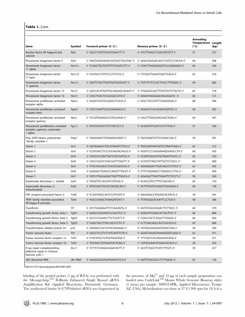
Table 1. List of primers for RT-PCR.
Gene Symbol Forward primer (59-39) Reverse primer (59-39)
AnnealingTemperature
(6C)Length(bp)
3-ketoacyl-CoA thiolase B Thiolase 59-TACGGTGAGTGATGGAGCAG-39 59-CACACAGTAGACGGCCTGAC-39 65 238
Active regulator of SIRT1 Aros 59-CATTGTCGAGACCACCTCAA-39 59-GATGGTTCCGCCAAACTAGA-39 59 458
Alkylglycerone phosphatesynthase
Agps 59-TTTTGGGAAACAAAAGCTCAA-39 59-TTGGAGCAACACACTTCAGG-39 56 250
Ataxia telangiectasia mutatedhomolog
Atm 59-GTGTGGCATTGATCTCACTGAGG-39 59-CTCAAATGTGTGCTTTGCCTAGC-39 62 574
ATP-binding cassette transporter,sub-family D, member 1,peroxisomal membrane protein
Abcd1 59-GAGGGAGGTTGGGAGGCAGT-39 59-GGTGGGAGCTGGGGATAAGG-39 67 465
ATP-binding cassette transporter,sub-family D, member 3,peroxisomal membrane protein
Abcd3 59-CTGGGCGTGAAATGACTAGATTGG-39 59-AGCTGCACATTGTCCAAGTACTCC-39 65 523
BCL2-associated agonist of celldeath
Bad 59-AGGACTTATCAGCCGAAGCA-39 59-GCTAAGCTCCTCCTCCATCC-39 61 420
Catalase Cat 59-ATGGTCTGGGACTTCTGGAGTCTTC-39 59-GTTTCCTCTCCTCCTCATTCAACAC-39 64 833
Cyclization recombinase Cre 59-CACCCTGTTACGTATAGC-39 59-CTAATCGCCATCTTCCAG-39 55 526
Cyclooxygenase-2 Cox2 59-TGCATGTGGCTGTGGATGTCATCA-39 59-CACTAAGACAGACCCGTCATCTCCA-39 66 449
DNA cross-link repair 1C,PSO2 homolog
Artemis 59-CCTGAGTGGCAGGAAGTCTC-39 59-CAGGCCACATACATCCACAG-39 61 379
Deleted in bladder cancer 1 Dbc1 59-ATCCACATGGTGATCGGAAT-39 59-CAGGCTGTACCCAAACACCT-39 58 372
Enoyl-Coenzyme A, hydratase Mfp1 59-ATGGCCAGATTTCAGGAATG-39 59-TGCCACTTTTGTTGATTTGC-39 60 211
Follicle stimulating hormonereceptor
Fshr 59-CCAGCCTTACCTACCCCAGT-39 59-CTGTGGTGTTCCCAGTGATG-39 62 345
Forkhead box O1 Foxo1 59-GGGAGAATGTTCGCTTTCTGGT-39 59-CCATCCTCATCAGGCACTTCTC-39 61 662
Forkhead box O3 Foxo3 59-CATCTCAAAGCTGGGTACCAGG-39 59-GCTCAAGGCCAGACTGGGAACT-39 63 541
Forkhead box O4 Foxo4 59-TTGTGCCTAGAAGAGAGTGCTG-39 59-ATTTGACACACTTCCCCACTCC-39 60 531
GATA binding protein 4 Gata4 59-CGGGTTGTTCAAACACCTTT-39 59-TGTGTGTGAAGGGGTGAAAA-39 58 617
Glutathione peroxidase 1 Gpx1 59-GGGACTACACCGAGATGAACGA-39 59-ACCATTCACTTCGCACTTCTCA-39 61 197
Glutathione S-transferase, alpha 1 Gsta1 59-GCAGACCAGAGCCATTCTCAACTAC-39 59-CTGCCAGGCTGTAGGAACTTCTTC-39 55 408
Glyceraldehyde-3-phosphatedehydrogenase
Gapdh 59-CACCATGGAGAAGGCCGGGG-39 59-GACGGACACATTGGGGGTAG-39 60 391
GlyceronephosphateO-acyltransferase
Gnpat 59-CCGTCTCCTTGAGACCTCTG-39 59-AGGTGTGGGAATCTGAGTGG-39 60 198
Heme oxygenase-1 Hmox1 59-GCACTATGTAAAGCGTCTCCACGAG-39 59-CCAGGCAAGATTCTCCCTTACAGAG-39 65 610
Inhibin alpha Inha 59-GCAATGGATGGGGAAGGTGG-39 59-GGTGGCTGCGTATGTGTTGG-39 65 238
Inhibitor of kappaB kinase beta Ikbkb 59-CAGGACACTGTGAAGGAGCA-39 59-TTGTACAGCAGCCATGAAGC-39 59 306
Inhibitor of kappaB kinase gamma Ikbkg 59-TGCCTTCAGAGCAGGGTACT-39 59-CTAAAGCTTGCCGATCCTG-39 59 390
Interleukin 1 alpha Il1a 59-CGTCAGGCAGAAGTTTGTCA-39 59-GTGCAAGTGACTCAGGGTGA-39 61 516
Interleukin 1 beta Il1b 59-GGACATGAGCACCTTCTTTTCC-39 59-GTGCCGTCTTTCATTACACAGG-39 63 321
Interleukin 6 Il6 59-GTTCTCTGGGAAATCGTGGA-39 59-GGAAATTGGGGTAGGAAGGA-39 61 339
Isopentenyl-diphosphatedelta isomerase
Idi1 59-TCTGTCTCGGTCTTCAGACAGGATG-39 59-AGTACCTGGGAGTCAGAGGAAGGTG-39 67 718
Luteinizing hormone/choriogonadotropin receptor
Lhcgr 59-ACCAAAAGCTGAGGCTGAGA-39 59-TACAGAAATGGGTTGGCACA-39 66 445
Meiotic recombination11 homolog A
Mre11 59-TGATGACGATGACCCTTTCA-39 59-ACAACGGAGGGGTTTTCTCT-39 57 368
Mitogen-activated protein 4kinase 4
Nik 59-GACAGCAGGGAGTAGCCTTG-39 59-GGAGAAAAGCCAGAGTGTCG-39 60 346
Nibrin Nbs1 59-TTCCTGCACGTTGAAGACAG-39 59-CTTGTGCTGCTTGGCATTTA-39 57 448
Nitric oxide synthase 2, inducible Nos2 59-GTGTTCCACCAGGAGATGTTG-39 59-CTCCTGCCCACTGAGTTCGTC-39 66 576
Nuclear factor erythroid 2-relatedfactor
Nrf2 59-CCACTGGTTTAGCCATCTCTCC-39 59-GTGGACATTAGCCCTTCCAAAC-39 64 364
Cre Recombinase-Mediated Stress in Sertoli Cells
PLoS ONE | www.plosone.org 5 July 2012 | Volume 7 | Issue 7 | e41097
labeling of the pooled probes (1 mg of RNA) was performed with
the MessageAmpTM II-Biotin Enhanced Single Round aRNA
Amplification Kit (Applied Biosystems, Darmstadt, Germany).
The synthesized biotin-16-UTP-labeled cRNA was fragmented in
the presence of Mg2+ and 10 mg of each sample preparation was
loaded onto CodeLinkTM Mouse Whole Genome Bioarray slides
(2 arrays per sample, 300033-6PK, Applied Microarrays, Tempe
AZ, USA). Hybridization was done at 37uC/300 rpm for 18 h in a
Table 1. Cont.
Gene Symbol Forward primer (59-39) Reverse primer (59-39)
AnnealingTemperature
(6C)Length(bp)
Nuclear factor NF-kappa-B p65subunit
Rela 59-GGCCTCATCCACATGAACTT-39 59-ATCTTGAGCTCGGCAGTGTT-39 57 337
Peroxisome biogenesis factor 5 Pex5 59-GAGTGAAGAAGCAGTGGCTGCATAC-39 59-GGACAGAGACAGCTCATCCCTACAA-39 64 508
Peroxisome biogenesis factor11 alpha
Pex11a 59-TCAGCTGCTGTGTTCTCAGTCCTT-39 59-GTACTTAGGAGGGTCCCGAGAGGA-39 64 420
Peroxisome biogenesis factor11 beta
Pex11b 59-GTATGCCTGTTCCCTTCTCG-39 59-CTCGGTTGAGGTGACTGACA-39 65 216
Peroxisome biogenesis factor11 gamma
Pex11c 59-GACTCTGCTTGGTGGTGGACACT-39 59-TGTCTCTCCCACTCACCTTTAGGC-39 64 682
Peroxisome biogenesis factor 13 Pex13 59-GACCACGTAGTTGCAAGAGCAGAGT-39 59-CTGAGGCAGCTTGTGTGTTCTACTG-39 65 718
Peroxisome biogenesis factor 14 Pex14 59-CACCTCACTCCGCAGCCATA-39 59-AGGATGAGGGGCAGCAGGTA -39 60 131
Peroxisome proliferator activatedreceptor alpha
Ppara 59-AGACCGTCACGGAGCTCACA-39 59-GGCCTGCCATCTCAGGAAAG-39 68 584
Peroxisome proliferator activatedreceptor beta
Pparb 59-CACCGAGTTCGCCAAGAACA-39 59-AGAGCCCGCAGAATGGTGTC-39 60 363
Peroxisome proliferator activatedreceptor gamma
Pparc 59-TCCGTAGAAGCCGTGCAAGA-39 59-CACCTTGGCGAACAGCTGAG-39 60 441
Peroxisome proliferative activatedreceptor, gamma, coactivator1 alpha
Pgc1a 59-ATGTGTCGCCTTCTTGCTCT-39 59-GCGGTATTCATCCCTCTTGA-39 57 350
Poly (ADP-ribose) polymerasefamily, member 1
Parp1 59-TGAGAAACTCGGAGGCAAGT-39 59-TGCAGAGTGTTCCAGACCAG-39 59 391
Sirtuin 1 Sirt1 59-GGTAGAGCCTGCATAGATCTTCA-39 59-TGGCAGTAATGGTCCTAACTGGG-39 62 512
Sirtuin 2 Sirt2 59-CCATGACCTCCCGCAGGACAGCG-39 59-GGGTCCCCAGGAAAGGGAGCCTA-39 69 505
Sirtuin 3 Sirt3 59-GTGCCCCGACTGCTCATCAATCG-39 59-CCGATCAACATGCTAGATTGCCC-39 65 523
Sirtuin 4 Sirt4 59-CACCCGGTCTGACGATTTGGCTT-39 59-CCGTGTTAGCTATTGCTCCTGCC-39 64 483
Sirtuin 5 Sirt5 59-TTGCTGCGACCTCACGTGGTGTG-39 59-GGAAGGACTTGACAGCCTCTTCC-39 65 505
Sirtuin 6 Sirt6 59-GGGGACTGAGCCCAGGTTTGCAT-39 59-CTTCTGGGAGCCTGGGGCCCTTA-39 67 494
Sirtuin 7 Sirt7 59-TATCCTAGGAGGCTGGTTTGGCA-39 59-GGAGGCTTAGTTAGATTCTCCCT-39 59 503
Superoxide dismutase 1, soluble Sod1 59-TGGGTTCCACGTCCATCAG-39 59-ACACCGTCCTTTCCAGCAG-39 63 153
Superoxide dismutase 2,mitochondrial
Sod2 59-ATGCAGCTGCACCACAGCAA-39 59-ACTTCAGTGCAGGCTGAAGAG-39 64 130
TNF receptor-associated factor 2 Traf2 59-CCAGTGACCACTCCATTGTG-39 59-GAGAAGCCATGAGCACAACA-39 59 341
TRAF family member-associatedNF-kappa B activator
Tank 59-AGCCCAGGCTAAAGATGAT-39 59-TTATGGGGTCAATTCCCTGA-39 58 306
Transferrin Trf 59-ATCTGGGAGATTCTCAAAGTG-39 59-AGTGTGGCAGGACTTCTTGCC-39 64 579
Transforming growth factor, beta 1 Tgfb1 59-GAGCCACAAACCCCGCCTCC-39 59-GGGGTTCGGGCACTGCTTCC-39 66 860
Transforming growth factor, beta 2 Tgfb2 59-GCCCCCGGACCTTCTCGTCT-39 59-CGGCCACTCTGGCTTTGGGG-39 66 924
Transforming growth factor, beta 3 Tgfb3 59-CAGCTGCCTTGCCACCCCTC-39 59-CCTCAACAGCCACTCGCGCA-39 65 966
Transformation related protein 53 p53 59-AGAGACCGCCGTACAGAAGA-39 59-TATGGCGGGAAGTAGACTGG-39 59 304
Tumor necrosis factor Tnf 59-GGCCTCCCTCTCATCAGTTCTA-39 59-GGGTCAGAGTAAAGGGGTCAGA-39 62 571
Tumor necrosis factor receptor 1a Tnfr1 59-CTGTATGCTGTGGTGGATGG-39 59-TTTCACCCACAGGGAGTAGG-39 59 371
Tumor necrosis factor receptor 1b Tnfr2 59-TGTAGCCTGTGGATGCTGAG-39 59-CATGCAAACATGGACACACA-39 58 324
X-ray repair complementingdefective repair in Chinesehamster cells 1
Xrcc1 59-TCTTCTCAAGGCGGACACTT-39 59-GCCTCTGCCTCATCTTTGTC-39 59 417
28S ribosomal RNA 28s rRNA 59-AAAGCGGGTGGTAAACTCCA-39 59-GGTTTCACGCCCTCTTGAAC-39 62 118
doi:10.1371/journal.pone.0041097.t001
Cre Recombinase-Mediated Stress in Sertoli Cells
PLoS ONE | www.plosone.org 6 July 2012 | Volume 7 | Issue 7 | e41097

Minitron shaker incubator (Infors AG, Bottmingen, Germany).
Thereafter, the microarrays were washed and incubated with Cy-
5-coupled streptavidine according to the manufacturer’s protocol
(Amersham Biosciences, Freiburg) to visualize hybridized com-
plexes. The labeled arrays were scanned using a GenePix 4000 B
scanner and GenePix Pro 4.0 software (Axon Instruments,
Table 2. List of primary antibodies.
Antigens
Speciesantibodiesraised in
Dilution(IF)
Dilution(WB) Supplier
ATP-binding cassette transporter, sub-family D,member 3, peroxisomal membrane protein, mouse
Rabbit,polyclonal
1:100 1:100 Gift from Alfred Volkl, University of Heidelberg, Germany
p53 binding protein 1 (53BP1), rabbit Rabbit,polyclonal
1:200 – Abcam plc, Cambridge, UK, Cat. No: ab21083
Catalase (CAT), mouse Rabbit,polyclonal
1:2,000 1:10,000 Gift from Denis I. Crane, School of Biomol. Biophys. Sci., Griffith Univ.,Nathan, Brisbane, Australia
Cleaved caspase-3, human Rabbit,monoclonal
– 1:1,000 New England Biolabs GmbH, Frankfurt am Main, Germany, Cat. No: 9664
Cre recombinase, P1 bacteriophage Rabbit,polyclonal
– 1:1,000 Covance, HISS Diagnostics GmbH, Freiburg, Germany, Cat. No: RPB-106C
Cyclooxygenase 2 (COX2), mouse Rabbit,polyclonal
– 1:1,000 Enzo Life Sciences GmbH, Loerrach, Germany, Cat. No: ALX-210-711
Glyceraldehyde 3-phosphate dehydrogenase(GAPDH), rabbit
Mouse,monoclonal
– 1:10,000 HyTest Ltd, Turku, Finland, Cat. No: 5G4
Heme oxygenase-1 (HMOX1), rat Rabbit,polyclonal
– 1:2,000 Assay Designs, Inc. Michigan, USA, Cat. No: SPA-895
Histone H3, human Rabbit,polyclonal
– 1:1,000 New England Biolabs GmbH, Frankfurt am Main, Germany, Cat. No: 9715
4-Hydroxynonenal (4-HNE), keyhole limpethemocyanin
Mouse,monoclonal
– 1:100 Cosmo Bio Co., LTD., Tokyo, Japan, Cat. No: MHN-020P
3-Ketoacyl-CoA thiolase B (Thiolase), mouse Rabbit,polyclonal
– 1:6,000 Gift from Paul P. von Veldhoven, Catholic University Leuven, Belgium
Nuclear factor erythroid 2-Related Factor (NRF2),human
Rabbit,polyclonal
1:300 1:400 Santa Cruz Biotechnology Inc., Heidelberg, Germany, Cat. No: sc-13032
Nuclear factor kB (NF-kB) p65, human Rabbit,polyclonal
– 1:1,000 New England Biolabs GmbH, Frankfurt am Main, Germany, Cat. No: 3034
Peroxisomal biogenesis factor 5 (PEX5), mouse Mouse,monoclonal
– 1:200 BD Transduction Laboratories, USA. Cat. No: 611594
Peroxisomal biogenesis factor 13 (PEX13), mouse Rabbit,polyclonal
1:2,000 1:6,000 Gift from Denis I. Crane (address see above)
Peroxisomal biogenesis factor 14 (PEX14), mouse Rabbit,polyclonal
1:4,000 1:20,000 Gift from Denis I. Crane (address see above)
Phospho-c-Jun (Ser63) II, human Rabbit,polyclonal
– 1:800 New England Biolabs GmbH, Frankfurt am Main, Germany, Cat. No:9261S
Phospho-NF-kB p65 (Ser536), human Rabbit,monoclonal
– 1:1,000 New England Biolabs GmbH, Frankfurt am Main, Germany, Cat. No: 3033
Phospho-p38 MAP Kinase (Thr180/Tyr182),human
Rabbit,polyclonal
– 1:1,000 New England Biolabs GmbH, Frankfurt am Main, Germany, Cat. No:9211S
Phospho-p44/42 MAPK (Erk1/2), human Mouse,monoclonal
- 1:1,000 New England Biolabs GmbH, Frankfurt am Main, Germany, Cat. No:9106S
Phospho-SAPK/JNK (Thr183/Tyr185), human Rabbit,polyclonal
– 1:1,000 New England Biolabs GmbH, Frankfurt am Main, Germany, Cat. No:9251S
p38 MAP Kinase, human Rabbit,polyclonal
– 1:1,000 New England Biolabs GmbH, Frankfurt am Main, Germany, Cat. No: 9212
Stress-activated protein kinase/Junamino-terminal kinase SAPK/JNK, human
Rabbit,polyclonal
– 1:1,000 New England Biolabs GmbH, Frankfurt am Main, Germany, Cat. No: 9252
Superoxide dismutase 2 (SOD2), rat Rabbit,polyclonal
1:5,000 1:6,000 Research Diagnostics, Inc., NJ, USA, Cat. No: RDI-RTSODMabR
a–tubulin, mouse Mouse,monoclonal
– 1:5,000 Sigma, Steinheim, Germany. Cat. No: T5168
Vimentin clone VIM 13.2, human Mouse,monoclonal
1:1,000 – Sigma, Steinheim, Germany. Cat. No: V5255
doi:10.1371/journal.pone.0041097.t002
Cre Recombinase-Mediated Stress in Sertoli Cells
PLoS ONE | www.plosone.org 7 July 2012 | Volume 7 | Issue 7 | e41097

Arlington, USA). Scanned Images were analyzed using the
CodeLink Expression Analysis software. The expression values
were normalized by quantile normalization [34] and log2
transformed. Technical replicates were averaged and fold changes
were calculated. The resulting gene list was subjected to the
Database for Annotation, Visualization and Integrated Discovery
(DAVID) [35] for annotation and overrepresentation analysis of
gene categories. Gene lists were analyzed with Ingenuity Pathway
Analysis (IngenuityH Systems, Redwood City, USA) to detect
altered canonical pathways. The microarray data was submitted to
the Gene Expression Omnibus (GEO) database at the NCBI/NIH
(http://www.nbci.nlm.nih.gov/projects/geo) and the accession
numbers from GEO are as follows: GSM700341, GSM700342,
GSM700343 and GSM700344. All data are MIAME compliant.
Protein AnalysesIsolation of enriched organelle and cytosolic fractions
from testes for analyses of peroxisomal and antioxidant
proteins. Testes were homogenized with a Potter-Elvehjem
homogenizer (Braun, Melsungen, Germany) in homogenization
medium (5 mM MOPS, pH 7.4, 250 mM sucrose, 1 mM EDTA,
0.1% [v/v] ethanol, 0.2 mM dithiothreitol, 1 mM 6-aminocapro-
nic acid, supplemented with 10% protease inhibitor mix M from
SERVA, Heidelberg, Germany) for 2 min with a single stroke at
1,000 rpm. The homogenate was centrifuged at 500 g for 10 min
Table 3. List of secondary antibodies and counterstaining of nuclei.
Secondary detection system used Host Method Dilution Supplier
anti-Mouse-IgG (whole molecule)-alkaline phosphatase Goat WB 1:20,000 Sigma, Steinheim, Germany. Cat. No: A3562
anti-Rabbit-IgG (whole molecule)-alkaline phosphatase Goat WB 1:20,000 Sigma, Steinheim, Germany. Cat. No: A3687
anti-Rabbit-IgG AlexaFluor488 Donkey IF 1:500 Molecular Probes/Invitrogen, Cat. No: A21206
anti-Mouse-IgG Texas Red Horse IF 1:1,000 Vertor laboratories, Inc, Burlingame, USA, Cat. No: TI-2000
Hoechst 33342 (1 mg/ml) nucleic acid staining – IF 1:750 Molecular Probes/Invitrogen, Carlsbad, CA, USA, Cat. No: A11007
TOTO-3 iodide – IF 1:750 Molecular Probes/Invitrogen, Carlsbad, CA, USA, Cat. No: T3604
doi:10.1371/journal.pone.0041097.t003
Figure 2. Peroxisome biogenesis and ROS metabolism are affected by Cre recombinase expression. A) Semiquantitative RT-PCR analysesfor the genes involved in peroxisome biogenesis (Pex5, Pex13, Pex14) and ROS metabolism (Cat, Gsta1, Gpx1, Sod1, Sod2, Hmox1) on cDNAs preparedfrom total testicular RNA (genotypes are indicated), Gapdh as control. For abbreviation of gene names see Table 1. B) Western blot analyses ofperoxisome biogenesis (PEX5p, PEX13p, PEX14p) and ROS metabolic (CAT, HMOX1, SOD2) proteins in cytosolic fractions (supernatant) and enrichedorganelles (pellet) of total testis samples (genotypes and protein masses are indicated). The abundance of a-tubulin was used as a loading control.20 mg of protein were loaded per lane.doi:10.1371/journal.pone.0041097.g002
Cre Recombinase-Mediated Stress in Sertoli Cells
PLoS ONE | www.plosone.org 8 July 2012 | Volume 7 | Issue 7 | e41097

Cre Recombinase-Mediated Stress in Sertoli Cells
PLoS ONE | www.plosone.org 9 July 2012 | Volume 7 | Issue 7 | e41097

to remove cell debris and nuclei. The supernatant was collected
and re-centrifuged at 120,000 g at 4uC for 30 min to yield the
mixed organelle pellet and the cytosolic supernatant, which were
stored at 280uC.
Isolation of nuclear extracts from testes for transcription
factor analyses. Nuclear extracts from testes were isolated with
the ProteoJETTM Cytoplasmic & Nuclear Protein Extraction Kit
according to the manufacturer’s protocol (Fermentas GmbH, St.
Leon-Rot, Germany). Briefly, testes were treated with the cell lysis
buffer and the lysate was centrifuged at 500 g to yield the nuclear
fraction. The supernatant was further centrifuged at 20,000 g for
15 min at 4uC to remove most organelles and yield an enriched
cytoplasmic fraction. The nuclear pellet was washed with the
Nuclei Wash Buffer provided in the kit and lysed in the Nuclei
Lysis Reagent by shaking for 15 min at 4uC with 1,200 rpm.
Nuclear lysates were centrifuged at 20,000 g for 5 min at 4uC to
obtain clear nuclear extracts.
Western blot analyses. Protein concentrations were deter-
mined by the Bradford protein assay (Bio-Rad, Munich, Germany)
using BSA as standard. Protein samples (20 mg) were separated on
12% SDS-PAGE and further processed for Western blot analyses
with the alkaline phosphatase-induced chemiluminescence tech-
nique as described by Nenicu and colleagues [28]. Primary and
secondary antibodies are listed in Tables 2 and 3. The blots were
exposured to Kodak Biomax MR Films. Bands on films were
quantified with a Bio-Rad Gel Doc 2000 system. All Western blot
analyses were performed three times and represent data from
three individual experiments. An example for the specificities of
the antibodies and method used is presented in Figure S2
(complete ABCD3 immunoblot).
Immunofluorescence analyses. Testes were removed from
three individual mice for every genotype and fixed by immersion
with 4% depolymerized paraformaldehyde (PFA), containing 2%
sucrose in phosphate-buffered saline (PBS) (150 mM NaCl,
13.1 mM K2HPO4, 5 mM KH2PO4, pH 7.4) at 4uC overnight.
The fixed testes samples were embedded into paraffin (Paraplast,
Sigma, St. Louis, MO, USA) using a Leica TP 1020 automated
vacuum infiltration tissue processor. Paraffin sections (4 mm) were
cut with a Leica RM2135 rotation microtome. The detailed
protocol for embedding procedures and subsequent immunoflu-
orescence analyses was described previously from our group [28].
Dilutions of the primary and secondary antibodies used are listed
in Tables 2 and 3. Negative controls were processed in parallel by
addition of TBST buffer instead of the first antibodies. Nuclei were
visualized with 1 mM TOTO-3 iodide for 10 min at room
temperature (Table 3). Samples were analyzed by confocal laser
scanning microscopy (CLSM) with a Leica TCS SP2 (Leica
Mikrosysteme Vertrieb GmbH, Wetzlar, Germany). Examples of
negative control sections without primary antibody are shown in
Figure S3.
TUNEL Test for Apoptosis DetectionApoptotic cells on paraffin sections were detected using the
ApopTagH Fluorescein In Situ Apoptosis Detection Kit (S7110,
Millipore GmbH, Schwalbach, Germany) by direct TdT-mediated
dUTP-biotin nick end labeling (TUNEL), according to the
manufacturer’s protocol. Testes from three animals were analyzed
for each genotype. For quantification of apoptosis rates in each
animal, TUNEL-positive cells in 300 cross-sections of seminiferous
tubules, obtained from 8 sections of different testis regions were
counted.
Statistical Analysis for Real-time PCRs and theMorphometric Measurements
The significance value of homogeneity of variances was 0.006
(less than 0.05), wherefore the differences between the relative Cre
transgene allelic abundances of real-time PCR results on genomic
DNA were analyzed with the Kruskal-Wallis test using SPSS
software.
The alterations of the apoptotic cell ratio between distinct
genotypes were analyzed with the ANOVA test using SPSS
software, because the significance value of homogeneity of
variances was 0.830 (more than 0.05). The significance of
differences between all genotypes was further analyzed with the
t-test.
Results
Our group is investigating the effects of tissue-specific perox-
isomal deficiency and we noted several unexplainable alterations
of peroxisomal gene expression in the heterozygous animals with
Cre-mediated PEX gene deletion. These alterations led us to reflect
whether the sole expression of Cre-recombinase in a wild type
background would affect the peroxisomal compartment and
induce oxidative and metabolic stress reactions. We have therefore
analyzed the effects of heterologous expression of Cre-recombinase
in the testis of mice expressing the Cre-recombinase under control
of the AMH promoter in Sertoli cells and used heterozygous and
homozygous AMH-Cre animals in comparison to wild type mice in
C57BL/6J genetic background to induce expression level-depen-
dent alterations in Sertoli cells.
Cre Expression in Sertoli Cells of the Testis Correlates withAllelic Abundance
Since also homozygous AMH-Cre animals were used for our
experiments, genomic sequencing with a PCR-based method for
walking in uncloned DNA was performed to unequivocally reveal
that the insertion of the transgene into the mouse genome would
not induce an undesired knockout of an endogenous gene by
affecting exon regions. The sequencing results revealed that only a
single copy of the transgene cassette, consisting of the human
AMH-promoter (3,504 bp), fused to the Cre-gene (1,254 bp),
followed by MmMt1 (1,372 bp), was inserted into the large intron 3
(98,309 bp) of the Plekha5 gene (ID: 109135) on chromosome 6 of
the mouse genome (Fig. 1A).
To confirm the correct genotypes of the animals (AMH-Cre/
AMH-Cre, AMH-Cre/Wt, and Wt/Wt) we have analyzed the
allelic abundance (Fig. 1B) of the Cre transgene in the testes by
Figure 3. Double-immunofluorescence analysis reveals that AMH-Cre-expression induces peroxisome proliferation and membraneprotein alterations. Double immunofluorescence for peroxisomal membrane proteins (green) and the intermediate filament protein vimentin asmarker for Sertoli cells (red) with counterstaining of nuclei with TOTO-3 iodide on paraffin sections from the testis of wild type (A, D, G, J, M, P),heterozygous AMH-Cre-transgenic mice (B, E, H, K, N, Q) and homozygous AMH-Cre-transgenic mice (C, F, I, L, O, R). Peroxisomes are proliferated andthe lipid transporter ABCD3 is increased in individual peroxisomes in Sertoli cells in the testis of AMH-Cre-transgenic animals in comparison to wildtype animals (A–F). A similar pattern is observed for PEX14p, a peroxisomal biogenesis protein with highest abundance in Sertoli cells, which wasstrongly induced in AMH-Cre-transgenic mice (M–R). In contrast, PEX13p, a peroxisomal biogenesis protein with highest abundance in germ cellperoxisomes is not increased in Sertoli cells and rather decreased in the germ cell population (G–L). Size of bars is: A, B, C, G, H, I, M, N, O: 25 mm; D, E,F, J, K, L, P, Q, R: 10 mm.doi:10.1371/journal.pone.0041097.g003
Cre Recombinase-Mediated Stress in Sertoli Cells
PLoS ONE | www.plosone.org 10 July 2012 | Volume 7 | Issue 7 | e41097

Cre Recombinase-Mediated Stress in Sertoli Cells
PLoS ONE | www.plosone.org 11 July 2012 | Volume 7 | Issue 7 | e41097

quantitative genomic PCR. Homozygous AMH-Cre, heterozygous
AMH-Cre and wild type animals without the Cre-transgene were
easily identified by quantitative genomic PCR. The levels of
mRNA expression (Fig. 1C) as well as the abundance of the Cre
recombinase protein in the total testes samples (Fig. 1D) correlated
well with the allelic abundance. In addition, RT-PCR analyses of
microdissected samples of distinct testicular cell types revealed that
the mRNA for Cre recombinase was only specifically expressed in
Sertoli cells in a similar pattern already observed in total testis
preparations, indicating that a comparison of the genotypes allows
for an analysis of Cre-mediated effects in a gene dosage-dependent
fashion in Sertoli cells (Fig. 1E).
Peroxisome Biogenesis and ROS Metabolism are Affectedby Cre Recombinase Expression
In order to assess alterations of the peroxisomal compartment,
we have analyzed the expression of genes involved in peroxisome
biogenesis, metabolism of reactive oxygen species (ROS) and
peroxisomal lipid metabolism or of genes involved in the
regulation of peroxisome-related gene expression and signal
transduction pathways. From the 32 peroxin genes (according to
the mouse nomenclature abbreviated as Pex), in the present study
mainly the mRNA expression and protein abundance for Pex5/
PEX5p, the shuttling receptor involved in the import of PTS1-
containing matrix proteins into the organelle, and for Pex13/
PEX13p as well as Pex14/PEX14p, two factors of the docking
complex on the peroxisomal membrane for matrix protein import
were analyzed. The Pex5 and Pex14 mRNA levels were increased
already in total testes preparations of homozygous AMH-Cre/
AMH-Cre animals compared to the wild type preparations (Fig. 2A)
whereas the one for Pex13 was not altered. Western blot analysis
for abundance of PEX14p revealed a genotype-correlated increase
with highest protein abundance in homozygous AMH-Cre animals.
Also immunofluorescence analysis for this protein revealed its
strong increase in Sertoli cells with concomitant induction of
peroxisome proliferation in this cell type (Fig. 3M, N, O, P, Q, R).
In contrast, PEX13p was most abundant in germ cells (Fig. 3G, H,
I, J, K, L) and appeared slightly downregulated already in
heterozygous Cre-transgenic mice (Fig. 3H, K) with homozygous
animals exhibiting a stronger germ cell-specific downregulation
(Fig. 3I, L). Comparable results for PEX13p were obtained also by
Western blot analysis of total testis homogenates (Fig. 2B).
Interestingly, PEX5p shifted from a primarily cytoplasmic
localization in wild type to the organelle pellet in Cre-transgenic
mice, suggesting the recruitment of this protein to the peroxisomal
membrane.
Moreover, the mRNA expression for catalase, the most
abundant antioxidative enzyme of the peroxisomal matrix, was
significantly increased in total RNA preparations from testis
already in heterozygous transgenic mice (Fig. 2A). Homozygous
AMH-Cre animals showed a very strong increase of catalase
mRNA expression. The results obtained on mRNA level could be
corroborated for catalase on the protein level by Western blot
analysis (Fig. 2B) as well as by immunofluorescence (Fig. 4A, B, C,
D, E, F), indicating a strong increase of catalase in Sertoli cells
(Fig. 4D, E, F). Similar effects were also noted on RNA and
protein levels for antioxidative enzymes of other compartments,
such as superoxide dismutase 2 (mitochondria) or heme oxygen-
ase-1 (endoplasmic reticulum), whereas the mRNA levels for
glutathion S-transferase 1 and glutathione peroxidase 1 were only
slightly elevated (Fig. 2A and 2B). The mRNA for NRF2, a redox-
dependent transcription factor regulating expression of most
antioxidative enzymes including catalase, was slightly elevated in
total RNA preparations from the testis (Fig. 5C). In contrast, the
mRNA for the transcription factor FOXO1 was slightly decreased
in homozygous AMH-Cre animals, whereas the ones for FOXO3
or FOXO4 were not significantly altered (Fig. 5C).
Immunofluorescence analysis revealed an increase in signal
intensity for peroxisomal catalase and mitochondrial SOD2
mainly in Sertoli cells of the seminiferous tubules (Fig. 4A, B, C,
D, E, F, G, H, I, J, K, L). Similarly NRF2 was also upregulated
(Fig. 4M, N, O, P, Q, R), but only partly translocated to the nuclei
of Sertoli cells (Fig. 4R). Furthermore, SOD2 and NRF2 were
significantly increased in spermatocytes, suggesting the presence of
oxidative stress also in germ cells (Fig. 4G, H, I, J, K, L, M, N, O,
P, Q, R).
Lipid Peroxidation Indicates Oxidative Stress in Cre-expressing Animals
As an indicator for oxidative stress and lipid peroxidation, we
have analyzed the 4-hydroxynonenal (4-HNE)-modification of
lysine residues in testes with distinct Cre-genotypes. The intensities
of several 4-HNE-modified protein bands (Fig. 5A) were increased
in organelle pellets from Cre-transgenic mice. Since 4-HNE
metabolites and lipid peroxides are PPAR ligands and oxidized
lipids are likely to be degraded in peroxisomes we have also
analyzed the expression levels of mRNAs for PPARs and enzymes
of peroxisomal b-oxidation. We found a significant upregulation of
PPARa mRNA in the testis of homozygous AMH-Cre animals,
whereas the mRNA expression levels for PPARb and PPARc in
total RNA preparations from testis were not altered (Fig. 5C). In
preparation from homozygous AMH-Cre testes, the strongest
mRNA increase of genes encoding proteins involved in peroxi-
somal lipid metabolism was noted for the ABC-transporter
ABCD3 (formerly PMP70), whereas the one for ABCD1 (formerly
ALDP) showed only a minor increase. Furthermore, we found a
strong induction of mRNAs encoding the multifunctional protein
1 (MFP1) and thiolase, two enzymes of the PPARa-induced
peroxisomal b-oxidation pathway 1, already in heterozygous
animals (Fig. 5C). In contrast, the mRNAs for enzymes of
peroxisomal cholesterol (isopentenyl-diphosphate delta isomerase,
IDI1) and ether lipid metabolism (glyceronephosphate O-acyl-
transferase, GNPAT and alkylglycerone phosphate synthase,
AGPS) were only mildly induced. The alterations of mRNA
expression levels of ABCD3 and thiolase were corroborated by
corresponding changes in protein abundance in Western blot
analyses of organelle fractions (Fig. 5B). Moreover, immunofluo-
rescence analysis revealed the strongest upregulation of ABCD3 in
Sertoli cells with only minor increases in germ and Leydig cells
(Fig. 3A, B, C, D, E, F).
Figure 4. Immunofluorescence analysis reveals an increased antioxidative response in AMH-Cre expression Sertoli cells. Doubleimmunofluorescence for peroxisomal catalase (CAT) (A–F), mitochondrial superoxide dismutase 2 (SOD2) (G–L) and the redox-sensitive transcriptionfactor NRF2 (M–R) with the intermediate filament protein vimentin as marker for Sertoli cells (red) and counterstaining of nuclei with TOTO-3 iodideon paraffin sections from the testis of wild type (A, D, G, J, M, P), heterozygous AMH-Cre-transgenic mice (B, E, H, K, N, Q) and homozygous AMH-Cre-transgenic mice (C, F, I, L, O, R). Whereas catalase is mainly present in Sertoli cells, SOD2 and NRF2 show highest abundance in spermatocytes of wildtype animals. Note that all three proteins were induced in Sertoli cells of AMH-Cre-transgenic mice (see higher magnification with regions of Sertolicells). Size of bars is: A, B, C, G, H, I, M, N, O: 25 mm; D, E, F, J, K, L, P, Q, R: 10 mm.doi:10.1371/journal.pone.0041097.g004
Cre Recombinase-Mediated Stress in Sertoli Cells
PLoS ONE | www.plosone.org 12 July 2012 | Volume 7 | Issue 7 | e41097

Figure 5. Alterations of gonadotropin receptors, paracrine regulators, proinflammatory genes, PPARs as well as lipid peroxidation.A) Western blot analysis of 4-HNE-modified lysine adducts in proteins of enriched organelle pellets of the testis (genotypes are indicated). The arrowson the right indicated the altered 4-HNE-modified proteins among the three genotypes. The abundance of a-tubulin was used as a loading control.25 mg of protein were loaded per lane. B) Western blot analyses of proteins involved in peroxisomal lipid metabolism (ABCD3), the PPARa-inducibleperoxisomal b-oxidation pathway 1 (Thiolase B) and COX2-protein in enriched organelle (pellet) and cytosolic fractions (supernatant) of the testis(genotypes and protein masses are indicated). The abundance of a-tubulin was used as a loading control. 20 mg of protein were loaded per lane. C)Semiquantitative RT-PCR analyses for PPARs (Ppara-, Pparb-, Pparc-mRNA), redox-dependent transcription factors (Nrf2-, Foxo1-, 3-, 4-mRNA) andgenes involved in peroxisomal lipid metabolism (Abcd1-, Abcd3-mRNA), PPARa-induced peroxisomal b-oxidation pathway 1 (Mfp1, Thiolase B),peroxisomal cholesterol (Idi1) and ether lipid metabolism (Gnpat, Agps). The abundance of the Gapdh mRNA was used as a loading control. D)Semiquantitative RT-PCR analyses for expression of mRNAs for gonadotropin receptors (Lhcgr, Fshr), inhibin a (Inha) and genes involved in paracrinesignaling (Il1a, I1lb, Il6, Tnfa, Tgfb1), as well as in proinflammatory pathways (Cox2, Nos2). The expression of the Gapdh mRNA was used as a loadingcontrol.doi:10.1371/journal.pone.0041097.g005
Cre Recombinase-Mediated Stress in Sertoli Cells
PLoS ONE | www.plosone.org 13 July 2012 | Volume 7 | Issue 7 | e41097
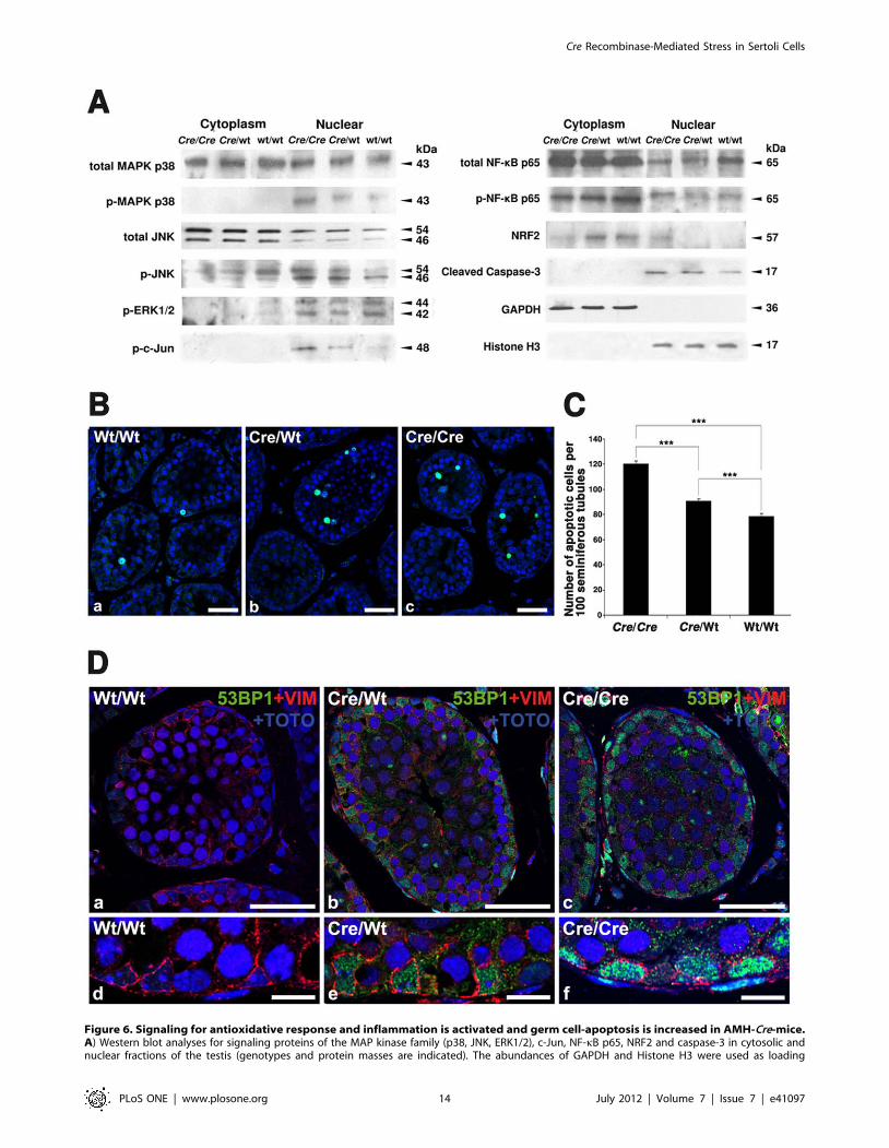
Figure 6. Signaling for antioxidative response and inflammation is activated and germ cell-apoptosis is increased in AMH-Cre-mice.A) Western blot analyses for signaling proteins of the MAP kinase family (p38, JNK, ERK1/2), c-Jun, NF-kB p65, NRF2 and caspase-3 in cytosolic andnuclear fractions of the testis (genotypes and protein masses are indicated). The abundances of GAPDH and Histone H3 were used as loading
Cre Recombinase-Mediated Stress in Sertoli Cells
PLoS ONE | www.plosone.org 14 July 2012 | Volume 7 | Issue 7 | e41097

Sertoli Cell-mediated Stress Induces Alterations inExpression of Gonadotropin Receptors, ParacrineRegulators and Proinflammatory Genes in AMH-Cre-transgenic Mice
As shown above, alterations of the abundance of the perox-
isomal ABCD3 and the mitochondrial SOD2 proteins were
observed also in germ and Leydig cells in AMH-Cre homozygous
animals, indicating that paracrine factors might be released from
Sertoli cells under oxidative stress conditions, influencing also
other testicular cells types. We have therefore investigated proteins
involved in endocrine and paracrine regulation of testis functions.
The mRNAs for gonadotropin receptors were strongly upregu-
lated in the testes of mice bearing the Cre-transgene (Fig. 5D).
Interestingly, the Lhcgr was induced only in homozygous animals,
whereas Fshr induction occurred already in heterozygous animals.
Furthermore, mRNAs for proteins involved in paracrine signaling,
such as IL1a, IL1b, IL6, TNFa and TGFb1, were also increased,
whereas the ones for TGFb2 and 3 were not altered. In addition,
the mRNAs of genes activated in proinflammatory conditions, like
the Cox2- and Nos2-genes, were also upregulated in the testes of
homozygous AMH-Cre mice (Fig. 5D). Similar to mRNA levels,
the COX2 protein abundance was also elevated in homozygous
AMH-Cre mice (Fig. 5B).
Signal Transduction Pathways Involved in Apoptosis,Antioxidative Response and Inflammation are Activatedin AMH-transgenic Mice
Already in H&E stained preparations an increase of altered
germ cells, reminiscent for apoptosis were observed, wherefore we
have analyzed the abundance and phosphorylation state of
different MAP kinase family signal transduction molecules, NF-
kB and activated caspase-3. In total testis preparations, the overall
abundance of MAPK p38 was not significantly altered in Cre-
transgenic- compared to wild type mice. The amount of the
phosphorylated p38 (pMAPK p38), however, was increased in
nuclear extracts from testes of Cre-transgenic mice (Fig. 6A).
Similarly, the relative abundance of the activated form of JNK
(mainly p46 JNK) was significantly increased in the nuclear
extracts in a dose-dependent manner (Fig. 6A). Furthermore, the
phosphorylated and activated form of c-Jun (p-c-Jun), downstream
of JNK signaling, was increased in a similar fashion. In contrast,
the abundance of phosphorylated ERK1/2, another member of
the MAPK-family, was not altered in Cre-transgenic mice
compared to the wild type animals (Fig. 6A).
As expected, NF-kB p65 was primarily localized in the cytosolic
fractions and only a minor amount was present in the nuclear
extracts. The ratio of phosphorylated (p-NF-kB p65) to unpho-
sphorylated NF-kB p65, however, was increased in the nuclear
fractions of Cre-transgenic mice, suggesting an activation of the
NF-kB pathway (Fig. 6A). NRF2, another ROS-activated signal
transduction molecule was primarily localized in the cytosolic
fraction in wild type mice. In homozygous Cre-transgenic mice, we
observed a significant shift of NRF2 into the nuclear fraction,
indicating its activation in these animals (Fig. 6A). Since signaling
molecules of three distinct pathways (MAPK p38, JNK and NF-
kB), implicated in the regulation of cell survival, proliferation and
apoptosis [36,37], were activated in Cre-transgenic animals, we
also analyzed the abundance of the cleaved form of caspase-3.
Indeed, we found that the amount of the activated caspase was
increased in nuclear extracts of testicular cells in AMH-Cre-
transgenic animals in a dose-dependent manner (Fig. 6A),
wherefore we analyzed the apoptosis rates in the seminiferous
tubules of AMH-Cre/AMH-Cre, AMH-Cre/Wt, and Wt/Wt mice
by TUNEL staining (Fig. 6B). Quantification of TUNEL positive
cells revealed a moderate but significant increase in number of
apoptotic germ cells already in heterozygous in comparison to wild
type testes (p,0.001) and a more pronounced increase in
homozygous Cre-transgenic testes (p,0.001) (Fig. 6C). No
apoptosis of Sertoli cells was noted, suggesting a higher capacity
of this cell type to counteract oxidative and cytokine-mediated
stress as well as Cre-induced DNA damage [7,8]. One of the
compensatory mechanisms could be a higher capacity of Sertoli
cells of transgenic AMH-Cre mice for DNA double-strand break
repair, wherefore we have also analyzed the recruitment of the p53
binding protein 1 (53BP1) by immunofluorescence analysis [38].
Indeed, an increased staining for the 53BP1 protein was observed
already in Sertoli cell nuclei of heterozygous AMH-Cre mice,
which was even more pronounced in homozygous AMH-Cre
animals (Fig. 6D).
Effects Observed on the Molecular Level in Total TestisPreparations could also be Verified in RNA Preparationsof Primary Sertoli Cell Cultures of Heterozygous AMH-CreAnimals
Generally heterozygous Cre mice are used for the generation of a
‘‘gene of interest (GOI)’’-knockout by the Cre-loxP technology (e.g.
promoter X-Cre/Wt/GOI-loxP_DY/Wt) in matings with mice
homozygous for the floxed GOI. To analyze whether all observed
gene alterations from total testis RNA preparations were specific
for Sertoli cells, we have used primary Sertoli cell cultures from
AMH-Cre/Wt mice and Wt/Wt mice for RT-PCR analyses.
Indeed, a comparable expression pattern was observed as in total
testis preparations for genes of peroxisomal and mitochondrial
proteins (Fig. 7B and 7C) and corresponding transcription factors
(Fig. 7D). The mRNAs for FSH receptor (Fig. 7A) and Sertoli cell
cytokines (Fig. 7E) were upregulated, with the Tnfa mRNA
showing the highest increase also in Sertoli cells as compared to
total testis preparations. Interestingly, in addition to the increase of
Ppara mRNA expression level, clear inductions of Pparc-, Nrf2- and
Foxo3-mRNAs were observed in the heterozygous Cre expressing
Sertoli cells (Fig. 7D). Moreover, the Gata4 mRNA (Fig. 7A),
encoding a transcription factor of Sertoli cells that is increased in
many testicular diseases in patients, was also clearly upregulated in
Sertoli cells with heterozygous Cre expression.
Since with primary Sertoli cells we have obtained similar results
as with complete testis preparations, we have done a microarray
analysis with CodeLinkTM Mouse Whole Genome Bioarray slides
to check for broad alterations in gene expression. Since the fold
changes of genes were only in a range of 5.043 (highest
upregulated gene: Agapat6) and 24.825 (lowest downregulated
controls. 20 mg of protein were loaded per lane. B) TUNEL staining in seminiferous tubules of paraffin sections of the testis from wild type,heterozygous and homozygous AMH-Cre animals. Size of bars is 10 mm. C) Statistical analysis of TUNEL stainings revealed an increase of germ cellapoptosis in parallel to Cre allelic abundance (genotypes and number of apoptotic cells per 100 seminiferous tubules are indicated), *** p,0.001. D)Double immunofluorescence for the p53 binding protein 1 (green) and the intermediate filament protein vimentin as marker for Sertoli cells (red)with counterstaining of nuclei with TOTO-3 iodide on paraffin sections from the testis of wild type (a,d), heterozygous AMH-Cre-transgenic mice (b,e)and homozygous AMH-Cre-transgenic mice (c,f). The 53BP1 protein was induced in Sertoli cells of AMH-Cre-transgenic mice (see higher magnificationwith regions of Sertoli cells). Size of bars is: a-c: 25 mm; d-f: 10 mm.doi:10.1371/journal.pone.0041097.g006
Cre Recombinase-Mediated Stress in Sertoli Cells
PLoS ONE | www.plosone.org 15 July 2012 | Volume 7 | Issue 7 | e41097

Cre Recombinase-Mediated Stress in Sertoli Cells
PLoS ONE | www.plosone.org 16 July 2012 | Volume 7 | Issue 7 | e41097
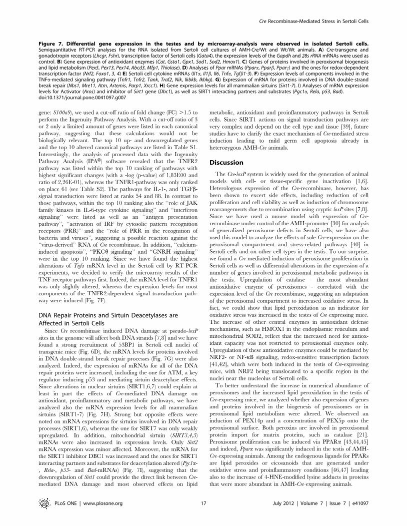
gene: S100a9), we used a cut-off ratio of fold change (FC) .1.5 to
perform the Ingenuity Pathway Analysis. With a cut-off ratio of 3
or 2 only a limited amount of genes were listed in each canonical
pathway, suggesting that these calculations would not be
biologically relevant. The top 10 up- and downregulated genes
and the top 10 altered canonical pathways are listed in Table S1.
Interestingly, the analysis of processed data with the Ingenuity
Pathway Analysis (IPAH) software revealed that the TNFR2
pathway was listed within the top 10 ranking of pathways with
highest significant changes (with a -log (p-value) of 1,83E00 and
ratio of 2,26E-01), whereas the TNFR1-pathway was only ranked
on place 61 (see Table S2). The pathways for IL-1-, and TGFb-
signal transduction were listed at ranks 54 and 88. In contrast to
those pathways, within the top 10 ranking also the ‘‘role of JAK
family kinases in IL-6-type cytokine signaling’’ and ‘‘interferon
signaling’’ were listed as well as an ‘‘antigen presentation
pathway’’, ‘‘activation of IRF by cytosolic pattern recognition
receptors (PRR)’’ and the ‘‘role of PRR in the recognition of
bacteria and viruses’’, suggesting a possible reaction against the
‘‘virus-derived’’ RNA of Cre recombinase. In addition, ‘‘calcium-
induced apoptosis’’, ‘‘PKCh signaling’’ and ‘‘GNRH signaling’’
were in the top 10 ranking. Since we have found the highest
alterations of Tnfa mRNA level in the Sertoli cell by RT-PCR
experiments, we decided to verify the microarray results of the
TNF-receptor pathways first. Indeed, the mRNA level for TNFR1
was only slightly altered, whereas the expression levels for most
components of the TNFR2-dependent signal transduction path-
way were induced (Fig. 7F).
DNA Repair Proteins and Sirtuin Deacetylases areAffected in Sertoli Cells
Since Cre recombinase induced DNA damage at pseudo-loxP
sites in the genome will affect both DNA strands [7,8] and we have
found a strong recruitment of 53BP1 in Sertoli cell nuclei of
transgenic mice (Fig. 6D), the mRNA levels for proteins involved
in DNA double-strand break repair processes (Fig. 7G) were also
analyzed. Indeed, the expression of mRNAs for all of the DNA
repair proteins were increased, including the one for ATM, a key
regulator inducing p53 and mediating sirtuin deacetylase effects.
Since alterations in nuclear sirtuins (SIRT1,6,7) could explain at
least in part the effects of Cre-mediated DNA damage on
antioxidant, proinflammatory and metabolic pathways, we have
analyzed also the mRNA expression levels for all mammalian
sirtuins (SIRT1-7) (Fig. 7H). Strong but opposite effects were
noted on mRNA expressions for sirtuins involved in DNA repair
processes (SIRT1,6), whereas the one for SIRT7 was only weakly
upregulated. In addition, mitochondrial sirtuin (SIRT3,4,5)
mRNAs were also increased in expression levels. Only Sirt2
mRNA expression was minor affected. Moreover, the mRNA for
the SIRT1 inhibitor DBC1 was increased and the ones for SIRT1
interacting partners and substrates for deacetylation altered (Pgc1a-
, Rela-, p53- and Bad-mRNAs) (Fig. 7I), suggesting that the
downregulation of Sirt1 could provide the direct link between Cre-
mediated DNA damage and most observed effects on lipid
metabolic, antioxidant and proinflammatory pathways in Sertoli
cells. Since SIRT1 actions on signal transduction pathways are
very complex and depend on the cell type and tissue [39], future
studies have to clarify the exact mechanism of Cre-mediated stress
induction leading to mild germ cell apoptosis already in
heterozygous AMH-Cre animals.
Discussion
The Cre-loxP system is widely used for the generation of animal
models with cell- or tissue-specific gene inactivation [1,6].
Heterologous expression of the Cre-recombinase, however, has
been shown to excert side effects, including reduction of cell
proliferation and cell viability as well as induction of chromosome
rearrangements due to recombination using cryptic loxP sites [7,8].
Since we have used a mouse model with expression of Cre-
recombinase under control of the AMH-promoter [30] for analysis
of generalized peroxisome defects in Sertoli cells, we have also
used this model to analyze the effects of sole Cre-expression on the
peroxisomal compartment and stress-related pathways [40] in
Sertoli cells and on other cell types in the testis. To our surprise,
we found a Cre-mediated induction of peroxisome proliferation in
Sertoli cells as well as differential alterations in the expression of a
number of genes involved in peroxisomal metabolic pathways in
the testis. Upregulation of catalase - the most abundant
antioxidative enzyme of peroxisomes - correlated with the
expression level of the Cre-recombinase, suggesting an adaptation
of the peroxisomal compartment to increased oxidative stress. In
fact, we could show that lipid peroxidation as an indicator for
oxidative stress was increased in the testes of Cre-expressing mice.
The increase of other central enzymes in antioxidant defense
mechanisms, such as HMOX1 in the endoplasmic reticulum and
mitochondrial SOD2, reflect that the increased need for antiox-
idant capacity was not restricted to peroxisomal enzymes only.
Upregulation of these antioxidative enzymes could be mediated by
NRF2- or NF-kB signaling, redox-sensitive transcription factors
[41,42], which were both induced in the testis of Cre-expressing
mice, with NRF2 being translocated to a specific region in the
nuclei near the nucleolus of Sertoli cells.
To better understand the increase in numerical abundance of
peroxisomes and the increased lipid peroxidation in the testis of
Cre-expressing mice, we analyzed whether also expression of genes
and proteins involved in the biogenesis of peroxisomes or in
peroxisomal lipid metabolism were altered. We observed an
induction of PEX14p and a concentration of PEX5p onto the
peroxisomal surface. Both peroxins are involved in peroxisomal
protein import for matrix proteins, such as catalase [21].
Peroxisome proliferation can be induced via PPARa [43,44,45]
and indeed, Ppara was significantly induced in the testis of AMH-
Cre-expressing animals. Among the endogenous ligands for PPARs
are lipid peroxides or eicosanoids that are generated under
oxidative stress and proinflammatory conditions [46,47] leading
also to the increase of 4-HNE-modified lysine adducts in proteins
that were more abundant in AMH-Cre-expressing animals.
Figure 7. Differential gene expression in the testes and by microarray-analysis were observed in isolated Sertoli cells.Semiquantitative RT-PCR analyses for the RNA isolated from Sertoli cell cultures of AMH-Cre/Wt and Wt/Wt animals. A) Cre-transgene andgonadotropin receptors (Lhcgr, Fshr), transcription factor of Sertoli cells (Gata4), the expression levels of the Gapdh and 28s rRNA mRNAs were used ascontrol. B) Gene expression of antioxidant enzymes (Cat, Gsta1, Gpx1, Sod1, Sod2, Hmox1). C) Genes of proteins involved in peroxisomal biogenesisand lipid metabolism (Pex5, Pex13, Pex14, Abcd3, Mfp1, Thiolase). D) Analyses of Ppar mRNAs (Ppara, Pparb, Pparc) and the ones for redox-dependenttranscription factor (Nrf2, Foxo1, 3, 4) E) Sertoli cell cytokine mRNAs (Il1a, Il1b, Il6, Tnfa, Tgfb1-3). F) Expression levels of components involved in theTNFa-mediated signaling pathway (Tnfr1, Tnfr2, Tank, Traf2, Nik, Ikbkb, Ikbkg). G) Expression of mRNA for proteins involved in DNA double-strandbreak repair (Nbs1, Mre11, Atm, Artemis, Parp1, Xrcc1). H) Gene expression levels for all mammalian sirtuins (Sirt1-7). I) Analyses of mRNA expressionlevels for Activator (Aros) and inhibitor of Sirt1 gene (Dbc1), as well as SIRT1 interacting partners and substrates (Pgc1a, Rela, p53, Bad).doi:10.1371/journal.pone.0041097.g007
Cre Recombinase-Mediated Stress in Sertoli Cells
PLoS ONE | www.plosone.org 17 July 2012 | Volume 7 | Issue 7 | e41097

PPARa induction leads to the transcriptional activation of genes
for peroxisomal b-oxidation enzymes [13,48] and this pathway in
turn is involved in control of the homeostasis of PPARa ligands by
their degradation [16,49]. We could show that the expressions of
the mRNAs for multifunctional protein 1 and the 3-ketoacyl-CoA
thiolase B, both enzymes of the PPARa-inducible peroxisomal b-
oxidation pathway 1, were induced in the testes of Cre-expressing
mice in addition to ABCD3, a transporter for peroxisomal b-
oxidation substrates. In addition to being PPARa ligands, secreted
eicosanoids such as prostaglandins or leukotrienes, as well as
cytokines are well-known mediators of proinflammarory signaling
that have also been implicated in Sertoli cell-mediated paracrine
regulation in the testis [50,51,52]. Indeed, the mRNAs for
proinflammatory cytokines (Il1a-, Il1b-, Il6- and Tnfa-mRNA)
and Nos2-mRNA, encoding the inducible form of the nitric oxide
synthases, were increased in the testes of AMH-Cre expressing
mice. We also observed that the amount of apoptosis of germ cells
correlated with an increase of Cre-expression. Since Cre was
specifically expressed in Sertoli cells as shown in microdissection
experiments, the induction of germ cell apoptosis must be an
indirect effect that could be mediated by the increased release of
cytokines and eicosanoids due to Cre-exerted stress in Sertoli cells.
Indeed, increased TGFb1 leads to the opening of the blood-testis-
barrier [53] and germ cell apoptosis [54]. Furthermore, TNFaperturbs cell junctions between Sertoli and germ cells and leads to
premature release of germ cells from the seminiferous epithelium
[55]. Interestingly, stress in Sertoli cells is frequently accompanied
with increased levels of Gata4- and Fshr-mRNA expression
[56,57,58,59], which were also observed in our study already in
heterozygous Cre expressing Sertoli cells. Indeed, upregulated
FSHR signaling could lead to enhanced cytokine production and
release from stressed Sertoli cells via the PKA pathway [60,61].
In addition to cytokines, also 4-HNE has been proposed to play
a role in cell signal transduction, in a variety of pathways affecting
cellular adhesion, the cell cycle and apoptosis [62,63]. 4-HNE, an
a, b-unsaturated hydroxyalkenal, which is produced by lipid
peroxidation of n-6 PUFA, is a potent cytotoxic and genotoxic
compound [64]. 4-HNE induced apoptosis in various cell lines was
accompanied by activation of JNK and caspase-3 [65,66]. Indeed,
our results indicate that major signaling pathways triggered by
oxidative stress (NF-kB, MAPK p38, JNK) are activated.
Furthermore, a significant activation of c-Jun and caspase-3 was
demonstrated in our study, suggesting that apoptotic germ cell
death might indeed be induced by these pathways also in Cre-
expressing animals.
Cell toxicity and effects on cell viability of Cre have been shown
to be inherent to its recombinase activity [7]. Cre-expression has
been shown to induce chromosomal rearrangements in genomes
that do not contain original loxP sequences, leading to the
discovery of cryptic loxP sites that were described in the yeast and
the mammalian genome [67,68]. As depicted in Figure S1, a
schematic overview made with the IPAH software, Cre-recombi-
nation induces DNA double-strand breaks that should induce the
recruitment of 53BP1 and of proteins of the related DNA damage
repair pathways [69]. Indeed, the recruitment of 53BP1 and an
induction in expression of the mRNAs for DNA repair proteins
was observed in this study. Interestingly, DBS1, a sensor for
double-strand breaks, is deacetylated by sirtuin 1 (SIRT1) and
thereby activated to induce the downstream DNA repair pathways
via the ATM protein [70,71]. By this mechanism SIRT1,
belonging to the nuclear sirtuins that are able to deacetylate
histones to control chromatin dynamics and DNA repair, is
recruited to damaged DNA regions [72]. In addition to SIRT1,
SIRT6 is also bound to chromatin and is involved in double-
strand break repair by regulating the C-terminal binding protein
(CtBP) and DNA protein kinase [73,74,75]. A deletion of Sirt6 has
been shown to lead to genomic instability, chromosome breaks
and -fusions, higher sensitivity to genotoxic agents, such a H2O2
and to severe metabolic defects and aging [69,76,77]. Alterations
of sirtuin levels could indeed explain part of the observed
phenotype in our study. Whereas Sirt6 was increased, probably
to promote genomic stability in Cre expressing cells, Sirt1 was
strongly decreased under chronic stress conditions in Sertoli cells,
possibly leading to derepression of NF-kB-dependent signaling
[78] and subsequently to cytokine release from Sertoli cells. Sertoli
cell-released TNFa could stimulate the TNFR2 pathway by an
autocrine mechanism, which is supported by the increased mRNA
levels of most TNFR2 signaling components in our study.
Oxidized lipids are strong ligands for PPARc, which can bind to
the promoter of the Sirt1 gene, repressing its transcriptional
activity [79]. Interestingly, the SIRT1 protein in turn deacetylates
besides PPARc also other proteins involved in metabolic
regulation, signal transduction and apoptosis (PPARa, PGC-1,
p53, NF-kB, FOXO3, BAD) [39,77,80,81]. Thus, a decrease of
SIRT1 could disturb cellular homeostasis and promote aging
[39,77]. In addition to a decrease of Sirt1 expression, an increase of
the expression level of Dbc1, encoding an inhibitor of SIRT1
activity [82,83,84], was observed in Sertoli cells of heterozygous
AMH-Cre mice. Furthermore, Sirt1 mRNA abundance is also
regulated post-transcriptionally by differential binding of the HuR
protein to the 39UTR [82,85,86,87]. Under oxidative stress
conditions HuR can be phosphorylated by MAPK p38 or the
check point kinase Chk2, leading to dissociation of the HuR-Sirt1-
mRNA-complex and its degradation [82,88]. Interestingly, Sirt1
deficient mouse embryonic fibroblasts are resistant to chronic and
sublethal doses of oxidative stress [39,89], suggesting that Sertoli
cells might be protected against apoptosis at least partially by this
mechanism as well as MAPK p38 activation [36].
MAPK p38 and c-Jun activation as well as peroxisomal
alterations correlated well with Cre-expression levels. Similar
dose-dependent correlations were found also for Fshr, Tnfa and
Tgfb1 mRNA expression levels in the testis of Cre expressing mice.
These findings suggest that high Cre levels that are obtained by
promoters with strong activity will lead to more Cre-induced side
effects. Indeed, several reports indicate that high levels of Cre-
expression might induce pathological alterations in diverse organ
systems in mice [8,9,11,90,91] and can even lead to cardiomyop-
athy [10], the molecular pathogenesis of which, however, has not
yet been published.
ConclusionsOur study indicates that the sole expression of Cre-recombinase
in AMH-Cre mice can lead to alterations of genes involved in
peroxisomal biogenesis, ROS- and lipid metabolism, stress
induced signal transduction pathways, expression of gonadotropin
receptors as well as proinflammatory cytokines and apoptosis. We
hypothesize that Cre-induced stress is mediated at least in part by
the increased DNA damage and transduced by sirtuins and the
repression release of important key regulators of signal transduc-
tion pathways (Figure S1). Therefore, wild type controls contain-
ing a heterozygous Cre genotype matched to the knockout animals,
have to be generated as control animals by separate breedings in
parallel, to insure that the analyzed phenotypic effects are due to
the deletion of the floxed alleles and not to Cre-induced side effects.
Cre Recombinase-Mediated Stress in Sertoli Cells
PLoS ONE | www.plosone.org 18 July 2012 | Volume 7 | Issue 7 | e41097

Supporting Information
Figure S1 Schematic overview of the model for regula-tion and transduction of AMH-Cre recombinase inducedstress in Sertoli and germ cells.(TIF)
Figure S2 Western blot of cytosolic and enrichedorganelle fractions of testes of AMH-Cre/AMH-Cre,AMH-Cre/Wt and Wt/Wt mice, probed with an antibodyagainst the peroxisomal membrane lipid transporterABCD3. The blot shows the high specificity of the antibody,
which is necessary to obtain an optimal and reliable staining for
the localization of organelles in tissue sections.
(TIF)
Figure S3 Negative control sections (without primaryantibody) of the testes of Wt/Wt and AMH-Cre/AMH-Cre mice, processed in parallel to double-immunofluo-rescence preparations presented in the manuscript,depicting only very few unspecific binding sites of thesecondary antibodies and revealing the high reliabilityof the immunofluorescence protocol used.(TIF)
Figure S4 Hematoxylin and eosin (H&E) staining oftestis sections of sexually mature adult animals (P120)with Wt/Wt (A, B), AMH-Cre/Wt (C, D) and AMH-Cre/AMH-Cre (E, F) genotypes. A, C, E: lower magnification
overviews (bar: 50 mm); B, D, F: corresponding partial higher
magnifications of the same regions (bar: 50 mm). Note the higher
amount of apoptotic germ cells in a semininiferous tubule
(seminiferous epithelium at stage 9) of a heterozygous Cre animal.
In homozygous Cre mice also a higher apoptosis rate was noted (in
stage 9 tubules, data not shown) and a mosaic pattern was
observed, with few degenerated seminiferous tubules exhibiting
severely impaired spermatogenesis located besides normal appear-
ing ones with regular spermatogenesis.
(TIF)
Sequence S1 Assembled sequence obtained by PCR-based genome walking proving the insertion of thetransgene in the intron 3 region of the Plekha5 gene onchromosome 6. Sequencing was started in both directions from
the AMH-Cre transgene cassette to obtain flanking regions at both
sides.
(DOC)
Table S1 Top 10 ranking of gene and canonical pathwayalterations in primary Cre-expressing Sertoli cells(AMH-Cre/Wt versus Wt/Wt) obtained by IngenuityPathway Analysis (IPAH software).
(DOC)
Table S2 List of Ingenuity Pathway Analysis (IPAHsoftware) - alterations of canonical pathways.
(XLS)
Acknowledgment
The excellent technical assistance of Elke Rodenberg-Frank, Gabriele
Thiele, Andrea Textor and Bianca Pfeiffer is gratefully acknowledged. We
also thank Vijith Vijayan for helpful discussions of the manuscript.
Author Contributions
Conceived and designed the experiments: YX GQ WF EBV. Performed
the experiments: YX SK GQ AN WF ST. Analyzed the data: YX SK GQ
ST AH HH GHL EBV. Contributed reagents/materials/analysis tools:
HH FG. Wrote the paper: YX GHL EBV.
References
1. Gu H, Zou YR, Rajewsky K (1993) Independent control of immunoglobulin
switch recombination at individual switch regions evidenced through Cre-loxP-
mediated gene targeting. Cell 73: 1155–1164.
2. Lobe CG, Nagy A (1998) Conditional genome alteration in mice. Bioessays 20:
200–208.
3. Sternberg N, Hamilton D (1981) Bacteriophage P1 site-specific recombination.
I. Recombination between loxP sites. J Mol Biol 150: 467–486.
4. Lewandoski M (2001) Conditional control of gene expression in the mouse. Nat
Rev Genet 2: 743–755.
5. Furuta Y, Behringer RR (2005) Recent innovations in tissue-specific gene
modifications in the mouse. Birth Defects Res C Embryo Today 75: 43–57.
6. Branda CS, Dymecki SM (2004) Talking about a revolution: The impact of site-
specific recombinases on genetic analyses in mice. Dev Cell 6: 7–28.
7. Loonstra A, Vooijs M, Beverloo HB, Allak BA, van Drunen E, et al. (2001)
Growth inhibition and DNA damage induced by Cre recombinase in
mammalian cells. Proc Natl Acad Sci USA 98: 9209–9214.
8. Pfeifer A, Brandon EP, Kootstra N, Gage FH, Verma IM (2001) Delivery of the
Cre recombinase by a self-deleting lentiviral vector: efficient gene targeting in
vivo. Proc Natl Acad Sci USA 98: 11450–11455.
9. Schmidt EE, Taylor DS, Prigge JR, Barnett S, Capecchi MR (2000) Illegitimate
Cre-dependent chromosome rearrangements in transgenic mouse spermatids.
Proc Natl Acad Sci USA 97: 13702–13707.
10. Buerger A, Rozhitskaya O, Sherwood MC, Dorfman AL, Bisping E, et al. (2006)
Dilated cardiomyopathy resulting from high-level myocardial expression of Cre-
recombinase. J Card Fail 12: 392–398.
11. Lee JY, Ristow M, Lin X, White MF, Magnuson MA, et al. (2006) RIP-Cre
revisited, evidence for impairments of pancreatic beta-cell function. J Biol Chem
281: 2649–2653.
12. Baumgart E, Fahimi HD, Steininger H, Grabenbauer M (2003) A review of
morphological techniques for detection of peroxisomal (and mitochondrial)
proteins and their corresponding mRNAs during ontogenesis in mice:
application to the PEX5-knockout mouse with Zellweger syndrome. Microsc
Res Tech 61: 121–138.
13. Baumgart E (1997) Application of in situ hybridization, cytochemical and
immunocytochemical techniques for the investigation of peroxisomes. A review
including novel data. Robert Feulgen Prize Lecture 1997. Histochem Cell Biol
108: 185–210.
14. Schrader M, Fahimi HD (2006) Peroxisomes and oxidative stress. BiochimBiophys Acta 1763: 1755–1766.
15. Masters CJ, Crane DI (1995) On the role of the peroxisome in ontogeny, ageingand degenerative disease. Mech Ageing Dev 80: 69–83.
16. Karnati S, Baumgart-Vogt E (2008) Peroxisomes in mouse and human lung:
their involvement in pulmonary lipid metabolism. Histochem Cell Biol 130:719–740.
17. Wanders RJ, Waterham HR (2006) Biochemistry of mammalian peroxisomesrevisited. Annu Rev Biochem 75: 295–332.
18. Van Veldhoven PP (2010) Biochemistry and genetics of inherited disorders ofperoxisomal fatty acid metabolism. J Lipid Res 51: 2863–2895.
19. Latruffe N, Cherkaoui Malki M, Nicolas-Frances V, Clemencet MC, Jannin B,et al. (2000) Regulation of the peroxisomal beta-oxidation-dependent pathway
by peroxisome proliferator-activated receptor alpha and kinases. BiochemPharmacol 60: 1027–1032.
20. del Rio LA, Corpas FJ, Sandalio LM, Palma JM, Barroso JB (2003) Plant
peroxisomes, reactive oxygen metabolism and nitric oxide. IUBMB Life 55: 71–81.
21. Girzalsky W, Saffian D, Erdmann R (2010) Peroxisomal protein translocation.Biochim Biophys Acta 1803: 724–731.
22. Baes M, Gressens P, Baumgart E, Carmeliet P, Casteels M, et al. (1997) A mousemodel for Zellweger syndrome. Nat Genet 17: 49–57.
23. Baumgart E, Vanhorebeek I, Grabenbauer M, Borgers M, Declercq PE, et al.(2001) Mitochondrial alterations caused by defective peroxisomal biogenesis in a
mouse model for Zellweger syndrome (PEX5 knockout mouse). Am J Pathol
159: 1477–1494.
24. Goldfischer S, Johnson AB, Essner E, Moore C, Ritch RH (1973) Peroxisomal
abnormalities in metabolic diseases. J Histochem Cytochem 21: 972–977.
25. Gould SJ, Valle D (2000) Peroxisome biogenesis disorders: genetics and cell
biology. Trends Genet 16: 340–345.
26. Baes M, Van Veldhoven PP (2006) Generalised and conditional inactivation of
Pex genes in mice. Biochim Biophys Acta 1763: 1785–1793.
27. Luers GH, Thiele S, Schad A, Volkl A, Yokota S, et al. (2006) Peroxisomes are
present in murine spermatogonia and disappear during the course ofspermatogenesis. Histochem Cell Biol 125: 693–703.
28. Nenicu A, Luers GH, Kovacs W, David M, Zimmer A, et al. (2007) Peroxisomesin human and mouse testis: differential expression of peroxisomal proteins in
Cre Recombinase-Mediated Stress in Sertoli Cells
PLoS ONE | www.plosone.org 19 July 2012 | Volume 7 | Issue 7 | e41097

germ cells and distinct somatic cell types of the testis. Biol Reprod 77: 1060–1072.
29. Dastig S, Nenicu A, Otte DM, Zimmer A, Seitz J, et al. (2011) Germ cells of
male mice express genes for peroxisomal metabolic pathways implicated in theregulation of spermatogenesis and the protection against oxidative stress.
Histochem Cell Biol 136: 413–425.
30. Lecureuil C, Fontaine I, Crepieux P, Guillou F (2002) Sertoli and granulosa cell-
specific Cre recombinase activity in transgenic mice. Genesis 33: 114–118.
31. Xavier F, Allard S (2003) Anti-Mullerian hormone, beta-catenin and Mullerianduct regression. Mol Cell Endocrinol 211: 115–121.
32. Siebert PD, Chenchik A, Kellogg DE, Lukyanov KA, Lukyanov SA (1995) Animproved PCR method for walking in uncloned genomic DNA. Nucleic Acids
Res 23: 1087–1088.
33. Miller SA, Dykes DD, Polesky HF (1988) A simple salting out procedure forextracting DNA from human nucleated cells. Nucleic Acids Res 16: 1215.
34. Bolstad BM, Irizarry RA, Astrand M, Speed TP (2003) A comparison of
normalization methods for high density oligonucleotide array data based onvariance and bias. Bioinformatics 19: 185–193.
35. Dennis GJ, Sherman BT, Hosack DA, Yang J, Gao W, et al. (2003) DAVID:Database for Annotation, Visualization, and Integrated Discovery. Genome Biol
4: P3.
36. Cargnello M, Roux PP (2011) Activation and function of the MAPKs and theirsubstrates, the MAPK-activated protein kinases. Microbiol Mol Biol Rev 75: 50–
83.
37. Chen F (2012) JNK-induced apoptosis, compensatory growth, and cancer stem
cells. Cancer Res 72: 379–386.
38. Noon AT, Goodarzi AA (2011) 53BP1-mediated DNA double strand breakrepair: insert bad pun here. DNA Repair (Amst) 10: 1071–1076.
39. Baur J (2010) Sirtuins and Oxidative Stress. Pathways (SABiosciences) 12: 12–
13.
40. Schlatt S, Meinhardt A, Nieschlag E (1997) Paracrine regulation of cellular
interactions in the testis: factors in search of a function. Eur J Endocrinol 137:107–117.
41. Nguyen T, Nioi P, Pickett CB (2009) The Nrf2-antioxidant response element
signaling pathway and its activation by oxidative stress. J Biol Chem 284: 13291–13295.
42. Surh YJ, Na HK (2008) NF-kappaB and Nrf2 as prime molecular targets forchemoprevention and cytoprotection with anti-inflammatory and antioxidant
phytochemicals. Genes Nutr 2: 313–317.
43. Lee SS, Pineau T, Drago J, Lee EJ, Owens JW, et al. (1995) Targeted disruptionof the alpha isoform of the peroxisome proliferator-activated receptor gene in
mice results in abolishment of the pleiotropic effects of peroxisome proliferators.Mol Cell Biol 15: 3012–3022.
44. Issemann I, Green S (1990) Activation of a member of the steroid hormone
receptor superfamily by peroxisome proliferators. Nature 347: 645–650.
45. Issemann I, Green S (1991) Cloning of novel members of the steroid hormone
receptor superfamily. J Steroid Biochem Mol Biol 40: 263–269.
46. Yaqoob P (2003) Fatty acids as gatekeepers of immune cell regulation. TrendsImmunol 24: 639–645.
47. Barrera G, Toaldo C, Pizzimenti S, Cerbone A, Pettazzoni P, et al. (2008) TheRole of PPAR Ligands in Controlling Growth-Related Gene Expression and
their Interaction with Lipoperoxidation Products. PPAR Res 2008: 524671.
48. Fahimi HD, Beier K, Lindauer M, Schad A, Zhan J, et al. (1996) Zonalheterogeneity of peroxisome proliferation in rat liver. Ann N Y Acad Sci 804:
341–361.
49. Hashimoto T, Fujita T, Usuda N, Cook W, Qi C, et al. (1999) Peroxisomal and
mitochondrial fatty acid beta-oxidation in mice nullizygous for both peroxisome
proliferator-activated receptor alpha and peroxisomal fatty acyl-CoA oxidase.Genotype correlation with fatty liver phenotype. J Biol Chem 274: 19228–
19236.
50. Jannini EA, Ulisse S, Cecconi S, Cironi L, Colonna R, et al. (1994) Follicle-
stimulating hormone-induced phospholipase A2 activity and eicosanoid
generation in rat Sertoli cells. Biol Reprod 51: 140–145.
51. Ishikawa T, Morris PL (2006) A multistep kinase-based sertoli cell autocrine-
amplifying loop regulates prostaglandins, their receptors, and cytokines.Endocrinology 147: 1706–1716.
52. Winnall WR, Ali U, O’Bryan MK, Hirst JJ, Whiley PA, et al. (2007) Constitutive
expression of prostaglandin-endoperoxide synthase 2 by somatic and spermato-genic cells is responsible for prostaglandin E2 production in the adult rat testis.
Biol Reprod 76: 759–768.
53. Konrad L, Luers GH, Volck-Badouin E, Keilani MM, Laible L, et al. (2006)
Analysis of the mRNA expression of the TGF-Beta family in testicular cells and
localization of the splice variant TGF-beta2B in testis. Mol Reprod Dev 73:1211–1220.
54. Konrad L, Keilani MM, Laible L, Nottelmann U, Hofmann R (2006) Effects ofTGF-betas and a specific antagonist on apoptosis of immature rat male germ
cells in vitro. Apoptosis 11: 739–748.
55. Li MW, Xia W, Mruk DD, Wang CQ, Yan HH, et al. (2006) Tumor necrosisfactor {alpha} reversibly disrupts the blood-testis barrier and impairs Sertoli-
germ cell adhesion in the seminiferous epithelium of adult rat testes. J Endocrinol190: 313–329.
56. Kyronlahti A, Euler R, Bielinska M, Schoeller EL, Moley KH, et al. (2011)
GATA4 regulates Sertoli cell function and fertility in adult male mice. Mol CellEndocrinol 333: 85–95.
57. LaVoie HA (2003) The role of GATA in mammalian reproduction. Exp Biol
Med (Maywood) 228: 1282–1290.
58. Allan CM, Garcia A, Spaliviero J, Zhang FP, Jimenez M, et al. (2004) Complete
Sertoli cell proliferation induced by follicle-stimulating hormone (FSH)
independently of luteinizing hormone activity: evidence from genetic models
of isolated FSH action. Endocrinology 145: 1587–1593.
59. Allan CM, Lim P, Robson M, Spaliviero J, Handelsman DJ (2009) Transgenic
mutant D567G but not wild-type human FSH receptor overexpression provides
FSH-independent and promiscuous glycoprotein hormone Sertoli cell signaling.
Am J Physiol Endocrinol Metab 296: E1022–1028.
60. Nenicu A, Okun JG, Hartmann MF, Wudy SA, Guillou F, et al. (2009)Molecular pathogenesis of male infertility due to peroxisome dysfunction in
Sertoli cells. Biol Reprod 81: Meeting Abstracts 682.
61. Chauvigne F, Verdura S, Mazon MJ, Duncan N, Zanuy S, et al. (2012) Follicle-
Stimulating Hormone and Luteinizing Hormone Mediate the Androgenic
Pathway in Leydig Cells of an Evolutionary Advanced Teleost. Biol Reprod.DOI:10.1095/biolreprod.112.100784. In press.
62. Zarkovic N, Zarkovic K, Schaur RJ, Stolc S, Schlag G, et al. (1999) 4-
Hydroxynonenal as a second messenger of free radicals and growth modifying
factor. Life Sci 65: 1901–1904.
63. Zarkovic N (2003) 4-hydroxynonenal as a bioactive marker of pathophysiolog-
ical processes. Mol Aspects Med 24: 281–291.
64. Esterbauer H, Schaur RJ, Zollner H (1991) Chemistry and biochemistry of 4-
hydroxynonenal, malonaldehyde and related aldehydes. Free Radic Biol Med
11: 81–128.
65. Awasthi YC, Sharma R, Cheng JZ, Yang Y, Sharma A, et al. (2003) Role of 4-
hydroxynonenal in stress-mediated apoptosis signaling. Mol Aspects Med 24:
219–230.
66. Chaudhary P, Sharma R, Sharma A, Vatsyayan R, Yadav S, et al. (2010)
Mechanisms of 4-Hydroxy-2-nonenal Induced Pro- and Anti-Apoptotic
Signaling. Biochemistry 49: 6263–6275.
67. Sauer B (1992) Identification of cryptic lox sites in the yeast genome by selection
for Cre-mediated chromosome translocations that confer multiple drugresistance. J Mol Biol 223: 911–928.
68. Thyagarajan B, Guimaraes MJ, Groth AC, Calos MP (2000) Mammalian
genomes contain active recombinase recognition sites. Gene 244: 47–54.
69. Lombard DB (2009) Sirtuins at the breaking point: SIRT6 in DNA repair. Aging(Albany NY) 1: 12–16.
70. Kinoshita T, Nagamatsu G, Kosaka T, Takubo K, Hotta A, et al. (2011) Ataxia-
telangiectasia mutated (ATM) deficiency decreases reprogramming efficiency
and leads to genomic instability in iPS cells. Biochem Biophys Res Commun
407: 321–326.
71. Yuan Z, Zhang X, Sengupta N, Lane WS, Seto E (2007) SIRT1 regulates the
function of the Nijmegen breakage syndrome protein. Mol Cell 27: 149–162.
72. Ozawa Y, Kubota S, Narimatsu T, Yuki K, Koto T, et al. (2010) Retinal agingand sirtuins. Ophthalmic Res 44: 199–203.
73. McCord RA, Michishita E, Hong T, Berber E, Boxer LD, et al. (2009) SIRT6
stabilizes DNA-dependent protein kinase at chromatin for DNA double-strand
break repair. Aging (Albany NY) 1: 109–121.
74. Kaidi A, Weinert BT, Choudhary C, Jackson SP (2010) Human SIRT6
promotes DNA end resection through CtIP deacetylation. Science 329: 1348–
1353.
75. Nakagawa T, Guarente L (2011) Sirtuins at a glance. J Cell Sci 124: 833–838.
76. Mostoslavsky R, Chua KF, Lombard DB, Pang WW, Fischer MR, et al. (2006)
Genomic instability and aging-like phenotype in the absence of mammalian
SIRT6. Cell 124: 315–329.
77. Purushotham A, Schug TT, Li X (2009) SIRT1 performs a balancing act on the
tight-rope toward longevity. Aging (Albany NY) 1: 669–673.
78. Chung S, Yao H, Caito S, Hwang JW, Arunachalam G, et al. (2010) Regulation
of SIRT1 in cellular functions: role of polyphenols. Arch Biochem Biophys 501:
79–90.
79. Han L, Zhou R, Niu J, McNutt MA, Wang P, et al. (2010) SIRT1 is regulated by
a PPAR{gamma}-SIRT1 negative feedback loop associated with senescence.
Nucleic Acids Res 38: 7458–7471.
80. Luo J, Nikolaev AY, Imai S, Chen D, Su F, et al. (2001) Negative control of p53
by Sir2alpha promotes cell survival under stress. Cell 107: 137–148.
81. Vaziri H, Dessain SK, Ng Eaton E, Imai SI, Frye RA, et al. (2001)
hSIR2(SIRT1) functions as an NAD-dependent p53 deacetylase. Cell 107:
149–159.
82. Kwon HS, Ott M (2008) The ups and downs of SIRT1. Trends Biochem Sci 33:517–525.
83. Kim JE, Chen J, Lou Z (2008) DBC1 is a negative regulator of SIRT1. Nature
451: 583–586.
84. Zhao W, Kruse JP, Tang Y, Jung SY, Qin J, et al. (2008) Negative regulation of
the deacetylase SIRT1 by DBC1. Nature 451: 587–590.
85. Michishita E, Park JY, Burneskis JM, Barrett JC, Horikawa I (2005)
Evolutionarily conserved and nonconserved cellular localizations and functions
of human SIRT proteins. Mol Biol Cell 16: 4623–4635.
86. Sasaki T, Maier B, Bartke A, Scrable H (2006) Progressive loss of SIRT1 with
cell cycle withdrawal. Aging Cell 5: 413–422.
87. Abdelmohsen K, Pullmann R, Jr., Lal A, Kim HH, Galban S, et al. (2007)
Phosphorylation of HuR by Chk2 regulates SIRT1 expression. Mol Cell 25:543–557.
Cre Recombinase-Mediated Stress in Sertoli Cells
PLoS ONE | www.plosone.org 20 July 2012 | Volume 7 | Issue 7 | e41097

88. Cuadrado A, Nebreda AR (2010) Mechanisms and functions of p38 MAPK
signalling. Biochem J 429: 403–417.89. Chua KF, Mostoslavsky R, Lombard DB, Pang WW, Saito S, et al. (2005)
Mammalian SIRT1 limits replicative life span in response to chronic genotoxic
stress. Cell Metab 2: 67–76.
90. Naiche LA, Papaioannou VE (2007) Cre activity causes widespread apoptosis
and lethal anemia during embryonic development. Genesis 45: 768–775.91. Whitsett JA, Perl AK (2006) Conditional control of gene expression in the
respiratory epithelium: A cautionary note. Am J Respir Cell Mol Biol 34: 519–
520.
Cre Recombinase-Mediated Stress in Sertoli Cells
PLoS ONE | www.plosone.org 21 July 2012 | Volume 7 | Issue 7 | e41097


![Transgenic Strategies for Sparse but Strong Expression of ... · TMP treatment, dCre is degraded and Cre-mediated recombination prevented [9]. In the presence of increasing doses](https://static.fdocuments.in/doc/165x107/5c68f5a409d3f2f5638c70e4/transgenic-strategies-for-sparse-but-strong-expression-of-tmp-treatment.jpg)