Targeted Imaging of Ewing Sarcoma in Preclinical Models...
11
Imaging, Diagnosis, Prognosis Targeted Imaging of Ewing Sarcoma in Preclinical Models Using a 64 Cu-Labeled Anti-CD99 Antibody Allison F. O'Neill 1 , Jason L.J. Dearling 2 , Yuchuan Wang 3 , Tanya Tupper 3 , Yanping Sun 3 , Jon C. Aster 4 , Monica L. Calicchio 5 , Antonio R. Perez-Atayde 5 , Alan B. Packard 2 , and Andrew L. Kung 6 Abstract Purpose: Ewing sarcoma is a tumor of the bone and soft tissue characterized by diffuse cell membrane expression of CD99 (MIC2). Single-site, surgically resectable disease is associated with an excellent 5-year event-free survival; conversely, patients with distant metastases have a poor prognosis. Noninvasive imaging is the standard approach to identifying sites of metastatic disease. We sought to develop a CD99-targeted imaging agent for staging Ewing sarcoma and other CD99-expressing tumors. Experimental Design: We identified a CD99 antibody with highly specific binding in vitro and labeled this antibody with 64 Cu. Mice with either subcutaneous Ewing sarcoma xenograft tumors or micrometastases were imaged with the 64 Cu-labeled anti-CD99 antibody and these results were compared with conventional MRI and 2[18F]fluoro-2-deoxy-D-glucose–positron emission tomography (FDG–PET) imaging. Results: 64 Cu-labeled anti-CD99 antibody demonstrated high avidity for the CD99-positive subcuta- neous tumors, with a high tumor-to-background ratio, greater than that demonstrated with FDG–PET. Micrometastases, measuring 1 to 2 mm on MRI, were not detected with FDG–PET but were readily visualized with the 64 Cu-labeled anti-CD99 antibody. Probe biodistribution studies demonstrated high specificity of the probe for CD99-positive tumors. Conclusions: 64 Cu-labeled anti-CD99 antibody can detect subcutaneous Ewing sarcoma tumors and metastatic sites with high sensitivity, outperforming FDG–PET in preclinical studies. This targeted radio- tracer may have important implications for the diagnosis, surveillance, and treatment of Ewing sarcoma. Similarly, it may impact the management of other CD99 positive tumors. Clin Cancer Res; 20(3); 678–87. Ó2013 AACR. Introduction Ewing sarcoma is a tumor of the bone or soft tissue affecting approximately 250 children, adolescents, and young adults each year (1, 2). Patients with localized disease are treated with chemotherapy, surgery for local control, and radiotherapy in patients for whom surgical margins remain positive (3). For patients with metastatic disease, chemotherapy and radiotherapy are the main- stays of treatment (4). Although patients with localized disease have a good prognosis with 70% 5-year event-free survival (EFS), those with metastatic or relapsed disease fare poorly with only 15% to 20% 5-year EFS (1, 2, 5). Accordingly, accurate assessment of the extent of disease at the time of diagnosis plays a critical role in directing appropriate therapy and assessing prognosis. Noninvasive studies including MRI, computed tomogra- phy (CT), or positron emission tomography (PET) are used to determine disease stage. Although these techniques are optimally used in different clinical scenarios, their limits of detection, constrained by system resolution, vary by modality: approximately millimeter range for CT and MRI, and 1 cm for PET (6–10). Apart from resolution limitation, the detection of micrometastases for molecular imaging modalities, such as PET, is highly dependent upon the signal-to-background ratio provided by the imaging probe. Micrometastatic disease that cannot be detected with exist- ing imaging modalities is a known risk for distant recur- rence, hence the need exists for new imaging approaches that can more accurately identify remote sites of disease with high specificity and sensitivity (11). Imaging also plays a primary role in assessing the resp- onse of patients to therapy. After completion of therapy, patients typically undergo surveillance with MRI of the primary disease site as well as chest CT, or serial chest Authors' Affiliations: 1 Department of Pediatric Oncology, Dana-Farber Cancer Institute, Boston Children's Hospital, and Harvard Medical School; 2 Division of Nuclear Medicine and Molecular Imaging, Department of Radiology, Boston Children's Hospital, and Harvard Medical School; 3 Lurie Family Imaging Center, Dana-Farber Cancer Institute; 4 Department of Pathology, Brigham and Women's Hospital, and Harvard Medical School; 5 Department of Pathology, Boston Children's Hospital, and Harvard Med- ical School, Boston, Massachusetts; and 6 Department of Pediatrics, Columbia University Medical Center, New York Note: Supplementary data for this article are available at Clinical Cancer Research Online (http://clincancerres.aacrjournals.org/). Corresponding Author: Andrew L. Kung, Columbia University Medical Center, 3959 Broadway, CHN 10-01, New York, NY 10032. Phone: 212- 342-4624; Fax: 212-305-8428; E-mail: [email protected] doi: 10.1158/1078-0432.CCR-13-1660 Ó2013 American Association for Cancer Research. Clinical Cancer Research Clin Cancer Res; 20(3) February 1, 2014 678 on July 27, 2018. © 2014 American Association for Cancer Research. clincancerres.aacrjournals.org Downloaded from Published OnlineFirst November 11, 2013; DOI: 10.1158/1078-0432.CCR-13-1660
Transcript of Targeted Imaging of Ewing Sarcoma in Preclinical Models...
Imaging, Diagnosis, Prognosis
Targeted Imaging of Ewing Sarcoma in Preclinical ModelsUsing a 64Cu-Labeled Anti-CD99 Antibody
Allison F. O'Neill1, Jason L.J. Dearling2, Yuchuan Wang3, Tanya Tupper3, Yanping Sun3, Jon C. Aster4,Monica L. Calicchio5, Antonio R. Perez-Atayde5, Alan B. Packard2, and Andrew L. Kung6
AbstractPurpose: Ewing sarcoma is a tumor of the bone and soft tissue characterized by diffuse cell membrane
expression of CD99 (MIC2). Single-site, surgically resectable disease is associated with an excellent 5-year
event-free survival; conversely, patientswith distantmetastases have a poor prognosis.Noninvasive imaging
is the standard approach to identifying sites of metastatic disease. We sought to develop a CD99-targeted
imaging agent for staging Ewing sarcoma and other CD99-expressing tumors.
Experimental Design: We identified a CD99 antibody with highly specific binding in vitro and labeled
this antibody with 64Cu. Mice with either subcutaneous Ewing sarcoma xenograft tumors ormicrometastases
were imaged with the 64Cu-labeled anti-CD99 antibody and these results were compared with conventional
MRI and 2[18F]fluoro-2-deoxy-D-glucose–positron emission tomography (FDG–PET) imaging.
Results: 64Cu-labeled anti-CD99 antibody demonstrated high avidity for the CD99-positive subcuta-
neous tumors, with a high tumor-to-background ratio, greater than that demonstrated with FDG–PET.
Micrometastases, measuring 1 to 2 mm on MRI, were not detected with FDG–PET but were readily
visualized with the 64Cu-labeled anti-CD99 antibody. Probe biodistribution studies demonstrated high
specificity of the probe for CD99-positive tumors.
Conclusions: 64Cu-labeled anti-CD99 antibody can detect subcutaneous Ewing sarcoma tumors and
metastatic sites with high sensitivity, outperforming FDG–PET in preclinical studies. This targeted radio-
tracer may have important implications for the diagnosis, surveillance, and treatment of Ewing sarcoma.
Similarly, it may impact the management of other CD99 positive tumors. Clin Cancer Res; 20(3); 678–87.
�2013 AACR.
IntroductionEwing sarcoma is a tumor of the bone or soft tissue
affecting approximately 250 children, adolescents, andyoung adults each year (1, 2). Patients with localizeddisease are treated with chemotherapy, surgery for localcontrol, and radiotherapy in patients for whom surgicalmargins remain positive (3). For patients with metastaticdisease, chemotherapy and radiotherapy are the main-stays of treatment (4). Although patients with localized
disease have a good prognosis with 70% 5-year event-freesurvival (EFS), those with metastatic or relapsed diseasefare poorly with only 15% to 20% 5-year EFS (1, 2, 5).Accordingly, accurate assessment of the extent of diseaseat the time of diagnosis plays a critical role in directingappropriate therapy and assessing prognosis.
Noninvasive studies including MRI, computed tomogra-phy (CT), or positron emission tomography (PET) are usedto determine disease stage. Although these techniques areoptimally used in different clinical scenarios, their limitsof detection, constrained by system resolution, vary bymodality: approximately millimeter range for CT and MRI,and 1 cm for PET (6–10). Apart from resolution limitation,the detection of micrometastases for molecular imagingmodalities, such as PET, is highly dependent upon thesignal-to-background ratio provided by the imaging probe.Micrometastatic disease that cannot be detected with exist-ing imaging modalities is a known risk for distant recur-rence, hence the need exists for new imaging approachesthat can more accurately identify remote sites of diseasewith high specificity and sensitivity (11).
Imaging also plays a primary role in assessing the resp-onse of patients to therapy. After completion of therapy,patients typically undergo surveillance with MRI of theprimary disease site as well as chest CT, or serial chest
Authors' Affiliations: 1Department of Pediatric Oncology, Dana-FarberCancer Institute, Boston Children's Hospital, and Harvard Medical School;2Division of Nuclear Medicine and Molecular Imaging, Department ofRadiology, BostonChildren'sHospital, andHarvardMedical School; 3LurieFamily Imaging Center, Dana-Farber Cancer Institute; 4Department ofPathology, Brigham and Women's Hospital, and Harvard Medical School;5Department of Pathology, Boston Children's Hospital, and Harvard Med-ical School, Boston, Massachusetts; and 6Department of Pediatrics,Columbia University Medical Center, New York
Note: Supplementary data for this article are available at Clinical CancerResearch Online (http://clincancerres.aacrjournals.org/).
Corresponding Author: Andrew L. Kung, Columbia University MedicalCenter, 3959 Broadway, CHN 10-01, New York, NY 10032. Phone: 212-342-4624; Fax: 212-305-8428; E-mail: [email protected]
doi: 10.1158/1078-0432.CCR-13-1660
�2013 American Association for Cancer Research.
ClinicalCancer
Research
Clin Cancer Res; 20(3) February 1, 2014678
on July 27, 2018. © 2014 American Association for Cancer Research. clincancerres.aacrjournals.org Downloaded from
Published OnlineFirst November 11, 2013; DOI: 10.1158/1078-0432.CCR-13-1660
X-rays, for evaluationof pulmonarynodules, a common siteof disease recurrence (12). AlthoughCT imaging is excellentat detecting abnormal lung lesions of small size, it cannotaccurately distinguish among scar tissue, vascular struc-tures, inflammation, infection, and malignant disease. Atargeted imaging approach for Ewing sarcoma would helpalleviate the diagnostic quandary posed by the finding ofsmall lung nodules detected using existing imaging app-roaches, both at the time of diagnosis and during post-treatment surveillance.By routine histologic staining, Ewing sarcoma cells are
similar to other so-called "small round blue cell tumors."The most specific diagnostic marker used for the histopath-ologic diagnosis of Ewing sarcoma is CD99 (MIC2), a 32-kDa integral membrane glycoprotein that is highlyexpressed in Ewing sarcoma (13, 14). CD99 plays a keyrole in cell adhesion, migration, and apoptosis in lympho-cytes, and seems to affect differentiation andmalignancy ofEwing cells (15, 16). In clinical Ewing sarcoma cancerspecimens, immunohistochemical staining of CD99demonstrates high expression in a diffuse membrane dis-tribution with nearly 100% of specimens staining positive,speaking to its use as a diagnostic tool (17). CD99 is alsoexpressed on circulating leukocytes; however, the degree ofCD99 expression in white blood cells as compared withEwing sarcoma cells is not well described (18, 19).Given a known high level of expression of CD99 at the
cell surface in Ewing sarcoma, we sought to create animaging probe targeting CD99 to allow detection of Ewingsarcoma micrometastases in a more sensitive and specificmanner than is possible with existing imaging modalities.Although therapeutic studies have targetedCD221 (insulin-like growth factor receptor), a well-researched Ewing sar-coma cell-surface protein, we opted to focus on CD99 giventhat the expression of CD221 has been shown to be variable(20). Given the routine use of PET imaging at diagnosis andduring surveillance, we chose to develop a 64Cu-labeled
anti-CD99 antibody, which may have utility not only inEwing sarcoma but also in other cancers in which CD99 isoverexpressed, including lymphomas, synovial cell sarco-ma, rhabdomyosarcoma, spindle cell tumors, andother softtissue sarcomas (21, 22).
Materials and MethodsCell culture and flow cytometry
TC32 Ewing sarcoma cells (CD99þ) were cultured inRPMI (Gibco); A673 Ewing sarcoma cells (CD99þ) andKelly neuroblastoma cells (CD99�) were cultured in Dul-becco’sModifiedEagleMedium, all supplementedwith10%FBS and 1% penicillin–streptomycin (Gibco) in 5% CO2 at37�C. A673 and TC32 cell lines were provided by Dr. ToddGolub (Broad Institute, Cambridge, MA). The Kelly cell linewas provided by Dr. Rani George (Dana-Farber CancerInstitute, Boston, MA). All Ewing sarcoma cell lines wereauthenticated via confirmation of a translocation of EWSand FLI1 genes by FISH for disruption of the EWS locus, andreverse transcriptase PCR cloning of the fusion junction(data not shown). TC32 cells were transduced with a lenti-virus containing the FUW-Luc-mCherry-puro construct toenable bioluminescence imaging (BLI; ref. 23). In prepara-tion for flow cytometry, cells were washed with PBS (Gibco;pH 7.0–7.2), dissociated from flasks using Cellstripper(Cellgro), a nonenzymatic dissociation solution, centri-fuged, and resuspended in PBS. Approximately 1 millioncells were aliquoted, filtered, and incubated on ice with 2mg/mLanti-CD99antibody (Abcam#23617and#48530)oranti-CD221 antibody (BD-556000, Abcam #16890) for 1hour. Cells were washed three times and incubated on icewith 2 mg/mL fluorescein isothiocyanate (FITC)-conjugatedsecondary anti-mouse immunoglobulin (IgG; BD-554001)for 1 hour. At the end of incubation, samples were againwashed with PBS andmaintained at 4�C until analyzed on aBecton Dickinson FACScan. A nonspecific IgG isotype con-trol was obtained from Southern Biotech (#0107-01).Human blood leukocytes for flow cytometry studies wereisolated using Ficoll-Paque density gradient separation aftercentrifugation for 30 minutes. Cells were stained with 1mg/mL phycoerythrin (PE)-conjugated anti-CD3 (Biolegend#317308), as per themanufacturer’s recommendations, andsubsequently labeled with anti-CD99 and an FITC-conju-gated secondary anti-mouse IgG as described earlier.
Inoculation of subcutaneous and micrometastaticlesions
TC32, A673, or Kelly cells in mid-logarithmic growthphase were trypsinized, centrifuged, and resuspended inPBS. NCr nude mice were obtained from the Charles RiverAnimal Facility (Wilmington, MA). NOD/SCID- IL2Rgnull
(NSG) mice were obtained from the Jackson Laboratory(Bar Harbor, ME). For subcutaneous xenografts, 2 to 3million TC32 or A673 cells and 4 to 6 million Kelly cellswere suspended in 100 mL of PBS and injected subcutane-ously in NCr-nude mice. For the experimental metastasismodel, 5,000 to 15,000 TC32 cells were suspended in 200mL of PBS and injected via tail vein into NSG mice. On the
Translational RelevanceIn this article, we describe the creation of a CD99-
targeted imaging probe that is highly sensitive forthe visualization of CD99-expressing tumors. The64Cu-labeled anti-CD99 probe is able to visualize 1- to2-mm lesions, in xenograft models of Ewing sarcomamicrometastases, below the threshold of detectionusing 2[18F]fluoro-2-deoxy-D-glucose–positron emis-sion tomography (FDG–PET). The performance of thecurrent probe establishes a rationale for the develop-ment of a CD99-targeted imaging probe for clinicaltranslation. This radiotracer may significantly impactthe determination of disease extent at diagnosis, inter-pretation of conventional imaging findings, and ulti-mate approach to therapy for patients with Ewingsarcoma. It may similarly impact the management ofother CD99 positive tumors.
Targeted Imaging of Ewing Sarcoma
www.aacrjournals.org Clin Cancer Res; 20(3) February 1, 2014 679
on July 27, 2018. © 2014 American Association for Cancer Research. clincancerres.aacrjournals.org Downloaded from
Published OnlineFirst November 11, 2013; DOI: 10.1158/1078-0432.CCR-13-1660
basis of pilot studies, 1 to 2 mm isolated liver metastasesappeared approximately 4 weeks after injection, whereaslarger secondary lung lesions grew at approximately 6weeksafter injection.
Antibody radiolabeling with 64CuChemicals and reagents were obtained from Sigma-
Aldrich unless otherwise specified. EDC(1-ethyl-3-(3-(dim-ethylamino)propyl)carbodiimide hydrochloride) was pur-chased fromPierce. Metal-na€�ve pipette tips were purchasedfrom Rainin Instrument and were used to prepare all sam-ples. Glass and plasticware were acid washed with 10%HNO3and rinsed thoroughlywithultrapurewater (>15MWresistivity; US Filter/Siemens Water Technologies). Ultra-pure water was also used in all buffer preparations. Metalcontaminants in buffers were decreased by passing througha Chelex-100 resin column (Bio-Rad Laboratories). 64Cuwas purchased from Washington University (St. Louis,MO).
The anti-CD99 antibody (Abcam #23617, DN16) and itsnonspecific IgG control were conjugated with the bifunc-tional chelator p-NH2-Bn-NOTA and radiolabeled with64Cu according to previously reported methods (24). Brief-ly, the antibodies were purified by high-performance liquidchromatography (HPLC) using a BioSep SEC-S3000 col-umn (Phenomonex) with an isocratic aqueous phase con-sisting of 0.1 mol/L sodium acetate, pH 5.0, at a flow rateof 1 mL/min. The antibodies were then concentrated to 8mg/mL in sodiumacetate buffer using centrifugal filter units(3,000 MWCO; Millipore). They were then mixed withp-NH2-Bn-NOTA (Macrocyclics) and later with EDC atreagent/antibody molar ratios of 250:1 and 500:1, respec-tively. The reactionwas diluted to aprotein concentrationof5 mg/mL with sodium acetate buffer, mixed by gentlepipetting, centrifuged to remove air bubbles, and placedin a water bath at 37�C for 30 minutes. The antibody wasseparated from unreacted p-NH2-Bn-NOTA using the sameHPLC conditions as before, concentrated in sodium acetatebuffer, aliquoted, and stored at�80�C. Flow cytometry wasperformed on TC32 cells to confirm antibody binding afterconjugation.
For radiolabeling, conjugated antibody aliquots werethawed, mixed with 64Cu at a ratio of 4:1 (mCi/mg), andincubated for 30 minutes at 25�C. TLC was used to assessradiolabeling. An aliquot (1 mL) of the radiolabeling reac-tion was added to 9 mL of phosphate buffer (0.1 mol/L, pH8, 100mmol/L EDTA). After 5minutes, a 1 mL aliquot of themixture was spotted on an instant TLC glassmicrofiber strip(Biodex Medical Systems), allowed to dry, and developedusing phosphate buffer/EDTA as the mobile phase. Usingthese conditions, the radiolabeled antibody remains at thebaseline and free 64Cu moves with the solvent front. TheTLC strip was cut in half and the radioactivity in each halfwas assayed. If the TLC assay showed that the radiochemicalpurity was less than 95%, the product was purified usingcentrifugal filter units (as described earlier). The radioim-munoconjugate was diluted with saline and sterile filtered(0.2 mmol/L filter) before injection.
Tumor volume measurements and BLIThe volumes of subcutaneous tumors were determined
by caliper measurements using the formula Vol¼ 0.5� L�W2. For PET studies, mice with 50 to 150mm3 tumors wereused to minimize necrosis. For the experimental metastasismodel, themice that underwent tail vein injection of tumorcells were serially imaged using BLI (IVIS Spectrum; CaliperLife Sciences) as previously described to identify mice withlung and liver lesions (25).
MRIMice were imaged with a Bruker 7T BioSpec system. Mice
were anesthetized with 1% to 1.5% isoflurane, and rapidacquisition with relaxation enhancement methods wereused to obtain the images with time to repetition in spin¼ 1200 ms, effective time to echo in spin¼ 20 ms; in planegeometry: field of view (FOV)¼ 25.6�19.2mm,matrix size¼ 256�192, and slice thickness ¼ 1 mm.
PET imagingFor 2[18F]fluoro-2-deoxy-D-glucose (FDG) imaging,
mice were fasted overnight (�12–16 hours) with freeaccess to water. Mice were warmed for at least an hour,anesthetized with inhaled sevoflurane, and intravenouslyinjected with approximately14 MBq (�400 mCi) of 18F-FDG in a volume of 250 mL through the lateral tail vein.Mice were maintained under anesthesia for a one-houruptake period and then scanned (350–650 keV energywindow, 10 minutes listmode acquisition, three-dimen-sional (3D) rebinning followed by ordered subsets expec-tation maximization-maximum a posteriori (OSEM-MAP)reconstruction) on a multimodality preclinical imagingsystem (Inveon). For selected mice, CT acquisitions (80kVp, 0.5mA, 220� rotation, 600ms/deg exposure time, 80-mm reconstruction pixel size) were also performed imme-diately before the PET imaging. The reconstructed PET/CTimages were analyzed using Inveon Research Workplace(Siemens Healthcare). Where applicable, tumor uptakewas quantified by the software and reported as PET stan-dardized uptake values (SUV).
For 64Cu PET studies, similar procedures were followedexcept that no fastingwas performed, and imaging occurredat multiple time points after tracer injection. Each mousewas intravenously injected with approximately 4 to 7 MBq(�100–200 mCi, 50 mg) of radiotracer (anti-CD99- or IgGconjugates) in a volume of 250 mL via the lateral tail vein. Atlater imaging time points (e.g., 48 hours), an acquisitiontime of 20minutes was used to compensate for diminishedcounting statistics due to radioisotope decay.
Biodistribution and washoutOne mouse with bilateral subcutaneous tumors was
imaged at 4, 14, 24, and 44 hours postinjection andtime–activity curves (TAC) were generated from regions-of-interest. Terminal tracer biodistribution studieswere alsocarried out at 24 and 48 hours after tracer injection (n¼ 3 ateach time point). Tumor, blood, and other selected tissueswere dissected and weighed. Radioactivity in the tissue
O'Neill et al.
Clin Cancer Res; 20(3) February 1, 2014 Clinical Cancer Research680
on July 27, 2018. © 2014 American Association for Cancer Research. clincancerres.aacrjournals.org Downloaded from
Published OnlineFirst November 11, 2013; DOI: 10.1158/1078-0432.CCR-13-1660

samples was assayed using a Packard Cobra II automatedgamma counter.
Immunohistochemistry of paraffin-embedded tissueExpression of CD99 in human Ewing sarcoma in murine
tissues was evaluated by immunohistochemical staining(MIC2 Covance antibody, dilution: 1/150) using a manualmethod. Formalin-fixed paraffin-embedded tissue sectionswere mounted on Superfrost plus charged microscopeslides. Tissues were incubated with primary antibody for60 minutes followed by secondary anti-mouse antibodylinked to horseradish peroxidase (HRP; Envision) for 30minutes, both at room temperature. Expression of CD99in Ewing sarcoma and human lymph node specimenswas evaluated by immunohistochemical staining (DN16Abcam antibody, dilution: 1/100) using an automatedmethod on a Ventana Discovery XT platform according tothemanufacturer’s instructions. Following the Closed LoopAssay Development protocol (Ventana Medical Systems),antibody staining was developed using the OmniMap DABanti-Mouse (HRP) detection kit (VentanaMedical Systems).In bothmurine and human tissue staining studies, standardquality control procedures were undertaken to optimizeantigen retrieval, primary antibody dilution, secondary
antibody detection, and other factors for both "signal andnoise." All human tumor specimens were studied in accor-dance with an Institutional Review Board–approved pro-tocol from Boston Children’s Hospital for the study ofdiscarded human sarcoma tissue (protocol #S10-12-0617).
Statistical analysisStatistical analysis was conducted using the Student two-
tailed t test. Data are expressed as means of two or moreexperiments � SEM.
ResultsImmunostaining of Ewing sarcoma cells
To develop a Ewing sarcoma-targeted imaging probe, wefirst sought to identify an antibody with high affinity forEwing sarcoma cells. On the basis of prior studies, weassessed antibodies targeting CD221 (insulin-like growthfactor receptor) and CD99 (MIC2) using flow cytometricanalysis after in vitro immunofluorescent staining (26, 27).Although the Ewing sarcoma cell line TC32 showed CD221positive staining (Fig. 1A), the degree of immunofluores-cence was less than that for CD99, which consistentlydemonstrated a 2.5-log shift in fluorescence (Fig. 1B). CD99staining was consistently positive in all Ewing sarcoma cell
CD99 [NOTA]
CD221 [3B7]
CD221 [αIR3]
IgG isotype control
Unstained TC32
CD99 [HCD99]
CD99 [DN16]
CD99 [DN16]
CD99 [HCD99]
A B
C D
Unstained TC32
IgG isotype control
IgG isotype control
Unstained Kelly Unstained TC32
IgG isotype control
CD99 [DN16]
Count
FITC-A FITC-A
FITC-A FITC-A
Count
Count
Count
Figure 1. Flow cytometric analysisof CD99 expression in Ewingsarcoma cells. A, TC32 Ewingsarcoma cells stained with anti-CD221 antibodies (3B7, aIR3). B,TC32 cells stained with anti-CD99antibodies (HCD99, DN16). C, Kellyneuroblastoma cells stained withanti-CD99 antibodies (HCD99,DN16). D, TC32 cells stained withanti-CD99 antibody (DN16)compared with DN16 conjugatedto NOTA. In all panels, unstainedand isotype-matched IgG arecontrols.
Targeted Imaging of Ewing Sarcoma
www.aacrjournals.org Clin Cancer Res; 20(3) February 1, 2014 681
on July 27, 2018. © 2014 American Association for Cancer Research. clincancerres.aacrjournals.org Downloaded from
Published OnlineFirst November 11, 2013; DOI: 10.1158/1078-0432.CCR-13-1660

lines tested (Supplementary Fig. 1), whereas no immuno-reactivity was found in cells without CD99 expression, suchas the neuroblastoma cell line Kelly (Fig. 1C). The mouseanti-CD99 monoclonal antibody DN16 was derivitized forall subsequent studies.
Creation of a 64Cu anti-CD99 probeWith the goal of enhancing tumor contrast and, in turn,
maximizing image resolution and sensitivity, we chose todevelop a PET probe for in vivo imaging. On the basis ofour prior studies, we first conjugated the bifunctional che-lator p-NH2-Bn-NOTA to the DN16 antibody (24). Afterpurification, retention of antibody affinity for CD99 wasverified by flow cytometry (Fig. 1D). Aliquots of NOTA-DN16 were radiolabeled with 64Cu to form a 64Cu-DN16probe, and the product with >95% radiochemical puritywas used for in vivo studies.
PET imaging of Ewing sarcoma xenograftsThe 64Cu-DN16 probewas first evaluated inmice bearing
subcutaneous xenograft tumors. Mice with A673 (CD99þ)xenograft tumors between 50 and 150 mm3 were injectedwith either 64Cu-DN16 or an isotype-matched 64Cu-IgGprobe. Both probes showed distribution through the bloodpool (Fig. 2A), but only the 64Cu-DN16 probe demonstrat-ed strong avidity for A673 xenografts with a statistically
significant difference (P ¼ 0.001) in maximum SUV valuescomparing the 64Cu-DN16 probe with 64Cu-IgG (Fig. 2A).
To further assess specificity, we established mice withbilateral xenograft tumors in which TC32 (CD99þ Ewingsarcoma) xenografts were established on the left flank, andKelly (CD99� neuroblastoma) xenografts were establishedon the right flank.Micewith similarly sized tumors (50–150mm3)were injectedwith 18F-FDG, followedbyPET imagingat 1 hour postinjection, to demonstrate the location oftumors and show equivalency for 18F-FDG uptake in thetwo tumor types (Fig. 2B). The samemicewere then injectedwith 64Cu-DN16 andPET imaged 24hours later. In contrastwith the 18F-FDG results (Fig. 2B), uptake of the 64Cu-DN16radiotracer was restricted to the CD99þ TC32 tumors (Fig.2C). Although tracer activity in the brain, kidneys, and heartwas apparent in animals injected with 18F-FDG (Fig. 2B), inmice injected with the 64Cu-DN16 probe, tracer back-ground was restricted to the blood pool (Fig. 2C). Together,these results demonstrate that the 64Cu-DN16 is highlyspecific for CD99-expressing tumors.
PET imaging of micrometastatic diseaseHaving established the specificity of 64Cu-DN16, we next
assessed the sensitivity of the probe. To establish amodel ofmicrometastatic disease, mice were injected intravenouslywith TC32 cells (transduced with Luc-mCherry-puro), and
SUV5
SUV0
SUV5
SUV0
A
SUV5
SUV0
IgG
18F
-FD
G64C
u-D
N16
B
DN16 DN16 IgG
C
n = 3n = 3
0
1
2
3
4
5
KellyTC32
P = 0.44
SU
Vm
ax
0
1
2
3
4
5
6
7
KellyTC32
n = 3
n = 3
P = 0.01
SU
Vm
ax
P = 0.001
SU
Vm
ax
Kelly
TC
32
0
2
4
6
8
10
Cu-DN16 Cu-IgG
n = 2
n = 3
Figure 2. PET imaging with a64Cu-labeled anti-CD99 antibodycompared with 18F-FDG. A, micewith A673 (CD99þ) xenografts(white arrows) were injected witheither 64Cu-DN16 or 64Cu-IgG, andPET imaged 24 hourspostinjection. Quantitated data,mean � SEM for n ¼ 5 animals. B,mice with TC32 (CD99þ)xenografts on the left and Kelly(CD99�) xenografts on the rightwere PET imaged one hour afterinjection of 18F-FDG PET. Data, asmean � SEM for n ¼ 3 animals. C,the same mice were injected with64Cu-DN16 and PET imaged 24hours after injection. Data, mean�SEMforn¼3 animals. Significancedetermined by the Student t test.
O'Neill et al.
Clin Cancer Res; 20(3) February 1, 2014 Clinical Cancer Research682
on July 27, 2018. © 2014 American Association for Cancer Research. clincancerres.aacrjournals.org Downloaded from
Published OnlineFirst November 11, 2013; DOI: 10.1158/1078-0432.CCR-13-1660
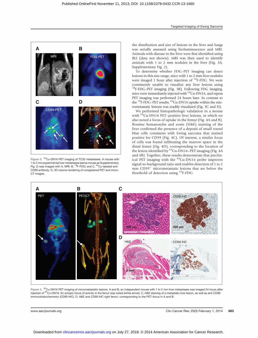
the distribution and size of lesions in the liver and lungswas serially assessed using bioluminescence and MRI.Animals with disease in the liver were first identified usingBLI (data not shown). MRI was then used to identifyanimals with 1 to 2 mm nodules in the liver (Fig. 3A,Supplementary Fig. 2).
To determine whether FDG–PET imaging can detectlesions in this size range, mice with 1 to 2mm liver noduleswere imaged 1 hour after injection of 18F-FDG. We wereconsistently unable to visualize any liver lesions using18F-FDG–PET imaging (Fig. 3B). Following FDG imaging,mice were immediately injected with 64Cu-DN16, and repeatPET imaging was performed 24 hours later. In contrast tothe 18F-FDG–PET results, 64Cu-DN16 uptake within the mic-rometastatic lesions was readily visualized (Fig. 3C and D).
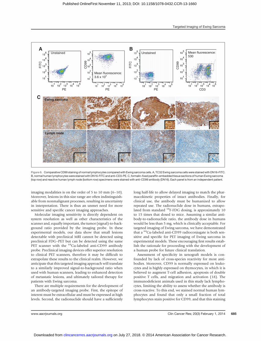
We performed histopathologic validation in a mousewith 64Cu-DN16 PET–positive liver lesions, in which wealso noted a focus of uptake in the femur (Fig. 4A and B).Routine hematoxylin and eosin (H&E) staining of theliver confirmed the presence of a deposit of small roundblue cells consistent with Ewing sarcoma that stainedpositive for CD99 (Fig. 4C). Of interest, a similar focusof cells was found infiltrating the marrow space in thedistal femur (Fig. 4D), corresponding to the location ofthe lesion identified by 64Cu-DN16–PET imaging (Fig. 4Aand 4B). Together, these results demonstrate that preclin-ical PET imaging with the 64Cu-DN16 probe improvessignal-to-background ratio and enables detection of 1 to 2mm CD99þ micrometastatic lesions that are below thethreshold of detection using 18F-FDG.
MRI FDG-PET
CD99-PET CD99-PET/CT
BA
DC
Figure 3. 64Cu-DN16 PET imaging of TC32 metastases. A mouse with1 to 2mmexperimental liver metastases (samemouse as SupplementaryFig. 2) was imaged with A, MRI; B, 18F-FDG; and C, 64Cu-labeled anti-CD99 antibody. D, 3D volume rendering of coregistered PET and micro-CT images.
A C
D
300 µm300 µm
3 mm3 mm
BPET PET/CT H&E CD99 IHC
H&E CD99 IHC
Figure 4. 64Cu-DN16 PET imaging of micrometastatic lesions. A and B, an independent mouse with 1 to 2 mm liver metastases was imaged 24 hours afterinjection of 64Cu-DN16. An ectopic focus of activity in the femur was noted (white arrow). C, H&E staining of a metastatic liver lesion, as well as anti-CD99immunohistochemistry (CD99 IHC). D, H&E and CD99 IHC right femur, corresponding to the PET-focus in A and B.
Targeted Imaging of Ewing Sarcoma
www.aacrjournals.org Clin Cancer Res; 20(3) February 1, 2014 683
on July 27, 2018. © 2014 American Association for Cancer Research. clincancerres.aacrjournals.org Downloaded from
Published OnlineFirst November 11, 2013; DOI: 10.1158/1078-0432.CCR-13-1660

Probe performanceTo more thoroughly define the biodistribution and phar-
macokinetics of the 64Cu-DN16 probe, we performed lon-gitudinal PET imaging and ex vivo validation in a subset ofanimals with subcutaneous andmetastatic xenograft tumors.TAC for major organs and xenograft tumors in one mousedemonstrated a progressive decrease in activity in the liver,heart, and spleen over time (Fig. 5A). The TC32 tumor had aprogressive increase in activity over the 40 hours of imaging(Fig.5A).Therewas likewisea small increase inactivitywithinthe CD99� Kelly tumor over time, perhaps due to enhancedpermeability and retention effects commonly associatedwithtumors (28). However, the absolute activity within the Kellytumor was substantially less than that previously shown inthe TC32 tumor at all points later than 4 hours (Fig. 5A).
Terminal tracer biodistribution studies were performed at24 and48hours after probe injection, with threemice at eachtime point (Fig. 5B). Each mouse had a unilateral TC32tumor,whereasonemousesacrificedat48hourshadbilateraltumorswith a TC32 tumor ononeflank andaKelly tumor onthe contralateral flank. Figures 5B andC validate our imagingresults (Figs. 2–4), demonstrating specificity of probe forCD99þ tumors and accumulation of probe over time.
Comparative staining of lymphocytes and Ewingsarcoma
Within normal tissues, CD99 expression is highly restrict-ed to lymphocytes and endothelial cells. To compare the
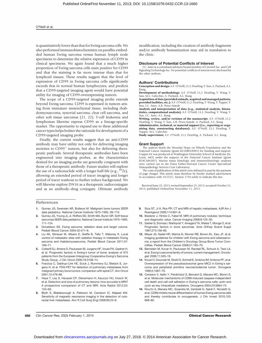
expression of CD99 in Ewing sarcoma cells with that inlymphocytes, we isolated normal human mononuclearcells and stained a normalized number of mononuclearand TC32 Ewing sarcoma cells with the DN16 antibody.Cells were also stained with a CD3 antibody to identifyT cells. CD99 expression was found on both CD3-positiveand -negative lymphocytes. Themeanfluorescence inEwingsarcoma cells (Fig. 6A) was nearly 100-fold higher than thestaining of normal human lymphocytes (Fig. 6B).
To further compare the expression of CD99 in clinicalspecimens, we performed immunohistochemistry on fourindependent human Ewing sarcoma and human lymphnode specimens. Staining in lymph nodes was limited toT-cell zones and occasional cells morphologically consis-tent with dendritic cells (Fig. 6C, bottom), whereas stainingin Ewing sarcoma cells was diffuse, membranous, andqualitatively more intense (Fig. 6C, top).
DiscussionIn the clinic, imaging plays a key role in the management
of patients with solid tumors, helping to determine theprimary site of disease, sites of dissemination, and responseto therapy. Although surgical sampling is used to determinethe extent of local spread in some forms of cancer (e.g.regional lymph node sampling), in most cases imaging isthe onlymodality used to determinewhether distantmetas-tases are present. Despite tremendous advances in the lastfew decades, the clinical limit of detection of current
A B
C
02468
101214161820
484032241680
% ID
/g
Time after injection (h)
TC32 (+)
Kelly (–)
Heart (blood
pool)
Liver (BKG)
0
5
10
15
20
25
30
35
0.150.120.080.240.060.03
Tumor weight (g)
% ID
/g
24 h
48 h
24 h
48 h
% ID
/g
Organs
0
5
10
15
20
25
30
35
Figure 5. 64Cu-DN16 biodistribution. A, TAC generated from serial imaging of a mouse with a subcutaneous TC32 (CD99þ) and Kelly (CD99�) xenografttumor following injection of 64Cu-DN16. B and C, terminal radiotracer biodistribution in mice with subcutaneous xenograft tumors, performed at 24 and48 hours postinjection.
O'Neill et al.
Clin Cancer Res; 20(3) February 1, 2014 Clinical Cancer Research684
on July 27, 2018. © 2014 American Association for Cancer Research. clincancerres.aacrjournals.org Downloaded from
Published OnlineFirst November 11, 2013; DOI: 10.1158/1078-0432.CCR-13-1660
imaging modalities is on the order of 5 to 10 mm (6–10).Moreover, lesions in this size range are often indistinguish-able from nonmalignant processes, resulting in uncertaintyin interpretation. There is thus an unmet need for moresensitive and specific cancer imaging approaches.Molecular imaging sensitivity is directly dependent on
system resolution as well as other characteristics of thescanner and, equally important, the tumor (signal)-to-back-ground ratio provided by the imaging probe. In theseexperimental models, our data show that small lesionsdetectable with preclinical MRI cannot be detected usingpreclinical FDG–PET but can be detected using the samePET scanner with the 64Cu-labeled anti-CD99 antibodyprobe. Preclinical imaging devices offer superior resolutionto clinical PET scanners, therefore it may be difficult toextrapolate these results to the clinical realm. However, weanticipate that this targeted imaging approach will translateto a similarly improved signal-to-background ratio whenused with human scanners, leading to enhanced detectionof metastatic lesions, and ultimately tailored therapy forpatients with Ewing sarcoma.There are multiple requirements for the development of
an antibody-targeted imaging probe. First, the epitope ofinterest must be extracellular andmust be expressed at highlevels. Second, the radionuclide should have a sufficiently
long half-life to allow delayed imaging to match the phar-macokinetic properties of intact antibodies. Finally, forclinical use, the antibody must be humanized to allowrepeated use. The radionuclide dose in humans, extrapo-lated from standard 18F-FDG dosing, is approximately 10to 15 times that dosed to mice. Assuming a similar anti-body-to-radionuclide ratio, the antibody dose in humanswould be less than 5 mg, which is clinically acceptable. Fortargeted imaging of Ewing sarcoma, we have demonstratedthat a 64Cu-labeled anti-CD99 radioconjugate is both sen-sitive and specific for PET imaging of Ewing sarcoma inexperimental models. These encouraging first results estab-lish the rationale for proceeding with the development ofa human probe for future clinical translation.
Assessment of specificity in xenograft models is con-founded by lack of cross-species reactivity for most anti-bodies. Moreover, CD99 is normally expressed on leuko-cytes and is highly expressed on thymocytes, in which it isbelieved to augment T-cell adhesion, apoptosis of doublepositive T cells, and migration and activation (18). Theimmunodeficient animals used in this study lack lympho-cytes, limiting the ability to assess whether the antibody iscross-reactive. To this end, we stained normal human lym-phocytes and found that only a small fraction of totallymphocytes stain positive for CD99, and that this staining
A B
CD
99
CD
99
CD3PEPE
FIT
C
FIT
C
PE
Mean fluorescence:
3.6 x 10⁴
Unstained Unstained
C
Mean fluorescence: 530
Ewing sarcoma
Lymph node
Figure 6. ComparativeCD99 staining of normal lymphocytes comparedwith Ewing sarcoma cells. A, TC32Ewing sarcoma cellswere stainedwithDN16-FITC.B, normal human lymphocyteswere stainedwithDN16-FITCandanti-CD3-PE.C, formalin-fixedparaffin-embedded tissue sections of humanEwing sarcoma(top row) and reactive human lymph node (bottom row) specimens were stained with anti-CD99 antibody (DN16). Each panel is from an independent patient.
Targeted Imaging of Ewing Sarcoma
www.aacrjournals.org Clin Cancer Res; 20(3) February 1, 2014 685
on July 27, 2018. © 2014 American Association for Cancer Research. clincancerres.aacrjournals.org Downloaded from
Published OnlineFirst November 11, 2013; DOI: 10.1158/1078-0432.CCR-13-1660
is quantitatively lower than that for Ewing sarcoma cells.Wealsoperformed immunohistochemistry onparaffin-embed-ded human Ewing sarcoma versus human lymph nodespecimens to determine the relative expression of CD99 inclinical specimens. We again found that a much higherproportion of Ewing sarcoma cells stain positive for CD99and that the staining is far more intense than that forlymphoid tissues. These results suggest that the level ofexpression of CD99 in Ewing sarcoma cells significantlyexceeds that in normal human lymphocytes, and predictsthat a CD99-targeted imaging agent would have potentialutility for imaging of CD99-overexpressing tumors.
The scope of a CD99-targeted imaging probe extendsbeyond Ewing sarcoma. CD99 is expressed in tumors aris-ing from immature mesenchymal tissue, including rhab-domyosarcoma, synovial sarcoma, clear cell sarcoma, andother soft tissue sarcomas (21, 22). T-cell leukemia andlymphomas likewise express CD99 as a lineage-specificmarker. The opportunity to expand use to these additionalcancer types helps bolster the rationale for development of aCD99-targeted imaging probe.
Finally, the current results suggest that an anti-CD99antibody may have utility not only for delivering imagingmoieties to CD99þ tumors, but also for delivering thera-peutic payloads. Several therapeutic antibodies have beenengineered into imaging probes, as the characteristicsdesired for an imaging probe are generally congruent withthose of a therapeutic antibody. Future studies will explorethe use of a radionuclide with a longer half-life (e.g., 89Zr),allowing an extended period of tracer imaging and longerperiod of tracer washout to further reduce background. Wewill likewise explore DN16 as a therapeutic radioconjugateand as an antibody–drug conjugate. Ultimate antibody
modification, including the creation of antibody fragmentsand/or antibody humanization may aid in translation tothe clinic.
Disclosure of Potential Conflicts of InterestJ.C. Aster is a consultant/advisory boardmember of CytomX Inc. and Cell
Signaling Technology Inc. No potential conflicts of interest were disclosed bythe other authors.
Authors' ContributionsConception anddesign:A.F.O’Neill, J.L.J. Dearling, Y. Sun, A. Packard, A.L.KungDevelopment of methodology: A.F. O’Neill, J.L.J. Dearling, Y. Wang, Y.Sun, M.L. Calicchio, A. Packard, A.L. KungAcquisitionofdata (provided animals, acquired andmanagedpatients,provided facilities, etc.):A.F.O’Neill, J.L.J. Dearling, Y.Wang, T. Tupper, Y.Sun, J.C. Aster, A.R. Perez-AtaydeAnalysis and interpretation of data (e.g., statistical analysis, biosta-tistics, computational analysis): A.F. O’Neill, J.L.J. Dearling, Y. Wang, Y.Sun, J.C. Aster, A.L. KungWriting, review, and/or revision of the manuscript: A.F. O’Neill, J.L.J.Dearling, Y. Wang, Y. Sun, A.R. Perez-Atayde, A. Packard, A.L. KungAdministrative, technical, or material support (i.e., reporting or orga-nizing data, constructing databases): A.F. O’Neill, J.L.J. Dearling, T.Tupper, M.L. CalicchioStudy supervision: A.F. O’Neill, J.L.J. Dearling, A. Packard, A.L. Kung
Grant SupportThe authors thank the Hyundai Hope on Wheels Foundation and the
National Cancer Institute (grant R21EB010085) for funding and support.Copper-64 was produced at Washington University School of Medicine (St.Louis, MO) under the support of the National Cancer Institute (grantR24CA86307). Murine tissue histologic and immunohistologic analyseswere carried out in the Dana Farber-Harvard Cancer Center SpecializedHistopathology Services Core Laboratory.
The costs of publication of this article were defrayed in part by the paymentof page charges. This article must therefore be hereby marked advertisementin accordance with 18 U.S.C. Section 1734 solely to indicate this fact.
Received June 25, 2013; revised September 25, 2013; acceptedOctober 21,2013; published OnlineFirst November 11, 2013.
References1. Gurney JG, Swensen AR, Bulterys M. Malignant bone tumors SEER
data pediatrics. National Cancer Institute 1975–1995, 99–110.2. Gurney JG, Young JL Jr, Roffers SD, Smith MA, Bunin GR. Soft tissue
sarcomas SEER data pediatrics. National Cancer Institute 1975–1995,111–124.
3. Donaldson SS. Ewing sarcoma: radiation dose and target volume.Pediatr Blood Cancer 2004;42:471–6.
4. Liu AK, Stinauer M, Albano E, Greffe B, Tello T, Maloney K. Localcontrol of metastatic sites with radiation therapy in metastatic Ewingsarcoma and rhabdomyosarcoma. Pediatr Blood Cancer 2011;57:169–71.
5. Cotterill SJ, Ahrens S, PaulussenM, Jurgens HF, Voute PA, Gadner H,et al. Prognostic factors in Ewing's tumor of bone: analysis of 975patients from the European Intergroup Cooperative Ewing's SarcomaStudy Group. J Clin Oncol 2000;18:3108–14.
6. Franzius C, Daldrup-Link HE, Sciuk J, Rummeny EJ, Bielack S, Jur-gens H, et al. FDG-PET for detection of pulmonary metastases frommalignant primary bone tumors: comparison with spiral CT. AnnOncol2001;12:479–86.
7. Heye T, Ley S, Heussel CP, Dienemann H, Kauczor HU, Hosch W,et al. Detection and size of pulmonary lesions: how accurate is MRI?A prospective comparison of CT and MRI. Acta Radiol 2012;53:153–60.
8. Blyth S, Blakeborough A, Peterson M, Cameron IC, Majeed AW.Sensitivity of magnetic resonance imaging in the detection of colo-rectal liver metastases. Ann R Coll Surg Engl 2008;90:25–8.
9. Sica GT, Ji H, Ros PR. CT and MRI of hepatic metastases. AJR Am JRoentgenol 2000;174:691–8.
10. Biederer J, Hintze C, Fabel M. MRI of pulmonary nodules: techniqueand diagnostic value. Cancer Imaging 2008;8:125–30.
11. Saeter G, Elomaa I,Wahlqvist Y, Alvegard TA,Wiebe T,MongeO, et al.Prognostic factors in bone sarcomas. Acta Orthop Scand Suppl1997;273:156–60.
12. Meyer JS, Nadel HR, Marina N, Womer RB, Brown KL, Eary JF, et al.Imaging guidelines for children with Ewing sarcoma and osteosarco-ma: a report from the Children's Oncology Group Bone Tumor Com-mittee. Pediatr Blood Cancer 2008;51:163–70.
13. Bernstein M, Kovar H, Paulussen M, Randall RL, Schuck A, Teot LA,et al. Ewing's sarcoma family of tumors: currentmanagement. Oncolo-gist 2006;11:503–19.
14. Kovar H, DworzakM, Strehl S, Schnell E, Ambros IM, Ambros PF, et al.Overexpression of the pseudoautosomal gene MIC2 in Ewing's sar-coma and peripheral primitive neuroectodermal tumor. Oncogene1990;5:1067–70.
15. Cerisano V, Aalto Y, Perdichizzi S, Bernard G, Manara MC, Benini S,et al. Molecular mechanisms of CD99-induced caspase-independentcell death and cell-cell adhesion in Ewing's sarcoma cells: actin andzyxin as key intracellular mediators. Oncogene 2004;23:5664–74.
16. Rocchi A, Manara MC, Sciandra M, Zambelli D, Nardi F, Nicoletti G,et al. CD99 inhibits neural differentiation of humanEwing sarcomacellsand thereby contributes to oncogenesis. J Clin Invest 2010;120:668–80.
O'Neill et al.
Clin Cancer Res; 20(3) February 1, 2014 Clinical Cancer Research686
on July 27, 2018. © 2014 American Association for Cancer Research. clincancerres.aacrjournals.org Downloaded from
Published OnlineFirst November 11, 2013; DOI: 10.1158/1078-0432.CCR-13-1660
17. Weidner N, Tjoe J. Immunohistochemical profile of monoclonal anti-body O13: antibody that recognizes glycoprotein p30/32MIC2 and isuseful in diagnosing Ewing's sarcoma and peripheral neuroepithe-lioma. Am J Surg Pathol 1994;18:486–94.
18. Choi EY, Park WS, Jung KC, Kim SH, Kim YY, Lee WJ, et al. Engage-ment of CD99 induces up-regulation of TCR and MHC class I and IImolecules on the surface of human thymocytes. J Immunol 1998;161:749–54.
19. Dubois SG, Epling CL, Teague J, Matthay KK, Sinclair E. Flow cyto-metric detection of Ewing sarcoma cells in peripheral blood and bonemarrow. Pediatr Blood Cancer 2010;54:13–8.
20. Asmane I, Watkin E, Alberti L, Duc A, Marec-Berard P, Ray-Coquard I,et al. Insulin-like growth factor type 1 receptor (IGF-1R) exclusivenuclear staining: a predictive biomarker for IGF-1R monoclonal anti-body (Ab) therapy in sarcomas. Eur J Cancer 2012;48:3027–35.
21. Llombart-Bosch A, Navarro S. Immunohistochemical detection ofEWS and FLI-1 proteins in Ewing sarcoma and primitive neuroecto-dermal tumors: comparative analysis with CD99 (MIC-2) expression.Appl Immunohistochem Mol Morphol 2001;9:255–60.
22. Perlman EJ, Dickman PS, Askin FB, Grier HE, Miser JS, Link MP.Ewing's sarcoma–routine diagnostic utilization of MIC2 analysis: aPediatric Oncology Group/Children's Cancer Group Intergroup Study.Hum Pathol 1994;25:304–7.
23. Kimbrel EA, Davis TN, Bradner JE, Kung AL. In vivo pharmacody-namic imaging of proteasome inhibition. Mol Imaging 2009;8:140–7.
24. Dearling JL, Voss SD, Dunning P, Snay E, Fahey F, Smith SV, et al.Imaging cancer usingPET–the effect of the bifunctional chelator on thebiodistribution of a (64)Cu-labeled antibody. Nucl Med Biol 2011;38:29–38.
25. Giubellino A, Woldemichael GM, Sourbier C, Lizak MJ, Powers JF,Tischler AS, et al. Characterization of twomousemodels of metastaticpheochromocytoma using bioluminescence imaging. Cancer Lett2012;316:46–52.
26. Scotlandi K, Benini S, Sarti M, Serra M, Lollini PL, Maurici D, et al.Insulin-like growth factor I receptor-mediated circuit in Ewing's sar-coma/peripheral neuroectodermal tumor: a possible therapeutic tar-get. Cancer Res 1996;56:4570–4.
27. Scotlandi K, Manara MC, Serra M, Marino MT, Ventura S, Garofalo C,et al. Expression of insulin-like growth factor system components inEwing's sarcoma and their association with survival. Eur J Cancer2011;47:1258–66.
28. Matsumura Y, Maeda H. A new concept for macromolecular thera-peutics in cancer chemotherapy: mechanism of tumoritropic accu-mulation of proteins and the antitumor agent smancs. Cancer Res1986;46:6387–92.
Targeted Imaging of Ewing Sarcoma
www.aacrjournals.org Clin Cancer Res; 20(3) February 1, 2014 687
on July 27, 2018. © 2014 American Association for Cancer Research. clincancerres.aacrjournals.org Downloaded from
Published OnlineFirst November 11, 2013; DOI: 10.1158/1078-0432.CCR-13-1660
2014;20:678-687. Published OnlineFirst November 11, 2013.Clin Cancer Res Allison F. O'Neill, Jason L.J. Dearling, Yuchuan Wang, et al.
Cu-Labeled Anti-CD99 Antibody64Targeted Imaging of Ewing Sarcoma in Preclinical Models Using a
Updated version
10.1158/1078-0432.CCR-13-1660doi:
Access the most recent version of this article at:
Material
Supplementary
http://clincancerres.aacrjournals.org/content/suppl/2013/11/11/1078-0432.CCR-13-1660.DC1
Access the most recent supplemental material at:
Cited articles
http://clincancerres.aacrjournals.org/content/20/3/678.full#ref-list-1
This article cites 26 articles, 5 of which you can access for free at:
E-mail alerts related to this article or journal.Sign up to receive free email-alerts
Subscriptions
Reprints and
To order reprints of this article or to subscribe to the journal, contact the AACR Publications Department at
Permissions
Rightslink site. Click on "Request Permissions" which will take you to the Copyright Clearance Center's (CCC)
.http://clincancerres.aacrjournals.org/content/20/3/678To request permission to re-use all or part of this article, use this link
on July 27, 2018. © 2014 American Association for Cancer Research. clincancerres.aacrjournals.org Downloaded from
Published OnlineFirst November 11, 2013; DOI: 10.1158/1078-0432.CCR-13-1660