system breaks on through to the clinical side Going non ...€¦mentation requires the ability to...
27
Full Terms & Conditions of access and use can be found at http://www.tandfonline.com/action/journalInformation?journalCode=ibmg20 Download by: [88.153.6.191] Date: 09 June 2017, At: 15:50 Critical Reviews in Biochemistry and Molecular Biology ISSN: 1040-9238 (Print) 1549-7798 (Online) Journal homepage: http://www.tandfonline.com/loi/ibmg20 Going non-viral: the Sleeping Beauty transposon system breaks on through to the clinical side Michael Hudecek, Zsuzsanna Izsvák, Sandra Johnen, Matthias Renner, Gabriele Thumann & Zoltán Ivics To cite this article: Michael Hudecek, Zsuzsanna Izsvák, Sandra Johnen, Matthias Renner, Gabriele Thumann & Zoltán Ivics (2017): Going non-viral: the Sleeping Beauty transposon system breaks on through to the clinical side, Critical Reviews in Biochemistry and Molecular Biology, DOI: 10.1080/10409238.2017.1304354 To link to this article: http://dx.doi.org/10.1080/10409238.2017.1304354 © 2017 The Author(s). Published by Informa UK Limited, trading as Taylor & Francis Group. Published online: 12 Apr 2017. Submit your article to this journal Article views: 693 View related articles View Crossmark data
Transcript of system breaks on through to the clinical side Going non ...€¦mentation requires the ability to...
Full Terms & Conditions of access and use can be found athttp://www.tandfonline.com/action/journalInformation?journalCode=ibmg20
Download by: [88.153.6.191] Date: 09 June 2017, At: 15:50
Critical Reviews in Biochemistry and Molecular Biology
ISSN: 1040-9238 (Print) 1549-7798 (Online) Journal homepage: http://www.tandfonline.com/loi/ibmg20
Going non-viral: the Sleeping Beauty transposonsystem breaks on through to the clinical side
Michael Hudecek, Zsuzsanna Izsvák, Sandra Johnen, Matthias Renner,Gabriele Thumann & Zoltán Ivics
To cite this article: Michael Hudecek, Zsuzsanna Izsvák, Sandra Johnen, Matthias Renner,Gabriele Thumann & Zoltán Ivics (2017): Going non-viral: the Sleeping Beauty transposon systembreaks on through to the clinical side, Critical Reviews in Biochemistry and Molecular Biology, DOI:10.1080/10409238.2017.1304354
To link to this article: http://dx.doi.org/10.1080/10409238.2017.1304354
© 2017 The Author(s). Published by InformaUK Limited, trading as Taylor & FrancisGroup.
Published online: 12 Apr 2017.
Submit your article to this journal
Article views: 693
View related articles
View Crossmark data
REVIEW ARTICLE
Going non-viral: the Sleeping Beauty transposon system breaks on throughto the clinical side
Michael Hudeceka, Zsuzsanna Izsv�akb, Sandra Johnenc, Matthias Rennerd, Gabriele Thumanne andZolt�an Ivicsd
aMedizinische Klinik und Poliklinik II, Universit€atsklinikum W€urzburg, W€urzburg, Germany; bMobile DNA, Max Delbr€uck Center forMolecular Medicine in the Helmholtz Association (MDC), Berlin, Germany; cDepartment of Ophthalmology, University Hospital RWTHAachen, Aachen, Germany; dDivision of Medical Biotechnology, Paul Ehrlich Institute, Langen, Germany; eD�epartement desNeurosciences Cliniques Service d’Ophthalmologie, Hopitaux Universitaires de Gen�eve, Gen�eve, Switzerland
ABSTRACTMolecular medicine has entered a high-tech age that provides curative treatments of complexgenetic diseases through genetically engineered cellular medicinal products. Their clinical imple-mentation requires the ability to stably integrate genetic information through gene transfer vec-tors in a safe, effective and economically viable manner. The latest generation of Sleeping Beauty(SB) transposon vectors fulfills these requirements, and may overcome limitations associated withviral gene transfer vectors and transient non-viral gene delivery approaches that are prevalent inongoing pre-clinical and translational research. The SB system enables high-level stable genetransfer and sustained transgene expression in multiple primary human somatic cell types,thereby representing a highly attractive gene transfer strategy for clinical use. Here we reviewseveral recent refinements of the system, including the development of optimized transposonsand hyperactive SB variants, the vectorization of transposase and transposon as mRNA and DNAminicircles (MCs) to enhance performance and facilitate vector production, as well as a detailedunderstanding of SB’s genomic integration and biosafety features. This review also provides aperspective on the regulatory framework for clinical trials of gene delivery with SB, and illustratesthe path to successful clinical implementation by using, as examples, gene therapy for age-related macular degeneration (AMD) and the engineering of chimeric antigen receptor (CAR)-modified T cells in cancer immunotherapy.
ARTICLE HISTORYReceived 5 January 2017Revised 22 February 2017Accepted 6 March 2017
KEYWORDSGene therapy vectors;minicircle; eye diseases;chimeric antigen receptor;stable gene transfer
Introduction: vectors in gene therapy andgenetic engineering
The ability to deliver natural or synthetic genes intohuman somatic cell types provides the technologicalbasis for gene therapy to treat inherited and acquiredgenetic diseases, and enables genetic engineering ofcellular medicinal products that are endowed withnovel properties and functions for use as diagnosticand therapeutic tools in medicine. The vast majority(�70%) of gene delivery systems used in ongoing clin-ical trials of gene therapy are based on viral vectors,and only a small proportion of trials utilizes non-viraldelivery systems, mainly non-integrating naked plasmidDNA, that only provide transient gene expression (GeneTherapy Clinical Trials Worldwide, http://www.abedia.com/wiley/vectors.php (Figure 1( A,B)).
This article will focus on non-viral stable gene trans-fer through the Sleeping Beauty (SB) transposon as aviable and ready-to-use alternative to viral gene transfervectors. We will review basic principles, and feature thekey scientific and technologic advances that have beenmade to position SB transposition as a preferred genedelivery system in translational and clinical medicine.The conceptual appeal of SB transposition for genedelivery is illustrated by examples of clinical applica-tions in gene therapy and genetic engineering, andcomplemented by a concise review of regulatoryaspects for clinical implementation.
Viral gene transfer vectors
Viral vector systems are attractive for gene delivery,because viruses have evolved the ability to cross
CONTACT Zolt�an Ivics [email protected] Paul Ehrlich Institute, Paul Ehrlich Str. 51-59, D-63225 Langen, GermanyThis article was originally published with errors. This version has been corrected. Please see Corrigendum. (http://dx.doi.org./10.1080/10409238.2017.1329969)� 2017 The Author(s). Published by Informa UK Limited, trading as Taylor & Francis Group.This is an Open Access article distributed under the terms of the Creative Commons Attribution-NonCommercial-NoDerivatives License (http://creativecommons.org/Licenses/by-nc-nd/4.0/), which permits non-commercial re-use, distribution, and reproduction in any medium, provided the original work is properly cited, and is not altered, transformed,or built upon in any way.
CRITICAL REVIEWS IN BIOCHEMISTRY AND MOLECULAR BIOLOGY, 2017http://dx.doi.org/10.1080/10409238.2017.1304354

through cellular membranes by infection, thereby deliv-ering nucleic acids to target cells. However, some viralvectors, including those derived from adenoviruses oradeno-associated viruses (AAVs), are not equipped forchromosomal integration, and thus remain largely epi-somal. Especially in cycling cells, episomal DNA is grad-ually lost requiring re-administration in vivo in order tosustain a desired level of transgene expression overtime. However, repeated delivery can provoke immuneresponses against vector-encoded proteins [reviewed in(Hartman et al., 2008)], which can manifest as loss ofthe desired therapeutic effect and potentially therapy-related adverse events. In contrast, retroviral vectorsintegrate their therapeutic cargo into the genome, andthus have the potential to confer long-term transgene
expression. Indeed, hematopoietic stem cell (HSC)-based gene therapy with integrating viral vectors hasclearly provided therapeutic benefit in primary immu-nodeficiencies (including SCID-X1, ADA-SCID), thalas-semia and leukodystrophies (Aiuti et al., 2013a; Biffiet al., 2013; Cartier et al., 2009; Cavazzana-Calvo et al.,2010). A concern with retroviral vectors is that chromo-somal integration may be associated with genotoxicityand mutagenic effects elicited by insertion of the vectorinto or near proto-oncogenes (Baum et al., 2004;Deichmann et al., 2007; Hacein-Bey-Abina et al., 2003;Hacein-Bey-Abina et al., 2008; Kustikova et al., 2005).Such risk is especially pronounced with gammaretroviralvectors based on the murine leukemia virus (MLV) thatpreferentially integrate into transcriptional regulatory
Figure 1. Viral and non-viral vectors in currently running gene therapy clinical trials. A) Percent contribution of different vectorsystems of currently running clinical trials. B) List of some of the most popular vector systems employed in clinical trials. The listalso contains the currently running trials with the Sleeping Beauty transposon (see colour version of this figure at www.tandfon-line.com/ibmg).
2 M. HUDECEK ET AL.
elements of active genes (Cattoglio et al., 2010; Cavazzaet al., 2013; De Ravin et al., 2014); in fact, severe adverseevents associated with vector integration have beenobserved in clinical trials for SCID-X1 (Hacein-Bey-Abinaet al., 2003; Hacein-Bey-Abina et al., 2008; Howe et al.,2008; Thrasher et al., 2006), X-CGD (Ott et al., 2006) andWAS (Braun et al., 2014).
Furthermore, HIV-derived lentiviral vectors are poten-tial mutagens due to their biased insertion into tran-scription units (Cavazza et al., 2013). Although lentiviralvectors appear to be safer than gammaretroviral vectorsin gene therapy, recent studies indicate that some HIVintegrations into genes associated with cancer or cellcycle regulation may confer a survival advantage ofHIV-infected cells, and thus a clonal imbalance of HIVintegrations in AIDS patients (Maldarelli et al., 2014;Wagner et al., 2014). A potential technical hurdle thatcan set limitations to vector design for clinical use ofretroviral vectors is that large transgenes may inhibitviral reverse transcription and packaging. Finally, thehigh costs associated with manufacture of clinical-graderetroviral vector batches seem incompatible with theiruse as gene delivery systems in large patient cohorts inroutine medical practice.
As a result, significant efforts have been made tocraft novel gene transfer vectors that exceed thequalities of currently available viral vectors in: i) effi-cacy: i.e. achieve high-level stable gene transfer atlow toxicity to the host cell; ii) safety: i.e. induce lowlevels of genotoxicity and possess a “safe” integra-tion profile, with a high proportion of integrationsinto genomic safe harbors (GSHs); iii) economic via-bility: i.e. are associated with acceptable cost pertreatment, and scalable/exportable vector productionto serve large numbers of patients.
Non-viral gene transfer vectors
Non-viral vector systems, including nucleic acid vectorssuch as plasmid DNA, generally suffer from inefficientcellular delivery, pronounced cellular toxicity and lim-ited duration of transgene expression due to the lack ofgenomic insertion and resulting degradation and/ordilution of the vector in transfected cell populations.Recent developments of non-viral delivery techniques,including liposomal formulations, nanoparticles,advanced electroporation methods such as nucleofec-tion and cell-penetrating peptides can significantlyenhance transfer of nucleic acids into therapeuticallyrelevant cell types. However, just like non-integratingviral systems, non-viral vectors do not provide long-term nuclear maintenance and transgene expression individing cell types such as stem cells. One class of non-
viral vector system that unites favorable characteristicsof integrating viral vectors (i.e. stable chromosomalintegration and long-lasting transgene expression) withthose of non-viral delivery systems (i.e. lower immuno-genicity, enhanced safety profile and reduced costs ofGMP manufacture) is transposon-based gene deliverysystems.
Non-viral gene transfer using the SleepingBeauty transposon
Jumping genes: transposon basics
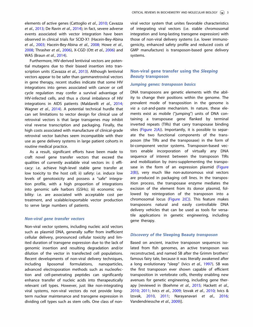
DNA transposons are genetic elements with the abil-ity to change their positions within the genome. Theprevalent mode of transposition in the genome isvia a cut-and-paste mechanism. In nature, these ele-ments exist as mobile (“jumping”) units of DNA con-taining a transposase gene flanked by terminalinverted repeats (TIRs) that carry transposase bindingsites (Figure 2(A)). Importantly, it is possible to separ-ate the two functional components of the trans-poson (the TIRs and the transposase) in the form ofbi-component vector systems. Transposon-based vec-tors enable incorporation of virtually any DNAsequence of interest between the transposon TIRsand mobilization by trans-supplementing the transpo-sase in the form of an expression plasmid (Figure2(B)), very much like non-autonomous viral vectorsare produced in packaging cell lines. In the transpos-ition process, the transposase enzyme mediates theexcision of the element from its donor plasmid, fol-lowed by reintegration of the transposon into achromosomal locus (Figure 2(C)). This feature makestransposons natural and easily controllable DNAdelivery vehicles that can be used as tools for versa-tile applications in genetic engineering, includinggene therapy.
Discovery of the Sleeping Beauty transposon
Based on ancient, inactive transposon sequences iso-lated from fish genomes, an active transposon wasreconstructed, and named SB after the Grimm brothers’famous fairy tale, because it was literally awakened aftera long evolutionary “sleep” (Ivics et al., 1997). SB wasthe first transposon ever shown capable of efficienttransposition in vertebrate cells, thereby enabling newavenues for genetic engineering, including gene ther-apy [reviewed in (Boehme et al., 2015; Hackett et al.,2010; 2011; Ivics et al., 2009; Izsvak et al., 2010; Ivics &Izsvak, 2010, 2011; Narayanavari et al., 2016;Vandendriessche et al., 2009)].
CRITICAL REVIEWS IN BIOCHEMISTRY AND MOLECULAR BIOLOGY 3
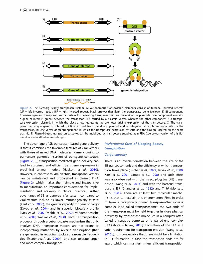
The advantage of SB transposon-based gene deliveryis that it combines the favorable features of viral vectorswith those of naked DNA molecules. Namely, owing topermanent genomic insertion of transgene constructs(Figure 2(C)), transposition-mediated gene delivery canlead to sustained and efficient transgene expression inpreclinical animal models (Hackett et al., 2010).However, in contrast to viral vectors, transposon vectorscan be maintained and propagated as plasmid DNA(Figure 2), which makes them simple and inexpensiveto manufacture, an important consideration for imple-mentation and scale-up in clinical practice. Furtheradvantages of SB as gene-transfer system compared toviral vectors include its lower immunogenicity in vivo(Yant et al., 2000), the greater capacity for genetic cargo(Zayed et al., 2004) and its superior biosafety profile(Ivics et al., 2007; Moldt et al., 2007; Vandendriesscheet al., 2009; Walisko et al., 2008). Because transpositionproceeds through a cut-and-paste mechanism that onlyinvolves DNA, transposon vectors are not prone toincorporating mutations by reverse transcription [thatare generated in retroviral stocks at reasonable frequen-cies (Menendez-Arias, 2009)], and can tolerate largerand more complex transgenes.
Performance facts of Sleeping Beautytransposition
Cargo capacity
There is an inverse correlation between the size of theSB transposon unit and the efficiency at which transpos-ition takes place (Fischer et al., 1999; Izsv�ak et al., 2000;Karsi et al., 2001; Lampe et al., 1998), and such effectwas also observed with the insect piggyBac (PB) trans-poson (Wang et al., 2014) and with the bacterial trans-posons IS1 (Chandler et al., 1982) and Tn10 (Morisatoet al., 1983). There are at least two molecular mecha-nisms that can explain this phenomenon. First, in orderto form a catalytically primed transposon/transposasecomplex (also called transpososome), the two ends ofthe transposon must be held together in close physicalproximity by transposase molecules in a complex oftencalled a synaptic complex or a paired-end complex(PEC) (Ivics & Izsvak, 2015). Formation of the PEC is astrict requirement for transposon excision (Wang et al.,2016b). It is conceivable that there might be a limitationin PEC formation in case the transposon ends are farapart, which can manifest in less efficient transposition
Figure 2. The Sleeping Beauty transposon system. A) Autonomous transposable elements consist of terminal inverted repeats(LIR¼ left inverted repeat; RIR¼ right inverted repeat, black arrows) that flank the transposase gene (yellow). B) Bi-component,trans-arrangement transposon vector system for delivering transgenes that are maintained in plasmids. One component containsa gene of interest (green) between the transposon TIRs carried by a plasmid vector, whereas the other component is a transpo-sase expression plasmid, in which the black arrow represents the promoter driving expression of the transposase. C) The trans-poson carrying a gene of interest (GOI) is excised from the donor plasmid and is integrated at a chromosomal site by thetransposase. D) One-vector or cis-arrangement, in which the transposase expression cassette and the GOI are located on the sameplasmid. E) Plasmid-based transposon cassettes can be mobilized by transposase supplied as mRNA (see colour version of this fig-ure at www.tandfonline.com/ibmg).
4 M. HUDECEK ET AL.

of larger transposons. A strategy to partially rescuetransposition of large transgene constructs is to shortenthe length of DNA that connects the transposon endsoutside the transposon (Izsv�ak et al., 2000). In case of aplasmid vector carrying the transposon unit, this meansshortening the plasmid backbone. By placing the trans-poson TIRs next to one another, bacterial artificial chro-mosomes (BACs) over 100 kb in length have beendemonstrated to transpose at reasonable efficiencies inhuman ES cells (Rostovskaya et al., 2012; Rostovskayaet al., 2013). The second mechanism limiting transpos-ition of large constructs is a suicidal transpositionalmechanism called autointegration (Wang et al., 2014).Autointegration means that following excision thetransposon ends attack a TA dinucleotide inside thetransposon, as opposed to a TA site in the target gen-ome. This will abort the reaction and effectively kill thetransposing element. Since larger transposons containmore potential target sites, they could be particularlyattractive targets for autointegration. Indeed, increasingsize was found to sensitize SB transposition for autoin-tegration (Wang et al., 2014).
Although larger transposons are expected to trans-pose less efficiently, there does not seem to exist anabsolute upper size limit for transposition, or at leastthis limit has not been experimentally reached. This isin sharp contrast to popular viral gene delivery systems,where packaging into infectious viral particles sets astrict limitation to the size of cargo to be incorporated.Indeed, recombinant vectors based on AAV are notcompetent to package vector genomes>5 kb (Hirschet al., 2016; Salganik et al., 2015), and both retroviraland lentiviral vectors undergo a severe loss of titerbeyond a vector size of �8–10 kb (Matrai et al., 2010;Sinn et al., 2005).
Transgene expression level and stability
Any transgene vector system should ideally providelong-term expression of transgenes. By using classical,plasmid-based, non-viral delivery approaches, expres-sion from the extrachromosomal plasmid rapidlydeclines following delivery. Transgenes delivered bynon-viral approaches often form long, repeated arrays(concatemers) that are targets for transcriptional silenc-ing by heterochromatin formation. In addition, long-term expression of transgenes delivered by retroviruseshas been shown to be compromised by transcriptionalsilencing (Jahner et al., 1982; Pannell & Ellis, 2001). Itwas shown that the zinc finger protein ZFP809 bridgesthe integrated proviral MLV genome and the tripartitemotif-containing 28 transcriptional co-repressor inembryonic stem cells (Wolf & Goff, 2009). Thus,
sequence elements in the vector itself can predisposethe cargo for silencing. Cut-and-paste DNA transpos-ition results in a single copy of the transgene per inser-tion locus; thus, concatemer-induced gene silencing isby definition a non-issue. Indeed, Grabundzija et al.found that transposon insertions delivered by the SBsystem only rarely (<2% of all insertions) undergosilencing in HeLa cells (Grabundzija et al., 2010), indicat-ing that it is unlikely that certain sequence motifs in thetransposon vector are recognized by mediators ofsilencing in the cell.
An additional factor that may provoke transgenesilencing is the cargo DNA, particularly the type of pro-moter used to drive expression of the gene of interest.Indeed, it was previously shown that transgene con-structs delivered into mouse cells using SB transpositioncan be subject to epigenetic regulation by CpG methy-lation. A determinant of epigenetic modifications of theintegrating transposon vector was found to be thecargo transgene construct itself, with viral promoter ele-ments playing a major role (Garrison et al., 2007).However, with careful promoter choice, several studieshave established that SB-mediated transposition pro-vides long-term expression in vivo. Notably, stable trans-gene expression from SB vectors was seen in mice aftergene delivery in the liver (Aronovich et al., 2009; Krenet al., 2009; Ohlfest et al., 2005b; Yant et al., 2000), lung(Belur et al., 2003; Liu et al., 2004), brain (Ohlfest et al.,2005a) and blood after hematopoietic reconstitutionin vivo (M�at�es et al., 2009; Xue et al., 2009). Thus, itappears that SB transposon vectors have the capacity toprovide long-term expression of transgenes bothin vitro and in vivo.
Sleeping Beauty transposition in pre-clinicalmodels
In ex vivo gene delivery, the therapeutic gene vector isintroduced into a selected cell population that hadbeen isolated from a donor, and the genetically engi-neered cells are transplanted into a patient. Dependingon whether the donor is the patient itself or anotherperson, we differentiate between autologous or allo-geneic cell products, respectively. Unlike viruses, trans-posons are not equipped to cross cell membranesthrough infection. Thus, it is necessary to combine thetransposon vectors composed of naked nucleic acids(DNA and mRNA) with technologies capable of efficientdelivery of these non-viral vectors into cells. Since theefficiency of transposition is dependent on the effi-ciency of uptake of the introduced nucleic acids intothe cell nuclei, delivery is a rate-limiting factor in trans-position, and is thus of paramount importance. In
CRITICAL REVIEWS IN BIOCHEMISTRY AND MOLECULAR BIOLOGY 5
principle, any technology developed for transferringnucleic acids into cells can be combined with transposonvectors. Unfortunately, there is no generally applicablemethod, and procedures must be established for eachcell type. In hard-to-transfect cells, including primaryhuman cell types, delivery of transposon-based vectorscan be significantly facilitated by nucleofection, a pro-cedure based on electroporation that transfers nucleicacids directly into the nucleus. Indeed, nucleofectionfacilitated transposition in CD34þ HSCs (Hollis et al.,2006; Izsvak et al., 2009; M�at�es et al., 2009; Sumiyoshiet al., 2009; Xue et al., 2009), primary T cells (Gogol-Doring et al., 2016; Huang et al., 2008, 2010; Singh et al.,2008) and human embryonic stem cells (Orban et al.,2009; Wilber et al., 2007). Importantly, in the context ofthe hematopoietic system, this ex vivo gene delivery pro-cedure apparently did not compromise the potential oftransposon-marked CD34þ cells to differentiate normallyinto the erythroid, megakaryocytic, granulocyte/mono-cyte/macrophage (M�at�es et al., 2009) as well as into theCD4þCD8þ T, CD19þ B, CD56þCD3� NK, and CD33þ
myeloid lineages (Xue et al., 2009). The robustness andfeasibility of this non-viral, transposon-based proceduresignificantly facilitates clinical realization of ex vivo stemcell therapy for the treatment of hematopoietic disordersand cancer, and is already successfully applied in humans(Kebriaei et al., 2016).
Refinement of Sleeping Beauty for clinicalapplications
There were three major areas of development andrefinement of SB technology bearing paramountimportance for clinical translation: i) increasing genetransfer rate and lowering toxicity; ii) defining suitablevectors to encode transposase and transposon;iii) establishing a biosafety profile that satisfied require-ments for clinical use.
Enhancing Sleeping Beauty’s performance index
Hyperactive SB transposases
In evolutionary terms, the SB transposon was a successfulelement with the ability of colonizing several fishgenomes millions of years ago (Ivics et al., 1996).However, even successful transposons have not beenselected for the highest possible activity. On the contrary,there is likely a strong selective pressure to avoid inser-tional mutagenesis of essential genes of their host. In anattempt to derive hyperactive transposase variants foradvanced genetic engineering, amino acid substitutions
spanning almost the entire SB transposase polypeptidehave been screened for eliciting a change in catalyticactivity. These amino acid replacements were conductedeither by systematic alanine-scanning (Wang et al.,2016b; Yant et al., 2004), by “transplanting” single aminoacids or small (2–7 aa) blocks of amino acids from relatedtransposases (Baus et al., 2005; Geurts et al., 2003; Zayedet al., 2004), and by replacement of selected amino acidresidues based on charge (Zayed et al., 2004). Theseapproaches generated transposase variants with i) nochange in activity; ii) reduced activity or iii) a relativelymodest increase of transposition activity. A second-gen-eration SB transposase called SB11 contains five aminoacid replacements (selected based on a phylogeneticcomparison to active Tc1/mariner transposases) over thefirst-generation transposase (Geurts et al., 2003). SB11 isabout 3-fold more active than the first-generation SBtransposase, and has been primarily employed in cur-rently running clinical trials based on CAR-engineered Tcells (Kebriaei et al., 2016) (Figure 1(B)).
Surprisingly, some combinations of hyperactivemutations were found to result in a significant reduc-tion of activity. Nevertheless, screening of a library ofpossible combinations yielded SB transposase variantswith significantly enhanced activities (M�at�es et al.,2009). The most hyperactive SB transposase versioncurrently available, SB100X, displays a �100-foldhyperactivity when compared to the originally resur-rected transposase (M�at�es et al., 2009). The hyper-activity of SB100X cannot be explained by alteredtransposase stability, nor by increased binding to thetransposon TIRs; instead, the particular combination ofmutations in SB100X appears to affect the foldingproperties of the transposase (M�at�es et al., 2009). TheSB100X transposase enables highly efficient germlinetransgenesis in relevant mammalian models, includingmice, rats, rabbits and pigs (Ivics et al., 2014a, 2014b,2014c). Moreover, the use of the SB100X systemyielded robust gene transfer efficiencies into humanHSCs (M�at�es et al., 2009; Xue et al., 2009), mesenchy-mal stem cells, muscle stem/progenitor cells (myo-blasts), iPSCs (Belay et al., 2010) and T cells (Jin et al.,2011). These cells are relevant targets for stem cellbiology and for regenerative medicine and gene- andcell-based therapies of complex genetic diseases.Thus, the SB100X hyperactive transposase holds greatpromise for of ex vivo and in vivo gene therapies.
Optimized transposons and transposon donorvectors
The first-generation SB transposon vector (called pT)was based on a naturally occurring sequence originally
6 M. HUDECEK ET AL.

isolated from the Tanichthys albonubes genome (Ivicset al., 1997). As with the transposase, although to alesser extent, mutagenesis of the SB TIR sequences hasbeen undertaken with the aim to increase the efficiencyof transposition. For example, flanking transgenes bytwo left TIRs of SB (instead of the canonical arrange-ment of one left TIR (LIR) and one right TIR (RIR) (Figure2(A))) was shown to enhance transposition by �3-fold,likely due to the presence of a transpositional enhancersequence located in LIR (Izsv�ak et al., 2002).Replacement of four base pairs in RIR and flanking bothTIRs by a doublet of TAs (i.e. by TATA sequences asopposed to the canonical TA sites) resulted in �3-foldincrease in transposition over pT (Cui et al., 2002).Incorporation of a multi-cloning-site consisting of sev-eral unique restriction enzymes sites available for clon-ing genes of interests into the optimized transposonsequences led to the pT2-generation SB transposonvector, which has become the most popular SB trans-poson vector in the scientific community. A combin-ation of the 2-left-TIR and 2-TA arrangements led to avector called pT3 (Yant et al., 2004). Finally, optimizationof SB transposase binding to its internal binding siteswithin the transposon TIRs by cyclic amplification andselection of targets (CASTing) recently yielded pT4 withmodest hyperactivity (Wang et al., 2016b). In addition toenhancing transposition per se, SB vectors of enhancedutility have been devised, for example by including cas-settes for constitutive or inducible expression of anygene of interest in combination with different antibioticselection markers and fluorescent reporters (Kowarzet al., 2015).
Vectorization of Sleeping Beauty components
Plasmid DNA to encode SB transposase andtransposon
The typical setup for delivery of the SB transposonsystem into cells is supplying the two components ofthe vector system (i.e. the engineered transposon car-rying a gene of interest and a transposase source) asconventional plasmids. Although both componentscan be placed on a single plasmid (Mikkelsen et al.,2003) (Figure 2(D)), this arrangement has not becomepopular, mainly because it does not allow carefultitration of transposase expression required for opti-mal transposition at a certain transposon dose.Because optimal transposon-to-transposase ratio cansubstantially vary dependent on cell type, most inves-tigators opt for the use of the 2-plasmid vector sys-tem (Figure 2(B)). Two recent developments that
address both the efficiency and safety of SB genedelivery are the use of mRNA-encoded SB100X andminicircle (MC) vectors to encode transposase/trans-poson. Both are key milestones toward the clinicaluse of SB transposition.
mRNA as a transient source of SB transposase
The transposase expression plasmid that is typicallyused to provide a transposase source in cultured celllines can be replaced by mRNA synthesized in in vitrotranscription reactions (Figure 2(E)). Co-delivery of SBtransposase-encoding mRNA with an SB transposonplasmid to somatic cells was originally tested in a mam-malian cell line in vitro and in the mouse liver in vivo,using an early version of the SB transposase, SB11(described above) (Wilber et al., 2006, 2007). By apply-ing mRNA for intracellular delivery in therapeuticallyrelevant cells ex vivo, some hurdles of gene transfer typ-ical for DNA-based vectors can be avoided. For example,nucleofection of primary human cells, including HSCsand T cells, with mRNA was shown to cause significantlyreduced cellular toxicity as compared to nucleofectionwith plasmid DNA (Monjezi et al., 2016; Wiehe et al.,2007). Second, upon transfection, mRNA translocatesinto the cytoplasm where it is readily available for thehost translational machinery and protein production.Finally, the implementation of an mRNA source for tran-sient delivery of the SB transposase increases the biosaf-ety of this approach, as mRNA does not bear the risk ofchromosomal integration. In contrast, it is known thattransfection of plasmid DNA is associated with a small,but non-negligible chance of spontaneous, illegitimatevector integration into the host genome (Wang et al.,2004). Genomic integration of the SB transposase cod-ing sequence into the genome represents a finite risk ina gene therapy application, because such event couldlead to genomic instability due to prolonged anduncontrollable transposase expression resulting in con-tinuous remobilization of the already integrated SBtransposon.
Minicircle DNA – minimalistic transposase andtransposon expression cassettes
A recent addition to the development of SB vectorswith enhanced utility for clinical applications is the useof MC vectors as carriers of the SB transposon compo-nents. The MC technology allows for a significant reduc-tion of SB vector size by removing most of thebackbone sequences from parental plasmids (Sharmaet al., 2013). The presence of bacterial backbone ele-ments is typically required for vector propagation in
CRITICAL REVIEWS IN BIOCHEMISTRY AND MOLECULAR BIOLOGY 7
Escherichia coli, but completely redundant or evenundesired for clinical applications. The first evidentadvantage of using MC vectors over plasmids is relatedto increased cell survival rates following nucleofectionof human T cells (Monjezi et al., 2016) (describedin detail below in “Cancer immunotherapy with tumor--reactive CAR T-cells”). Unmethylated CpG motifs, whichare highly enriched in the bacterial backbone ofexogenously delivered plasmids, were shown to triggerstrong inflammatory responses through toll-like recep-tor-9 (Chuang et al., 2002; Hemmi et al., 2000) and/orinterferon induction (Huerfano et al., 2013). Activationof these cellular sensors might be a conceivable explan-ation for the observed cytotoxicity and thus for the lossof a large fraction of cells transfected with plasmid DNAfrom the rest of the proliferating cell culture. Indeed,removal of CpG motifs from plasmid DNA vectors wasdemonstrated to reduce inflammatory responses uponpulmonary gene delivery (Hyde et al., 2008), and vectorCpG methylation was able to lower immune responsive-ness towards “non-self” DNA and led to delayed clear-ance of transfected cells in vivo (Reyes-Sandoval & Ertl,2004). Along with decreased levels of cytotoxicity,nucleofection of T cells with SB transposon componentssupplied as MCs resulted in enhanced transient genedelivery and more efficient stable genome modificationas compared to conventional plasmid vectors (Monjeziet al., 2016) (described in detail below in “Cancerimmunotherapy with tumor-reactive CAR T-cells”).
The MC components are likely more efficient intransfection than plasmids because, due to their smallersize, they cross cellular membranes more efficientlythan plasmids (Chabot et al., 2013; Darquet et al., 1997).Second, MC vectors may support enhanced transcrip-tion of transgene cassettes. Indeed, enhanced and sus-tained transgene expression has been seen in episomalgene therapy applications with MC vectors with con-comitant gene silencing commencing rapidly afterhydrodynamic injection of parental plasmid vectorsin vivo (Chen et al., 2003; Osborn et al., 2011). Covalentlinkage of bacterial DNA sequences to a eukaryoticexpression cassette has been suggested to facilitate thespreading of repressive chromatin formed primarily onthe bacterial backbone, leading in turn to rapid loss oftransgene expression from plasmid DNA vectors (Chenet al., 2004, 2008). Finally, the elevated levels of trans-position observed with MC vectors are likely supported,at least in part, by the relatively short, �200 bp distancebetween the SB transposon ends in the MC-based trans-poson vector, owing to the depletion of the bacterialplasmid backbone. Indeed, SB transposition was shownto be far more efficient when the length of DNAsequence outside the transposon unit was shortened,
likely by aiding transposon/transposase complex forma-tion, as discussed above (Izsv�ak et al., 2000). In additionto efficacy, the MC technology also offers biosafetyadvantages over conventional plasmid DNA vectors.Namely, the absence of bacterial plasmid backbone ele-ments in therapeutic vectors is highly relevant in clinicalapplications, because antibiotic resistance genesincluded in a therapeutic cell product may raise safetyconcerns.
Establishing a biosafety profile
Genome-wide distribution of Sleeping Beautyinsertions
With any vector that integrates into chromosomes in asemi-random manner comes the potential risk of inser-tional mutagenesis leading to transcriptional activationor inactivation of cellular genes (Baum et al., 2004).Such genotoxic effects can have devastating conse-quences for the cell and the whole organism, includingthe development of cancer, as discussed above in “Viralgene transfer vectors”.
SB displays considerable specificity in target siteselection at the primary DNA sequence level in that TAdinucleotides are near-obligate target sites (Vigdal et al.,2002). A palindromic AT-repeat consensus sequencewith bendability and hydrogen bonding potential wasfound to constitute preferred target sites (Vigdal et al.,2002). It was later shown that a characteristic deform-ation of the DNA sequence may be a recognition signalfor target selection (Liu et al., 2005). Although character-ization of the target site selection properties of differentvector systems still falls short of predicting the actualrisk of insertional oncogenesis in a clinical trial, it ishighly useful for ranking the different vector types anddesigns according to their genotoxic potential (Naldini,2011). We have previously undertaken a comparativestudy addressing target site selection properties ofthe SB and PB transposons as well as MLV-derivedgammaretroviral and HIV-derived lentiviral systems inprimary human CD4þ T cells. Our bioinformatic analyzesincluded mapping against the T cell genome withrespect to proximity to genes, transcriptional start sites(TSSs), CpG islands, DNaseI hypersensitive sites, chroma-tin marks and transcriptional status of genes. The SBtransposon displayed the least deviation from randomwith respect to genome-wide distribution: no apparentbias was seen for either heterochromatin marks oreuchromatin marks and only a weak correlation withtranscriptional status of targeted genes was detected(Gogol-Doring et al., 2016). Our compiled datasets alsoallowed us to rank these vector systems with respect to
8 M. HUDECEK ET AL.
their projected relative “safety” based on the frequen-cies of integration into GSHs that were previously pro-posed to satisfy five criteria: (i) distance of at least 50 kbfrom the 5’-end of any gene, (ii) distance of at least300 kb from any cancer-related gene, (iii) distance of atleast 300 kb from any microRNA (miRNA), (iv) locationoutside a transcription unit and (v) location outsideultraconserved regions of the human genome(Papapetrou et al., 2011; Sadelain et al., 2012). Our ana-lyzes collectively established a favorable integrationprofile of the SB transposon, suggesting that SB mightbe safer for therapeutic gene delivery than the integrat-ing viral vectors that are currently used in clinical trials.Importantly, no SB-associated adverse effects have beenobserved in preclinical animal studies (Fernando &Fletcher, 2006; Hackett et al., 2010; Ivics & Izsvak, 2006;Izsvak et al., 2010).
Vector-associated transcriptional activity
Integration of a vector into a gene or its regulatory ele-ments can knock out the gene, overexpress the gene oralter its spatio/temporal expression pattern. The majorunderlying mechanism, by which insertional mutagen-esis manifests in gene therapy appears to be transcrip-tional trans-activation of oncogenes (Baum et al., 2004;Deichmann et al., 2007) by the retroviral long terminalrepeats (LTRs) that carry strong enhancer/promoter ele-ments. Due to their intrinsic preference for integratinginto TSSs (see above), MLV-derived gammaretroviralvectors are especially prone to transcriptionally upregu-late endogenous genes. Indeed, it has been demon-strated that MLV-based gammaretroviral insertions,although they target GSHs>2-fold more frequentlythan HIV-based lentiviral vectors (Gogol-Doring et al.,2016), were approximately 3-fold more likely to triggertransformation of primary HSCs in a cell-based immor-talization assay (Modlich et al., 2009). This suggests thatan MLV insertion next to a TSSs tends to be more geno-toxic than an HIV insertion in a gene body, in line withthe expectation that gain-of-function due to oncogeneupregulation has a more profound effect on cellularhomeostasis than loss-of-function due to knockoutmutations (unless the affected gene is haplo-insuffi-cient). To ameliorate vector-induced insertional onco-genesis, second-generation, self-inactivating (SIN)vectors have been developed for both gammaretroviraland lentiviral vectors. These vectors lack transcriptionalenhancer/promopter elements in their LTRs(Schambach et al., 2006, 2007), and therefore they areexpected to have an enhanced safety profile in clinicalapplications. Indeed, SIN vectors result in reduced
clonal immortalization in cell-based genotoxicity assays(Modlich et al., 2009).
Unlike the LTRs of retroviruses, the TIRs of SB vectorshave negligible enhancer/promoter activity (Moldt et al.,2007; Walisko et al., 2008). The left TIR of SB is separatedfrom the transposase coding sequence by a 5’-UTR of160 bp stretch of DNA with no apparent function in thetransposition reaction (Zayed et al., 2004). As measuredby transient reporter assays, transcription driven by the5’-UTR of SB is �18-fold higher than transcription of apromoter-less sequence, and �5-fold higher than tran-scription driven by a TATA-box minimal promoter(Walisko et al., 2008). Importantly, the 5’-UTR is not pre-sent in SB transposon vectors (because it is dispensablefor transposition). Thus, contemporary SB vectors aretranscriptionally neutral, and therefore it is expectedthat their mutagenic potential will largely depend ontheir cargo (including the transcriptional regulatory ele-ments that drive transgene expression).
Beyond the genomic integration pattern and thetranscriptional activities associated with a given vector,a third determinant of risk stemming from vector inte-gration is the type of cell, in which therapeutic genetransfer is executed. In contrast to HSC-based genetherapy, leukemia was never observed in preclinical ani-mal models or clinical trials involving gene transfer intoperipheral blood-derived T lymphocytes (Newrzelaet al., 2008; Recchia et al., 2006). Thus, mature T cellsseem to be less susceptible to transformation by geno-toxic events than are HSCs. It is likely that other typesof terminally differentiated cells, including retinal pig-ment epithelium (RPE) cells isolated from the eye arealso more refractory to oncogenic transformation thanstem cells (Balaggan et al., 2012), thereby presentingattractive targets for Phase I clinical trials.
Clinical application of Sleeping Beauty genedelivery
Overview
The past few years have seen a steadily growing interestin applying SB transposition in gene therapy to provideinnovative and potentially curative treatments for gen-etic disorders [reviewed in (Boehme et al., 2015; Essneret al., 2005; Hackett et al., 2005, 2010, 2011; Ivics &Izsvak, 2006, 2011; Izsvak & Ivics, 2004; Izsvak et al.,2010)]. Prime examples for the use of SB in gene therapyinclude the treatment of hematologic disorders, lyso-somal storage diseases, pulmonary disorders, dermato-logic diseases, a variety of metabolic disorders,neurologic disorders, muscle disorders and cancer(Table 1).
CRITICAL REVIEWS IN BIOCHEMISTRY AND MOLECULAR BIOLOGY 9
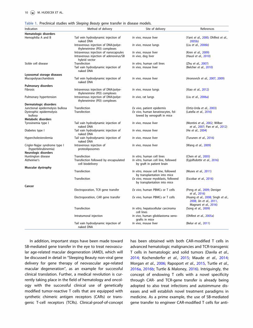
In addition, important steps have been made towardSB-mediated gene transfer in the eye to treat neovascu-lar age-related macular degeneration (AMD), which willbe discussed in detail in “Sleeping Beauty non-viral genedelivery for gene therapy of neovascular age-relatedmacular degeneration”, as an example for successfulclinical translation. Further, a medical revolution is cur-rently taking place in the field of hematology and oncol-ogy with the successful clinical use of geneticallymodified tumor-reactive T cells that are equipped withsynthetic chimeric antigen receptors (CARs) or trans-genic T-cell receptors (TCRs). Clinical-proof-of-concept
has been obtained with both CAR-modified T cells inadvanced hematologic malignancies and TCR-transgenicT cells in hematologic and solid tumors (Davila et al.,2014; Kochenderfer et al., 2015; Maude et al., 2014;Morgan et al., 2006; Rapoport et al., 2015, Turtle et al.,2016a, 2016b; Turtle & Maloney, 2016). Intriguingly, theconcept of endowing T cells with a novel specificitythrough CAR- and TCR-gene transfer is already beingadopted to also treat infections and autoimmune dis-eases and will establish novel treatment paradigms inmedicine. As a prime example, the use of SB-mediatedgene transfer to engineer CAR-modified T cells for anti-
Table 1. Preclinical studies with Sleeping Beauty gene transfer in disease models.Indication Method of delivery Site of delivery References
Hematologic disordersHemophilia A and B Tail vein hydrodynamic injection of
naked DNAIn vivo, mouse liver (Yant et al., 2000; Ohlfest et al.,
2005b)Intravenous injection of DNA/polye-
thyleneimine (PEI) complexesIn vivo, mouse lungs (Liu et al., 2006b)
Intravenous injection of nanocapsules In vivo, mouse liver (Kren et al., 2009)Intravenous injection of adenovirus/SB
hybrid vectorIn vivo, dog liver (Hausl et al., 2010)
Sickle cell disease Transfection In vitro, human cell lines (Zhu et al., 2007)Tail vain hydrodynamic injection of
naked DNAIn vivo, mouse liver (Belcher et al., 2010)
Lysosomal storage diseasesMucopolysaccharidosis Tail vein hydrodynamic injection of
naked DNAIn vivo, mouse liver (Aronovich et al., 2007, 2009)
Pulmonary disordersFibrosis Intravenous injection of DNA/polye-
thyleneimine (PEI) complexesIn vivo, mouse lungs (Xiao et al., 2012)
Pulmonary hypertension Intravenous injection of DNA/polye-thyleneimine (PEI) complexes
In vivo, rat lungs (Liu et al., 2006a)
Dermatologic disordersJunctional epidermolysis bullosa Transfection Ex vivo, patient epidermis (Ortiz-Urda et al., 2003)Dystrophic epidermolysis
bullosaTransfection Ex vivo, human keratinocytes, fol-
lowed by xenograft in mice(Latella et al., 2016)
Metabolic disordersTyrosinemia type I Tail vain hydrodynamic injection of
naked DNAIn vivo, mouse liver (Montini et al., 2002; Wilber
et al., 2007; Pan et al., 2012)Diabetes type I Tail vain hydrodynamic injection of
naked DNAIn vivo, mouse liver (He et al., 2004)
Hypercholesterolemia Tail vain hydrodynamic injection ofnaked DNA
In vivo, mouse liver (Turunen et al., 2016)
Crigler-Najjar syndrome type I(hyperbilirubinemia)
Intravenous injection ofproteoliposomes
In vivo, mouse liver (Wang et al., 2009)
Neurologic disordersHuntington disease Transfection In vitro, human cell lines (Chen et al., 2005)Alzheimer’s Transfection followed by encapsulated
cell biodeliveryIn vitro, human cell line, followed
by graft in patient brain(Eyjolfsdottir et al., 2016)
Muscular dystrophyTransfection In vitro, mouse cell line, followed
by transplantation into mice(Muses et al., 2011)
Transfection Ex vivo, mouse myoblasts, followedby transplantation into mice
(Escobar et al., 2016)
CancerElectroporation, TCR gene transfer Ex vivo, human PBMCs or T cells (Peng et al., 2009; Deniger
et al., 2016)Electroporatiion, CAR gene transfer Ex vivo, human PBMCs or T cells (Huang et al., 2008; Singh et al.,
2008; Jin et al., 2011,Magnani et al., 2016)
Transfection In vitro, hepatocellular carcinomacell lines
(Song et al., 2009)
Intratumoral injection In vivo, human glioblastoma xeno-grafts in mice
(Ohlfest et al., 2005a)
Tail vain hydrodynamic injection ofnaked DNA
In vivo, mouse liver (Belur et al., 2011)
10 M. HUDECEK ET AL.

cancer therapy will be discussed in detail in “Cancerimmunotherapy with tumor-reactive CAR T-cells”.
Sleeping Beauty non-viral gene delivery for genetherapy of neovascular age-related maculardegeneration
TargetAMD is an international consortium of univer-sities, research institutes and commercial organizationsfunded by the European Commission to execute aPhase Ia/IIb clinical trial for the treatment of neovascu-lar AMD by transplantation of genetically modified,autologous retinal pigment epithelial (RPE) or iris pig-ment epithelial (IPE) cells that overexpress pigment epi-thelial-derived factor (PEDF) to the subretinal space ofthe eye. Specifically, RPE or IPE cells isolated from theperipheral retina or obtained from an iris biopsy of apatient will be transfected with an SB transposon vectorcarrying a PEDF expression cassette and transplantedback into the same patient during one surgical sessionlasting about 60min.
AMD is a major cause of acquired irreversibleblindness in adults
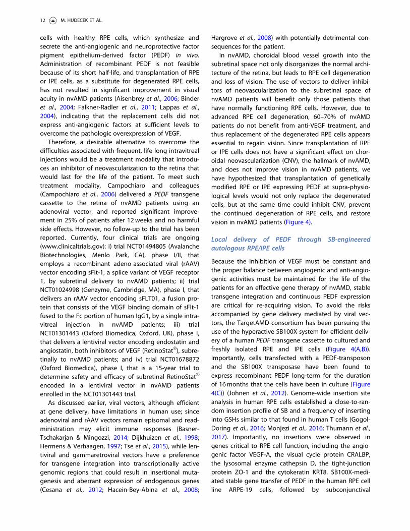
AMD refers to the degeneration of RPE cells in the mac-ula area (Figure 3), which is essential for central vision.With over 13 million people affected, AMD is the fourthmost common cause of blindness after cataract,
retinopathy of prematurity, and glaucoma in the worldand the leading cause of irreversible blindness in peo-ple aged>50 years in developed countries (Velez-Montoya et al., 2014). There are two forms of AMD:avascular AMD (avAMD) or geographic atrophy (GA)and neovascular AMD (nvAMD), which is characterizedby the subretinal invasion of choroidal vessels. WhereasavAMD is a slow progressing disorder, in which photo-receptor degeneration follows RPE cell degeneration,nvAMD progresses rapidly to blindness following RPEcell degeneration. No treatments are available foravAMD.
Limited clinical benefit of AMD fromantibody-mediated VEGF blockade
Since overexpression of vascular endothelial growth fac-tor (VEGF) has been shown to be responsible for thedevelopment of subretinal neovascularization underly-ing the development of nvAMD (Ohno-Matsui et al.,2001; Velez-Montoya et al., 2014), its current treatment,which is effective in 30–40% of patients, is based onmonthly, life-long, intravitreal injections of inhibitors ofVEGF. The three most commonly used anti-VEGFs arebevacizumab (AvastinVR , Genentech, San Francisco, CA),its Fab fragment ranibizumab (LucentisVR , Genentech)and aflibercept (EyleaVR , Regeneron, Tarrytown, NY),which is a recombinant fusion protein consisting of theextracellular binding domains of human VEGF receptors1 and 2 fused to the Fc portion of the human IgG1immunoglobulin.
The two major limitations of anti-VEGFs are the lackof improvement of vision in 60–70% of patients andadverse effects (Nesmith et al., 2014) including morerapid progression of GA (Enslow et al., 2016), increasedintraocular pressure, retinal detachment, endophthalmi-tis, photoreceptor cell death and thinning of the innerneuronal layer of the retina (Saint-Geniez et al., 2008;Scott & Bressler, 2013). In addition to the adverseeffects, the issues associated with logistics of blind orlow-vision patients traveling to a clinic on a monthlybasis result in a significant number of patients discon-tinuing treatment, which has been reported to be from57% over five years (Boulanger-Scemama et al., 2015) toas high as 71% within 24months (Lad et al., 2014).Therefore, it is evident that new approaches arerequired for the efficient treatment of nvAMD.
One-shot AMD treatment: Sleeping Beauty-basedPEDF gene therapy
The evidence that nvAMD is caused by RPE cell degen-eration has engendered the hypothesis that the condi-tion could be treated by replacing the degenerated RPE
Figure 3. Neovascular age-related macular degeneration(nvAMD). In nvAMD choroidal neovascularization (CNV) due toan imbalance of pro- and anti-angiogenic factors like VEGFand PEDF leads to severe hemorrhages damaging the retinalpigment epithelium (RPE) and the neural retina. The fundu-scopic image illustrates such a bleeding (arrow) in advancednvAMD (see colour version of this figure at www.tandfonline.com/ibmg).
CRITICAL REVIEWS IN BIOCHEMISTRY AND MOLECULAR BIOLOGY 11
cells with healthy RPE cells, which synthesize andsecrete the anti-angiogenic and neuroprotective factorpigment epithelium-derived factor (PEDF) in vivo.Administration of recombinant PEDF is not feasiblebecause of its short half-life, and transplantation of RPEor IPE cells, as a substitute for degenerated RPE cells,has not resulted in significant improvement in visualacuity in nvAMD patients (Aisenbrey et al., 2006; Binderet al., 2004; Falkner-Radler et al., 2011; Lappas et al.,2004), indicating that the replacement cells did notexpress anti-angiogenic factors at sufficient levels toovercome the pathologic overexpression of VEGF.
Therefore, a desirable alternative to overcome thedifficulties associated with frequent, life-long intravitrealinjections would be a treatment modality that introdu-ces an inhibitor of neovascularization to the retina thatwould last for the life of the patient. To meet suchtreatment modality, Campochiaro and colleagues(Campochiaro et al., 2006) delivered a PEDF transgenecassette to the retina of nvAMD patients using anadenoviral vector, and reported significant improve-ment in 25% of patients after 12weeks and no harmfulside effects. However, no follow-up to the trial has beenreported. Currently, four clinical trials are ongoing(www.clinicaltrials.gov): i) trial NCT01494805 (AvalancheBiotechnologies, Menlo Park, CA), phase I/II, thatemploys a recombinant adeno-associated viral (rAAV)vector encoding sFlt-1, a splice variant of VEGF receptor1, by subretinal delivery to nvAMD patients; ii) trialNCT01024998 (Genzyme, Cambridge, MA), phase I, thatdelivers an rAAV vector encoding sFLT01, a fusion pro-tein that consists of the VEGF binding domain of sFlt-1fused to the Fc portion of human IgG1, by a single intra-vitreal injection in nvAMD patients; iii) trialNCT01301443 (Oxford Biomedica, Oxford, UK), phase I,that delivers a lentiviral vector encoding endostatin andangiostatin, both inhibitors of VEGF (RetinoStatVR ), subre-tinally to nvAMD patients; and iv) trial NCT01678872(Oxford Biomedica), phase I, that is a 15-year trial todetermine safety and efficacy of subretinal RetinoStatVR
encoded in a lentiviral vector in nvAMD patientsenrolled in the NCT01301443 trial.
As discussed earlier, viral vectors, although efficientat gene delivery, have limitations in human use; sinceadenoviral and rAAV vectors remain episomal and read-ministration may elicit immune responses (Basner-Tschakarjan & Mingozzi, 2014; Dijkhuizen et al., 1998;Hermens & Verhaagen, 1997; Tse et al., 2015), while len-tiviral and gammaretroviral vectors have a preferencefor transgene integration into transcriptionally activegenomic regions that could result in insertional muta-genesis and aberrant expression of endogenous genes(Cesana et al., 2012; Hacein-Bey-Abina et al., 2008;
Hargrove et al., 2008) with potentially detrimental con-sequences for the patient.
In nvAMD, choroidal blood vessel growth into thesubretinal space not only disorganizes the normal archi-tecture of the retina, but leads to RPE cell degenerationand loss of vision. The use of vectors to deliver inhibi-tors of neovascularization to the subretinal space ofnvAMD patients will benefit only those patients thathave normally functioning RPE cells. However, due toadvanced RPE cell degeneration, 60–70% of nvAMDpatients do not benefit from anti-VEGF treatment, andthus replacement of the degenerated RPE cells appearsessential to regain vision. Since transplantation of RPEor IPE cells does not have a significant effect on chor-oidal neovascularization (CNV), the hallmark of nvAMD,and does not improve vision in nvAMD patients, wehave hypothesized that transplantation of geneticallymodified RPE or IPE expressing PEDF at supra-physio-logical levels would not only replace the degeneratedcells, but at the same time could inhibit CNV, preventthe continued degeneration of RPE cells, and restorevision in nvAMD patients (Figure 4).
Local delivery of PEDF through SB-engineeredautologous RPE/IPE cells
Because the inhibition of VEGF must be constant andthe proper balance between angiogenic and anti-angio-genic activities must be maintained for the life of thepatients for an effective gene therapy of nvAMD, stabletransgene integration and continuous PEDF expressionare critical for re-acquiring vision. To avoid the risksaccompanied by gene delivery mediated by viral vec-tors, the TargetAMD consortium has been pursuing theuse of the hyperactive SB100X system for efficient deliv-ery of a human PEDF transgene cassette to cultured andfreshly isolated RPE and IPE cells (Figure 4(A,B)).Importantly, cells transfected with a PEDF-transposonand the SB100X transposase have been found toexpress recombinant PEDF long-term for the durationof 16months that the cells have been in culture (Figure4(C)) (Johnen et al., 2012). Genome-wide insertion siteanalysis in human RPE cells established a close-to-ran-dom insertion profile of SB and a frequency of insertinginto GSHs similar to that found in human T cells (Gogol-Doring et al., 2016; Monjezi et al., 2016; Thumann et al.,2017). Importantly, no insertions were observed ingenes critical to RPE cell function, including the angio-genic factor VEGF-A, the visual cycle protein CRALBP,the lysosomal enzyme cathepsin D, the tight-junctionprotein ZO-1 and the cytokeratin KRT8. SB100X-medi-ated stable gene transfer of PEDF in the human RPE cellline ARPE-19 cells, followed by subconjunctival
12 M. HUDECEK ET AL.
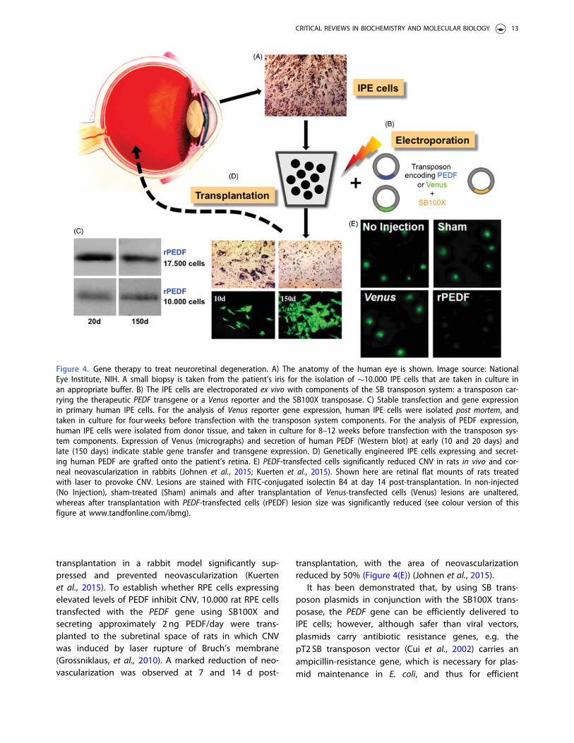
transplantation in a rabbit model significantly sup-pressed and prevented neovascularization (Kuertenet al., 2015). To establish whether RPE cells expressingelevated levels of PEDF inhibit CNV, 10.000 rat RPE cellstransfected with the PEDF gene using SB100X andsecreting approximately 2 ng PEDF/day were trans-planted to the subretinal space of rats in which CNVwas induced by laser rupture of Bruch’s membrane(Grossniklaus, et al., 2010). A marked reduction of neo-vascularization was observed at 7 and 14 d post-
transplantation, with the area of neovascularizationreduced by 50% (Figure 4(E)) (Johnen et al., 2015).
It has been demonstrated that, by using SB trans-poson plasmids in conjunction with the SB100X trans-posase, the PEDF gene can be efficiently delivered toIPE cells; however, although safer than viral vectors,plasmids carry antibiotic resistance genes, e.g. thepT2 SB transposon vector (Cui et al., 2002) carries anampicillin-resistance gene, which is necessary for plas-mid maintenance in E. coli, and thus for efficient
Figure 4. Gene therapy to treat neuroretinal degeneration. A) The anatomy of the human eye is shown. Image source: NationalEye Institute, NIH. A small biopsy is taken from the patient’s iris for the isolation of �10.000 IPE cells that are taken in culture inan appropriate buffer. B) The IPE cells are electroporated ex vivo with components of the SB transposon system: a transposon car-rying the therapeutic PEDF transgene or a Venus reporter and the SB100X transposase. C) Stable transfection and gene expressionin primary human IPE cells. For the analysis of Venus reporter gene expression, human IPE cells were isolated post mortem, andtaken in culture for four weeks before transfection with the transposon system components. For the analysis of PEDF expression,human IPE cells were isolated from donor tissue, and taken in culture for 8–12 weeks before transfection with the transposon sys-tem components. Expression of Venus (micrographs) and secretion of human PEDF (Western blot) at early (10 and 20 days) andlate (150 days) indicate stable gene transfer and transgene expression. D) Genetically engineered IPE cells expressing and secret-ing human PEDF are grafted onto the patient’s retina. E) PEDF-transfected cells significantly reduced CNV in rats in vivo and cor-neal neovascularization in rabbits (Johnen et al., 2015; Kuerten et al., 2015). Shown here are retinal flat mounts of rats treatedwith laser to provoke CNV. Lesions are stained with FITC-conjugated isolectin B4 at day 14 post-transplantation. In non-injected(No Injection), sham-treated (Sham) animals and after transplantation of Venus-transfected cells (Venus) lesions are unaltered,whereas after transplantation with PEDF-transfected cells (rPEDF) lesion size was significantly reduced (see colour version of thisfigure at www.tandfonline.com/ibmg).
CRITICAL REVIEWS IN BIOCHEMISTRY AND MOLECULAR BIOLOGY 13
manufacturing of the plasmid vector. The presence ofantibiotic resistance markers in gene therapy vectors isa matter of concern, since there is a risk that the anti-biotic resistance genes could elicit resistance to patho-genic bacteria by horizontal gene transfer and residuesof antibiotics could contaminate the final product, plac-ing patients with severe hypersensitivity to antibioticsat risk (Solensky, 2003). In fact, regulatory agencies rec-ommend that antibiotic resistance genes be eliminatedfrom gene therapy treatments whenever possible. Thus,the TargetAMD consortium resorted to apply a variantof MC technology in free of antibiotic markers (pFAR)miniplasmids (Marie et al., 2010). Like MCs, pFAR-basedvactors lack antibiotic-resistance genes, thereby signifi-cantly enhancing the safety profile of nonviral genedelivery in clinical settings.
TargetAMD: Phase I clinical trial of SB-based PEDFgene therapy launched in 2017
The ultimate goal of TargetAMD is to perform a PhaseIa/IIb clinical trial in which IPE cells will be isolated froman iris biopsy of a nvAMD patient, transfected with thePEDF gene and transplanted to the subretinal space ofthe same patient during a single surgical session lastingapproximately one hour (Figure 4). Since the isolationof IPE or RPE cells from a patient’s biopsy yields a lim-ited number of cells, and since safety is a major concernof any therapy, and of gene therapy in particular, wehave developed a protocol for the efficient delivery ofthe PEDF gene in as few as 5.000–10.000 freshly isolatedIPE and RPE cells from human donor eyes by vectorizingthe PEDF-encoding SB transposon and the SB100Xtransposase in pFAR miniplasmids (Marie et al., 2010;Thumann et al., 2017). Using this protocol, a standardoperating procedure has been established that i) con-sistently shows highly efficient transfer of the PEDFgene in RPE and IPE cells obtained from donor eyes, ii)enables expression of recombinant PEDF at high levelsof recombinant protein in cultured PEDF-transfectedcells, and iii) allows for sustained transgene expression(for over one year that the cells have been in culture) ingenetically engineered cells. The robustness of this pro-cedure is coupled with salient safety features includinga close-to-random transgene integration profile of theSB transposon in human IPE and RPE cells and the lackof antibiotic resistance genes in vector components.Based on the results described here and approval bythe Swissmedic regulatory agency, TargetAMD willbegin patient recruitment for the phase Ia/IIb clinicaltrial in the spring of 2017 and expects to complete thefirst European in-man clinical trial using SB transposonand pFAR technologies.
Cancer immunotherapy with tumor-reactive CART-cells
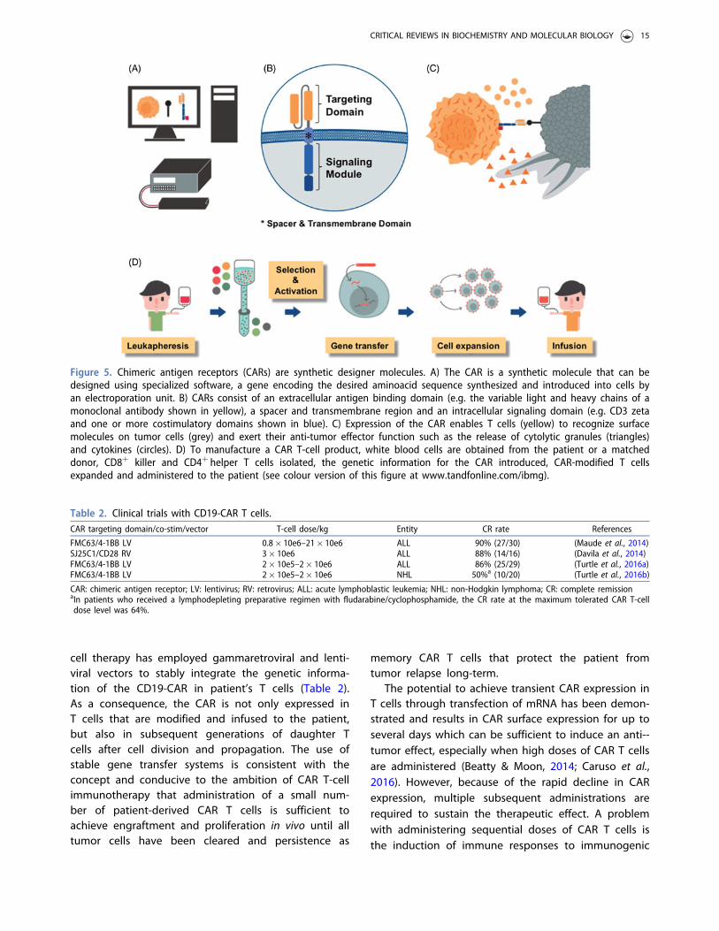
Adoptive immunotherapy with tumor-reactive T cellsthat are engineered by gene transfer to express asynthetic CAR is emerging as an effective and poten-tially curative treatment for advanced malignancies.CARs are designer molecules comprised of an extra-cellular antigen binding domain, most commonly thevariable light and heavy chains of a monoclonal anti-body, a spacer and transmembrane region thatanchors the receptor on the T-cell surface and pro-vides reach and flexibility for binding the target epi-tope, and an intracellular signaling module, mostcommonly CD3 zeta and one or more costimulatorydomains, that mediate T-cell activation after antigenbinding (Sadelain et al., 2013; Srivastava & Riddell,2015) (Figure 5). The genetic information required forencoding a CAR is �1–2 kb depending on bindingdomain configuration, spacer length and the numberof costimulatory moieties.
A medical revolution: CAR T-cell immunotherapy forB-cell malignancies
The most advanced clinical development is the use ofCARs specific for the B-lineage marker CD19 that isexpressed on B-cell acute and chronic lymphocytic leuke-mia and B-cell lymphoma. Several groups have reportedup to 90% complete remissions in patients with chemo-therapy- and radiotherapy-refractory B-cell acutelymphoblastic leukemia (ALL) and>60% complete remis-sions in patients with non-Hodgkin lymphoma (NHL)after administration of autologous (i.e. patient-derived)CD19-CAR T cells (Davila et al., 2014; Kochenderfer et al.,2015; Lee et al., 2015; Maude et al., 2014; Turtle et al.,2016a, 2016b; Turtle & Maloney, 2016) (Table 2). Theseresults are considered by many to be a medical break-through, given the advanced disease stage and failure ofconventional treatments in many patients that wereincluded in these clinical trials. Side effects of CD19-CART-cell therapy are a consequence of the strong anti-tumor immune response and include tumor lysis syn-drome due to the rapid destruction of a large number oftumor cells, cytokine release syndrome due to the rapidrelease of cytokines by CAR T cells and other immunecells, and the depletion of normal B-cells due to theirphysiologic expression of CD19.
Viral gene transfer vectors dominate pre-clinical andclinical studies with CAR T cells
The overwhelming majority of pre-clinical work andclinical trials that reported efficacy of CD19-CAR T-
14 M. HUDECEK ET AL.
cell therapy has employed gammaretroviral and lenti-viral vectors to stably integrate the genetic informa-tion of the CD19-CAR in patient’s T cells (Table 2).As a consequence, the CAR is not only expressed inT cells that are modified and infused to the patient,but also in subsequent generations of daughter Tcells after cell division and propagation. The use ofstable gene transfer systems is consistent with theconcept and conducive to the ambition of CAR T-cellimmunotherapy that administration of a small num-ber of patient-derived CAR T cells is sufficient toachieve engraftment and proliferation in vivo until alltumor cells have been cleared and persistence as
memory CAR T cells that protect the patient fromtumor relapse long-term.
The potential to achieve transient CAR expression inT cells through transfection of mRNA has been demon-strated and results in CAR surface expression for up toseveral days which can be sufficient to induce an anti--tumor effect, especially when high doses of CAR T cellsare administered (Beatty & Moon, 2014; Caruso et al.,2016). However, because of the rapid decline in CARexpression, multiple subsequent administrations arerequired to sustain the therapeutic effect. A problemwith administering sequential doses of CAR T cells isthe induction of immune responses to immunogenic
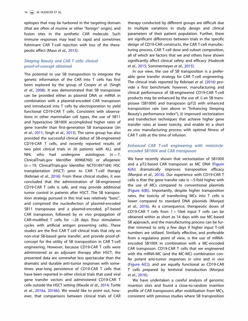
Table 2. Clinical trials with CD19-CAR T cells.CAR targeting domain/co-stim/vector T-cell dose/kg Entity CR rate References
FMC63/4-1BB LV 0.8� 10e6–21� 10e6 ALL 90% (27/30) (Maude et al., 2014)SJ25C1/CD28 RV 3� 10e6 ALL 88% (14/16) (Davila et al., 2014)FMC63/4-1BB LV 2� 10e5–2� 10e6 ALL 86% (25/29) (Turtle et al., 2016a)FMC63/4-1BB LV 2� 10e5–2� 10e6 NHL 50%a (10/20) (Turtle et al., 2016b)
CAR: chimeric antigen receptor; LV: lentivirus; RV: retrovirus; ALL: acute lymphoblastic leukemia; NHL: non-Hodgkin lymphoma; CR: complete remissionaIn patients who received a lymphodepleting preparative regimen with fludarabine/cyclophosphamide, the CR rate at the maximum tolerated CAR T-celldose level was 64%.
Figure 5. Chimeric antigen receptors (CARs) are synthetic designer molecules. A) The CAR is a synthetic molecule that can bedesigned using specialized software, a gene encoding the desired aminoacid sequence synthesized and introduced into cells byan electroporation unit. B) CARs consist of an extracellular antigen binding domain (e.g. the variable light and heavy chains of amonoclonal antibody shown in yellow), a spacer and transmembrane region and an intracellular signaling domain (e.g. CD3 zetaand one or more costimulatory domains shown in blue). C) Expression of the CAR enables T cells (yellow) to recognize surfacemolecules on tumor cells (grey) and exert their anti-tumor effector function such as the release of cytolytic granules (triangles)and cytokines (circles). D) To manufacture a CAR T-cell product, white blood cells are obtained from the patient or a matcheddonor, CD8þ killer and CD4þ helper T cells isolated, the genetic information for the CAR introduced, CAR-modified T cellsexpanded and administered to the patient (see colour version of this figure at www.tandfonline.com/ibmg).
CRITICAL REVIEWS IN BIOCHEMISTRY AND MOLECULAR BIOLOGY 15
epitopes that may be harbored in the targeting domain(that are often of murine or other “foreign” origin), andfusion sites in the synthetic CAR molecule. Suchimmune responses may lead to rapid and sometimesfulminant CAR T-cell rejection with loss of the thera-peutic effect (Maus et al., 2013).
Sleeping Beauty and CAR T cells: clinicalproof-of-concept obtained
The potential to use SB transposition to integrate thegenetic information of the CAR into T cells has firstbeen explored by the group of Cooper et al. (Singhet al., 2008). It was demonstrated that SB transposasecan be provided either as plasmid DNA or mRNA incombination with a plasmid-encoded CAR transposonand introduced into T cells by electroporation to yieldfunctional CD19-CAR T cells. Consistent with observa-tions in other mammalian cell types, the use of SB11and hyperactive SB100X accomplished higher rates ofgene transfer than first-generation SB transposase (Jinet al., 2011; Singh et al., 2013). The same group has alsoprovided the successful clinical debut of SB-engineeredCD19-CAR T cells, and recently reported results oftwo pilot clinical trials in 26 patients with ALL andNHL who had undergone autologous (n¼ 7,ClinicalTrials.gov Identifier 00968760) or allogeneic(n¼ 19, ClinicalTrials.gov Identifier NCT01497184) HSCtransplantation (HSCT) prior to CAR T-cell therapy(Kebriaei et al., 2016). From these clinical studies, it wasconcluded that the administration of SB-engineeredCD19-CAR T cells is safe, and may provide additionaltumor control in patients after HSCT. The SB transpos-ition strategy pursued in this trial was relatively “basic”,and comprised the nucleofection of plasmid-encodedSB11 transposase and a plasmid-encoded, pT-basedCAR transposon, followed by ex vivo propagation ofCAR-modified T cells for �28 days (four stimulationcycles with artificial antigen presenting cells). Thesestudies are the first CAR T cell clinical trials that rely onnon-viral SB-based gene transfer, and provide proof-of-concept for the utility of SB transposition in CAR T-cellengineering. However, because CD19-CAR T cells wereadministered as an adjuvant therapy after HSCT, thepresented data are somewhat less spectacular than thedramatic and durable anti-tumor responses with some-times year-long persistence of CD19-CAR T cells thathave been reported in other clinical trials that used viralgene transfer vectors and administered CD19-CAR Tcells outside the HSCT setting (Maude et al., 2014; Turtleet al., 2016a, 2016b). We would like to point out, how-ever, that comparisons between clinical trials of CAR
therapy conducted by different groups are difficult dueto multiple variations in study design and clinicalparameters of their patient population. Further, thereare significant differences between trials in the specificdesign of CD19-CAR constructs, the CAR T-cell manufac-turing process, CAR T-cell dose and subset composition,all of which are factors that we and others have shownsignificantly affect clinical safety and efficacy (Hudeceket al., 2015; Sommermeyer et al., 2015).
In our view, the use of SB transposition is a prefer-able gene transfer strategy for CAR T-cell engineering.The clinical trials reported by Kebriaei et al. (2016) pro-vide a first benchmark; however, manufacturing andclinical performance of SB-engineered CD19-CAR T-cellproducts may be enhanced by the use of: i) an SB trans-posase (SB100X) and transposon (pT2) with enhancedtransposition rate (see above in “Enhancing SleepingBeauty’s performance index”); ii) improved vectorizationand transfection techniques that achieve higher genetransfer rates at lower toxicity, and enable iii) a shortex vivo manufacturing process with optimal fitness ofCAR T cells at the time of infusion.
Enhanced CAR T-cell engineering with minicircle-encoded SB100X and CAR transposon
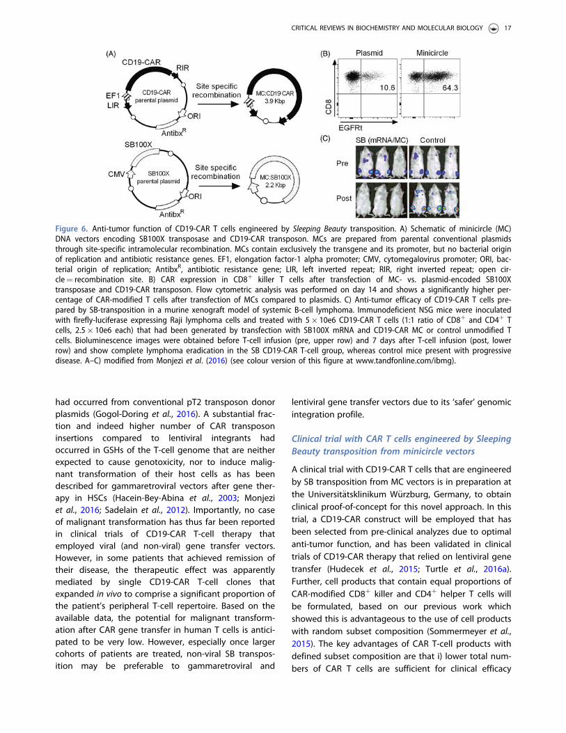
We have recently shown that vectorization of SB100Xand a pT2-based CAR transposon as MC DNA (Figure6(A)) dramatically improves transposition efficacy(Monjezi et al., 2016). Our experience with CD19-CAR Tcells is that the gene transfer rate is �5-fold higher withthe use of MCs compared to conventional plasmids(Figure 6(B)). Importantly, despite higher transpositionrates, the toxicity of transfecting MCs into T cells islower compared to standard DNA plasmids (Monjeziet al., 2016). As a consequence, therapeutic doses ofCD19-CAR T cells from 1� 10e6 input T cells can beobtained within as short as 14 days with our MC-basedSB approach, and the manufacturing process can be fur-ther trimmed to only a few days if higher input T-cellnumbers are utilized. Similarly effective, and preferablefrom a regulatory point of view, is the use of mRNA-encoded SB100X in combination with a MC-encodedCAR transposon. CD19-CAR T cells that we engineeredwith the mRNA-MC (and the MC-MC) combination con-fer potent anti-tumor responses in vitro and in vivo(Figure 6(C)), and are equally functional as CD19-CART cells prepared by lentiviral transduction (Monjeziet al., 2016).
We have undertaken a careful analysis of genomicinsertion sites and found a close-to-random insertionprofile of CAR transposons after mobilization from MCs,consistent with previous studies where SB transposition
16 M. HUDECEK ET AL.
had occurred from conventional pT2 transposon donorplasmids (Gogol-Doring et al., 2016). A substantial frac-tion and indeed higher number of CAR transposoninsertions compared to lentiviral integrants hadoccurred in GSHs of the T-cell genome that are neitherexpected to cause genotoxicity, nor to induce malig-nant transformation of their host cells as has beendescribed for gammaretroviral vectors after gene ther-apy in HSCs (Hacein-Bey-Abina et al., 2003; Monjeziet al., 2016; Sadelain et al., 2012). Importantly, no caseof malignant transformation has thus far been reportedin clinical trials of CD19-CAR T-cell therapy thatemployed viral (and non-viral) gene transfer vectors.However, in some patients that achieved remission oftheir disease, the therapeutic effect was apparentlymediated by single CD19-CAR T-cell clones thatexpanded in vivo to comprise a significant proportion ofthe patient’s peripheral T-cell repertoire. Based on theavailable data, the potential for malignant transform-ation after CAR gene transfer in human T cells is antici-pated to be very low. However, especially once largercohorts of patients are treated, non-viral SB transpos-ition may be preferable to gammaretroviral and
lentiviral gene transfer vectors due to its ‘safer’ genomicintegration profile.
Clinical trial with CAR T cells engineered by SleepingBeauty transposition from minicircle vectors
A clinical trial with CD19-CAR T cells that are engineeredby SB transposition from MC vectors is in preparation atthe Universit€atsklinikum W€urzburg, Germany, to obtainclinical proof-of-concept for this novel approach. In thistrial, a CD19-CAR construct will be employed that hasbeen selected from pre-clinical analyzes due to optimalanti-tumor function, and has been validated in clinicaltrials of CD19-CAR therapy that relied on lentiviral genetransfer (Hudecek et al., 2015; Turtle et al., 2016a).Further, cell products that contain equal proportions ofCAR-modified CD8þ killer and CD4þ helper T cells willbe formulated, based on our previous work whichshowed this is advantageous to the use of cell productswith random subset composition (Sommermeyer et al.,2015). The key advantages of CAR T-cell products withdefined subset composition are that i) lower total num-bers of CAR T cells are sufficient for clinical efficacy
Figure 6. Anti-tumor function of CD19-CAR T cells engineered by Sleeping Beauty transposition. A) Schematic of minicircle (MC)DNA vectors encoding SB100X transposase and CD19-CAR transposon. MCs are prepared from parental conventional plasmidsthrough site-specific intramolecular recombination. MCs contain exclusively the transgene and its promoter, but no bacterial originof replication and antibiotic resistance genes. EF1, elongation factor-1 alpha promoter; CMV, cytomegalovirus promoter; ORI, bac-terial origin of replication; AntibxR, antibiotic resistance gene; LIR, left inverted repeat; RIR, right inverted repeat; open cir-cle¼ recombination site. B) CAR expression in CD8þ killer T cells after transfection of MC- vs. plasmid-encoded SB100Xtransposase and CD19-CAR transposon. Flow cytometric analysis was performed on day 14 and shows a significantly higher per-centage of CAR-modified T cells after transfection of MCs compared to plasmids. C) Anti-tumor efficacy of CD19-CAR T cells pre-pared by SB-transposition in a murine xenograft model of systemic B-cell lymphoma. Immunodeficient NSG mice were inoculatedwith firefly-luciferase expressing Raji lymphoma cells and treated with 5� 10e6 CD19-CAR T cells (1:1 ratio of CD8þ and CD4þ Tcells, 2.5� 10e6 each) that had been generated by transfection with SB100X mRNA and CD19-CAR MC or control unmodified Tcells. Bioluminescence images were obtained before T-cell infusion (pre, upper row) and 7 days after T-cell infusion (post, lowerrow) and show complete lymphoma eradication in the SB CD19-CAR T-cell group, whereas control mice present with progressivedisease. A–C) modified from Monjezi et al. (2016) (see colour version of this figure at www.tandfonline.com/ibmg).
CRITICAL REVIEWS IN BIOCHEMISTRY AND MOLECULAR BIOLOGY 17
which reduces the risk for side effects and shortens themanufacturing process; and ii) because of less product-to-product variability between patients, the time to andlevel of CAR T-cell engraftment and proliferation ismore consistent and predictable, and allows establish-ing dose-response relationships as well as immunepharmacokinetic and pharmacodynamic parameters forCAR T cells as medicinal products. We have determinedthe copy number of the CD19-CAR transposon in bothCD8þ and CD4þ T cells, and found it to be in an accept-able range (n¼ 6 and n¼ 5, respectively), similar to thatobtained with gammaretroviral and lentiviral vectors(Monjezi et al., 2016).
For future clinical trials, there is a pre-clinical devel-opment pipeline with CARs specific for target antigensin other hematologic and solid tumors, including aSLAMF7-specific CAR in multiple myeloma, and a ROR1-specific CAR in lymphoma, breast and lung cancer(Berger et al., 2015; Hudecek et al., 2013).Importantly, the MC-encoded SB transposon can accom-modate additional genetic cargo without compromisingtransposition efficacy. Our CAR transposon includes anEGFRt marker that was developed in Mike Jensen’slaboratory and serves multiple functions, e.g. to selectCAR-modified T cells to further refine the infusion prod-uct, and to detect CAR T cells in blood and tissue sam-ples (Wang et al., 2011). Further, CD19-CAR_EGFRtT cells can be depleted through administration of ananti-EGFR antibody to mitigate toxicity or terminate thetherapeutic effect after tumor clearance. We haverecently shown in a pre-clinical model that depletion ofCD19-CAR_EGFRt T cells after lymphoma eradicationreverses B-cell aplasia as the major long-term side effectand adds another layer of safety to CD19-CAR T-celltherapy (Paszkiewicz et al., 2016).
Regulatory considerations for clinical trialapproval
Currently, the main focus of clinical development of theSB transposon/transposase system is on ex vivo genedelivery into primary cells. Thus, from a regulatory pointof view, the actual investigational medicinal product(IMP) administered to humans are the genetically modi-fied cells, which in the European Union (EU) are classi-fied as gene therapy medicinal product (GTMP) in mostcases. In the EU, GTMPs belong to the group ofadvanced therapy medicinal products (ATMPs). Basicguidance on the regulatory requirements for this typeof products regarding quality and non-clinical aspects,as well as on clinical development is given in the“Guideline on quality, non-clinical and clinical aspects of
medicinal products containing genetically modifiedcells” (EMA/CAT/GTWP/671639/2008), which wasreleased in 2012. However, in this guideline only limitedinformation is provided on the new technologies forgenetic modification of cells, such as the transposon/transposase system.
For genetically modified cells, both the cells to bemodified as well as the vector encoding the transposonand the therapeutic gene are considered to be startingmaterials, and need to be manufactured according tothe principles of GMP. However, in case of primary celldonation, procurement and testing of the cells can beperformed outside of a GMP environment. The princi-ples of testing quality parameters during manufacturingand for release of the final product are, for the cellularcomponent, similar to most cell therapies, irrespectivewhether the cells are genetically modified or not, butmay depend on the cell type employed.
One important aspect that is specific for transposon-based genetic modification of cells is to monitor theintegration profile of the transposon and the number ofvector copies integrated into the host genome. As dis-cussed above, genomic integration may have functionalconsequences, and such impact on the genetic stabilityof the cells and their proliferation and differentiationcapacity needs to be analyzed and documented. In add-ition, potential toxic effects by the transposase need tobe taken into account. In many cases the potency ofsuch products is directly linked to the number or per-centage of gene-modified cells as well as to the levelsof expression of the therapeutic sequence. It is import-ant to determine these parameters already in early clin-ical phases as release criteria for each batch. Increasingclinical experience would then allow the developmentof biological assays indicative for the functional activityof the therapy and related to the clinical efficacy.
In contrast to classical integrating vectors such asretro- or lentiviral vectors, which deliver and insert thetherapeutic sequence into the host genome in a “one-shot” process, the transposon in theory is able to berepeatedly excised and integrated at different loci incase of persistent transposase activity. Therefore, a crit-ical issue is the presence of the transposase at the timeof administration of the IMP. Delivery of the transposaseas protein might thus be preferable to delivery viatransposase-encoding mRNA or DNA; however, this willmost probably be at the expense of efficacy of the gen-etic modification. Currently, transcriptionally regulatedtransposase expression systems are under evaluation, toallow for highly efficient delivery of the transposasegene and concurrent switch-off of gene expression priorto administration of the cell to the patient (Cocchiarella
18 M. HUDECEK ET AL.

et al., 2016). In such a case, full function of the systemincluding control of leakage needs to be demonstratedbefore clinical application.
As for all other gene modifying systems, the risk ofinsertional oncogenesis due to semi-random genomicintegration is an important regulatory concern prior tohuman use. Transposons integrate their payload, similarto retro- and lentiviruses, in a more or less random fash-ion bearing the risk of activating a proto-oncogene orinactivating a tumor suppressor gene, as discussedabove. However, additional factors may contribute tothe risk of oncogenesis, such as the presence andstrength of transcriptional regulatory elements, thetherapeutic gene, the vector dose, the target cell popu-lation, and the indication to be treated (Aiuti et al.,2013b). Thus, the need and, if so, the extent of integra-tion studies, genotoxicity and tumorigenicity studiesstrongly depends on the above parameters employedin context of the transposon/transposase delivery sys-tem. The current clinical database available for retro- orlentiviral vector-transduced T cells used in cancerimmunotherapy, for example, justifies that integrationstudies for this type of therapy are omitted, taking intoaccount also the life-threatening nature of the diseasewith poor treatment alternatives. For the transposon/transposase system, clinical experience is more limited,and thus a decision on the need for integration and/ortumorigenicity studies is on a case-by-case basis, andshould follow a risk-based approach. In any case, use oftransient delivery of the transposase in the form ofmRNA or recombinant protein is preferred over transpo-sase coding sequences in the form of DNA to limittransposase activity to reduce the risk of insertionalmutagenesis.
Perspective: Sleeping Beauty – a possible newgold standard for gene transfer!
The first clinical application of the SB system is currentlyongoing using autologous T cells gene-modified withSB vectors carrying a CAR to render the T cells cytotoxicspecifically toward CD19-positive lymphoid tumors(Huang et al., 2008; Kebriaei et al., 2016; Singh et al.,2008). Lymphocytes represent a suitable initial platformfor testing new gene transfer systems, as T cells can begenetically modified using viral and non-viralapproaches without apparent resulting genotoxicity.The advantage of using the SB system for genetic modi-fication includes i) the ease and reduced cost associatedwith manufacturing of clinical-grade, plasmid-basedvectors compared with recombinant viral vectors, ii)scalability: SB vectors can be manufactured in any
quantity, iii) easy quality control for clinical use, and iii)indefinite storage with absolute fidelity.
A major hurdle in ex vivo delivery of the transposoncomponents into relevant primary cell types is the tox-icity of contemporary transfection/electroporation pro-tocols. In situations, where target cells are scarce and/orculturing and expansion of the transfected cells isimpossible or cannot be solved without compromisingcell identity and grafting potential, cytotoxicity of thetransfection procedures is a serious issue that mayundermine clinical applications. However, recent experi-mental data indicate that by employing transposon cas-settes vectorized as MCs and by providing thetransposase in the form of mRNA cellular toxicity can bereduced (Monjezi et al., 2016), thereby positioning clin-ical applications with SB well within reach, at least inthe area of T cell engineering.
Alternatively, the development of hybrid vector sys-tems combining the natural ability of viruses to traversecell mebranes with efficient genomic insertion medi-ated by the SB system can be a promising strategy.Indeed, components of the SB transposon have beenincorporated into integrase-defective lentiviral particlesthat showed efficient gene transfer in a range of humancell types and an insertion profile favorable to conven-tional lentiviral vectors (Moldt et al., 2011; Staunstrupet al., 2009; Vink et al., 2009). Hybrid adenovirus/SB vec-tors (Boehme et al., 2016; Yant et al., 2002) have beenused to efficiently deliver SB transposon vectorsexpressing FIX into the liver in a hemophilic dog model(Hausl et al., 2010). Recently, a new approach was estab-lished based on a hybrid adenovirus/SB vector targetedto CD46, a receptor expressed on HSCs. The system hasbeen shown to yield up to 10% stable gene transfer inHSCs following in vivo transduction, thereby alleviatingthe need for myeloablation and transplantation (Richteret al., 2016). Retroviral vectors disabled in generating acDNA copy of the retroviral vector have been shown todeliver the SB transposase mRNA into target cells withan impressive efficiency (Galla et al., 2011). Herpes sim-plex virus vectors with a tropism to infect neural pro-genitor cells have been used to target SB insertions inthe central nervous system in an in utero gene deliverysystem in the mouse (Bowers et al., 2006; de Silva et al.,2010). Finally, similar to adenovirus, baculoviral vectorscan efficiently transduce many cell types, but can onlyprovide transient expression in the absence of stablegenomic integration. Hybrid baculovirus/SB vectorshave been shown to confer prolonged transgeneexpression in human cells both in vitro and in vivo inintratumoral injections in xenograft mice (Luo et al.,2012), as well as in vivo in the mouse eye (Turunenet al., 2014). Note that, although these hybrid virus/SB
CRITICAL REVIEWS IN BIOCHEMISTRY AND MOLECULAR BIOLOGY 19

transposon vectors may provide advantage from a tech-nical point of view (non-toxic delivery of the SB vectorcomponents into target cells), their application wouldlose all the advantages of a fully non-viral system thatwe outlined above.
Ex vivo modification of T cells is currently limited to asmall number of centers with the required infrastructureand expertise in the logistics of cell isolation, activation,gene transfer, expansion and cryopreservation steps. Tosimplify procedures and widen applicability for clinicaltherapies, automation of these procedures is beingdeveloped. The CliniMACS ProdigyVR (Miltenyi Biotec,Bergisch Gladbach, Germany) device delivers automatedcell processing in a closed GMP-compliant system, andhas recently been adapted for lentiviral transduction ofT cells (Mock et al., 2016). The advantage of the system isthat it can standardize the complete manufacturing pro-cess of cellular products. Future generations of the sys-tem adapted to non-viral vector delivery may advancemanufacturing of SB-engineered cellular products.
Alternative technologies for genetic engineering inclinically relevant cell types are rapidly advancing.Designer nucleases, including zinc finger nucleases(ZFNs), transcription activator like effector nucleases(TALENs) and clustered regularly interspaced short pal-indromic repeat (CRISPR)/Cas nucleases are excellenttools for genome engineering and gene editing(Doudna & Charpentier, 2014). The common advantageof these technologies is that they enable site-specificengineering of the genome, thereby allowing introduc-tion of facile and highly specific changes in the geneticcode. These technologies are making their way towardtherapeutic applications (Yu et al., 2016); for example,site-specific gene addition at reasonable efficiencies hasrecently been demonstrated by mRNA encoding ZFNand a homology template delivered as AAV in bothHSCs (Wang et al., 2015) and T cells (Wang et al., 2016a).One key difference that sets designer nucleases apartfrom a transposon is that they are specialized andhighly active at cutting the genome at a specificsequence, but transgene addition at the cut site isdependent on double strand break repair mechanismsof the cell. In contrast, transposons are gene integration“machines” that evolved to insert themselves into thegenome; an attribute that forms the basis of applyingthem for robust gene transfer.
Finally, the sequence “space” of the SB transposaseremains largely unexplored for hyperactive phenotypes.Importantly, the crystal structure of SB100X’s catalyticdomain has recently been solved (Voigt et al., 2016).The structure was highly informative with respect torationalizing the biochemical basis of hyperactivity ofalready existing SB mutants, and can likely be used in
the future for structure-based design of next generationSB transposases for therapeutic gene delivery.
Acknowledgments
M.H. is a member of the “Young Scholar Program” (JungesKolleg) of the Bavarian Academy of Sciences (BayerischeAkademie der Wissenschaften).
Disclosure statement
M.H., Z.Iz. and Z.Iv. are inventors on patents related toSleeping Beauty gene transfer technology.
Funding
M.H. was supported by the German Cancer Aid (DeutscheKrebshilfe e.V., Max Eder Programm 110313), the MyelomaCrowd Research Initiative and the Interdisciplinary Center forClinical Research (IZKF) W€urzburg (Projects Z/4–109 and D-244). The TargetAMD project has received funding from theEuropean Union’s Seventh Framework Programme forresearch, technological development and demonstrationunder grant agreement no 305134. Z.Iv. was supported bythe Center for Cell and Gene Therapy of the LOEWE (Landes-Offensive zur Entwicklung Wissenschaftlich-€okonomischerExzellenz) program in Hessen, Germany.
References
Aisenbrey S, Lafaut BA, Szurman P, et al. (2006). Iris pigmentepithelial translocation in the treatment of exudativemacular degeneration: a 3-year follow-up. ArchOphthalmol 124:183–8.
Aiuti A, Biasco L, Scaramuzza S, et al. (2013a). Lentiviral hem-atopoietic stem cell gene therapy in patients with Wiskott-Aldrich syndrome. Science 341:1233151.
Aiuti A, Cossu G, De Felipe P, et al. (2013b). The committeefor advanced therapies’ of the European medicines agencyreflection paper on management of clinical risks derivingfrom insertional mutagenesis. Hum Gene Ther Clin Dev24:47–54.
Aronovich EL, Bell JB, Belur LR, et al. (2007). Prolongedexpression of a lysosomal enzyme in mouse liver aftersleeping beauty transposon-mediated gene delivery: impli-cations for non-viral gene therapy of mucopolysaccharido-ses. J Gene Med 9:403–15.
Aronovich EL, Bell JB, Khan SA, et al. (2009). Systemiccorrection of storage disease in MPS I NOD/SCID miceusing the sleeping beauty transposon system. Mol Ther17:1136–44.
Balaggan KS, Duran Y, Georgiadis A, et al. (2012). Absence ofocular malignant transformation after sub-retinal deliveryof rAAV2/2 or integrating lentiviral vectors in p53-deficientmice. Gene Ther 19:182–8.
Basner-Tschakarjan E, Mingozzi F. (2014). Cell-mediatedimmunity to AAV vectors, evolving concepts and potentialsolutions. Front Immunol 5:350.
20 M. HUDECEK ET AL.

Baum C, Kalle CV, Staal FJT, et al. (2004). Chance or neces-sity? Insertional mutagenesis in gene therapy and its con-sequences. Mol Ther 9:5–13.
Baus J, Liu L, Heggestad AD, et al. (2005). Hyperactive trans-posase mutants of the sleeping beauty transposon. MolTher 12:1148–56.
Beatty GL, Moon EK. (2014). Chimeric antigen receptor T cellsare vulnerable to immunosuppressive mechanisms presentwithin the tumor microenvironment. Oncoimmunology3:e970027.
Belay E, Matrai J, Acosta-Sanchez A, et al. (2010). Novelhyperactive transposons for genetic modification ofinduced pluripotent and adult stem cells: a nonviral para-digm for coaxed differentiation. Stem Cells (Dayton, Ohio)28:1760–71.
Belcher JD, Vineyard JV, Bruzzone CM, et al. (2010). Hemeoxygenase-1 gene delivery by Sleeping Beauty inhibits vas-cular stasis in a murine model of sickle cell disease. J MolMed 88:665–75.
Belur LR, Frandsen JL, Dupuy AJ, et al. (2003). Gene insertionand long-term expression in lung mediated by the sleep-ing beauty transposon system. Mol Ther 8:501–7.
Belur LR, Podetz-Pedersen KM, Sorenson BS, et al. (2011).Inhibition of angiogenesis and suppression of colorectalcancer metastatic to the liver using the sleeping beautytransposon system. Mol Cancer 10:14.
Berger C, Sommermeyer D, Hudecek M, et al. (2015). Safety oftargeting ROR1 in primates with chimeric antigen receptor-modified T cells. Cancer Immunol Res 3:206–16.
Biffi A, Montini E, Lorioli L, et al. (2013). Lentiviral hematopoi-etic stem cell gene therapy benefits metachromatic leuko-dystrophy. Science 341:1233158.
Binder S, Krebs I, Hilgers RD, et al. (2004). Outcome of trans-plantation of autologous retinal pigment epithelium inage-related macular degeneration: a prospective trial.Invest Ophthalmol Vis Sci 45:4151–60.
Boehme P, Doerner J, Solanki M, et al. (2015). The sleepingbeauty transposon vector system for treatment of rare gen-etic diseases: an unrealized hope? Curr Gene Ther 15:255–65.
Boehme P, Zhang W, Solanki M, et al. (2016). A high-capacityadenoviral hybrid vector system utilizing the hyperactivesleeping beauty transposase SB100X for enhanced integra-tion. Mol Ther Nucleic Acids 5:e337.
Boulanger-Scemama E, Querques G, About F, et al. (2015).Ranibizumab for exudative age-related macular degener-ation: a five year study of adherence to follow-up in areal-life setting. J Fr Ophtalmol 38:620–7.
Bowers WJ, Mastrangelo MA, Howard DF, et al. (2006).Neuronal precursor-restricted transduction via in uteroCNS gene delivery of a novel bipartite HSV amplicon/trans-posase hybrid vector. Mol Ther 13:580–8.
Braun CJ, Boztug K, Paruzynski A, et al. (2014). Gene therapyfor Wiskott-Aldrich syndrome–long-term efficacy and gen-otoxicity. Sci Transl Med 6:227ra33.
Campochiaro PA, Nguyen QD, Shah SM, et al. (2006).Adenoviral vector-delivered pigment epithelium-derivedfactor for neovascular age-related macular degeneration:results of a phase I clinical trial. Hum Gene Ther17:167–76.
Cartier N, Hacein-Bey-Abina S, Bartholomae CC, et al. (2009).Hematopoietic stem cell gene therapy with a lentiviral vec-tor in X-linked adrenoleukodystrophy. Science 326:818–23.
Caruso HG, Torikai H, Zhang L, et al. (2016). Redirecting T-cellspecificity to EGFR using mRNA to self-limit expression ofchimeric antigen receptor. J Immunother 39:205–17.
Cattoglio C, Pellin D, Rizzi E, et al. (2010). High-definitionmapping of retroviral integration sites identifies activeregulatory elements in human multipotent hematopoieticprogenitors. Blood 116:5507–17.
Cavazza A, Moiani A, Mavilio F. (2013). Mechanisms of retro-viral integration and mutagenesis. Hum Gene Ther24:119–31.
Cavazzana-Calvo M, Payen E, Negre O, et al. (2010).Transfusion independence and HMGA2 activation aftergene therapy of human beta-thalassaemia. Nature467:318–22.
Cesana D, Sgualdino J, Rudilosso L, et al. (2012). Whole tran-scriptome characterization of aberrant splicing eventsinduced by lentiviral vector integrations. J Clin Invest122:1667–76.
Chabot S, Orio J, Schmeer M, et al. (2013). Minicircle DNAelectrotransfer for efficient tissue-targeted gene delivery.Gene Ther 20:62–8.
Chandler M, Clerget M, Galas DJ. (1982). The transpositionfrequency of IS1-flanked transposons is a function of theirsize. J Mol Biol 154:229–43.
Chen Z-Y, He C-Y, Ehrhardt A, Kay MA. (2003). Minicircle DNAvectors devoid of bacterial DNA result in persistent andhigh-level transgene expression in vivo. Mol Ther8:495–500.
Chen Z-Y, Riu E, He C-Y, et al. (2008). Silencing of episomaltransgene expression in liver by plasmid bacterial back-bone DNA is independent of CpG methylation. Mol Ther16:548–56.
Chen ZJ, Kren BT, Wong PY, et al. (2005). Sleeping Beauty-mediated down-regulation of huntingtin expression byRNA interference. Biochem Biophys Res Commun329:646–52.
Chen ZY, He CY, Meuse L, Kay MA. (2004). Silencing of episo-mal transgene expression by plasmid bacterial DNA ele-ments in vivo. Gene Ther 11:856–64.
Chuang T-H, Lee J, Kline L, et al. (2002). Toll-like receptor 9mediates CpG-DNA signaling. J Leukoc Biol 71:538–44.
Cocchiarella F, Latella MC, Basile V, et al. (2016).Transcriptionally regulated and nontoxic delivery of thehyperactive sleeping beauty transposase. Mol TherMethods Clin Dev 3:16038.
Cui Z, Geurts AM, Liu G, et al. (2002). Structure-function ana-lysis of the inverted terminal repeats of the sleepingbeauty transposon. J Mol Biol 318:1221–35.
Darquet AM, Cameron B, Wils P, et al. (1997). A new DNAvehicle for nonviral gene delivery: supercoiled minicircle.Gene Ther 4:1341–9.
Davila ML, Riviere I, Wang X, et al. (2014). Efficacy and tox-icity management of 19–28z CAR T cell therapy in B cellacute lymphoblastic leukemia. Sci Transl Med 6:224–5.
De Ravin SS, Su L, Theobald N, et al. (2014). Enhancers aremajor targets for murine leukemia virus vector integration.J Virol 88:4504–13.
De Silva S, Mastrangelo MA, Lotta LT Jr, et al. (2010).Extending the transposable payload limit of sleepingbeauty (SB) using the herpes simplex virus (HSV)/SB ampli-con-vector platform. Gene Ther 17:424–31.
CRITICAL REVIEWS IN BIOCHEMISTRY AND MOLECULAR BIOLOGY 21
Deichmann A, Hacein-Bey-Abina S, Schmidt M, et al. (2007).Vector integration is nonrandom and clustered and influ-ences the fate of lymphopoiesis in SCID-X1 gene therapy.J Clin Invest 117:2225–32.
Deniger DC, Pasetto A, Tran E, et al. (2016). Stable, nonviralexpression of mutated tumor neoantigen-specific T-cellreceptors using the sleeping beauty transposon/transpo-sase system. Mol Ther 24:1078–89.
Dijkhuizen PA, Pasterkamp RJ, Hermens WT, et al. (1998).Adenoviral vector-mediated gene delivery to injured ratperipheral nerve. J Neurotrauma 15:387–97.
Doudna JA, Charpentier E. (2014). Genome editing. The newfrontier of genome engineering with CRISPR-Cas9. Science346:1258096.
Enslow R, Bhuvanagiri S, Vegunta S, et al. (2016). Associationof anti-VEGF injections with progression of geographicatrophy. Ophthalmol Eye Dis 8:31–2.
Escobar H, Schowel V, Spuler S, et al. (2016). Full-length dys-ferlin transfer by the hyperactive sleeping beauty transpo-sase restores dysferlin-deficient muscle. Mol Ther NucleicAcids 5:e277.
Essner JJ, Mcivor RS, Hackett PB. (2005). Awakening genetherapy with sleeping beauty transposons. Curr OpinPharmacol 5:513–9.
Eyjolfsdottir H, Eriksdotter M, Linderoth B, et al. (2016).Targeted delivery of nerve growth factor to the cholinergicbasal forebrain of Alzheime’s disease patients: applicationof a second-generation encapsulated cell biodeliverydevice. Alzheimers Res Ther 8:30.
Falkner-Radler CI, Krebs I, Glittenberg C, et al. (2011). Humanretinal pigment epithelium (RPE) transplantation: outcomeafter autologous RPE-choroid sheet and RPE cell-suspen-sion in a randomised clinical study. Br J Ophthalmol95:370–5.
Fernando S, Fletcher BS. (2006). Sleeping beauty transposon-mediated nonviral gene therapy. BioDrugs 20:219–29.
Fischer SE, Van Luenen HG, Plasterk RH. (1999). Cis require-ments for transposition of Tc1-like transposons in C. ele-gans. Mol Gen Genet 262:268–74.
Galla M, Schambach A, Falk CS, et al. (2011). Avoiding cyto-toxicity of transposases by dose-controlled mRNA delivery.Nucleic Acids Res 39:7147–60.
Garrison BS, Yant SR, Mikkelsen JG, Kay MA. (2007).Postintegrative gene silencing within the sleeping beautytransposition system. Mol Cell Biol 27:8824–33.
Geurts AM, Yang Y, Clark KJ, et al. (2003). Gene transfer intogenomes of human cells by the sleeping beauty trans-poson system. Mol Ther 8:108–17.
Gogol-Doring A, Ammar I, Gupta S. (2016). Genome-wideprofiling reveals remarkable parallels between insertionsite selection properties of the MLV retrovirus and thepiggyBac transposon in primary human CD4(þ) T cells.Mol Ther 24:592–606.
Grabundzija I, Irgang M, Mates L, et al. (2010). Comparativeanalysis of transposable element vector systems in humancells. Mol Ther 18:1200–9.
Grossniklaus HE, Kang SJ, Berglin L. (2010). Animal models ofchoroidal and retinal neovascularization. Prog Retin EyeRes 29:500–19.
Hacein-Bey-Abina S, Garrigue A, Wang GP, et al. (2008).Insertional oncogenesis in 4 patients after retrovirus-medi-ated gene therapy of SCID-X1. J Clin Invest 118:3132–42.
Hacein-Bey-Abina S, Von Kalle C, Schmidt M, et al. (2003).LMO2-associated clonal T cell proliferation in two patientsafter gene therapy for SCID-X1. Science 302:415–9.
Hackett PB, Ekker SC, Largaespada DA, Mcivor RS. (2005).Sleeping beauty transposon-mediated gene therapy forprolonged expression. Adv Genet 54:189–232.
Hackett PB Jr., Aronovich EL, Hunter D, et al. (2011). Efficacyand safety of Sleeping Beauty transposon-mediated genetransfer in preclinical animal studies. Curr Gene Ther11:341–9.
Hackett PB, Largaespada DA, Cooper LJN. (2010). A trans-poson and transposase system for human application. MolTher 18:674–83.
Hargrove PW, Kepes S, Hanawa H, et al. (2008). Globin lentivi-ral vector insertions can perturb the expression ofendogenous genes in beta-thalassemic hematopoieticcells. Mol Ther 16:525–33.
Hartman ZC, Appledorn DM, Amalfitano A. (2008).Adenovirus vector induced innate immune responses:impact upon efficacy and toxicity in gene therapy and vac-cine applications. Virus Res 132:1–14.
Hausl MA, Zhang W, Muther N, et al. (2010). Hyperactivesleeping beauty transposase enables persistent phenotypiccorrection in mice and a canine model for hemophilia B.Mol Ther 18:1896–906.
He C-X, Shi D, Wu W-J, et al. (2004). Insulin expression in liv-ers of diabetic mice mediated by hydrodynamics-basedadministration. World J Gastroenterol 10:567–72.
Hemmi H, Takeuchi O, Kawai T, et al. (2000). A toll-like recep-tor recognizes bacterial DNA. Nature 408:740–5.
Hermens WT, Verhaagen J. (1997). Adenoviral vector-medi-ated gene expression in the nervous system of immuno-competent Wistar and T cell-deficient nude rats:preferential survival of transduced astroglial cells in nuderats. Hum Gene Ther 8:1049–63.
Hirsch ML, Wolf SJ, Samulski RJ. (2016). Delivering transgenicDNA eExceeding the carrying capacity of AAV vectors.Methods Mol Biol 1382:21–39.
Hollis RP, Nightingale SJ, Wang X, et al. (2006). Stablegene transfer to human CD34(þ) hematopoietic cells usingthe sleeping beauty transposon. Exp Hematol 34:1333–43.
Howe SJ, Mansour MR, Schwarzwaelder K, et al. (2008).Insertional mutagenesis combined with acquired somaticmutations causes leukemogenesis following gene therapyof SCID-X1 patients. J Clin Invest 118:3143–50.
Huang X, Guo H, Kang J, et al. (2008). Sleeping beauty trans-poson-mediated engineering of human primary T cells fortherapy of CD19þ lymphoid malignancies. Mol Ther16:580–9.
Huang X, Guo H, Tammana S, et al. (2010). Gene transfer effi-ciency and genome-wide integration profiling of sleepingbeauty, Tol2, and piggyBac transposons in human primaryT cells. Mol Ther 18:1803–13.
Hudecek M, Lupo-Stanghellini MT, Kosasih PL, et al. (2013).Receptor affinity and extracellular domain modificationsaffect tumor recognition by ROR1-specific chimeric antigenreceptor T cells. Clin Cancer Res 19:3153–64.
Hudecek M, Sommermeyer D, Kosasih PL, et al. (2015). Thenonsignaling extracellular spacer domain of chimeric anti-gen receptors is decisive for in vivo antitumor activity.Cancer Immunol Res 3:125–35.
22 M. HUDECEK ET AL.

Huerfano S, Ryabchenko B, Forstov�a J. (2013). Nucleofectionof expression vectors induces a robust interferon responseand inhibition of cell proliferation. DNA Cell Biol32:467–79.
Hyde SC, Pringle IA, Abdullah S, et al. (2008). CpG-free plas-mids confer reduced inflammation and sustained pulmon-ary gene expression. Nat Biotechnol 26:549–51.
Ivics Z, Garrels W, M�at�es L, et al. (2014a). Germline transgene-sis in pigs by cytoplasmic microinjection of SleepingBeauty transposons. Nat Protoco 9:810–27.
Ivics Z, Hackett PB, Plasterk RH, Izsvak Z. (1997). Molecularreconstruction of sleeping beauty, a Tc1-like transposonfrom fish, and its transposition in human cells. Cell91:501–10.
Ivics Z, Hiripi L, Hoffmann OI, et al. (2014b). Germline trans-genesis in rabbits by pronuclear microinjection of SleepingBeauty transposons. Nat Protoc 9:794–809.
Ivics Z, Izsvak Z. (2006). Transposons for gene therapy! CurrGene Ther 6:593–607.
Ivics Z, Izsvak Z. (2010). The expanding universe of trans-poson technologies for gene and cell engineering. MobDNA 1:25.
Ivics Z, Izsvak Z. (2011). Nonviral gene delivery with thesleeping beauty transposon system. Hum Gene Ther22:1043–51.
Ivics Z, Izsvak Z. (2015). Sleeping beauty transposition.Microbiol Spectr 3:MDNA3.
Ivics Z, Izsv�ak Z, Minter A, Hackett PB. (1996). Identification offunctional domains and evolution of Tc1-like transposableelements. Proc Natl Acad Sci. USA 93:5008–13.
Ivics Z, Katzer A, Stuwe EE, et al. (2007). Targeted sleepingbeauty transposition in human cells. Mol Ther 15:1137–44.
Ivics Z, Li MA, Mates L, et al. (2009). Transposon-mediatedgenome manipulation in vertebrates. Nat Methods6:415–22.
Ivics Z, Mates L, Yau TY, et al. (2014c). Germline transgenesisin rodents by pronuclear microinjection of sleeping beautytransposons. Nat Protoc 9:773–93.
Izsvak Z, Chuah MK, Vandendriessche T, Ivics Z. (2009).Efficient stable gene transfer into human cells by thesleeping beauty transposon vectors. Methods 49:287–97.
Izsvak Z, Hackett PB, Cooper LJN, Ivics Z. (2010). Translatingsleeping beauty transposition into cellular therapies: victo-ries and challenges. Bioessays 32:756–67.
Izsvak Z, Ivics Z. (2004). Sleeping beauty transposition: biol-ogy and applications for molecular therapy. Mol Ther9:147–56.
Izsv�ak Z, Ivics Z, Plasterk RH. (2000). Sleeping beauty, a widehost-range transposon vector for genetic transformation invertebrates. J Mol Biol 302:93–102.
Izsv�ak Z, Khare D, Behlke J, et al. (2002). Involvement of abifunctional, paired-like DNA-binding domain and a trans-positional enhancer in sleeping beauty transposition. J BiolChem 277:34581–8.
Jahner D, Stuhlmann H, Stewart CL, et al. (1982). De novomethylation and expression of retroviral genomes duringmouse embryogenesis. Nature 298:623–8.
Jin Z, Maiti S, Huls H, et al. (2011). The hyperactive sleepingbeauty transposase SB100X improves the genetic modifica-tion of T cells to express a chimeric antigen receptor.Gene Ther 18:849–56.
Johnen S, Djalali-Talab Y, Kazanskaya O, et al. (2015).Antiangiogenic and neurogenic activities of sleepingbeauty-mediated PEDF-transfected RPE cells in vitro and invivo. Biomed Res Int 2015:863845.
Johnen S, Izsvak Z, Stocker M, et al. (2012). Sleepingbeauty transposon-mediated transfection of retinal andiris pigment epithelial cells. Invest Ophthalmol Vis Sci53:4787–96.
Karsi A, Moav B, Hackett P, Liu Z. (2001). Effects of insert sizeon transposition efficiency of the sleeping beauty trans-poson in mouse cells. Mar Biotechnol 3:241–5.
Kebriaei P, Singh H, Huls MH, et al. (2016). Phase I trials usingsleeping beauty to generate CD19-specific CAR T cells.J Clin Invest 126:3363–76.
Kochenderfer JN, Dudley ME, Kassim SH, et al. (2015).Chemotherapy-refractory diffuse large B-cell lymphomaand indolent B-cell malignancies can be effectively treatedwith autologous T cells expressing an anti-CD19 chimericantigen receptor. J Clin Oncol 33:540–9.
Kowarz E, Loscher D, Marschalek R. (2015). Optimized sleep-ing beauty transposons rapidly generate stable transgeniccell lines. Biotechnol J 10:647–53.
Kren BT, Unger GM, Sjeklocha L, et al. (2009). Nanocapsule-delivered sleeping beauty mediates therapeutic Factor VIIIexpression in liver sinusoidal endothelial cells of hemo-philia A mice. J Clin Invest 119:2086–99.
Kuerten D, Johnen S, Harmening N, et al. (2015).Transplantation of PEDF-transfected pigment epithelialcells inhibits corneal neovascularization in a rabbit model.Graefes Arch Clin Exp Ophthalmol 253:1061–9.
Kustikova O, Fehse B, Modlich U, et al. (2005). Clonal domin-ance of hematopoietic stem cells triggered by retroviralgene marking. Science (New York, N.Y.) 308:1171–4.
Lad EM, Hammill BG, Qualls LG, et al. (2014). Anti-VEGF treat-ment patterns for neovascular age-related macular degen-eration among medicare beneficiaries. Am J Ophthalmol158:537–43 e2.
Lampe DJ, Grant TE, Robertson HM. (1998). Factors affectingtransposition of the Himar1 mariner transposon in vitro.Genetics 149:179–87.
Lappas A, Foerster AM, Weinberger AW, et al. (2004).Translocation of iris pigment epithelium in patients withexudative age-related macular degeneration: long-termresults. Graefes Arch Clin Exp Ophthalmol 242:638–47.
Latella MC, Cocchiarella F, De Rosa L, et al. (2016). Correctionof recessive dystrophic epidermolysis bullosa by trans-poson-mediated integration of COL7A1 in transplantablepatient-derived primary keratinocytes. J Invest Dermatol, inpress.
Lee DW, Kochenderfer JN, Stetler-Stevenson M, et al. (2015).T cells expressing CD19 chimeric antigen receptors foracute lymphoblastic leukaemia in children and youngadults: a phase 1 dose-escalation trial. Lancet 385:517–28.
Liu G, Geurts AM, Yae K, et al. (2005). Target-site preferencesof sleeping beauty transposons. J Mol Biol 346:161–73.
Liu L, Liu H, Visner G, Fletcher BS. (2006a). Sleeping beauty-mediated eNOS gene therapy attenuates monocrotaline-induced pulmonary hypertension in rats. FASEB J 20:2594–6.
Liu L, Mah C, Fletcher BS. (2006b). Sustained FVIII expressionand phenotypic correction of hemophilia A in neonatal
CRITICAL REVIEWS IN BIOCHEMISTRY AND MOLECULAR BIOLOGY 23

mice using an endothelial-targeted Sleeping Beauty trans-poson. Mol Ther 13:1006–15.
Liu L, Sanz S, Heggestad AD, et al. (2004). Endothelial target-ing of the sleeping beauty transposon within lung. MolTher 10:97–105.
Luo WY, Shih YS, Hung CL, et al. (2012). Development of thehybrid sleeping beauty: baculovirus vector for sustainedgene expression and cancer therapy. Gene Ther 19:844–51.
Magnani CF, Turazzi N, Benedicenti F, et al. (2016).Immunotherapy of acute leukemia by chimericantigen receptor-modified lymphocytes using an improvedsleeping beauty transposon platform. Oncotarget7:51581–97.
Maldarelli F, Wu X, Su L, et al. (2014). HIV latency. SpecificHIV integration sites are linked to clonal expansion andpersistence of infected cells. Science 345:179–83.
Marie C, Vandermeulen G, Quiviger M, et al. (2010). pFARs,plasmids free of antibiotic resistance markers, display high-level transgene expression in muscle, skin and tumourcells. J Gene Med 12:323–32.
M�at�es L, Chuah MK, Belay E, et al. (2009). Molecular evolutionof a novel hyperactive sleeping beauty transposase ena-bles robust stable gene transfer in vertebrates. Nat Genet,41:753–61.
Matrai J, Chuah MK, Vandendriessche T. (2010). Recentadvances in lentiviral vector development and applica-tions. Mol Ther 18:477–90.
Maude SL, Frey N, Shaw PA, et al. (2014). Chimeric antigenreceptor T cells for sustained remissions in leukemia.N Engl J Med 371:1507–17.
Maus MV, Haas AR, Beatty GL, et al. (2013). T cells expressingchimeric antigen receptors can cause anaphylaxis inhumans. Cancer Immunol Res 1:26–31.
Menendez-Arias L. (2009). Mutation rates and intrinsic fidelityof retroviral reverse transcriptases. Viruses 1:1137–65.
Mikkelsen JG, Yant SR, Meuse L, et al. (2003). Helper-Independent sleeping beauty transposon-transposase vec-tors for efficient nonviral gene delivery and persistentgene expression in vivo. Mol Ther 8:654–65.
Mock U, Nickolay L, Philip B, et al. (2016). Automated manu-facturing of chimeric antigen receptor T cells for adoptiveimmunotherapy using CliniMACS prodigy. Cytotherapy18:1002–11.
Modlich U, Navarro S, Zychlinski D, et al. (2009). Insertionaltransformation of hematopoietic cells by self-inactivatinglentiviral and gammaretroviral vectors. Mol Ther 17:1919–28.
Moldt B, Miskey C, Staunstrup NH, et al. (2011). Comparativegenomic integration profiling of sleeping beauty transpo-sons mobilized with high efficacy from integrase-defectivelentiviral vectors in primary human cells. Mol Ther19:1499–510.
Moldt B, Yant SR, Andersen PR, et al. (2007). Cis-acting generegulatory activities in the terminal regions of sleepingbeauty DNA transposon-based vectors. Hum Gene Ther18:1193–204.
Monjezi R, Miskey C, Gogishvili T, et al. (2016). Enhanced CART-cell engineering using non-viral Sleeping Beauty trans-position from minicircle vectors. Leukemia 31:186–94.
Montini E, Held PK, Noll M, et al. (2002). In vivo correction ofmurine tyrosinemia type I by DNA-mediated transposition.Mol Ther 6:69.
Morgan RA, Dudley ME, Wunderlich JR, et al. (2006). Cancerregression in patients after transfer of genetically engi-neered lymphocytes. Science 314:126–9.
Morisato D, Way JC, Kim HJ, Kleckner N. (1983). Tn10 transpo-sase acts preferentially on nearby transposon ends in vivo.Cell 32:799–807.
Muses S, Morgan JE, Wells DJ. (2011). Restoration of dys-trophin expression using the sleeping beauty transposon.PLoS Curr 3:RRN1296.
Naldini L. (2011). Ex vivo gene transfer and correction forcell-based therapies. Nat Rev Genet 12:301–15.
Narayanavari SA, Chilkunda SS, Ivics Z, Izsvak Z. (2016).Sleeping beauty transposition: from biology to applica-tions. Crit Rev Biochem Mol Biol 52:18–44.
Nesmith BL, Ihnen M, Schaal S. (2014). Poor responders tobevacizumab pharmacotherapy in age-related maculardegeneration and in diabetic macular edema demonstrateincreased risk for obstructive sleep apnea. Retina34:2423–30.
Newrzela S, Cornils K, Li Z, et al. (2008). Resistance of matureT cells to oncogene transformation. Blood 112:2278–86.
Ohlfest JR, Demorest ZL, Motooka Y, et al. (2005a).Combinatorial antiangiogenic gene therapy by nonviralgene transfer using the sleeping beauty transposon causestumor regression and improves survival in mice bearingintracranial human glioblastoma. Mol Ther 12:778–88.
Ohlfest JR, Frandsen JL, Fritz S, et al. (2005b). Phenotypic cor-rection and long-term expression of factor VIII in hemo-philic mice by immunotolerization and nonviral genetransfer using the Sleeping Beauty transposon system.Blood 105:2691–8.
Ohno-Matsui K, Morita I, Tombran-Tink J, et al. (2001). Novelmechanism for age-related macular degeneration: an equi-librium shift between the angiogenesis factors VEGF andPEDF. J Cell Physiol 189:323–33.
Orban TI, Apati A, Nemeth A, et al. (2009). Applying a“double-feature” promoter to identify cardiomyocytes dif-ferentiated from human embryonic stem cells followingtransposon-based gene delivery. Stem Cells 27:1077–87.
Ortiz-Urda S, Lin Q, Yant SR, et al. (2003). Sustainable correc-tion of junctional epidermolysis bullosa via transposon-mediated nonviral gene transfer. Gene Ther 10:1099–104.
Osborn MJ, Mcelmurry RT, Lees CJ, et al. (2011). MinicircleDNA-based gene therapy coupled with immune modula-tion permits long-term expression of a-L-Iduronidase inmice with mucopolysaccharidosis type I. Mol Ther19:450–60.
Ott MG, Schmidt M, Schwarzwaelder K, et al. (2006).Correction of X-linked chronic granulomatous disease bygene therapy, augmented by insertional activation ofMDS1-EVI1, PRDM16 or SETBP1. Nat Med 12:401–9.
Pan XJ, Ma ZZ, Zhang QJ, et al. (2012). Sleeping beauty trans-poson system is a reliable gene delivery tool for hereditarytyrosinaemia type 1 disease gene therapy: size of the for-eign gene decides the timing of stable integration intothe host chromosomes. J Int Med Res 40:1850–9.
Pannell D, Ellis J. (2001). Silencing of gene expression: impli-cations for design of retrovirus vectors. Rev Med Virol11:205–17.
Papapetrou EP, Lee G, Malani N, et al. (2011). Genomic safeharbors permit high beta-globin transgene expression in
24 M. HUDECEK ET AL.
thalassemia induced pluripotent stem cells. Nat Biotechnol29:73–8.
Paszkiewicz PJ, Frassle SP, Srivastava S, et al. (2016). Targetedantibody-mediated depletion of murine CD19 CAR T cellspermanently reverses B cell aplasia. J Clin Invest 126:4262–72.
Peng PD, Cohen CJ, Yang S, et al. (2009). Efficient nonviralsleeping beauty transposon-based TCR gene transfer toperipheral blood lymphocytes confers antigen-specificantitumor reactivity. Gene Ther 16:1042–9.
Rapoport AP, Stadtmauer EA, Binder-Scholl GK, et al. (2015).NY-ESO-1-specific TCR-engineered T cells mediate sus-tained antigen-specific antitumor effects in myeloma. NatMed 21:914–21.
Recchia A, Bonini C, Magnani Z, et al. (2006). Retroviral vectorintegration deregulates gene expression but has no conse-quence on the biology and function of transplanted Tcells. Proc Natl Acad Sci USA 103:1457–62.
Reyes-Sandoval A, Ertl HC. (2004). CpG methylation of a plas-mid vector results in extended transgene product expres-sion by circumventing induction of immune responses.Mol Ther 9:249–61.
Richter M, Saydaminova K, Yumul R, et al. (2016). In vivotransduction of primitive mobilized hematopoietic stemcells after intravenous injection of integrating adenovirusvectors. Blood 128:2206–17.
Rostovskaya M, Fu J, Obst M, et al. (2012). Transposon-medi-ated BAC transgenesis in human ES cells. Nucleic AcidsRes 40:e150.
Rostovskaya M, Naumann R, Fu J, et al. (2013). Transposonmediated BAC transgenesis via pronuclear injection ofmouse zygotes. Genesis 51:135–41.
Sadelain M, Brentjens R, Riviere I. (2013). The basic principlesof chimeric antigen receptor design. Cancer Discov3:388–98.
Sadelain M, Papapetrou EP, Bushman FD. (2012). Safe har-bours for the integration of new DNA in the human gen-ome. Nat Rev Cancer 12:51–8.
Saint-Geniez M, Maharaj AS, Walshe TE, et al. (2008).Endogenous VEGF is required for visual function: evidencefor a survival role on muller cells and photoreceptors.PLoS One 3:e3554.
Salganik M, Hirsch ML, Samulski RJ. (2015). Adeno-associatedvirus as a mammalian DNA vector. Microbiol Spectr3:MDNA3-0052-2014.
Schambach A, Bohne J, Chandra S, et al. (2006). Equalpotency of gammaretroviral and lentiviral SIN vectors forexpression of O6-methylguanine-DNA methyltransferase inhematopoietic cells. Mol Ther 13:391–400.
Schambach A, Galla M, Maetzig T, et al. (2007). Improvingtranscriptional termination of self-inactivating gamma-retroviral and lentiviral vectors. Mol Ther 15:1167–73.
Scott AW, Bressler SB. (2013). Long-term follow-up of vascularendothelial growth factor inhibitor therapy for neovascularage-related macular degeneration. Curr Opin Ophthalmol24:190–6.
Sharma N, Cai Y, Bak RO, et al. (2013). Efficient sleepingbeauty DNA transposition from DNA minicircles. Mol TherNucleic Acids 2:e74.
Singh H, Figliola MJ, Dawson MJ, et al. (2013). Manufactureof clinical-grade CD19-specific T cells stably expressing
chimeric antigen receptor using sleeping beauty systemand artificial antigen presenting cells. PLoS One 8:e64138.
Singh H, Manuri PR, Olivares S, et al. (2008). Redirecting spe-cificity of T-cell populations for CD19 using the sleepingbeauty system. Cancer Res 68:2961–71.
Sinn PL, Sauter SL, Mccray PB Jr. (2005). Gene therapy pro-gress and prospects: development of improved lentiviraland retroviral vectors–design, biosafety, and production.Gene Ther 12:1089–98.
Solensky R. (2003). Hypersensitivity reactions to beta-lactamantibiotics. Clin Rev Allergy Immunol 24:201–20.
Sommermeyer D, Hudecek M, Kosasih PL, et al. (2015).Chimeric antigen receptor-modified T cells derived fromdefined CD8þ and CD4þ subsets confer superior antitu-mor reactivity in vivo. Leukemia 30:492–500.
Song J, Kim C, Ochoa ER. (2009). Sleeping beauty-mediatedsuicide gene therapy of hepatocellular carcinoma. BiosciBiotechnol Biochem 73:165–8.
Srivastava S, Riddell SR. (2015). Engineering CAR-T cells:design concepts. Trends Immunol 36:494–502.
Staunstrup NH, Moldt B, Mates L, et al. (2009). Hybrid lenti-virus-transposon vectors with a random integration profilein human cells. Mol Ther 17:1205–14.
Sumiyoshi T, Holt NG, Hollis RP, et al. (2009). Stable trans-gene expression in primitive human CD34þ hematopoieticstem/progenitor cells, using the sleeping beauty trans-poson system. Hum Gene Ther 20:1607–26.
Thrasher AJ, Gaspar HB, Baum C, et al. (2006). Gene therapy:X-SCID transgene leukaemogenicity. Nature 443:E5–6. dis-cussion E6–7.
Thumann G, Harmening N, Prat-Souteyrand C, et al. (2017).Engineering of PEDF-expressing primary pigment epithelialcells by the sleeping beauty transposon system deliveredby pFAR4 miniplasmids. Mol Ther Nucleic Acids 6:302–14.
Tse LV, Moller-Tank S, Asokan A. (2015). Strategies to circum-vent humoral immunity to adeno-associated viral vectors.Expert Opin Biol Ther 15:845–55.
Turtle CJ, Hanafi LA, Berger C, et al. (2016a). CD19 CAR-T cellsof defined CD4þ:CD8þ composition in adult B cell ALLpatients. J Clin Invest 126:2123–38.
Turtle CJ, Hanafi LA, Berger C, et al. (2016b). Immunotherapyof non-Hodgkin’s lymphoma with a defined ratio of CD8þand CD4þ CD19-specific chimeric antigen receptor-modi-fied T cells. Sci Transl Med 8:355ra116.
Turtle CJ, Maloney DG. (2016). Clinical trials of CD19-targetedCAR-modified T cell therapy; a complex and varied land-scape. Expert Rev Hematol 9:719–21.
Turunen TA, Kurkipuro J, Heikura T, et al. (2016). Sleepingbeauty transposon vectors in liver-directed gene deliveryof LDLR and VLDLR for gene therapy of familial hyperchol-esterolemia. Mol Ther 24:620–35.
Turunen TA, Laakkonen JP, Alasaarela L, et al. (2014).Sleeping beauty-baculovirus hybrid vectors for long-termgene expression in the eye. J Gene Med 16:40–53.
Vandendriessche T, Ivics Z, Izsvak Z, Chuah MKL. (2009).Emerging potential of transposons for gene therapy andgeneration of induced pluripotent stem cells. Blood114:1461–8.
Velez-Montoya R, Oliver SC, Olson JL, et al. (2014). Currentknowledge and trends in age-related macular degener-ation: genetics, epidemiology, and prevention. Retina34:423–41.
CRITICAL REVIEWS IN BIOCHEMISTRY AND MOLECULAR BIOLOGY 25

Vigdal TJ, Kaufman CD, Izsv�ak Z, et al. (2002). Common phys-ical properties of DNA affecting target site selection ofsleeping beauty and other Tc1/mariner transposable ele-ments. J Mol Biol 323:441–52.
Vink CA, Gaspar HB, Gabriel R, et al. (2009). Sleeping beautytransposition from nonintegrating lentivirus. Mol Ther17:1197–204.
Voigt F, Wiedemann L, Zuliani C, et al. (2016). Sleepingbeauty transposase structure allows rational design ofhyperactive variants for genetic engineering. Nat Commun7:11126.
Wagner TA, Mclaughlin S, Garg K, et al. (2014). HIV latency.Proliferation of cells with HIV integrated into cancer genescontributes to persistent infection. Science 345:570–3.
Walisko O, Schorn A, Rolfs F, et al. (2008). Transcriptionalactivities of the sleeping beauty transposon and shieldingits genetic cargo with insulators. Mol Ther 16:359–69.
Wang J, Declercq JJ, Hayward SB, et al. (2016a). Highly effi-cient homology-driven genome editing in human T cellsby combining zinc-finger nuclease mRNA and AAV6 donordelivery. Nucleic Acids Res 44:e30.
Wang J, Exline CM, Declercq JJ, et al. (2015). Homology-driven genome editing in hematopoietic stem and pro-genitor cells using ZFN mRNA and AAV6 donors. NatBiotechnol 33:1256–63.
Wang X, Chang WC, Wong CW, et al. (2011). A transgene-encoded cell surface polypeptide for selection, in vivotracking, and ablation of engineered cells. Blood118:1255–63.
Wang X, Sarkar DP, Mani P, et al. (2009). Long-termreduction of jaundice in Gunn rats by nonviral liver-targeted delivery of sleeping beauty transposon.Hepatology 50:815–24.
Wang Y, Pryputniewicz-Dobrinska D, Nagy EE, et al. (2016b).Regulated complex assembly safeguards the fidelity ofsleeping beauty transposition. Nucleic Acids Res45:311–26.
Wang Y, Wang J, Devaraj A, et al. (2014). Suicidal autointegra-tion of sleeping beauty and piggyBac transposons ineukaryotic cells. PLoS Genet 10:e1004103.
Wang Z, Troilo PJ, Wang X, et al. (2004). Detection of integra-tion of plasmid DNA into host genomic DNA followingintramuscular injection and electroporation. Gene Ther11:711–21.
Wiehe JM, Ponsaerts P, Rojewski MT, et al. (2007). mRNA-mediated gene delivery into human progenitor cells pro-motes highly efficient protein expression. J Cell Mol Med11:521–30.
Wilber A, Frandsen JL, Geurts JL, et al. (2006). RNA as asource of transposase for sleeping beauty-mediated geneinsertion and expression in somatic cells and tissues. MolTher 13:625–30.
Wilber A, Wangensteen KJ, Chen Y, et al. (2007). MessengerRNA as a source of transposase for sleeping beauty trans-poson-mediated correction of hereditary tyrosinemia typeI. Mol Ther 15:1280–7.
Wolf D, Goff SP. (2009). Embryonic stem cells use ZFP809 tosilence retroviral DNAs. Nature 458:1201–4.
Xiao J, Meng XM, Huang XR, et al. (2012). miR-29 inhibitsbleomycin-induced pulmonary fibrosis in mice. Mol Ther20:1251–60.
Xue X, Huang X, Nodland SE, et al. (2009). Stable gene trans-fer and expression in cord blood-derived CD34þhematopoietic stem and progenitor cells by ahyperactive sleeping beauty transposon system. Blood114:1319–30.
Yant SR, Ehrhardt A, Mikkelsen JG, et al. (2002). Transpositionfrom a gutless adeno-transposon vector stabilizes trans-gene expression in vivo. Nat Biotechnol 20:999–1005.
Yant SR, Meuse L, Chiu W, et al. (2000). Somatic integrationand long-term transgene expression in normal andhaemophilic mice using a DNA transposon system. NatGenet 25:35–41.
Yant SR, Park J, Huang Y, et al. (2004). Mutational analysisof the N-terminal DNA-binding domain of sleepingbeauty transposase: critical residues for DNA bindingand hyperactivity in mammalian cells. Mol Cell Biol24:9239–47.
Yu KR, Natanson H, Dunbar CE. (2016). Gene editing ofhuman hematopoietic stem and progenitor cells: promiseand potential hurdles. Hum Gene Ther, in press.
Zayed H, Izsvak Z, Walisko O, Ivics Z. (2004). Development ofhyperactive sleeping beauty transposon vectors by muta-tional analysis. Mol Ther 9:292–304.
Zhu J, Kren BT, Park CW, et al. (2007). Erythroid-specificexpression of beta-globin by the sleeping beauty trans-poson for Sickle cell disease. Biochemistry 46:6844–58.
26 M. HUDECEK ET AL.