SYNTHESIS AND CHARACTERIZATIONS OF Co3O4 BASED...
224
SYNTHESIS AND CHARACTERIZATIONS OF Co3O4 BASED NANOCOMPOSITE FOR ELECTROCHEMICAL SENSOR APPLICATIONS SHAHID MEHMOOD FACULTY OF SCIENCE UNIVERSITY OF MALAYA KUALA LUMPUR 2018
Transcript of SYNTHESIS AND CHARACTERIZATIONS OF Co3O4 BASED...

SYNTHESIS AND CHARACTERIZATIONS OF Co3O4 BASED NANOCOMPOSITE FOR ELECTROCHEMICAL
SENSOR APPLICATIONS
SHAHID MEHMOOD
FACULTY OF SCIENCE UNIVERSITY OF MALAYA
KUALA LUMPUR
2018
SYNTHESIS AND CHARACTERIZATIONS OF Co3O4
BASED NANOCOMPOSITE FOR ELECTROCHEMICAL
SENSOR APPLICATIONS
SHAHID MEHMOOD
THESIS SUBMITTED IN FULFILMENT OF THE
REQUIREMENTS FOR THE DEGREE OF DOCTOR OF
PHILOSOPHY
DEPARTMENT OF PHYSICS
FACULTY OF SCIENCE
UNIVERSITY OF MALAYA
KUALA LUMPUR
2018

ii
UNIVERSITY OF MALAYA
ORIGINAL LITERARY WORK DECLARATION
Name of Candidate: Shahid Mehmood (I.C/Passport No: WQ0158091)
Registration/Matric No: SHC160025
Name of Degree: Doctor of Philosophy
Title of Thesis: SYNTHESIS AND CHARACTERIZATIONS OF Co3O4 BASED
NANOCOMPOSITE FOR ELECTROCHEMICAL SENSOR APPLICATIONS
Field of Study: Experimental Physics
I do solemnly and sincerely declare that:
(1) I am the sole author/writer of this Work;
(2) This Work is original;
(3) Any use of any work in which copyright exists was done by way of fair dealing
and for permitted purposes and any excerpt or extract from, or reference to or
reproduction of any copyright work has been disclosed expressly and
sufficiently and the title of the Work and its authorship have been
acknowledged in this Work;
(4) I do not have any actual knowledge nor do I ought reasonably to know that the
making of this work constitutes an infringement of any copyright work;
(5) I hereby assign all and every rights in the copyright to this Work to the
University of Malaya (“UM”), who henceforth shall be owner of the copyright
in this Work and that any reproduction or use in any form or by any means
whatsoever is prohibited without the written consent of UM having been first
had and obtained;
(6) I am fully aware that if in the course of making this Work I have infringed any
copyright whether intentionally or otherwise, I may be subject to legal action
or any other action as may be determined by UM.
Candidate’s Signature Date:
Subscribed and solemnly declared before,
Witness’s Signature Date:
Name: Dr. Chiu Wee Siong
Designation: Senior Lecturer
kaunter
Highlight
iii
ABSTRACT
Cobalt oxide (Co3O4), a metal oxide semiconductor has attained intensive interest and
widely investigated due to its extraordinary characteristics such as facile synthetic
methodologies, excellent catalytic properties, diverse morphologies and multiple
applications. In recent years, enhancing the properties of Co3O4 by incorporating it into a
conducting platform such as (in our case) graphene for its viable commercial applications,
has been the focus of research, to explore its abilities towards the electrochemical sensing
of target molecules. For an effective and sensitive sensing of a target molecule, Co3O4
with different morphologies was synthesized by a facile single-step hydrothermal
method. The higher electrocatalytic performance was observed by Co3O4 nanocubes with
low limit of detection i.e. 0.93 M with the sensitivity value of 0.0485 ± 0.00063 µA.µM-
1 for the detection of 4-nitrophenol. 4-NP is an important toxic phenol-based nitro-
compound that can be found in the waste-water released by the chemical and
pharmaceutical industries. The Co3O4 with cubical morphology was selected as a model
nanostructure due to its higher catalytic performance and Co3O4 based nanocomposites
were synthesized for further studies. Graphene, a two-dimensional allotrope of carbon
with 2D honeycomb crystal lattice, having single atom thickness, large theoretical surface
area with high conductivity at room temperature and wide electrochemical window, has
attracted much attention by many researchers, was used as a conducting platform for
Co3O4 nanocubes enrichment and to facilitate the electron transfer process. A Co3O4
based nanocomposite with different wt. % (2,4,8 and 12 wt.%) of graphene oxide (GO)
was synthesized by the hydrothermal method and named as rGO-Co3O4 nanocomposite.
The rGO-Co3O4 nanocomposite was used for the sensitive and selective detection of
biological molecule serotonin (5-HT) a monoamine, present in enterochromaffin cells
SYNTHESIS AND CHARACTERIZATIONS OF Co3O4 BASED
NANOCOMPOSITE FOR ELECTROCHEMICAL SENSOR
APPLICATIONS

iv
located in the colonic mucosal epithelium widely distributed in the central nervous
system. The rGO-Co3O4-4 % nanocomposite has shown higher current value of 36 A
with a lower potential value of 0.31 V. the limit of detection was found to be 1.128 M.
It is known that transition metals have high catalytic activity, high conductivity and ability
to sense the target molecules. Hence, the deposition of a minimal amount of metal
nanoparticle was proven to be an electrochemical signal enhancer in sensor application.
Therefore, a nanocomposite consists of reduced graphene oxide, cobalt oxide and gold
nanoparticle (rGO-Co3O4@Au) was synthesized by the same single-step hydrothermal
method and utilized for the detection of hydrazine, a toxic, colorless and flammable
molecule. Higher electrocatalytic performance was observed with a low limit of detection
i.e. 0.443 M for Hydrazine detection. Platinum (Pt) is considered a promising sensing
element, due to its conducting nature and large surface area which boosts the
electrochemical signal of the target analyte. Therefore, Co3O4 nanocubes deposited with
Pt nanoparticle incorporated with graphene into a nanocomposite (rGO-Co3O4@Pt) was
prepared by hydrothermal reaction and used as a sensor for the detection of nitric oxide
(NO), a very important biological molecule responsible for vasodilation and blood
pressure regulation in the nervous and cardiovascular systems of mammalian physiology.
A limit of detection of 1.73 M was calculated with a sensitivity value of 0.58304 A.M-
1. It will be worth mentioning that all the nanocomposites were synthesized for the first
time by single step hydrothermal technique.
Keywords: cobalt oxide, graphene oxide, hydrothermal, electrochemical sensor.
v
ABSTRAK
Kobalt oksida (Co3O4) merupakan satu semikonduktor logam oksida yang menarik minat
yang tinggi dan banyak dikaji kerana ciri luar biasanya seperti metodologi sintetik yang
mudah, sifat pemangkin yang sangat baik, kepelbagaian morfologi dan aplikasi. Sejak
beberapa tahun yang lalu, penambahbaikan sifat Co3O4 dengan memasukkan konduktor
tunjang seperti grafin untuk aplikasi komersil yang berdaya maju, telah menjadi tumpuan
penyelidikan, untuk meneroka keupayanya ke arah pengesanan elektrokimia molekul
sasaran. Untuk penderiaan yang berkesan dan sensitif terhadap molekul sasaran, Co3O4
dengan morfologi yang berbeza telah disintesis dengan kaedah hidroterma sangat mudah
(satu langkah). Prestasi elektropemangkin yang lebih tinggi telah dilihat apabila
nanokubus Co3O4 mengesan 4-nitrofenol (4-NP) dengan kadar pengesanan yang rendah
iaitu 0.93 M dan nilai kepekaan 0.0485 ± 0.00063 µA.µM-1. 4-NP merupakan sebatian
penting yang boleh didapati di dalam sisa industri kimia dan farmaseutikal. Co3O4 dengan
morfologi kubus dipilih sebagai model struktur nano untuk kajian seterusnya. Grafin,
alotrop dua dimensi karbon dengan kekisi kristal sarang lebah 2D, mempunyai ketebalan
atom tunggal, luas permukaan teoretikal yang besar serta kekonduksian yang tinggi pada
suhu bilik, telah digunakan sebagai konduktor tunjang kepada nanokubus Co3O4 dalam
memfasilitasikan proses pemindahan elektron. Nanokomposit berasaskan Co3O4 dengan
komposisi berat (wt %) grafin oksida (GO) yang berbeza. (2, 4, 8 dan 12 %) disintesis
mengunakan kaedah hidroterma dan dinamakan sebagai nanokomposit rGO-Co3O4.
Nanokomposit rGO-Co3O4 digunakan untuk pengesanan sensitif dan selektif serotonin,
satu molekul biologi (5-HT) monoamine yang terdapat di sel enterkhromafin yang
terletak di epitel mukosa kolon yang tersebar luas dalam sistem saraf pusat.
Nanokomposit yang mengandungi 4 % rGO-Co3O4 menunjukkan pengaliran arus yang
tinggi iaitu 36 A, nilai potensi yang rendah iaitu 0.31 V dan kadar pengesanan iaitu
SINTESIS DAN PENCIRIAN KOMPOSIT NANO BERASASKAN Co3O4
UNTUK APLIKASI PENGESAN ELEKTROKIMIA

vi
1.128 M. Logam peralihan secara umumnya mempunyai aktiviti pemangkin yang tinggi,
kekonduksian yang tinggi dan keupayaan untuk mengesan molekul sasaran. Didapati
juga, pemendapan nanopartikel logam pada kadar yang minimum terbukti menambah
isyarat elektrokimia dalam aplikasi sensor. Oleh itu, nanokomposit yang terdiri daripada
grafin oksida terturun, kobalt oksida dan nanopartikel emas (rGO-Co3O4 @ Au) disintesis
melalui kaedah hidroterma yang sama dan digunakan untuk mengesan hidrazin, satu
molekul toksik, tidak berwarna dan mudah terbakar. Prestasi elektrokatalik yang tinggi
serta kadar pengesanan yang sangat rendah untuk hidrazin iaitu 0.443 M ditemui.
Platinum (Pt) diterima umum sebagai unsur penderiaan yang teguh atas sifat
kekonduksian dan luas permukaan yang dapat meningkatkan isyarat elektrokimia dari
molekul sasaran. Oleh itu, nanokubus Co3O4 dimendapkan bersama nanopartikel Pt yang
telah digabungkan dengan grafin oksida terturun untuk menghasilkan nanokomposit
(rGO-Co3O4@ Pt) melalui tindak balas hidroterma dan digunakan sebagai sensor untuk
pengesanan nitrik oksida (NO), satu molekul biologi yang sangat penting terhadap
vasodilasi dan peraturan tekanan darah dalam sistem saraf dan kardiovaskular fisiologi
mamalia. Kadar pengesanan sebanyak 1.73 M dan nilai sensitif sebanyak 0.58304
A.M-1 telah dikira. Perlu dinyatakan bahawa semua nanokomposit telah disintesis buat
kali pertama dengan teknik hidroterma yang melibatkan satu langkah sahaja.
Kata kunci: kobalt oksida, grafin oksida, hidrotermal, pengesan elektrokimia
vii
ACKNOWLEDGEMENTS
All Praises be to Allah, His Majesty for His uncountable blessings, the Allah who
is the most Gracious and the most Merciful. The best prayers and peace be unto his best
messenger Mohammad, his pure descendant, family and his noble companions. Firstly,
I would like to express my sincere gratitude to my advisors Dr. Huang Nay Ming, Dr.
Chiu Wee Siong and Professor Dr. Wan Jeffrey Basirun for their continuous support
during my Ph.D. study and related research, their critical review which allowed me to
enhance the quality of my research writings, and immense knowledge. Next, I would like
to express my gratitude to Dr. Alagarsamy Pandikumar, Dr. Perumal Rameshkumar, Dr.
Sohail Ahmed for their guidance and discussions. I would like to extend my gratitude to
my teachers: Mr. Amir Shehzad, Mr. Rehman wali khattak, Mr. Nazir Muhammad, Mr.
Waheed Qureshi and Mr. Rashid Iqbal. Sincere thanks to Norazriena Binti Yusoff,
Numan Arshed, Syed Tawab Shah, Syed Shahabuddin, Eraj Humayun Mirza, Chong Mee
Yoke and all who supported me. I thankfully acknowledge the funding source, High
Impact Research grant (UM.C/625/1/HIR/MOHE/05), supported me during my PhD
work and Low Dimensional Materials Research Center’s (LDMRC) members. Finally,
my sincere gratitude e goes to my deceased father for his hard work, undemanding love,
innumerable sacrifices and unconditional support throughout my life. It is possible for me
to complete my studies and fulfil his dream because of his sacrifices which he made for
me to reach until this point. I feel honored and deeply indebted for the support,
encouragement, sacrifices, patience and prayers of my loving mother, sisters, brothers,
aunties and my dearest Rafia. Without their love and support over the years none of this
would have been possible. They have always been there for me and I am gratified for
everything they have helped me to achieve.
Shahid Mehmood
January 2018
viii
TABLE OF CONTENTS
ABSTRACT .................................................................................................................... iii
ABSTRAK ....................................................................................................................... v
ACKNOWLEDGEMENTS .......................................................................................... vii
TABLE OF CONTENTS ............................................................................................. viii
LIST OF FIGURES ...................................................................................................... xii
LIST OF TABLES ....................................................................................................... xvi
LIST OF SYMBOLS AND ABBREVIATIONS ...................................................... xvii
LIST OF APPENDICES ............................................................................................. xix
CHAPTER 1: INTRODUCTION .................................................................................. 1
1.1 Background .............................................................................................................. 1
1.1.1 Nanotechnology ........................................................................................... 1
1.2 Aim and Objectives ................................................................................................. 4
1.3 Structure of Thesis ................................................................................................... 6
CHAPTER 2: LITERATURE REVIEW ...................................................................... 8
2.1 Metal Oxide Nanoparticles ...................................................................................... 8
2.1.1 Synthesis Processes of Metal Oxides Nanoparticle ..................................... 8
2.1.1.1 Chemical Process/Solution Phase Growth Processes ......... 10
(a) Co-precipitation Process.................................................... 11
(b) Sol-gel Process ................................................................... 11
(c) Electrochemical Deposition Process ................................. 12
(d) Sonochemical Method ........................................................ 12
(e) Hydrothermal or Solvothermal Process ............................. 13
(f) Template Process ............................................................... 13
2.1.1.2 Physical Process / Vapor Phase Growth Processes ............ 14
(a) Thermal Evaporation Method ............................................ 15
(b) Pulsed Laser Deposition .................................................... 15
(c) Sputtering Process.............................................................. 16
(d) Mechanical Attrition .......................................................... 16
(e) Metal-organic Chemical Vapor Deposition ....................... 17
ix
(f) Chemical Vapor Deposition and Chemical Vapor
Condensation ...................................................................... 18
2.1.2 Properties of Metal Oxides ........................................................................ 18
2.1.2.1 Surface Properties ............................................................... 18
2.1.2.2 Electrical Properties ............................................................ 19
2.1.2.3 Optical Properties ............................................................... 21
2.1.2.4 Redox Properties ................................................................. 21
2.1.2.5 Magnetic Properties ............................................................ 22
2.1.2.6 Other Properties .................................................................. 23
2.2 Cobalt Oxide .......................................................................................................... 23
2.3 Graphene Oxide and Graphene .............................................................................. 26
2.3.1 Graphene .................................................................................................... 26
2.3.2 Graphene Oxide ......................................................................................... 29
2.3.3 Synthesis of Graphene Oxide .................................................................... 32
2.4 Metal Nanoparticles ............................................................................................... 33
2.5 Cobalt Oxide Based Nanocomposites ................................................................... 35
2.5.1 Synthesis of Cobalt Oxide and its Composites with Graphene Oxide ...... 35
2.5.1.1 Hydrothermal Method ........................................................ 35
2.5.2 Application of Cobalt Oxide Based Nanocomposite ................................. 36
2.5.2.1 Electrochemical Detection/Sensing of Target Molecules ... 36
CHAPTER 3: MORPHOLOGY DEPENDENT ELECTROCATALYTIC
PROPERTIES OF HYDROTHERMALLY SYNTHESIZED COBALT OXIDE
NANOSTRUCTURES .................................................................................................. 40
3.1 Introduction............................................................................................................ 40
3.2 Experimental Section ............................................................................................. 43
3.2.1 Materials .................................................................................................... 43
3.2.2 Synthesis of Co3O4 Nanostructures with Different Morphologies ............ 43
3.2.3 Modified Electrode Preparation and Electrochemical Measurements ....... 44
3.2.4 Characterization Techniques ...................................................................... 45
3.3 Results and Discussions ......................................................................................... 45
3.3.1 Morphological Characterization of Co3O4 Nanostructures ........................ 45
3.3.2 XRD and Raman Analyses of Co3O4 Nanostructures ................................ 47
x
3.3.3 Electrochemical Iimpedance Spectroscopy Analysis ................................ 49
3.3.4 Electrocatalytic Reduction of 4-Nitrophenol ............................................. 52
3.3.5 Square Wave Voltammetric Detection of 4-Nitrophenol .......................... 56
3.4 Conclusion ............................................................................................................. 58
CHAPTER 4: AMPEROMETRIC DETECTION OF DEPRESSION
BIOMARKER USING A GLASSY CARBON ELECTRODE MODIFIED
WITH NANOCOMPOSITE OF COBALT OXIDE NANOCUBES
INCORPORATED INTO REDUCED GRAPHENE OXIDE .................................. 60
4.1 Introduction............................................................................................................ 60
4.2 Experimental Section ............................................................................................. 63
4.2.1 Materials .................................................................................................... 63
4.2.2 Synthesis of rGO-Co3O4 Nanocomposite .................................................. 63
4.2.3 Preparation of Modified Electrode and Electrochemical Measurements .. 64
4.2.4 Characterization Techniques ...................................................................... 65
4.3 Results and Discussions ......................................................................................... 65
4.3.1 Morphological Characterization of the rGO-Co3O4 Nanocomposites ....... 65
4.3.2 XRD and Raman Analysis ......................................................................... 69
4.3.3 Electrocatalysis of 5-HT ............................................................................ 71
4.3.4 Amperometric Detection of 5-HT .............................................................. 76
4.4 Conclusions ........................................................................................................... 80
CHAPTER 5: AN ELECTROCHEMICAL SENSING PLATFORM OF
COBALT OXIDE@GOLD NANOCUBES INTERLEAVED REDUCED
GRAPHENE OXIDE FOR THE SELECTIVE DETERMINATION OF
HYDRAZINE ................................................................................................................ 81
5.1 Introduction............................................................................................................ 81
5.2 Experimental Methods ........................................................................................... 84
5.2.1 Materials .................................................................................................... 84
5.2.2 Synthesis of rGO-Co3O4@Au Nanocomposite ......................................... 84
5.2.3 Characterization Techniques ...................................................................... 85
5.2.4 Electrochemical Measurements ................................................................. 85
5.3 Results and Discussions ......................................................................................... 86
xi
5.3.1 Formation, Morphology and Elemental Mapping Analysis of rGO-
Co3O4@Au Nanocomposite....................................................................... 86
5.3.2 XRD and Raman Analyses ........................................................................ 91
5.3.3 Electrocatalytic Oxidation of Hydrazine ................................................... 94
5.3.4 Amperometric Detection of Hydrazine .................................................... 100
5.3.5 Application to Real Sample Analysis ...................................................... 105
5.3.6 Conclusions .............................................................................................. 106
CHAPTER 6: AN ELECTROCHEMICAL SENSING PLATFORM BASED
ON REDUCED GRAPHENE OXIDE-COBALT OXIDE
NANOCUBES@PLATINUM NANOCOMPOSITE FOR NITRIC OXIDE
DETECTION ............................................................................................................... 107
6.1 Introduction.......................................................................................................... 107
6.2 Experimental Methods ......................................................................................... 110
6.2.1 Materials .................................................................................................. 110
6.2.2 Synthesis of rGO-Co3O4@Pt Nanocomposite ......................................... 110
6.2.3 Electrochemical Measurements ............................................................... 111
6.2.4 Characterization Techniques .................................................................... 111
6.3 Results and Discussions ....................................................................................... 111
6.3.1 Morphological Characterization of rGO-Co3O4@Pt Nanocomposite ..... 111
6.3.2 XRD and Raman analyses ....................................................................... 115
6.3.3 Electrochemistry of the Redox Marker [Fe(CN)6]3-/4- and
Electrochemical Impedance Spectroscopy Analysis ............................... 119
6.3.4 Electrocatalysis of Nitric Oxide (NO) ..................................................... 122
6.3.5 Amperometric Detection of NO .............................................................. 126
6.4 Conclusions ......................................................................................................... 132
CHAPTER 7: CONCLUSION AND FUTURE WORK ......................................... 133
REFERENCES ............................................................................................................ 137
LIST OF PUBLICATIONS AND PAPERS PRESENTED .................................... 163

xii
Figure 1.1: Flow chart of research studies……………………………………… 7
Figure 2.1: The spinel structure of cobalt (II, III) oxide.………………………... 25
Figure 2.2: Co3O4 normal spinel structure coordination geometry (a)
tetrahedral coordination geometry Co(II) (b) distorted octahedral
coordination geometry Co(III) and (c) distorted tetrahedral
coordination geometry of O…………………………...…………… 26
Figure 2.3: Carbon materials as fullerenes (0D), carbon nanotubes (CNTs) (1D)
and graphite (3D) can be derived from single layer graphene (2D) .... 27
Figure 2.4: (a) Armchair and zig-zag edges in graphene, (b) sp2 hybridization
illustrated in graphene………………………………………………. 28
Figure 2.5: Graphene oxide with functional groups. A: Epoxy bridges, B:
Hydroxyl groups, C: Pairwise carboxyl groups…………….............. 30
Figure 2.6: Schematic representation for synthesis of GO using Simplified
Hummers’ method…………………………………………………. 33
Figure 3.1: FESEM images of different morphologies of Co3O4 (a) Nanocubes,
(b) Nanowires, (c) Nanobundles, (d) Nanoplates and (e) Nanoflower.
46
Figure 3.2: TEM image of Co3O4 nanocubes. Inset shows the TEM image at
higher magnification………………………………………………... 47
Figure 3.3: XRD patterns of Co3O4 nanostructures……………………………. 48
Figure 3.4: Raman spectra of Co3O4 nanostructures……………………………. 49
Figure 3.5: Normal and expanded views of Nyquist plots (a & b) and Bode
impedance plots (c) obtained for bare GC and GC electrodes
modified using Co3O4 nanostructures with different morphologies
for 1 mM K3[Fe(CN)6] in 0.1 M KCl a: bare GC, b: nanowires, c:
nanoplates, d: nanocubes, e: nanoflowers and f: nanostrips
nanobundles………………………………........................................ 51
Figure 3.6: Cyclic voltammograms recorded at bare GC and GC electrodes
modified using Co3O4 nanostructures with different morphologies in
presence of 100 µM 4-NP in 0.1 M PBS (pH 7) at scan rate of 50
mVs–1…………………...................................................................... 53
Figure 3.7: Cyclic voltammogram recorded at Co3O4 nanocubes modified
electrode in presence 100 µM 4-NP in 0.1 M PBS (pH 7) at scan rate
of 50 mV.s–1……................................................................................ 54
LIST OF FIGURES
xiii
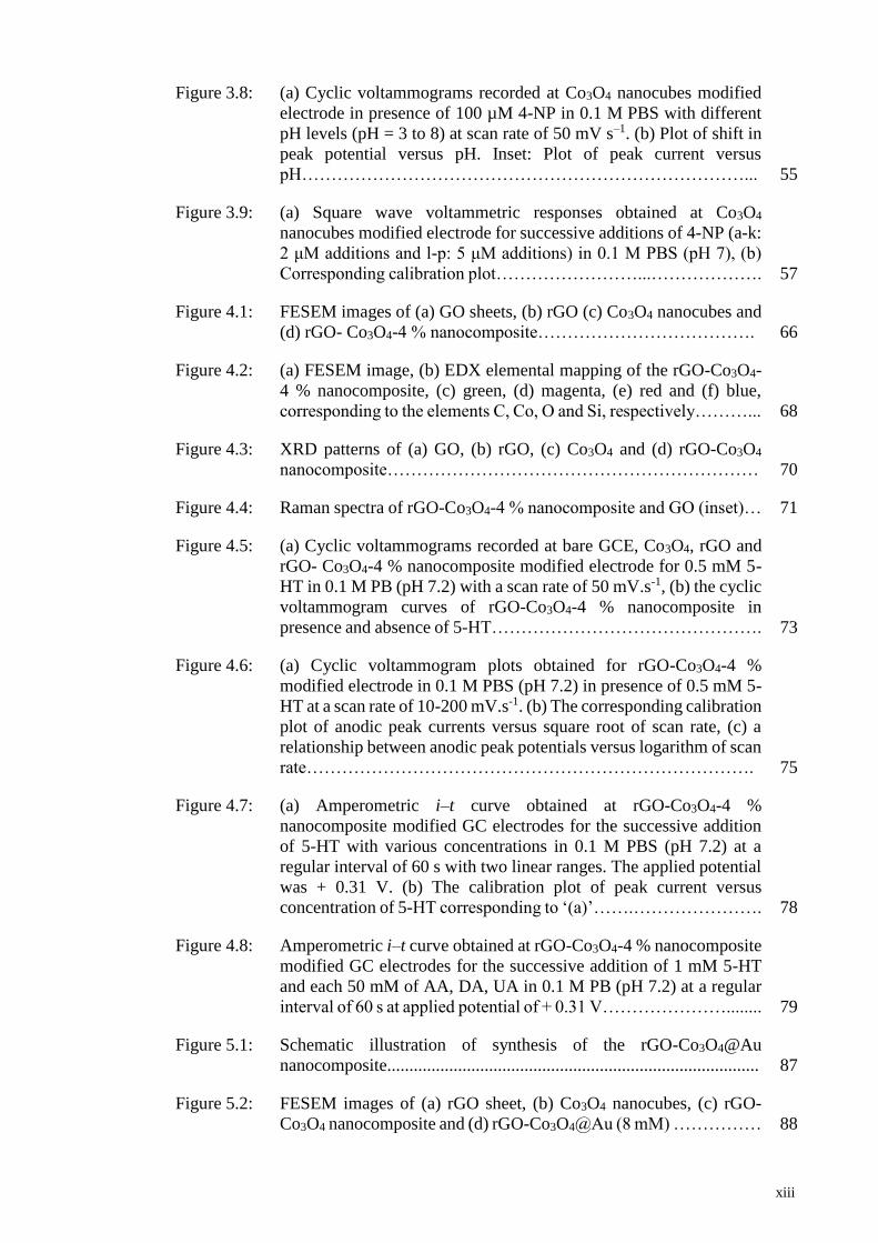
Figure 3.8: (a) Cyclic voltammograms recorded at Co3O4 nanocubes modified
electrode in presence of 100 µM 4-NP in 0.1 M PBS with different
pH levels (pH = 3 to 8) at scan rate of 50 mV s–1. (b) Plot of shift in
peak potential versus pH. Inset: Plot of peak current versus
pH…………………………………………………………………... 55
Figure 3.9: (a) Square wave voltammetric responses obtained at Co3O4
nanocubes modified electrode for successive additions of 4-NP (a-k:
2 μM additions and l-p: 5 μM additions) in 0.1 M PBS (pH 7), (b)
Corresponding calibration plot……………………...………………. 57
Figure 4.1: FESEM images of (a) GO sheets, (b) rGO (c) Co3O4 nanocubes and
(d) rGO- Co3O4-4 % nanocomposite………………………………. 66
Figure 4.2: (a) FESEM image, (b) EDX elemental mapping of the rGO-Co3O4-
4 % nanocomposite, (c) green, (d) magenta, (e) red and (f) blue,
corresponding to the elements C, Co, O and Si, respectively………... 68
Figure 4.3: XRD patterns of (a) GO, (b) rGO, (c) Co3O4 and (d) rGO-Co3O4
nanocomposite……………………………………………………… 70
Figure 4.4: Raman spectra of rGO-Co3O4-4 % nanocomposite and GO (inset)… 71
Figure 4.5: (a) Cyclic voltammograms recorded at bare GCE, Co3O4, rGO and
rGO- Co3O4-4 % nanocomposite modified electrode for 0.5 mM 5-
HT in 0.1 M PB (pH 7.2) with a scan rate of 50 mV.s-1, (b) the cyclic
voltammogram curves of rGO-Co3O4-4 % nanocomposite in
presence and absence of 5-HT………………………………………. 73
Figure 4.6: (a) Cyclic voltammogram plots obtained for rGO-Co3O4-4 %
modified electrode in 0.1 M PBS (pH 7.2) in presence of 0.5 mM 5-
HT at a scan rate of 10-200 mV.s-1. (b) The corresponding calibration
plot of anodic peak currents versus square root of scan rate, (c) a
relationship between anodic peak potentials versus logarithm of scan
rate…………………………………………………………………. 75
Figure 4.7: (a) Amperometric i–t curve obtained at rGO-Co3O4-4 %
nanocomposite modified GC electrodes for the successive addition
of 5-HT with various concentrations in 0.1 M PBS (pH 7.2) at a
regular interval of 60 s with two linear ranges. The applied potential
was + 0.31 V. (b) The calibration plot of peak current versus
concentration of 5-HT corresponding to ‘(a)’…….…………………. 78
Figure 4.8: Amperometric i–t curve obtained at rGO-Co3O4-4 % nanocomposite
modified GC electrodes for the successive addition of 1 mM 5-HT
and each 50 mM of AA, DA, UA in 0.1 M PB (pH 7.2) at a regular
interval of 60 s at applied potential of + 0.31 V…………………........ 79
Figure 5.1: Schematic illustration of synthesis of the rGO-Co3O4@Au
nanocomposite.................................................................................... 87
Figure 5.2: FESEM images of (a) rGO sheet, (b) Co3O4 nanocubes, (c) rGO-
Co3O4 nanocomposite and (d) rGO-Co3O4@Au (8 mM) …………… 88
xiv
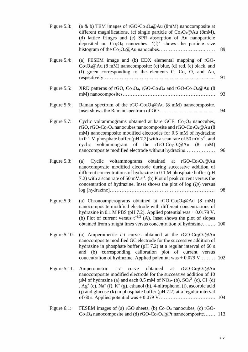
Figure 5.3: (a & b) TEM images of rGO-Co3O4@Au (8mM) nanocomposite at
different magnifications, (c) single particle of Co3O4@Au (8mM),
(d) lattice fringes and (e) SPR absorption of Au nanoparticle
deposited on Co3O4 nanocubes. ‘(f)’ shows the particle size
histogram of the Co3O4@Au nanocubes……………………….…… 89
Figure 5.4: (a) FESEM image and (b) EDX elemental mapping of rGO-
Co3O4@Au (8 mM) nanocomposite: (c) blue, (d) red, (e) black, and
(f) green corresponding to the elements C, Co, O, and Au,
respectively…………………………………………………………. 91
Figure 5.5: XRD patterns of rGO, Co3O4, rGO-Co3O4 and rGO-Co3O4@Au (8
mM) nanocomposites……………………….………………………. 93
Figure 5.6: Raman spectrum of the rGO-Co3O4@Au (8 mM) nanocomposite.
Inset shows the Raman spectrum of GO……………………………. 94
Figure 5.7: Cyclic voltammograms obtained at bare GCE, Co3O4 nanocubes,
rGO, rGO-Co3O4 nanocubes nanocomposite and rGO-Co3O4@Au (8
mM) nanocomposite modified electrodes for 0.5 mM of hydrazine
in 0.1 M phosphate buffer (pH 7.2) with a scan rate of 50 mV s-1. and
cyclic voltammogram of the rGO-Co3O4@Au (8 mM)
nanocomposite modified electrode without hydrazine……………… 96
Figure 5.8: (a) Cyclic voltammograms obtained at rGO-Co3O4@Au
nanocomposite modified electrode during successive addition of
different concentrations of hydrazine in 0.1 M phosphate buffer (pH
7.2) with a scan rate of 50 mV.s-1. (b) Plot of peak current versus the
concentration of hydrazine. Inset shows the plot of log (Ip) versus
log [hydrazine]……………………………………………………… 98
Figure 5.9: (a) Chronoamperograms obtained at rGO-Co3O4@Au (8 mM)
nanocomposite modified electrode with different concentrations of
hydrazine in 0.1 M PBS (pH 7.2). Applied potential was + 0.0179 V.
(b) Plot of current versus t−1/2 (A). Inset shows the plot of slopes
obtained from straight lines versus concentration of hydrazine….…. 100
Figure 5.10: (a) Amperometric i–t curves obtained at the rGO-Co3O4@Au
nanocomposite modified GC electrode for the successive addition of
hydrazine in phosphate buffer (pH 7.2) at a regular interval of 60 s
and (b) corresponding calibration plot of current versus
concentration of hydrazine. Applied potential was + 0.079 V……… 102
Figure 5.11: Amperometric i–t curve obtained at rGO-Co3O4@Au
nanocomposite modified electrode for the successive addition of 10
µM of hydrazine (a) and each 0.5 mM of NO3- (b), SO42- (c), Cl- (d)
, Ag+ (e), Na+ (f), K+ (g), ethanol (h), 4-nitrophenol (i), ascorbic acid
(j) and glucose (k) in phosphate buffer (pH 7.2) at a regular interval
of 60 s. Applied potential was + 0.079 V……………………………. 104
Figure 6.1: FESEM images of (a) rGO sheets, (b) Co3O4 nanocubes, (c) rGO-
Co3O4 nanocomposite and (d) rGO-Co3O4@Pt nanocomposite……. 113

xv
Figure 6.2: FESEM image (a) and EDX elemental mapping (b) of rGO-
Co3O4@Pt nanocomposite: black (c), green (d), blue (e) and red (f)
corresponding to the elements O, Co, C and Pt, respectively….……. 114
Figure 6.3: XRD pattern of rGO-Co3O4@Pt nanocomposite……………….…... 116
Figure 6.4: (a) Raman spectra of the rGO sheet (Inset: Raman spectrum of GO
sheet) and (b) rGO-Co3O4@Pt nanocomposite (Inset: expanded view
of Raman modes of Co3O4…………………….……………………. 118
Figure 6.5: Cyclic voltammograms obtained for bare GC, Co3O4 nanocubes,
rGO, rGO-Co3O4 nanocomposite and rGO-Co3O4@Pt
nanocomposite modified GC electrodes for 1 mM K3[Fe(CN)6] in
0.1 M KCl at a scan rate of 50 mV.s-1……………………………….. 120
Figure 6.6: Nyquist plots obtained for bare GC (black) Co3O4 nanocubes (red),
rGO (blue), rGO-Co3O4 nanocomposite (pink) and rGO-Co3O4@Pt
nanocomposite (green) modified GC electrodes for 1 mM
K3[Fe(CN)6] in 0.1 M KCl. The frequency range was 0.01 Hz to 10
kHz…………………………………………………………………. 122
Figure 6.7: Cyclic voltammograms recorded at bare GC, Co3O4 nanocubes,
rGO, rGO-Co3O4 nanocomposite, rGO-Pt nanocomposite and rGO-
Co3O4@Pt nanocompositen modified electrodes for 5 mM of NO2-
in 0.1 M PBS (pH 2.5) with a scan rate of 50 mVs-1……….………… 124
Figure 6.8: Cyclic voltammograms at rGO-Co3O4@Pt nanocomposite modified
electrode during successive addition of different concentrations of
NO2- in 0.1 M PBS (pH 2.5) with a scan rate of 50 mV.s-1…………... 125
Figure 6.9: (a) Amperometric i–t curves obtained at bare GC, Co3O4 nanocubes,
rGO, rGO-Co3O4 nanocomposite and rGO-Co3O4@Pt
nanocomposite modified GC electrodes for the successive addition
of 1 mM NO2- in 0.1 M PBS (pH 2.5) at a regular interval of 60 s
and (b) corresponding calibration plots of current versus
concentration of NO2-. Applied potentials were the peak potentials
obtained from Figure 6.7…………………….……………………… 127
Figure 6.10: (a) Amperometric i–t curves obtained at the rGO-Co3O4@Pt
nanocomposite modified GC electrodes for the successive addition
of NO2- with various concentrations in 0.1 M PBS (pH 2.5) at a
regular interval of 60 s. Inset: expanded view of the i–t curve
obtained for the successive addition of 10 µM NO2-. The applied
potential was + 0.84 V. (b) Calibration plot of peak current versus
concentration of NO2- corresponding to ‘A’. Inset: the expanded
view of linear calibration plot corresponding to 10 µM NO2-
addition……………………………………………………………... 129
Figure 6.11: Amperometric i–t curve obtained at rGO-Co3O4@Pt nanocomposite
modified GC electrode for the successive addition of 10 µM NO2-
and each 1 mM of DA, AA, UA, glucose, urea and NaCl in 0.1 M
PBS (pH 2.5) at a regular interval of 60 s. Applied potential was +
0.84 V…………………………………………………………….…. 131
xvi
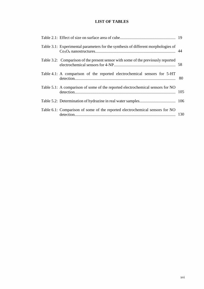
LIST OF TABLES
Table 2.1: Effect of size on surface area of cube...................................................... 19
Table 3.1: Experimental parameters for the synthesis of different morphologies of
Co3O4 nanostructures.............................................................................. 44
Table 3.2: Comparison of the present sensor with some of the previously reported
electrochemical sensors for 4-NP............................................................ 58
Table 4.1: A comparison of the reported electrochemical sensors for 5-HT
detection.................................................................................................. 80
Table 5.1: A comparison of some of the reported electrochemical sensors for NO
detection.................................................................................................. 105
Table 5.2: Determination of hydrazine in real water samples................................... 106
Table 6.1: Comparison of some of the reported electrochemical sensors for NO
detection.................................................................................................. 130
xvii
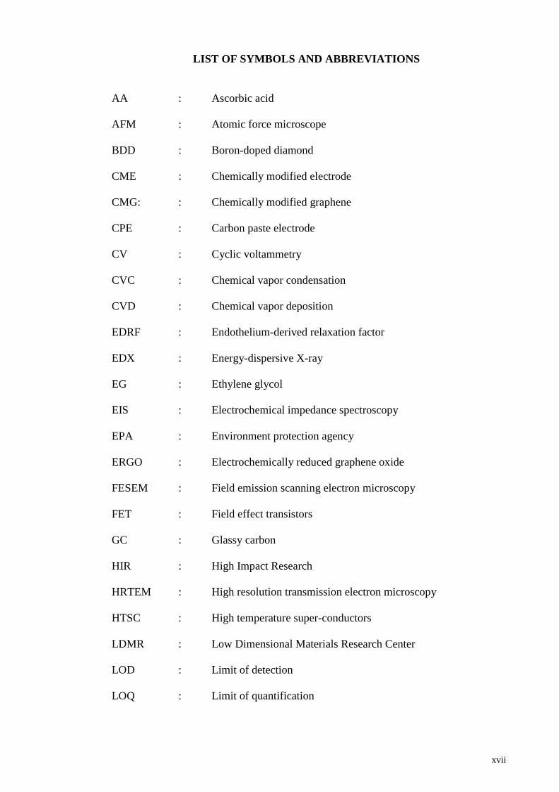
LIST OF SYMBOLS AND ABBREVIATIONS
AA : Ascorbic acid
AFM : Atomic force microscope
BDD : Boron-doped diamond
CME : Chemically modified electrode
CMG: : Chemically modified graphene
CPE : Carbon paste electrode
CV : Cyclic voltammetry
CVC : Chemical vapor condensation
CVD : Chemical vapor deposition
EDRF : Endothelium-derived relaxation factor
EDX : Energy-dispersive X-ray
EG : Ethylene glycol
EIS : Electrochemical impedance spectroscopy
EPA : Environment protection agency
ERGO : Electrochemically reduced graphene oxide
FESEM : Field emission scanning electron microscopy
FET : Field effect transistors
GC : Glassy carbon
HIR : High Impact Research
HRTEM : High resolution transmission electron microscopy
HTSC : High temperature super-conductors
LDMR : Low Dimensional Materials Research Center
LOD : Limit of detection
LOQ : Limit of quantification
xviii
MOCVD : Metal-organic chemical vapor deposition
ORR : Oxygen reduction reaction
PBS : Phosphate buffer solution
SCE : Saturated calomel electrode
SPR : Surface plasmon resonance
STM : Scanning tunneling microscope
SWV : Square wave voltammetry
UA : Uric acid
UM : University of Malaya
VLS : Vapor-Liquid-Solid
VS : Vapor-Solid
WE : Working electrode
XRD : X-ray diffraction

xix
LIST OF APPENDICES
Appendix 1: CV recorded at GC/Co3O4 nanocubes............................................... 164
Appendix 2: EDX spectrum of rGO-Co3O4-4 % nanocomposite........................... 164
Appendix 3: Raman spectra of pure Co3O4 and rGO (inset)................................... 165
Appendix 4: CV recorded for rGO-Co3O4 nanocomposites................................... 166
Appendix 5: (a) CV obtained at rGO-Co3O4-4 % nanocomposite for various
concentration, (b) shows the corresponding calibration plot of
serotonine concentrations versus current........................................... 167
Appendix 6: EDX spectrum of the rGO-Co3O4@Au nanocomposite.................... 168
Appendix 7: XRD pattern of GO........................................................................... 168
Appendix 8: XRD pattern of a) rGO-Co3O4@Au (2 mM), b) rGO-Co3O4@Au
(4 mM), c) rGO-Co3O4@Au (6 mM), d) rGO-Co3O4@Au (8 mM),
e) rGO-Co3O4@Au (10 mM)............................................................ 169
Appendix 9: Raman spectra of rGO, and Co3O4 nanocube (inset)......................... 169
Appendix 10: Cyclic voltammograms recorded at rGO-Co3O4@Au
nanocomposite with different amounts of Au modified electrodes
for 0.5 mM of hydrazine in 0.1 M PBS at a scan rate of 50 mV s-1... 170
Appendix 11: (a) Cyclic voltammograms obtained at rGO-Co3O4@Au 8 mM
nanocomposite modified electrode for 0.5 mM hydrazine in 0.1 M
phosphate buffer with different scan rates a: 10 mVs-1, b: 25 mVs-
1, c: 50 mVs-1, d: 75 mVs-1, e: 100 mVs-1, f: 125 mVs-1, g: 150 mVs-
1, h: 175 mVs-1, i: 200 mVs-1 Inset: Plot of peak current versus
square root of scan rate and (b) the corresponding calibration plot
of log for different scan rate versus peak current............................... 171
Appendix 12: FESEM images of rGO-Co3O4 nanocomposite................................. 172
Appendix 13: EDX spectrum of rGO-Co3O4@Pt nanocomposite........................... 172
Appendix 14: (a) Bode phase plots (A) and Bode impedance plots (log Z vs. log
f) (b) obtained for bare GC, Co3O4 nanocubes, rGO, rGO-Co3O4
nanocomposite and rGO-Co3O4@Pt nanocomposite modified GC
electrodes for 1 mM K3[Fe(CN)6] in 0.1 M KCl................................ 173
Appendix 15: Cyclic voltammograms recorded at rGO-Co3O4@Pt
nanocomposite modified electrode in the absence (a) and presence
(b) of 5 mM NO2- in 0.1 M PBS (pH 2.5) at a scan rate of 50 mV s-
1......................................................................................................... 174
xx
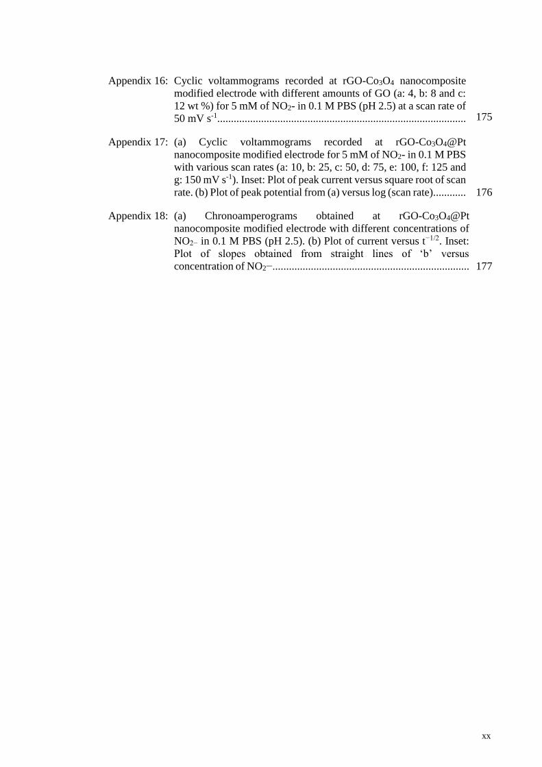
Appendix 16:
Cyclic voltammograms recorded at rGO-Co3O4 nanocomposite
modified electrode with different amounts of GO (a: 4, b: 8 and c:
12 wt %) for 5 mM of NO2- in 0.1 M PBS (pH 2.5) at a scan rate of
50 mV s-1...........................................................................................
175
Appendix 17: (a) Cyclic voltammograms recorded at rGO-Co3O4@Pt
nanocomposite modified electrode for 5 mM of NO2- in 0.1 M PBS
with various scan rates (a: 10, b: 25, c: 50, d: 75, e: 100, f: 125 and
g: 150 mV s-1). Inset: Plot of peak current versus square root of scan
rate. (b) Plot of peak potential from (a) versus log (scan rate)............ 176
Appendix 18: (a) Chronoamperograms obtained at rGO-Co3O4@Pt
nanocomposite modified electrode with different concentrations of
NO2− in 0.1 M PBS (pH 2.5). (b) Plot of current versus t−1/2. Inset:
Plot of slopes obtained from straight lines of ‘b’ versus
concentration of NO2−........................................................................ 177

1
CHAPTER 1: INTRODUCTION
1.1 Background
1.1.1 Nanotechnology
The introduction to nanoscience and nanotechnology has revolutionized the whole
world. Recently, great efforts have been paid to materials that were thought of being
inactive in bulk form for different technological applications. Now it is proven that these
bulk materials can exhibit extraordinary physical and chemical properties at nanoscale
dimensions (Zhang et al., 2008). The idea to synthesize materials at nanoscale dimensions
was presented by Richard Feynman during his lecture “There is Plenty of Room at the
Bottom” in 1959 at an American Physical Society meeting in Caltech. He said that with
nanoscale components, it will be possible to successfully manipulate and control
materials on the atomic and molecular size for electronic and mechanical systems; the
development of technologies into such small systems would be created from combined
fields such as chemistry, biology and physics. Similarly, the term “nanotechnology” was
first introduced in 1974 by a Japanese scientist Norio Taniguchi at The International
Conference on Production Engineering, Tokyo, from 26-29th August. He said that
“nanotechnology” is the process of separation, integration and deformation of material by
using one atom or one molecule. The idea of nanotechnology had been applied in 1980s
by Gerd Binning and Heinrich Rohrer in the invention of scanning tunneling microscope
(STM) which they won the Nobel Prize in Physics in 1986 (Demuth et al., 1986).
Furthermore, the idea of nanotechnology was also used in the development of the atomic
force microscope (AFM), invented by Calvin Quate and Christoph Gerber.
2
More attention are being paid by many researchers from the past few decades to
develop nanostructures because of their unique properties, such as:
1. The large surface area to volume ratio which increases the surface reactivity of
nanomaterials which is useful for chemical and sensing applications.
2. Increased optical emission and absorption due to the electron transfer from one
state to another state which is useful for optoelectronic nanodevices.
It has been also found that the magnetic, optic, catalytic and electronic properties
of nanomaterial strongly depend on their crystallinity, size, structure and morphology
(Rahman et al., 2009).
Recently, nanostructured semiconductors have attracted much attention owing to
their technological applications and fascinating properties (Ng et al., 2003). The transition
metal oxides have been studied intensively and it was found that these metal oxides play
a very crucial role in the field of chemistry, physics and material science, and are widely
used in these fields (Fernandez-Garcia et al., 2004; Sun et al., 2015). A large variety of
oxide compounds can be formed from metal elements that can adopt a vast number of
structural geometries with an electronic structure that can exhibit metallic, semiconductor
or insulator character. Metal oxide nanostructures due to their widespread structural,
physical and chemical properties and functionalities, stand out as one of the most
common, diverse and richest class of material, and among the most versatile groups of
semiconductor nanostructures. Metal oxides proved to be very promising for a variety of
technological applications due to their unique and tunable optical (Cho et al., 2017),
optoelectronic (Allag et al., 2016), magnetic (Xiao et al., 2008), electrical (Cho et al.,
2017), mechanical (Cinthia et al., 2015), thermal (Bala et al., 2009), catalytic (Li et al.,
2017) and photochemical (Stroyuk et al., 2005) properties . Metal oxides nanostructures
have been at the heart of many dramatic advances in materials science. For example, these
3
metal oxides have been used as chemical sensors (Shahid et al., 2015) , gas sensors and
biosensor (Dalkıran et al., 2017; Xu et al., 2017), fuel cells (Shahid et al., 2014),
supercapacitors (Numan et al., 2016), secondary battery materials (Park et al., 2006),
solar cells (Baek et al., 2017), alkaline and lithium ion batteries (Chen et al., 2017),
piezoelectric (Jeong et al., 2006), lasers (Pravinraj et al., 2017), solar absorbers (Shimizu
et al., 2014) and so on. Hence, it was observed that metal oxide nanostructures have been
explored widely by researchers, therefore the understanding of metal oxide
nanostructures is the topic of main interest in term of their synthesis, properties and
applications.
In recent years, cobalt oxide amongst the various types of metal oxides has attracted
intensive attention from researchers due to its tremendous electrical, optical, magnetic
and transport properties (Mini et al., 2016; Xiao et al., 2008). Due to the properties such
as well-defined electrochemical redox activity (Ming-Jay et al., 2009), low cost, stable
chemical state (Xue et al., 2014) and high theoretical capacity (890 mA hg1) (Shahid et
al., 2015), cobalt oxide nanostructures are considered as very promising candidate in the
field of material science for electrochemical applications. Nevertheless, Co3O4
nanostructures have great potential as anode materials for electrochemical devices
(Shahid et al., 2017), rechargeable electronic devices (Numan et al., 2016), Li ion
batteries (Xue et al., 2014), gas sensors (Li et al., 2010), and high-temperature selective
solar-radiation absorbers (Choudhury et al., 1983).
However, Co3O4 is type of semiconductor which suffers from poor conductivity,
low ion transport problem, larger band gap, low electrocatalytic activity and low stability
as compared to metals. To mitigate these issues, researchers have utilized other material
such as metals and conducting platform (graphene, carbon nanotubes and conducting
polymers) and fabricated nanocomposites with Co3O4 which enhances the physical and

4
physiochemical properties of Co3O4. Therefore, Co3O4 based nanocomposite has attained
immense interest in the field of electrochemistry. The major reason towards the interest
in Co3O4 nanostructures and Co3O4 based nanocomposites is due to their technological
application in various fields stated above and versatile morphological structures of Co3O4
which help in boosting the electrocatalytic performance of the nanocomposite.
In recent years, Co3O4 nanostructures and Co3O4 based nanocomposites have been
used intensively as efficient electrode material for electrochemical applications, such as
sensing of water pollutants like phenol based compounds, dyes, bleaches, salts, pesticides,
insecticides, metals, toxins produced by bacteria and human or animal drugs etc.
Moreover, Co3O4 and its nanocomposites are also used to detect the biological molecules
such as serotonin (5-HT), dopamine (DA), ascorbic acid (AA), uric acid (UA) and nitric
oxide (NO) etc. Thus, Co3O4 based nanocomposites provide promising features towards
sensing of target molecules.
1.2 Aim and Objectives
Co3O4 is a very suitable candidate for various applications especially
electrochemical applications. In view of these facts, different structures of Co3O4 could
contribute to electrochemical sensing of target molecule, since the performance of
nanomaterial significantly depends on the size, morphology, crystallinity and distribution
of the particles. Similarly, the specific structure of Co3O4 with higher electrochemical
performance can be used to fabricate a nanocomposite with graphene, which further
contributes to the electrochemical sensing of target molecules, by increasing the effective
surface area of the nanocomposite. Moreover, Co3O4 behaves like impurities on graphene
matrix which increases the defect level and creates an interlayer spacing between the
different layers of GO, which allows the electrolytes to diffuse between the layer of GO.
This could ultimately increase the electrocatalytic activity of the nanocomposites through

5
the interaction of the HOMO-LUMO of graphene with the d-orbital electrons in the Co
atom which significantly enhances the electrocatalytic performance of the
nanocomposite. Metal atoms can be doped into Co3O4 nanoparticles to boost the
performance, as stated above that metals have significant role in improving the
performance of the sensor material. So, the main objectives of the reseach are:
A) Investigation of optimized parameters for the synthesis of different structures of
metal oxides.
B) To Optimize the parameter for the synthesis graphene-metal oxide
nanocomposite.
C) Deposition of metal nanoparticle on the surface of metal oxides to further
enhance the electrocatalytic performance of electrode material.
D) To investigate the performance of electrode material for the detection of target
molecules.
Based on the objectives stated above, the main research focus of the thesis are:
1) Synthesis of different nanostructures of Co3O4 by using simple one step
hydrothermal route by varying the temperature and time. The synthesized
material can be used as a catalyst for the sensing of a water contaminant such as
4-NP.
2) Synthesis of a specific nanostructure of Co3O4 as a composite with rGO chosen
from different synthesized nanostructures which is based on the
performance/electrocatalytic activity for electrochemical sensor studies of
serotonin a biological molecule. To synthesize the rGO-Co3O4 nanocomposite,
ammonia is used which promotes the reduction of GO into rGO and precipitation
of Co3O4. The same one-step hydrothermal route can be used for the synthesis of
rGO-Co3O4 nanocomposite.
6
3) A ternary nanocomposite can be synthesized by the addition of Au i.e. rGO-
Co3O4@Au by using the same synthesis route as mentioned above in the 2nd
hypothesis. The nanocomposite can be used for the electrochemical sensing of
hydrazine.
4) The synthesis of ternary nanocomposite by using the hydrothermal route as stated
above and by replacing Au with Pt i.e. rGO-Co3O4@Pt since Pt is a more efficient
catalyst compared to Au. The electrochemical studies can be carried out for the
sensing of nitric oxide (NO) a very important physiological molecule.
1.3 Structure of Thesis
This thesis is divided into seven chapters, chapter one includes the background
studies of the metal oxides and their nanocomposites used for various applications based
on their properties. Chapter one further includes the aims and objectives of the thesis.
Chapter two provides a brief literature review on metal oxide and metals nanoparticles,
graphene and graphene oxide and metal oxide based nanocomposite and their
applications. Chapter three, four, five and six presents with the research work conducted
during the Ph.D. candidature period which includes experimental details, and results and
discussions involving different type of Co3O4 based nanocomposites and their
electrochemical applications:
1. Morphology dependent electrocatalytic properties of hydrothermally synthesized
cobalt oxide nanostructure.
2. Amperometric detection of depression biomarker using a glassy carbon electrode
modified with nanocomposite of cobalt oxide nanocubes incorporated into reduced
graphene oxide
3. An electrochemical sensing platform of cobalt oxide@gold nanocubes interleaved
reduced graphene oxide for the selective determination of hydrazine.
7
4. An electrochemical sensing platform based on reduced graphene oxide-cobalt
oxide nanocubes@platinum nanocomposite for nitric oxide detection.
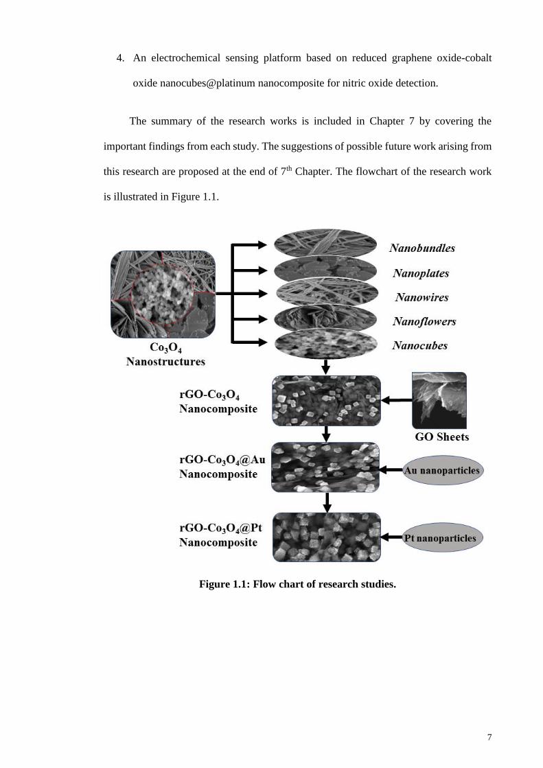
The summary of the research works is included in Chapter 7 by covering the
important findings from each study. The suggestions of possible future work arising from
this research are proposed at the end of 7th Chapter. The flowchart of the research work
is illustrated in Figure 1.1.
Figure 1.1: Flow chart of research studies.
8
CHAPTER 2: LITERATURE REVIEW
2.1 Metal Oxide Nanoparticles
Metal oxides are an important class of materials which are widely used as catalysts
in addition to a wide range of diverse applications in materials science, chemical sensing,
microelectronics, nanotechnology, environmental decontamination, analytical chemistry,
solid state chemistry and fuel cells.
The oxides of metals such as iron, nickel, cobalt, copper and zinc have many
important applications, such as magnetic storage media, solar energy conversion,
electronics, semiconductor and catalysis. The use and performance for different
properties and applications are however, strongly influenced by the crystalline structure,
the morphology and the size of the particles. Therefore, it is very important to develop
methods for the synthesis of metal oxide nanoparticles where the particle size and the
crystal structure of the products can be controlled.
2.1.1 Synthesis Processes of Metal Oxides Nanoparticle
The principal necessity of any innovative research in the field of nanometal oxides
is the preparation of the material. The design of an efficient method for the synthesis of
metal oxide nanoparticles, is a present-day challenge. The synthetic methodologies for
metal oxide nanoparticles can be subdivided into the following categories:
The synthesis of nanomaterials with desired morphology and composition is the
most challenging task in the field of nanoscience and nanotechnology. In the past several
decades, the synthesis of metal oxides nanostructures has stimulated great interest due to
their novel properties which provide intense research efforts to fabricate efficient
miniaturized devices in various nanoelectronics and photonics applications. Thus, various
fabrication techniques have been explored in the literature for the synthesis of these metal
oxide nano-structures.
9
Methods for fabricating nanomaterials can be generally subdivided into two groups:
top-down methods and bottom-up methods. In the first method, nanomaterials are derived
from a bulk substrate and obtained by progressive removal of the material, until the
desired nanomaterials are obtained. A simple way to illustrate the top-down method is to
think of carving a statue out of a large block of marble. Bottom-up methods work in the
opposite direction: the nanomaterials, such as a nanocoating, are obtained starting from
the atomic or molecular precursors and gradually assembling it until the desired structure
is formed.
In both methods two requisites are fundamental: the control of the fabrication
conditions (e.g. energy of the electron beam) and the control of the environmental
conditions (presence of dust, contaminants, etc.). For these reasons, nanotechnology
requires highly sophisticated fabrication tools that are mostly operated in vacuum or
clean-room laboratories.
An overview is given on the experimental techniques used for the synthesis and
characterization of nanomaterials. The synthesis of nanomaterials with desired
morphology and composition is the most challenging task in nanoscience and
nanotechnology. In several decades, the synthesis and characterization of metal oxide
nanostructures have stimulated great interest due to their novel properties which provide
intense research efforts to fabricate efficient miniaturized devices in various
nanoelectronics and photonics applications. Therefore, various fabrication techniques
have been explored in the literature for the synthesis of these metal oxide nanostructures
but typically, they can be divided into two categories: (1) solution phase growth processes
and (2) vapor phase growth processes.

10
2.1.1.1 Chemical Process/Solution Phase Growth Processes
Solution phase growth process is the successful and generic method for the
synthesis of various nanostructures. Unlike the vapor phase synthesis, this method
provides different environment for the growth of the nanostructures. Thus, it considerably
reduces the cost and complexity of the fabrication of nanostructures. Although large
yields of desired nanomaterials are produced based on the solution method, but it also
produces large amount of impurities, which in turn could hamper the applications of the
products. However, the obtained products can be cleaned and hence the impurities can be
decreased by the filtration and washing of the obtained products. In this way, pure
products make this technique commercially applicable for nanostructured formations. To
develop strategies that control and confine the growths, several approaches have been
used and is reported in the literature, such as sol-gel, electrochemical deposition,
surfactant assisted growth process, sonochemical, solvothermal, chemical precipitation
methods etc.
Chemical method for the synthesis of metal oxide nanoparticles is referred to as
bottom-up approach where the nanoparticle synthesis is achieved by the chemical
reduction of metal salts, electrochemical procedures or from the metastable
organometallic compounds by their precisely controlled decomposition. Numerous kinds
of stabilizers such as surfactants, donor ligands, polymeric compounds etc. are utilized
for controlling the growth of nanoparticles and restraining them from agglomeration. The
important types of chemical method for the synthesis of metal oxide nanoparticles are
discussed as follows:
11
(a) Co-precipitation Process
Metal oxides are prepared using the chemical precipitation route. The selection of
proper reactants is the most important factor in any chemical synthesis process. For this
purpose, an extensive knowledge on the chemical reactivities of the reagents, and the
reaction mechanism is required. The morphology and the composition of a nanomaterial
can be controlled efficiently if each reaction step is fully understood. The chemical
reaction could be initiated by mixing the reactants in a beaker or in a round-bottom flask.
The concentration of reactants, reaction time and order of addition of reactants to the
solution, temperature, pH, viscosity and surface tension of the solution are the parameters
which must be controlled. When the reaction products are supersaturated, spontaneous
nucleation occurs and subsequently, it passes through the growth mechanism.
Nanomaterials, with different morphology, can be prepared during this step if proper care
is taken. The major difficulty in the chemical precipitation method is the contamination,
particularly due to the by-product generated in chemical reaction. The optimization
procedure is certainly a tedious task. Numerous experiments at different parameters must
be investigated to achieve the desired results. Even working conditions such as stirring
speed, vibration, exposure to light, cleanliness of glassware etc. can significantly affect
the quality of nanomaterial produced. Hence, the synthesis of nanomaterials of desired
morphology and composition through chemical methods is considered to be an art and
also a skill. (Ajayan et al., 2000)
(b) Sol-gel Process
Sol-gel chemistry has recently evolved into a general and powerful approach for
preparing inorganic materials. This method typically entails hydrolysis of a solution of a
precursor molecule to obtain, first a suspension of colloidal particles (the sol) and then a
gel composed of an aggregate of sol particles. The gel is then thermally treated to yield
12
the desired material. This method is a versatile solution process for preparing ceramic and
glass materials (Interrante et al., 1997).
(c) Electrochemical Deposition Process
This method has been widely used for the fabrication of metallic nanowires in
porous structures and is convenient for the fabrication of metal oxide nanostructures.
Electrodeposition uses dissolved precursors, especially in aqueous solution, is a low cost
and a scalable technique, well suited to produce large scale semiconductor thin films.
Recently, the electrochemical deposition has attracted much attention due to its short
reaction times and low cost. Yang et al. in 2007 reported the synthesis of highly ordered
ZnO ultrathin nanorod and hierarchical nanobelt arrays on zinc substrate with an
electrochemical route in mixed H2O2 and NaOH solution (Yang et al., 2007) Different
materials produced with this method using porous or non-porous structures, substrates
and metal foils etc. are reported in the literature (Rout et al., 2006).
(d) Sonochemical Method
This method of synthesizing materials has proven to be a valuable technique for
producing novel materials with uncommon properties. The sonochemical method of
synthesis basically arises from the acoustic cavitation phenomenon, this phenomenon
includes formation, growth and collapse of bubbles in the aqueous solution (Thompson
et al., 1999). The process occurs under extreme reaction conditions, for example high
pressure greater than 500 atm, very high temperature more than 5000K, and cooling rate
(>1010 K/s, attained during cavity collapse). This method leads to many unique properties
of the irradiated solution, resulting in the formation of nanostructures via the chemical
reaction. A variety of nanostructures are already prepared by the sonochemical method
and reported in the literature (Dhas et al., 1997; Kumar et al., 2000).
13
(e) Hydrothermal or Solvothermal Process
The hydrothermal synthesis is a method of synthesis of single crystals which
depends on the solubility of minerals in hot water under high pressure. The crystal growth
occurs in an apparatus consisting of a steel pressure vessel called autoclave, in which a
nutrient is supplied along with water. A gradient of temperature is maintained at the
opposite ends of the growth chamber so that the hotter end dissolves the nutrient and the
cooler end causes the seeds to grow continuously. The hydrothermal process was initiated
in the middle of the 19th century by geologists and was aimed at laboratory simulations
of natural hydrothermal phenomena. In the 20th century, the hydrothermal synthesis was
clearly identified as an important method for material synthesis, predominantly in the
fields of hydrometallurgy and single crystal growth (Byrappa et al., 2012). Advantages
of the hydrothermal synthesis method include the ability to synthesize crystals of
substances which are unstable near to the melting point, and the ability to synthesize large
crystals of high quality. The solubility of many oxides in hydrothermal solutions of salts
is much higher than in pure water; such salts are called mineralizers. Among the
disadvantages are the high cost of equipment and the inability to control the crystal
growth process (O'Donoghue, 1983).
(f) Template Process
Amongst the numerous synthetic route for the synthesis of controlled sized metal
oxide nanoparticles, the template technique is one of the encouraging approaches to
prepare nanoscale metal oxides. In the template based nanoparticles synthesis, porous
materials comprising of uniform void spaces are utilized as the host to trap nanoparticles
as the guest. The template methods are commonly used in some of the previously stated
approaches and specifically applies to two types of templates, soft-templates (surfactants)
and hard templates (porous solid materials such as silica or carbon).
14
2.1.1.2 Physical Process / Vapor Phase Growth Processes
Physical method is also generally known as the top-down approach for the synthesis
of nanoscale material. In this approach, the bulk material is transformed into nanomaterial
by using physical forces such as milling, grinding, vapor phase deposition etc. Some of
the important physical approaches for the synthesis of nanomaterials are discussed below:
For the growth of a group of nanostructures, vapor phase deposition is the most
versatile technique. In vapor-phase synthesis of nanoparticles, the vapor phase mixture is
thermodynamically unstable relative to the formation of solid material in nanoparticulate
form. This includes the usual situation of a supersaturated vapor. It also includes a third
process ‘chemical supersaturation’ where it is thermodynamically favorable for the vapor
phase molecules to react chemically to form a condensed phase. If the degree of
supersaturation and reaction/condensation kinetics are sufficient, the particles will
nucleate homogeneously. Once nucleation occurs, the remaining supersaturation can be
relieved by condensation or reaction of the vapor-phase molecules on the resulting
particles, thus particle growth will supersede further nucleation. Therefore, to prepare
smaller particles, a high degree of supersaturation by inducing a high nucleation density
is necessary, and immediately quenching the system, either by removing the source of
supersaturation or slowing the kinetics, to stop the growth of the particles. In most cases,
this happens rapidly (milliseconds to seconds) in a relatively uncontrolled fashion, and
lends itself to continuous or quasi continuous operation.
To control the diameter, the aspect ratio and crystallinity of nanomaterial, diverse
techniques have been explored such as thermal evaporation, pulse laser deposition (PLD),
metal organic chemical vapor deposition (MOCVD), sputtering process, thermal
chemical vapor deposition, cyclic chemical vapor deposition (CFCVD) etc. Generally,
two growth mechanisms have been explored for the formation of these metal oxide
15
nanostructures by the aforesaid techniques: (a) Vapor-Liquid-Solid (VLS) and (b) Vapor-
Solid (VS) process.
(a) Thermal Evaporation Method
Various nanostructured materials are grown by the thermal evaporation process. In
this technique, there is a need of high temperature thermal furnace, used for vaporizing
the source material to facilitate the deposition of the nanostructures at relatively lower
temperatures. The vapor species of the source materials are generated first by physical or
chemical methods, and subsequently condensed under certain conditions such as
temperature and pressure on silicon substrate. Numerous nanomaterials have been grown
by this method which ranges from elemental nanowires to a variety of semiconductor
materials (Frohlich et al., 2006; Greene et al., 2003; Wang et al., 2002). Generally, the
thermal evaporation process contains a horizontal quartz tube furnace with rotary pump
and gas supply system.
(b) Pulsed Laser Deposition
A pulsed-laser beam leads to a rapid removal of material from a solid target and to the
formation of an energetic plasma plume, which then condenses onto a substrate. In
contrast to the simplicity of the technique, the mechanisms in PLD including ablation,
plasma formation and plume propagation, as well as nucleation and growth are rather
complex. In the laser ablation process, the photons are converted first into electronic
excitations and then into thermal, chemical and mechanical energy (Kelly et al., 1994;
Miotello et al., 1999) resulting in the rapid removal of material from the surface. This
process has been studied extensively because of its importance in laser machining.
Heating rates as high as 1011 K s-1 and instantaneous gas pressures of 10–500 atm are
observed at the target surface (Kelly et al., 1994).
16
PLD has been used extensively in the growth of high-temperature curates and
numerous other complex oxides, including materials that cannot be obtained via an
equilibrium route. Earlier on, it has been shown that the growth process of materials from
a PLD plume are fundamentally different compared to thermal evaporation (Sankur et al.,
1989). The method has been successful for the film synthesis of Y-type magnetoplumbite
(with a c-axis lattice parameter of 43.5 Å) (Ohkubo et al., 2001) and garnets with 160
atoms per unit cell (Willmott et al., 2000).
(c) Sputtering Process
A final means of vaporizing a solid is via sputtering with a beam of inert gas ions.
Urban et al. in 2002 demonstrated the formation of a dozen different metal nanoparticles
using magnetron sputtering of metal targets. They formed collimated beams of the
nanoparticles and deposited them as nanostructured films on silicon substrates. This
process must be carried out at relatively lower pressures (~1 mTorr), which makes further
processing of the nanoparticles in aerosol form difficult. It is largely driven by the
momentum exchange between ions and atoms in the material, due to collisions. The
surface diffusion process is usually used to explain the formation of nanoscale islands or
rod growth during the sputtering process. Recently this method has been used for the
synthesis of various nanostructures such as ZnO, W, Si, B, etc. (Cao et al., 2001; Cao et
al., 2002; Karabacak et al., 2003).
(d) Mechanical Attrition
One of the important physical method for the synthesis of nanoparticles is
mechanical attrition or mechanical milling of bulk material to produce low dimensional
materials. This technique yields nanoparticles by using milling equipment which are
categorized as “low energy milling” and “high energy milling” based on the material to
be transformed into nanomaterials. The main goal of the milling technique is the reduction

17
of particle size and merging of particles in new phases. In contrast to the several
procedures cited earlier, mechanical attrition leads to the formation of nanostructures
through the structural decomposition of crude grained structures, instead of cluster
assembly due to mechanical deformation. The ball milling and rod milling systems are
potent tools to produce numerous advanced materials. Mechanical attrition is a distinctive
technique which can be carried out at room temperature. The procedure of mechanical
attrition has been performed on high energy mills, vibratory type mill, centrifugal type
mill and low energy tumbling mill. Some of the important milling techniques includes
attrition ball mill, planetary ball mill, vibrating ball mill, low energy tumbling mill and
high energy ball mill.
(e) Metal-organic Chemical Vapor Deposition
This technique MOCVD is widely used for the preparation of epitaxial structures
by atom deposition on a wafer substrate. The operational principle of this method is
simple and has been extensively used for various thin film growths. For the specific
crystal growth, the desired atoms, which are bonded with complex organic gas molecules
are passed over a hot semiconductor wafer. Due to the heat, the complex organic
molecules decompose and are deposited as the desired atoms, via layer by layer
deposition onto the substrate surface. The undesired remnants are removed or deposited
on the walls of the reactor. By varying the composition of the gas, crystal properties
approaching the atomic scale can be achieved. Using this technique, layers of the
precisely controlled thickness can be obtained, which is important for the fabrication of
materials with specific optical and electrical properties. By MOCVD, it is possible to
build a range of semiconductor photodetectors and lasers. Furthermore, scientists are
recently inclined to grow nanostructures with this technique, in addition to thin film
growth. Various semiconductor nanostructures have been synthesized by this technique
as reported in the literature (Baxter et al., 2005; Kang et al., 2006; Su et al., 2005)
18
(f) Chemical Vapor Deposition and Chemical Vapor Condensation
A CVD is well renowned technique where a solid material is deposited on a pre-
heated surface of a substrate through a chemical reaction from the vapor or gas phase.
This process need an appropriate amount of activation energy to initiate the nanoparticle
synthesis which can be supplied through numerous ways. In thermal CVD, the energy to
initiate the reaction is provided by elevated temperature (up to 900 °C). In plasma CVD,
the reaction is triggered by plasma at temperatures between 300 and 700 °C. In laser
equipped CVD technique, the pyrolysis of bulk solid takes place upon the adsorption of
heat from laser thermal energy which eventually leads to nanoscale material synthesis. In
photo-laser equipped CVD, the ultra violet radiation induces the chemical reaction which
has adequate amount of photon energy to breakdown the chemical bond in the reactant
molecules. An alternative method known as CVC was established in Germany in 1994.
It comprises pyrolysis of the metal-organic precursors into vapors, under reduced pressure
atmosphere. Nanoparticles of metal oxides such as ZrO2, Y2O3 and nano-whiskers have
been synthesized by the CVC method (Chang et al., 1994; Rajput, 2015).
2.1.2 Properties of Metal Oxides
2.1.2.1 Surface Properties
Little is known about the surface structures of transition metal oxides, but their bulk
crystal structures are well researched. The physical and chemical properties of any
material depend mainly on its surface properties regardless of its bulk or nanoscale nature.
The surface of any type of material is different compared to the bulk, as the movement
and exchange of matter and energy occur through an area called the interface. In addition,
they also can either initiate or terminate a chemical reaction, like in the case of catalysts.
When a bulk solid material is further segmented into nano-regime material, the total
collective surface area is significantly enhanced although the total volume remains the
same. Therefore, the surface-to-volume ratio of the material is increased as compared to
19
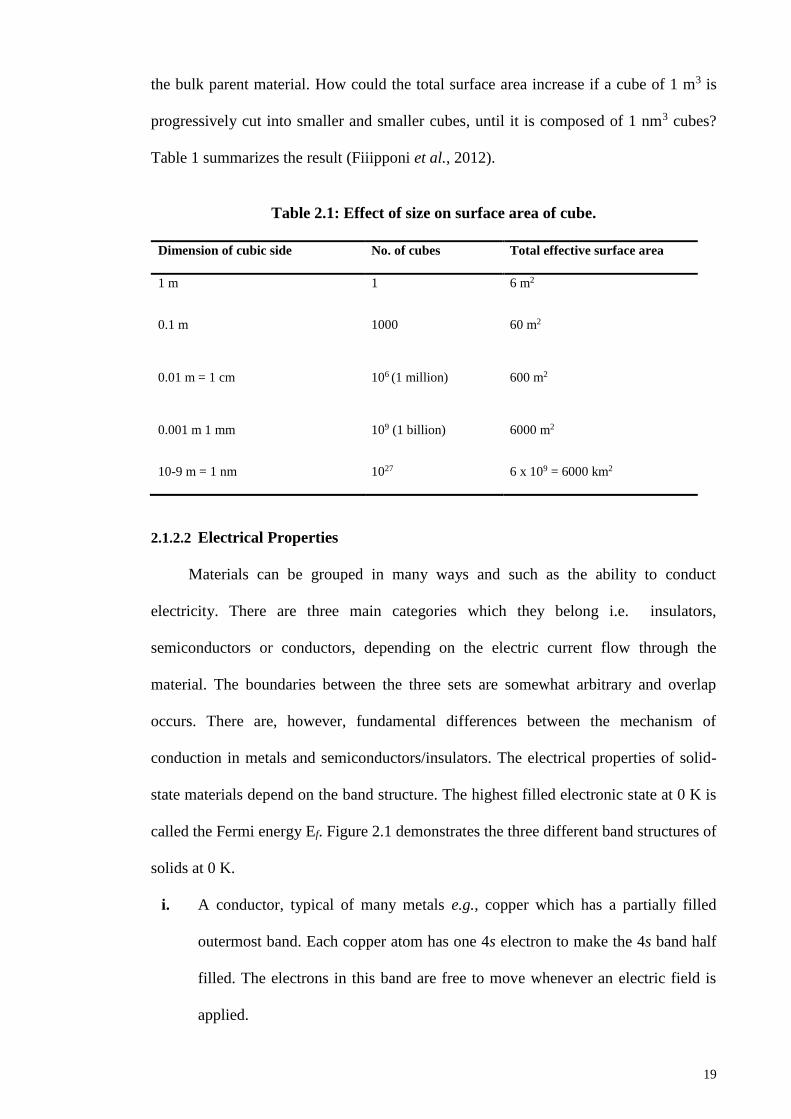
the bulk parent material. How could the total surface area increase if a cube of 1 m3 is
progressively cut into smaller and smaller cubes, until it is composed of 1 nm3 cubes?
Table 1 summarizes the result (Fiiipponi et al., 2012).
Table 2.1: Effect of size on surface area of cube.
Dimension of cubic side No. of cubes Total effective surface area
1 m 1 6 m2
0.1 m 1000 60 m2
0.01 m = 1 cm 106 (1 million) 600 m2
0.001 m 1 mm 109 (1 billion) 6000 m2
10-9 m = 1 nm 1027 6 x 109 = 6000 km2
2.1.2.2 Electrical Properties
Materials can be grouped in many ways and such as the ability to conduct
electricity. There are three main categories which they belong i.e. insulators,
semiconductors or conductors, depending on the electric current flow through the
material. The boundaries between the three sets are somewhat arbitrary and overlap
occurs. There are, however, fundamental differences between the mechanism of
conduction in metals and semiconductors/insulators. The electrical properties of solid-
state materials depend on the band structure. The highest filled electronic state at 0 K is
called the Fermi energy Ef. Figure 2.1 demonstrates the three different band structures of
solids at 0 K.
i. A conductor, typical of many metals e.g., copper which has a partially filled
outermost band. Each copper atom has one 4s electron to make the 4s band half
filled. The electrons in this band are free to move whenever an electric field is
applied.
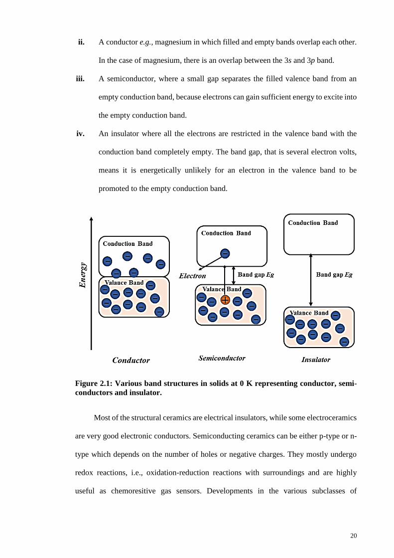
20
ii. A conductor e.g., magnesium in which filled and empty bands overlap each other.
In the case of magnesium, there is an overlap between the 3s and 3p band.
iii. A semiconductor, where a small gap separates the filled valence band from an
empty conduction band, because electrons can gain sufficient energy to excite into
the empty conduction band.
iv. An insulator where all the electrons are restricted in the valence band with the
conduction band completely empty. The band gap, that is several electron volts,
means it is energetically unlikely for an electron in the valence band to be
promoted to the empty conduction band.
Figure 2.1: Various band structures in solids at 0 K representing conductor, semi-
conductors and insulator.
Most of the structural ceramics are electrical insulators, while some electroceramics
are very good electronic conductors. Semiconducting ceramics can be either p-type or n-
type which depends on the number of holes or negative charges. They mostly undergo
redox reactions, i.e., oxidation-reduction reactions with surroundings and are highly
useful as chemoresitive gas sensors. Developments in the various subclasses of
21
electroceramics have paralleled the growth of emerging technologies, examples are such
as optical properties.
2.1.2.3 Optical Properties
Additionally, metal oxides display optical characteristics and have shown ground
state electronic structures, as well as numerous excitations of charge, spin, orbital and
lattice degrees of freedom. The optical features of metal oxides have broadened their field
of technological applications such as optical and optoelectronic devices, by applying the
optical responses, magneto-optical effect, photo-refractive effect and elasto-optic effect.
Moreover, the conductivity of a semiconductor depends on the current or voltage applied
to a control electrode or to the intensity of irradiation by infrared, visible light, ultraviolet
(UV) or X-ray.
2.1.2.4 Redox Properties
Metal oxides are one of the most important and widely employed classes of solid
catalysts, either as active phases or as supports. Metal oxides especially containing
transition metals possess redox properties in addition to their acidic and basic nature. This
is due to the interaction with reactant molecules such as CO, H2, and O2 which could lead
to electron transfer from the surface to the adsorbed species and modify the valence state
of the metal centers.
Metal oxides are used because of their acid-base and redox properties and constitute
the largest family of heterogeneous catalysis (Brückner, 2003; Henrich et al., 1996; Henry
et al., 1998; Kung, 1989; Noguera, 1996; Védrine, 2002; Zecchina et al., 2001). The three
key features of metal oxides, which are essential for applications in catalysis, are
(i) Coordination environment of the surface atoms.
(ii) Redox properties of metal oxides.
(iii) Oxidation state of the surface.
22
The control of surface coordination environment can be done by the choice of
crystal plane exposition and by the preparation methods employed; however,
specification of redox properties is mainly due to the type of oxide. Many oxide catalysts
correspond to more or less complex transition metal oxides containing cations of available
oxidation state which introduce redox properties in addition to acid-base properties. The
acid-base properties of the oxides are usually interrelated to their redox behavior. In
particular, the redox behavior of the metal oxide being used and the effect of various
additives on its redox properties are the principal factors for the formulation of a catalyst
for oxidation and related reactions (Fierro, 2005).
2.1.2.5 Magnetic Properties
Inorganic solids that have magnetic effects other than diamagnetism (a property of
all substances), are characterized by the presence of unpaired electrons, usually located
on metal cations. Just like electronic properties, the magnetic properties of three
dimensional solids result from the interaction of metal centers. Magnetic behavior is thus
restricted to the compounds having transition metals and lanthanides due to the presence
of unpaired d and f electrons, respectively. In a discrete molecule in solution, the unpaired
electrons are totally independent and are randomly oriented. The metal oxides show a
variety of phenomena, such as magnetism, dielectricity, superconductivity etc., which are
remarkably sensitive to their chemical compositions, crystal structures, carrier
concentrations and the applied external field. Long time ago, the study of magnetic
properties in metal oxides was done for fundamental aspects as well as applications
(Adler, 1968; Tsuda et al., 2013). Magnetic oxides, especially ferrites e.g., MgFe2O4, are
new materials which are used in transformer cores, magnetic recording and information
storage devices, etc. After the discovery of high temperature super-conductors (HTSC)
cupperates, metal oxides has been studied from the modern point of view and the most

23
attractive compound after HTSC is the manganites with perovskite structure (R1-
xAxMnO3), whose study started as early as the 1950s (Jonker et al., 1950).
2.1.2.6 Other Properties
In addition to the properties discussed earlier, metal oxides exhibit distinctive
chemical, mechanical, catalytic and adsorption properties. Metal oxide nanoparticles have
been extensively employed for industrial applications in the field of catalysis as active
compositions or as supports materials. Metal oxides possess photocatalytic abilities which
can be exploited to solve the present-day energy crises. In 1972, Fujishima and Honda
first reported the photocatalytic splitting of water using TiO2, which was the first
photocatalyst used for water splitting and the commencement of a new field of modern
heterogeneous photocatalysis (Fujishima et al., 1972; Ni et al., 2007). Currently, metal
oxide nanoparticles, owing to large surface to volume ratio and enhanced surface binding
properties have been employed as adsorbent to remove environmental pollutants. Metal
oxide nanoparticles exhibit stability towards radioactive radiations, thermal and
mechanical changes and are exploited for the irreversible, selective and efficient removal
of large amounts of pollutants from contaminated water. Thus, metal oxide in nanoscale
have demonstrated unique and distinctive properties and have been applied extensively
in the field of chemical, nuclear-energy, pharmaceutical, food, bioengineering, dairy,
water treatment and electronic industries.
2.2 Cobalt Oxide
Cobalt oxide (Co3O4), a magnetic p-type semiconductor, is an important class of
inorganic metal oxide which belongs to normal spinel structure based on a cubic close
packing array of oxide ions. Spinel cobalt oxide (Co3O4) nanomaterials have been widely
explored recently, as they possess facile synthetic methodologies, excellent catalytic
properties and diverse morphologies (Jiao et al., 2010). Thus, Co3O4 appears as a
24
promising candidate for various applications such as fuel cells, lithium ion batteries,
photocatalysis, artificial photosynthesis, gas sensors, etc., due to its eclectic abundance
and economic cost (Hu et al., 2008; Jiao et al., 2009; Li et al., 2005; Shahid et al., 2014).
Cobalt (II, III) oxide is one of two well characterized cobalt oxides. As shown in
scheme 1, cobalt (II, III) oxide is a mixed valence compound, and its formula is
sometimes written as CoIICoIII2O4 and sometimes as CoO & Co2O3. Like Fe3O4, Co3O4
has a spinel structure. The Co2+ occupies the tetragonal 8(a) sites, while Co3+ occupies
the octahedral 16(d) sites. The 32(e) sites are occupied by 32 O2- ions. As known, the
spinel minerals have the generic formula AB2O4, where A is a cation with a +2 charge
and B is a cation with +3 charges. The oxygen atoms in a spinel are arranged in a cubic
close-packed structure, and the cations A and B occupy some or all of the octahedral and
tetrahedral sites in the lattice (Wang et al., 2012).

25
Figure 2.1: The spinel structure of cobalt (II, III) oxide.
The difference in oxygen defects, oxygen holes and oxygen adsorbed in different
states of cobalt in Co3O4 (a mixed valance material that is formally CoII CoIII2 O4) are
thought to be the reason for high activity and selectivity of this metal oxide catalysts
(Sharifi et al., 2013).

26
The Co3O4 adopts the normal spinel structure, with Co2+ ions in tetrahedral
interstices and Co3+ ions in the octahedral interstices of the cubic close-packed lattice of
oxide anions (Greenwood et al., 1997).
Figure 2.2: Co3O4 normal spinel structure coordination geometry (a) tetrahedral
coordination geometry Co(II) (b) distorted octahedral coordination geometry
Co(III) and (c) distorted tetrahedral coordination geometry of O.
Co3O4 is an important form among the various cobalt oxides based on its distinctive
structural features and properties (Shi et al., 2012). It has been demonstrated that these
nanostructured transition metal oxides have even more attractive applications such as
heterogeneous catalysts, gas sensors, lithium ion batteries, electrochromic devices, solar
energy absorbers, ceramic pigments and optical devices, etc. (Ando et al., 2004; Chou et
al., 2008; Li et al., 2005; Lou et al., 2008; Makhlouf, 2002; Rahman et al., 2012; Wu et
al., 2003). Further details and synthesis procedures of cobalt oxide is explained briefly in
chapter 3, 4, 5 and 6.
2.3 Graphene Oxide and Graphene
2.3.1 Graphene
Graphene a two-dimensional material is one of the allotropes of carbon. It is an
isolated single layer in graphite composed of sp2 hybridized carbon atoms. It is composed
of sp2 hybridized carbon atoms arranged in a 2D honeycomb crystal lattice having C-C
(a) (c) (b)
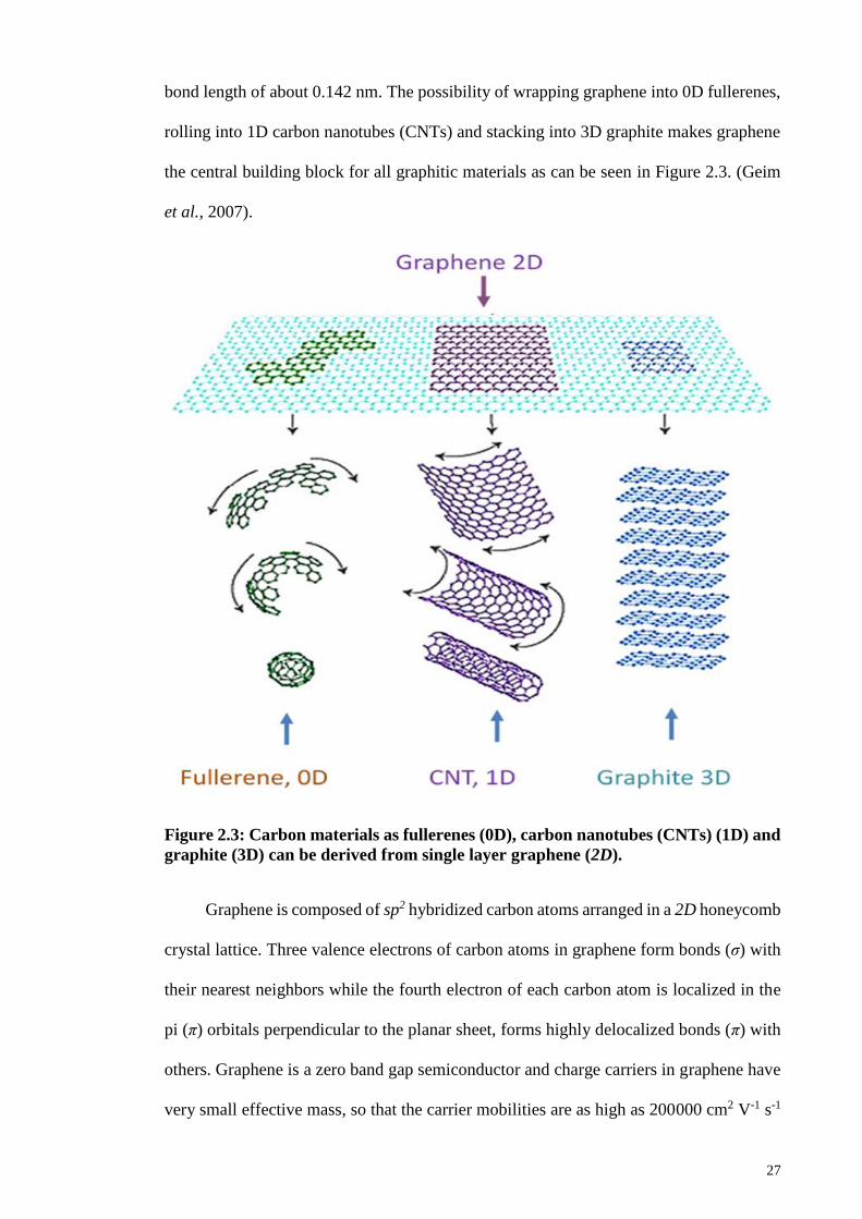
27
bond length of about 0.142 nm. The possibility of wrapping graphene into 0D fullerenes,
rolling into 1D carbon nanotubes (CNTs) and stacking into 3D graphite makes graphene
the central building block for all graphitic materials as can be seen in Figure 2.3. (Geim
et al., 2007).
Figure 2.3: Carbon materials as fullerenes (0D), carbon nanotubes (CNTs) (1D) and
graphite (3D) can be derived from single layer graphene (2D).
Graphene is composed of sp2 hybridized carbon atoms arranged in a 2D honeycomb
crystal lattice. Three valence electrons of carbon atoms in graphene form bonds (σ) with
their nearest neighbors while the fourth electron of each carbon atom is localized in the
pi (π) orbitals perpendicular to the planar sheet, forms highly delocalized bonds (π) with
others. Graphene is a zero band gap semiconductor and charge carriers in graphene have
very small effective mass, so that the carrier mobilities are as high as 200000 cm2 V-1 s-1

28
at a carrier density of 1012 cm−2 (Du et al., 2008). Electrons can flow through graphene
more easily compared to copper metal. The edges of graphene are also known as an
armchair or a zig-zag edge, due to the individual lattice arrangement on the atomic scale
(see Figure 6.1(a)).
Figure 2.4: Armchair and zig-zag edges in graphene (a), sp2 hybridization illustrated
in graphene (b).
The opacity of a single graphene layer is 2.3 % so its optical transparency is 97.7
% observed in the visible range, but decreases linearly as the number of layers increases
The mechanical properties of graphene have been investigated by numerical simulations
and experimental measurements using AFM (Van Lier et al., 2000). Graphene is one of
the strongest materials with a mechanical strength higher than diamond and over 300
times greater than a steel film of the same thickness (Lee et al., 2008). Reported values
for defect-free graphene are Young’s modulus of 1.0 TPa and a fracture strength of 130
GPa that are higher than CNTs (Van Lier et al., 2000). Graphene is flexible and
stretchable up to 20 % of its initial length. In addition to these outstanding properties,
graphene has a thermal conductance (>5000 W/m K) (Chen et al., 2012) that is also higher
than all the other carbon structures and theoretical surface area of 2600 m2/g (Stankovich
et al., 2007).

29
2.3.2 Graphene Oxide
During the last few years, chemically modified graphene (CMG) has attracted great
interest in the perspective of several applications such as sensors, energy related
materials, polymer composites, field effect transistors (FET), paper-like materials and
biomedical relevance due to the remarkable mechanical, thermal and electrical, properties
(Park et al., 2009). Chemically modified graphene oxide has been a favorable route to
obtain mass produced CMG platelets. Like graphite, graphite oxide also has a layered
structure, but in graphite oxide the carbon planes are highly decorated by oxygen
containing functional groups. These functional groups not only make the layers
hydrophilic, but they also expand the interlayer distance. With moderate ultrasonication,
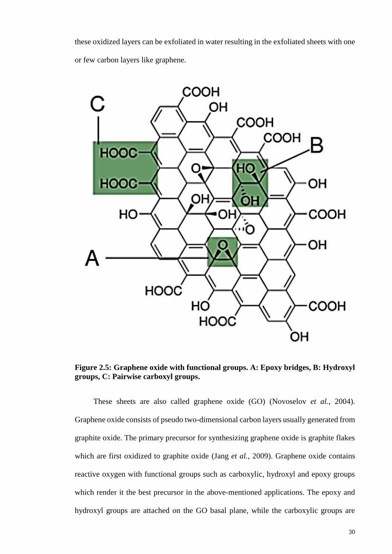
30
these oxidized layers can be exfoliated in water resulting in the exfoliated sheets with one
or few carbon layers like graphene.
Figure 2.5: Graphene oxide with functional groups. A: Epoxy bridges, B: Hydroxyl
groups, C: Pairwise carboxyl groups.
These sheets are also called graphene oxide (GO) (Novoselov et al., 2004).
Graphene oxide consists of pseudo two-dimensional carbon layers usually generated from
graphite oxide. The primary precursor for synthesizing graphene oxide is graphite flakes
which are first oxidized to graphite oxide (Jang et al., 2009). Graphene oxide contains
reactive oxygen with functional groups such as carboxylic, hydroxyl and epoxy groups
which render it the best precursor in the above-mentioned applications. The epoxy and
hydroxyl groups are attached on the GO basal plane, while the carboxylic groups are
31
present on the edges of graphene oxide. However, due to these functional groups, GO is
strongly hydrophilic in nature and disperse easily in water and intercalation of water
molecules readily occur between the GO sheets. Depending upon the relative humidity
within the stacked GO sheets, the interlayer spacing between GO sheets varies
significantly from 0.6 to 1.2 nm (Buchsteiner et al., 2006). Consequently, the interaction
between GO sheets is weakened, while the inter-sheet spacing is increased which in turn
enables the exfoliation of GO sheets. Mechanical stirring, thermal shock, ultra-sonication
in water, or polar solvents are used to exfoliate GO into two dimensional individual
nanosheets (Hu et al., 2010). However, many studies claimed that too much
ultrasonication could decrease the lateral dimension of the GO sheets. The resulting
individual GO sheets are mostly single or few layer sheets that disperse readily in water
to make a stable colloidal GO suspension. The GO suspension stability originates from
the negative electrostatic repulsion due to the ionization of phenolic hydroxyl groups and
carboxylic groups (Li et al., 2008). Because of the introduction of oxygen functional
groups on the carbon basal planes, the thickness of single-layer GO sheets has been
reported approximately between 1–1.4 nm. In other words, the individual GO sheet
thickness is approximately three times greater than an ideal single graphene layer (de
Moraes et al., 2015). Indeed, the graphite oxide exfoliation into individual GO sheet can
also occur in polar organic solvents such as N-methylpyrolidine (NMP), ethylene glycol
(EG) and N, N-dimethylformamide (DMF). It forms a non-aqueous colloidal suspension
that is analogous to aqueous GO colloidal suspension (Paredes et al., 2008). Generally,
the GO sheet concentration, dispersed in water, is up to 3 mg/ml. The aqueous GO
colloidal suspension offers an appropriate setting for an electrochemical method of
converting GO into electrochemically reduced graphene oxide (ERGO). However, the
properties of ERGO are different from pristine graphene because of various residual
oxygen containing functional groups on the carbon basal plane (Viinikanoja et al., 2012).
32
2.3.3 Synthesis of Graphene Oxide
The oxidation of graphite is carried out by mixing graphite flakes into
H2SO4:H3PO4 (320:80 mL) and KMnO4 (18 g) with continuous stirring. After the slow
addition of all materials, the one-pot mixture was left stirring for 3 days to allow the
oxidation of graphite. The color of the mixture changed from dark purplish green to dark
brown. Later, H2O2 solution was added to stop the oxidation process in the presence of
ice to control the temperature, and the color of the mixture changed to bright yellow,
indicating a high oxidation level of the graphite. The graphite oxide product was washed
three times with 1 M of HCl aqueous solution and repeatedly with deionized water until
a pH of 4–5 was achieved. The washing process was carried out using simple decantation
of supernatant via a centrifugation technique with a centrifugation force of 11,500 g.
During the washing process with deionized water, the graphite oxide experienced
exfoliation, which resulted in the thickening of the graphene solution, forming a GO gel.
The synthesis of graphene oxide can be summarized into three steps:
1. In first step, the graphite flakes are treated with strong acids and a strong oxidizing
agent i.e. KMnO4, the product obtained after the treatment is named as graphite
oxide since it has oxygen containing hydroxyl and epoxide groups across the basal
planes of graphite oxide and carbonyl groups situated at the edges of lattice.
2. In the second step, the oxidation will be stopped by H2O2 and ice, and the washing
process will be carried out to remove the acids and to raise the pH of graphite
oxide, i.e. the washing process will also contribute in the exfoliation of graphite
oxide.
3. Lastly, the sonication of the washed product will be carried out to exfoliate the
graphite oxide to graphene oxide.
The following figure shows the synthesis process of GO.
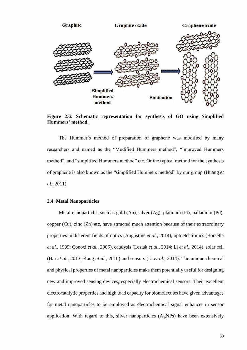
33
Figure 2.6: Schematic representation for synthesis of GO using Simplified
Hummers’ method.
The Hummer’s method of preparation of graphene was modified by many
researchers and named as the “Modified Hummers method”, “Improved Hummers
method”, and “simplified Hummers method” etc. Or the typical method for the synthesis
of graphene is also known as the “simplified Hummers method” by our group (Huang et
al., 2011).
2.4 Metal Nanoparticles
Metal nanoparticles such as gold (Au), silver (Ag), platinum (Pt), palladium (Pd),
copper (Cu), zinc (Zn) etc, have attracted much attention because of their extraordinary
properties in different fields of optics (Augustine et al., 2014), optoelectronics (Borsella
et al., 1999; Conoci et al., 2006), catalysis (Lesiak et al., 2014; Li et al., 2014), solar cell
(Hai et al., 2013; Kang et al., 2010) and sensors (Li et al., 2014). The unique chemical
and physical properties of metal nanoparticles make them potentially useful for designing
new and improved sensing devices, especially electrochemical sensors. Their excellent
electrocatalytic properties and high load capacity for biomolecules have given advantages
for metal nanoparticles to be employed as electrochemical signal enhancer in sensor
application. With regard to this, silver nanoparticles (AgNPs) have been extensively
34
investigated as an effective electrocatalyst for electrochemical sensor applications
(Yusoff et al., 2017). AgNPs continue to gather enormous attention because they require
low production cost, are environmentally friendly, have low toxicity and good
biocompatibility. Moreover, AgNPs possesses advantages of excellent catalytic activity,
high conductivity and high surface energy, which makes them a promising catalyst
material. Furthermore, the high surface to volume ratio allows the exposition of large
fraction of metal atoms to the reactant molecules and is very much desirable for sensor
applications (Rastogi et al., 2014). Besides that, they are also the best conductors among
all the noble metals (Jiang et al., 2013). Due to these properties, AgNPs may facilitate
more efficient electron transfer than the other noble metal nanoparticles. Besides AgNPs,
gold nanoparticles (AuNPs) have recently drawn increasing attention from many
researchers in the field of sensors (Yusoff et al., 2017) due to their novel chemical, optical
and physical properties such as high effective surface to volume ratio, excellent electrical
and heat conductivity, and strong absorption in the visible and near infrared wavelength
region (380 to 750 nm). Among the important physical properties of AuNPs are the
surface plasmon resonance (SPR) and the ability to quench fluorescence (Yeh et al.,
2012). Besides that, they have excellent biocompatibility and low toxicity which are
suitable for biotechnology applications (Khlebtsov et al., 2011). AuNPs also exhibit high
chemical stability and inertness under physiological conditions as well as excellent
electrocatalytic properties. All these properties make AuNPs an attractive material for
electrochemical and biological devices. More interestingly, the properties of AuNPs can
be controlled by tuning the shape and size (Jain et al., 2006). Because of its smaller size,
Au could provide high active surface area, thus, improve the electron transfer process.
This will lead to the enhancement in sensitivity and signal to noise ratio, therefore
improves the analytical performance. Another noble metal that has the potential of a
catalyst in electrochemical sensors is palladium nanoparticles (PdNPs) (Liu et al., 2016).
35
PdNPs have attracted extensive attention because of their good chemical and physical
properties, such as wear and corrosion resistance, as well as good stability. Their high
specific surface area could increase the mass transport and enhance the electron transfer
kinetics, thus improves the electrocatalytic activity. PtNPs have been used for
electrochemical sensor application in composite with graphene and metal oxides, due to
their potential electrical communication, resulting in an efficient electron transfer process
at the modified electrode. The Pt nanoparticles also provide a larger surface area for the
effective interaction of target molecules and thereby improves the electron-transfer
kinetics and the electrocatalytic performance (Shahid et al., 2015).
2.5 Cobalt Oxide Based Nanocomposites
Nanocomposites are material composites where at least one of the phases shows
dimensions in the nanoscale range (1 nm = 10-9 m). Nanocomposite materials have
emerged as suitable alternatives to overcome limitations of microcomposites and
monolithic, while posing preparation challenges related to the control of elemental
composition and stoichiometry in the nanocluster phase. They are the advanced materials
of the 21st century due to the unique design and property combinations that are not found
in conventional composites. The general understanding of these properties is yet to be
reached, although the first inference about them was reported as early as 1992.
2.5.1 Synthesis of Cobalt Oxide and its Composites with Graphene Oxide
Co3O4 nanoparticles and Co3O4 based nanocomposite can be synthesized through
many routes such as mechanical, sonication, electrochemical, chemical,
hydrothermal/solvothermal methods of synthesis and so on.
2.5.1.1 Hydrothermal Method
Co3O4 nanoparticles and its nanocomposite have been synthesized with graphene,
carbon nanotubes, metals and polymers etc. by hydrothermal routes. The hydrothermal
36
synthesis is generally defined as crystal synthesis or growth under high temperature and
water pressure conditions from substances which are insoluble in normal temperature and
pressure (<100 °C, <1 atm). Since the ionic product (Kw) has a maximum value around
250–300 °C, the hydrothermal synthesis is usually carried out below 300 °C. The critical
temperature and water pressure are 374 °C and 22.1 MPa, respectively. The solvent
properties for many compounds, such as dielectric constant and solubility, change
dramatically under supercritical conditions. The dielectric constant of water is 78 at room
temperature where polar inorganic salts can be dissolved in water. The dielectric constant
of water decreases with increasing temperature and decreasing pressure. When the
dielectric constant is below 10 under supercritical conditions, the contribution of the
dielectric constant to the reaction rates increases based on the electrostatic theory. Thus,
supercritical water gives a favorable reaction medium for particle formation, owing to the
enhancement of the reaction rate and large supersaturation, based on the nucleation
theory, due to the lower solubility. The formation mechanism of metal oxide
nanoparticles from metal nitrate solution is as follows: First, the hydrated metal ions are
hydrolyzed to metal hydroxide. Then, the metal hydroxides precipitate as metal oxides
through the dehydration step.
Hydrolysis is regarded as an electrostatic reaction between metal ions and hydroxyl ions.
2.5.2 Application of Cobalt Oxide Based Nanocomposite
2.5.2.1 Electrochemical Detection/Sensing of Target Molecules
Electrochemical techniques have been used to measure the concentration of
biomolecules due to the direct transformation of electrochemical information produced
OHx
MOOHM
xHNOOHMOxHNOM
xx
x
22
3223
2)(
)()(

37
by biochemical mechanism into an analytically useful signal. Electrochemical biosensors
have advantages such as high sensitivity and selectivity towards the electroactive target
molecules, rapid and accurate response and most importantly it is portable and
inexpensive compared to another existing biosensor.
Besides that, it also offers advantages of wide linear response range, good stability
and reproducibility. There are two basic components of an electrochemical sensor which
works together as a working or sensing electrode, that are a chemical recognition system
and physicochemical transducer. Other than the working electrode (WE), the reference
and counter electrodes are also required in this sensor and are enclosed in the sensor
housing in contact with a liquid electrolyte and biomolecules. As a biosensor, the
recognition layers must interact with the target biomolecules and the physicochemical
transducer will translate the bio-recognition event into a useful electrical signal which can
be detected by electrochemical workstation. Amperometry, cyclic voltammetry (CV) and
potentiometry are some of the examples of electrical signal resulting from the
transduction of a biological signal. One of the most important components in
electrochemical sensing technique is the WE because it is the place where all the
electrochemical oxidation and reduction occur. There are various types of WE that have
been commercialized such as platinum (Pt), gold (Au), mercury (Hg) and carbon
electrode. Even though Pt electrode demonstrates good electrochemical inertness, the
high cost for production and contamination from the presence of small amounts of water
or acid in the electrolyte limits the application. The catalytic reduction of protons to form
hydrogen gas (hydrogen evolution) at low negative overpotential obscures any useful
analytical signal.
Another metal electrode that behaves almost like Pt electrode is the Au electrode.
The Au electrode provides good electron transfer kinetics and a wide anodic potential
38
range; however, it exhibits weakness in the positive potential range due to the oxidation
of the surface. The mercury electrode is another type of WE that has been used in
electrochemical sensing technique due to its high hydrogen overvoltage which can extend
the cathodic potential window. Besides that, it also possesses highly reproducible,
renewable and smooth surface, which is very beneficial in electrochemical analysis. The
most common form of mercury electrode is the dropping mercury electrode, hanging
mercury drop electrode and mercury film electrode. Nevertheless, the toxicity and limited
anodic range of mercury limits the applications. Among the different types of electrodes,
the carbon electrode such as carbon paste electrode (CPE) and glassy carbon (GC)
electrode has been commonly used as WE, due to the wider negative and anodic potential
range compared to other electrodes. Moreover, carbon electrodes also have a low
background current, rich surface chemistry as well as comparative chemical inertness.
Therefore, researchers commonly utilize the carbon electrode as the WE in
electrochemical sensing.
Razmi et al. reported the amperometric detection of acetaminophen by an
electrochemical sensor based on cobalt oxide nanoparticles. They reported the suitability
of a carbon ceramic electrode for the uniform formation of cobalt oxide nanoparticles
with an average size of approximately 70 nm by a simple and inexpensive CV method.
They demonstrated that the electrode exhibited good electrocatalytic activity toward the
oxidation of acetaminophen in an alkaline medium because of the good stability, short
response time, low detection limit, high sensitivity and relatively low operational
potential (Razmi et al., 2010). Batsile et al. reported the catalytic activity of mesoporous
cobalt oxides with controlled porosity and crystallite size evaluation using the reduction
of 4-nitrophenol. They claimed that mesoporous cobalt oxide can be a cost effective
catalyst for the reduction of 4-nitrophenol and also for other environmentally hazardous
phenolic organic compounds (Mogudi et al., 2016). Dinesh et al. reported an in-situ
39
electrochemical synthesis of reduced graphene oxide-cobalt oxide nanocomposite
modified electrode for the selective sensing of depression biomarker in the presence of
AA and DA. They demonstrated that for the first time, a new route of synthesis i.e. in-
situ electrochemical synthesis of RGO/Co3O4 nanocomposite without any binder, organic
solvent, strong reducing agents and bulk deposition methods. The synthesized
nanocomposite is utilized for the selective detection of serotonin in the presence of other
coexisting species like AA and DA with a very low detection limit and remarkable current
sensitivity in physiological conditions (Dinesh et al., 2017). Thi et al. synthesized cobalt
oxide/reduced graphene oxide composites for electrochemical capacitor and sensor
applications. In their report, reduced graphene oxide sheets decorated with cobalt oxide
nanoparticles (Co3O4/rGO) were produced using a hydrothermal method without
surfactants and used for non-enzymatic H2O2 sensing. The developed sensor showed good
sensing ability toward the detection of H2O2 with limit of detection (LOD) = 0.62 mM
and the sensitivity was 28,500 mAmM-1 cm-2 (Nguyen et al., 2016). Other than this, other
material based on metal, metal oxides and graphene have been prepared for a sensitive
and selective determination of water contaminants and biological molecules. Numerous
reports are available on Co3O4 based nanocomposites for the detection of water
contaminants as well as biological molecules due to the unique properties of Co3O4 as
discussed earlier in this Chapter and Chapter 1.

40
CHAPTER 3: MORPHOLOGY DEPENDENT ELECTROCATALYTIC
PROPERTIES OF HYDROTHERMALLY SYNTHESIZED COBALT OXIDE
NANOSTRUCTURES1
3.1 Introduction
The introduction of nanoscience and nanotechnology and the ability to synthesize
various nanomaterials have breathed new life into catalysis science. In recent years,
nanostructured metal oxides have attracted much attention because of their applications
in electronics, optics, magnetic storage devices, and electrochemical sensors for
environmental analyses (Poizot et al., 2000; Rahman et al., 2009; Zhang et al., 2008).
Nanostructured of metal oxides such as TiO2, Fe3O4, NiO, MnO2 and Co3O4 have been
used in various applications such as lithium ion batteries, supercapacitors, solar cells, fuel
cells and catalysis to overcome the high cost of noble metals (Huang et al., 2009; Liu,
2008; Zhai et al., 2009). Recently, electrodes modified with metal oxide nanostructures
have been thoroughly investigated for the electrochemical determination of several
biologically important analytes because of their interesting electrocatalytic properties
(Asif et al., 2011; Jiang et al., 2014). Among the various metal oxides, the Co3O4
nanostructure shows some interesting magnetic, optical and transport properties (Ahmed
et al., 2002; Takada et al., 2001), and are considered to be one of the more promising
materials for electrochemical applications because of their well-defined electrochemical
redox activity, high theoretical capacity (890 mAh g−1), low cost and stable chemical state
(Xue et al., 2014).
1 This chapter is published as: Shahid, M. M., Rameshkumar, P., & Huang, N. M. (2015). Morphology
dependent electrocatalytic properties of hydrothermally synthesized cobalt oxide nanostructures. Ceramics
International, 41(10), 13210-13217.

41
Further, Co3O4 nanoparticles have shown their potential utility in anode materials
for rechargeable Li ion batteries (Rahman et al., 2009), electronic devices (Cheng et al.,
1998), gas sensors (Li et al., 2005), electrochromic devices (Xia et al., 2010) and high-
temperature selective solar-radiation absorbers (Shalini et al., 2001). With the goal of
profitable utilization, much effort has been exhausted to develop synthetic techniques for
growing Co3O4 nanostructures, including hydrothermal (Rahman et al., 2009), pulsed
laser deposition (Ahmed et al., 2002), chemical vapour deposition (Cheng et al., 1998)
and radiolysis (Alrehaily et al., 2013). Generally, hydrothermal synthesis is highly
preferred for the synthesis of metal oxide nanostructures because of its simplicity. An
appropriate amount of powdered reagents and water are placed in a Teflon-lined
autoclave and heated without stirring from moderate to high temperatures and pressures
for the desired time. Further, it is possible to predict the optimum reaction conditions
using the electrolyte thermodynamics, and the problem of impurities can be overcome by
varying the ratios of the precursors in the hydrothermal synthesis (Lencka et al., 2000).
Recently, more attention has been given to controlling the morphology of the metal oxide
nanostructures in the synthesis, as the novel functionalities of nanostructures depend not
only on their compositions but also on their shapes and sizes.
Phenol-based nitro-compounds are extensively used in the pharmaceutical and
chemical industries and are considered to be toxicants, which cause damage to organisms
and plants even at a very low concentration and can create various problems in humans
(Madhu et al., 2014; Wang et al., 2012). As an example, 4-nitrophenol (4-NP) is one of
the important toxic phenol-based nitro-compounds that can be found in the waste-water
released by the chemical and pharmaceutical industries, and it is a common intermediate
in the production of analgesics, leather products, dyes and pharmaceuticals. Acute
exposure to 4-NP can cause headache, fever, breathing problems and even death at a high
level of exposure. It also tends to remain in agricultural crops, vegetables, fruits and water
42
sources when used as an ingredient in a fertilizer or pesticide (Madhu et al., 2014;
Santhiago et al., 2014). Because of its high stability in water, low biodegradation, high
toxicity and persistence, 4-NP is on the priority list of the US environment protection
agency (EPA) (Liu et al., 2014; Madhu et al., 2014; Tang et al., 2013; Yang et al., 2012).
Based on the facts described above, it is vitally important to develop simple and reliable
techniques for the detection of trace amounts of 4-NP in environmental water samples.
Out of the various available techniques, electrochemical techniques have drawn much
attention because of the advantages such as simplicity, excellent selectivity and
sensitivity, easy operation with a rapid response, and cost effectiveness (Madhu et al.,
2014; Tang et al., 2013). In addition, it is well known that nitro groups can be
electrochemically reduced easily in aromatic or heterocyclic compounds, thus permit the
sensitive determination of 4-NP through electrochemical methods utilizing a very good
electrocatalytic material (Liu et al., 2009; Zeng et al., 2014). Metal oxides such as TiO2
(Lin et al., 2013; Shaoqing et al., 2010), Fe3O4 (Du et al., 2012), NiO (Pan et al., 2012),
MnO2 (Wu et al., 2014), and Co3O4 (Pan et al., 2013) were previously exploited for the
detection of 4-NP.
In this study, nanostructured Co3O4 with different morphologies (nanocubes,
nanowires, nanobundles, nanoplates and nanoflowers) were synthesized using the
hydrothermal process and their electrochemical properties in the electrochemical
reduction of 4-NP were investigated. The nanostructures were characterized by field
emission scanning electron microscopy (FESEM), X-ray diffraction (XRD), and Raman
spectroscopy. The electrochemical impedance spectroscopy (EIS) measurements showed
the lowest charge transfer resistance (Rct) value for Co3O4 nanocubes toward the
[Fe(CN)6]3-/4- redox couple among all the modified electrodes. The Co3O4 nanocubes
modified electrode showed higher electrocatalytic activity toward the electrochemical
reduction of 4-NP, and the detection of 4-NP was performed at the same modified
43
electrode using square wave voltammetry (SWV). A linear relationship was observed
between the current response and the concentration (R2 = 0.997), and LOD was 0.93 µM.
3.2 Experimental Section
3.2.1 Materials
Cobalt acetate (Co(CH3COO)2.4H2O) was purchased from Sigma Aldrich. Ethanol
(99.8 %) and urea (99 %) were received from Systerm Malaysia. NaOH (99 %), while
ammonia (25 %) and 4-NP (> 98 %) were purchased from R & M chemicals. Distilled
water was used throughout the experiments unless stated.
3.2.2 Synthesis of Co3O4 Nanostructures with Different Morphologies
Co3O4 nanostructures with different morphology were synthesized using a simple
one-step hydrothermal process with the aid of a Teflon-lined stainless-steel autoclave
with a total capacity of 100 mL filled to 75 % with the solution. Co3O4 nanocubes were
synthesized using the same procedure as previously reported by our group (Shahid et al.,
2014). During the synthesis of different morphologies, the cobalt precursor was dissolved
in deionized water, and structure-directing agents such as ethanol, ammonia, NaOH and
urea were added drop-wise under stirring. After the formation of a homogeneous slurry,
it was transferred to the autoclave for the hydrothermal process at different temperatures.
The total reaction volume was maintained as 75 mL using distilled water for the synthesis
of the Co3O4 nanostructures. After the hydrothermal treatment, the solid Co3O4 product
was collected, washed with DI water and ethanol, and dried in a hot air oven at 60 °C for
24 h to evaporate the water content. The dried solid product was then crushed to obtain a
powder form and was further used in the electrochemical experiments. The experimental
details of the synthesis of the Co3O4 nanostructures with different morphologies are
summarized in Table 3.1.
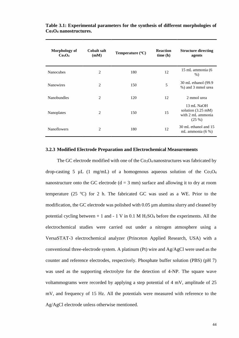
44
Table 3.1: Experimental parameters for the synthesis of different morphologies of
Co3O4 nanostructures.
Morphology of
Co3O4
Cobalt salt
(mM) Temperature (oC)
Reaction
time (h)
Structure directing
agents
Nanocubes 2 180 12 15 mL ammonia (6
%)
Nanowires 2 150 5 30 mL ethanol (99.9
%) and 3 mmol urea
Nanobundles 2 120 12 2 mmol urea
Nanoplates 2 150 15
13 mL NaOH
solution (3.25 mM)
with 2 mL ammonia
(25 %)
Nanoflowers 2 180 12 30 mL ethanol and 15
mL ammonia (6 %)
3.2.3 Modified Electrode Preparation and Electrochemical Measurements
The GC electrode modified with one of the Co3O4 nanostructures was fabricated by
drop-casting 5 µL (1 mg/mL) of a homogenous aqueous solution of the Co3O4
nanostructure onto the GC electrode (d = 3 mm) surface and allowing it to dry at room
temperature (25 oC) for 2 h. The fabricated GC was used as a WE. Prior to the
modification, the GC electrode was polished with 0.05 µm alumina slurry and cleaned by
potential cycling between + 1 and - 1 V in 0.1 M H2SO4 before the experiments. All the
electrochemical studies were carried out under a nitrogen atmosphere using a
VersaSTAT-3 electrochemical analyzer (Princeton Applied Research, USA) with a
conventional three-electrode system. A platinum (Pt) wire and Ag/AgCl were used as the
counter and reference electrodes, respectively. Phosphate buffer solution (PBS) (pH 7)
was used as the supporting electrolyte for the detection of 4-NP. The square wave
voltammograms were recorded by applying a step potential of 4 mV, amplitude of 25
mV, and frequency of 15 Hz. All the potentials were measured with reference to the
Ag/AgCl electrode unless otherwise mentioned.
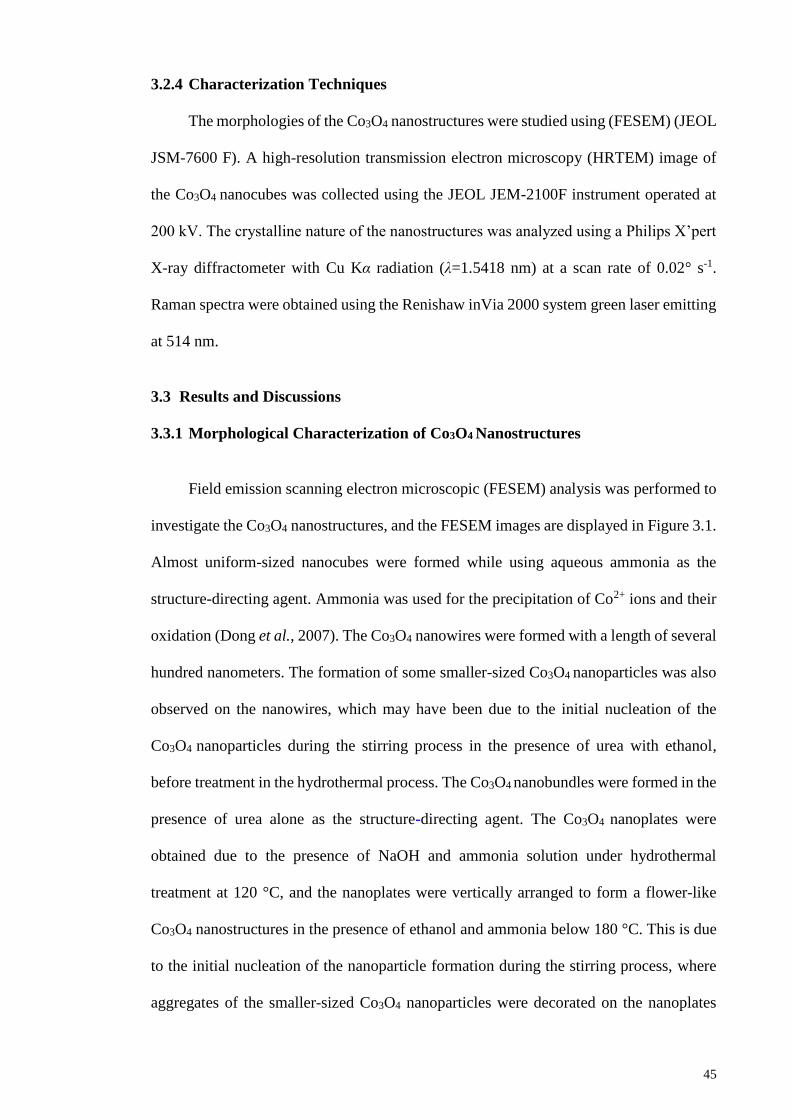
45
3.2.4 Characterization Techniques
The morphologies of the Co3O4 nanostructures were studied using (FESEM) (JEOL
JSM-7600 F). A high-resolution transmission electron microscopy (HRTEM) image of
the Co3O4 nanocubes was collected using the JEOL JEM-2100F instrument operated at
200 kV. The crystalline nature of the nanostructures was analyzed using a Philips X’pert
X-ray diffractometer with Cu Kα radiation (λ=1.5418 nm) at a scan rate of 0.02° s-1.
Raman spectra were obtained using the Renishaw inVia 2000 system green laser emitting
at 514 nm.
3.3 Results and Discussions
3.3.1 Morphological Characterization of Co3O4 Nanostructures
Field emission scanning electron microscopic (FESEM) analysis was performed to
investigate the Co3O4 nanostructures, and the FESEM images are displayed in Figure 3.1.
Almost uniform-sized nanocubes were formed while using aqueous ammonia as the
structure-directing agent. Ammonia was used for the precipitation of Co2+ ions and their
oxidation (Dong et al., 2007). The Co3O4 nanowires were formed with a length of several
hundred nanometers. The formation of some smaller-sized Co3O4 nanoparticles was also
observed on the nanowires, which may have been due to the initial nucleation of the
Co3O4 nanoparticles during the stirring process in the presence of urea with ethanol,
before treatment in the hydrothermal process. The Co3O4 nanobundles were formed in the
presence of urea alone as the structure-directing agent. The Co3O4 nanoplates were
obtained due to the presence of NaOH and ammonia solution under hydrothermal
treatment at 120 °C, and the nanoplates were vertically arranged to form a flower-like
Co3O4 nanostructures in the presence of ethanol and ammonia below 180 °C. This is due
to the initial nucleation of the nanoparticle formation during the stirring process, where
aggregates of the smaller-sized Co3O4 nanoparticles were decorated on the nanoplates
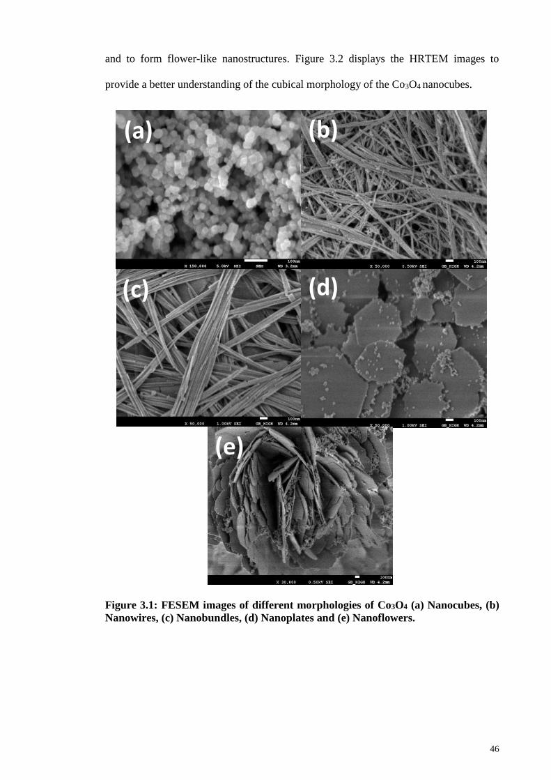
46
and to form flower-like nanostructures. Figure 3.2 displays the HRTEM images to
provide a better understanding of the cubical morphology of the Co3O4 nanocubes.
Figure 3.1: FESEM images of different morphologies of Co3O4 (a) Nanocubes, (b)
Nanowires, (c) Nanobundles, (d) Nanoplates and (e) Nanoflowers.
(a)
(e)
(d) (c)
(b)
47
Figure 3.2: TEM image of Co3O4 nanocubes. Inset shows the TEM image at higher
magnification.
3.3.2 XRD and Raman Analyses of Co3O4 Nanostructures
The crystalline nature of the Co3O4 nanostructures was studied by recording the
XRD patterns, as shown in Figure 3.3. The characteristic diffraction peaks at 31.1°, 37.0°,
44.6°, 55.6°, 59.2°, and 65.1° correspond to the (220), (311), (222), (400), (422), and
(511) diffraction planes of the face-centered cubic Co3O4 spinel phase, respectively
(JCPDS card No. 42-1467) (Yao et al., 2013). All the morphologies showed the well-
identified diffraction peaks of Co3O4.

48
Figure 3.3: XRD patterns of Co3O4 nanostructures
The Raman spectra of all the morphologies of the Co3O4 structures are shown in
Figure 3.4. These show peaks at 192, 476, 516, 612, and 680 cm-1, which correspond to
the Eg, F1
2m, F12g and A1g modes of Co3O4, respectively (Liu et al., 2007). The Raman
peak positions of all the synthesized Co3O4 structures are almost identical to one another.
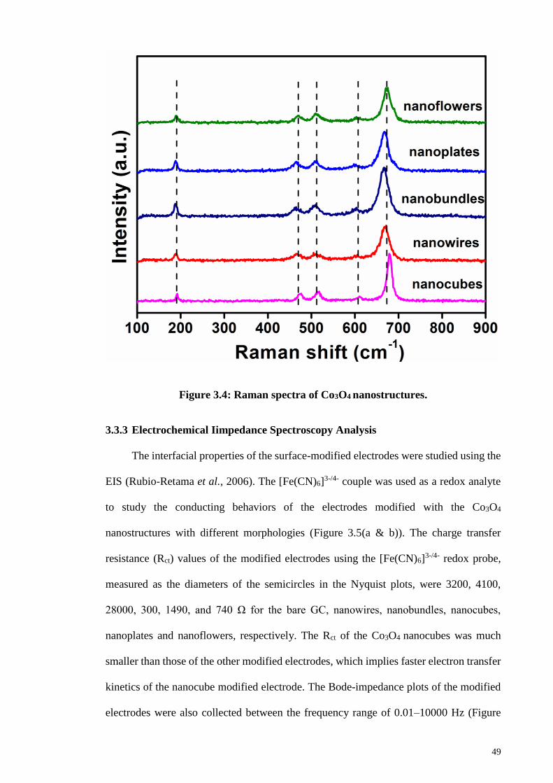
49
Figure 3.4: Raman spectra of Co3O4 nanostructures.
3.3.3 Electrochemical Iimpedance Spectroscopy Analysis
The interfacial properties of the surface-modified electrodes were studied using the
EIS (Rubio-Retama et al., 2006). The [Fe(CN)6]3-/4- couple was used as a redox analyte
to study the conducting behaviors of the electrodes modified with the Co3O4
nanostructures with different morphologies (Figure 3.5(a & b)). The charge transfer
resistance (Rct) values of the modified electrodes using the [Fe(CN)6]3-/4- redox probe,
measured as the diameters of the semicircles in the Nyquist plots, were 3200, 4100,
28000, 300, 1490, and 740 Ω for the bare GC, nanowires, nanobundles, nanocubes,
nanoplates and nanoflowers, respectively. The Rct of the Co3O4 nanocubes was much
smaller than those of the other modified electrodes, which implies faster electron transfer
kinetics of the nanocube modified electrode. The Bode-impedance plots of the modified
electrodes were also collected between the frequency range of 0.01–10000 Hz (Figure

50
3.5(c)). The phase peaks that appears between the frequency range of 100–1000 Hz
indicates the charge-transfer resistance of the modified electrodes. The less intense peak
obtained for the Co3O4 nanocubes modified electrode reveals a faster electron transfer
process at the modified electrode surface, and the phase angles of all the electrodes were
less than 90° at higher frequencies, which suggests that the electrodes did not behave like
an ideal capacitor (Matemadombo et al., 2007).
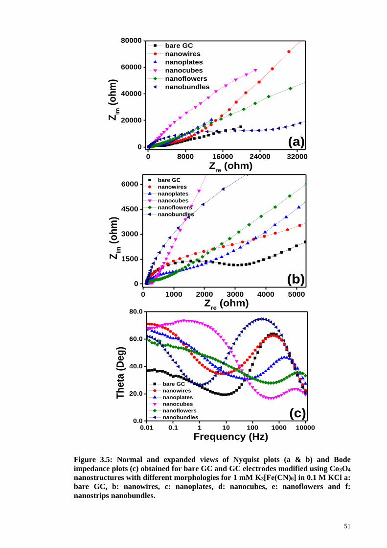
51
Figure 3.5: Normal and expanded views of Nyquist plots (a & b) and Bode
impedance plots (c) obtained for bare GC and GC electrodes modified using Co3O4
nanostructures with different morphologies for 1 mM K3[Fe(CN)6] in 0.1 M KCl a:
bare GC, b: nanowires, c: nanoplates, d: nanocubes, e: nanoflowers and f:
nanostrips nanobundles.
0 8000 16000 24000 32000
0
20000
40000
60000
80000
Zim
(o
hm
)
Zre
(ohm)
bare GC
nanowires
nanoplates
nanocubes
nanoflowers
nanobundles
(a)
0 1000 2000 3000 4000 5000
0
1500
3000
4500
6000
Zim
(o
hm
)
Zre
(ohm)
bare GC
nanowires
nanoplates
nanocubes
nanoflowers
nanobundles
(b)
0.01 0.1 1 10 100 1000 100000.0
20.0
40.0
60.0
80.0
bare GC
nanowires
nanoplates
nanocubes
nanoflowers
nanobundles
Th
eta
(D
eg
)
Frequency (Hz)
(c)

52
3.3.4 Electrocatalytic Reduction of 4-Nitrophenol
The electrocatalytic reduction of 4-NP was chosen as a model system to study the
electrocatalytic behaviors of the Co3O4 nanostructures modified electrodes with the
different morphologies. Irrespective of the morphology, all the Co3O4 nanostructures
showed the increased catalytic current for the reduction of 4-NP (Figure 3.6). Among the
morphologies, Co3O4 nanocubes displayed the highest catalytic current (-15 µA) for the
reduction of 100 µM 4-NP. The Co3O4 nanowires and nanobundles showed smaller
catalytic currents of -8.3 and -7.4 µA, respectively, at more negative potentials for the
same concentration of 4-NP. The higher catalytic performance of the nanocubes can be
attributed to the uniformly formed smaller-sized nanostructures compared to the other
morphologies. Notably, the Co3O4 nanocube modified electrode did not show any
electrochemical feature in the absence of 4-NP (Appendix 1). The Co3O4 nanoplates and
nanoflowers produced almost the same catalytic responses for the reduction of 4-NP in
terms of the peak potential and catalytic current. The bare GC electrode also showed the
minimum catalytic current for the reduction of 4-NP in the given potential window.
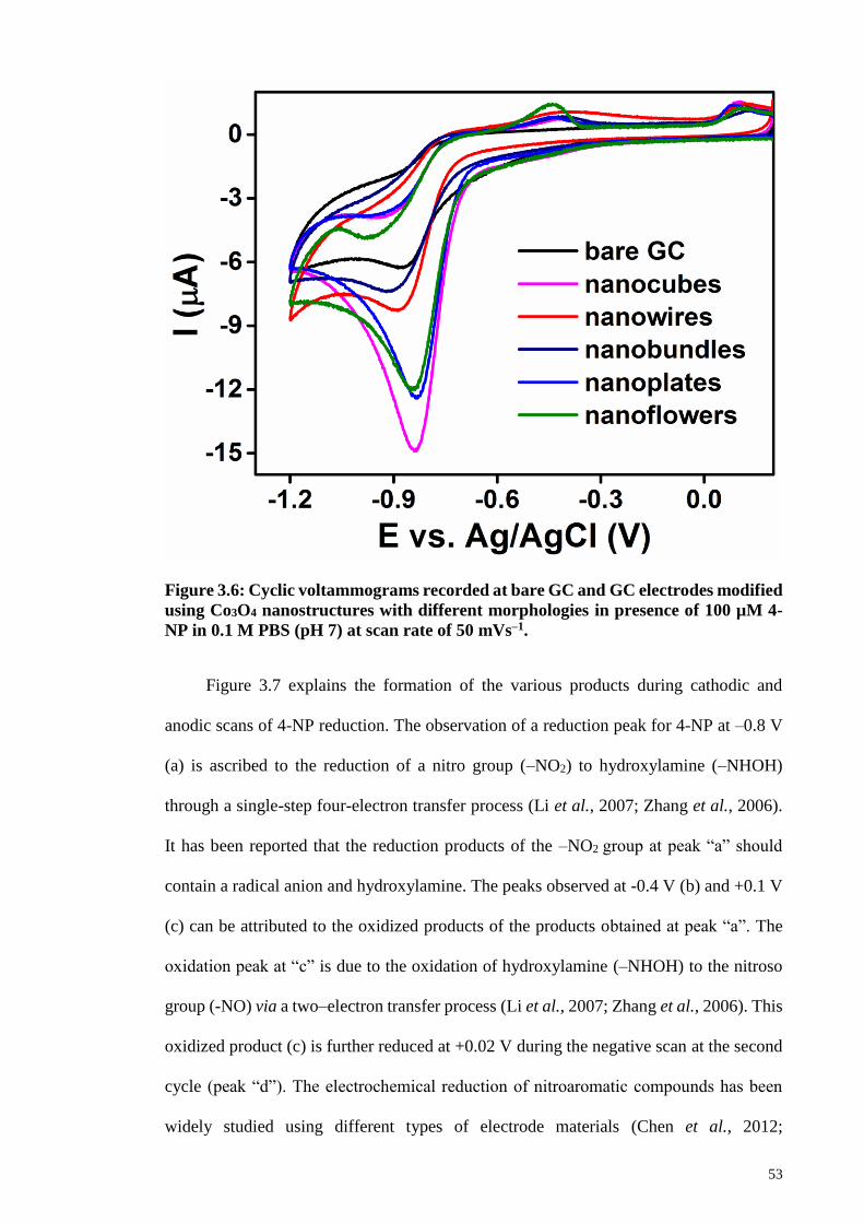
53
Figure 3.6: Cyclic voltammograms recorded at bare GC and GC electrodes modified
using Co3O4 nanostructures with different morphologies in presence of 100 µM 4-
NP in 0.1 M PBS (pH 7) at scan rate of 50 mVs–1.
Figure 3.7 explains the formation of the various products during cathodic and
anodic scans of 4-NP reduction. The observation of a reduction peak for 4-NP at –0.8 V
(a) is ascribed to the reduction of a nitro group (–NO2) to hydroxylamine (–NHOH)
through a single-step four-electron transfer process (Li et al., 2007; Zhang et al., 2006).
It has been reported that the reduction products of the –NO2 group at peak “a” should
contain a radical anion and hydroxylamine. The peaks observed at -0.4 V (b) and +0.1 V
(c) can be attributed to the oxidized products of the products obtained at peak “a”. The
oxidation peak at “c” is due to the oxidation of hydroxylamine (–NHOH) to the nitroso
group (-NO) via a two–electron transfer process (Li et al., 2007; Zhang et al., 2006). This
oxidized product (c) is further reduced at +0.02 V during the negative scan at the second
cycle (peak “d”). The electrochemical reduction of nitroaromatic compounds has been
widely studied using different types of electrode materials (Chen et al., 2012;

54
Maduraiveeran et al., 2009; Yuan et al., 2014). Generally, the electroreduction of Ar-NO2
to Ar-NH2 via a six-electron transfer process requires an acidic electrolyte, and the
formation of Ar-NHOH is facilitated in a neutral or alkaline medium through a four-
electron transfer process (Zhang et al., 2000). The dependence of the peak current and
peak potential on the pH variation of PBS for the reduction of 4-NP was studied using the
nanocubes modified electrode.
Figure 3.7: Cyclic voltammogram recorded at Co3O4 nanocubes modified electrode
in presence 100 µM 4-NP in 0.1 M PBS (pH 7) at scan rate of 50 mV.s–1.
Figure 3.8(a) displays voltammograms of the 4-NP reduction in 0.1 M PBS at
various pH values, where the reduction peak potential is shifted toward more negative
value when the pH is increased from 3 to 8. The plot of the reduction peak potential versus
pH shows a linear relation (Figure 3.8(b)) with a slope of -54 mV/pH, which suggests that
an equal number of protons and electrons are involved in the electrochemical reduction
of 4-NP (Xu et al., 2014). Further, the catalytic current observed for the reduction of 4-

55
NP at pH 7 is higher than those observed at other pH solutions (Figure 3.8(b) (inset)).
Since most waste-water has a neutral pH, it is advantageous to perform the catalytic
reduction and detection of 4-NP at pH 7. Moreover, adjusting the pH of the PBS to much
lower values requires the addition of acid, which may increase the cost of the catalytic
process.
Figure 3.8: (a) Cyclic voltammograms recorded at Co3O4 nanocubes modified
electrode in presence of 100 µM 4-NP in 0.1 M PBS with different pH levels (pH = 3
to 8) at scan rate of 50 mV s–1. (b) Plot of shift in peak potential versus pH. Inset:
Plot of peak current versus pH.
-1.2 -0.9 -0.6 -0.3 0.0
-16
-12
-8
-4
0
I (
A)
E vs. Ag/AgCl (V)
pH 3
pH 4
pH 5
pH 6
pH 7
pH 8
pH = 3 to 8(a)
3 4 5 6 7 8 9
-1.0
-0.9
-0.8
-0.7
-0.6
3 4 5 6 7 8-9
-10
-11
-12
-13
-14
I (
A)
pH
Ep (
V)
pH
(b)
56
3.3.5 Square Wave Voltammetric Detection of 4-Nitrophenol
The Co3O4 nanocubes modified electrode was chosen to detect the lowest detected
concentration of 4-NP because of its better performance in catalysis. The square wave
voltammetric technique was used to study the sensing ability of the modified electrode
and Figure 3.9(a) displays the voltammetric responses observed for successive additions
of 4-NP at the nanocubes modified electrode dipped in 0.1 M PBS (pH 7). During the
successive additions (both 2 and 5 µM additions), the peak current corresponding to the
reduction of 4-NP increased at the potential of – 0.83 mV, and the plot of the peak current
difference (Id) versus the concentration of 4-NP showed a linear relation (R2=0.997)
(Figure 3.9(b)). After some additions of 4-NP, the voltammogram showed the formation
of a pre-peak at a less negative potential, due to the strong adsorption of 4-NP at the
Co3O4 nanocubes modified electrode surface. It is well known that a split in the peak is
observed in the square wave voltammograms, due to the adsorption of redox analytes
(Rameshkumar et al., 2014). The sensitivity and LOD of the modified electrode were
0.0485±0.00063 µA/µM and 0.93 µM, respectively, for the detection of 4-NP. The
voltammetric responses were reproducible for different GC electrodes modified with
Co3O4 nanocubes and for repeated experiments. The analytical performance of the Co3O4
nanocubes modified GC electrode toward the detection of 4-NP was compared with those
of different electrode materials (Table 3.2), and a good LOD was attained without
employing any polymeric binder material or tedious electrode modification process.
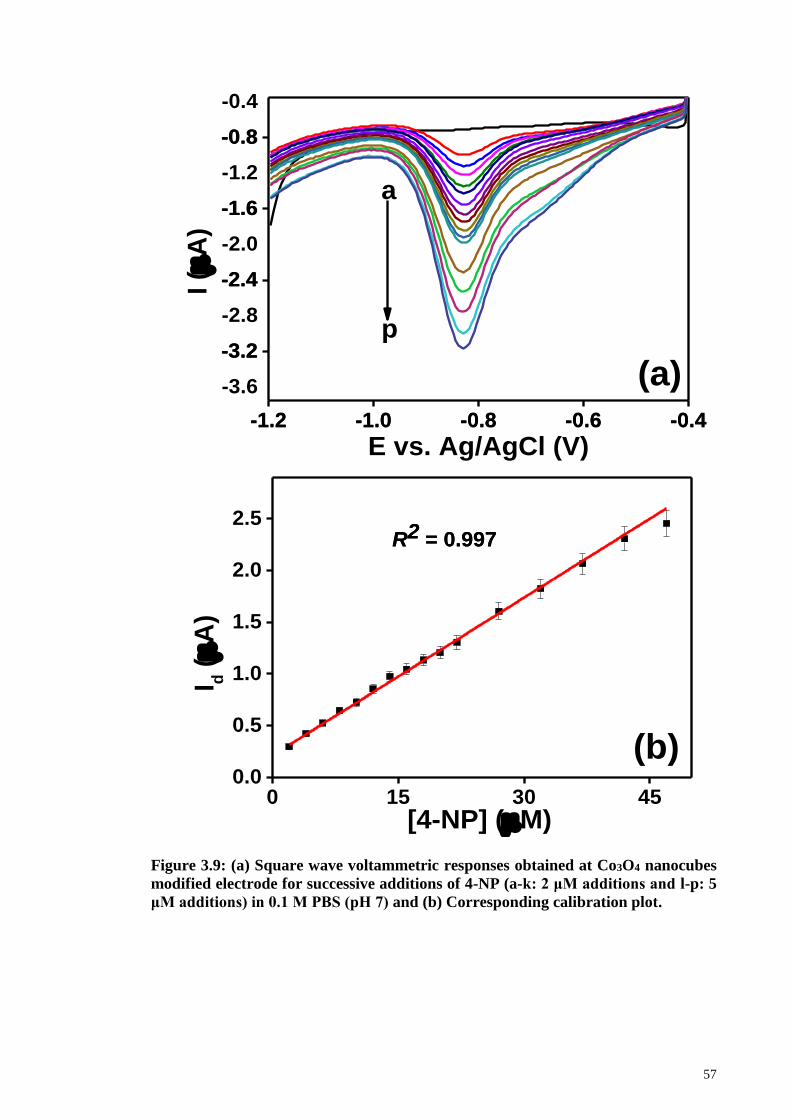
57
Figure 3.9: (a) Square wave voltammetric responses obtained at Co3O4 nanocubes
modified electrode for successive additions of 4-NP (a-k: 2 μM additions and l-p: 5
μM additions) in 0.1 M PBS (pH 7) and (b) Corresponding calibration plot.
0 15 30 450.0
0.5
1.0
1.5
2.0
2.5
(b)
I d (
A)
[4-NP] (M)
R2 = 0.997R2 = 0.997
-1.2-1.2 -1.0-1.0 -0.8-0.8 -0.6-0.6 -0.4-0.4
-3.6
-3.2-3.2
-2.8
-2.4-2.4
-2.0
-1.6-1.6
-1.2
-0.8-0.8
-0.4
I (
A)
E vs. Ag/AgCl (V)
a
p
(a)
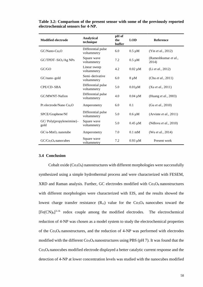
58
Table 3.2: Comparison of the present sensor with some of the previously reported
electrochemical sensors for 4-NP.
3.4 Conclusion
Cobalt oxide (Co3O4) nanostructures with different morphologies were successfully
synthesized using a simple hydrothermal process and were characterized with FESEM,
XRD and Raman analysis. Further, GC electrodes modified with Co3O4 nanostructures
with different morphologies were characterized with EIS, and the results showed the
lowest charge transfer resistance (Rct) value for the Co3O4 nanocubes toward the
[Fe(CN)6]3-/4- redox couple among the modified electrodes. The electrochemical
reduction of 4-NP was chosen as a model system to study the electrochemical properties
of the Co3O4 nanostructures, and the reduction of 4-NP was performed with electrodes
modified with the different Co3O4 nanostructures using PBS (pH 7). It was found that the
Co3O4 nanocubes modified electrode displayed a better catalytic current response and the
detection of 4-NP at lower concentration levels was studied with the nanocubes modified
Modified electrode Analytical
technique
pH of
the
buffer
LOD Reference
GC/Nano-Cu2O Differential pulse
voltammetry 6.0 0.5 µM (Yin et al., 2012)
GC/TPDT–SiO2/Ag NPs Square wave
voltammetry 7.2 0.5 µM
(Rameshkumar et al.,
2014)
GC/GO Linear sweep
voltammetry 4.2 0.02 µM (Li et al., 2012)
GC/nano–gold Semi–derivative
voltammetry 6.0 8 µM (Chu et al., 2011)
CPE/CD–SBA Differential pulse
voltammetry 5.0 0.01µM (Xu et al., 2011)
GC/MWNT-Nafion Differential pulse
voltammetry 4.0 0.04 µM (Huang et al., 2003)
Pt electrode/Nano Cu2O Amperometry 6.0 0.1 (Gu et al., 2010)
SPCE/Graphene/Nf Differential pulse
voltammetry 5.0 0.6 µM (Arvinte et al., 2011)
GC/ Poly(propyleneimine)–
gold
Square wave
voltammetry 5.0 0.45 µM (Ndlovu et al., 2010)
GC/α-MnO2 nanotube Amperometry 7.0 0.1 mM (Wu et al., 2014)
GC/Co3O4 nanocubes Square wave
voltammetry 7.2 0.93 µM Present work

59
electrode using SWV. A LOD of 0.93 µM was achieved using the Co3O4 nanocubes, and
the current responses were reproducible for the detection of 4-NP.
60
CHAPTER 4: AMPEROMETRIC DETECTION OF DEPRESSION
BIOMARKER USING A GLASSY CARBON ELECTRODE MODIFIED WITH
NANOCOMPOSITE OF COBALT OXIDE NANOCUBES INCORPORATED
INTO REDUCED GRAPHENE OXIDE
4.1 Introduction
Serotonin (5-hydroxytryptamine, 5-HT) is a monoamine, present in
enterchromaffin cells located in the colonic mucosal epithelium widely distributed in the
central nervous system. It has an enormous biological importance, the deficiency of 5-HT
leads to mental disorders such as Alzheimer’s disease, infantile autism, mental
retardation, mood, sleep disorders, appetite and depression (Abbaspour et al., 2011; Wang
et al., 2013). A series of studies were carried out on human and animal which shows the
effect of changes in motility and 5-HT signaling in ageing in terms of, obesity and
gastrointestinal diseases such as inflammatory bowel diseases (Morris et al., 2016).
Therefore, the sensitive and selective detection of 5-HT with a measurement levels is of
great worth and can contribute in understanding the role of 5-HT in depression and other
neurological disorders. There are plenty of analytical techniques available which are
being used for the detection of 5-HT, these techniques includes fluorimetry (Panholzer et
al., 1999), enzyme immunoassay (Chauveau et al., 1991) radioimmunoassay (Jeon et al.,
1992) chemiluminescence (Tsunoda et al., 1999) and mass spectrometry (Middelkoop et
al., 1993).
Although these techniques are available and has been used for the detection of 5-
HT but there are some limitations in using these technique as they are time-consuming
and often require sample pre-treatment. On the other hand, electrochemical methods are
highly efficient for the detection of a target molecule. But one of the main requirements
is that the analyte must be electrochemically active. Since 5-HT is an electroactive

61
compound, it can be determined by electrochemical methods (Anithaa et al., 2017; Crespi
et al., 1991; Dinesh et al., 2017; Fagan-Murphy et al., 2012; Gueell et al., 2010; Gupta et
al., 2014; Liu et al., 2011; Liu et al., 2010; Ran et al., 2015; Wang et al., 2013).
Nonetheless, the electrochemical technique also endures some issues that limit the
detection of 5-HT. The first is that the concentration of 5-HT is very low under
physiological conditions. Secondly, the co-existence of other biological molecules like
such as AA, DA and UA, which have their oxidation potential nearby to each other and
almost overlaps with that of 5-HT at conventional solid electrodes. These concerns make
the detection of 5-HT a much more challenging task. A conventional unmodified
electrode will not be able to detect this type of molecule in a challenging environment. In
recent years, the surface modification of the conventional electrode by special coatings
has been the state-of-the-art area in the development of electrochemical sensors (Gupta
et al., 2014; Niu et al., 2013; Xu et al., 2012). To resolve these problems, one of the most
common ways is the utilization of a modified electrode to improve the sensitivity of 5-
HT detection and remove the interference from AA and DA and UA in the 5-HT
determination. So, carbon-based electrodes are widely used in voltammetric analysis due
to their several advantages such as low cost, chemical stability, wide potential window,
good electrocatalytic activity and easy availability. The biocompatibility of carbon
electrode makes them a suitable candidate to study the biologically relevant redox
systems and in vivo analysis. A GC electrode, polycrystalline boron doped diamond
electrode and carbon nanotubes are materials which present a wide range of
characteristics that are interesting to compare. However, GCE is one of the widely used
WE material in electrochemistry.
In recent years, the construction of a competitive sensor material is a difficult and
challenging job. A list of nanocomposite material has been used to modify the GCE
surface for electrochemical detection of target molecules (Numan et al., 2017; Shahid et
62
al., 2015; Yusoff et al., 2017). The different types of materials which has been used for
the detection of 5-HT are GC and boron-doped diamond (BDD) electrodes (Fagan-
Murphy et al., 2012), MWCNTs–CS–poly(p-ABSA) (Ran et al., 2015), CUCR/GCE
(Wang et al., 2013), Nafion/Ni(OH)2/MWCNT/GC (Babaei et al., 2013), rGO-
Co3O4/GCE (Dinesh et al., 2017) and 100 kGy GI-WO3/GCE (Anithaa et al., 2017). In
this report, reduced graphene oxide and cobalt oxide (rGO-Co3O4) nanocomposite
synthesized by a very simple single step hydrothermal route, was utilized for the detection
of 5-HT. As it is well known that graphene, a two-dimensional carbon sheet having
single-atom thickness, large theoretical surface area (2630 m2.g1) with high conductivity
at room temperature (106 s.cm1), with a wide electrochemical window has a great
contribution in catalysis science (Shahid et al., 2017). Graphene sheets are the excellent
host material for growing nanomaterials for high performance electrochemical
applications (Kim et al., 2009; Zhou et al., 2010). Until now large number of graphene
based materials has been synthesized for applications in fuel cell (Shahid et al., 2017),
electrochemical sensor (Thanh et al., 2016) , supercapacitors (Numan et al., 2016;
Rafique et al., 2017), solar cells (Rafique et al., 2017) and Li-ion batteries (He et al.,
2010), etc. The Co3O4 nanocubes have been utilized with graphene oxide as a composite
material due to its extraordinary characteristics (Shahabuddin et al., 2015).
Our group has used rGO-Co3O4 nanocomposite as a cathode and anode material for
fuel cell applications (Shahid et al., 2014; Shahid et al., 2017). Until now, there are no
reports on rGO-Co3O4 nanocomposite prepared by the hydrothermal method utilized for
the detection of 5-HT. The presence of the rGO-Co3O4 nanocomposite was characterized
by FESEM, energy-dispersive X-ray spectroscopy (EDX) mapping, XRD and Raman
analyses. The rGO-Co3O4 nanocomposite was utilized for the sensitive and selective
detection of 5-HT, in the presence of other co-existing interference molecules such as AA
and DA and UA. The LOD and limit of quantification (LOQ) were 1.128M and 3.760
63
M, respectively, with a sensitivity of 0.133A.M-1 on a rGO-Co3O4 nanocomposite
modified GCE. Furthermore, the as prepared nanocomposite modified electrode was
stable, reproducible and showed excellent selectivity toward the detection of 5-HT.
4.2 Experimental Section
4.2.1 Materials
Graphite flakes were purchased from Asbury Inc. (USA). Sulfuric acid (H2SO4, 98
%), phosphoric acid (H3PO4), hydrochloric acid (HCl, 35 %), potassium permanganate
(KMnO4, > 99 %) and ammonia solution (NH3, 25 %) were purchased from R & M
Chemicals. Cobalt acetate tetrahydrate Co(CH3COO)2.4H2O was purchased from Sigma
Aldrich. Hydrogen peroxide (H2O2, 35 %) was obtained from Systerm, Malaysia.
Serotonin hydrochloride was obtained from abcr GmbH Germany. DI water was used
throughout the experimental work.
4.2.2 Synthesis of rGO-Co3O4 Nanocomposite
GO was prepared by a simplified Hummers method route (Ming, 2010). Graphite
flakes (3 g), H2SO4 (360 mL), H3PO4 (40 mL), and KMnO4 (18 g) were mixed under
stirring at room temperature. The mixture was stirred for three days to achieve the
complete oxidation of the graphite. The color of the mixture changed from dark green to
dark brown. Ice containing H2O2 solution was used to stop the oxidation process and
control the temperature. The color of the mixture changed to bright yellow, indicating a
high level of oxidation of the graphite oxide. The formed graphite oxide was washed three
times with 1 M HCl aqueous solution and repeatedly several times with de-ionized water
to achieve a pH of 5-6. The washing process was carried out using a simple decantation
of supernatant via the centrifugation technique. During the process of washing with de-
ionized water, the graphite oxide was exfoliated, which resulted in the thickening of the
GO solution and finally formation of the GO gel. In the typical procedure for the synthesis
64
of rGO-Co3O4 nanocomposites, the GO was synthesized by a simplified Hummer's
method (Shahid et al., 2015). A 1 mmol of Co (CH3COO)2.4H2O was mixed in 12 mL of
DI water and added drop by drop into a GO solution of 1 mg.mL under continuous
stirring. Four samples of rGO-Co3O4 nanocomposites were prepared with the same
procedure but with different wt. % of GO (2, 4, 8, and 12 wt. %) and stirred for 2 h to
obtain a homogeneous solution. After that, 15 mL of 6 % ammonia was slowly added
drop-wise into the above reaction mixture under vigorous stirring to raise the pH to almost
10. Then, the as prepared 75 mL of the reaction mixture was transferred into a 100 mL of
Teflon lined stainless steel autoclave and subjected to hydrothermal treatment at 180 C
for 12 h. Finally, the obtained precipitate of rGO-Co3O4 nanocomposite was washed
many times with DI water, followed by ethanol to remove the extra impurities and dried
in a hot air oven at 60 oC. For comparison, unaided Co3O4 and rGO were prepared using
the same method but without the presence of GO and Co(CH3COO)2.4H2O, respectively.
4.2.3 Preparation of Modified Electrode and Electrochemical Measurements
Prior to the modification of GC electrode, it was polished mechanically and
electrochemically in both ways. In the mechanical cleaning, 0.5 µm alumina slurry was
used and the GC electrode was gently rubbed on alumina coated cloth. After that the GC
electrode was sonicated for less than 5 min to remove the adherent particles on its surface.
The electrochemical cleaning was carried out by potential cycling between - 1 and + 1 V
in 0.1 M H2SO4, and washing with DI water after sonicating for 5 min. The rGO-Co3O4
nanocomposite modified GC electrode was prepared by using a well sonicated
homogenous solution with an optimized concentration of 1 mg.mL-1 using DI water as
the solvent. The electrochemical studies for the 5-HT was carried out using an Autolab
analyzer fitted with a conventional three-electrode system in a 0.1 M phosphate buffer
(PB) (pH 7.2) as the supporting electrolyte. The GC electrode with an area of 0.707 cm2,
modified with 5 L of as syntheised ink was used as the WE, while a saturated calomel
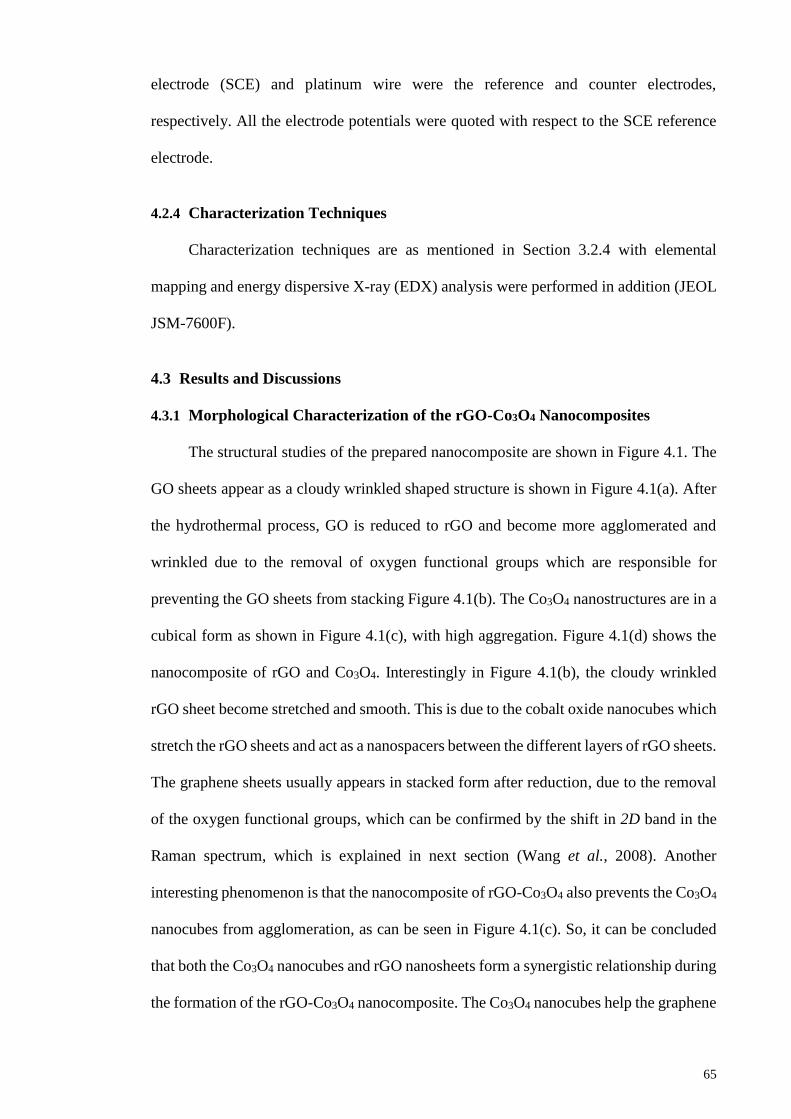
65
electrode (SCE) and platinum wire were the reference and counter electrodes,
respectively. All the electrode potentials were quoted with respect to the SCE reference
electrode.
4.2.4 Characterization Techniques
Characterization techniques are as mentioned in Section 3.2.4 with elemental
mapping and energy dispersive X-ray (EDX) analysis were performed in addition (JEOL
JSM-7600F).
4.3 Results and Discussions
4.3.1 Morphological Characterization of the rGO-Co3O4 Nanocomposites
The structural studies of the prepared nanocomposite are shown in Figure 4.1. The
GO sheets appear as a cloudy wrinkled shaped structure is shown in Figure 4.1(a). After
the hydrothermal process, GO is reduced to rGO and become more agglomerated and
wrinkled due to the removal of oxygen functional groups which are responsible for
preventing the GO sheets from stacking Figure 4.1(b). The Co3O4 nanostructures are in a
cubical form as shown in Figure 4.1(c), with high aggregation. Figure 4.1(d) shows the
nanocomposite of rGO and Co3O4. Interestingly in Figure 4.1(b), the cloudy wrinkled
rGO sheet become stretched and smooth. This is due to the cobalt oxide nanocubes which
stretch the rGO sheets and act as a nanospacers between the different layers of rGO sheets.
The graphene sheets usually appears in stacked form after reduction, due to the removal
of the oxygen functional groups, which can be confirmed by the shift in 2D band in the
Raman spectrum, which is explained in next section (Wang et al., 2008). Another
interesting phenomenon is that the nanocomposite of rGO-Co3O4 also prevents the Co3O4
nanocubes from agglomeration, as can be seen in Figure 4.1(c). So, it can be concluded
that both the Co3O4 nanocubes and rGO nanosheets form a synergistic relationship during
the formation of the rGO-Co3O4 nanocomposite. The Co3O4 nanocubes help the graphene

66
sheet to stretch and prevents the stacking of the graphene sheets thus increases the
effective surface area of the graphene, while the graphene sheets also prevented the Co3O4
nanocubes from agglomerations due to strong interaction between the Co3O4 and the rGO
through interfacial Co–O–C bonds formed by the high reactivity of sp2 carbon atoms of
rGO with the electron-rich oxygen species of Co3O4 (Shahid et al., 2015). The use of
ammonia facilitated the reduction of graphene oxide and also played an important role in
the precipitation of cobalt Co2+ ions and their oxidation (Dong et al., 2007). In addition,
the density of Co3O4 nanocubes decreased when the wt. % of GO is varied from lower to
higher concentration (2 wt. % to 12 wt. %). This is because of the presence of a sufficient
number of graphene sheet which help to optimize the best nanocomposite for detection
of 5-HT, as explained in electrocatalysis of 5-HT section.
Figure 4.1: FESEM images of (a) GO sheets, (b) rGO (c) Co3O4 nanocubes and (d)
rGO-Co3O4-4 % nanocomposite.
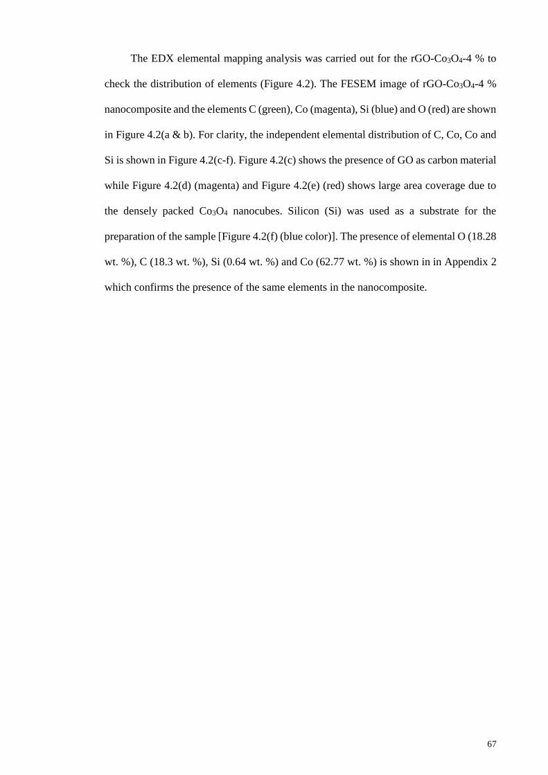
67
The EDX elemental mapping analysis was carried out for the rGO-Co3O4-4 % to
check the distribution of elements (Figure 4.2). The FESEM image of rGO-Co3O4-4 %
nanocomposite and the elements C (green), Co (magenta), Si (blue) and O (red) are shown
in Figure 4.2(a & b). For clarity, the independent elemental distribution of C, Co, Co and
Si is shown in Figure 4.2(c-f). Figure 4.2(c) shows the presence of GO as carbon material
while Figure 4.2(d) (magenta) and Figure 4.2(e) (red) shows large area coverage due to
the densely packed Co3O4 nanocubes. Silicon (Si) was used as a substrate for the
preparation of the sample [Figure 4.2(f) (blue color)]. The presence of elemental O (18.28
wt. %), C (18.3 wt. %), Si (0.64 wt. %) and Co (62.77 wt. %) is shown in in Appendix 2
which confirms the presence of the same elements in the nanocomposite.
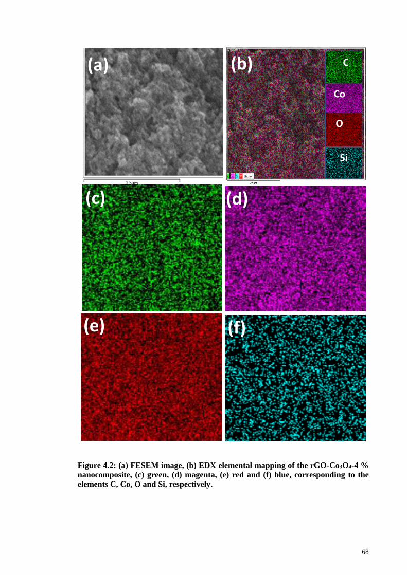
68
Figure 4.2: (a) FESEM image, (b) EDX elemental mapping of the rGO-Co3O4-4 %
nanocomposite, (c) green, (d) magenta, (e) red and (f) blue, corresponding to the
elements C, Co, O and Si, respectively.
O
Si
C
Co
(a) (b)
(c) (d)
(e) (f)
69
4.3.2 XRD and Raman Analysis
The XRD analysis was conducted to evaluate the crystalline nature of the rGO-
Co3O4-4 % nanocomposite (Figure 4.3). The diffraction peaks at 2values of 19.1°,
31.2°, 37.0°, 38.7°, 45.0°, 59.4°, 65.4° and 77.4° correspond to the crystal planes of (111),
(2 2 0), (3 1 1), (222), (4 0 0), (5 1 1), (4 4 0) and (5 3 3) of the face centered cubic Co3O4
(JCPDS Card No. 42-1467) (Song et al., 2013). The observed peaks for rGO-Co3O4-4 %
nanocomposite has higher intensity (Figure 4.3d) as compared to the peaks obtained for
unaided Co3O4 (Figure 4.3c). The high intensity peaks show that the presence of graphene
in the nanocomposite prevented the Co3O4 from agglomeration, which in turn appears as
highly crystalline nanostructures, and the XRD results agrees well with the FESEM
results. The high intensity diffraction peak of GO at 10.6° corresponds to lattice plane of
(001) which confirms the presence of GO in Figure 4.3a (Chen et al., 2010) . Additionally,
the disappearance of the high intensity peak of GO at 10° in Figure 4.3b and the
appearance of other two peaks at 25.5° and 43.2° corresponds to lattice plane of (002)
and (100) respectively, which indicates the reduction of GO to rGO. Interestingly, the
absence of rGO peak in the rGO-Co3O4-4 % nanocomposite confirms the exfoliation of
the graphene sheets as well as the interleaved Co3O4 nanocubes into different layers of
rGO sheets, which can be seen in the FESEM (Figure 4.1(d)).

70
Figure 4.3: XRD patterns of (a) GO, (b) rGO, (c) Co3O4 and (d) rGO-Co3O4
nanocomposite.
The Raman spectra of GO and rGO-Co3O4-4 % nanocomposite is shown in the
Figure 4.4. The rGO-Co3O4-4 % nanocomposite shows the D (1349 cm-1) and G (1601
cm-1) band of reduced graphene oxide with higher intensity of D/G > 1 which confirms
the reduction of GO into rGO. The D band in Raman spectra refers to the defects which
arises from the vibration of sp3 carbon atoms while the G band arises from sp2 hybridized
carbon atoms (Yusoff et al., 2017). The slight shifting of the G band to 1601 cm-1 for
rGO-Co3O4-4 % nanocomposite also confirms the reduction of GO into rGO, as shown
in Appendix 3 (inset). The successful synthesis of rGO-Co3O4-4 % nanocomposite can
be supported further by the Raman modes in Figure 4.4, which shows different values at
194, 482, 525 and 691 cm-1 which corresponds to the F2g2, Eg, F2g
1 and A1g Raman modes
respectively, of the Co3O4 along with rGO peaks (Jiang et al., 2016). The Raman mode
are given in Appendix 3 which further justify the formation of pure Co3O4 nanocubes.
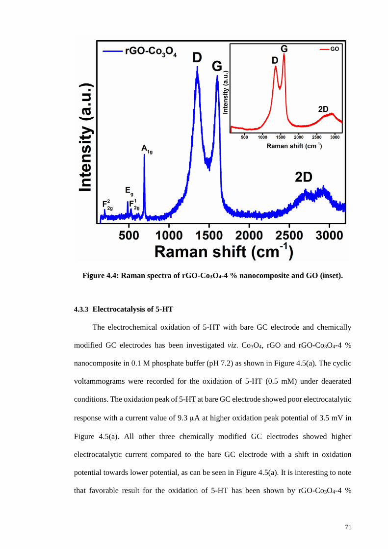
71
Figure 4.4: Raman spectra of rGO-Co3O4-4 % nanocomposite and GO (inset).
4.3.3 Electrocatalysis of 5-HT
The electrochemical oxidation of 5-HT with bare GC electrode and chemically
modified GC electrodes has been investigated viz. Co3O4, rGO and rGO-Co3O4-4 %
nanocomposite in 0.1 M phosphate buffer (pH 7.2) as shown in Figure 4.5(a). The cyclic
voltammograms were recorded for the oxidation of 5-HT (0.5 mM) under deaerated
conditions. The oxidation peak of 5-HT at bare GC electrode showed poor electrocatalytic
response with a current value of 9.3 A at higher oxidation peak potential of 3.5 mV in
Figure 4.5(a). All other three chemically modified GC electrodes showed higher
electrocatalytic current compared to the bare GC electrode with a shift in oxidation
potential towards lower potential, as can be seen in Figure 4.5(a). It is interesting to note
that favorable result for the oxidation of 5-HT has been shown by rGO-Co3O4-4 %
72
modified GC electrode with the highest current (36 mA) and is four times higher
compared to the bare GC electrode with an oxidation potential of 3.1 mV in Figure 4.5(a).
The considerable higher oxidation peak current of 5-HT at rGO-Co3O4-4 % modified GC
electrode shows a faster electron transfer process facilitated at the modified electrode.
The poor electrocatalytic performance of the Co3O4 modified GC electrode was due to
the Co3O4 nanocubes aggregation, which prevent faster electron transfer kinetics. The
higher electrocatalytic detection of 5-HT was shown by the rGO modified GC electrode
with lower oxidation potential, due to the higher electrical conduction of the rGO. The
cyclic voltammogram in Figure 4.5(b) shows the comparison of rGO-Co3O4-4 %
modified GC electrode in the presence and absence of 0.5 mM of 5-HT. There was no
peak observed in the absence of 5-HT at the modified GC electrode, while a well resolved
oxidation peak appears in the presence of 5-HT, which further confirms the
electrocatalytic oxidation of 5-HT at the chemically modified electrode (CME).
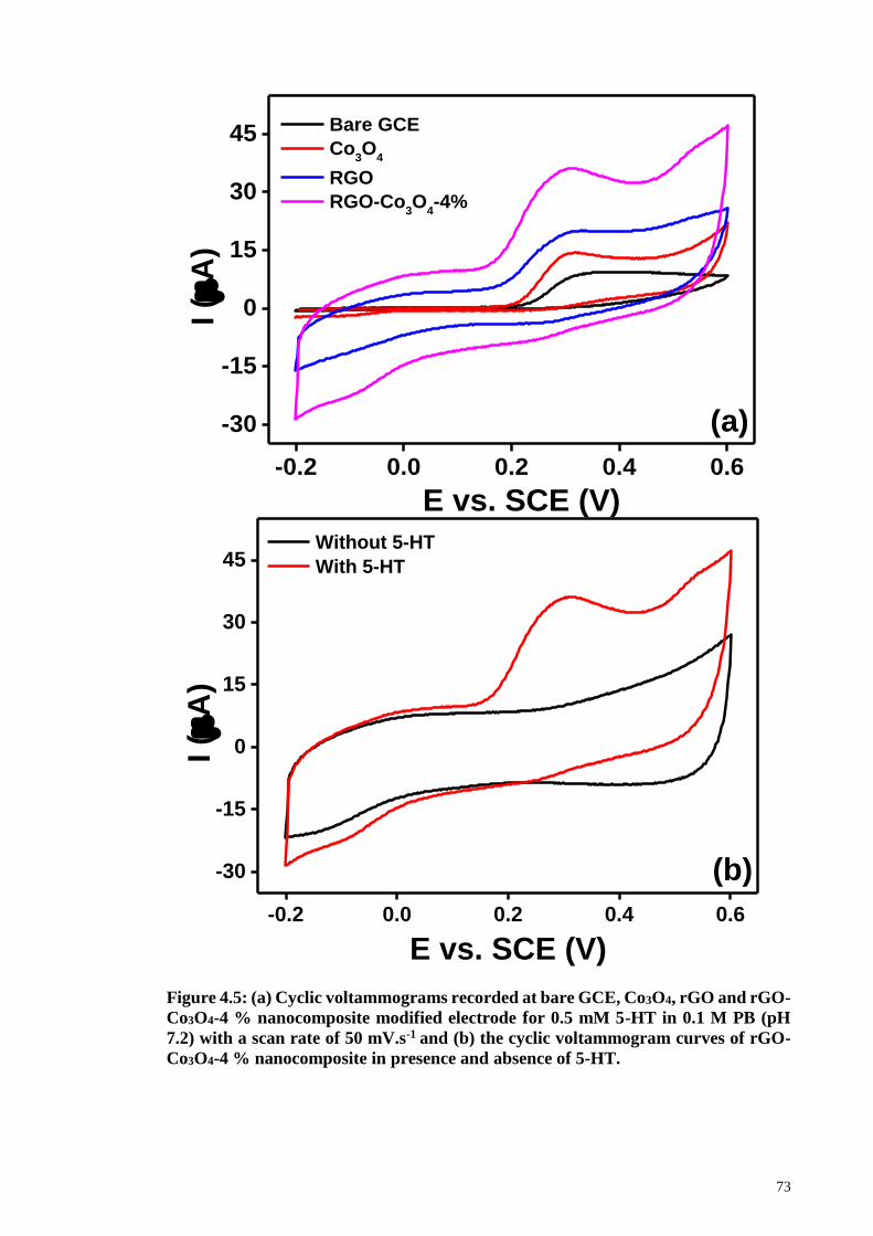
73
Figure 4.5: (a) Cyclic voltammograms recorded at bare GCE, Co3O4, rGO and rGO-
Co3O4-4 % nanocomposite modified electrode for 0.5 mM 5-HT in 0.1 M PB (pH
7.2) with a scan rate of 50 mV.s-1 and (b) the cyclic voltammogram curves of rGO-
Co3O4-4 % nanocomposite in presence and absence of 5-HT.
-0.2 0.0 0.2 0.4 0.6
-30
-15
0
15
30
45
I (
A)
E vs. SCE (V)
Bare GCE
Co3O
4
RGO
RGO-Co3O
4-4%
(a)
-0.2 0.0 0.2 0.4 0.6
-30
-15
0
15
30
45
I (
A)
E vs. SCE (V)
Without 5-HT
With 5-HT
(b)

74
Appendix 4 shows the electrocatalytic performance comparison of the rGO-Co3O4
modified GC electrode at different concentrations of GO (2 wt. % to 12 wt. %). The rGO-
Co3O4-4 % modified GC electrode gave the best electrocatalytic activity. On the other
hand, the rGO-Co3O4-2 % modified GC electrode shows poor electrocatalytic effect due
to the lower amount of graphene nanosheets which eventually allowed the Co3O4 to
aggregate on the surface of the graphene sheets. The increase in the concentration of GO
to 8 and 12 wt. % allows a sufficient number of graphene sheets to decrease the density
of the Co3O4 nanocubes. Therefore, the rGO-Co3O4 modified GC with 8 and 12 wt. % of
GO gave poor electrocatalytic activity compared to the rGO-Co3O4-4 % modified GC
electrode.
The influence of the scan rate on the electrocatalytic oxidation of 5-HT was
examined for the rGO-Co3O4-4 % modified GC electrode from 10-200 mV.s-1 in the
presence of 0.5 mM 5-HT in 0.1 M PB (pH 7.2), as shown in Figure 4.6(a). The peak
current for oxidation of 5-HT was found to be linear with the square root of the scan rate
(1/2) (Figure 4.6(b)) which indicates a typical diffusion controlled process. The positive
shift in oxidation peak potential with the increase of the scan rate indicates the slow
kinetics of the interfacial electron transfer of 5-HT (Abbaspour et al., 2011). The
irreversible electrooxidation of 5-HT is also supported by the linear relation of the peak
potential (Ep) and the log () (Figure 4.6(c)).

75
Figure 4.6: (a) Cyclic voltammogram plots obtained for rGO-Co3O4-4 % modified
electrode in 0.1 M PBS (pH 7.2) in presence of 0.5 mM 5-HT at a scan rate of 10-200
mV.s-1. (b) The corresponding calibration plot of anodic peak currents versus square
root of scan rate, (c) a relationship between anodic peak potentials versus logarithm
of scan rate.
-0.2 0.0 0.2 0.4 0.6
-50
0
50
100
I(
A)
E vs. SCE (V)
10 mVs-1
25 mVs-1
50 mVs-1
75 mVs-1
100 mVs-1
125 mVs-1
150 mVs-1
175mVs-1
200 mVs-1
(a)
4 6 8 10 12 14
10
20
30
40
50
60
70
(b)
I(
A)
1/2
(mVs-1)1/2
1.00 1.25 1.50 1.75 2.00 2.25
0.30
0.32
0.34
0.36
0.38
(c)
Ep
(V
)
log () (mVs-1)
76
Appendix 5(a) shows the cyclic voltammograms carried out for the rGO-Co3O4-4
% modified GC electrode in 0.1 M PB (pH 7.2) at scan rate of 50 mV.s-1, with increasing
concentration of 5-HT from 0.5 mM to 3 mM. The oxidation peak current is found to
increase linearly with a slight shift in the oxidation peak current by increasing the 5-HT
concentration. The plot of peak current versus concentration of 5-HT showed a linear
response (Appendix 5(b)) which indicates the efficient electrocatalytic ability of the
modified electrode.
4.3.4 Amperometric Detection of 5-HT
The rGO-Co3O4-4 % modified GC electrode was chosen for further amperometric
i–t curve due to the outstanding electrochemical behavior and good electrocatalytic
oxidation of 5-HT. Figure 4.7(a) shows the typical amperometric curve for the rGO-
Co3O4-4 % modified GC electrode with successive addition of 1 M of 5-HT
concentration and was increased to 2 M after 10 injections. The 5-HT solution was
spiked after a regular interval of 60 s, once the amperometric curve become stable with
an applied potential of + 0.31 V. An obvious enhancement in current response for the
successive injection of 5-HT was obtained and the corresponding linear relationship
between the current response and 5-HT concentration is shown in Figure 4.7(b). There
are two linear ranges plotted ranging from 1 M to 10 M and 12 M to 22 M based on
the results obtained from the amperometric i–t curve. The LOD and LOQ were calculated
from the first linear range and found to be 1.128M and 3.760 M respectively, with a
S/N ratio of 3. The values for LOD and LOQ were calculated by the following equations
(4.1) and (4.2):
LOD = 3σ/m (4.1)
LOQ = 10σ/m (4.2)

77
where σ is the residual standard deviation of the linear regression and m is the slope of
the regression line (Reddaiah et al., 2012; Shrivastava et al., 2011). The sensitivity value
from the slope of the line was 1.337 M.M-1. The comparisons in Table 1 compiles the
analytical parameters for the electrochemical detection of 5-HT using different types of
modified GC electrodes reported in the literature.

78
Figure 4.7: (a) Amperometric i–t curve obtained at rGO-Co3O4-4 % nanocomposite
modified GC electrodes for the successive addition of 5-HT with various
concentrations in 0.1 M PBS (pH 7.2) at a regular interval of 60 s with two linear
ranges. The applied potential was +0.31 V. (b) The calibration plot of peak current
versus concentration of 5-HT corresponding to ‘(a)’.
The selectivity of the prepared sensor was tested in the presence of interfering
species such as AA, UA, and DA which is naturally found with 5-HT in the central
nervous system, as can be seen in Figure 4.8. These species were added one after the
0 200 400 600 800 1000
0.0
0.5
1.0
1.5
2.0
20 M
12 M
10 M
5 M
I (
A)
t (s)
1 M
(a)
0 3 6 9 12 15 18 21
0.4
0.8
1.2
1.6
2.0
I (
A)
Serotonin (M)
(b)

79
other, after three successive additions of 5-HT. The results obtained from the selectivity
experiments suggests that the rGO-Co3O4 modified GC electrode didn’t show any
response for the AA, DA and UA even with a 50-fold higher concentration. The
interference studies show only a small disturbance upon the addition of the spikes of AA.
This could arise from the dynamic conditions present due to the almost overlapping
potential of the AA and 5-HT. To further confirm the selectivity of the sensor, the 5-HT
solution was again spiked and an obvious increase in current values was obtained.
However, the present sensor still maintains the high selectivity towards the sensing of 5-
HT, even in the presence of 50-fold higher concentration of interference molecules.
Figure 4.8: Amperometric i–t curve obtained at rGO-Co3O4-4 % nanocomposite
modified GC electrodes for the successive addition of 1 M 5-HT and each 50 M of
AA, DA, UA in 0.1 M PB (pH 7.2) at a regular interval of 60 s at applied potential of
+ 0.31 V.
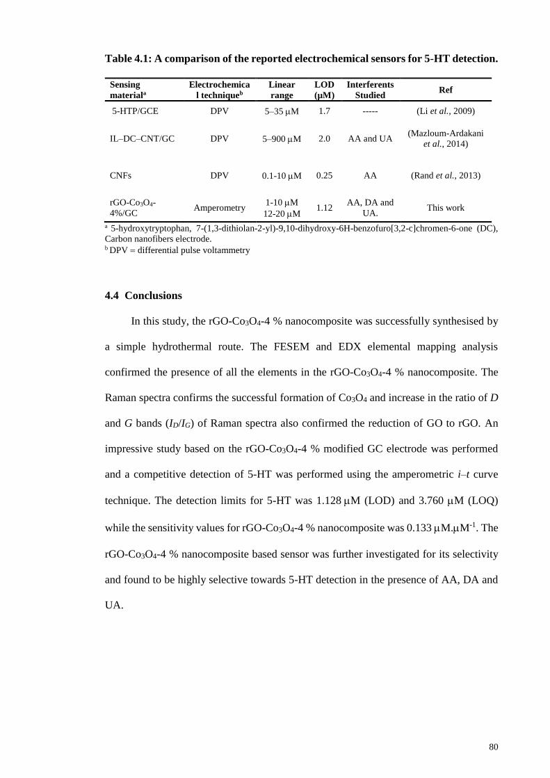
80
Table 4.1: A comparison of the reported electrochemical sensors for 5-HT detection.
Sensing
materiala
Electrochemica
l techniqueb
Linear
range
LOD
(µM)
Interferents
Studied Ref
5-HTP/GCE DPV 5–35 M 1.7 ----- (Li et al., 2009)
IL–DC–CNT/GC DPV 5–900 M 2.0 AA and UA (Mazloum-Ardakani
et al., 2014)
CNFs DPV 0.1-10 M 0.25 AA (Rand et al., 2013)
rGO-Co3O4-
4%/GC Amperometry
1-10 M
12-20 M 1.12
AA, DA and
UA. This work
a 5-hydroxytryptophan, 7-(1,3-dithiolan-2-yl)-9,10-dihydroxy-6H-benzofuro[3,2-c]chromen-6-one (DC),
Carbon nanofibers electrode. b DPV differential pulse voltammetry
4.4 Conclusions
In this study, the rGO-Co3O4-4 % nanocomposite was successfully synthesised by
a simple hydrothermal route. The FESEM and EDX elemental mapping analysis
confirmed the presence of all the elements in the rGO-Co3O4-4 % nanocomposite. The
Raman spectra confirms the successful formation of Co3O4 and increase in the ratio of D
and G bands (ID/IG) of Raman spectra also confirmed the reduction of GO to rGO. An
impressive study based on the rGO-Co3O4-4 % modified GC electrode was performed
and a competitive detection of 5-HT was performed using the amperometric i–t curve
technique. The detection limits for 5-HT was 1.128M (LOD) and 3.760 M (LOQ)
while the sensitivity values for rGO-Co3O4-4 % nanocomposite was 0.133 M.M-1. The
rGO-Co3O4-4 % nanocomposite based sensor was further investigated for its selectivity
and found to be highly selective towards 5-HT detection in the presence of AA, DA and
UA.

81
CHAPTER 5: AN ELECTROCHEMICAL SENSING PLATFORM OF COBALT
OXIDE@GOLD NANOCUBES INTERLEAVED REDUCED GRAPHENE
OXIDE FOR THE SELECTIVE DETERMINATION OF HYDRAZINE2
5.1 Introduction
Hydrazine is a toxic, colorless and flammable molecule (Krittayavathananon et al.,
2014). It is classified as a human mutagenic and carcinogen in group B2 by the EPA,
United States. It could cause severe injury of lungs, liver, nervous system, spinal cord,
temporary blindness and dizziness, pneumonia and kidney damage (Cui et al., 2014; Cui
et al., 2014). In addition, acute exposure to hydrazine could result in death (Choudhary et
al., 1997). Hydrazine and its offshoots are well-known environmental toxic pollutants,
are widely used in rocket fuel (Zhang et al., 2015), fuel cell systems (Madhu et al., 2015),
photographic chemicals, insecticides, herbicides, emulsifiers, blowing agents, textile dyes
and corrosion inhibitors in various chemical, pharmaceutical and agricultural industries
(Ding et al., 2015; Madhu et al., 2015; Wang et al., 2010). Since hydrazine is a suspected
carcinogenic and mutagenic agent, the detection of hydrazine in biological systems has
attracted considerable attention in recent decades (Karimi-Maleh et al., 2014). There are
numerous analytical techniques available for the sensitive determination of hydrazine
such as spectrophotometry (Ensafi et al., 1998; Watt et al., 1952), amperometry (Jayasri
et al., 2007; Mallela et al., 1977), titrimetry (Budkuley, 1992), chemiluminescence
(Safavi et al., 2002), fluorimetry (Safavi et al., 1995), and especially electroanalytical
methods (Afzali et al., 2011; Khalilzadeh et al., 2009; Yi et al., 2009). The techniques
2 This chapter is published as: Shahid, M. M., Rameshkumar, P., Basirunc, W. J., Wijayantha, U., Chiu,
W. S., Khiew, P. S., & Huang, N. M. (2018). An electrochemical sensing platform of cobalt oxide@ gold
nanocubes interleaved reduced graphene oxide for the selective determination of hydrazine. Electrochimica
Acta, 259, 606-616.
82
other than electroanalytical methods are complicated, more laborious and also are unable
to determine the real time concentration of hydrazine (Liu et al., 2014). Electrochemical
techniques offer the opportunity for a portable, economical, sensitive and rapid method
for the determination of hydrazine molecule (Karuppiah et al., 2014). The
electrochemical oxidation of hydrazine on carbon electrode produces nitrogen and water
that do not cause environmental pollution and have been investigated widely (Antoniadou
et al., 1989; Karimi-Maleh et al., 2014). However, the electrochemical oxidation of
hydrazine occurs at higher oxidation overpotential due to the sluggish decomposition at
the bare electrode surface (Zhang et al., 2010). Therefore, several attempts have been
made to circumvent this problem by using CME surface with new class of materials such
as metals, metal oxides and their nanocomposites supported by conducting platform
(Ding et al., 2015; Rastogi et al., 2014). The CME have the ability to decrease the
oxidation overpotential and helps in increasing the oxidation current based on their easy,
economical and labor-free operation along with sufficient sensitivity and selectivity
(Gholamian et al., 2012; Liu et al., 2014).
Nanotechnology-driven materials have attracted extensive attention in the recent
years due to their unique structures and catalytic properties (Gholamian et al., 2012;
Khoei et al., 2011). The modification of an electrode by the incorporation of
nanomaterials in conjunction with one another to form novel composites such as Pt-
Cu@silicon (Ensafi et al., 2016), TiO2–Pt, Pd-modified TiO2 (Yi et al., 2011) and
Au@Pd (Dutta et al., 2015) is a topic of interest to enhance the sensitivity, selectivity and
stability of the electrochemical sensing assay (Ganjali et al., 2010). The synergistic effect
of these materials plays a vital role in the determination of the target analyte (Kimmel et
al., 2011). Therefore, a considerable attention has also been focused to develop highly
conducting materials for supporting nanosized particles such as Ag/PPy/GCE (Ghanbari,
2014), ZnO/MWCNTs/GCE (Fang et al., 2009), AuNPs/GO (Lu et al., 2011) and ZnO–
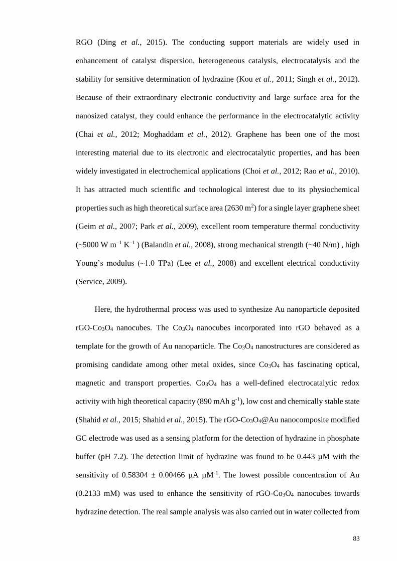
83
RGO (Ding et al., 2015). The conducting support materials are widely used in
enhancement of catalyst dispersion, heterogeneous catalysis, electrocatalysis and the
stability for sensitive determination of hydrazine (Kou et al., 2011; Singh et al., 2012).
Because of their extraordinary electronic conductivity and large surface area for the
nanosized catalyst, they could enhance the performance in the electrocatalytic activity
(Chai et al., 2012; Moghaddam et al., 2012). Graphene has been one of the most
interesting material due to its electronic and electrocatalytic properties, and has been
widely investigated in electrochemical applications (Choi et al., 2012; Rao et al., 2010).
It has attracted much scientific and technological interest due to its physiochemical
properties such as high theoretical surface area (2630 m2) for a single layer graphene sheet
(Geim et al., 2007; Park et al., 2009), excellent room temperature thermal conductivity
(~5000 W m–1 K–1 ) (Balandin et al., 2008), strong mechanical strength (~40 N/m) , high
Young’s modulus (~1.0 TPa) (Lee et al., 2008) and excellent electrical conductivity
(Service, 2009).
Here, the hydrothermal process was used to synthesize Au nanoparticle deposited
rGO-Co3O4 nanocubes. The Co3O4 nanocubes incorporated into rGO behaved as a
template for the growth of Au nanoparticle. The Co3O4 nanostructures are considered as
promising candidate among other metal oxides, since Co3O4 has fascinating optical,
magnetic and transport properties. Co3O4 has a well-defined electrocatalytic redox
activity with high theoretical capacity (890 mAh g-1), low cost and chemically stable state
(Shahid et al., 2015; Shahid et al., 2015). The rGO-Co3O4@Au nanocomposite modified
GC electrode was used as a sensing platform for the detection of hydrazine in phosphate
buffer (pH 7.2). The detection limit of hydrazine was found to be 0.443 µM with the
sensitivity of 0.58304 ± 0.00466 µA µM-1. The lowest possible concentration of Au
(0.2133 mM) was used to enhance the sensitivity of rGO-Co3O4 nanocubes towards
hydrazine detection. The real sample analysis was also carried out in water collected from
84
different sources and the good recoveries were found. The nanocomposite displayed good
sensitivity even in the presence of 50-fold higher concentration of interferent species.
5.2 Experimental Methods
5.2.1 Materials
All chemicals used in this study were of analytical grade and were used as received
without further purification. The graphite flakes were purchased from Asbury Inc. (USA).
Potassium permanganate (KMnO4, > 99 %), sulphuric acid (H2SO4, 98 %), phosphoric
acid (H3PO4, 98 %), hydrochloric acid (HCl, 35 %), and ammonia solution (NH3, 25 %)
were obtained from R & M Chemicals. Cobalt acetate tetrahydrate (Co(CH3COO)2.4H2O)
was purchased from Sigma Aldrich. Hydrogen tetrachloroaurate (III) trihydrate
(HAuCl4⋅3H2O) precursor was obtained from abcr GmbH & CO. KG. Hydrogen peroxide
(H2O2, 35 %) was purchased from Systerm. Hydrazine hydrate (N2H4.H2O, 50-60 %) was
procured from Sigma Aldrich.
5.2.2 Synthesis of rGO-Co3O4@Au Nanocomposite
To prepare the rGO-Co3O4@Au nanocomposites, 1 mmol of Co(CH3COO)2.4H2O
solution was first prepared in 10 mL of DI water. The solution of Co(CH3COO)2.4H2O
was slowly added into the well-sonicated 8 wt. % GO (1 mg/mL) solution under stirring.
The solution was allowed to stir for 1 h, so that the cobalt ions could intercalate in between
the different layers of graphene sheets. After that, 2 mL of Au (8 mM) solution was drop-
wise added to the reaction mixture and stirred for 30 min. A 15 mL ammonia (6 %)
solution was added into the above solution and left for another 15 min. Finally, the
mixture (75 mL) was transferred into Teflon-lined container with a volume of 100 mL
and was subjected to hydrothermal treatment for 12 h at 180 oC. The autoclave was cooled
down to room temperature and the precipitation was collected and washed with DI water
and ethanol several times. The washed product was dried in hot air oven at 60 oC for 24
85
h and the rGO-Co3O4@Au nanocomposite powder was collected for further analysis. For
the optimization of the Au content, the rGO-Co3O4@Au nanocomposites were prepared
with 2, 4, 6, 8 and 10 mM of Au by following the same procedure and the nanocomposites
were named as rGO-Co3O4@Au (2 mM), rGO-Co3O4@Au (4 mM), rGO-Co3O4@Au (6
mM), rGO-Co3O4@Au (8 mM) and rGO-Co3O4@Au (10 mM). The controlled materials
such as rGO, Co3O4 and rGO-Co3O4 nanocomposite was prepared in the absence of the
other components.
5.2.3 Characterization Techniques
Characterization techniques are as mentioned in Section 4.2.4.
5.2.4 Electrochemical Measurements
A conventional three electrode electrochemical cell was used to carry out all
electrochemical studies. The CME was prepared by drop-casting 5 µL of rGO-
Co3O4@Au nanocomposite (1 mg/mL) on GCE surface (d = 3 mm) and allowing it to dry
at room temperature (25 oC). The rGO-Co3O4@Au coated GCE was used as the WE.
Prior to the electrochemical experiments, the GCE was cleaned electrochemically in 0.5
M H2SO4 and mechanically polished on 0.05 micron alumina slurry. Nitrogen purged 0.1
M phosphate buffer (pH 7.2) was used as the electrolyte throughout the electrochemical
studies. A platinum wire and SCE were the counter and reference electrodes, respectively.
Hydrazine hydrate (N2H4.H2O) was used as the analyte for detection at the rGO-
Co3O4@Au modified GCE surface. All the electrochemical studies were carried out by
using PAR-VersaSTAT-3 Electrochemical Workstation.

86
5.3 Results and Discussions
5.3.1 Formation, Morphology and Elemental Mapping Analysis of rGO-Co3O4@Au
Nanocomposite
In the synthesis of the rGO-Co3O4@Au nanocomposite, the Co2+ ions were strongly
adsorbed on the surface of highly negatively charged GO due to the electrostatic attraction
with oxygen functional groups of GO (Jokar et al., 2014). The Co2+ ions forms
coordination with the ammonia present in the reaction mixture and thus, releases the
[Co(NH3)6]2+ ions. During the hydrothermal treatment, both the oxidation of
[Co(NH3)6]2+ ions as well as the reduction of GO into rGO occurred with the aid of
ammonia (Shahid et al., 2015; Yao et al., 2012). The nucleation of Co3O4 nanostructure
formation was controlled in the presence of ammonia and thereby, it directed the growth
of Co3O4 nanocubes under the proposed experimental conditions. In addition, these
Co3O4 nanocubes acted as nanospacers in preventing the restacking of rGO sheets. The
Co3O4 nanocubes could strongly attach onto the rGO sheets because of the interaction
between Co3O4 and rGO through the interfacial Co-O-C bonds (Liang et al., 2011).
During the hydrothermal process, the reduction of Au3+ ions also occurred, and the Au
nanoparticles were deposited on the surface of the Co3O4 nanocubes. The schematic
illustration of the formation of rGO-Co3O4@Au nanocomposite is shown in Figure 5.1.
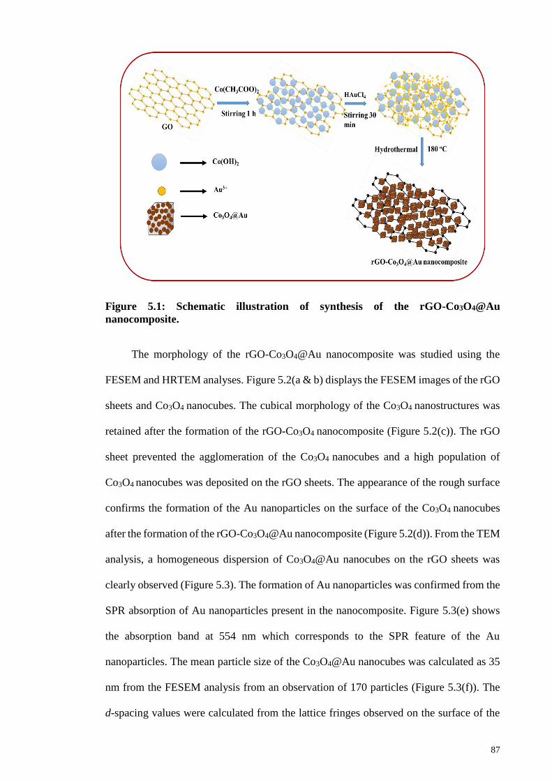
87
Figure 5.1: Schematic illustration of synthesis of the rGO-Co3O4@Au
nanocomposite.
The morphology of the rGO-Co3O4@Au nanocomposite was studied using the
FESEM and HRTEM analyses. Figure 5.2(a & b) displays the FESEM images of the rGO
sheets and Co3O4 nanocubes. The cubical morphology of the Co3O4 nanostructures was
retained after the formation of the rGO-Co3O4 nanocomposite (Figure 5.2(c)). The rGO
sheet prevented the agglomeration of the Co3O4 nanocubes and a high population of
Co3O4 nanocubes was deposited on the rGO sheets. The appearance of the rough surface
confirms the formation of the Au nanoparticles on the surface of the Co3O4 nanocubes
after the formation of the rGO-Co3O4@Au nanocomposite (Figure 5.2(d)). From the TEM
analysis, a homogeneous dispersion of Co3O4@Au nanocubes on the rGO sheets was
clearly observed (Figure 5.3). The formation of Au nanoparticles was confirmed from the
SPR absorption of Au nanoparticles present in the nanocomposite. Figure 5.3(e) shows
the absorption band at 554 nm which corresponds to the SPR feature of the Au
nanoparticles. The mean particle size of the Co3O4@Au nanocubes was calculated as 35
nm from the FESEM analysis from an observation of 170 particles (Figure 5.3(f)). The
d-spacing values were calculated from the lattice fringes observed on the surface of the

88
Co3O4@Au nanocubes. On the other hand, the small lattice fringes come under
observation belongs to the Au nanoparticles in Figure 5.3(d) with a lattice distance of
0.25 nm corresponding to the Au (1 1 1) plane. This also confirms the formation of Au
nanoparticle on the surface of Co3O4 nanocubes.
Figure 5.2: FESEM images of (a) rGO sheet, (b) Co3O4 nanocubes, (c) rGO-Co3O4
nanocomposite and (d) rGO-CO3O4@Au (8 mM).
(a)
(d)
(b)
(c)

89
Figure 5.3: (a & b) TEM images of rGO-Co3O4@Au (8mM) nanocomposite at
different magnifications, (c) single particle of Co3O4@Au (8mM), (d) lattice fringes
and (e) SPR absorption of Au nanoparticle deposited on Co3O4 nanocubes. ‘(f)’
shows the particle size histogram of the Co3O4@Au nanocubes.
(d) (c)
(b) (a)
300 450 600 750 900
Ab
so
rba
nce
(a.u
.)
Wavelength (nm)
20 25 30 35 40 45 500
5
10
15
20Mean 35.240
Std.Dev. 4.289
Fre
qu
en
cy
Particle size (nm)
(f) (e)
(111)
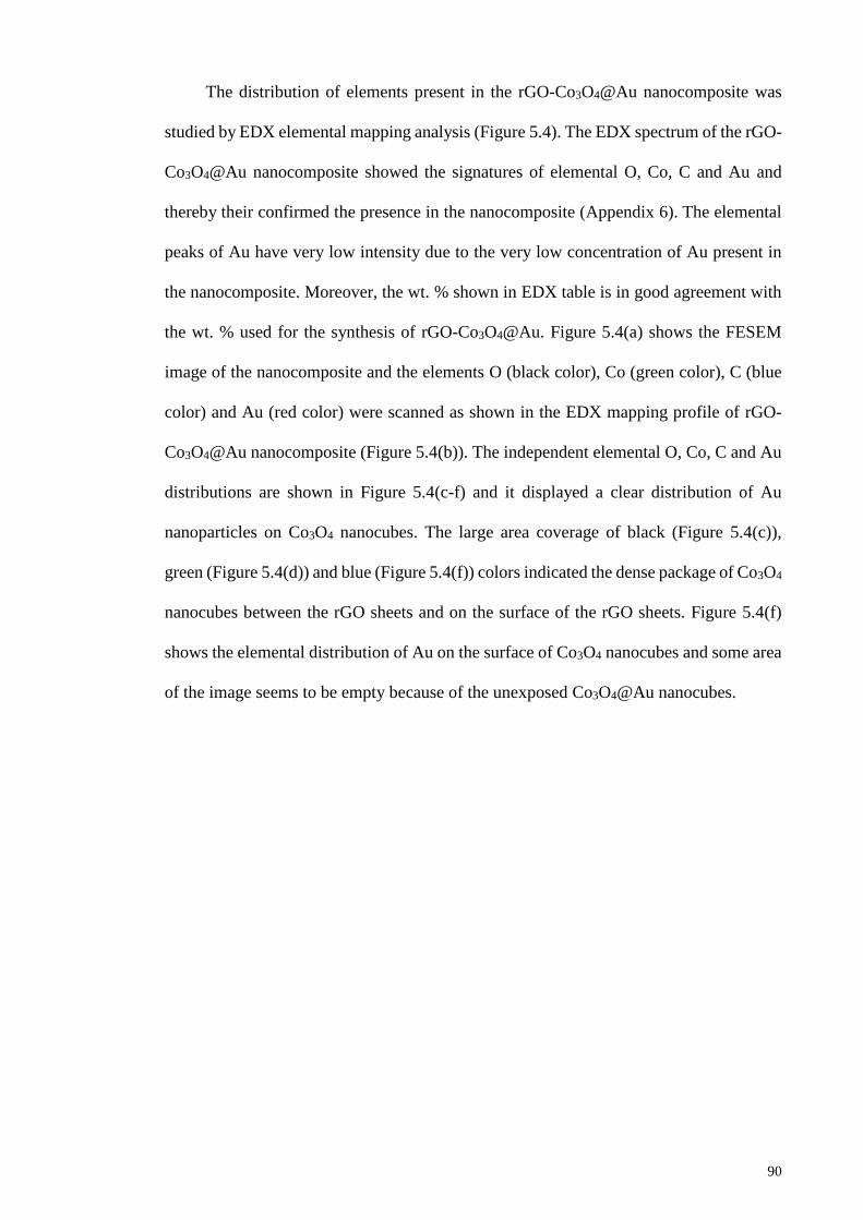
90
The distribution of elements present in the rGO-Co3O4@Au nanocomposite was
studied by EDX elemental mapping analysis (Figure 5.4). The EDX spectrum of the rGO-
Co3O4@Au nanocomposite showed the signatures of elemental O, Co, C and Au and
thereby their confirmed the presence in the nanocomposite (Appendix 6). The elemental
peaks of Au have very low intensity due to the very low concentration of Au present in
the nanocomposite. Moreover, the wt. % shown in EDX table is in good agreement with
the wt. % used for the synthesis of rGO-Co3O4@Au. Figure 5.4(a) shows the FESEM
image of the nanocomposite and the elements O (black color), Co (green color), C (blue
color) and Au (red color) were scanned as shown in the EDX mapping profile of rGO-
Co3O4@Au nanocomposite (Figure 5.4(b)). The independent elemental O, Co, C and Au
distributions are shown in Figure 5.4(c-f) and it displayed a clear distribution of Au
nanoparticles on Co3O4 nanocubes. The large area coverage of black (Figure 5.4(c)),
green (Figure 5.4(d)) and blue (Figure 5.4(f)) colors indicated the dense package of Co3O4
nanocubes between the rGO sheets and on the surface of the rGO sheets. Figure 5.4(f)
shows the elemental distribution of Au on the surface of Co3O4 nanocubes and some area
of the image seems to be empty because of the unexposed Co3O4@Au nanocubes.
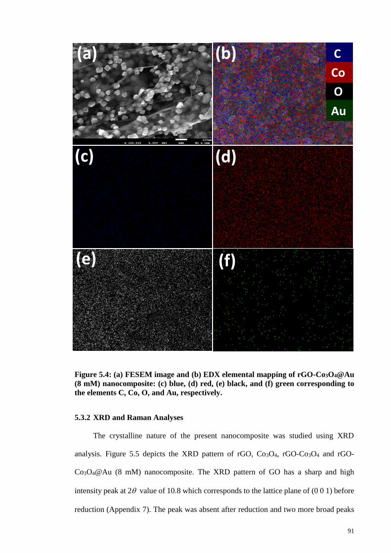
91
Figure 5.4: (a) FESEM image and (b) EDX elemental mapping of rGO-Co3O4@Au
(8 mM) nanocomposite: (c) blue, (d) red, (e) black, and (f) green corresponding to
the elements C, Co, O, and Au, respectively.
5.3.2 XRD and Raman Analyses
The crystalline nature of the present nanocomposite was studied using XRD
analysis. Figure 5.5 depicts the XRD pattern of rGO, Co3O4, rGO-Co3O4 and rGO-
Co3O4@Au (8 mM) nanocomposite. The XRD pattern of GO has a sharp and high
intensity peak at 2 value of 10.8 which corresponds to the lattice plane of (0 0 1) before
reduction (Appendix 7). The peak was absent after reduction and two more broad peaks
O
Au
C
Co
(a)
F (e)
(d) (c)
(b)
(f)

92
appeared at the 2 values of 26.0 and 43.1 corresponding to the (0 0 2) and (1 0 0) lattice
planes, respectively, thus showed the disorderedly stacking of the rGO in Figure 5.5
(Wang et al., 2008). All the other peaks observed at 31.3°, 36.9°, 44.8°, 55.7°, 59.3°,
65.2°and 77.3° correspond to (2 2 0), (3 1 1), (4 0 0), (4 2 2), (4 4 0), (5 1 1 ) and (5 3 3)
crystal planes of face centered cubic Co3O4, respectively (JCPDS Card No. 42-1467)
(Wu et al., 2010). There is no peak observed for the rGO due to its very thin and well
exfoliated sheets, the Co3O4 nanocubes behaves like nanospacers between the different
layers of rGO sheets as confirmed by FESEM analysis. This confirms that the Co3O4
nanocubes intercalate quiet well and prevents the rGO sheets from restacking after
reduction and there is no bulk graphene observed in XRD of rGO-Co3O4 (Wu et al., 2010).
The XRD pattern recorded for rGO-Co3O4@Au (8 mM) nanocomposite in Figure 5.5
showed that the four Au peaks observed at 38.2°, 44.4° ,64.7° and 77.7° which correspond
to the Au lattice planes (1 1 1), (2 0 0), (2 2 0) and (3 1 1) have similar 2θ values with
Co3O4. These Au peaks merged with the Co3O4 peaks and it could be seen from the XRD
pattern of rGO-Co3O4@Au nanocomposites that with the increase in Au contents from 2
mM to 10 mM the Au peaks became more intense in the rGO-Co3O4@Au
nanocomposites (Appendix 8) (JCPDS 04-0784) (Li et al., 2012). So, the existence of
these peaks in the XRD pattern is a further evidence of Au presence/deposition on Co3O4
nanocubes, in favor of the FESEM and HRTEM images in Figure 5.2 & 5.3 respectively.
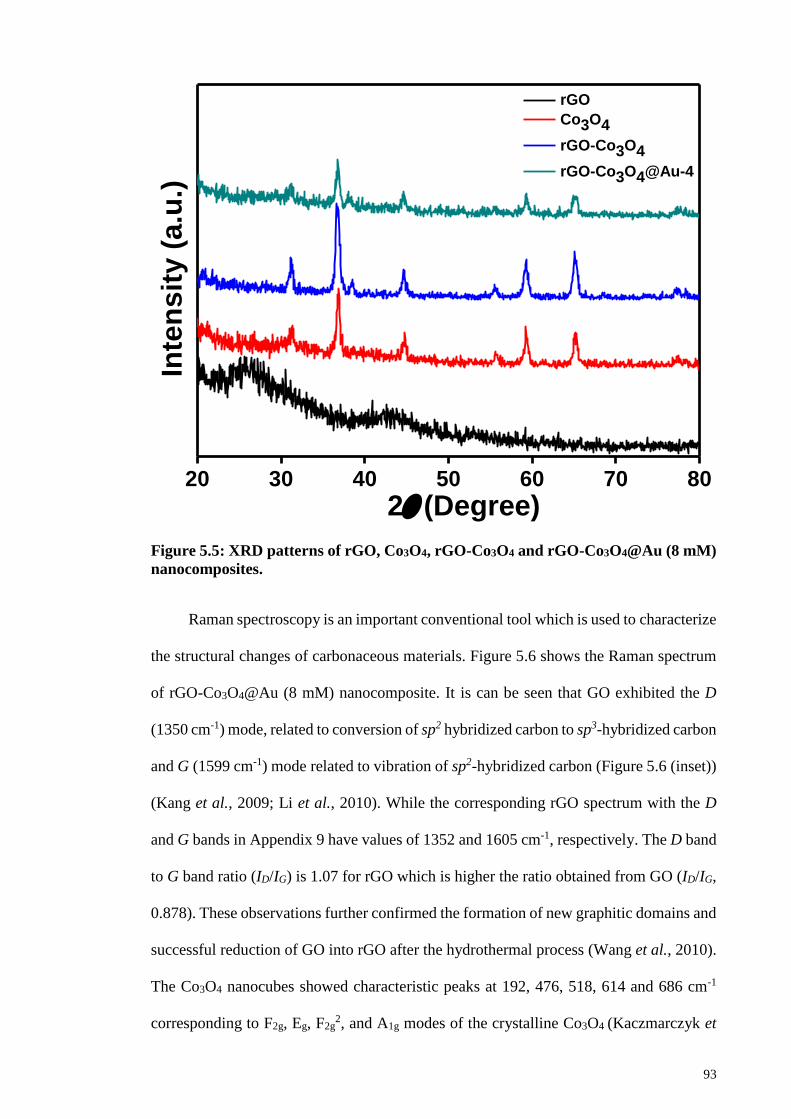
93
20 30 40 50 60 70 80
10 20 30 40 50 60 70 80
rGO
Co3O4
rGO-Co3O4
rGO-Co3O4@Au-4
Inte
nsit
y (
a.u
.)
2(Degree)
Figure 5.5: XRD patterns of rGO, Co3O4, rGO-Co3O4 and rGO-Co3O4@Au (8 mM)
nanocomposites.
Raman spectroscopy is an important conventional tool which is used to characterize
the structural changes of carbonaceous materials. Figure 5.6 shows the Raman spectrum
of rGO-Co3O4@Au (8 mM) nanocomposite. It is can be seen that GO exhibited the D
(1350 cm-1) mode, related to conversion of sp2 hybridized carbon to sp3-hybridized carbon
and G (1599 cm-1) mode related to vibration of sp2-hybridized carbon (Figure 5.6 (inset))
(Kang et al., 2009; Li et al., 2010). While the corresponding rGO spectrum with the D
and G bands in Appendix 9 have values of 1352 and 1605 cm-1, respectively. The D band
to G band ratio (ID/IG) is 1.07 for rGO which is higher the ratio obtained from GO (ID/IG,
0.878). These observations further confirmed the formation of new graphitic domains and
successful reduction of GO into rGO after the hydrothermal process (Wang et al., 2010).
The Co3O4 nanocubes showed characteristic peaks at 192, 476, 518, 614 and 686 cm-1
corresponding to F2g, Eg, F2g2, and A1g modes of the crystalline Co3O4 (Kaczmarczyk et
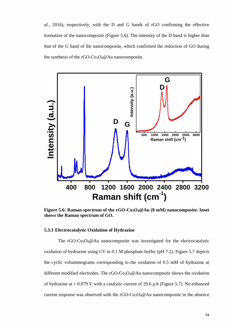
94
al., 2016), respectively, with the D and G bands of rGO confirming the effective
formation of the nanocomposite (Figure 5.6). The intensity of the D band is higher than
that of the G band of the nanocomposite, which confirmed the reduction of GO during
the synthesis of the rGO-Co3O4@Au nanocomposite.
400 800 1200 1600 2000 2400 2800 3200
500 1000 1500 2000 2500 3000
GIn
ten
sit
y (
a.u
.)
Inte
ns
ity
(a
.u.)
Raman shift (cm-1)
D
DG
Raman shift (cm-1)
Figure 5.6: Raman spectrum of the rGO-Co3O4@Au (8 mM) nanocomposite. Inset
shows the Raman spectrum of GO.
5.3.3 Electrocatalytic Oxidation of Hydrazine
The rGO-Co3O4@Au nanocomposite was investigated for the electrocatalytic
oxidation of hydrazine using CV in 0.1 M phosphate buffer (pH 7.2). Figure 5.7 depicts
the cyclic voltammograms corresponding to the oxidation of 0.5 mM of hydrazine at
different modified electrodes. The rGO-Co3O4@Au nanocomposite shows the oxidation
of hydrazine at + 0.079 V with a catalytic current of 29.6 µA (Figure 5.7). No enhanced
current response was observed with the rGO-Co3O4@Au nanocomposite in the absence

95
of hydrazine (Figure 5.7). The bare GCE and rGO did not produce faradaic current
response from the oxidation of hydrazine (Figure 5.7). However, the cyclic
voltammogram of Co3O4 nanocubes seems to exhibit the anodic peak of hydrazine
oxidation at higher positive potential (Figure 5.7). The hydrazine oxidation was highly
facilitated at the rGO-Co3O4 nanocubes modified electrode surface and the electrode
showed well resolved voltammetric behavior with enormous negative shift in the
potential. It showed a catalytic current of 21.57 µA at the peak potential of +0.089 V
(Figure 5.7). Graphene is highly conducting platform with higher electron transfer
kinetics and it prevents the Co3O4 nanocubes from agglomeration (Figure 5.2), so higher
electrocatalytic activity is expected due to the synergistic effect of rGO-Co3O4
nanocomposite (Shahid et al., 2014). Furthermore, the deposition of very low
concentration of Au nanoparticles on the surface of the Co3O4 nanocubes improved the
electrocatalytic activity of the rGO-Co3O4 nanocomposite towards the oxidation of
hydrazine. The increase of catalytic current and the decrease of overpotential were
attributed to the synergetic effect of the rGO-Co3O4 nanocomposite and the highly
conductive Au nanoparticles. The surface roughness and increased surface area of the
rGO-Co3O4@Au nanocomposite also played a crucial role for the enhanced electron
transfer process towards the catalytic performance. The cyclic voltammograms were
recorded with rGO-Co3O4@Au nanocomposite consisting of different concentration of
Au (2, 4, 6, 8 and 10 mM) for the electrocatalytic oxidation of hydrazine shown in
Appendix 10. It was found that the rGO-Co3O4@Au (8 mM) nanocomposite showed the
best catalytic response towards the oxidation of hydrazine.
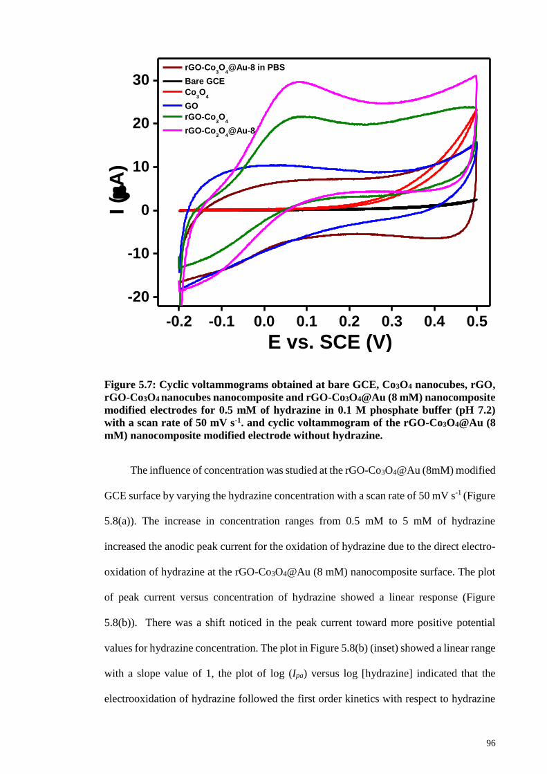
96
-0.2 -0.1 0.0 0.1 0.2 0.3 0.4 0.5
-20
-10
0
10
20
30 rGO-Co
3O
4@Au-8 in PBS
Bare GCE
Co3O
4
GO
rGO-Co3O
4
rGO-Co3O
4@Au-8
I (
A)
E vs. SCE (V)
Figure 5.7: Cyclic voltammograms obtained at bare GCE, Co3O4 nanocubes, rGO,
rGO-Co3O4 nanocubes nanocomposite and rGO-Co3O4@Au (8 mM) nanocomposite
modified electrodes for 0.5 mM of hydrazine in 0.1 M phosphate buffer (pH 7.2)
with a scan rate of 50 mV s-1. and cyclic voltammogram of the rGO-Co3O4@Au (8
mM) nanocomposite modified electrode without hydrazine.
The influence of concentration was studied at the rGO-Co3O4@Au (8mM) modified
GCE surface by varying the hydrazine concentration with a scan rate of 50 mV s-1 (Figure
5.8(a)). The increase in concentration ranges from 0.5 mM to 5 mM of hydrazine
increased the anodic peak current for the oxidation of hydrazine due to the direct electro-
oxidation of hydrazine at the rGO-Co3O4@Au (8 mM) nanocomposite surface. The plot
of peak current versus concentration of hydrazine showed a linear response (Figure
5.8(b)). There was a shift noticed in the peak current toward more positive potential
values for hydrazine concentration. The plot in Figure 5.8(b) (inset) showed a linear range
with a slope value of 1, the plot of log (Ipa) versus log [hydrazine] indicated that the
electrooxidation of hydrazine followed the first order kinetics with respect to hydrazine
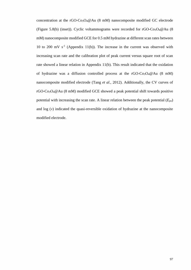
97
concentration at the rGO-Co3O4@Au (8 mM) nanocomposite modified GC electrode
(Figure 5.8(b) (inset)). Cyclic voltammograms were recorded for rGO-Co3O4@Au (8
mM) nanocomposite modified GCE for 0.5 mM hydrazine at different scan rates between
10 to 200 mV s-1 (Appendix 11(b)). The increase in the current was observed with
increasing scan rate and the calibration plot of peak current versus square root of scan
rate showed a linear relation in Appendix 11(b). This result indicated that the oxidation
of hydrazine was a diffusion controlled process at the rGO-Co3O4@Au (8 mM)
nanocomposite modified electrode (Tang et al., 2012). Additionally, the CV curves of
rGO-Co3O4@Au (8 mM) modified GCE showed a peak potential shift towards positive
potential with increasing the scan rate. A linear relation between the peak potential (Epa)
and log (ν) indicated the quasi-reversible oxidation of hydrazine at the nanocomposite
modified electrode.
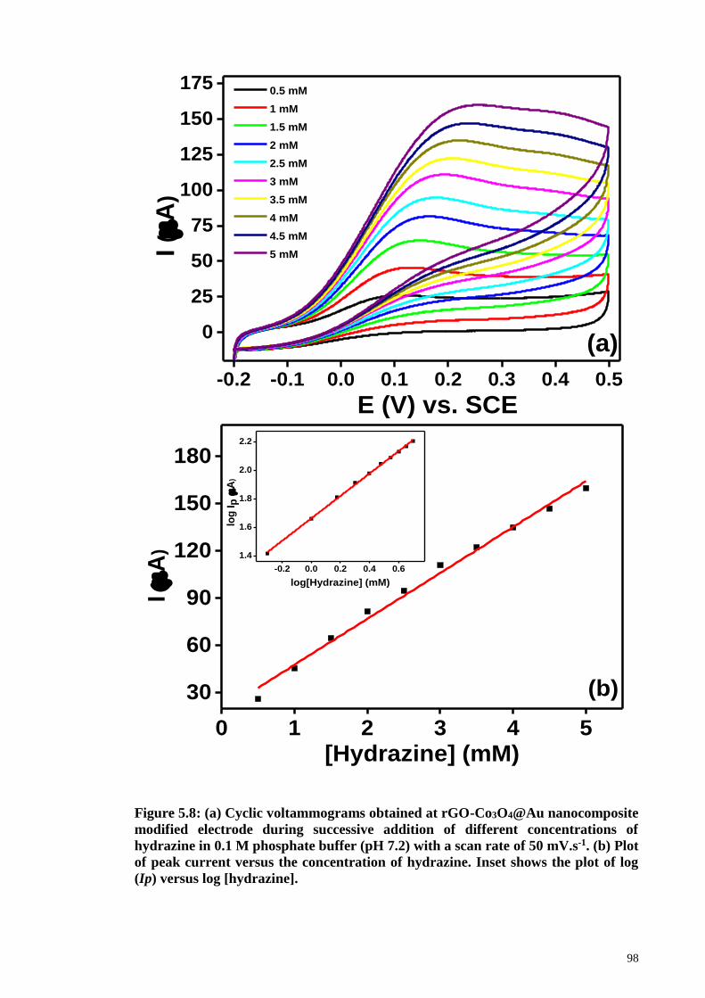
98
Figure 5.8: (a) Cyclic voltammograms obtained at rGO-Co3O4@Au nanocomposite
modified electrode during successive addition of different concentrations of
hydrazine in 0.1 M phosphate buffer (pH 7.2) with a scan rate of 50 mV.s-1. (b) Plot
of peak current versus the concentration of hydrazine. Inset shows the plot of log
(Ip) versus log [hydrazine].
-0.2 -0.1 0.0 0.1 0.2 0.3 0.4 0.5
0
25
50
75
100
125
150
175
I (
A)
E (V) vs. SCE
0.5 mM
1 mM
1.5 mM
2 mM
2.5 mM
3 mM
3.5 mM
4 mM
4.5 mM
5 mM
(a)
0 1 2 3 4 5
30
60
90
120
150
180
-0.2 0.0 0.2 0.4 0.6
1.4
1.6
1.8
2.0
2.2
I (
A)
[Hydrazine] (mM)
(b)
log
Ip
(A
)
log[Hydrazine] (mM)

99
The diffusion coefficient (D) of hydrazine during the electrocatalysis was
calculated using the Cottrell equation (equation. 5.1).
𝐼 = 𝑛𝐹𝐷1/2A𝐶0𝜋−1/2𝑡−1/2 (5.1)
Where n is the number of electron involved per hydrazine molecule during oxidation, F
is the Faraday constant, A is the geometric area of the electrode, C0 is the concentration
of hydrazine, and t is time. The amperometric i-t curves were collected at the
nanocomposite modified electrode for the different concentrations of hydrazine (Figure
5.9(a)) and the plot of peak current versus t-1/2 showed a linear relation (Figure 5.9(b)).
The slopes of the obtained linear lines were plotted against the hydrazine concentrations
(Figure 5.9(b) (inset)) and from this plot D was determined to be 0.782 × 10⁻⁶ cm2.s-1.
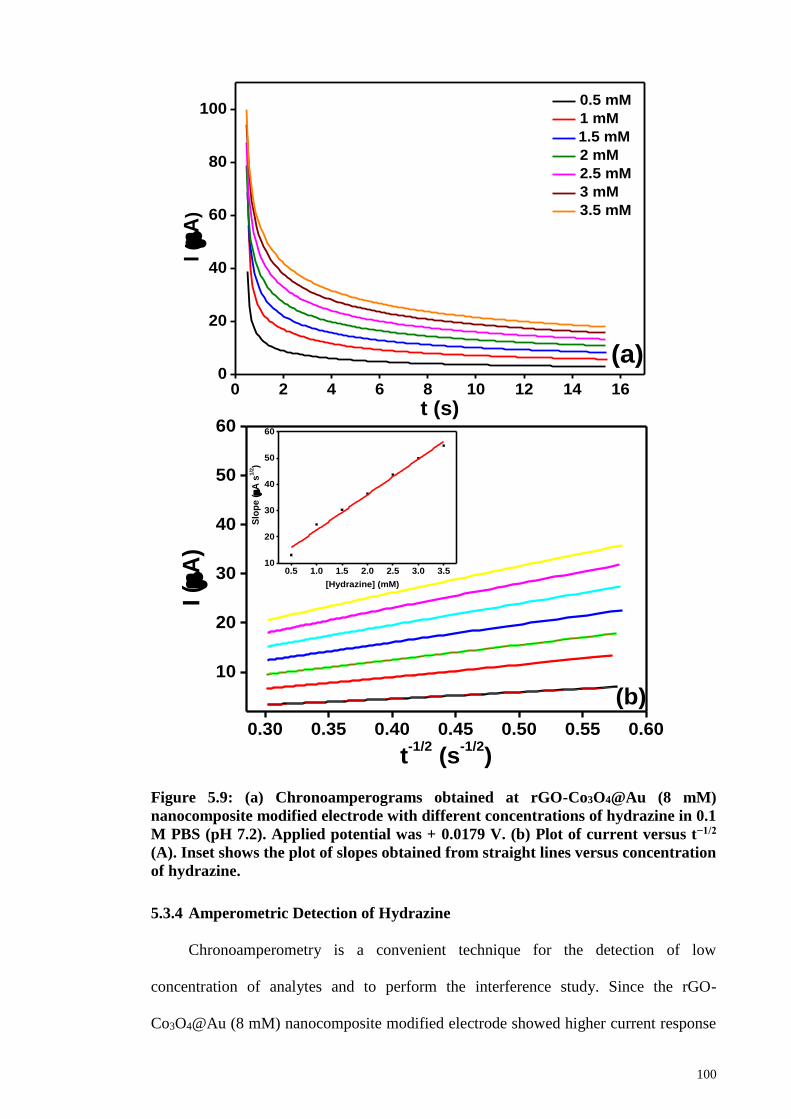
100
Figure 5.9: (a) Chronoamperograms obtained at rGO-Co3O4@Au (8 mM)
nanocomposite modified electrode with different concentrations of hydrazine in 0.1
M PBS (pH 7.2). Applied potential was + 0.0179 V. (b) Plot of current versus t−1/2
(A). Inset shows the plot of slopes obtained from straight lines versus concentration
of hydrazine.
5.3.4 Amperometric Detection of Hydrazine
Chronoamperometry is a convenient technique for the detection of low
concentration of analytes and to perform the interference study. Since the rGO-
Co3O4@Au (8 mM) nanocomposite modified electrode showed higher current response
0 2 4 6 8 10 12 14 160
20
40
60
80
100
I (
A)
t (s)
0.5 mM
1 mM
1.5 mM
2 mM
2.5 mM
3 mM
3.5 mM
(a)
0.30 0.35 0.40 0.45 0.50 0.55 0.60
10
20
30
40
50
60
0.5 1.0 1.5 2.0 2.5 3.0 3.510
20
30
40
50
60
(b)
I (
A)
t-1/2
(s-1/2
)
Slo
pe
(A
s1
/2)
[Hydrazine] (mM)

101
in the CV, it was used as the amperometric sensor for the detection of hydrazine at low
concentration levels. Figure 5.10(a) depicted the amperometric response of the rGO-
Co3O4@Au (8 mM) nanocomposite for the successive additions of hydrazine at an
applied potential of + 0.079 V. The current response was measured from the successive
injection of 10 µM and 20 µM concentration of hydrazine at the time interval of 60 s in a
continuously stirred 0.1 M phosphate buffer (pH 7.2). The rGO-Co3O4@Au (8mM)
modified GCE exhibited a significant and quick amperometric response towards each
addition of hydrazine. The current reached its steady-state within 3 s indicating the fast
electrooxidation of hydrazine at the rGO-Co3O4@Au (8 mM) modified GCE surface. The
response current increased linearly for each addition of hydrazine over the range between
10 and 740 µM. The corresponding regression plot of current response versus
concentration of hydrazine with a linear relation is shown in Figure 5.10(b). The LOD
was calculated as 0.443 µM from the expression LOD = 3σ/slope, where, σ is the standard
deviation (Devasenathipathy et al., 2014). The sensitivity of the sensor was found to be
0.58304 ± 0.00466 µA µM–1 from the slope of the linear regression. Repeated
measurements were carried out to check the stability of the sensor and the current
responses with negligible decrement were observed at different days. The electrocatalytic
activity of the rGO-Co3O4 nanocomposite towards the detection of hydrazine was
improved by the deposition of minimum amount of Au on the Co3O4 nanocubes. The
LOD can further be improved by optimizing the rGO content in the nanocomposite
because it mainly alters the diffusion layer thickness of the nanocomposite modified
electrode.
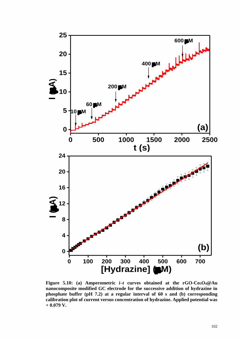
102
Figure 5.10: (a) Amperometric i–t curves obtained at the rGO-Co3O4@Au
nanocomposite modified GC electrode for the successive addition of hydrazine in
phosphate buffer (pH 7.2) at a regular interval of 60 s and (b) corresponding
calibration plot of current versus concentration of hydrazine. Applied potential was
+ 0.079 V.
0 500 1000 1500 2000 2500
0
5
10
15
20
25600 M
400 M
200 M
60 M
I (
A)
t (s)
10 M
(a)
0 100 200 300 400 500 600 700
0
4
8
12
16
20
24
(b)
[Hydrazine] (M)
I (
A)

103
The selectivity of the rGO-Co3O4@Au (8 mM) nanocomposite is an important
aspect of the sensor performance. This was assessed by studying the sensor response in
the presence of interferents in the same phosphate buffer and the change in current
response was monitored. Figure 5.11 shows the amperometric current responses of
hydrazine (a) and various interferents such as NO3- (b), SO4
2- (c), Cl- (d), Ag+ (e), Na+ (f),
K+ (g), ethanol (h), 4-nitrophenol (i), AA (j) and glucose for studying selectivity of rGO-
Co3O4@Au (8 mM) nanocomposite. For the first two injections of hydrazine, the
nanocomposite behaved efficiently and showed a current response for the hydrazine
oxidation. There was no response shown by the rGO-Co3O4@Au (8 mM) for the rest of
the interferents even at 50-fold higher concentration. Again, for the reconfirmation of
selective behavior of rGO-Co3O4@Au (8 mM) nanocomposite, hydrazine was injected
into homogeneously stirred 0.1 M phosphate buffer and it showed a rapid current response
suggesting that the present rGO-Co3O4@Au (8 mM) nanocomposite was more selective
towards hydrazine oxidation. Moreover, the same magnitude of the current response for
the addition of hydrazine was observed and it sustained the steady state current after the
3 s of response time. Two other interferent molecules such as ascorbic acid and glucose
were injected after hydrazine and there was no current observed for these molecules. This
study revealed the good selectivity of the rGO-Co3O4@Au nanocomposite towards
hydrazine sensing. Table 1 shows the comparison of analytical performance of the present
nanocomposite with other reported nanomaterials. The present work stands for the novel
synthesis of the rGO-Co3O4@Au nanocomposite using one-pot hydrothermal synthesis
and its fast response towards the detection of hydrazine. The proposed modified electrode
displayed a satisfactory performance in terms of the detection limit and good selectivity.
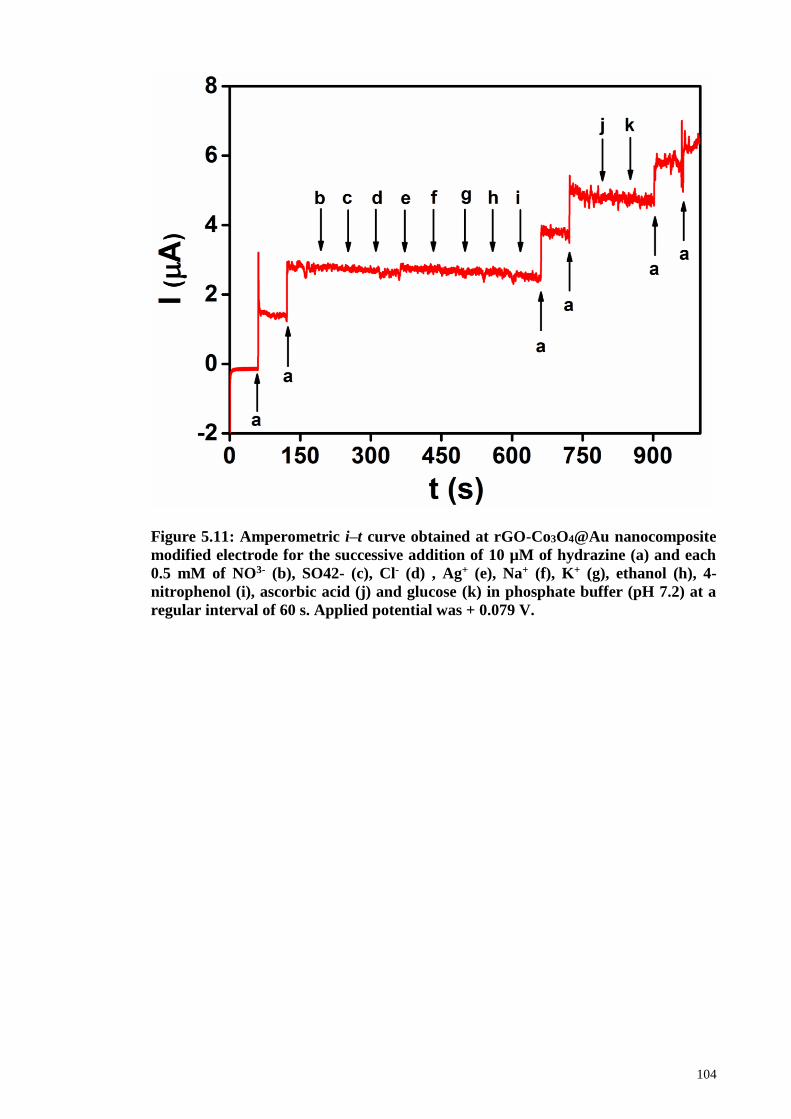
104
Figure 5.11: Amperometric i–t curve obtained at rGO-Co3O4@Au nanocomposite
modified electrode for the successive addition of 10 µM of hydrazine (a) and each
0.5 mM of NO3- (b), SO42- (c), Cl- (d) , Ag+ (e), Na+ (f), K+ (g), ethanol (h), 4-
nitrophenol (i), ascorbic acid (j) and glucose (k) in phosphate buffer (pH 7.2) at a
regular interval of 60 s. Applied potential was + 0.079 V.
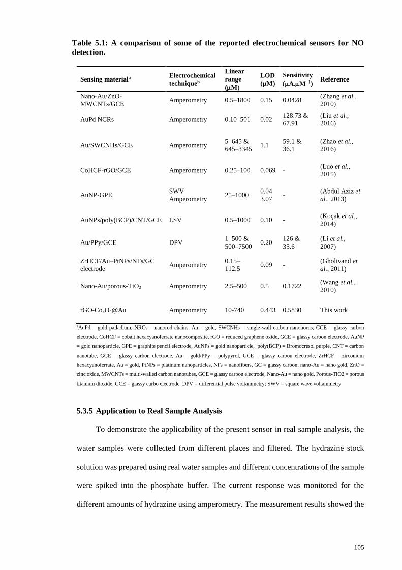
105
Table 5.1: A comparison of some of the reported electrochemical sensors for NO
detection.
Sensing materiala Electrochemical
techniqueb
Linear
range
(M)
LOD
(µM)
Sensitivity
(A.M−1) Reference
Nano-Au/ZnO-
MWCNTs/GCE Amperometry 0.5–1800 0.15 0.0428
(Zhang et al.,
2010)
AuPd NCRs Amperometry 0.10–501 0.02 128.73 &
67.91
(Liu et al.,
2016)
Au/SWCNHs/GCE Amperometry 5–645 &
645–3345 1.1
59.1 &
36.1
(Zhao et al.,
2016)
CoHCF-rGO/GCE Amperometry 0.25–100 0.069 - (Luo et al.,
2015)
AuNP-GPE SWV
Amperometry 25–1000
0.04
3.07 -
(Abdul Aziz et
al., 2013)
AuNPs/poly(BCP)/CNT/GCE LSV 0.5–1000 0.10 - (Koçak et al.,
2014)
Au/PPy/GCE DPV 1–500 &
500–7500 0.20
126 &
35.6
(Li et al.,
2007)
ZrHCF/Au–PtNPs/NFs/GC
electrode Amperometry
0.15–
112.5 0.09 -
(Gholivand et
al., 2011)
Nano-Au/porous-TiO2 Amperometry 2.5–500 0.5 0.1722 (Wang et al.,
2010)
rGO-Co3O4@Au Amperometry 10-740 0.443 0.5830 This work
aAuPd = gold palladium, NRCs = nanorod chains, Au = gold, SWCNHs = single-wall carbon nanohorns, GCE = glassy carbon
electrode, CoHCF = cobalt hexacyanoferrate nanocomposite, rGO = reduced graphene oxide, GCE = glassy carbon electrode, AuNP
= gold nanoparticle, GPE = graphite pencil electrode, AuNPs = gold nanoparticle, poly(BCP) = Bromocresol purple, CNT = carbon
nanotube, GCE = glassy carbon electrode, Au = gold/PPy = polypyrol, GCE = glassy carbon electrode, ZrHCF = zirconium
hexacyanoferrate, Au = gold, PtNPs = platinum nanoparticles, NFs = nanofibers, GC = glassy carbon, nano-Au = nano gold, ZnO =
zinc oxide, MWCNTs = multi-walled carbon nanotubes, GCE = glassy carbon electrode, Nano-Au = nano gold, Porous-TiO2 = porous
titanium dioxide, GCE = glassy carbo electrode, DPV = differential pulse voltammetry; SWV = square wave voltammetry
5.3.5 Application to Real Sample Analysis
To demonstrate the applicability of the present sensor in real sample analysis, the
water samples were collected from different places and filtered. The hydrazine stock
solution was prepared using real water samples and different concentrations of the sample
were spiked into the phosphate buffer. The current response was monitored for the
different amounts of hydrazine using amperometry. The measurement results showed the
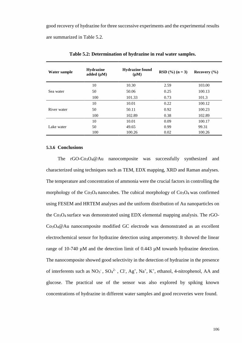
106
good recovery of hydrazine for three successive experiments and the experimental results
are summarized in Table 5.2.
Table 5.2: Determination of hydrazine in real water samples.
5.3.6 Conclusions
The rGO-Co3O4@Au nanocomposite was successfully synthesized and
characterized using techniques such as TEM, EDX mapping, XRD and Raman analyses.
The temperature and concentration of ammonia were the crucial factors in controlling the
morphology of the Co3O4 nanocubes. The cubical morphology of Co3O4 was confirmed
using FESEM and HRTEM analyses and the uniform distribution of Au nanoparticles on
the Co3O4 surface was demonstrated using EDX elemental mapping analysis. The rGO-
Co3O4@Au nanocomposite modified GC electrode was demonstrated as an excellent
electrochemical sensor for hydrazine detection using amperometry. It showed the linear
range of 10-740 µM and the detection limit of 0.443 µM towards hydrazine detection.
The nanocomposite showed good selectivity in the detection of hydrazine in the presence
of interferents such as NO3- , SO4
2- , Cl-, Ag+, Na+, K+, ethanol, 4-nitrophenol, AA and
glucose. The practical use of the sensor was also explored by spiking known
concentrations of hydrazine in different water samples and good recoveries were found.
Water sample Hydrazine
added (µM)
Hydrazine found
(µM) RSD (%) (n = 3) Recovery (%)
Sea water
10 10.30 2.59 103.00
50 50.06 0.25 100.13
100 101.33 0.73 101.3
River water
10 10.01 0.22 100.12
50 50.11 0.92 100.23
100 102.89 0.38 102.89
Lake water
10 10.01 0.09 100.17
50 49.65 0.99 99.31
100 100.26 0.02 100.26
107
CHAPTER 6: AN ELECTROCHEMICAL SENSING PLATFORM BASED ON
REDUCED GRAPHENE OXIDE-COBALT OXIDE NANOCUBES@PLATINUM
NANOCOMPOSITE FOR NITRIC OXIDE DETECTION3
6.1 Introduction
Nitric oxide (NO) is one of the smallest and simplest biologically important
molecules in nature with distinctive and fascinating chemistry.(Taha, 2003) Ignarro,
Furchgott and Murad identified NO as endothelium-derived relaxation factor (EDRF)
which is responsible for vasodilation and blood pressure regulation in the nervous and
cardiovascular systems of mammalian physiology (Furchgott, 1999; Guix et al., 2005;
Guo et al., 2012; Ignarro et al., 1987). NO plays extremely important physiological roles
as an endogenously-produced antimicrobial agent, (Fang, 1997) as a signaling molecule
capable of modulating cytokine production (Schwentker et al., 2002) and also, it plays a
key role in wound healing (Luo et al., 2005) and in immune response (Bogdan, 2001).
The multi-tasking NO is actually produced endogenously by a class of heme-containing
enzymes called nitric oxide synthases (Hetrick et al., 2009; Moncada et al., 1993). Due
to the extensive interest in NO from a biochemical and a medical perspective, it is vitally
important to monitor the concentration level of NO in physiological system very closely.
The concentration of NO varies in the human body from sub-nanomolar to micromolar
levels (Privett et al., 2010). Some parameters are very important for the effective
detection of NO such as adequate sensitivity, fast response time, wide dynamic range and
high selectivity toward NO over the interfering species. Due to the irresistible complexity
3 This chapter has been published Shahid, M. M., Rameshkumar, P., Pandikumar, A., Lim, H. N., Ng,
Y. H., & Huang, N. M. (2015). An electrochemical sensing platform based on a reduced graphene oxide–
cobalt oxide nanocube@ platinum nanocomposite for nitric oxide detection. Journal of Materials
Chemistry A, 3(27), 14458-14468.
108
of biological systems, these parameters are often challenging. Besides all the mentioned
challenges, the chemical properties of NO make the detection very complex. Fortunately,
some analytical techniques including spectroscopic and electrochemical methods are
often used for the detection of NO. Among the various methods, electrochemical method
is an efficient analytical technique to detect NO because of its long term high calibration
stability, fast response, good sensitivity, better selectivity and simplicity(Bedioui et al.,
2003).
In recent years, constructing a competent electrochemical sensor is highly pursued
among researchers for the sensitive detection of NO. Recently, efforts have been made to
increase the sensitivity of the electrochemical detection of NO by engineering the
electrode with functional nanomaterials (Privett et al., 2010; Wang et al., 2005). Different
type of nanomaterials including single-walled carbon nanotubes (SWCNTs) (Li et al.,
2006), multi-walled carbon nanotubes (MWCNTs) (Wu et al., 2002), nano-alumina (He
et al., 2009), Nafion-nickel (II) porphyrin film (Malinski et al., 1992) and gold
nanoparticles (Thangavel et al., 2008) etc. have been previously used for the detection of
NO. Graphene based nanocomposite materials as electrochemical sensors have also been
reported for the detection of NO (Jayabal et al., 2014; Li et al., 2011; Wang et al., 2014).
Graphene is a two dimensional carbon sheet having single atom thickness, large
theoretical surface area (2630 m2 g-1) with high conductivity at room temperature (106 s
cm-1) and wide electrochemical window (Chang et al., 2013; Zhu et al., 2010). Graphene
nanosheet is an excellent host material for growing nanomaterials for high performance
electrochemical applications (Kim et al., 2009; Zhou et al., 2010). To date, a large number
of graphene based nanocomposite materials has been synthesized for different
applications such as energy storage (Mahmood et al., 2014), biosensor (Karuppiah et al.,
2014) and electrocatlaytic (Choi et al., 2012) applications etc. A number of reports on
metals, metal oxides and semiconductor nanoparticles grown on graphene nanosheets and

109
various electrocatalytic applications have been reported (Guo et al., 2009; Kou et al.,
2011; Qu et al., 2011; Wu et al., 2010). Among the transition metal oxides, cobalt oxide
(Co3O4) gathered the attention of many researchers due to its high surface area to volume
ratio, high ratio of surface atoms, good chemical stability and it is expected to meet the
requirements of future energy applications (Lu et al., 2010). Owing these properties,
unaided Co3O4 nanoparticles have been used in many reports for variety of applications
(Chen et al., 2013; Farhadi et al., 2014; Farhadi et al., 2013; Mu et al., 2013). However,
only few attempts have been made to synthesize graphene-cobalt oxide nanocomposite
and used for electrocatalytic applications (Karuppiah et al., 2014); (Pan et al., 2013; Xiao
et al., 2013). For the synthesis of graphene-metal oxide nanocomposites, hydrothermal
synthesis is highly preferred because an appropriate amount of powdered reagents and
water are placed in a Teflon-lined autoclave and heated without stirring from moderate to
high temperatures and pressures for the desired time, with the possibility of predicting the
optimum reaction conditions by electrolyte thermodynamics (Lencka et al., 2000).
In this work, the electrochemical sensing platform based on one-pot hydrothermally
synthesized rGO-Co3O4@Pt nanocomposite for the detection of in-situ generated NO was
investigated. The formation of rGO-Co3O4@Pt nanocomposite was confirmed by
FESEM, EDX mapping, XRD and Raman analyses. The rGO-Co3O4@Pt nanocomposite
modified GC electrode displayed better catalytic performance toward the oxidation of NO
compared to the other controlled modified electrode investigated in this work. The
detection of lower concentration of NO was studied using amperometric i-t curve
technique and the LOD was 1.73 µM with a signal-to-noise (S/N) ratio ~ 3. The use of
lower concentration of Pt improved the sensitivity of the rGO-Co3O4@Pt nanocomposite
modified electrode. Moreover, the nanocomposite modified electrode was stable,
reproducible and showed an excellent selectivity toward the detection of NO in the
presence of 100-fold higher concentration of other physiologically important interferents.
110
6.2 Experimental Methods
6.2.1 Materials
Graphite flakes was purchased from Asbury Inc. (USA). Potassium permanganate
(KMnO4, >99 %), sulphuric acid (H2SO4, 98 %), hydrochloric acid (HCl, 35 %), and
ammonia solution (NH3, 25 %) were received from R & M Chemicals. Cobalt acetate
tetrahydrate (Co(CH3COO)2.4H2O) and potassium tetrachloroplatinate (II) (K2PtCl4)
were obtained from Sigma Aldrich and Acros Organics, respectively. Hydrogen peroxide
(H2O2) and sodium nitrite (NaNO2) were obtained from Systerm and Merck, respectively.
All the chemicals used in this study were of analytical grade. Doubly distilled water was
used to prepare the solutions for all the experiments.
6.2.2 Synthesis of rGO-Co3O4@Pt Nanocomposite
First, GO was prepared by following the simplified Hummer’s method.(Huang et
al., 2011) For the preparation of rGO-Co3O4@Pt nanocomposite, 12 mL of 1 mmol of
Co(CH3COO)2.4H2O solution was mixed with GO solution (8 wt. %) and stirred for 1.5
h to get a homogeneous solution. To this solution, 1 mL of 10 mM K2PtCl4 was added
slowly with stirring. This was followed by the drop-wise addition of 15 mL of 7.5 %
ammonia into the above reaction mixture under vigorous stirring. Then, 75 mL of the
reaction mixture was transferred to a 100 mL Teflon-lined stainless steel autoclave for
the hydrothermal treatment at 180 °C for 12 h. After the hydrothermal treatment, the
precipitate of rGO-Co3O4@Pt nanocomposite was washed five times with deionized
water and ethanol and dried in a hot air oven at 60 °C. The solid product of the
nanocomposite was collected and used for further studies. The synthesis of rGO, Co3O4
nanocubes and rGO-Co3O4 nanocomposite were followed by using a similar method. For
the optimization of GO, rGO-Co3O4 nanocomposite was prepared using different
amounts of GO (4 and 12 %). The rGO-Co3O4 nanocomposite used in this work contains
8 wt. % of GO, unless mentioned otherwise.
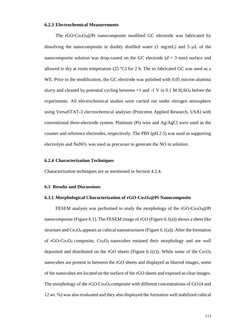
111
6.2.3 Electrochemical Measurements
The rGO-Co3O4@Pt nanocomposite modified GC electrode was fabricated by
dissolving the nanocomposite in doubly distilled water (1 mg/mL) and 5 L of the
nanocomposite solution was drop-casted on the GC electrode (d = 3 mm) surface and
allowed to dry at room temperature (25 oC) for 2 h. The so fabricated GC was used as a
WE. Prior to the modification, the GC electrode was polished with 0.05 micron alumina
slurry and cleaned by potential cycling between +1 and -1 V in 0.1 M H2SO4 before the
experiments. All electrochemical studies were carried out under nitrogen atmosphere
using VersaSTAT-3 electrochemical analyser (Princeton Applied Research, USA) with
conventional three-electrode system. Platinum (Pt) wire and Ag/AgCl were used as the
counter and reference electrodes, respectively. The PBS (pH 2.5) was used as supporting
electrolyte and NaNO2 was used as precursor to generate the NO in solution.
6.2.4 Characterization Techniques
Characterization techniques are as mentioned in Section 4.2.4.
6.3 Results and Discussions
6.3.1 Morphological Characterization of rGO-Co3O4@Pt Nanocomposite
FESEM analysis was performed to study the morphology of the rGO-Co3O4@Pt
nanocomposite (Figure 6.1). The FESEM image of rGO (Figure 6.1(a)) shows a sheet like
structure and Co3O4 appears as cubical nanostructures (Figure 6.1(a)). After the formation
of rGO-Co3O4 composite, Co3O4 nanocubes retained their morphology and are well
deposited and distributed on the rGO sheets (Figure 6.1(c)). While some of the Co3O4
nanocubes are present in between the rGO sheets and displayed as blurred images, some
of the nanocubes are located on the surface of the rGO sheets and exposed as clear images.
The morphology of the rGO-Co3O4 composite with different concentrations of GO (4 and
12 wt. %) was also evaluated and they also displayed the formation well stabilized cubical
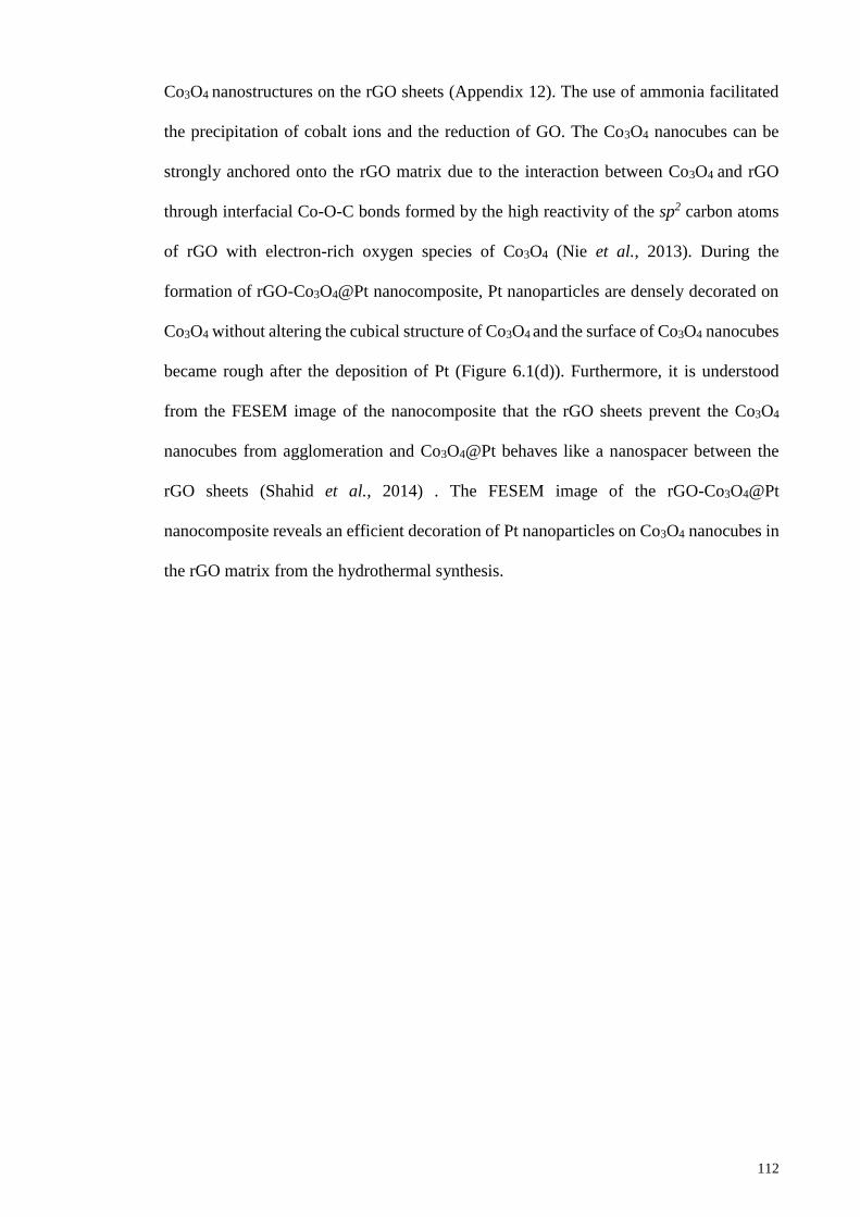
112
Co3O4 nanostructures on the rGO sheets (Appendix 12). The use of ammonia facilitated
the precipitation of cobalt ions and the reduction of GO. The Co3O4 nanocubes can be
strongly anchored onto the rGO matrix due to the interaction between Co3O4 and rGO
through interfacial Co-O-C bonds formed by the high reactivity of the sp2 carbon atoms
of rGO with electron-rich oxygen species of Co3O4 (Nie et al., 2013). During the
formation of rGO-Co3O4@Pt nanocomposite, Pt nanoparticles are densely decorated on
Co3O4 without altering the cubical structure of Co3O4 and the surface of Co3O4 nanocubes
became rough after the deposition of Pt (Figure 6.1(d)). Furthermore, it is understood
from the FESEM image of the nanocomposite that the rGO sheets prevent the Co3O4
nanocubes from agglomeration and Co3O4@Pt behaves like a nanospacer between the
rGO sheets (Shahid et al., 2014) . The FESEM image of the rGO-Co3O4@Pt
nanocomposite reveals an efficient decoration of Pt nanoparticles on Co3O4 nanocubes in
the rGO matrix from the hydrothermal synthesis.
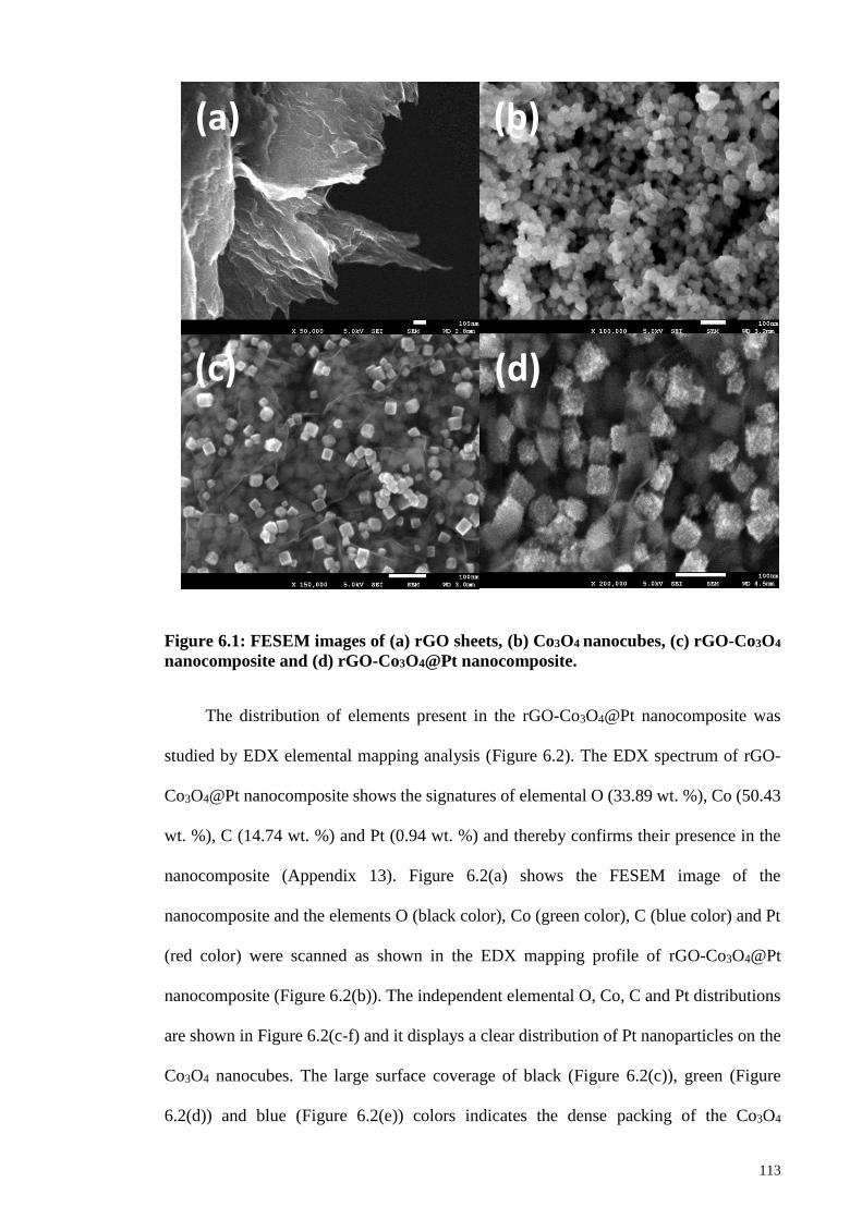
113
Figure 6.1: FESEM images of (a) rGO sheets, (b) Co3O4 nanocubes, (c) rGO-Co3O4
nanocomposite and (d) rGO-Co3O4@Pt nanocomposite.
The distribution of elements present in the rGO-Co3O4@Pt nanocomposite was
studied by EDX elemental mapping analysis (Figure 6.2). The EDX spectrum of rGO-
Co3O4@Pt nanocomposite shows the signatures of elemental O (33.89 wt. %), Co (50.43
wt. %), C (14.74 wt. %) and Pt (0.94 wt. %) and thereby confirms their presence in the
nanocomposite (Appendix 13). Figure 6.2(a) shows the FESEM image of the
nanocomposite and the elements O (black color), Co (green color), C (blue color) and Pt
(red color) were scanned as shown in the EDX mapping profile of rGO-Co3O4@Pt
nanocomposite (Figure 6.2(b)). The independent elemental O, Co, C and Pt distributions
are shown in Figure 6.2(c-f) and it displays a clear distribution of Pt nanoparticles on the
Co3O4 nanocubes. The large surface coverage of black (Figure 6.2(c)), green (Figure
6.2(d)) and blue (Figure 6.2(e)) colors indicates the dense packing of the Co3O4
(a)
(d) (c)
(b)
114
nanocubes between the rGO sheets and on the surface of the rGO sheets. Figure 6.2(f)
shows the elemental distribution of Pt on the surface of Co3O4 nanocubes and some area
of the image seems to be empty due to the unexposed Co3O4@Pt nanocubes.
Figure 6.2: FESEM image (a) and EDX elemental mapping (b) of rGO-Co3O4@Pt
nanocomposite: black (c), green (d), blue (e) and red (f) corresponding to the
elements O, Co, C and Pt, respectively.
Mix (a)
(e)
(d) (c)
(b)
(f)
O
Pt
C
Co
115
6.3.2 XRD and Raman analyses
The crystalline nature of the rGO-Co3O4@Pt nanocomposite was evaluated by
XRD analysis (Figure 6.3). The diffraction peaks observed at 31.2°, 36.8°, 44.7°, 55.5°,
59.2°, 65.1° and 77.2° correspond to the crystal planes of (2 2 0), (3 1 1), (4 0 0), (4 2 2),
(4 4 0), (5 1 1 ) and (5 3 3) of the face centred cubic Co3O4 (JCPDS Card No. 42-1467)
(Song et al., 2013). The observed 2θ values are in good agreement with the standard
database values. The diffraction peaks of Pt nanoparticles might be obscured from the
noise and the intense Co3O4 diffraction peaks might mask the peaks of Pt since a small
amount of Pt was used in the synthesis of rGO-Co3O4@Pt nanocomposite. Pt possesses
2θ values of 39.2°, 45.6°, 66.5° and 80.1° corresponding to the (1 1 1), (2 0 0), (2 2 0)
and (3 1 1) crystal planes (JCPDS Card No. 88-2343) and these 2θ values are closer to
those of Co3O4. No distinguishable peak was observed for the carbon diffraction of rGO
due to the very thin layer of the rGO sheet.
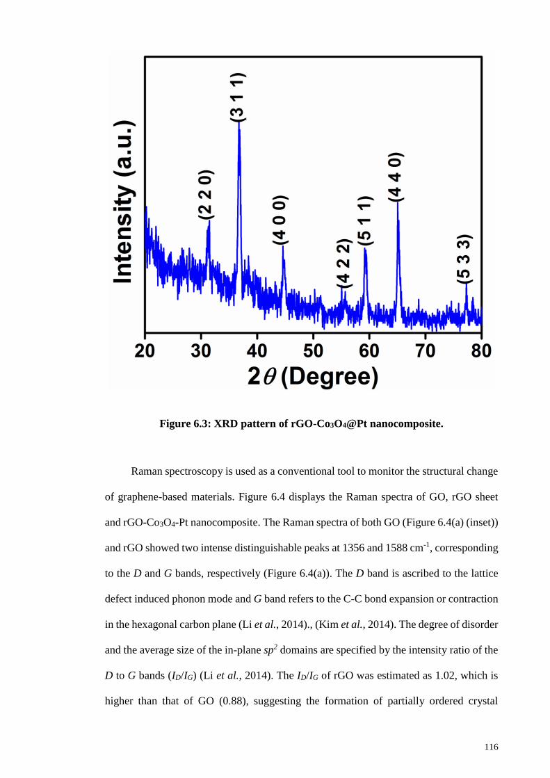
116
Figure 6.3: XRD pattern of rGO-Co3O4@Pt nanocomposite.
Raman spectroscopy is used as a conventional tool to monitor the structural change
of graphene-based materials. Figure 6.4 displays the Raman spectra of GO, rGO sheet
and rGO-Co3O4-Pt nanocomposite. The Raman spectra of both GO (Figure 6.4(a) (inset))
and rGO showed two intense distinguishable peaks at 1356 and 1588 cm-1, corresponding
to the D and G bands, respectively (Figure 6.4(a)). The D band is ascribed to the lattice
defect induced phonon mode and G band refers to the C-C bond expansion or contraction
in the hexagonal carbon plane (Li et al., 2014)., (Kim et al., 2014). The degree of disorder
and the average size of the in-plane sp2 domains are specified by the intensity ratio of the
D to G bands (ID/IG) (Li et al., 2014). The ID/IG of rGO was estimated as 1.02, which is
higher than that of GO (0.88), suggesting the formation of partially ordered crystal
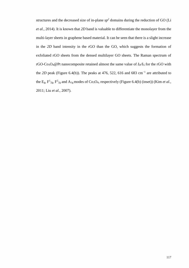
117
structures and the decreased size of in-plane sp2 domains during the reduction of GO (Li
et al., 2014). It is known that 2D band is valuable to differentiate the monolayer from the
multi-layer sheets in graphene based material. It can be seen that there is a slight increase
in the 2D band intensity in the rGO than the GO, which suggests the formation of
exfoliated rGO sheets from the densed multilayer GO sheets. The Raman spectrum of
rGO-Co3O4@Pt nanocomposite retained almost the same value of ID/IG for the rGO with
the 2D peak (Figure 6.4(b)). The peaks at 476, 522, 616 and 683 cm−1 are attributed to
the Eg, F1
2g, F2
2g and A1g modes of Co3O4, respectively (Figure 6.4(b) (inset)) (Kim et al.,
2011; Liu et al., 2007).
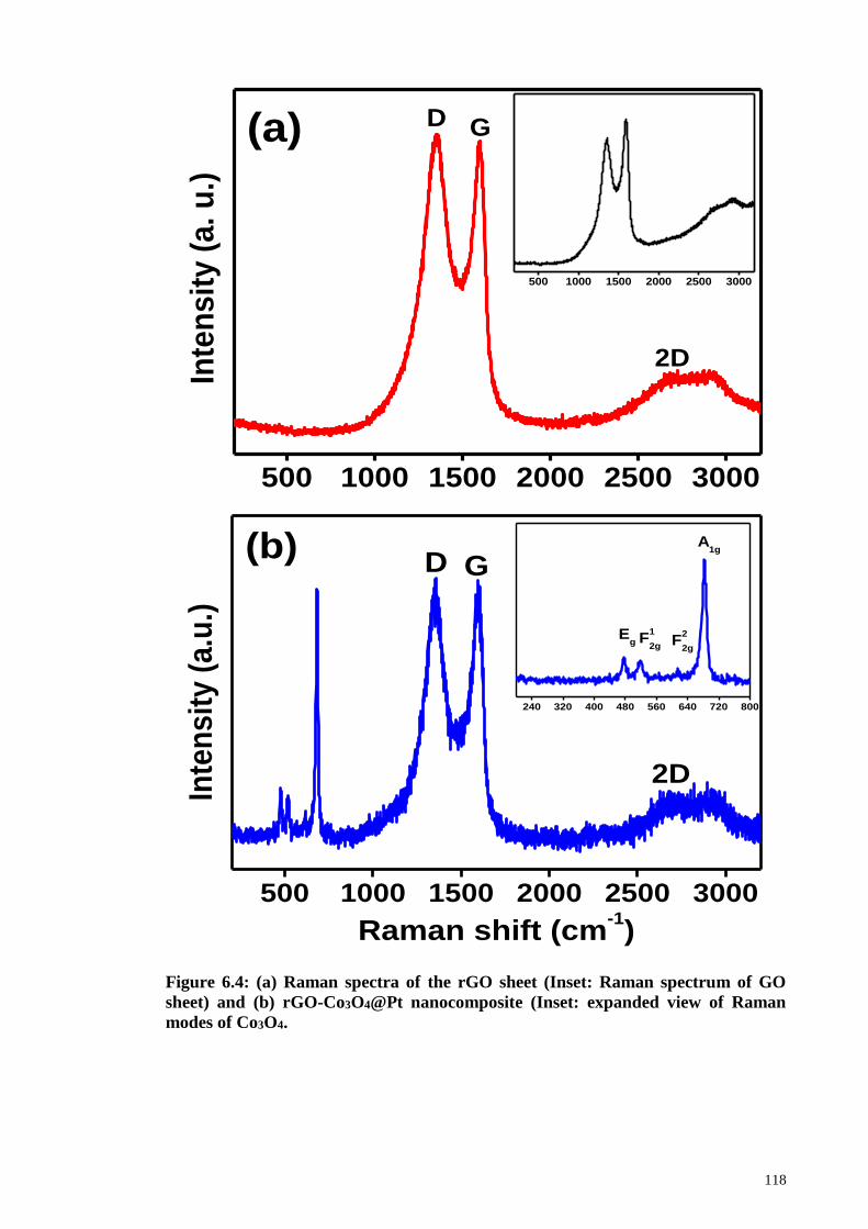
118
Figure 6.4: (a) Raman spectra of the rGO sheet (Inset: Raman spectrum of GO
sheet) and (b) rGO-Co3O4@Pt nanocomposite (Inset: expanded view of Raman
modes of Co3O4.
500 1000 1500 2000 2500 3000
500 1000 1500 2000 2500 3000
D G
2D
Inte
nsit
y (
a. u
.)
(a)
500 1000 1500 2000 2500 3000
240 320 400 480 560 640 720 800
F2
2g
Inte
nsit
y (
a.u
.)
Raman shift (cm-1)
2D
(b)
F1
2g
GDA
1g
Eg
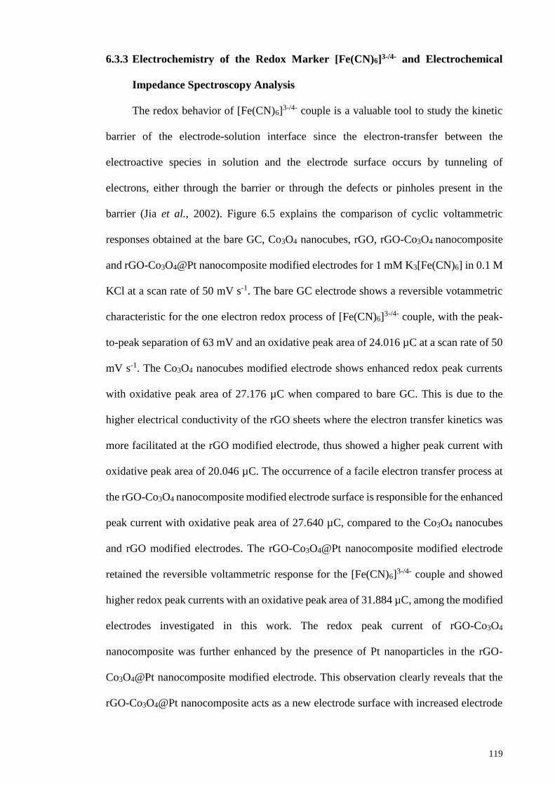
119
6.3.3 Electrochemistry of the Redox Marker [Fe(CN)6]3-/4- and Electrochemical
Impedance Spectroscopy Analysis
The redox behavior of [Fe(CN)6]3-/4- couple is a valuable tool to study the kinetic
barrier of the electrode-solution interface since the electron-transfer between the
electroactive species in solution and the electrode surface occurs by tunneling of
electrons, either through the barrier or through the defects or pinholes present in the
barrier (Jia et al., 2002). Figure 6.5 explains the comparison of cyclic voltammetric
responses obtained at the bare GC, Co3O4 nanocubes, rGO, rGO-Co3O4 nanocomposite
and rGO-Co3O4@Pt nanocomposite modified electrodes for 1 mM K3[Fe(CN)6] in 0.1 M
KCl at a scan rate of 50 mV s-1. The bare GC electrode shows a reversible votammetric
characteristic for the one electron redox process of [Fe(CN)6]3-/4- couple, with the peak-
to-peak separation of 63 mV and an oxidative peak area of 24.016 µC at a scan rate of 50
mV s-1. The Co3O4 nanocubes modified electrode shows enhanced redox peak currents
with oxidative peak area of 27.176 µC when compared to bare GC. This is due to the
higher electrical conductivity of the rGO sheets where the electron transfer kinetics was
more facilitated at the rGO modified electrode, thus showed a higher peak current with
oxidative peak area of 20.046 µC. The occurrence of a facile electron transfer process at
the rGO-Co3O4 nanocomposite modified electrode surface is responsible for the enhanced
peak current with oxidative peak area of 27.640 µC, compared to the Co3O4 nanocubes
and rGO modified electrodes. The rGO-Co3O4@Pt nanocomposite modified electrode
retained the reversible voltammetric response for the [Fe(CN)6]3-/4- couple and showed
higher redox peak currents with an oxidative peak area of 31.884 µC, among the modified
electrodes investigated in this work. The redox peak current of rGO-Co3O4
nanocomposite was further enhanced by the presence of Pt nanoparticles in the rGO-
Co3O4@Pt nanocomposite modified electrode. This observation clearly reveals that the
rGO-Co3O4@Pt nanocomposite acts as a new electrode surface with increased electrode
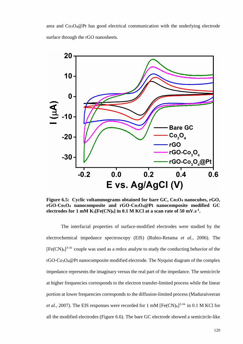
120
area and Co3O4@Pt has good electrical communication with the underlying electrode
surface through the rGO nanosheets.
Figure 6.5: Cyclic voltammograms obtained for bare GC, Co3O4 nanocubes, rGO,
rGO-Co3O4 nanocomposite and rGO-Co3O4@Pt nanocomposite modified GC
electrodes for 1 mM K3[Fe(CN)6] in 0.1 M KCl at a scan rate of 50 mV.s-1.
The interfacial properties of surface-modified electrodes were studied by the
electrochemical impedance spectroscopy (EIS) (Rubio-Retama et al., 2006). The
[Fe(CN)6]3-/4- couple was used as a redox analyte to study the conducting behavior of the
rGO-Co3O4@Pt nanocomposite modified electrode. The Nyquist diagram of the complex
impedance represents the imaginary versus the real part of the impedance. The semicircle
at higher frequencies corresponds to the electron transfer-limited process while the linear
portion at lower frequencies corresponds to the diffusion-limited process (Maduraiveeran
et al., 2007). The EIS responses were recorded for 1 mM [Fe(CN)6]3-/4- in 0.1 M KCl for
all the modified electrodes (Figure 6.6). The bare GC electrode showed a semicircle-like
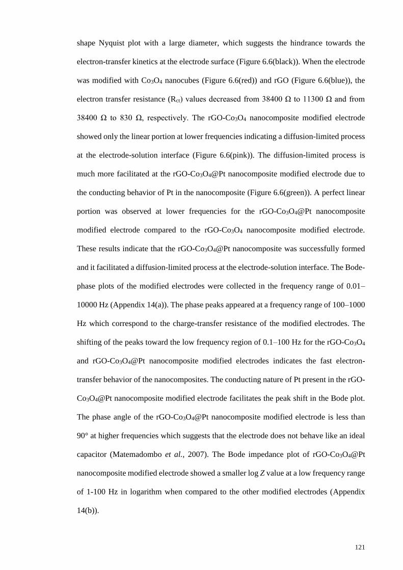
121
shape Nyquist plot with a large diameter, which suggests the hindrance towards the
electron-transfer kinetics at the electrode surface (Figure 6.6(black)). When the electrode
was modified with Co3O4 nanocubes (Figure 6.6(red)) and rGO (Figure 6.6(blue)), the
electron transfer resistance (Rct) values decreased from 38400 Ω to 11300 Ω and from
38400 Ω to 830 Ω, respectively. The rGO-Co3O4 nanocomposite modified electrode
showed only the linear portion at lower frequencies indicating a diffusion-limited process
at the electrode-solution interface (Figure 6.6(pink)). The diffusion-limited process is
much more facilitated at the rGO-Co3O4@Pt nanocomposite modified electrode due to
the conducting behavior of Pt in the nanocomposite (Figure 6.6(green)). A perfect linear
portion was observed at lower frequencies for the rGO-Co3O4@Pt nanocomposite
modified electrode compared to the rGO-Co3O4 nanocomposite modified electrode.
These results indicate that the rGO-Co3O4@Pt nanocomposite was successfully formed
and it facilitated a diffusion-limited process at the electrode-solution interface. The Bode-
phase plots of the modified electrodes were collected in the frequency range of 0.01–
10000 Hz (Appendix 14(a)). The phase peaks appeared at a frequency range of 100–1000
Hz which correspond to the charge-transfer resistance of the modified electrodes. The
shifting of the peaks toward the low frequency region of 0.1–100 Hz for the rGO-Co3O4
and rGO-Co3O4@Pt nanocomposite modified electrodes indicates the fast electron-
transfer behavior of the nanocomposites. The conducting nature of Pt present in the rGO-
Co3O4@Pt nanocomposite modified electrode facilitates the peak shift in the Bode plot.
The phase angle of the rGO-Co3O4@Pt nanocomposite modified electrode is less than
90° at higher frequencies which suggests that the electrode does not behave like an ideal
capacitor (Matemadombo et al., 2007). The Bode impedance plot of rGO-Co3O4@Pt
nanocomposite modified electrode showed a smaller log Z value at a low frequency range
of 1-100 Hz in logarithm when compared to the other modified electrodes (Appendix
14(b)).
122
0 50000 100000 150000 200000
0
50000
100000
150000
200000
150 1800
30
60
90
-Zim
(o
hm
)
Zre
(ohm)
600 1200 1800
0
600
1200
Zim
(o
hm
)
Zre
(ohm)
0 8000 16000
0
6000
12000
Zim
(o
hm
)
Zre
(ohm)
-Zim
(o
hm
)
Zre
(ohm)
Figure 6.6: Nyquist plots obtained for bare GC (black) Co3O4 nanocubes (red), rGO
(blue), rGO-Co3O4 nanocomposite (pink) and rGO-Co3O4@Pt nanocomposite
(green) modified GC electrodes for 1 mM K3[Fe(CN)6] in 0.1 M KCl. The frequency
range was 0.01 Hz to 10 kHz.
6.3.4 Electrocatalysis of Nitric Oxide (NO)
The electrocatlytic oxidation of NO was carried out in 0.1 M PBS (pH 2.5). NaNO2
was used as precursor to produce NO in PBS to study the electrocatalytic activity of the
rGO-Co3O4@Pt nanocomposite modified electrode. In acidic solution (pH<4), NaNO2
can generate NO by the disproportionation reaction (Equation 6.1) (Thangavel et al.,
2008)., (Beltramo et al., 2003). The addition of a known amount of NaNO2 into the bulk
electrolyte solution at pH<4 generates a series of concentrations of NO (Thangavel et al.,
2008).
3HONO H+ + 2NO + NO3⁻ + H2O (6.1)
Figure 6.7 displays the cyclic voltammetric responses of the modified electrodes used in
this work for the oxidation of NO in 0.1 PBS (pH 2.5) containing 5 mM NO2- ions. The
rGO-Co3O4@Pt nanocomposite modified GC electrode showed catalytic NO oxidation
peak at + 0.84 V in 0.1 M PBS containing 5 mM of NO2- at a scan rate of 50 mV s-1.
However, the nanocomposite modified electrode did not show any voltammetric response
123
in the absence of NO2- (Appendix 15). The Co3O4 nanocubes and rGO sheet modified
electrodes showed the oxidation peak for NO at almost the same potential (~ + 0.95 V)
with smaller difference in the peak current. The rGO-Co3O4 nanocomposite modified
electrode shifted the oxidation peak potential of NO (+0.87 V) towards less positive
potential with a small increment in the peak current. For comparison, the rGO-Pt
nanocomposite modified electrode was also fabricated and it showed a peak current of
102 µA at the peak potential of + 0.87 V for NO oxidation. From these results, it can be
concluded that the rGO-Co3O4@Pt nanocomposite modified electrode displayed a
synergistic catalytic effect of the Co3O4 nanocubes and Pt nanostructures toward NO
oxidation. The Co3O4 nanocubes provided a large surface area for Pt deposition and
thereby enhanced the electrocatalytic activity of rGO-Co3O4@Pt nanocomposite
modified electrode. The rGO-Co3O4@Pt nanocomposite modified GC electrode
efficiently shifted the catalytic peak potential of NO with a peak current of 119 µA, when
compared to the other controlled modified electrodes investigated in this work. This
reveals that the Pt nanoparticles present in the nanocomposite are in good electrical
communication with the rGO-Co3O4 which results in an efficient electron-transfer
process at the modified electrode toward NO oxidation. The Pt nanoparticles also provide
a larger surface area for the effective interaction of NO and thereby improved the
electron-transfer kinetics and the electrocatalytic performance. The NO oxidation at the
rGO-Co3O4@Pt nanocomposite modified electrode possibly proceeds through an
electrochemical reaction followed by a chemical reaction (Li et al., 2006). During the
electrochemical oxidation of NO, one electron from the NO molecule transfers to the
electrode with the formation of the nitrosonium ion (NO+) (Li et al., 2006). The bare GC
electrode also displayed a voltammetric response for the oxidation of NO at +0.94 V with
a smaller peak current (79 µA) than the other modified electrodes. The electrocatalytic
oxidation of NO was performed using rGO-Co3O4 nanocomposite containing different

124
amounts of GO (4, 8 and 12 wt. % GO) (Appendix 16). The rGO-Co3O4 nanocomposite
prepared using 8 wt. % GO showed better performance toward the oxidation of NO and
the rGO-Co3O4@Pt was the preferred nanocomposite. The reproducibility and
repeatability of the nanocomposite modified electrode were checked by recording the
voltammograms with different electrodes and good reproducible and repeatable results
were observed. The stability of the nanocomposite modified electrode was studied for NO
oxidation by recording the voltammogram of the same modified electrode after one week
and the electrode showed only less than 5 % decrement in the peak current. The modified
electrode was stored at room temperature (25 °C) during the period of stability
measurements. This infers that the present nanocomposite modified electrode was stable
towards the electrocatalytic oxidation of NO.
Figure 6.7: Cyclic voltammograms recorded at bare GC, Co3O4 nanocubes, rGO,
rGO-Co3O4 nanocomposite, rGO-Pt nanocomposite and rGO-Co3O4@Pt
nanocompositen modified electrodes for 5 mM of NO2- in 0.1 M PBS (pH 2.5) with a
scan rate of 50 mVs-1.
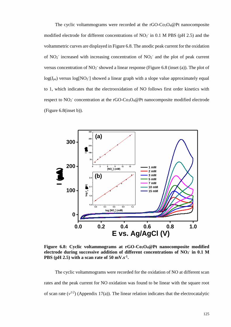
125
The cyclic voltammograms were recorded at the rGO-Co3O4@Pt nanocomposite
modified electrode for different concentrations of NO2- in 0.1 M PBS (pH 2.5) and the
voltammetric curves are displayed in Figure 6.8. The anodic peak current for the oxidation
of NO2- increased with increasing concentration of NO2
- and the plot of peak current
versus concentration of NO2- showed a linear response (Figure 6.8 (inset (a)). The plot of
log(Ipa) versus log[NO2-] showed a linear graph with a slope value approximately equal
to 1, which indicates that the electrooxidation of NO follows first order kinetics with
respect to NO2- concentration at the rGO-Co3O4@Pt nanocomposite modified electrode
(Figure 6.8(inset b)).
0.0 0.2 0.4 0.6 0.8 1.0
0
100
200
300
0.0 0.3 0.6 0.9 1.21.5
1.8
2.1
2.4
log
Ip (A
)
log [NO2
-] (mM)
0 3 6 9 12 15
70
140
210
280
350
I (
)
[NO2
-] (mM)
I (
)
E vs. Ag/AgCl (V)
1 mM
2 mM
3 mM
5 mM
7 mM
10 mM
15 mM
(a)
(b)
Figure 6.8: Cyclic voltammograms at rGO-Co3O4@Pt nanocomposite modified
electrode during successive addition of different concentrations of NO2- in 0.1 M
PBS (pH 2.5) with a scan rate of 50 mV.s-1.
The cyclic voltammograms were recorded for the oxidation of NO at different scan
rates and the peak current for NO oxidation was found to be linear with the square root
of scan rate (ν1/2) (Appendix 17(a)). The linear relation indicates that the electrocatalytic
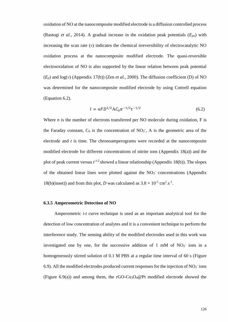
126
oxidation of NO at the nanocomposite modified electrode is a diffusion controlled process
(Rastogi et al., 2014). A gradual increase in the oxidation peak potentials (Epa) with
increasing the scan rate (ν) indicates the chemical irreversibility of electrocatalytic NO
oxidation process at the nanocomposite modified electrode. The quasi-reversible
electrooxidation of NO is also supported by the linear relation between peak potential
(Ep) and log(ν) (Appendix 17(b)) (Zen et al., 2000). The diffusion coefficient (D) of NO
was determined for the nanocomposite modified electrode by using Cottrell equation
(Equation 6.2).
𝐼 = 𝑛𝐹𝐷1/2A𝐶0𝜋−1/2𝑡−1/2 (6.2)
Where n is the number of electrons transferred per NO molecule during oxidation, F is
the Faraday constant, C0 is the concentration of NO2-, A is the geometric area of the
electrode and t is time. The chronoamperograms were recorded at the nanocomposite
modified electrode for different concentrations of nitrite ions (Appendix 18(a)) and the
plot of peak current versus t-1/2 showed a linear relationship (Appendix 18(b)). The slopes
of the obtained linear lines were plotted against the NO2- concentrations (Appendix
18(b)(inset)) and from this plot, D was calculated as 3.8 × 10⁻⁵ cm2.s-1.
6.3.5 Amperometric Detection of NO
Amperometric i-t curve technique is used as an important analytical tool for the
detection of low concentration of analytes and it is a convenient technique to perform the
interference study. The sensing ability of the modified electrodes used in this work was
investigated one by one, for the successive addition of 1 mM of NO2- ions in a
homogeneously stirred solution of 0.1 M PBS at a regular time interval of 60 s (Figure
6.9). All the modified electrodes produced current responses for the injection of NO2- ions
(Figure 6.9(a)) and among them, the rGO-Co3O4@Pt modified electrode showed the
127
highest response for every injection of NO2- ions with a good linear range of 1 mM-14
mM (Figure 6.9(b)).
Figure 6.9: (a) Amperometric i–t curves obtained at bare GC, Co3O4 nanocubes,
rGO, rGO-Co3O4 nanocomposite and rGO-Co3O4@Pt nanocomposite modified GC
electrodes for the successive addition of 1 mM NO2- in 0.1 M PBS (pH 2.5) at a
regular interval of 60 s and (b) corresponding calibration plots of current versus
concentration of NO2-. Applied potentials were the peak potentials obtained from
Figure 6.7.
200 400 600 800
0
100
200
300
400
I (
)
t (s)
bare GC
Co3O
4
rGO
rGO-Co3O
4
rGO-Co3O
4@Pt
(a)
0 2 4 6 8 10 12 14 16
0
100
200
300
400
bare GC
Co3O
4
rGO
rGO-Co3O
4
rGO-Co3O
4@Pt
I (
)
[NO2
-] (mM)
(b)
128
The presence of Pt nanoparticles on the electrode effectively enhanced the sensing
ability of the rGO-Co3O4@Pt nanocomposite toward the detection of NO. The rGO-
Co3O4@Pt nanocomposite was chosen as an electrochemical sensor material for the
detection of lower concentration levels of NO in PBS. The amperometric i-t curve was
obtained at the rGO-Co3O4@Pt modified electrode for the successive addition of NO2-
ions with different concentrations in a homogeneously stirred solution of 0.1 M PBS with
an applied potential of +0.84 V (Figure 6.10). For each addition of NO2- with a sample
interval of 60 s, a significant current response was observed (Figure 6.10(a)) and it
suggests that the rGO-Co3O4@Pt efficiently promoted the oxidation of NO in 0.1 M PBS
(pH 2.5). The plot of current response versus concentration of NO2- showed a linear
relation for the concentration range of 10 µM to 650 µM (Figure 6.10(b)). Repeated
measurements were performed for the detection of NO at lower concentration levels and
the current response was reproduced at the nanocomposite modified electrode. The
nanocomposite showed the sensitivity of 0.026±0.0002 µA/µM and the LOD was
calculated as 1.73 µM for the detection of NO. The comparison of the analytical
performance of the present rGO-Co3O4@Pt nanocomposite modified GC electrode with
some of the reported GC electrode based electrochemical sensors toward the detection of
NO is shown in Table 6.1.
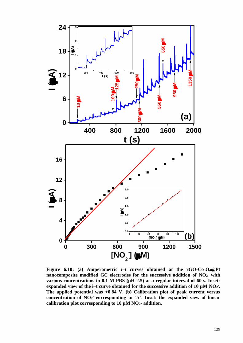
129
Figure 6.10: (a) Amperometric i–t curves obtained at the rGO-Co3O4@Pt
nanocomposite modified GC electrodes for the successive addition of NO2- with
various concentrations in 0.1 M PBS (pH 2.5) at a regular interval of 60 s. Inset:
expanded view of the i–t curve obtained for the successive addition of 10 µM NO2-.
The applied potential was +0.84 V. (b) Calibration plot of peak current versus
concentration of NO2- corresponding to ‘A’. Inset: the expanded view of linear
calibration plot corresponding to 10 µM NO2- addition.
400 800 1200 1600 20000
6
12
18
24
13
50
M
95
0
M
65
0
M
55
0
M
12
5
M
30
0
M
25
0
M
10
0
M
200 400 600 800
0
1
2
3
I (
A)
t (s)
I (
A)
t (s)
10
M
(a)
0 300 600 900 1200 15000
4
8
12
16
0 20 40 60 80 1000.0
0.6
1.2
1.8
2.4
3.0
I (
A)
[NO2
-] (M)
I (
A)
[NO2
-] (M)
(b)

130
Table 6.1: Comparison of some of the reported electrochemical sensors for NO
detection.
Modified GC
electrodea
Techniqueb
Linear range
(µM)
LOD
(µM)
Reference
GC/PAM/SDS/Cyt c Amperometry 0.80 μM– 95 0.1 (Chen et al., 2009)
GC/PNMP-b-
PGMA/Hb
DPV 0.45 µM–10 0.32 (Jia et al., 2009)
GC/EDAS(TiO2-Au)
nps
SWV 1 µM– 60 1.0
(Pandikumar et al.,
2011)
GC/DNA/Cyt c Amperometry 0.6 µM– 8 0.1 (Liu et al., 2007)
GC/PtNP/AB Amperometry 0.18 μM– 120 0.05 (Zheng et al., 2012)
GC/AuNP-ERGO Amperometry up to 3.38 0.133 (Ting et al., 2013)
GC/SWNT/PVP–Os–
EA
Amperometry 0.2 μM– 40 0.05 (Fei et al., 2011)
GC/G-Nf SWV 50 µM– 450 11.61 (Yusoff et al., 2015)
GC/Hb–CPB/PAM CV 9.8 µM– 100 9.3 (He et al., 2006)
GC/rGO-Co3O4@Pt Amperometry 10 µM– 650 1.73 Present work
a PAM = polyacrylamide; SDS = sodium dodecyl sulfate; Cyt c = cytochrome c; PNMP-b-PGMA = poly
[N-(2-methacryloyloxyethyl) pyrrolidone]-blockpoly [glycidyl methacrylate]; Hb = hemoglobin; EDAS =
N-[3-(trimethoxysilyl)propyl]ethylene diamine; TiO2-Au = titanium dioxide-gold nanocomposite; DNA =
deoxyribonucleic acid; PtNP = platinum nanoparticle; AB = acetylene black; AuNP = gold nanoparticle;
ERGO = electrochemically reduced graphene oxide; SWNT = single-walled carbon nanotube; PVP–Os–
EA = Os-bipyridine complex and poly(4-vinylpyridine) (PVP) partially quaternized with 2-
bromoethylamine (EA) functionalities; G = graphene; Nf = Nafion; Hb-CPB = hemoglobin-cetylpyridinium
bromide. b DPV = differential pulse voltammetry; SWV = square wave voltammetry; CV = cyclic
voltammetry
The selectivity of the rGO-Co3O4@Pt nanocomposite for the detection NO was
investigated by injecting various possible physiological interferents in the same
homogeneously stirred PBS containing NO and the change in current response was
observed. Figure 6.11 explains the continuously recorded amperometric i–t curve
response for the successive additions of NO2- and interferents in a homogeneously stirred
0.1 M PBS (pH 2.5). The current response of the interferents such as DA, AA and UA
was studied by adding them in succession, after the few successive additions of NO2- (10

131
µM) in the same stirred PBS. However, the added interferents did not change the current
response even with a 100-fold higher concentration. Again, the injection of NO2- in the
same solution displayed almost the same magnitude of current response for the oxidation
of NO. After few successive additions of NO2-, more interferents such as glucose, urea
and NaCl were added in succession. The i-t curve was recorded with a sampling interval
of 60 s and the addition of these interferents did not produce increase in the current
response. However, the introduction of 10 µM NO2- to the same solution again resulted
in a clear and quick response. These results indicated that the present sensor possesses
good selectivity and sensitivity towards the determination of NO2- even in the presence
of 100-fold excess of common physiological interferents.
Figure 6.11: Amperometric i–t curve obtained at rGO-Co3O4@Pt nanocomposite
modified GC electrode for the successive addition of 10 µM NO2- and each 1 mM of
DA, AA, UA, glucose, urea and NaCl in 0.1 M PBS (pH 2.5) at a regular interval of
60 s. Applied potential was + 0.84 V.
132
6.4 Conclusions
Co3O4@Pt nanocubes was successfully synthesized and incorporated to the rGO
sheets using the hydrothermal synthesis. The reduction of GO to rGO was confirmed from
the increase in the ratio of D and G bands (ID/IG) from the Raman spectra. The
morphology of the Co3O4 and the deposition of Pt on Co3O4 nanocubes were confirmed
by the FESEM analysis and the EDX elemental mapping analysis confirmed the presence
of all the elements of the rGO-Co3O4@Pt nanocomposite. The rGO-Co3O4@Pt
nanocomposite modified GC electrode shifted the oxidation overpotential of the in-situ
generated NO toward smaller positive potential with enhanced catalytic peak current
when compared to the other modified electrodes. The higher catalytic effect of the rGO-
Co3O4@Pt nanocomposite was attributed to the synergistic effect of the Co3O4 nanocubes
and Pt nanoparticles present in the rGO matrix. The detection of NO was performed using
amperometric i-t curve technique at the various modified electrodes and among them, the
rGO-Co3O4@Pt nanocomposite modified electrode showed better performance with the
lowest detection limit of 1.73 µM. Also, the nanocomposite modified electrode displayed
good selectivity toward NO even in the presence of 100-fold higher concentration of other
physiologically analytes. The current sensor was stable and reproducible and adds further
credits to the rGO-Co3O4 nanocomposite based electrochemical sensors in the
contemporary research.
133
CHAPTER 7: CONCLUSION AND FUTURE WORK
The present study highlights the typical synthesis of Co3O4 different nanostructures
and Co3O4 based nanocomposites for the electrochemical detection of target molecules
for the sensor applications. The Co3O4 different nanostructures together with Co3O4
nanocomposites were designed based upon their inherent properties such as structural
affects, catalysis, charge transfer abilities in nanocomposites and sensing capabilities of
the materials. The synthesized materials were comprehensively characterized by various
techniques such as FESEM fitted with EDX elemental mapping analysis, high resolution
transmission electron microscopy (HRTEM), XRD and Raman spectroscopy. The
synthesized nanomaterials have demonstrated improved physical and chemical properties
for electrochemical sensors applications. Adding into it, the facile method of synthesizing
nanomaterials by the hydrothermal technique, their thermal stability, higher
electrocatalytic activity and ability to detect the target molecule has made them a very
sophisticated material for electrochemical sensors applications.
Co3O4 with five different nanostructures were prepared via the hydrothermal route
and utilized for the sensing of 4-NP, a water contaminant. The cubical structure of Co3O4
presented optimistic results among the other nanostructures (nanocubes, nanowires,
nanobundles, nanoplates and nanoflowers) based on their structure, size and crystallinity.
All the structures of Co3O4 were tested by using cyclic voltammogram techniques for the
reduction of 4-NP. A higher current was recorded for 4-NP reduction with a well-defined
peak at lower potential as compared to the other nanostructures. So Co3O4 with cubical
morphology was selected as potential catalyst for the detection of 4-NP. The sensitivity
and the lowest LOD for 4-NP were 0.0485±0.00063 µA/µM and 0.93 µM respectively.
Based on the results reported in chapter three, Co3O4 with cubical structure was selected
as potential candidate to fabricate nanocomposites with graphene for another sensor

134
application. Hence, rGO-Co3O4 nanocomposite was prepared by using the same
hydrothermal route. In this study, the rGO-Co3O4 nanocomposites were prepared with
different wt. % of GO (2, 4, 8, 12 and wt. %). The prepared composition with 4 wt. % of
GO concentrations was found to be the optimized composition by testing it through cyclic
voltammogram technique. All nanocomposites were analyzed to oxidase a depression
biomarker called serotonin (5-HT) but promising peak current with lower potential value
for the oxidation of biomolecules was displayed by rGO-Co3O4-4 %. It was noticed that
2 wt. % of GO results in the agglomeration of Co3O4 nanocubes and thus agglomerated
particles appears on the surface of graphene sheets. The increase of the GO wt. % provides
sufficient numbers of sheets to accommodate the Co3O4 nanocubes and hence prevent
them from agglomeration for increased electrolyte diffusion into the rGO-Co3O4-4 %
matrix and boost the catalytic process. Further increase in the GO wt. % results in an
increased of stacking graphene sheets lowers the density of Co3O4 which produces poor
electrocatalytic activity. So rGO-Co3O4 was used as an efficient electrode material to
detect 5-HT with a LOD of 1.128M. Interference studies were also investigated in the
presence of co-existing molecules such as AA, DA and UA and sensor was found to be
highly selective towards 5-HT. In addition, gold deposited Co3O4 nanocubes incorporated
into reduced graphene oxide nanocomposites were synthesised by hydrothermal method.
In this research, the enhancement in catalytic performance of rGO-Co3O4 nanocomposite
was tuned by varying the concentration (2, 4, 6, 8 and 10 mM) of Au nanoparticles. It
was observed that rGO-Co3O4@Au nanocomposite with 8 mM concentration of Au
nanoparticle has shown enhanced electrochemical performance. The studies were carried
out for the detection of hydrazine, a toxic, colorless and flammable molecule which
causes severe injury to the lungs, liver, nervous system and spinal cord etc. The
electrochemical detecion of hydrazine was obtained at rGO-Co3O4@Au modified GC
electrode and the LOD was 0.443 µM and 0.58304 ± 0.00466 µA µM–1. For selectivity
135
test, the sensor material was tested in the presence of various interfering molecules
mentioned in chapter 5. There was no signal detected for the interference species which
confirms the selectivity of the rGO-Co3O4@Au. Real sample analysis was also studied
for the water collected from different areas in Malaysia. Finally, platinum nanoparticle
deposited Co3O4 nanocubes incorporated into reduced graphene oxide was prepared by
hydrothermal method. In this method, rGO-Co3O4 nanocomposite with different
concentration of GO (2,4,8 and 12 wt. %) were first tested for electrochemical oxidation
of NO and it was found that rGO-Co3O4 with 8 wt. % of GO showed enhanced
electrochemcal signal compared to other compositions. So rGO-Co3O4 nanocomposite
with 8 wt. % of GO was deposited with platinum nanoparticles and utilized for the
detection of NO, a biomolecule responsible for vasodilation and blood pressure regulation
in the nervous and cardiovascular systems in mammalian physiology. The detection of
NO was carried out on rGO-Co3O4@Pt modified GC electrode and LOD was 1.73 M
for the detection of NO. Since NO is a biological molecule which is found togather with
other biological molecules, so the detection of NO in the presence of these molecules is
very important to check the selectivity of the sensor. It was found that the rGO-Co3O4@Pt
modified GC electrode is highly selective towards sensing of NO.
Future Work
The current research work focuses on the enhancement of Co3O4 nanostruructures
as a catalyst for electrochmical sensor applications. In this work, we have used graphene
as a superhighway for electrical conduction and Co3O4 was used as catalyst material.
Graphene facilitated the efficient electron transfer at the interface of the electrode and
electrolyte, thus took part in boosting the electrochemical signals. For further enrichment
of Co3O4 nanocubes, some metal nanoparticles were deposited onto the Co3O4 nanocubes.
From these results, the proposed future work is listed below:
136
1. Five different nanostructures of Co3O4, have been synthesized but only one type
of nanostructured Co3O4 was utilized for electrochemical sensors applications
based on the performance. The other four nanostructures are still yet to be
explored.
2. From the literature, nanowires are considered very promising materials for
supercapacitors electrode materials. Nanowires are mostly synthesised by
electrodeposition, electrophoretic deposition and chemical vapour deposition
methods (e.g. AACVD, MOCVD and HWCVD), but were synthesized by the
hydrothermal method in this research.
3. The Co3O4 nanowires can be utilized as composite materials with graphene, carbon
nanotubes and metal nanoparticles by the same synthetic methods, especially for
electrochemcial sensors, supercapacitors and solar cell applications.
4. Similarly, other nanostructures can be utilized for different electrochemical
applications or by fabrication into nanocomposites to achieve higher
electrocatlaytic performance.
137
REFERENCES
Abbaspour, A., & Noori, A. (2011). A cyclodextrin host–guest recognition approach to
an electrochemical sensor for simultaneous quantification of serotonin and
dopamine. Biosensors and Bioelectronics, 26(12), 4674-4680.
Abdul Aziz, M., & Kawde, A. N. (2013). Gold nanoparticle-modified graphite pencil
electrode for the high-sensitivity detection of hydrazine. Talanta, 115, 214-221.
Adler, D. (1968). Insulating and metallic states in transition metal oxides. Solid State
Physics, 21, 1-113.
Afzali, D., Karimi-Maleh, H., & Khalilzadeh, M. A. (2011). Sensitive and selective
determination of phenylhydrazine in the presence of hydrazine at a ferrocene-
modified carbon nanotube paste electrode. Environmental Chemistry Letters,
9(3), 375-381.
Ahmed, S. R., Ogale, S., Papaefthymiou, G. C., Ramesh, R., & Kofinas, P. (2002).
Magnetic properties of CoFe2O4 nanoparticles synthesized through a block
copolymer nanoreactor route. Applied Physics Letters, 80(9), 1616-1618.
Ajayan, P., & Nalwa, H. (2000). Handbook of Nanostructured Materials and
Nanotechnology. Academic Press, New York.
Allag, A., Saâd, R., Ouahab, A., Attouche, H., & Kouidri, N. (2016). Optoelectronic
properties of SnO2 thin films sprayed at different deposition times. Chinese
Physics B, 25(4), 046801.
Alrehaily, L., Joseph, J., Biesinger, M., Guzonas, D., & Wren, J. (2013). Gamma-
radiolysis-assisted cobalt oxide nanoparticle formation. Physical Chemistry
Chemical Physics, 15(3), 1014-1024.
Ando, M., Kadono, K., Kamada, K., & Ohta, K. (2004). Third-order nonlinear optical
responses of nanoparticulate Co3O4 films. Thin Solid Films, 446(2), 271-276.
Anithaa, A. C., Asokan, K., & Sekar, C. (2017). Highly sensitive and selective serotonin
sensor based on gamma ray irradiated tungsten trioxide nanoparticles. Sensors
and Actuators B, 238, 667-675.
Antoniadou, S., Jannakoudakis, A., & Theodoridou, E. (1989). Electrocatalytic reactions
on carbon fibre electrodes modified by hemine II. Electro-oxidation of hydrazine.
Synthetic Metals, 30(3), 295-304.
Arvinte, A., Mahosenaho, M., Pinteala, M., Sesay, A.-M., & Virtanen, V. (2011).
Electrochemical oxidation of p-nitrophenol using graphene-modified electrodes,
and a comparison to the performance of MWNT-based electrodes. Microchimica
Acta, 174(3-4), 337-343.
Asif, M. H., Elinder, F., & Willander, M. (2011). Electrochemical biosensors based on
ZnO nanostructures to measure intracellular metal ions and glucose. Journal of
Analytical & Bioanalytical Techniques 7, doi: 10.4172/2155-9872.S7-003.
138
Augustine, A. K., Nampoori, V., & Kailasnath, M. (2014). Rapid synthesize of gold
nanoparticles by microwave irradiation method and its application as an optical
limiting material. Optik-International Journal for Light and Electron Optics,
125(22), 6696-6699.
Babaei, A., & Taheri, A. R. (2013). Nafion/Ni(OH)2 nanoparticles-carbon nanotube
composite modified glassy carbon electrode as a sensor for simultaneous
determination of dopamine and serotonin in the presence of ascorbic acid. Sensors
and Actuators B: Chemical, 176, 543-551.
Baek, H. R., & Eo, I. S. (2017). Electrochemical properties of TiO2-metal oxide
composites for dye-sensitized solar cell. Current Applied Physics, 17(6), 854-857.
Bala, H., Fu, W., Yu, Y., Yang, H., & Zhang, Y. (2009). Preparation, optical properties,
magnetic properties and thermal stability of core–shell structure cobalt/zinc oxide
nanocomposites. Applied Surface Science, 255(7), 4050-4055.
Balandin, A. A., Ghosh, S., Bao, W., Calizo, I., Teweldebrhan, D., Miao, F., & Lau, C.
N. (2008). Superior thermal conductivity of single-layer graphene. Nano Letters,
8(3), 902-907.
Baxter, J. B., & Aydil, E. S. (2005). Nanowire-based dye-sensitized solar cells. Applied
Physics Letters, 86(5), 053114.
Bedioui, F., & Villeneuve, N. (2003). Electrochemical nitric oxide sensors for biological
samples–principle, selected examples and applications. Electroanalysis, 15(1), 5-
18.
Beltramo, G., & Koper, M. (2003). Nitric oxide reduction and oxidation on stepped Pt [n
(111)×(111)] electrodes. Langmuir, 19(21), 8907-8915.
Bogdan, C. (2001). Nitric oxide and the immune response. Nature Immunology, 2(10),
907-916.
Borsella, E., Cattaruzza, E., De Marchi, G., Gonella, F., Mattei, G., Mazzoldi, P.,
Quaranta, A., Battaglin, G., & Polloni, R. (1999). Synthesis of silver clusters in
silica-based glasses for optoelectronics applications. Journal of Non-Crystalline
Solids, 245(1), 122-128.
Brückner, A. (2003). Looking on Heterogeneous Catalytic Systems from Different
Perspectives: Multitechnique Approaches as a New Challenge for In Situ Studies.
Catalysis Reviews, 45(1), 97-150.
Buchsteiner, A., Lerf, A., & Pieper, J. (2006). Water dynamics in graphite oxide
investigated with neutron scattering. The Journal of Physical Chemistry B,
110(45), 22328-22338.
Budkuley, J. S. (1992). Determination of hydrazine and sulphite in the presence of one
another. Microchimica Acta, 108(1-2), 103-105.
Byrappa, K., & Yoshimura, M. (2012). Handbook of hydrothermal technology: William
Andrew.

139
Cao, L., Zhang, Z., Sun, L., Gao, C., He, M., Wang, Y., Li, Y., Zhang, X., Li, G., &
Zhang, J. (2001). Well‐Aligned Boron Nanowire Arrays. Advanced Materials,
13(22), 1701-1704.
Cao, L. M., Hahn, K., Scheu, C., Rühle, M., Wang, Y. Q., Zhang, Z., Gao, C. X., Li, Y.
C., Zhang, X. Y., He, M., Sun, L. L., & Wang, W. K. (2002). Template-catalyst-
free growth of highly ordered boron nanowire arrays. Applied Physics Letters,
80(22), 4226-4228.
Chai, S.-H., Howe, J. Y., Wang, X., Kidder, M., Schwartz, V., Golden, M. L., Overbury,
S. H., Dai, S., & Jiang, D.-e. (2012). Graphitic mesoporous carbon as a support of
promoted Rh catalysts for hydrogenation of carbon monoxide to ethanol. Carbon,
50(4), 1574-1582.
Chang, H., & Wu, H. (2013). Graphene-based nanocomposites: preparation,
functionalization, and energy and environmental applications. Energy &
Environmental Science, 6(12), 3483.
Chang, W., Skandan, G., Hahn, H., Danforth, S. C., & Kear, B. H. (1994). Chemical
vapor condensation of nanostructured ceramic powders. Nanostructured
Materials, 4(3), 345-351.
Chauveau, J., Fert, V., Morel, A., & Delaage, M. (1991). Rapid and specific enzyme
immunoassay of serotonin. Clinical Chemistry, 37(7), 1178-1184.
Chen, C.-H., Chen, Y.-C., & Lin, M.-S. (2013). Amperometric determination of NADH
with Co3O4 nanosheet modifiedelectrode. Biosensors and Bioelectronics, 42, 379-
384.
Chen, C.-M., Zhang, Q., Yang, M.-G., Huang, C.-H., Yang, Y.-G., & Wang, M.-Z.
(2012). Structural evolution during annealing of thermally reduced graphene
nanosheets for application in supercapacitors. Carbon, 50(10), 3572-3584.
Chen, M., Zhou, W., Qi, M., Zhang, J., Yin, J., & Chen, Q. (2017). Reconstruction of
copper shell on metal oxides as enhanced nanoarrays electrodes for lithium ion
batteries. Materials Research Bulletin, 86, 308-312.
Chen, S., Wu, Q., & Mishra, C. (2012). New graphene-related materials on the horizon.
Nature Materials, 11, 203-207.
Chen, S., Zhu, J., & Wang, X. (2010). One-Step Synthesis of Graphene-Cobalt Hydroxide
Nanocomposites and Their Electrochemical Properties. Journal of Physical
Chemistry C, 114(27), 11829-11834.
Chen, X., Long, H.-Y., Wu, W.-L., & Yang, Z.-S. (2009). Direct electrochemical
behavior of cytochrome c on sodium dodecyl sulfate modified electrode and its
application to nitric oxide biosensor. Thin Solid Films, 517(8), 2787-2791.
Cheng, C.-S., Serizawa, M., Sakata, H., & Hirayama, T. (1998). Electrical conductivity
of Co3O4 films prepared by chemical vapour deposition. Materials Chemistry and
Physics, 53(3), 225-230.

140
Cho, Y., Parmar, N. S., Nahm, S., & Choi, J.-W. (2017). Full range optical and electrical
properties of Zn-doped SnO2 and oxide/metal/oxide multilayer thin films
deposited on flexible PET substrate. Journal of Alloys and Compounds, 694, 217-
222.
Choi, H.-J., Jung, S.-M., Seo, J.-M., Chang, D. W., Dai, L., & Baek, J.-B. (2012).
Graphene for energy conversion and storage in fuel cells and supercapacitors.
Nano Energy, 1(4), 534-551.
Choi, Y., Gu, M., Park, J., Song, H. K., & Kim, B. S. (2012). Graphene multilayer
supported gold nanoparticles for efficient electrocatalysts toward methanol
oxidation. Advanced Energy Materials, 2(12), 1510-1518.
Chou, S.-L., Wang, J.-Z., Liu, H.-K., & Dou, S.-X. (2008). Electrochemical deposition
of porous Co3O4 nanostructured thin film for lithium-ion battery. Journal of
Power Sources, 182(1), 359-364.
Choudhary, G., IIansen, H., Donkin, S., & Kirman, C. (1997). Toxicological profile for
hydrazines. US Department of Health and Human Service, 5.
Choudhury, C., Bansal, N. K., & Sehgal, H. K. (1983). Heat loss optimisation of a
concentric cylindrical solar collector employing a cobalt oxide selective absorber.
Applied Energy, 14(2), 143-159.
Chu, L., Han, L., & Zhang, X. (2011). Electrochemical simultaneous determination of
nitrophenol isomers at nano-gold modified glassy carbon electrode. Journal of
Applied Electrochemistry, 41(6), 687-694.
Cinthia, A. J., Priyanga, G. S., Rajeswarapalanichamy, R., & Iyakutti, K. (2015).
Structural, electronic and mechanical properties of alkaline earth metal oxides
MO (M=Be, Mg, Ca, Sr, Ba). Journal of Physics and Chemistry of Solids, 79, 23-
42.
Conoci, S., Petralia, S., Samorì, P., Raymo, F. M., Di Bella, S., & Sortino, S. (2006).
Optically transparent, ultrathin Pt films as versatile metal substrates for molecular
optoelectronics. Advanced Functional Materials, 16(11), 1425-1432.
Crespi, F., Baumeyer, T., Moebius, C., & Dittrich, J. (1991). Simultaneous in vivo
monitoring of dopamine and serotonin by differential pulse conditioning
voltammetry with NA-CRO microbiosensors. Fresenius Journal of Analytical
Chemistry, 341(10), 644.
Cui, L., Ji, C., Peng, Z., Zhong, L., Zhou, C., Yan, L., Qu, S., Zhang, S., Huang, C., &
Qian, X. (2014). Unique tri-output optical probe for specific and ultrasensitive
detection of hydrazine. Analytical Chemistry, 86(9), 4611-4617.
Cui, L., Peng, Z., Ji, C., Huang, J., Huang, D., Ma, J., Zhang, S., Qian, X., & Xu, Y.
(2014). Hydrazine detection in the gas state and aqueous solution based on the
Gabriel mechanism and its imaging in living cells. Chemical Communications,
50(12), 1485-1487.
141
Dalkıran, B., Esra Erden, P., & Kılıç, E. (2017). Amperometric biosensors based on
carboxylated multiwalled carbon nanotubes-metal oxide nanoparticles-7,7,8,8-
tetracyanoquinodimethane composite for the determination of xanthine. Talanta,
167, 286-295.
de Moraes, A. C. M., Andrade, P. F., de Faria, A. F., Simões, M. B., Salomão, F. C. C.
S., Barros, E. B., do Carmo Gonçalves, M., & Alves, O. L. (2015). Fabrication of
transparent and ultraviolet shielding composite films based on graphene oxide and
cellulose acetate. Carbohydrate Polymers, 123, 217-227.
Demuth, J., Hamers, R., Tromp, R., & Welland, M. (1986). A scanning tunneling
microscope for surface science studies. IBM Journal of Research and
Development, 30(4), 396-402.
Devasenathipathy, R., Mani, V., & Chen, S.-M. (2014). Highly selective amperometric
sensor for the trace level detection of hydrazine at bismuth nanoparticles
decorated graphene nanosheets modified electrode. Talanta, 124, 43-51.
Dhas, N. A., Koltypin, Y., & Gedanken, A. (1997). Sonochemical preparation and
characterization of ultrafine chromium oxide and manganese oxide powders.
Chemistry of Materials, 9(12), 3159-3163.
Dinesh, B., Veeramani, V., Chen, S.-M., & Saraswathi, R. (2017). In situ electrochemical
synthesis of reduced graphene oxide-cobalt oxide nanocomposite modified
electrode for selective sensing of depression biomarker in the presence of ascorbic
acid and dopamine. Journal of Electroanalytical Chemistry, 786, 169-176.
Ding, J., Zhu, S., Zhu, T., Sun, W., Li, Q., Wei, G., & Su, Z. (2015). Hydrothermal
synthesis of zinc oxide-reduced graphene oxide nanocomposites for an
electrochemical hydrazine sensor. RSC Advances, 5(29), 22935-22942.
Dong, Y., He, K., Yin, L., & Zhang, A. (2007). A facile route to controlled synthesis of
Co3O4 nanoparticles and their environmental catalytic properties.
Nanotechnology, 18(43), 435602.
Du, X., He, J., Zhu, J., Sun, L., & An, S. (2012). Ag-deposited silica-coated Fe3O4
magnetic nanoparticles catalyzed reduction of p-nitrophenol. Applied Surface
Science, 258(7), 2717-2723.
Du, X., Skachko, I., Barker, A., & Andrei, E. Y. (2008). Approaching ballistic transport
in suspended graphene. Nature Nanotechnology, 3(8), 491-495.
Dutta, S., Ray, C., Mallick, S., Sarkar, S., Roy, A., & Pal, T. (2015). Au@ Pd core–shell
nanoparticles-decorated reduced graphene oxide: a highly sensitive and selective
platform for electrochemical detection of hydrazine. RSC Advances, 5(64), 51690-
51700.
Ensafi, A. A., Abarghoui, M. M., & Rezaei, B. (2016). Facile synthesis of Pt-Cu@ silicon
nanostructure as a new electrocatalyst supported matrix, electrochemical
detection of hydrazine and hydrogen peroxide. Electrochimica Acta, 190, 199-
207.

142
Ensafi, A. A., & Rezaei, B. (1998). Flow injection determination of hydrazine with
fluorimetric detection. Talanta, 47(3), 645-649.
Fagan-Murphy, A., Watt, F., Morgan, K. A., & Patel, B. A. (2012). Influence of different
biological environments on the stability of serotonin detection on carbon-based
electrodes Journal of Electroanalytical Chemistry, 684, 1-5.
Fang, B., Zhang, C., Zhang, W., & Wang, G. (2009). A novel hydrazine electrochemical
sensor based on a carbon nanotube-wired ZnO nanoflower-modified electrode.
Electrochimica Acta, 55(1), 178-182.
Fang, F. C. (1997). Perspectives series: host/pathogen interactions. Mechanisms of nitric
oxide-related antimicrobial activity. Journal of Clinical Investigation, 99(12),
2818.
Farhadi, S., Pourzare, K., & Bazgir, S. (2014). Co3O4 nanoplates: Synthesis,
characterization and study of optical and magnetic properties. Journal of Alloys
and Compounds, 587, 632-637.
Farhadi, S., Safabakhsh, J., & Zaringhadam, P. (2013). Synthesis, characterization, and
investigation of optical and magnetic properties of cobalt oxide (Co3O4)
nanoparticles. Journal of Nanostructure in Chemistry, 3(1), 1-9.
Fei, J., Hu, S., & Shiu, K.-K. (2011). Amperometric determination of nitric oxide at a
carbon nanotube modified electrode with redox polymer coating. Journal of Solid
State Electrochemistry, 15(3), 519-523.
Fernandez-Garcia, M., Martinez-Arias, A., Hanson, J., & Rodriguez, J. (2004).
Nanostructured oxides in chemistry: characterization and properties. Chemical
Reviews, 104(9), 4063-4104.
Fierro, J. L. G. (2005). Metal oxides: chemistry and applications: CRC press, Broken
Sound Pkwy NW.
Fiiipponi, L., & Sutherland, D. (2012). Nanotechnologies: principles, applications,
implications and hands-on activities: A compendium for educators: European
Union, Directorate General for Research and Innovation.
Frohlich, K., Luptak, R., Dobrocka, E., Husekova, K., Cico, K., Rosova, A., Lukosius,
M., Abrutis, A., Pisecny, P., & Espinos, J. (2006). Characterization of rare earth
oxides based MOSFET gate stacks prepared by metal-organic chemical vapour
deposition. Materials Science in Semiconductor Processing, 9(6), 1065-1072.
Fujishima, A., & Honda, K. (1972). Electrochemical photolysis of water at a
semiconductor electrode. Nature, 238(5358), 37-38.
Furchgott, R. F. (1999). Endothelium‐Derived Relaxing Factor: Discovery, Early Studies,
and Identifcation as Nitric Oxide (Nobel Lecture). Angewandte Chemie
International Edition, 38(13‐14), 1870-1880.
Ganjali, M. R., Motakef-Kazami, N., Faridbod, F., Khoee, S., & Norouzi, P. (2010).
Determination of Pb 2+ ions by a modified carbon paste electrode based on multi-

143
walled carbon nanotubes (MWCNTs) and nanosilica. Journal of Hazardous
Materials, 173(1), 415-419.
Geim, A. K., & Novoselov, K. S. (2007). The rise of graphene. Nature materials, 6(3),
183-191.
Ghanbari, K. (2014). Fabrication of silver nanoparticles–polypyrrole composite modified
electrode for electrocatalytic oxidation of hydrazine. Synthetic Metals, 195, 234-
240.
Gholamian, F., Sheikh-Mohseni, M. A., & Naeimi, H. (2012). Simultaneous
determination of phenylhydrazine and hydrazine by a nanostructured
electrochemical sensor. Materials Science and Engineering: C, 32(8), 2344-2348.
Gholivand, M. B., & Azadbakht, A. (2011). A novel hydrazine electrochemical sensor
based on a zirconium hexacyanoferrate film-bimetallic Au-Pt inorganic-organic
hybrid nanocomposite onto glassy carbon-modified electrode. Electrochimica
Acta, 56(27), 10044-10054.
Greene, L. E., Law, M., Goldberger, J., Kim, F., Johnson, J. C., Zhang, Y., Saykally, R.
J., & Yang, P. (2003). Low‐temperature wafer‐scale production of ZnO nanowire
arrays. Angewandte Chemie International Edition, 42(26), 3031-3034.
Greenwood, N., & Earnshaw, A. (1997). Chemistry of the Elements 2nd Edition:
Butterworth-Heinemann.
Gu, Y.-e., Zhang, Y., Zhang, F., Wei, J., Wang, C., Du, Y., & Ye, W. (2010). Investigation
of photoelectrocatalytic activity of Cu2O nanoparticles for p-nitrophenol using
rotating ring-disk electrode and application for electrocatalytic determination.
Electrochimica Acta, 56(2), 953-958.
Gueell, A. G., Meadows, K. E., Unwin, P. R., & MacPherson, J. V. (2010). Trace
voltammetric detection of serotonin at carbon electrodes: comparison of glassy
carbon, boron doped diamond and carbon nanotube network electrodes. Physical
Chemistry Chemical Physics, 12(34), 10108-10114.
Guix, F., Uribesalgo, I., Coma, M., & Munoz, F. (2005). The physiology and
pathophysiology of nitric oxide in the brain. Progress in Neurobiology, 76(2),
126-152.
Guo, C. X., Ng, S. R., Khoo, S. Y., Zheng, X., Chen, P., & Li, C. M. (2012). RGD-peptide
functionalized graphene biomimetic live-cell sensor for real-time detection of
nitric oxide molecules. ACS Nano, 6(8), 6944-6951.
Guo, S., Dong, S., & Wang, E. (2009). Three-dimensional Pt-on-Pd bimetallic
nanodendrites supported on graphene nanosheet: facile synthesis and used as an
advanced nanoelectrocatalyst for methanol oxidation. ACS Nano, 4(1), 547-555.
Gupta, P., & Goyal, R. N. (2014). Polymelamine modified edge plane pyrolytic graphite
sensor for the electrochemical assay of serotonin. Talanta, 120, 17-22.

144
Hai, H. T., Takamura, H., & Koike, J. (2013). Oxidation behavior of Cu–Ag core–shell
particles for solar cell applications. Journal of Alloys and Compounds, 564, 71-
77.
He, Q., Zheng, D., & Hu, S. (2009). Development and application of a nano-alumina
based nitric oxide sensor. Microchimica Acta, 164(3-4), 459-464.
He, X., & Zhu, L. (2006). Direct electrochemistry of hemoglobin in cetylpyridinium
bromide film: Redox thermodynamics and electrocatalysis to nitric oxide.
Electrochemistry Communications, 8(4), 615-620.
He, Y.-S., Bai, D.-W., Yang, X., Chen, J., Liao, X.-Z., & Ma, Z.-F. (2010). A Co(OH)2-
graphene nanosheets composite as a high performance anode material for
rechargeable lithium batteries. Electrochemistry Communications, 12(4), 570-
573.
Henrich, V. E., & Cox, P. A. (1996). The Surface Science of Metal Oxides: Cambridge
university press, New York.
Henry, P. F., & Weller, M. T. (1998). Ca2AuN: A nitride containing infinite zigzag gold
chains. Angewandte Chemie International Edition, 37(20), 2855-2857.
7.1.1 Hetrick, E. M., & Schoenfisch, M. H. (2009). Analytical chemistry of nitric oxide.
Annual Review of Analytical Chemistry (Palo Alto Calif), 2, 409-433.
Hu, H., Wang, X., Wang, J., Wan, L., Liu, F., Zheng, H., Chen, R., & Xu, C. (2010).
Preparation and properties of graphene nanosheets–polystyrene nanocomposites
via in situ emulsion polymerization. Chemical Physics Letters, 484(4), 247-253.
Hu, L., Peng, Q., & Li, Y. (2008). Selective synthesis of Co3O4 nanocrystal with different
shape and crystal plane effect on catalytic property for methane combustion.
Journal of the American Chemical Society, 130(48), 16136-16137.
Huang, J., & Wan, Q. (2009). Gas sensors based on semiconducting metal oxide one-
dimensional nanostructures. Sensors, 9(12), 9903-9924.
Huang, N. M., Lim, H. N., Chia, C. H., Yarmo, M. A., & Muhamad, M. R. (2011). Simple
room-temperature preparation of high-yield large-area graphene oxide.
International Journal of Nanomedicine, 6, 3443-3448.
Huang, W., Yang, C., & Zhang, S. (2003). Simultaneous determination of 2-nitrophenol
and 4-nitrophenol based on the multi-wall carbon nanotubes Nafion-modified
electrode. Analytical and Bioanalytical Chemistry, 375(5), 703-707.
Ignarro, L. J., Buga, G. M., Wood, K. S., Byrns, R. E., & Chaudhuri, G. (1987).
Endothelium-derived relaxing factor produced and released from artery and vein
is nitric oxide. Proceedings of the National Academy of Sciences, 84(24), 9265-
9269.
Interrante, L. V., & Hampden-Smith, M. J. (1997). Chemistry of advanced materials: an
overview. Chemistry of Advanced Materials: An Overview, by Leonard V.
Interrante (Editor), Mark J. Hampden-Smith (Editor),. Wiley-VCH, 1997., 592.

145
Jain, P. K., Lee, K. S., El-Sayed, I. H., & El-Sayed, M. A. (2006). Calculated absorption
and scattering properties of gold nanoparticles of different size, shape, and
composition: applications in biological imaging and biomedicine. Journal of
Physical Chemistry B, 110(14), 7238-7248.
Jang, J. Y., Kim, M. S., Jeong, H. M., & Shin, C. M. (2009). Graphite oxide/poly (methyl
methacrylate) nanocomposites prepared by a novel method utilizing
macroazoinitiator. Composites Science and Technology, 69(2), 186-191.
Jayabal, S., Viswanathan, P., & Ramaraj, R. (2014). Reduced graphene oxide–gold
nanorod composite material stabilized in silicate sol–gel matrix for nitric oxide
sensor. RSC Advances, 4(63), 33541-33548.
Jayasri, D., & Narayanan, S. S. (2007). Amperometric determination of hydrazine at
manganese hexacyanoferrate modified graphite–wax composite electrode.
Journal of Hazardous Materials, 144(1), 348-354.
Jeon, H.-K., Nohta, H., & Ohkura, Y. (1992). High-performance liquid chromatographic
determination of catecholamines and their precursor and metabolites in human
urine and plasma by postcolumn derivatization involving chemical oxidation
followed by fluorescence reaction. Analytical Biochemistry, 200(2), 332-338.
Jeong, S. J., Song, J. S., Min, B. K., Lee, W. J., & Park, E. C. (2006). Characteristics of
Piezoelectric Multilayer Devices Containing Metal-Oxide Multicomponent
Electrode. Ferroelectrics, 338(1), 9-16.
Jia, J., Wang, B., Wu, A., Cheng, G., Li, Z., & Dong, S. (2002). A method to construct a
third-generation horseradish peroxidase biosensor: self-assembling gold
nanoparticles to three-dimensional sol-gel network. Analytical Chemistry, 74(9),
2217-2223.
Jia, S., Fei, J., Deng, J., Cai, Y., & Li, J. (2009). Direct electrochemistry and
electrocatalysis of hemoglobin immobilized in an amphiphilic diblock copolymer
film. Sensors and Actuators B: Chemical, 138(1), 244-250.
Jiang, L., Gu, S., Ding, Y., Jiang, F., & Zhang, Z. (2014). Facile and novel
electrochemical preparation of a graphene–transition metal oxide nanocomposite
for ultrasensitive electrochemical sensing of acetaminophen and phenacetin.
Nanoscale, 6(1), 207-214.
Jiang, Y., Zheng, B., Du, J., Liu, G., Guo, Y., & Xiao, D. (2013). Electrophoresis
deposition of Ag nanoparticles on TiO2 nanotube arrays electrode for hydrogen
peroxide sensing. Talanta, 112, 129-135.
Jiang, Z., Jiang, Z.-J., Maiyalagan, T., & Manthiram, A. (2016). Cobalt oxide-coated N-
and B-doped graphene hollow spheres as bifunctional electrocatalysts for oxygen
reduction and oxygen evolution reactions. Journal of Materials Chemistry A,
4(16), 5877-5889.
Jiao, F., & Frei, H. (2009). Nanostructured Cobalt Oxide Clusters in Mesoporous Silica
as Efficient Oxygen‐Evolving Catalysts. Angewandte Chemie International
Edition, 48(10), 1841-1844.

146
Jiao, F., & Frei, H. (2010). Nanostructured cobalt and manganese oxide clusters as
efficient water oxidation catalysts. Energy & Environmental Science, 3(8), 1018-
1027.
Jokar, E., & Shahrokhian, S. (2014). Growth control of cobalt oxide nanoparticles on
reduced graphene oxide for enhancement of electrochemical capacitance.
International Journal of Hydrogen Energy, 39(36), 21068-21075.
Jonker, G., & Van Santen, J. (1950). Physica (Utrecht) 16, 337 (1950). CrossRef Google
Scholar.
Kaczmarczyk, J., Zasada, F., Janas, J., Indyka, P., Piskorz, W., Kotarba, A., & Sojka, Z.
(2016). Thermodynamic Stability, Redox Properties and Reactivity of Mn3O4,
Fe3O4, and Co3O4 Model Catalysts for N2O Decomposition–Resolving the Origins
of Steady Turnover. ACS Catalysis.
Kang, H., Kulkarni, A., Stankovich, S., Ruoff, R. S., & Baik, S. (2009). Restoring
electrical conductivity of dielectrophoretically assembled graphite oxide sheets by
thermal and chemical reduction techniques. Carbon, 47(6), 1520-1525.
Kang, M.-G., Park, H. J., Ahn, S. H., & Guo, L. J. (2010). Transparent Cu nanowire mesh
electrode on flexible substrates fabricated by transfer printing and its application
in organic solar cells. Solar Energy Materials and Solar Cells, 94(6), 1179-1184.
Kang, T.-T., Liu, X., Zhang, R. Q., Hu, W. G., Cong, G., Zhao, F.-A., & Zhu, Q. (2006).
InN nanoflowers grown by metal organic chemical vapor deposition. Applied
Physics Letters, 89(7), 071113.
Karabacak, T., Mallikarjunan, A., Singh, J. P., Ye, D., Wang, G.-C., & Lu, T.-M. (2003).
β-phase tungsten nanorod formation by oblique-angle sputter deposition. Applied
Physics Letters, 83(15), 3096-3098.
Karimi-Maleh, H., Moazampour, M., Ensafi, A. A., Mallakpour, S., & Hatami, M. (2014).
An electrochemical nanocomposite modified carbon paste electrode as a sensor
for simultaneous determination of hydrazine and phenol in water and wastewater
samples. Environmental Science and Pollution Research, 21(9), 5879-5888.
Karuppiah, C., Palanisamy, S., Chen, S.-M., Ramaraj, S. K., & Periakaruppan, P. (2014).
A novel and sensitive amperometric hydrazine sensor based on gold nanoparticles
decorated graphite nanosheets modified screen printed carbon electrode.
Electrochimica Acta, 139, 157-164.
Karuppiah, C., Palanisamy, S., Chen, S.-M., Veeramani, V., & Periakaruppan, P. (2014).
A novel enzymatic glucose biosensor and sensitive non-enzymatic hydrogen
peroxide sensor based on graphene and cobalt oxide nanoparticles composite
modified glassy carbon electrode. Sensors and Actuators B: Chemical, 196, 450-
456.
Kelly, R., Miotello, A., Chrisey, D., & Hubler, G. (1994). Pulsed Laser Deposition of
Thin Films. by DB Chrisey and GK Hubler (Wiley, New York, 1994) p, 55.
147
Khalilzadeh, M. A., & Karimi-Maleh, H. (2009). Sensitive and selective determination
of phenylhydrazine in the presence of hydrazine at a ferrocene monocarboxylic
acid modified carbon nanotube paste electrode. Analytical Letters, 43(1), 186-
196.
Khlebtsov, N., & Dykman, L. (2011). Biodistribution and toxicity of engineered gold
nanoparticles: a review of in vitro and in vivo studies. Chemical Society Reviews,
40(3), 1647-1671.
Khoei, A., Ban, E., Banihashemi, P., & Qomi, M. A. (2011). Effects of temperature and
torsion speed on torsional properties of single-walled carbon nanotubes. Materials
Science and Engineering: C, 31(2), 452-457.
Kim, H., Seo, D.-H., Kim, S.-W., Kim, J., & Kang, K. (2011). Highly reversible
Co3O4/graphene hybrid anode for lithium rechargeable batteries. Carbon, 49(1),
326-332.
Kim, K., Zhao, Y., Jang, H., Lee, S., Kim, J., Kim, K., & Ahn, J. (2009). P. kim, J.-Y.
Choi and BH Hong. Nature, 457, 706.
Kim, K. S., Zhao, Y., Jang, H., Lee, S. Y., Kim, J. M., Kim, K. S., Ahn, J.-H., Kim, P.,
Choi, J.-Y., & Hong, B. H. (2009). Large-scale pattern growth of graphene films
for stretchable transparent electrodes. Nature, 457(7230), 706-710.
Kim, Y., Noh, Y., Lim, E. J., Lee, S., Choi, S. M., & Kim, W. B. (2014). Star-shaped
Pd@ Pt core–shell catalysts supported on reduced graphene oxide with superior
electrocatalytic performance. Journal of Materials Chemistry A, 2(19), 6976-
6986.
Kimmel, D. W., LeBlanc, G., Meschievitz, M. E., & Cliffel, D. E. (2011).
Electrochemical sensors and biosensors. Analytical Chemistry, 84(2), 685-707.
Koçak, S., & Aslışen, B. (2014). Hydrazine oxidation at gold nanoparticles and
poly(bromocresol purple) carbon nanotube modified glassy carbon electrode.
Sensors and Actuators B: Chemical, 196, 610-618.
Kou, R., Shao, Y., Mei, D., Nie, Z., Wang, D., Wang, C., Viswanathan, V. V., Park, S.,
Aksay, I. A., & Lin, Y. (2011). Stabilization of electrocatalytic metal
nanoparticles at metal− metal oxide− graphene triple junction points. Journal of
the American Chemical Society, 133(8), 2541-2547.
Krittayavathananon, A., Srimuk, P., Luanwuthi, S., & Sawangphruk, M. (2014).
Palladium nanoparticles decorated on reduced graphene oxide rotating disk
electrodes toward ultrasensitive hydrazine detection: effects of particle size and
hydrodynamic diffusion. Analytical Chemistry, 86(24), 12272-12278.
Kumar, R. V., Diamant, Y., & Gedanken, A. (2000). Sonochemical synthesis and
characterization of nanometer-size transition metal oxides from metal acetates.
Chemistry of Materials, 12(8), 2301-2305.
Kung, H. H. (1989). Transition Metal Oxides: Surface Chemistry and Catalysis (Vol. 45):
Elsevier, Amsterdam.
148
Lee, C., Wei, X., Kysar, J. W., & Hone, J. (2008). Measurement of the elastic properties
and intrinsic strength of monolayer graphene. Science, 321(5887), 385-388.
Lencka, M. M., Oledzka, M., & Riman, R. E. (2000). Hydrothermal synthesis of sodium
and potassium bismuth titanates. Chemistry of Materials, 12(5), 1323-1330.
Lesiak, M., Binczarski, M., Karski, S., Maniukiewicz, W., Rogowski, J., Szubiakiewicz,
E., Berlowska, J., Dziugan, P., & Witońska, I. (2014). Hydrogenation of furfural
over Pd–Cu/Al2O3 catalysts. The role of interaction between palladium and
copper on determining catalytic properties. Journal of Molecular Catalysis A:
Chemical, 395, 337-348.
Li, C. C., Chen, L. B., Li, Q. H., & Wang, T. H. (2012). Seed-free, aqueous synthesis of
gold nanowires. CrystEngComm, 14(22), 7549-7551.
Li, C. M., Zang, J., Zhan, D., Chen, W., Sun, C. Q., Teo, A. L., Chua, Y., Lee, V., &
Moochhala, S. (2006). Electrochemical detection of nitric oxide on a
SWCNT/RTIL composite gel microelectrode. Electroanalysis, 18(7), 713-718.
Li, D., Müller, M. B., Gilje, S., Kaner, R. B., & Wallace, G. G. (2008). Processable
aqueous dispersions of graphene nanosheets. Nature Nanotechnology, 3(2), 101-
105.
Li, J., Kuang, D., Feng, Y., Zhang, F., Xu, Z., & Liu, M. (2012). A graphene oxide-based
electrochemical sensor for sensitive determination of 4-nitrophenol. Journal of
Hazardous Materials, 201, 250-259.
Li, J., & Lin, X. (2007). Electrocatalytic oxidation of hydrazine and hydroxylamine at
gold nanoparticle—polypyrrole nanowire modified glassy carbon electrode.
Sensors and Actuators B: Chemical, 126(2), 527-535.
Li, S.-J., Du, J.-M., Zhang, J.-P., Zhang, M.-J., & Chen, J. (2014). A glassy carbon
electrode modified with a film composed of cobalt oxide nanoparticles and
graphene for electrochemical sensing of H2O2. Microchimica Acta, 181(5-6), 631-
638.
Li, S.-S., Zheng, J.-N., Ma, X., Hu, Y.-Y., Wang, A.-J., Chen, J.-R., & Feng, J.-J. (2014).
Facile synthesis of hierarchical dendritic PtPd nanogarlands supported on reduced
graphene oxide with enhanced electrocatalytic properties. Nanoscale, 6(11),
5708-5713.
Li, W.-Y., Xu, L.-N., & Chen, J. (2005). Co3O4 nanomaterials in lithium‐ion batteries and
gas sensors. Advanced Functional Materials, 15(5), 851-857.
Li, W., Geng, X., Guo, Y., Rong, J., Gong, Y., Wu, L., Zhang, X., Li, P., Xu, J., & Cheng,
G. (2011). Reduced graphene oxide electrically contacted graphene sensor for
highly sensitive nitric oxide detection. ACS Nano, 5(9), 6955-6961.
Li, W., Gong, B., Wu, P., Yang, S., Yang, Q., Dang, Z., & Zhu, N. (2017). Evaluation of
the physiochemical properties and catalytic performance of mixed metal oxides-
carbon nanotubes nanohybrids containing carbon nanotubes with different
diameters. Applied Clay Science, 135, 95-102.
149
Li, W., Jung, H., Hoa, N. D., Kim, D., Hong, S.-K., & Kim, H. (2010). Nanocomposite
of cobalt oxide nanocrystals and single-walled carbon nanotubes for a gas sensor
application. Sensors and Actuators B: Chemical, 150(1), 160-166.
Li, X., Wang, L., Wu, Q., Chen, Z., & Lin, X. (2014). A nonenzymatic hydrogen peroxide
sensor based on Au–Ag nanotubes and chitosan film. Journal of Electroanalytical
Chemistry, 735, 19-23.
Li, Y.-P., Cao, H.-B., Liu, C.-M., & Zhang, Y. (2007). Electrochemical reduction of
nitrobenzene at carbon nanotube electrode. Journal of Hazardous Materials,
148(1), 158-163.
Li, Y., Huang, X., Chen, Y., Wang, L., & Lin, X. (2009). Simultaneous determination of
dopamine and serotonin by use of covalent modification of 5-hydroxytryptophan
on glassy carbon electrode. Microchimica Acta, 164(1-2), 107-112.
Li, Z., Yao, Y., Lin, Z., Moon, K.-S., Lin, W., & Wong, C. (2010). Ultrafast, dry
microwave synthesis of graphene sheets. Journal of Materials Chemistry, 20(23),
4781-4783.
7.1.2 Liang, Y., Li, Y., Wang, H., Zhou, J., Wang, J., Regier, T., & Dai, H. (2011).
Co3O4 nanocrystals on graphene as a synergistic catalyst for oxygen reduction
reaction. Nature Materials, 10(10), 780-786.
Lin, X., Chen, Y., & Li, S. (2013). Spectrophotometric determination of trace p-
nitrophenol enriched by 1-hydroxy-2-naphthoic acid-modified nanometer TiO2 in
water. Analytical Methods, 5(22), 6480-6485.
Liu, A. (2008). Towards development of chemosensors and biosensors with metal-oxide-
based nanowires or nanotubes. Biosensors and Bioelectronics, 24(2), 167-177.
Liu, B., Wang, T., Yin, C., & Wei, Z. (2014). Electrochemical analysis of p-nitrophenol
in acidic or alkaline medium using silver nanoparticle decorated multi-walled
carbon nanotubes. Journal of Materials Science, 49(15), 5398-5405.
Liu, H.-C., & Yen, S.-K. (2007). Characterization of electrolytic Co3O4 thin films as
anodes for lithium-ion batteries. Journal of Power Sources, 166(2), 478-484.
Liu, M., Hu, J., Ma, T., Wang, S., & Ding, H. (2011). Application of a disposable screen-
printed electrode to depression diagnosis for laboratory rats based on blood
serotonin detection. Analytical Sciences, 27(8), 839-843.
Liu, M., Xiang, J., Zhou, J., & Ding, H. (2010). A disposable amperometric sensor for
rapid detection of serotonin in the blood and brain of the depressed mice based on
Nafion membrane-coated colloidal gold screen-printed electrode. Journal of
Electroanalytical Chemistry, 640(1), 1-7.
Liu, X., Zhu, H., & Yang, X. (2014). An electrochemical sensor for dopamine based on
poly(o-phenylenediamine) functionalized with electrochemically reduced
graphene oxide. RSC Advances, 4(8), 3706-3712.
150
Liu, Y.-C., Zhao, J., Wu, W.-L., & Yang, Z.-S. (2007). Direct electrochemical behavior
of cytochrome c on DNA modified glassy carbon electrode and its application to
nitric oxide biosensor. Electrochimica Acta, 52(14), 4848-4852.
Liu, Y., Chen, S.-S., Wang, A.-J., Feng, J.-J., Wu, X., & Weng, X. (2016). An ultra-
sensitive electrochemical sensor for hydrazine based on AuPd nanorod alloy
nanochains. Electrochimica Acta, 195, 68-76.
Liu, Z., Du, J., Qiu, C., Huang, L., Ma, H., Shen, D., & Ding, Y. (2009). Electrochemical
sensor for detection of p-nitrophenol based on nanoporous gold. Electrochemistry
Communications, 11(7), 1365-1368.
Lou, X. W., Deng, D., Lee, J. Y., Feng, J., & Archer, L. A. (2008). Self‐supported
formation of needlelike Co3O4 nanotubes and their application as lithium‐ion
battery electrodes. Advanced Materials, 20(2), 258-262.
Lu, W., Ning, R., Qin, X., Zhang, Y., Chang, G., Liu, S., Luo, Y., & Sun, X. (2011).
Synthesis of Au nanoparticles decorated graphene oxide nanosheets: Noncovalent
functionalization by TWEEN 20 in situ reduction of aqueous chloroaurate ions
for hydrazine detection and catalytic reduction of 4-nitrophenol. Journal of
Hazardous Materials, 197, 320-326.
Lu, Y., Wang, Y., Zou, Y., Jiao, Z., Zhao, B., He, Y., & Wu, M. (2010). Macroporous
Co3O4 platelets with excellent rate capability as anodes for lithium ion batteries.
Electrochemistry Communications, 12(1), 101-105.
Luo, J.-d., & Chen, A. F. (2005). Nitric oxide: a newly discovered function on wound
healing. Acta Pharmacologica Sinica, 26(3), 259-264.
Luo, X., Pan, J., Pan, K., Yu, Y., Zhong, A., Wei, S., Li, J., Shi, J., & Li, X. (2015). An
electrochemical sensor for hydrazine and nitrite based on graphene–cobalt
hexacyanoferrate nanocomposite: Toward environment and food detection.
Journal of Electroanalytical Chemistry, 745, 80-87.
Madhu, R., Dinesh, B., Chen, S.-M., Saraswathi, R., & Mani, V. (2015). An
electrochemical synthesis strategy for composite based ZnO microspheres–Au
nanoparticles on reduced graphene oxide for the sensitive detection of hydrazine
in water samples. RSC Advances, 5(67), 54379-54386.
Madhu, R., Karuppiah, C., Chen, S.-M., Veerakumar, P., & Liu, S.-B. (2014).
Electrochemical detection of 4-nitrophenol based on biomass derived activated
carbons. Analytical Methods, 6(14), 5274-5280.
Maduraiveeran, G., & Ramaraj, R. (2007). Gold nanoparticles embedded in silica sol–gel
matrix as an amperometric sensor for hydrogen peroxide. Journal of
Electroanalytical Chemistry, 608(1), 52-58.
Maduraiveeran, G., & Ramaraj, R. (2009). Potential sensing platform of silver
nanoparticles embedded in functionalized silicate shell for nitroaromatic
compounds. Analytical Chemistry, 81(18), 7552-7560.
151
Mahmood, N., Zhang, C., Yin, H., & Hou, Y. (2014). Graphene-based nanocomposites
for energy storage and conversion in lithium batteries, supercapacitors and fuel
cells. Journal of Materials Chemistry A, 2(1), 15-32.
Makhlouf, S. A. (2002). Magnetic properties of Co3O4 nanoparticles. Journal of
Magnetism and Magnetic Materials, 246(1), 184-190.
Malinski, T., & Taha, Z. (1992). Nitric oxide release from a single cell measured in situ
by a porphyrinic-based microsensor. Nature, 358(6388), 676-678.
Mallela, S., & Khandelwal, B. (1977). Use of the rotating platinum electrode for
microdetermination of hydrazine, hydroxylamine, nitrite, ascorbic acid, oxalic
acid and thiourea with manganese (III) pyrophosphate. Microchimica Acta, 68(3-
4), 245-248.
Matemadombo, F., & Nyokong, T. (2007). Characterization of self-assembled
monolayers of iron and cobalt octaalkylthiosubstituted phthalocyanines and their
use in nitrite electrocatalytic oxidation. Electrochimica Acta, 52(24), 6856-6864.
Mazloum-Ardakani, M., & Khoshroo, A. (2014). High sensitive sensor based on
functionalized carbon nanotube/ionic liquid nanocomposite for simultaneous
determination of norepinephrine and serotonin. Journal of Electroanalytical
Chemistry, 717-718, 17-23.
Middelkoop, C. M., Dekker, G. A., Kraayenbrink, A. A., & Popp-Snijders, C. (1993).
Platelet-poor plasma serotonin in normal and preeclamptic pregnancy. Clinical
Chemistry, 39(8), 1675-1678.
Ming-Jay, D., Fu-Lu, H., Sun, I. W., Wen-Ta, T., & Jeng-Kuei, C. (2009). An entirely
electrochemical preparation of a nano-structured cobalt oxide electrode with
superior redox activity. Nanotechnology, 20(17), 175602.
Mini, V., Archana, K., Raghu, S., Sharanappa, C., & Devendrappa, H. (2016).
Nanostructured multifunctional core/shell ternary composite of polyaniline-
chitosan-cobalt oxide: Preparation, electrical and optical properties. Materials
Chemistry and Physics, 170, 90-98.
Miotello, A., & Kelly, R. (1999). Laser-induced phase explosion: new physical problems
when a condensed phase approaches the thermodynamic critical temperature.
Applied Physics A, 69(1), S67-S73.
Moghaddam, R. B., & Pickup, P. G. (2012). Support effects on the oxidation of ethanol
at Pt nanoparticles. Electrochimica Acta, 65, 210-215.
Mogudi, B. M., Ncube, P., & Meijboom, R. (2016). Catalytic activity of mesoporous
cobalt oxides with controlled porosity and crystallite sizes: Evaluation using the
reduction of 4-nitrophenol. Applied Catalysis B: Environmental, 198, 74-82.
Moncada, S., & Higgs, A. (1993). The L-arginine-nitric oxide pathway. The New England
Journal of Medicine, 329(27), 2002-2012.
152
Morris, R., Fagan-Murphy, A., MacEachern, S. J., Covill, D., & Patel, B. A. (2016).
Electrochemical fecal pellet sensor for simultaneous real-time ex vivo detection
of colonic serotonin signalling and motility. Scientific reports, 6, 23442.
Mu, J., Zhang, L., Zhao, M., & Wang, Y. (2013). Co3O4 nanoparticles as an efficient
catalase mimic: Properties, mechanism and its electrocatalytic sensing application
for hydrogen peroxide. Journal of Molecular Catalysis A: Chemical, 378, 30-37.
Ndlovu, T., Arotiba, O. A., Krause, R. W., & Mamba, B. B. (2010). Electrochemical
detection of o-nitrophenol on a poly (propyleneimine)-gold nanocomposite
modified glassy carbon electrode. International Journal of Electrochemical
Science, 5, 117-1186.
Ng, H. T., Li, J., Smith, M. K., Nguyen, P., Cassell, A., Han, J., & Meyyappan, M. (2003).
Growth of Epitaxial Nanowires at the Junctions of Nanowalls. Science,
300(5623), 1249-1249.
Nguyen, T. T., Nguyen, V. H., Deivasigamani, R. K., Kharismadewi, D., Iwai, Y., &
Shim, J.-J. (2016). Facile synthesis of cobalt oxide/reduced graphene oxide
composites for electrochemical capacitor and sensor applications. Solid State
Sciences, 53, 71-77.
Ni, M., Leung, M. K., Leung, D. Y., & Sumathy, K. (2007). A review and recent
developments in photocatalytic water-splitting using TiO2 for hydrogen
production. Renewable and Sustainable Energy Reviews, 11(3), 401-425.
Nie, R., Shi, J., Du, W., Ning, W., Hou, Z., & Xiao, F.-S. (2013). A sandwich N-doped
graphene/Co 3 O 4 hybrid: an efficient catalyst for selective oxidation of olefins
and alcohols. Journal of Materials Chemistry A, 1(32), 9037-9045.
Niu, L. M., Lian, K. Q., Shi, H. M., Wu, Y. B., Kang, W. J., & Bi, S. Y. (2013).
Characterization of an ultrasensitive biosensor based on a nano-Au/DNA/nano-
Au/poly (SFR) composite and its application in the simultaneous determination
of dopamine, uric acid, guanine, and adenine. Sensors and Actuators B: Chemical,
178, 10-18.
Noguera, C. (1996). Physics and chemistry at oxide surfaces: Cambridge University
Press, Orsay.
Novoselov, K. S., Geim, A. K., Morozov, S. V., Jiang, D., Zhang, Y., Dubonos, S. V.,
Grigorieva, I. V., & Firsov, A. A. (2004). Electric field effect in atomically thin
carbon films. Science, 306(5696), 666-669.
Numan, A., Duraisamy, N., Saiha Omar, F., Mahipal, Y. K., Ramesh, K., & Ramesh, S.
(2016). Enhanced electrochemical performance of cobalt oxide nanocube
intercalated reduced graphene oxide for supercapacitor application. RSC
Advances, 6(41), 34894-34902.
Numan, A., Shahid, M. M., Omar, F. S., Ramesh, K., & Ramesh, S. (2017). Facile
fabrication of cobalt oxide nanograin-decorated reduced graphene oxide
composite as ultrasensitive platform for dopamine detection. Sensors and
Actuators B: Chemical, 238, 1043-1051.
153
O'Donoghue, M. (1983). A guide to Man-made Gemstones: Van Nostrand Reinhold
Company.
Ohkubo, I., Matsumoto, Y., Hasegawa, T., Ueno, K., Itaka, K., Ahmet, P., Chikyow, T.,
Kawasaki, M., & Koinuma, H. (2001). Pulsed laser epitaxy and magnetic
properties of single phase Y-type magnetoplumbite thin films. Japanese Journal
of Applied Physics, 40(12B), L1343.
Pan, L., Li, L., & Chen, Y. (2012). Synthesis of NiO nanomaterials with various
morphologies and their electrocatalytic performances for p-nitrophenol reduction.
Journal of sol-gel science and technology, 62(3), 364-369.
Pan, L., Tang, J., & Wang, F. (2013). Synthesis and electrocatalytic performance for p-
nitrophenol reduction of rod-like Co3O4 and Ag/Co3O4 composites. Materials
Research Bulletin, 48(7), 2648-2653.
Pan, L., Zhao, H., Shen, W., Dong, X., & Xu, J. (2013). Surfactant-assisted synthesis of
a Co3O4/reduced graphene oxide composite as a superior anode material for Li-
ion batteries. Journal of Materials Chemistry A, 1(24), 7159-7166.
Pandikumar, A., & Ramaraj, R. (2011). Aminosilicate sol-gel embedded core-shell (TiO2-
Au) nps nanomaterials modified electrode for the electrochemical detection of
nitric oxide. Indian Journal of Chemistry, 50A, 1388-1393.
Panholzer, T. J., Beyer, J., & Lichtwald, K. (1999). Coupled-column liquid
chromatographic analysis of catecholamines, serotonin, and metabolites in human
urine. Clinical Chemistry, 45(2), 262-268.
Paredes, J., Villar-Rodil, S., Martinez-Alonso, A., & Tascon, J. (2008). Graphene oxide
dispersions in organic solvents. Langmuir, 24(19), 10560-10564.
Park, D.-H., Lim, S.-T., & Hwang, S.-J. (2006). A soft chemical route to multicomponent
lithium transition metal oxide nanowires as promising cathode materials for
lithium secondary batteries. Electrochimica Acta, 52(4), 1462-1466.
Park, S., & Ruoff, R. S. (2009). Chemical methods for the production of graphenes.
Nature Nanotechnology, 4(4), 217-224.
Poizot, P., Laruelle, S., Grugeon, S., Dupont, L., & Tarascon, J. (2000). Nano-sized
transition-metal oxides as negative-electrode materials for lithium-ion batteries.
Nature, 407(6803), 496-499.
Pravinraj, S., Vijayakumar, M., & Marimuthu, K. (2017). Enhanced luminescence
behaviour of Eu3+ doped heavy metal oxide telluroborate glasses for Laser and
LED applications. Physica B: Condensed Matter, 509, 84-93.
Privett, B. J., Shin, J. H., & Schoenfisch, M. H. (2010). Electrochemical nitric oxide
sensors for physiological measurements. Chemical Society Reviews, 39(6), 1925-
1935.
154
Qu, Q., Yang, S., & Feng, X. (2011). 2D Sandwich‐like Sheets of Iron Oxide Grown on
Graphene as High Energy Anode Material for Supercapacitors. Advanced
Materials, 23(46), 5574-5580.
Rafique, S., Abdullah, S. M., Shahid, M. M., Ansari, M. O., & Sulaiman, K. (2017).
Significantly improved photovoltaic performance in polymer bulk heterojunction
solar cells with graphene oxide/PEDOT: PSS double decked hole transport layer.
Scientific Reports, 7, doi: 10.1038/srep39555.
Rahman, M. M., Khan, S. B., Faisal, M., Rub, M. A., Al-Youbi, A. O., & Asiri, A. M.
(2012). Electrochemical determination of olmesartan medoxomil using
hydrothermally prepared nanoparticles composed SnO2– Co3O4 nanocubes in
tablet dosage forms. Talanta, 99, 924-931.
Rahman, M. M., Wang, J.-Z., Deng, X.-L., Li, Y., & Liu, H.-K. (2009). Hydrothermal
synthesis of nanostructured Co3O4 materials under pulsed magnetic field and with
an aging technique, and their electrochemical performance as anode for lithium-
ion battery. Electrochimica Acta, 55(2), 504-510.
Rahman, M. M., Wang, J.-Z., Deng, X.-L., Li, Y., & Liu, H.-K. (2009). Hydrothermal
synthesis of nanostructured Co3O4 materials under pulsed magnetic field and with
an aging technique, and their electrochemical performance as anode for lithium-
ion battery. Electrochimica Acta, 55(2), 504-510.
Rajput, N. (2015). Methods of preparation of nanoparticles-A review. International
Journal Of Advances in Engineering & Technology, 7(6), 1806-1811.
Rameshkumar, P., & Ramaraj, R. (2014). Electroanalysis of nitrobenzene derivatives and
nitrite ions using silver nanoparticles deposited silica spheres modified electrode.
Journal of Electroanalytical Chemistry, 731, 72-77.
Ran, G., Chen, C., & Gu, C. (2015). Serotonin sensor based on a glassy carbon electrode
modified with multiwalled carbon nanotubes, chitosan and poly(p-
aminobenzenesulfonate). Microchimica Acta, 182(7), 1323-1328.
Rand, E., Periyakaruppan, A., Tanaka, Z., Zhang, D. A., Marsh, M. P., Andrews, R. J.,
Lee, K. H., Chen, B., Meyyappan, M., & Koehne, J. E. (2013). A carbon nanofiber
based biosensor for simultaneous detection of dopamine and serotonin in the
presence of ascorbicacid. Biosensors and Bioelectronics, 42, 434-438.
Rao, C., Sood, A., Voggu, R., & Subrahmanyam, K. (2010). Some novel attributes of
graphene. The Journal of Physical Chemistry Letters, 1(2), 572-580.
Rastogi, P. K., Ganesan, V., & Krishnamoorthi, S. (2014a). Palladium nanoparticles
decorated gaur gum based hybrid material for electrocatalytic hydrazine
determination. Electrochimica Acta, 125, 593-600.
Rastogi, P. K., Ganesan, V., & Krishnamoorthi, S. (2014b). A promising electrochemical
sensing platform based on a silver nanoparticles decorated copolymer for sensitive
nitrite determination. Journal of Materials Chemistry A, 2(4), 933-943.
155
Razmi, H., & Habibi, E. (2010). Amperometric detection of acetaminophen by an
electrochemical sensor based on cobalt oxide nanoparticles in a flow injection
system. Electrochimica Acta, 55(28), 8731-8737.
Reddaiah, K., Reddy, T. M., & Raghu, P. (2012). Electrochemical investigation of L-
dopa and simultaneous resolution in the presence of uric acid and ascorbic acid at
a poly (methyl orange) film coated electrode: A voltammetric study. Journal of
Electroanalytical Chemistry, 682, 164-171.
Rout, C. S., Krishna, S. H., Vivekchand, S., Govindaraj, A., & Rao, C. (2006). Hydrogen
and ethanol sensors based on ZnO nanorods, nanowires and nanotubes. Chemical
Physics Letters, 418(4), 586-590.
Rubio-Retama, J., Hernando, J., Lopez-Ruiz, B., Härtl, A., Steinmüller, D., Stutzmann,
M., Lopez-Cabarcos, E., & Antonio Garrido, J. (2006). Synthetic nanocrystalline
diamond as a third-generation biosensor support. Langmuir, 22(13), 5837-5842.
Safavi, A., & Ensafi, A. A. (1995). Kinetic spectrophotometric determination of
hydrazine. Analytica Chimica Acta, 300(1), 307-311.
Safavi, A., & Karimi, M. A. (2002). Flow injection chemiluminescence determination of
hydrazine by oxidation with chlorinated isocyanurates. Talanta, 58(4), 785-792.
Sankur, H., Gunning, W., DeNatale, J., & Flintoff, J. (1989). High‐quality optical and
epitaxial Ge films formed by laser evaporation. Journal of Applied Physics, 65(6),
2475-2478.
Santhiago, M., Henry, C. S., & Kubota, L. T. (2014). Low cost, simple three dimensional
electrochemical paper-based analytical device for determination of p-nitrophenol.
Electrochimica Acta, 130, 771-777.
Schwentker, A., Vodovotz, Y., Weller, R., & Billiar, T. R. (2002). Nitric oxide and wound
repair: role of cytokines? Nitric oxide, 7(1), 1-10.
Service, R. F. (2009). Materials science. Carbon sheets an atom thick give rise to graphene
dreams. Science (New York, NY), 324(5929), 875-877.
Shahabuddin, S., Sarih, N. M., Ismail, F. H., Shahid, M. M., & Huang, N. M. (2015).
Synthesis of chitosan grafted-polyaniline/ Co3O4 nanocube nanocomposites and
their photocatalytic activity toward methylene blue dye degradation. RSC
Advances, 5(102), 83857-83867.
Shahid, M. M., Pandikumar, A., Golsheikh, A. M., Huang, N. M., & Lim, H. N. (2014).
Enhanced electrocatalytic performance of cobalt oxide nanocubes incorporating
reduced graphene oxide as a modified platinum electrode for methanol oxidation.
RSC Advances, 4(107), 62793-62801.
Shahid, M. M., Rameshkumar, P., Basirun, W. J., Juan, J. C., & Huang, N. M. (2017).
Cobalt oxide nanocubes interleaved reduced graphene oxide as an efficient
electrocatalyst for oxygen reduction reaction in alkaline medium. Electrochimica
Acta, 237, 61-68.

156
Shahid, M. M., Rameshkumar, P., & Huang, N. M. (2015). Morphology dependent
electrocatalytic properties of hydrothermally synthesized cobalt oxide
nanostructures. Ceramics International, 41(10), 13210-13217.
Shahid, M. M., Rameshkumar, P., Pandikumar, A., Lim, H. N., Ng, Y. H., & Huang, N.
M. (2015). An electrochemical sensing platform based on a reduced graphene
oxide-cobalt oxide nanocube@platinum nanocomposite for nitric oxide detection.
Journal of Materials Chemistry A, 3(27), 14458-14468.
Shalini, K., Mane, A. U., Shivashankar, S., Rajeswari, M., & Choopun, S. (2001).
Epitaxial growth of Co3O4 films by low temperature, low pressure chemical
vapour deposition. Journal of Crystal Growth, 231(1), 242-247.
Shaoqing, Y., Jun, H., & Jianlong, W. (2010). Radiation-induced catalytic degradation of
p-nitrophenol (PNP) in the presence of TiO2 nanoparticles. Radiation Physics and
Chemistry, 79(10), 1039-1046.
Sharifi, S., Shakur, H., Mirzaei, A., & Hosseini, M. (2013). Characterization of cobalt
oxide Co3O4 nanoparticles prepared by various methods: Effect of calcination
temperatures on size, dimension and catalytic decomposition of hydrogen
peroxide. International Journal of Nanoscience and Nanotechnology, 9(1), 51-58.
Shi, R., Chen, G., Ma, W., Zhang, D., Qiu, G., & Liu, X. (2012). Shape-controlled
synthesis and characterization of cobalt oxides hollow spheres and octahedra.
Dalton Transactions, 41(19), 5981-5987.
Shimizu, M., Suzuki, M., Iguchi, F., & Yugami, H. (2014). High-temperature Solar
Selective Absorbers Using Transparent Conductive Oxide Coated Metal. Energy
Procedia, 57, 418-426.
Shrivastava, A., & Gupta, V. (2011). Methods for the determination of limit of detection
and limit of quantitation of the analytical methods. Chronicles of Young Scientists,
2(1), 21-21.
Singh, B., Dempsey, E., Dickinson, C., & Laffir, F. (2012). Inside/outside Pt
nanoparticles decoration of functionalised carbon nanofibers (Pt 19.2/f-CNF 80.8)
for sensitive non-enzymatic electrochemical glucose detection. Analyst, 137(7),
1639-1648.
Song, Z., Zhang, Y., Liu, W., Zhang, S., Liu, G., Chen, H., & Qiu, J. (2013).
Hydrothermal synthesis and electrochemical performance of Co3O4/reduced
graphene oxide nanosheet composites for supercapacitors. Electrochimica Acta,
112, 120-126.
Stankovich, S., Dikin, D. A., Piner, R. D., Kohlhaas, K. A., Kleinhammes, A., Jia, Y.,
Wu, Y., Nguyen, S. T., & Ruoff, R. S. (2007). Synthesis of graphene-based
nanosheets via chemical reduction of exfoliated graphite oxide. Carbon, 45(7),
1558-1565.
Stroyuk, A. L., Shvalagin, V. V., & Kuchmii, S. Y. (2005). Photochemical synthesis and
optical properties of binary and ternary metal–semiconductor composites based

157
on zinc oxide nanoparticles. Journal of Photochemistry and Photobiology A:
Chemistry, 173(2), 185-194.
Su, J., Gherasimova, M., Cui, G., Tsukamoto, H., Han, J., Onuma, T., Kurimoto, M.,
Chichibu, S., Broadbridge, C., & He, Y. (2005). Growth of AlGaN nanowires by
metalorganic chemical vapor deposition. Applied Physics Letters, 87(18), 183108.
Sun, M., Liu, H., Liu, Y., Qu, J., & Li, J. (2015). Graphene-based transition metal oxide
nanocomposites for the oxygen reduction reaction. Nanoscale, 7(4), 1250-1269.
Taha, Z. H. (2003). Nitric oxide measurements in biological samples. Talanta, 61(1), 3-
10.
Takada, S., Fujii, M., Kohiki, S., Babasaki, T., Deguchi, H., Mitome, M., & Oku, M.
(2001). Intraparticle magnetic properties of Co3O4 nanocrystals. Nano Letters,
1(7), 379-382.
Tang, Y.-Y., Kao, C.-L., & Chen, P.-Y. (2012). Electrochemical detection of hydrazine
using a highly sensitive nanoporous gold electrode. Analytica Chimica Acta, 711,
32-39.
Tang, Y., Huang, R., Liu, C., Yang, S., Lu, Z., & Luo, S. (2013). Electrochemical
detection of 4-nitrophenol based on a glassy carbon electrode modified with a
reduced graphene oxide/Au nanoparticle composite. Analytical Methods, 5(20),
5508-5514.
Thangavel, S., & Ramaraj, R. (2008). Polymer membrane stabilized gold nanostructures
modified electrode and its application in nitric oxide detection. The Journal of
Physical Chemistry C, 112(50), 19825-19830.
Thanh, T. D., Balamurugan, J., Kim, N. H., Lee, S. H., & Lee, J. H. (2016). Effective
seed-assisted synthesis of gold nanoparticles anchored nitrogen-doped graphene
for electrochemical detection of glucose and dopamine. Biosensors and
Bioelectronics, 81, 259-267.
Thompson, L., & Doraiswamy, L. (1999). Sonochemistry: science and engineering.
Industrial & Engineering Chemistry Research, 38(4), 1215-1249.
Ting, S. L., Guo, C. X., Leong, K. C., Kim, D.-H., Li, C. M., & Chen, P. (2013). Gold
nanoparticles decorated reduced graphene oxide for detecting the presence and
cellular release of nitric oxide. Electrochimica Acta, 111, 441-446.
Tsuda, N., Nasu, K., Fujimori, A., & Siratori, K. (2013). Electronic Conduction in Oxides
(Vol. 94): Springer Science & Business Media, Hiedelberg.
Tsunoda, M., Takezawa, K., Santa, T., & Imai, K. (1999). Simultaneous automatic
determination of catecholamines and their 3-O-methyl metabolites in rat plasma
by high-performance liquid chromatography using peroxyoxalate
chemiluminescence reaction. Analytical Biochemistry, 269(2), 386-392.

158
Van Lier, G., Van Alsenoy, C., Van Doren, V., & Geerlings, P. (2000). Ab initio study
of the elastic properties of single-walled carbon nanotubes and graphene.
Chemical Physics Letters, 326(1), 181-185.
Védrine, J. C. (2002). The Role of Redox, Acid-Base and Collective Properties and of
Cristalline State of Heterogeneous Catalysts in the Selective Oxidation of
Hydrocarbons. Topics in Catalysis, 21(1), 97-106.
Viinikanoja, A., Wang, Z., Kauppila, J., & Kvarnström, C. (2012). Electrochemical
reduction of graphene oxide and its in situ spectroelectrochemical
characterization. Physical Chemistry Chemical Physics, 14(40), 14003-14009.
Wang, F., Wu, Y., Lu, K., & Ye, B. (2013). A simple but highly sensitive and selective
calixarene-based voltammetric sensor for serotonin. Electrochimica Acta, 87,
756-762.
Wang, G., Yang, J., Park, J., Gou, X., Wang, B., Liu, H., & Yao, J. (2008). Facile
synthesis and characterization of graphene nanosheets. The Journal of Physical
Chemistry C, 112(22), 8192-8195.
Wang, G., Zhang, C., He, X., Li, Z., Zhang, X., Wang, L., & Fang, B. (2010). Detection
of hydrazine based on Nano-Au deposited on Porous-TiO2 film. Electrochimica
Acta, 55(24), 7204-7210.
Wang, J., Xie, Y., Zhang, Z., Li, J., Li, C., Zhang, L., Xing, Z., Xu, R., & Zhang, X.
(2010). Photocatalytic degradation of organic dyes by Er3+:YAlO3/TiO2
composite under solar light. Environmental Chemistry Letters, 8(1), 87-93.
Wang, S., & Lin, X. (2005). Electrodeposition of Pt–Fe (III) nanoparticle on glassy
carbon electrode for electrochemical nitric oxide sensor. Electrochimica Acta,
50(14), 2887-2891.
Wang, X., Tian, W., Zhai, T., Zhi, C., Bando, Y., & Golberg, D. (2012). Cobalt(ii,iii)
oxide hollow structures: fabrication, properties and applications. Journal of
Materials Chemistry, 22(44), 23310-23326.
Wang, Y., Jin, A., & Zhang, Z. (2002). Cu/SiO 2− x nanowires with compositional
modulation structure grown via thermal evaporation. Applied Physics Letters,
81(23), 4425-4427.
Wang, Y., Song, B., Xu, J., & Hu, S. (2014). An amperometric sensor for nitric oxide
based on a glassy carbon electrode modified with graphene, Nafion, and
electrodeposited gold nanoparticles. Microchimica Acta, 182(3-4), 711-718.
Wang, Y., Wan, Y., & Zhang, D. (2010). Reduced graphene sheets modified glassy
carbon electrode for electrocatalytic oxidation of hydrazine in alkaline media.
Electrochemistry Communications, 12(2), 187-190.
Watt, G. W., & Chrisp, J. D. (1952). Spectrophotometric method for determination of
hydrazine. Analytical Chemistry, 24(12), 2006-2008.
159
Willmott, P., Manoravi, P., & Holliday, K. (2000). Production and characterization of
Nd, Cr: GSGG thin films on Si (001) grown by pulsed laser ablation. Applied
Physics A: Materials Science & Processing, 70(4), 425-429.
Wu, F.-H., Zhao, G.-C., & Wei, X.-W. (2002). Electrocatalytic oxidation of nitric oxide
at multi-walled carbon nanotubes modified electrode. Electrochemistry
Communications, 4(9), 690-694.
Wu, J., Wang, Q., Umar, A., Sun, S., Huang, L., Wang, J., & Gao, Y. (2014). Highly
sensitive p-nitrophenol chemical sensor based on crystalline α-MnO2 nanotubes.
New Journal of Chemistry, 38(9), 4420-4426.
Wu, R.-J., Hu, C.-H., Yeh, C.-T., & Su, P.-G. (2003). Nanogold on powdered cobalt oxide
for carbon monoxide sensor. Sensors and Actuators B: Chemical, 96(3), 596-601.
Wu, Z.-S., Ren, W., Wen, L., Gao, L., Zhao, J., Chen, Z., Zhou, G., Li, F., & Cheng, H.-
M. (2010). Graphene anchored with Co3O4 nanoparticles as anode of lithium ion
batteries with enhanced reversible capacity and cyclic performance. ACS nano,
4(6), 3187-3194.
Wu, Z. S., Wang, D. W., Ren, W., Zhao, J., Zhou, G., Li, F., & Cheng, H. M. (2010).
Anchoring Hydrous RuO2 on Graphene Sheets for High‐Performance
Electrochemical Capacitors. Advanced Functional Materials, 20(20), 3595-3602.
Xia, X., Tu, J., Zhang, J., Xiang, J., Wang, X., & Zhao, X. (2010). Fast electrochromic
properties of self-supported Co 3 O 4 nanowire array film. Solar Energy Materials
and Solar Cells, 94(2), 386-389.
Xiao, J., Kuang, Q., Yang, S., Xiao, F., Wang, S., & Guo, L. (2013). Surface Structure
Dependent Electrocatalytic Activity of Co3O4 Anchored on Graphene Sheets
toward Oxygen Reduction Reaction. Scientific reports, 3.
Xiao, S. Q., Wang, H., Zhao, Z. C., Xia, Y. X., & Wang, Z. H. (2008). Magnetic and
transport properties in metal-oxide-semiconductor structures of Co3Mn2O/SiO2
/Si. Journal of Physics D: Applied Physics, 41(4), 045005.
Xu, C.-X., Huang, K.-J., Fan, Y., Wu, Z.-W., Li, J., & Gan, T. (2012). Simultaneous
electrochemical determination of dopamine and tryptophan using a TiO2-
graphene/poly (4-aminobenzenesulfonic acid) composite film based platform.
Materials Science and Engineering: C, 32(4), 969-974.
Xu, G., Li, B., Wang, X., & Luo, X. (2014). Electrochemical sensor for nitrobenzene
based on carbon paste electrode modified with a poly (3, 4-
ethylenedioxythiophene) and carbon nanotube nanocomposite. Microchimica
Acta, 181(3-4), 463-469.
Xu, M., Ye, L., Wang, J., Wei, Z., & Cheng, S. (2017). Quality tracing of peanuts using
an array of metal-oxide based gas sensors combined with chemometrics methods.
Postharvest Biology and Technology, 128, 98-104.
Xu, X., Liu, Z., Zhang, X., Duan, S., Xu, S., & Zhou, C. (2011). β-Cyclodextrin
functionalized mesoporous silica for electrochemical selective sensor:
160
Simultaneous determination of nitrophenol isomers. Electrochimica Acta, 58,
142-149.
Xue, P., Zhang, L., Zhang, L., Feng, X., Zhang, Y., Hao, W., Wang, H., & Zheng, H.
(2014). In-plane Vacancy-Induced Growth of Ultra-High Loading Cobalt Oxide-
Graphene Composite for High-Performance Lithium-Ion Batteries.
Electrochimica Acta, 136, 330-339.
Yang, J., Liu, G., Lu, J., Qiu, Y., & Yang, S. (2007). Electrochemical route to the
synthesis of ultrathin ZnO nanorod/nanobelt arrays on zinc substrate. Applied
Physics Letters, 90(10), 103109.
Yang, W., Gao, Z., Wang, J., Wang, B., Liu, Q., Li, Z., Mann, T., Yang, P., Zhang, M.,
& Liu, L. (2012). Synthesis of reduced graphene nanosheet/urchin-like
manganese dioxide composite and high performance as supercapacitor electrode.
Electrochimica Acta, 69, 112-119.
Yao, Y., Yang, Z., Sun, H., & Wang, S. (2012). Hydrothermal synthesis of Co3O4–
graphene for heterogeneous activation of peroxymonosulfate for decomposition
of phenol. Industrial & Engineering Chemistry Research, 51(46), 14958-14965.
Yao, Z., Yue, R., Zhai, C., Jiang, F., Wang, H., Du, Y., Wang, C., & Yang, P. (2013).
Electrochemical layer-by-layer fabrication of a novel three-dimensional
Pt/graphene/carbon fiber electrode and its improved catalytic performance for
methanol electrooxidation in alkaline medium. International Journal of Hydrogen
Energy, 38(15), 6368-6376.
Yeh, M.-H., Sun, C.-L., Su, J.-S., Lin, L.-Y., Lee, C.-P., Chen, C.-Y., Wu, C.-G., Vittal,
R., & Ho, K.-C. (2012). A low-cost counter electrode of ITO glass coated with a
graphene/Nafion® composite film for use in dye-sensitized solar cells. Carbon,
50(11), 4192-4202.
Yi, Q., Niu, F., & Yu, W. (2011). Pd-modified TiO2 electrode for electrochemical
oxidation of hydrazine, formaldehyde and glucose. Thin Solid Films, 519(10),
3155-3161.
Yi, Q., & Yu, W. (2009). Nanoporous gold particles modified titanium electrode for
hydrazine oxidation. Journal of Electroanalytical Chemistry, 633(1), 159-164.
Yin, H., Zhou, Y., Ai, S., Ma, Q., Zhu, L., & Lu, L. (2012). Electrochemical oxidation
determination and voltammetric behaviour of 4-nitrophenol based on Cu2O
nanoparticles modified glassy carbon electrode. International Journal of
Environmental Analytical Chemistry, 92(6), 742-754.
Yuan, C.-X., Fan, Y.-R., Guo, H.-X., Zhang, J.-X., Wang, Y.-L., Shan, D.-L., & Lu, X.-
Q. (2014). A new electrochemical sensor of nitro aromatic compound based on
three-dimensional porous Pt–Pd nanoparticles supported by graphene–
multiwalled carbon nanotube composite. Biosensors and Bioelectronics, 58, 85-
91.
161
Yusoff, N., Pandikumar, A., Huang, N. M., & Lim, H. N. (2015). Facile synthesis of
nanosized graphene/Nafion hybrid materials and their application in
electrochemical sensing of nitric oxide. Analytical Methods, 7(8), 3537-3544.
Yusoff, N., Rameshkumar, P., Mehmood, M. S., Pandikumar, A., Lee, H. W., & Huang,
N. M. (2017). Ternary nanohybrid of reduced graphene oxide-nafion@ silver
nanoparticles for boosting the sensor performance in non-enzymatic
amperometric detection of hydrogen peroxide. Biosensors and Bioelectronics, 87,
1020-1028.
Yusoff, N., Rameshkumar, P., Shahid, M. M., Huang, S.-T., & Huang, N. M. (2017).
Amperometric detection of nitric oxide using a glassy carbon electrode modified
with gold nanoparticles incorporated into a nanohybrid composed of reduced
graphene oxide and nafion. Microchimica Acta, 184(9), 3291–3299.
Zecchina, A., Scarano, D., Bordiga, S., Spoto, G., & Lamberti, C. (2001). Surface
structures of oxides and halides and their relationships to catalytic properties.
Advances in Catalysis, 46, 265-397.
Zen, J.-M., Kumar, A. S., & Wang, H.-F. (2000). A dual electrochemical sensor for nitrite
and nitric oxide. Analyst, 125(12), 2169-2172.
Zeng, Y., Zhou, Y., Zhou, T., & Shi, G. (2014). A novel composite of reduced graphene
oxide and molecularly imprinted polymer for electrochemical sensing 4-
nitrophenol. Electrochimica Acta, 130, 504-511.
Zhai, T., Fang, X., Liao, M., Xu, X., Zeng, H., Yoshio, B., & Golberg, D. (2009). A
comprehensive review of one-dimensional metal-oxide nanostructure
photodetectors. Sensors, 9(8), 6504-6529.
Zhang, C., Wang, G., Ji, Y., Liu, M., Feng, Y., Zhang, Z., & Fang, B. (2010).
Enhancement in analytical hydrazine based on gold nanoparticles deposited on
ZnO-MWCNTs films. Sensors and Actuators B: Chemical, 150(1), 247-253.
Zhang, C., Yang, J., & Wu, Z. (2000). Electroreduction of nitrobenzene on titanium
electrode implanted with platinum. Materials Science and Engineering: B, 68(3),
138-142.
Zhang, H.-X., Cao, A.-M., Hu, J.-S., Wan, L.-J., & Lee, S.-T. (2006). Electrochemical
sensor for detecting ultratrace nitroaromatic compounds using mesoporous SiO2-
modified electrode. Analytical Chemistry, 78(6), 1967-1971.
Zhang, J., Gao, W., Dou, M., Wang, F., Liu, J., Li, Z., & Ji, J. (2015). Nanorod-
constructed porous Co3O4 nanowires: highly sensitive sensors for the detection of
hydrazine. Analyst, 140(5), 1686-1692.
Zhang, Y., Chen, Y., Wang, T., Zhou, J., & Zhao, Y. (2008). Synthesis and magnetic
properties of nanoporous Co3O4 snanoflowers. Microporous and Mesoporous
Materials, 114(1), 257-261.

162
Zhao, S., Wang, L., Wang, T., Han, Q., & Xu, S. (2016). A high-performance hydrazine
electrochemical sensor based on gold nanoparticles/single-walled carbon
nanohorns composite film. Applied Surface Science, 369, 36-42.
Zheng, D., Liu, X., Zhou, D., & Hu, S. (2012). Sensing of nitric oxide using a glassy
carbon electrode modified with an electrocatalytic film composed of dihexadecyl
hydrogen phosphate, platinum nanoparticles, and acetylene black. Microchimica
Acta, 176(1-2), 49-55.
Zhou, Y.-G., Chen, J.-J., Wang, F.-b., Sheng, Z.-H., & Xia, X.-H. (2010). A facile
approach to the synthesis of highly electroactive Pt nanoparticles on graphene as
an anode catalyst for direct methanol fuel cells. Chemical Communications,
46(32), 5951-5953.
7.1.3 Zhu, Y., Murali, S., Cai, W., Li, X., Suk, J. W., Potts, J. R., & Ruoff, R. S. (2010).
Graphene and graphene oxide: synthesis, properties, and applications. Advanced
Materials, 22(35), 3906-3924.
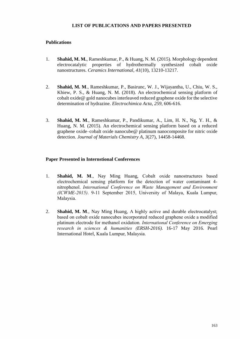
163
LIST OF PUBLICATIONS AND PAPERS PRESENTED
Publications
1. Shahid, M. M., Rameshkumar, P., & Huang, N. M. (2015). Morphology dependent
electrocatalytic properties of hydrothermally synthesized cobalt oxide
nanostructures. Ceramics International, 41(10), 13210-13217.
2. Shahid, M. M., Rameshkumar, P., Basirunc, W. J., Wijayantha, U., Chiu, W. S.,
Khiew, P. S., & Huang, N. M. (2018). An electrochemical sensing platform of
cobalt oxide@ gold nanocubes interleaved reduced graphene oxide for the selective
determination of hydrazine. Electrochimica Acta, 259, 606-616.
3. Shahid, M. M., Rameshkumar, P., Pandikumar, A., Lim, H. N., Ng, Y. H., &
Huang, N. M. (2015). An electrochemical sensing platform based on a reduced
graphene oxide–cobalt oxide nanocube@ platinum nanocomposite for nitric oxide
detection. Journal of Materials Chemistry A, 3(27), 14458-14468.
Paper Presented in Internstional Conferences
1. Shahid, M. M., Nay Ming Huang, Cobalt oxide nanostructures based
electrochemical sensing platform for the detection of water contaminant 4-
nitrophenol. International Conference on Waste Management and Environment
(ICWME-2015). 9-11 September 2015, University of Malaya, Kuala Lumpur,
Malaysia.
2. Shahid, M. M., Nay Ming Huang, A highly active and durable electrocatalyst;
based on cobalt oxide nanocubes incorporated reduced graphene oxide a modified
platinum electrode for methanol oxidation. International Conference on Emerging
research in sciences & humanities (ERSH-2016). 16-17 May 2016. Pearl
International Hotel, Kuala Lumpur, Malaysia.
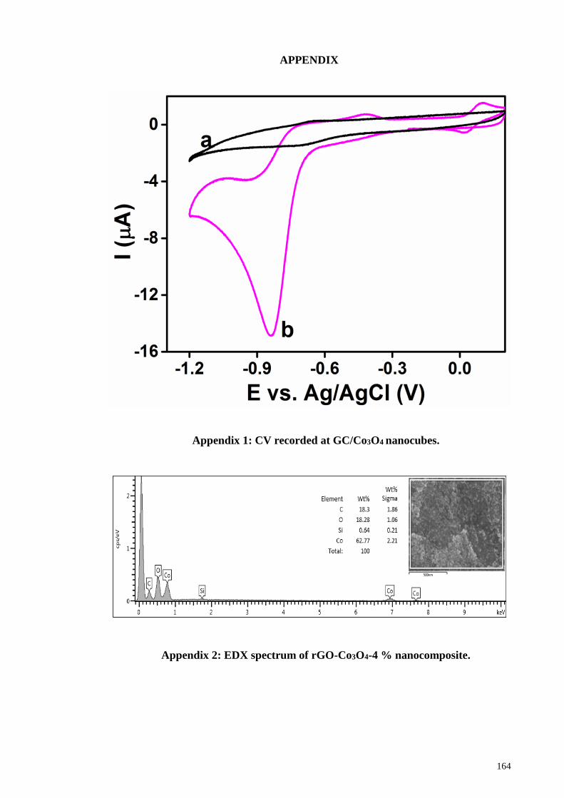
164
APPENDIX
Appendix 1: CV recorded at GC/Co3O4 nanocubes.
Appendix 2: EDX spectrum of rGO-Co3O4-4 % nanocomposite.
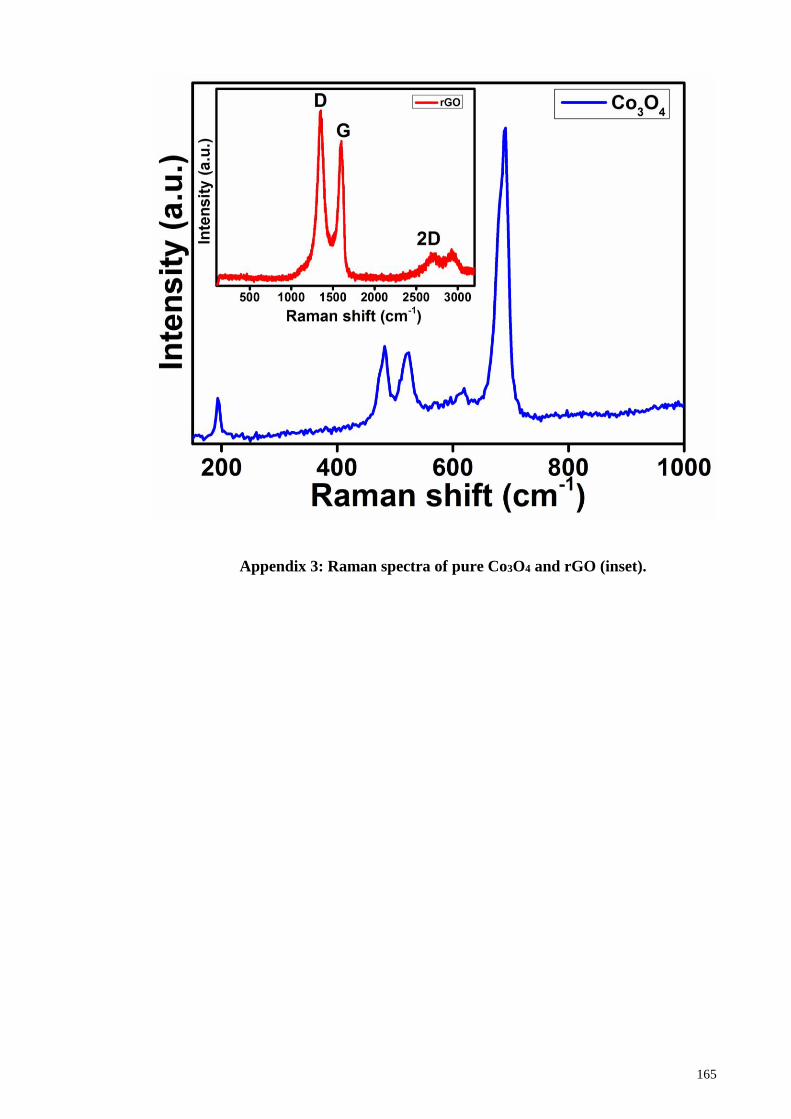
165
Appendix 3: Raman spectra of pure Co3O4 and rGO (inset).
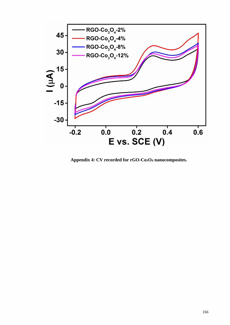
166
Appendix 4: CV recorded for rGO-Co3O4 nanocomposites.
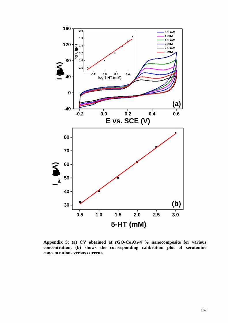
167
Appendix 5: (a) CV obtained at rGO-Co3O4-4 % nanocomposite for various
concentration, (b) shows the corresponding calibration plot of serotonine
concentrations versus current.
-0.2 0.0 0.2 0.4 0.6-40
0
40
80
120
160
-0.2 0.0 0.2 0.4
1.5
1.6
1.7
1.8
1.9
2.0
I (
A)
E vs. SCE (V)
0.5 mM
1 mM
1.5 mM
2 mM
2.5 mM
3 mM
(a)
log
Ip (
A)
log 5-HT (mM)
0.5 1.0 1.5 2.0 2.5 3.0
30
40
50
60
70
80
I pa (
A)
5-HT (mM)
(b)
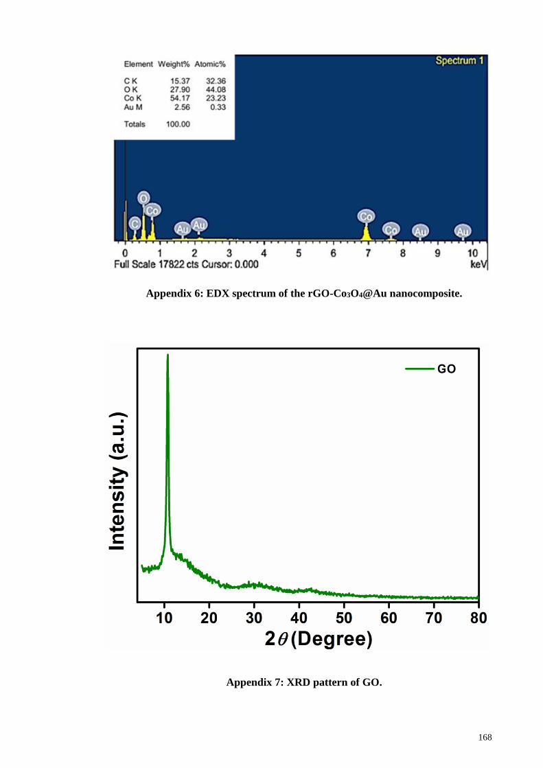
168
Appendix 6: EDX spectrum of the rGO-Co3O4@Au nanocomposite.
Appendix 7: XRD pattern of GO.
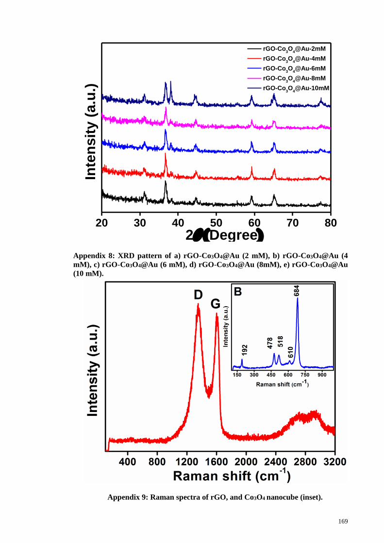
169
20 30 40 50 60 70 80
rGO-Co3O
4@Au-2mM
rGO-Co3O
4@Au-4mM
rGO-Co3O
4@Au-6mM
rGO-Co3O
4@Au-8mM
rGO-Co3O
4@Au-10mM
Inte
nsit
y (
a.u
.)
2(Degree)
Appendix 8: XRD pattern of a) rGO-Co3O4@Au (2 mM), b) rGO-Co3O4@Au (4
mM), c) rGO-Co3O4@Au (6 mM), d) rGO-Co3O4@Au (8mM), e) rGO-Co3O4@Au
(10 mM).
Appendix 9: Raman spectra of rGO, and Co3O4 nanocube (inset).
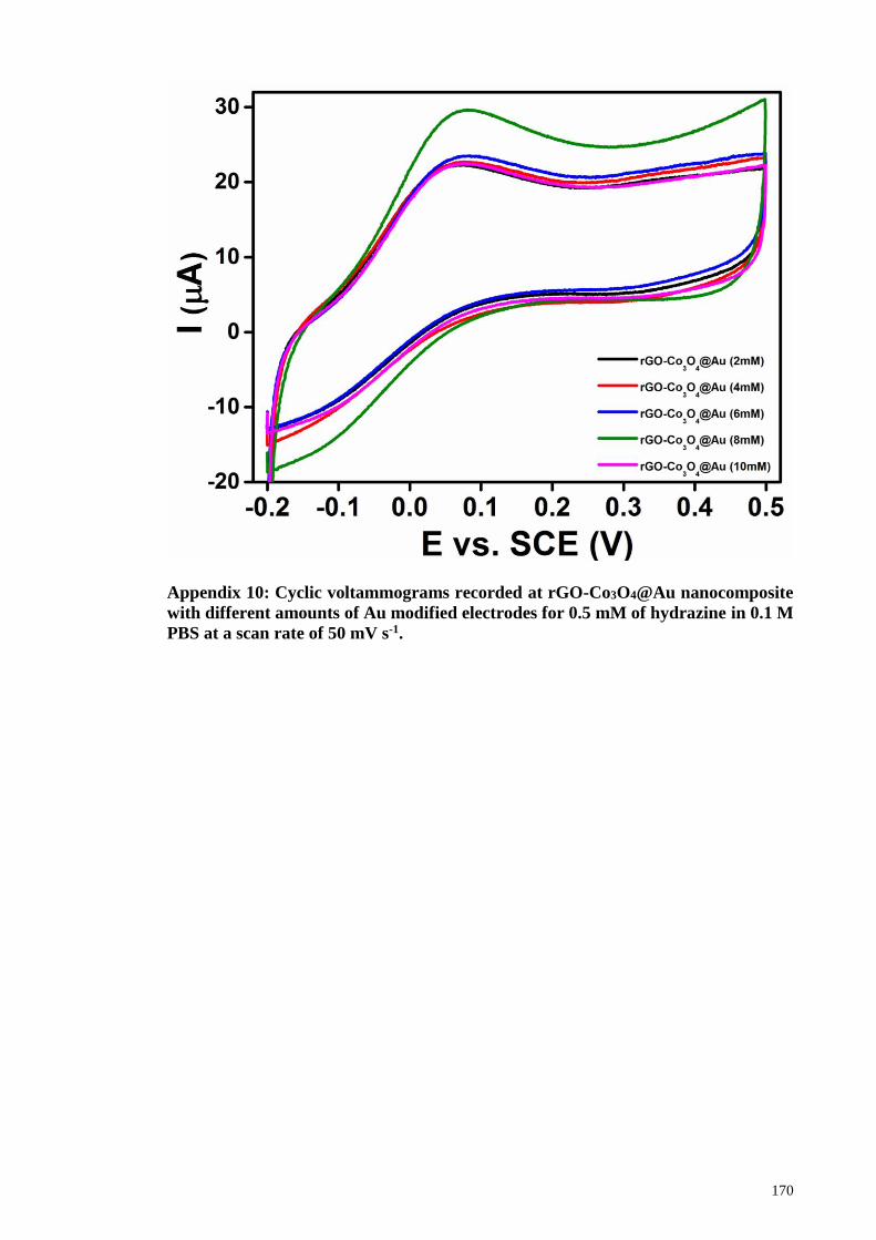
170
Appendix 10: Cyclic voltammograms recorded at rGO-Co3O4@Au nanocomposite
with different amounts of Au modified electrodes for 0.5 mM of hydrazine in 0.1 M
PBS at a scan rate of 50 mV s-1.
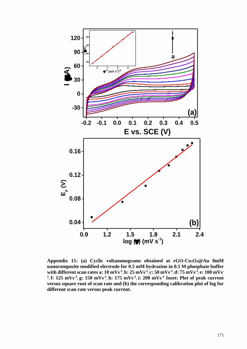
171
Appendix 11: (a) Cyclic voltammograms obtained at rGO-Co3O4@Au 8mM
nanocomposite modified electrode for 0.5 mM hydrazine in 0.1 M phosphate buffer
with different scan rates a: 10 mVs-1, b: 25 mVs-1
, c: 50 mVs-1, d: 75 mVs-1
, e: 100 mVs-
1, f: 125 mVs-1
, g: 150 mVs-1, h: 175 mVs-1
, i: 200 mVs-1 Inset: Plot of peak current
versus square root of scan rate and (b) the corresponding calibration plot of log for
different scan rate versus peak current.
-0.2 -0.1 0.0 0.1 0.2 0.3 0.4 0.5
-30
0
30
60
90
120
2 4 6 8
20
30
40
50
i
I (
A)
E vs. SCE (V)
a
(a)
I (
A)
1/2
(mV s-1)
1/2
0.9 1.2 1.5 1.8 2.1 2.4
0.04
0.08
0.12
0.16
Ep (
V)
log () (mV s-1)
(b)
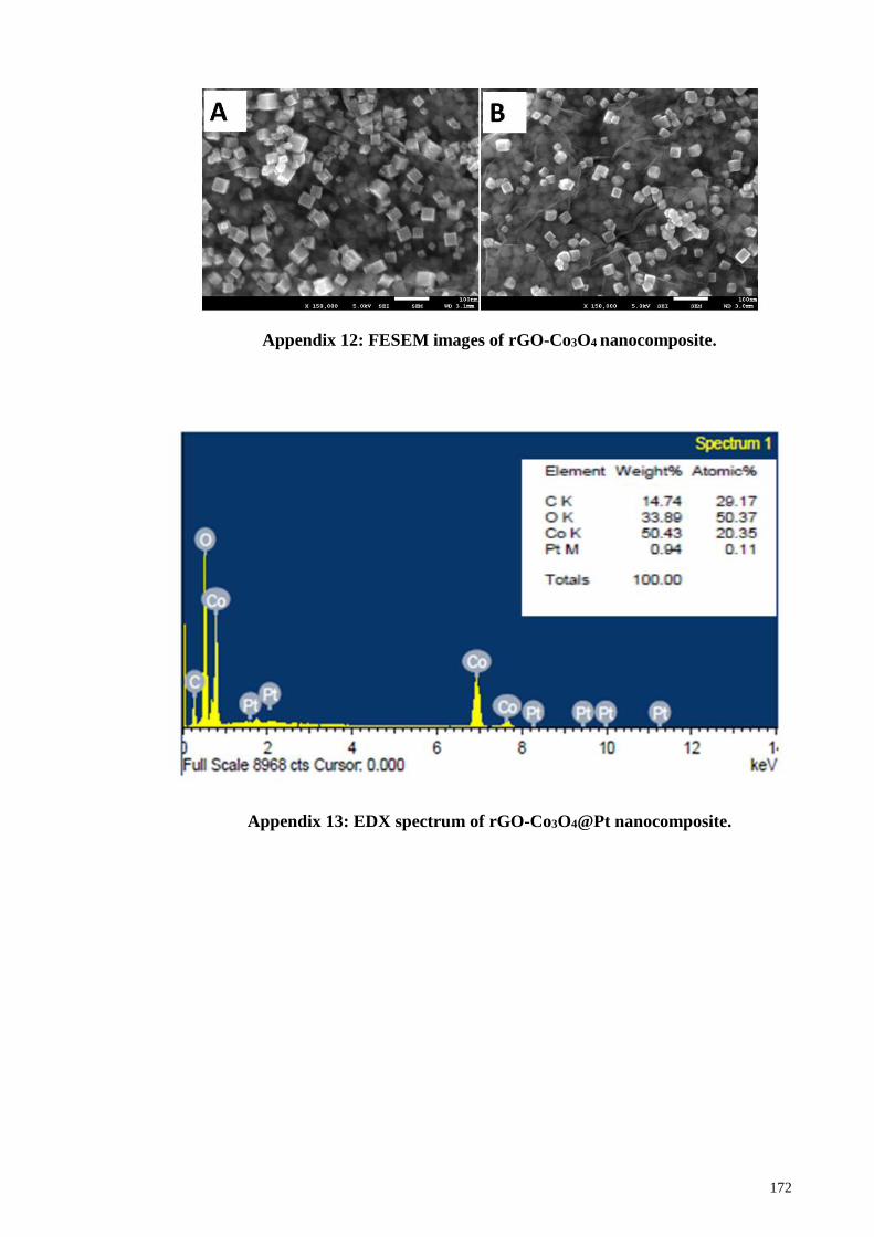
172
Appendix 12: FESEM images of rGO-Co3O4 nanocomposite.
Appendix 13: EDX spectrum of rGO-Co3O4@Pt nanocomposite.
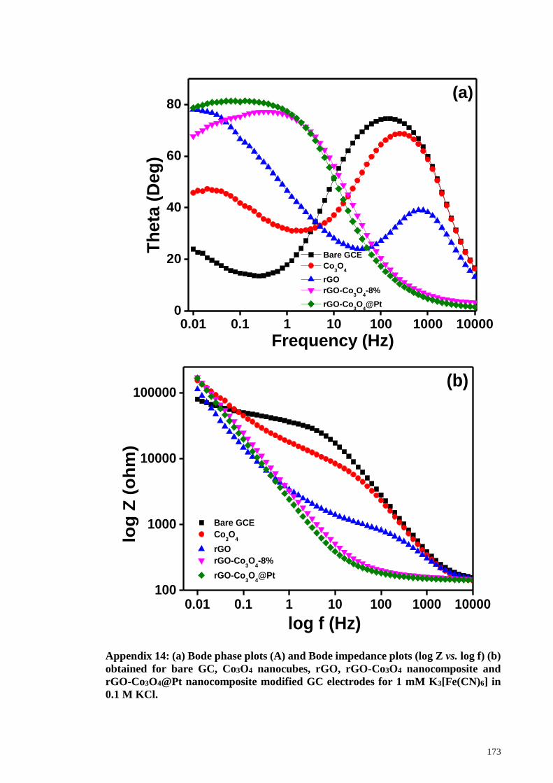
173
Appendix 14: (a) Bode phase plots (A) and Bode impedance plots (log Z vs. log f) (b)
obtained for bare GC, Co3O4 nanocubes, rGO, rGO-Co3O4 nanocomposite and
rGO-Co3O4@Pt nanocomposite modified GC electrodes for 1 mM K3[Fe(CN)6] in
0.1 M KCl.
0.01 0.1 1 10 100 1000 100000
20
40
60
80
Bare GCE
Co3O
4
rGO
rGO-Co3O
4-8%
rGO-Co3O
4@Pt
Th
eta
(D
eg
)
Frequency (Hz)
(a)
0.01 0.1 1 10 100 1000 10000100
1000
10000
100000
Bare GCE
Co3O
4
rGO
rGO-Co3O
4-8%
rGO-Co3O
4@Pt
log
Z (
oh
m)
log f (Hz)
(b)
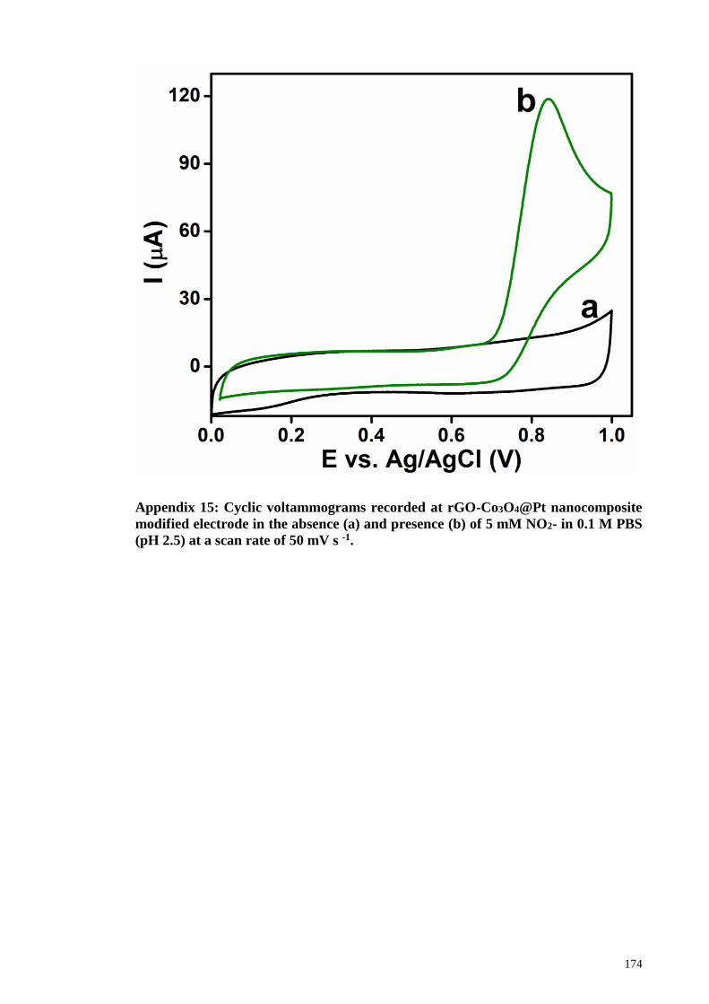
174
Appendix 15: Cyclic voltammograms recorded at rGO-Co3O4@Pt nanocomposite
modified electrode in the absence (a) and presence (b) of 5 mM NO2- in 0.1 M PBS
(pH 2.5) at a scan rate of 50 mV s -1.
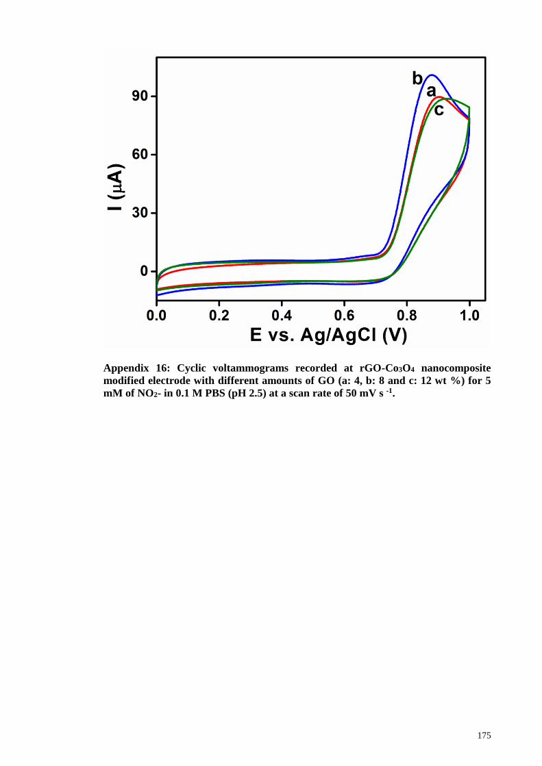
175
Appendix 16: Cyclic voltammograms recorded at rGO-Co3O4 nanocomposite
modified electrode with different amounts of GO (a: 4, b: 8 and c: 12 wt %) for 5
mM of NO2- in 0.1 M PBS (pH 2.5) at a scan rate of 50 mV s -1.
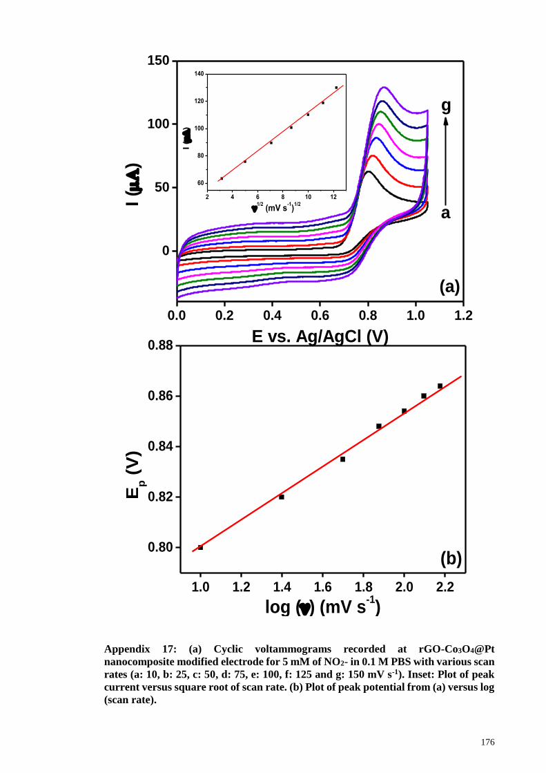
176
Appendix 17: (a) Cyclic voltammograms recorded at rGO-Co3O4@Pt
nanocomposite modified electrode for 5 mM of NO2- in 0.1 M PBS with various scan
rates (a: 10, b: 25, c: 50, d: 75, e: 100, f: 125 and g: 150 mV s-1). Inset: Plot of peak
current versus square root of scan rate. (b) Plot of peak potential from (a) versus log
(scan rate).
0.0 0.2 0.4 0.6 0.8 1.0 1.2
0
50
100
150
2 4 6 8 10 12
60
80
100
120
140
I (
)
1/2
(mV s-1)
1/2
I (
)
E vs. Ag/AgCl (V)
a
g
(a)
1.0 1.2 1.4 1.6 1.8 2.0 2.2
0.80
0.82
0.84
0.86
0.88
Ep (
V)
log () (mV s-1)
(b)
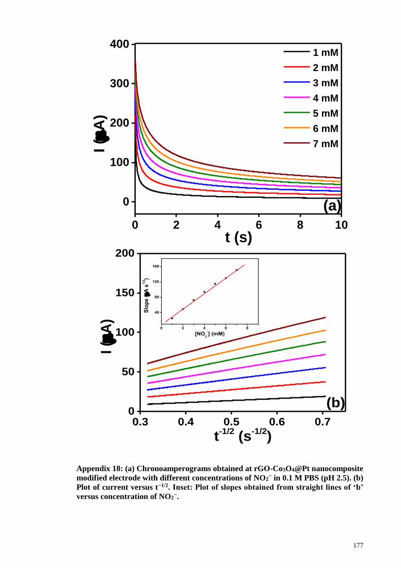
177
Appendix 18: (a) Chronoamperograms obtained at rGO-Co3O4@Pt nanocomposite
modified electrode with different concentrations of NO2− in 0.1 M PBS (pH 2.5). (b)
Plot of current versus t−1/2. Inset: Plot of slopes obtained from straight lines of ‘b’
versus concentration of NO2−.
0.3 0.4 0.5 0.6 0.70
50
100
150
200
0 2 4 6 8
40
80
120
160
Slo
pe
(A
s1
/2)
[NO2
-] (mM)
I (
A)
t-1/2
(s-1/2
)
(b)
0 2 4 6 8 10
0
100
200
300
400
I (
A)
t (s)
1 mM
2 mM
3 mM
4 mM
5 mM
6 mM
7 mM
(a)

178
Abbaspour, A., & Noori, A. (2011). A cyclodextrin host–guest recognition approach to
an electrochemical sensor for simultaneous quantification of serotonin and dopamine.
Biosensors and Bioelectronics, 26(12), 4674-4680.
Abdul Aziz, M., & Kawde, A. N. (2013). Gold nanoparticle-modified graphite pencil
electrode for the high-sensitivity detection of hydrazine. Talanta, 115, 214-221.
Adler, D. (1968). Insulating and metallic states in transition metal oxides. Solid State
Physics, 21, 1-113.
Afzali, D., Karimi-Maleh, H., & Khalilzadeh, M. A. (2011). Sensitive and selective
determination of phenylhydrazine in the presence of hydrazine at a ferrocene-
modified carbon nanotube paste electrode. Environmental chemistry letters, 9(3),
375-381.
Ahmed, S. R., Ogale, S., Papaefthymiou, G. C., Ramesh, R., & Kofinas, P. (2002).
Magnetic properties of cofe2o4 nanoparticles synthesized through a block
copolymer nanoreactor route. Applied physics letters, 80(9), 1616-1618.
Ajayan, P., & Nalwa, H. (2000). Handbook of nanostructured materials and
nanotechnology. Academic Press, New York.
Allag, A., Saâd, R., Ouahab, A., Attouche, H., & Kouidri, N. (2016). Optoelectronic
properties of sno 2 thin films sprayed at different deposition times. Chinese
Physics B, 25(4), 046801.
Alrehaily, L., Joseph, J., Biesinger, M., Guzonas, D., & Wren, J. (2013). Gamma-
radiolysis-assisted cobalt oxide nanoparticle formation. Physical Chemistry
Chemical Physics, 15(3), 1014-1024.
Ando, M., Kadono, K., Kamada, K., & Ohta, K. (2004). Third-order nonlinear optical
responses of nanoparticulate co 3 o 4 films. Thin Solid Films, 446(2), 271-276.
Anithaa, A. C., Asokan, K., & Sekar, C. (2017). Highly sensitive and selective serotonin
sensor based on gamma ray irradiated tungsten trioxide nanoparticles. Sensors
and Actuators B, 238, 667-675.
Antoniadou, S., Jannakoudakis, A., & Theodoridou, E. (1989). Electrocatalytic reactions
on carbon fibre electrodes modified by hemine ii. Electro-oxidation of hydrazine.
Synthetic metals, 30(3), 295-304.
Arvinte, A., Mahosenaho, M., Pinteala, M., Sesay, A.-M., & Virtanen, V. (2011).
Electrochemical oxidation of p-nitrophenol using graphene-modified electrodes,
and a comparison to the performance of mwnt-based electrodes. Microchimica
Acta, 174(3-4), 337-343.
Asif, M. H., Elinder, F., & Willander, M. (2011). Electrochemical biosensors based on
zno nanostructures to measure intracellular metal ions and glucose. Journal of
Analytical & Bioanalytical Techniques 7.
Augustine, A. K., Nampoori, V., & Kailasnath, M. (2014). Rapid synthesize of gold
nanoparticles by microwave irradiation method and its application as an optical

179
limiting material. Optik-International Journal for Light and Electron Optics,
125(22), 6696-6699.
Babaei, A., & Taheri, A. R. (2013). Nafion/ni(oh)2 nanoparticles-carbon nanotube
composite modified glassy carbon electrode as a sensor for simultaneous
determination of dopamine and serotonin in the presence of ascorbic acid. Sensors
and Actuators B: Chemical, 176, 543-551.
Baek, H. R., & Eo, I. S. (2017). Electrochemical properties of tio2-metal oxide
composites for dye-sensitized solar cell. Current Applied Physics, 17(6), 854-857.
Bala, H., Fu, W., Yu, Y., Yang, H., & Zhang, Y. (2009). Preparation, optical properties,
magnetic properties and thermal stability of core–shell structure cobalt/zinc oxide
nanocomposites. Applied Surface Science, 255(7), 4050-4055.
Balandin, A. A., Ghosh, S., Bao, W., Calizo, I., Teweldebrhan, D., Miao, F., & Lau, C.
N. (2008). Superior thermal conductivity of single-layer graphene. Nano Letters,
8(3), 902-907.
Baxter, J. B., & Aydil, E. S. (2005). Nanowire-based dye-sensitized solar cells. Applied
physics letters, 86(5), 053114.
Bedioui, F., & Villeneuve, N. (2003). Electrochemical nitric oxide sensors for biological
samples–principle, selected examples and applications. Electroanalysis, 15(1), 5-
18.
Beltramo, G., & Koper, M. (2003). Nitric oxide reduction and oxidation on stepped pt [n
(111)×(111)] electrodes. Langmuir, 19(21), 8907-8915.
Bogdan, C. (2001). Nitric oxide and the immune response. Nature immunology, 2(10),
907-916.
Borsella, E., Cattaruzza, E., De Marchi, G., Gonella, F., Mattei, G., Mazzoldi, P.,
Quaranta, A., Battaglin, G., & Polloni, R. (1999). Synthesis of silver clusters in
silica-based glasses for optoelectronics applications. Journal of non-crystalline
solids, 245(1), 122-128.
Brückner, A. (2003). Looking on heterogeneous catalytic systems from different
perspectives: Multitechnique approaches as a new challenge for in situ studies.
Catalysis Reviews, 45(1), 97-150.
Buchsteiner, A., Lerf, A., & Pieper, J. (2006). Water dynamics in graphite oxide
investigated with neutron scattering. The Journal of Physical Chemistry B,
110(45), 22328-22338.
Budkuley, J. S. (1992). Determination of hydrazine and sulphite in the presence of one
another. Microchimica Acta, 108(1-2), 103-105.
Byrappa, K., & Yoshimura, M. (2012). Handbook of hydrothermal technology: William
Andrew.
180
Cao, L., Zhang, Z., Sun, L., Gao, C., He, M., Wang, Y., Li, Y., Zhang, X., Li, G., &
Zhang, J. (2001). Well‐aligned boron nanowire arrays. Advanced Materials,
13(22), 1701-1704.
Cao, L. M., Hahn, K., Scheu, C., Rühle, M., Wang, Y. Q., Zhang, Z., Gao, C. X., Li, Y.
C., Zhang, X. Y., He, M., Sun, L. L., & Wang, W. K. (2002). Template-catalyst-
free growth of highly ordered boron nanowire arrays. Applied physics letters,
80(22), 4226-4228.
Chai, S.-H., Howe, J. Y., Wang, X., Kidder, M., Schwartz, V., Golden, M. L., Overbury,
S. H., Dai, S., & Jiang, D.-e. (2012). Graphitic mesoporous carbon as a support of
promoted rh catalysts for hydrogenation of carbon monoxide to ethanol. carbon,
50(4), 1574-1582.
Chang, H., & Wu, H. (2013). Graphene-based nanocomposites: Preparation,
functionalization, and energy and environmental applications. Energy &
Environmental Science, 6(12), 3483.
Chang, W., Skandan, G., Hahn, H., Danforth, S. C., & Kear, B. H. (1994). Chemical
vapor condensation of nanostructured ceramic powders. Nanostructured
Materials, 4(3), 345-351.
Chauveau, J., Fert, V., Morel, A., & Delaage, M. (1991). Rapid and specific enzyme
immunoassay of serotonin. Clinical chemistry, 37(7), 1178-1184.
Chen, C.-H., Chen, Y.-C., & Lin, M.-S. (2013). Amperometric determination of nadh
with co 3 o 4 nanosheet modifiedelectrode. Biosensors and Bioelectronics, 42,
379-384.
Chen, C.-M., Zhang, Q., Yang, M.-G., Huang, C.-H., Yang, Y.-G., & Wang, M.-Z.
(2012). Structural evolution during annealing of thermally reduced graphene
nanosheets for application in supercapacitors. carbon, 50(10), 3572-3584.
Chen, M., Zhou, W., Qi, M., Zhang, J., Yin, J., & Chen, Q. (2017). Reconstruction of
copper shell on metal oxides as enhanced nanoarrays electrodes for lithium ion
batteries. Materials Research Bulletin, 86, 308-312.
Chen, S., Wu, Q., & Mishra, C. (2012). New graphene-related materials on the horizon.
Nat. Mater, 11, 203-207.
Chen, S., Zhu, J., & Wang, X. (2010). One-step synthesis of graphene-cobalt hydroxide
nanocomposites and their electrochemical properties. Journal of Physical
Chemistry C, 114(27), 11829-11834.
Chen, X., Long, H.-Y., Wu, W.-L., & Yang, Z.-S. (2009). Direct electrochemical
behavior of cytochrome c on sodium dodecyl sulfate modified electrode and its
application to nitric oxide biosensor. Thin Solid Films, 517(8), 2787-2791.
Cheng, C.-S., Serizawa, M., Sakata, H., & Hirayama, T. (1998). Electrical conductivity
of co 3 o 4 films prepared by chemical vapour deposition. Materials Chemistry
and Physics, 53(3), 225-230.

181
Cho, Y., Parmar, N. S., Nahm, S., & Choi, J.-W. (2017). Full range optical and electrical
properties of zn-doped sno2 and oxide/metal/oxide multilayer thin films deposited
on flexible pet substrate. Journal of Alloys and Compounds, 694, 217-222.
Choi, H.-J., Jung, S.-M., Seo, J.-M., Chang, D. W., Dai, L., & Baek, J.-B. (2012).
Graphene for energy conversion and storage in fuel cells and supercapacitors.
Nano Energy, 1(4), 534-551.
Choi, Y., Gu, M., Park, J., Song, H. K., & Kim, B. S. (2012). Graphene multilayer
supported gold nanoparticles for efficient electrocatalysts toward methanol
oxidation. Advanced Energy Materials, 2(12), 1510-1518.
Chou, S.-L., Wang, J.-Z., Liu, H.-K., & Dou, S.-X. (2008). Electrochemical deposition
of porous co 3 o 4 nanostructured thin film for lithium-ion battery. Journal of
Power Sources, 182(1), 359-364.
Choudhary, G., IIansen, H., Donkin, S., & Kirman, C. (1997). Toxicological profile for
hydrazines. US Department of Health and Human Service, 5.
Choudhury, C., Bansal, N. K., & Sehgal, H. K. (1983). Heat loss optimisation of a
concentric cylindrical solar collector employing a cobalt oxide selective absorber.
Applied Energy, 14(2), 143-159.
Chu, L., Han, L., & Zhang, X. (2011). Electrochemical simultaneous determination of
nitrophenol isomers at nano-gold modified glassy carbon electrode. Journal of
Applied Electrochemistry, 41(6), 687-694.
Cinthia, A. J., Priyanga, G. S., Rajeswarapalanichamy, R., & Iyakutti, K. (2015).
Structural, electronic and mechanical properties of alkaline earth metal oxides mo
(m=be, mg, ca, sr, ba). Journal of Physics and Chemistry of Solids, 79, 23-42.
Conoci, S., Petralia, S., Samorì, P., Raymo, F. M., Di Bella, S., & Sortino, S. (2006).
Optically transparent, ultrathin pt films as versatile metal substrates for molecular
optoelectronics. Advanced Functional Materials, 16(11), 1425-1432.
Crespi, F., Baumeyer, T., Moebius, C., & Dittrich, J. (1991). Simultaneous in vivo
monitoring of dopamine and serotonin by differential pulse conditioning
voltammetry with na-cro microbiosensors. Fresenius. J. Anal. Chem., 341(10),
644.
Cui, L., Ji, C., Peng, Z., Zhong, L., Zhou, C., Yan, L., Qu, S., Zhang, S., Huang, C., &
Qian, X. (2014). Unique tri-output optical probe for specific and ultrasensitive
detection of hydrazine. Analytical chemistry, 86(9), 4611-4617.
Cui, L., Peng, Z., Ji, C., Huang, J., Huang, D., Ma, J., Zhang, S., Qian, X., & Xu, Y.
(2014). Hydrazine detection in the gas state and aqueous solution based on the
gabriel mechanism and its imaging in living cells. Chemical Communications,
50(12), 1485-1487.
Dalkıran, B., Esra Erden, P., & Kılıç, E. (2017). Amperometric biosensors based on
carboxylated multiwalled carbon nanotubes-metal oxide nanoparticles-7,7,8,8-

182
tetracyanoquinodimethane composite for the determination of xanthine. Talanta,
167, 286-295.
de Moraes, A. C. M., Andrade, P. F., de Faria, A. F., Simões, M. B., Salomão, F. C. C.
S., Barros, E. B., do Carmo Gonçalves, M., & Alves, O. L. (2015). Fabrication of
transparent and ultraviolet shielding composite films based on graphene oxide and
cellulose acetate. Carbohydrate polymers, 123, 217-227.
Demuth, J., Hamers, R., Tromp, R., & Welland, M. (1986). A scanning tunneling
microscope for surface science studies. IBM journal of research and development,
30(4), 396-402.
Devasenathipathy, R., Mani, V., & Chen, S.-M. (2014). Highly selective amperometric
sensor for the trace level detection of hydrazine at bismuth nanoparticles
decorated graphene nanosheets modified electrode. Talanta, 124, 43-51.
Dhas, N. A., Koltypin, Y., & Gedanken, A. (1997). Sonochemical preparation and
characterization of ultrafine chromium oxide and manganese oxide powders.
Chemistry of materials, 9(12), 3159-3163.
Dinesh, B., Veeramani, V., Chen, S.-M., & Saraswathi, R. (2017). In situ electrochemical
synthesis of reduced graphene oxide-cobalt oxide nanocomposite modified
electrode for selective sensing of depression biomarker in the presence of ascorbic
acid and dopamine. J. Electroanal. Chem., 786, 169-176.
Ding, J., Zhu, S., Zhu, T., Sun, W., Li, Q., Wei, G., & Su, Z. (2015). Hydrothermal
synthesis of zinc oxide-reduced graphene oxide nanocomposites for an
electrochemical hydrazine sensor. RSC Advances, 5(29), 22935-22942.
Dong, Y., He, K., Yin, L., & Zhang, A. (2007). A facile route to controlled synthesis of
co3o4 nanoparticles and their environmental catalytic properties.
Nanotechnology, 18(43), 435602.
Du, X., He, J., Zhu, J., Sun, L., & An, S. (2012). Ag-deposited silica-coated fe3o4
magnetic nanoparticles catalyzed reduction of p-nitrophenol. Applied Surface
Science, 258(7), 2717-2723.
Du, X., Skachko, I., Barker, A., & Andrei, E. Y. (2008). Approaching ballistic transport
in suspended graphene. Nature nanotechnology, 3(8), 491-495.
Dutta, S., Ray, C., Mallick, S., Sarkar, S., Roy, A., & Pal, T. (2015). Au@ pd core–shell
nanoparticles-decorated reduced graphene oxide: A highly sensitive and selective
platform for electrochemical detection of hydrazine. RSC Advances, 5(64), 51690-
51700.
Ensafi, A. A., Abarghoui, M. M., & Rezaei, B. (2016). Facile synthesis of pt-cu@ silicon
nanostructure as a new electrocatalyst supported matrix, electrochemical
detection of hydrazine and hydrogen peroxide. Electrochimica Acta, 190, 199-
207.
Ensafi, A. A., & Rezaei, B. (1998). Flow injection determination of hydrazine with
fluorimetric detection. Talanta, 47(3), 645-649.

183
Fagan-Murphy, A., Watt, F., Morgan, K. A., & Patel, B. A. (2012). Influence of different
biological environments on the stability of serotonin detection on carbon-based
electrodes. J. Electroanal. Chem., 684, 1-5.
Fang, B., Zhang, C., Zhang, W., & Wang, G. (2009). A novel hydrazine electrochemical
sensor based on a carbon nanotube-wired zno nanoflower-modified electrode.
Electrochimica Acta, 55(1), 178-182.
Fang, F. C. (1997). Perspectives series: Host/pathogen interactions. Mechanisms of nitric
oxide-related antimicrobial activity. Journal of Clinical Investigation, 99(12),
2818.
Farhadi, S., Pourzare, K., & Bazgir, S. (2014). Co 3 o 4 nanoplates: Synthesis,
characterization and study of optical and magnetic properties. Journal of Alloys
and Compounds, 587, 632-637.
Farhadi, S., Safabakhsh, J., & Zaringhadam, P. (2013). Synthesis, characterization, and
investigation of optical and magnetic properties of cobalt oxide (co3o4)
nanoparticles. Journal of Nanostructure in Chemistry, 3(1), 1-9.
Fei, J., Hu, S., & Shiu, K.-K. (2011). Amperometric determination of nitric oxide at a
carbon nanotube modified electrode with redox polymer coating. Journal of Solid
State Electrochemistry, 15(3), 519-523.
Fernandez-Garcia, M., Martinez-Arias, A., Hanson, J., & Rodriguez, J. (2004).
Nanostructured oxides in chemistry: Characterization and properties. Chemical
Reviews, 104(9), 4063-4104.
Fierro, J. L. G. (2005). Metal oxides: Chemistry and applications: CRC press.
Fiiipponi, L., & Sutherland, D. (2012). Nanotechnologies: Principles, applications,
implications and hands-on activities: A compendium for educators: European
Union, Directorate General for Research and Innovation.
Frohlich, K., Luptak, R., Dobrocka, E., Husekova, K., Cico, K., Rosova, A., Lukosius,
M., Abrutis, A., Pisecny, P., & Espinos, J. (2006). Characterization of rare earth
oxides based mosfet gate stacks prepared by metal-organic chemical vapour
deposition. Materials science in semiconductor processing, 9(6), 1065-1072.
Fujishima, A., & Honda, K. (1972). Electrochemical photolysis of water at a
semiconductor electrode. nature, 238(5358), 37-38.
Furchgott, R. F. (1999). Endothelium‐derived relaxing factor: Discovery, early studies,
and identifcation as nitric oxide (nobel lecture). Angewandte Chemie
International Edition, 38(13‐14), 1870-1880.
Ganjali, M. R., Motakef-Kazami, N., Faridbod, F., Khoee, S., & Norouzi, P. (2010).
Determination of pb 2+ ions by a modified carbon paste electrode based on multi-
walled carbon nanotubes (mwcnts) and nanosilica. Journal of hazardous
materials, 173(1), 415-419.

184
Geim, A. K., & Novoselov, K. S. (2007). The rise of graphene. Nature materials, 6(3),
183-191.
Ghanbari, K. (2014). Fabrication of silver nanoparticles–polypyrrole composite modified
electrode for electrocatalytic oxidation of hydrazine. Synthetic metals, 195, 234-
240.
Gholamian, F., Sheikh-Mohseni, M. A., & Naeimi, H. (2012). Simultaneous
determination of phenylhydrazine and hydrazine by a nanostructured
electrochemical sensor. Materials Science and Engineering: C, 32(8), 2344-2348.
Gholivand, M. B., & Azadbakht, A. (2011). A novel hydrazine electrochemical sensor
based on a zirconium hexacyanoferrate film-bimetallic au-pt inorganic-organic
hybrid nanocomposite onto glassy carbon-modified electrode. Electrochim. Acta,
56(27), 10044-10054.
Greene, L. E., Law, M., Goldberger, J., Kim, F., Johnson, J. C., Zhang, Y., Saykally, R.
J., & Yang, P. (2003). Low‐temperature wafer‐scale production of zno nanowire
arrays. Angewandte Chemie International Edition, 42(26), 3031-3034.
Greenwood, N., & Earnshaw, A. (1997). Chemistry of the elements 2nd edition:
Butterworth-Heinemann.
Gu, Y.-e., Zhang, Y., Zhang, F., Wei, J., Wang, C., Du, Y., & Ye, W. (2010). Investigation
of photoelectrocatalytic activity of cu 2 o nanoparticles for p-nitrophenol using
rotating ring-disk electrode and application for electrocatalytic determination.
Electrochimica Acta, 56(2), 953-958.
Gueell, A. G., Meadows, K. E., Unwin, P. R., & MacPherson, J. V. (2010). Trace
voltammetric detection of serotonin at carbon electrodes: Comparison of glassy
carbon, boron doped diamond and carbon nanotube network electrodes. Phys.
Chem. Chem. Phys., 12(34), 10108-10114.
Guix, F., Uribesalgo, I., Coma, M., & Munoz, F. (2005). The physiology and
pathophysiology of nitric oxide in the brain. Progress in neurobiology, 76(2), 126-
152.
Guo, C. X., Ng, S. R., Khoo, S. Y., Zheng, X., Chen, P., & Li, C. M. (2012). Rgd-peptide
functionalized graphene biomimetic live-cell sensor for real-time detection of
nitric oxide molecules. ACS nano, 6(8), 6944-6951.
Guo, S., Dong, S., & Wang, E. (2009). Three-dimensional pt-on-pd bimetallic
nanodendrites supported on graphene nanosheet: Facile synthesis and used as an
advanced nanoelectrocatalyst for methanol oxidation. ACS nano, 4(1), 547-555.
Gupta, P., & Goyal, R. N. (2014). Polymelamine modified edge plane pyrolytic graphite
sensor for the electrochemical assay of serotonin. Talanta, 120, 17-22.
Hai, H. T., Takamura, H., & Koike, J. (2013). Oxidation behavior of cu–ag core–shell
particles for solar cell applications. Journal of Alloys and Compounds, 564, 71-
77.

185
He, Q., Zheng, D., & Hu, S. (2009). Development and application of a nano-alumina
based nitric oxide sensor. Microchimica Acta, 164(3-4), 459-464.
He, X., & Zhu, L. (2006). Direct electrochemistry of hemoglobin in cetylpyridinium
bromide film: Redox thermodynamics and electrocatalysis to nitric oxide.
Electrochemistry Communications, 8(4), 615-620.
He, Y.-S., Bai, D.-W., Yang, X., Chen, J., Liao, X.-Z., & Ma, Z.-F. (2010). A co(oh)2-
graphene nanosheets composite as a high performance anode material for
rechargeable lithium batteries. Electrochem. Commun., 12(4), 570-573.
Henrich, V. E., & Cox, P. A. (1996). The surface science of metal oxides: Cambridge
university press.
Henry, P. F., & Weller, M. T. (1998). Ca2aun: A nitride containing infinite zigzag gold
chains. Angewandte Chemie International Edition, 37(20), 2855-2857.
Hetrick, E. M., & Schoenfisch, M. H. (2009). Analytical chemistry of nitric oxide. Annual
review of analytical chemistry (Palo Alto, Calif.), 2, 409.
Hu, H., Wang, X., Wang, J., Wan, L., Liu, F., Zheng, H., Chen, R., & Xu, C. (2010).
Preparation and properties of graphene nanosheets–polystyrene nanocomposites
via in situ emulsion polymerization. Chemical Physics Letters, 484(4), 247-253.
Hu, L., Peng, Q., & Li, Y. (2008). Selective synthesis of co3o4 nanocrystal with different
shape and crystal plane effect on catalytic property for methane combustion.
Journal of the American Chemical Society, 130(48), 16136-16137.
Huang, J., & Wan, Q. (2009). Gas sensors based on semiconducting metal oxide one-
dimensional nanostructures. Sensors, 9(12), 9903-9924.
Huang, N., Lim, H., Chia, C., Yarmo, M., & Muhamad, M. (2011). Simple room-
temperature preparation of high-yield large-area graphene oxide. International
Journal of Nanomedicine, 6, 3443.
Huang, N. M., Lim, H. N., Chia, C. H., Yarmo, M. A., & Muhamad, M. R. (2011). Simple
room-temperature preparation of high-yield large-area graphene oxide.
International Journal of Nanomedicine, 6, 3443-3448.
Huang, W., Yang, C., & Zhang, S. (2003). Simultaneous determination of 2-nitrophenol
and 4-nitrophenol based on the multi-wall carbon nanotubes nafion-modified
electrode. Analytical and bioanalytical chemistry, 375(5), 703-707.
Ignarro, L. J., Buga, G. M., Wood, K. S., Byrns, R. E., & Chaudhuri, G. (1987).
Endothelium-derived relaxing factor produced and released from artery and vein
is nitric oxide. Proceedings of the National Academy of Sciences, 84(24), 9265-
9269.
Interrante, L. V., & Hampden-Smith, M. J. (1997). Chemistry of advanced materials: An
overview. Chemistry of Advanced Materials: An Overview, by Leonard V.
Interrante (Editor), Mark J. Hampden-Smith (Editor), pp. 592. ISBN 0-471-
18590-6. Wiley-VCH, December 1997., 592.
186
Jain, P. K., Lee, K. S., El-Sayed, I. H., & El-Sayed, M. A. (2006). Calculated absorption
and scattering properties of gold nanoparticles of different size, shape, and
composition: Applications in biological imaging and biomedicine. J. Phys. Chem.
B, 110(14), 7238-7248.
Jang, J. Y., Kim, M. S., Jeong, H. M., & Shin, C. M. (2009). Graphite oxide/poly (methyl
methacrylate) nanocomposites prepared by a novel method utilizing
macroazoinitiator. Composites Science and Technology, 69(2), 186-191.
Jayabal, S., Viswanathan, P., & Ramaraj, R. (2014). Reduced graphene oxide–gold
nanorod composite material stabilized in silicate sol–gel matrix for nitric oxide
sensor. RSC Advances, 4(63), 33541-33548.
Jayasri, D., & Narayanan, S. S. (2007). Amperometric determination of hydrazine at
manganese hexacyanoferrate modified graphite–wax composite electrode.
Journal of hazardous materials, 144(1), 348-354.
Jeon, H.-K., Nohta, H., & Ohkura, Y. (1992). High-performance liquid chromatographic
determination of catecholamines and their precursor and metabolites in human
urine and plasma by postcolumn derivatization involving chemical oxidation
followed by fluorescence reaction. Analytical biochemistry, 200(2), 332-338.
Jeong, S. J., Song, J. S., Min, B. K., Lee, W. J., & Park, E. C. (2006). Characteristics of
piezoelectric multilayer devices containing metal-oxide multicomponent
electrode. Ferroelectrics, 338(1), 9-16.
Jia, J., Wang, B., Wu, A., Cheng, G., Li, Z., & Dong, S. (2002). A method to construct a
third-generation horseradish peroxidase biosensor: Self-assembling gold
nanoparticles to three-dimensional sol-gel network. Analytical chemistry, 74(9),
2217-2223.
Jia, S., Fei, J., Deng, J., Cai, Y., & Li, J. (2009). Direct electrochemistry and
electrocatalysis of hemoglobin immobilized in an amphiphilic diblock copolymer
film. Sensors and Actuators B: Chemical, 138(1), 244-250.
Jiang, L., Gu, S., Ding, Y., Jiang, F., & Zhang, Z. (2014). Facile and novel
electrochemical preparation of a graphene–transition metal oxide nanocomposite
for ultrasensitive electrochemical sensing of acetaminophen and phenacetin.
Nanoscale, 6(1), 207-214.
Jiang, Y., Zheng, B., Du, J., Liu, G., Guo, Y., & Xiao, D. (2013). Electrophoresis
deposition of ag nanoparticles on tio 2 nanotube arrays electrode for hydrogen
peroxide sensing. Talanta, 112, 129-135.
Jiang, Z., Jiang, Z.-J., Maiyalagan, T., & Manthiram, A. (2016). Cobalt oxide-coated n-
and b-doped graphene hollow spheres as bifunctional electrocatalysts for oxygen
reduction and oxygen evolution reactions. Journal of Materials Chemistry A,
4(16), 5877-5889.
Jiao, F., & Frei, H. (2009). Nanostructured cobalt oxide clusters in mesoporous silica as
efficient oxygen‐evolving catalysts. Angewandte Chemie International Edition,
48(10), 1841-1844.

187
Jiao, F., & Frei, H. (2010). Nanostructured cobalt and manganese oxide clusters as
efficient water oxidation catalysts. Energy & Environmental Science, 3(8), 1018-
1027.
Jokar, E., & Shahrokhian, S. (2014). Growth control of cobalt oxide nanoparticles on
reduced graphene oxide for enhancement of electrochemical capacitance.
International Journal of Hydrogen Energy, 39(36), 21068-21075.
Jonker, G., & Van Santen, J. (1950). Physica (utrecht) 16, 337 (1950). CrossRef Google
Scholar.
Kaczmarczyk, J., Zasada, F., Janas, J., Indyka, P., Piskorz, W., Kotarba, A., & Sojka, Z.
(2016). Thermodynamic stability, redox properties and reactivity of mn3o4,
fe3o4, and co3o4 model catalysts for n2o decomposition–resolving the origins of
steady turnover. ACS Catalysis.
Kang, H., Kulkarni, A., Stankovich, S., Ruoff, R. S., & Baik, S. (2009). Restoring
electrical conductivity of dielectrophoretically assembled graphite oxide sheets by
thermal and chemical reduction techniques. carbon, 47(6), 1520-1525.
Kang, M.-G., Park, H. J., Ahn, S. H., & Guo, L. J. (2010). Transparent cu nanowire mesh
electrode on flexible substrates fabricated by transfer printing and its application
in organic solar cells. Solar Energy Materials and Solar Cells, 94(6), 1179-1184.
Kang, T.-T., Liu, X., Zhang, R. Q., Hu, W. G., Cong, G., Zhao, F.-A., & Zhu, Q. (2006).
Inn nanoflowers grown by metal organic chemical vapor deposition. Applied
physics letters, 89(7), 071113.
Karabacak, T., Mallikarjunan, A., Singh, J. P., Ye, D., Wang, G.-C., & Lu, T.-M. (2003).
Β-phase tungsten nanorod formation by oblique-angle sputter deposition. Applied
physics letters, 83(15), 3096-3098.
Karimi-Maleh, H., Moazampour, M., Ensafi, A. A., Mallakpour, S., & Hatami, M. (2014).
An electrochemical nanocomposite modified carbon paste electrode as a sensor
for simultaneous determination of hydrazine and phenol in water and wastewater
samples. Environmental Science and Pollution Research, 21(9), 5879-5888.
Karuppiah, C., Palanisamy, S., Chen, S.-M., Ramaraj, S. K., & Periakaruppan, P. (2014).
A novel and sensitive amperometric hydrazine sensor based on gold nanoparticles
decorated graphite nanosheets modified screen printed carbon electrode.
Electrochimica Acta, 139, 157-164.
Karuppiah, C., Palanisamy, S., Chen, S. M., Veeramani, V., & Periakaruppan, P. (2014).
A novel enzymatic glucose biosensor and sensitive non-enzymatic hydrogen
peroxide sensor based on graphene and cobalt oxide nanoparticles composite
modified glassy carbon electrode. Sensors and Actuators B-Chemical, 196, 450-
456.
Kelly, R., Miotello, A., Chrisey, D., & Hubler, G. (1994). Pulsed laser deposition of thin
films. by DB Chrisey and GK Hubler (Wiley, New York, 1994) p, 55.
188
Khalilzadeh, M. A., & Karimi-Maleh, H. (2009). Sensitive and selective determination
of phenylhydrazine in the presence of hydrazine at a ferrocene monocarboxylic
acid modified carbon nanotube paste electrode. Analytical Letters, 43(1), 186-
196.
Khlebtsov, N., & Dykman, L. (2011). Biodistribution and toxicity of engineered gold
nanoparticles: A review of in vitro and in vivo studies. Chemical Society Reviews,
40(3), 1647-1671.
Khoei, A., Ban, E., Banihashemi, P., & Qomi, M. A. (2011). Effects of temperature and
torsion speed on torsional properties of single-walled carbon nanotubes. Materials
Science and Engineering: C, 31(2), 452-457.
Kim, H., Seo, D.-H., Kim, S.-W., Kim, J., & Kang, K. (2011). Highly reversible co 3 o
4/graphene hybrid anode for lithium rechargeable batteries. carbon, 49(1), 326-
332.
Kim, K., Zhao, Y., Jang, H., Lee, S., Kim, J., Kim, K., & Ahn, J. (2009). P. Kim, j.-y.
Choi and bh hong. nature, 457, 706.
Kim, K. S., Zhao, Y., Jang, H., Lee, S. Y., Kim, J. M., Kim, K. S., Ahn, J.-H., Kim, P.,
Choi, J.-Y., & Hong, B. H. (2009). Large-scale pattern growth of graphene films
for stretchable transparent electrodes. nature, 457(7230), 706-710.
Kim, Y., Noh, Y., Lim, E. J., Lee, S., Choi, S. M., & Kim, W. B. (2014). Star-shaped
pd@ pt core–shell catalysts supported on reduced graphene oxide with superior
electrocatalytic performance. Journal of Materials Chemistry A, 2(19), 6976-
6986.
Kimmel, D. W., LeBlanc, G., Meschievitz, M. E., & Cliffel, D. E. (2011).
Electrochemical sensors and biosensors. Analytical chemistry, 84(2), 685-707.
Koçak, S., & Aslışen, B. (2014). Hydrazine oxidation at gold nanoparticles and
poly(bromocresol purple) carbon nanotube modified glassy carbon electrode.
Sensors and Actuators B: Chemical, 196, 610-618.
Kou, R., Shao, Y., Mei, D., Nie, Z., Wang, D., Wang, C., Viswanathan, V. V., Park, S.,
Aksay, I. A., & Lin, Y. (2011). Stabilization of electrocatalytic metal
nanoparticles at metal− metal oxide− graphene triple junction points. Journal of
the American Chemical Society, 133(8), 2541-2547.
Krittayavathananon, A., Srimuk, P., Luanwuthi, S., & Sawangphruk, M. (2014).
Palladium nanoparticles decorated on reduced graphene oxide rotating disk
electrodes toward ultrasensitive hydrazine detection: Effects of particle size and
hydrodynamic diffusion. Analytical chemistry, 86(24), 12272-12278.
Kumar, R. V., Diamant, Y., & Gedanken, A. (2000). Sonochemical synthesis and
characterization of nanometer-size transition metal oxides from metal acetates.
Chemistry of materials, 12(8), 2301-2305.
Kung, H. H. (1989). Transition metal oxides: Surface chemistry and catalysis (Vol. 45):
Elsevier.
189
Lee, C., Wei, X., Kysar, J. W., & Hone, J. (2008). Measurement of the elastic properties
and intrinsic strength of monolayer graphene. Science, 321(5887), 385-388.
Lencka, M. M., Oledzka, M., & Riman, R. E. (2000). Hydrothermal synthesis of sodium
and potassium bismuth titanates. Chemistry of materials, 12(5), 1323-1330.
Lesiak, M., Binczarski, M., Karski, S., Maniukiewicz, W., Rogowski, J., Szubiakiewicz,
E., Berlowska, J., Dziugan, P., & Witońska, I. (2014). Hydrogenation of furfural
over pd–cu/al 2 o 3 catalysts. The role of interaction between palladium and
copper on determining catalytic properties. Journal of Molecular Catalysis A:
Chemical, 395, 337-348.
Li, C. C., Chen, L. B., Li, Q. H., & Wang, T. H. (2012). Seed-free, aqueous synthesis of
gold nanowires. CrystEngComm, 14(22), 7549-7551.
Li, C. M., Zang, J., Zhan, D., Chen, W., Sun, C. Q., Teo, A. L., Chua, Y., Lee, V., &
Moochhala, S. (2006). Electrochemical detection of nitric oxide on a swcnt/rtil
composite gel microelectrode. Electroanalysis, 18(7), 713-718.
Li, D., Müller, M. B., Gilje, S., Kaner, R. B., & Wallace, G. G. (2008). Processable
aqueous dispersions of graphene nanosheets. Nature nanotechnology, 3(2), 101-
105.
Li, J., Kuang, D., Feng, Y., Zhang, F., Xu, Z., & Liu, M. (2012). A graphene oxide-based
electrochemical sensor for sensitive determination of 4-nitrophenol. Journal of
hazardous materials, 201, 250-259.
Li, J., & Lin, X. (2007). Electrocatalytic oxidation of hydrazine and hydroxylamine at
gold nanoparticle—polypyrrole nanowire modified glassy carbon electrode.
Sensors and Actuators B: Chemical, 126(2), 527-535.
Li, S.-J., Du, J.-M., Zhang, J.-P., Zhang, M.-J., & Chen, J. (2014). A glassy carbon
electrode modified with a film composed of cobalt oxide nanoparticles and
graphene for electrochemical sensing of h2o2. Microchimica Acta, 181(5-6), 631-
638.
Li, S.-S., Zheng, J.-N., Ma, X., Hu, Y.-Y., Wang, A.-J., Chen, J.-R., & Feng, J.-J. (2014).
Facile synthesis of hierarchical dendritic ptpd nanogarlands supported on reduced
graphene oxide with enhanced electrocatalytic properties. Nanoscale, 6(11),
5708-5713.
Li, W.-Y., Xu, L.-N., & Chen, J. (2005). Co3o4 nanomaterials in lithium‐ion batteries
and gas sensors. Advanced Functional Materials, 15(5), 851-857.
Li, W., Geng, X., Guo, Y., Rong, J., Gong, Y., Wu, L., Zhang, X., Li, P., Xu, J., & Cheng,
G. (2011). Reduced graphene oxide electrically contacted graphene sensor for
highly sensitive nitric oxide detection. ACS nano, 5(9), 6955-6961.
Li, W., Gong, B., Wu, P., Yang, S., Yang, Q., Dang, Z., & Zhu, N. (2017). Evaluation of
the physiochemical properties and catalytic performance of mixed metal oxides-
carbon nanotubes nanohybrids containing carbon nanotubes with different
diameters. Applied Clay Science, 135, 95-102.
190
Li, W., Jung, H., Hoa, N. D., Kim, D., Hong, S.-K., & Kim, H. (2010). Nanocomposite
of cobalt oxide nanocrystals and single-walled carbon nanotubes for a gas sensor
application. Sensors and Actuators B: Chemical, 150(1), 160-166.
Li, X., Wang, L., Wu, Q., Chen, Z., & Lin, X. (2014). A nonenzymatic hydrogen peroxide
sensor based on au–ag nanotubes and chitosan film. Journal of Electroanalytical
Chemistry, 735, 19-23.
Li, Y.-P., Cao, H.-B., Liu, C.-M., & Zhang, Y. (2007). Electrochemical reduction of
nitrobenzene at carbon nanotube electrode. Journal of hazardous materials,
148(1), 158-163.
Li, Y., Huang, X., Chen, Y., Wang, L., & Lin, X. (2009). Simultaneous determination of
dopamine and serotonin by use of covalent modification of 5-hydroxytryptophan
on glassy carbon electrode. Microchimica Acta, 164(1-2), 107-112.
Li, Z., Yao, Y., Lin, Z., Moon, K.-S., Lin, W., & Wong, C. (2010). Ultrafast, dry
microwave synthesis of graphene sheets. Journal of Materials Chemistry, 20(23),
4781-4783.
Liang, Y., Li, Y., Wang, H., Zhou, J., Wang, J., Regier, T., & Dai, H. (2011). Co3o4
nanocrystals on graphene as a synergistic catalyst for oxygen reduction reaction.
Nat Mater, 10(10), 780-786.
Lin, X., Chen, Y., & Li, S. (2013). Spectrophotometric determination of trace p-
nitrophenol enriched by 1-hydroxy-2-naphthoic acid-modified nanometer tio2 in
water. Analytical Methods, 5(22), 6480.
Liu, A. (2008). Towards development of chemosensors and biosensors with metal-oxide-
based nanowires or nanotubes. Biosensors and Bioelectronics, 24(2), 167-177.
Liu, B., Wang, T., Yin, C., & Wei, Z. (2014). Electrochemical analysis of p-nitrophenol
in acidic or alkaline medium using silver nanoparticle decorated multi-walled
carbon nanotubes. Journal of Materials Science, 49(15), 5398-5405.
Liu, H.-C., & Yen, S.-K. (2007). Characterization of electrolytic co 3 o 4 thin films as
anodes for lithium-ion batteries. Journal of Power Sources, 166(2), 478-484.
Liu, M., Hu, J., Ma, T., Wang, S., & Ding, H. (2011). Application of a disposable screen-
printed electrode to depression diagnosis for laboratory rats based on blood
serotonin detection. Anal. Sci., 27(8), 839-843.
Liu, M., Xiang, J., Zhou, J., & Ding, H. (2010). A disposable amperometric sensor for
rapid detection of serotonin in the blood and brain of the depressed mice based on
nafion membrane-coated colloidal gold screen-printed electrode. Journal of
Electroanalytical Chemistry, 640(1), 1-7.
Liu, X., Zhu, H., & Yang, X. (2014). An electrochemical sensor for dopamine based on
poly(o-phenylenediamine) functionalized with electrochemically reduced
graphene oxide. RSC Adv., 4(8), 3706-3712.
191
Liu, Y.-C., Zhao, J., Wu, W.-L., & Yang, Z.-S. (2007). Direct electrochemical behavior
of cytochrome c on DNA modified glassy carbon electrode and its application to
nitric oxide biosensor. Electrochimica Acta, 52(14), 4848-4852.
Liu, Y., Chen, S.-S., Wang, A.-J., Feng, J.-J., Wu, X., & Weng, X. (2016). An ultra-
sensitive electrochemical sensor for hydrazine based on aupd nanorod alloy
nanochains. Electrochimica Acta, 195, 68-76.
Liu, Z., Du, J., Qiu, C., Huang, L., Ma, H., Shen, D., & Ding, Y. (2009). Electrochemical
sensor for detection of p-nitrophenol based on nanoporous gold. Electrochemistry
Communications, 11(7), 1365-1368.
Lou, X. W., Deng, D., Lee, J. Y., Feng, J., & Archer, L. A. (2008). Self‐supported
formation of needlelike co3o4 nanotubes and their application as lithium‐ion
battery electrodes. Advanced Materials, 20(2), 258-262.
Lu, W., Ning, R., Qin, X., Zhang, Y., Chang, G., Liu, S., Luo, Y., & Sun, X. (2011).
Synthesis of au nanoparticles decorated graphene oxide nanosheets: Noncovalent
functionalization by tween 20 in situ reduction of aqueous chloroaurate ions for
hydrazine detection and catalytic reduction of 4-nitrophenol. Journal of
hazardous materials, 197, 320-326.
Lu, Y., Wang, Y., Zou, Y., Jiao, Z., Zhao, B., He, Y., & Wu, M. (2010). Macroporous co
3 o 4 platelets with excellent rate capability as anodes for lithium ion batteries.
Electrochemistry Communications, 12(1), 101-105.
Luo, J.-d., & Chen, A. F. (2005). Nitric oxide: A newly discovered function on wound
healing. Acta Pharmacologica Sinica, 26(3), 259-264.
Luo, X., Pan, J., Pan, K., Yu, Y., Zhong, A., Wei, S., Li, J., Shi, J., & Li, X. (2015). An
electrochemical sensor for hydrazine and nitrite based on graphene–cobalt
hexacyanoferrate nanocomposite: Toward environment and food detection.
Journal of Electroanalytical Chemistry, 745, 80-87.
Madhu, R., Dinesh, B., Chen, S.-M., Saraswathi, R., & Mani, V. (2015). An
electrochemical synthesis strategy for composite based zno microspheres–au
nanoparticles on reduced graphene oxide for the sensitive detection of hydrazine
in water samples. RSC Advances, 5(67), 54379-54386.
Madhu, R., Karuppiah, C., Chen, S.-M., Veerakumar, P., & Liu, S.-B. (2014).
Electrochemical detection of 4-nitrophenol based on biomass derived activated
carbons. Analytical Methods, 6(14), 5274-5280.
Maduraiveeran, G., & Ramaraj, R. (2007). Gold nanoparticles embedded in silica sol–gel
matrix as an amperometric sensor for hydrogen peroxide. Journal of
Electroanalytical Chemistry, 608(1), 52-58.
Maduraiveeran, G., & Ramaraj, R. (2009). Potential sensing platform of silver
nanoparticles embedded in functionalized silicate shell for nitroaromatic
compounds. Analytical chemistry, 81(18), 7552-7560.
192
Mahmood, N., Zhang, C., Yin, H., & Hou, Y. (2014). Graphene-based nanocomposites
for energy storage and conversion in lithium batteries, supercapacitors and fuel
cells. Journal of Materials Chemistry A, 2(1), 15-32.
Makhlouf, S. A. (2002). Magnetic properties of co 3 o 4 nanoparticles. Journal of
magnetism and magnetic materials, 246(1), 184-190.
Malinski, T., & Taha, Z. (1992). Nitric oxide release from a single cell measured in situ
by a porphyrinic-based microsensor.
Mallela, S., & Khandelwal, B. (1977). Use of the rotating platinum electrode for
microdetermination of hydrazine, hydroxylamine, nitrite, ascorbic acid, oxalic
acid and thiourea with manganese (iii) pyrophosphate. Microchimica Acta, 68(3-
4), 245-248.
Matemadombo, F., & Nyokong, T. (2007). Characterization of self-assembled
monolayers of iron and cobalt octaalkylthiosubstituted phthalocyanines and their
use in nitrite electrocatalytic oxidation. Electrochimica Acta, 52(24), 6856-6864.
Mazloum-Ardakani, M., & Khoshroo, A. (2014). High sensitive sensor based on
functionalized carbon nanotube/ionic liquid nanocomposite for simultaneous
determination of norepinephrine and serotonin. Journal of Electroanalytical
Chemistry, 717-718, 17-23.
Middelkoop, C. M., Dekker, G. A., Kraayenbrink, A. A., & Popp-Snijders, C. (1993).
Platelet-poor plasma serotonin in normal and preeclamptic pregnancy. Clinical
chemistry, 39(8), 1675-1678.
Ming-Jay, D., Fu-Lu, H., Sun, I. W., Wen-Ta, T., & Jeng-Kuei, C. (2009). An entirely
electrochemical preparation of a nano-structured cobalt oxide electrode with
superior redox activity. Nanotechnology, 20(17), 175602.
Ming, H. N. (2010). Simple room-temperature preparation of high-yield large-area
graphene oxide. International Journal of Nanomedicine, 6.
Mini, V., Archana, K., Raghu, S., Sharanappa, C., & Devendrappa, H. (2016).
Nanostructured multifunctional core/shell ternary composite of polyaniline-
chitosan-cobalt oxide: Preparation, electrical and optical properties. Materials
Chemistry and Physics, 170, 90-98.
Miotello, A., & Kelly, R. (1999). Laser-induced phase explosion: New physical problems
when a condensed phase approaches the thermodynamic critical temperature.
Applied Physics A, 69(1), S67-S73.
Moghaddam, R. B., & Pickup, P. G. (2012). Support effects on the oxidation of ethanol
at pt nanoparticles. Electrochimica Acta, 65, 210-215.
Mogudi, B. M., Ncube, P., & Meijboom, R. (2016). Catalytic activity of mesoporous
cobalt oxides with controlled porosity and crystallite sizes: Evaluation using the
reduction of 4-nitrophenol. Applied Catalysis B: Environmental, 198, 74-82.
193
Moncada, S., & Higgs, A. (1993). The l-arginine-nitric oxide pathway. The New England
journal of medicine, 329(27), 2002-2012.
Morris, R., Fagan-Murphy, A., MacEachern, S. J., Covill, D., & Patel, B. A. (2016).
Electrochemical fecal pellet sensor for simultaneous real-time ex vivo detection
of colonic serotonin signalling and motility. 6, 23442.
Mu, J., Zhang, L., Zhao, M., & Wang, Y. (2013). Co 3 o 4 nanoparticles as an efficient
catalase mimic: Properties, mechanism and its electrocatalytic sensing application
for hydrogen peroxide. Journal of Molecular Catalysis A: Chemical, 378, 30-37.
Ndlovu, T., Arotiba, O. A., Krause, R. W., & Mamba, B. B. (2010). Electrochemical
detection of o-nitrophenol on a poly (propyleneimine)-gold nanocomposite
modified glassy carbon electrode. International Journal of Electrochemical
Science, 5, 117-1186.
Ng, H. T., Li, J., Smith, M. K., Nguyen, P., Cassell, A., Han, J., & Meyyappan, M. (2003).
Growth of epitaxial nanowires at the junctions of nanowalls. Science, 300(5623),
1249-1249.
Nguyen, T. T., Nguyen, V. H., Deivasigamani, R. K., Kharismadewi, D., Iwai, Y., &
Shim, J.-J. (2016). Facile synthesis of cobalt oxide/reduced graphene oxide
composites for electrochemical capacitor and sensor applications. Solid State
Sciences, 53, 71-77.
Ni, M., Leung, M. K., Leung, D. Y., & Sumathy, K. (2007). A review and recent
developments in photocatalytic water-splitting using tio2 for hydrogen
production. Renewable and Sustainable Energy Reviews, 11(3), 401-425.
Nie, R., Shi, J., Du, W., Ning, W., Hou, Z., & Xiao, F.-S. (2013). A sandwich n-doped
graphene/co 3 o 4 hybrid: An efficient catalyst for selective oxidation of olefins
and alcohols. Journal of Materials Chemistry A, 1(32), 9037-9045.
Niu, L. M., Lian, K. Q., Shi, H. M., Wu, Y. B., Kang, W. J., & Bi, S. Y. (2013).
Characterization of an ultrasensitive biosensor based on a nano-au/DNA/nano-
au/poly (sfr) composite and its application in the simultaneous determination of
dopamine, uric acid, guanine, and adenine. Sensors and Actuators B: Chemical,
178, 10-18.
Noguera, C. (1996). Physics and chemistry at oxide surfaces: Cambridge University
Press.
Novoselov, K. S., Geim, A. K., Morozov, S. V., Jiang, D., Zhang, Y., Dubonos, S. V.,
Grigorieva, I. V., & Firsov, A. A. (2004). Electric field effect in atomically thin
carbon films. Science, 306(5696), 666-669.
Numan, A., Duraisamy, N., Saiha Omar, F., Mahipal, Y. K., Ramesh, K., & Ramesh, S.
(2016). Enhanced electrochemical performance of cobalt oxide nanocube
intercalated reduced graphene oxide for supercapacitor application. RSC
Advances, 6(41), 34894-34902.

194
Numan, A., Shahid, M. M., Omar, F. S., Ramesh, K., & Ramesh, S. (2017). Facile
fabrication of cobalt oxide nanograin-decorated reduced graphene oxide
composite as ultrasensitive platform for dopamine detection. Sensors and
Actuators B: Chemical, 238, 1043-1051.
O'Donoghue, M. (1983). A guide to man-made gemstones: Van Nostrand Reinhold
Company.
Ohkubo, I., Matsumoto, Y., Hasegawa, T., Ueno, K., Itaka, K., Ahmet, P., Chikyow, T.,
Kawasaki, M., & Koinuma, H. (2001). Pulsed laser epitaxy and magnetic
properties of single phase y-type magnetoplumbite thin films. Japanese journal
of applied physics, 40(12B), L1343.
Pan, L., Li, L., & Chen, Y. (2012). Synthesis of nio nanomaterials with various
morphologies and their electrocatalytic performances for p-nitrophenol reduction.
Journal of sol-gel science and technology, 62(3), 364-369.
Pan, L., Tang, J., & Wang, F. (2013). Synthesis and electrocatalytic performance for p-
nitrophenol reduction of rod-like co3o4 and ag/co3o4 composites. Materials
Research Bulletin, 48(7), 2648-2653.
Pan, L., Zhao, H., Shen, W., Dong, X., & Xu, J. (2013). Surfactant-assisted synthesis of
a co 3 o 4/reduced graphene oxide composite as a superior anode material for li-
ion batteries. Journal of Materials Chemistry A, 1(24), 7159-7166.
Pandikumar, A., & Ramaraj, R. (2011). Aminosilicate sol-gel embedded core-shell (tio
2-au) nps nanomaterials modified electrode for the electrochemical detection of
nitric oxide.
Panholzer, T. J., Beyer, J., & Lichtwald, K. (1999). Coupled-column liquid
chromatographic analysis of catecholamines, serotonin, and metabolites in human
urine. Clinical chemistry, 45(2), 262-268.
Paredes, J., Villar-Rodil, S., Martinez-Alonso, A., & Tascon, J. (2008). Graphene oxide
dispersions in organic solvents. Langmuir, 24(19), 10560-10564.
Park, D.-H., Lim, S.-T., & Hwang, S.-J. (2006). A soft chemical route to multicomponent
lithium transition metal oxide nanowires as promising cathode materials for
lithium secondary batteries. Electrochimica Acta, 52(4), 1462-1466.
Park, S., & Ruoff, R. S. (2009). Chemical methods for the production of graphenes.
Nature nanotechnology, 4(4), 217-224.
Poizot, P., Laruelle, S., Grugeon, S., Dupont, L., & Tarascon, J. (2000). Nano-sized
transition-metal oxides as negative-electrode materials for lithium-ion batteries.
nature, 407(6803), 496-499.
Pravinraj, S., Vijayakumar, M., & Marimuthu, K. (2017). Enhanced luminescence
behaviour of eu3+ doped heavy metal oxide telluroborate glasses for laser and led
applications. Physica B: Condensed Matter, 509, 84-93.

195
Privett, B. J., Shin, J. H., & Schoenfisch, M. H. (2010). Electrochemical nitric oxide
sensors for physiological measurements. Chemical Society Reviews, 39(6), 1925-
1935.
Qu, Q., Yang, S., & Feng, X. (2011). 2d sandwich‐like sheets of iron oxide grown on
graphene as high energy anode material for supercapacitors. Advanced Materials,
23(46), 5574-5580.
Rafique, S., Abdullah, S. M., Shahid, M. M., Ansari, M. O., & Sulaiman, K. (2017).
Significantly improved photovoltaic performance in polymer bulk heterojunction
solar cells with graphene oxide/pedot: Pss double decked hole transport layer.
Scientific reports, 7.
Rahman, M. M., Khan, S. B., Faisal, M., Rub, M. A., Al-Youbi, A. O., & Asiri, A. M.
(2012). Electrochemical determination of olmesartan medoxomil using
hydrothermally prepared nanoparticles composed sno 2–co 3 o 4 nanocubes in
tablet dosage forms. Talanta, 99, 924-931.
Rahman, M. M., Wang, J.-Z., Deng, X.-L., Li, Y., & Liu, H.-K. (2009). Hydrothermal
synthesis of nanostructured co3o4 materials under pulsed magnetic field and with
an aging technique, and their electrochemical performance as anode for lithium-
ion battery. Electrochimica Acta, 55(2), 504-510.
Rahman, M. M., Wang, J.-Z., Deng, X.-L., Li, Y., & Liu, H.-K. (2009). Hydrothermal
synthesis of nanostructured co 3 o 4 materials under pulsed magnetic field and
with an aging technique, and their electrochemical performance as anode for
lithium-ion battery. Electrochimica Acta, 55(2), 504-510.
Rajput, N. (2015). Methods of preparation of nanoparticles-a review. International
Journal Of Advances In Engineering & Technology, 7(6), 1806.
Rameshkumar, P., & Ramaraj, R. (2014). Electroanalysis of nitrobenzene derivatives and
nitrite ions using silver nanoparticles deposited silica spheres modified electrode.
Journal of Electroanalytical Chemistry, 731, 72-77.
Ran, G., Chen, C., & Gu, C. (2015). Serotonin sensor based on a glassy carbon electrode
modified with multiwalled carbon nanotubes, chitosan and poly(p-
aminobenzenesulfonate). Microchimica Acta, 182(7), 1323-1328.
Rand, E., Periyakaruppan, A., Tanaka, Z., Zhang, D. A., Marsh, M. P., Andrews, R. J.,
Lee, K. H., Chen, B., Meyyappan, M., & Koehne, J. E. (2013). A carbon nanofiber
based biosensor for simultaneous detection of dopamine and serotonin in the
presence of ascorbicacid. Biosensors and Bioelectronics, 42, 434-438.
Rao, C., Sood, A., Voggu, R., & Subrahmanyam, K. (2010). Some novel attributes of
graphene. The Journal of Physical Chemistry Letters, 1(2), 572-580.
Rastogi, P. K., Ganesan, V., & Krishnamoorthi, S. (2014). Palladium nanoparticles
decorated gaur gum based hybrid material for electrocatalytic hydrazine
determination. Electrochimica Acta, 125, 593-600.

196
Rastogi, P. K., Ganesan, V., & Krishnamoorthi, S. (2014). A promising electrochemical
sensing platform based on a silver nanoparticles decorated copolymer for sensitive
nitrite determination. Journal of Materials Chemistry A, 2(4), 933-943.
Razmi, H., & Habibi, E. (2010). Amperometric detection of acetaminophen by an
electrochemical sensor based on cobalt oxide nanoparticles in a flow injection
system. Electrochimica Acta, 55(28), 8731-8737.
Reddaiah, K., Reddy, T. M., & Raghu, P. (2012). Electrochemical investigation of l-dopa
and simultaneous resolution in the presence of uric acid and ascorbic acid at a
poly (methyl orange) film coated electrode: A voltammetric study. Journal of
Electroanalytical Chemistry, 682, 164-171.
Rout, C. S., Krishna, S. H., Vivekchand, S., Govindaraj, A., & Rao, C. (2006). Hydrogen
and ethanol sensors based on zno nanorods, nanowires and nanotubes. Chemical
Physics Letters, 418(4), 586-590.
Rubio-Retama, J., Hernando, J., Lopez-Ruiz, B., Härtl, A., Steinmüller, D., Stutzmann,
M., Lopez-Cabarcos, E., & Antonio Garrido, J. (2006). Synthetic nanocrystalline
diamond as a third-generation biosensor support. Langmuir, 22(13), 5837-5842.
Safavi, A., & Ensafi, A. A. (1995). Kinetic spectrophotometric determination of
hydrazine. Analytica chimica acta, 300(1), 307-311.
Safavi, A., & Karimi, M. A. (2002). Flow injection chemiluminescence determination of
hydrazine by oxidation with chlorinated isocyanurates. Talanta, 58(4), 785-792.
Sankur, H., Gunning, W., DeNatale, J., & Flintoff, J. (1989). High‐quality optical and
epitaxial ge films formed by laser evaporation. Journal of Applied Physics, 65(6),
2475-2478.
Santhiago, M., Henry, C. S., & Kubota, L. T. (2014). Low cost, simple three dimensional
electrochemical paper-based analytical device for determination of p-nitrophenol.
Electrochimica Acta, 130, 771-777.
Schwentker, A., Vodovotz, Y., Weller, R., & Billiar, T. R. (2002). Nitric oxide and wound
repair: Role of cytokines? Nitric oxide, 7(1), 1-10.
Service, R. F. (2009). Materials science. Carbon sheets an atom thick give rise to graphene
dreams. Science (New York, NY), 324(5929), 875.
Shahabuddin, S., Sarih, N. M., Ismail, F. H., Shahid, M. M., & Huang, N. M. (2015).
Synthesis of chitosan grafted-polyaniline/co 3 o 4 nanocube nanocomposites and
their photocatalytic activity toward methylene blue dye degradation. RSC
Advances, 5(102), 83857-83867.
Shahid, M. M., Pandikumar, A., Golsheikh, A. M., Huang, N. M., & Lim, H. N. (2014).
Enhanced electrocatalytic performance of cobalt oxide nanocubes incorporating
reduced graphene oxide as a modified platinum electrode for methanol oxidation.
RSC Adv., 4(107), 62793-62801.
197
Shahid, M. M., Rameshkumar, P., Basirun, W. J., Juan, J. C., & Huang, N. M. (2017).
Cobalt oxide nanocubes interleaved reduced graphene oxide as an efficient
electrocatalyst for oxygen reduction reaction in alkaline medium. Electrochimica
Acta, 237, 61-68.
Shahid, M. M., Rameshkumar, P., & Huang, N. M. (2015). Morphology dependent
electrocatalytic properties of hydrothermally synthesized cobalt oxide
nanostructures. Ceramics International, 41(10), 13210-13217.
Shahid, M. M., Rameshkumar, P., Pandikumar, A., Lim, H. N., Ng, Y. H., & Huang, N.
M. (2015). An electrochemical sensing platform based on a reduced graphene
oxide-cobalt oxide nanocube@platinum nanocomposite for nitric oxide detection.
Journal of Materials Chemistry A, 3(27), 14458-14468.
Shalini, K., Mane, A. U., Shivashankar, S., Rajeswari, M., & Choopun, S. (2001).
Epitaxial growth of co 3 o 4 films by low temperature, low pressure chemical
vapour deposition. Journal of crystal growth, 231(1), 242-247.
Shaoqing, Y., Jun, H., & Jianlong, W. (2010). Radiation-induced catalytic degradation of
p-nitrophenol (pnp) in the presence of tio2 nanoparticles. Radiation Physics and
Chemistry, 79(10), 1039-1046.
Sharifi, S., Shakur, H., Mirzaei, A., & Hosseini, M. (2013). Characterization of cobalt
oxide co3o4 nanoparticles prepared by various methods: Effect of calcination
temperatures on size, dimension and catalytic decomposition of hydrogen
peroxide. International Journal of Nanoscience and Nanotechnology, 9(1), 51-58.
Shi, R., Chen, G., Ma, W., Zhang, D., Qiu, G., & Liu, X. (2012). Shape-controlled
synthesis and characterization of cobalt oxides hollow spheres and octahedra.
Dalton Transactions, 41(19), 5981-5987.
Shimizu, M., Suzuki, M., Iguchi, F., & Yugami, H. (2014). High-temperature solar
selective absorbers using transparent conductive oxide coated metal. Energy
Procedia, 57, 418-426.
Shrivastava, A., & Gupta, V. (2011). Methods for the determination of limit of detection
and limit of quantitation of the analytical methods. Chronicles of Young Scientists,
2(1), 21-21.
Singh, B., Dempsey, E., Dickinson, C., & Laffir, F. (2012). Inside/outside pt
nanoparticles decoration of functionalised carbon nanofibers (pt 19.2/f-cnf 80.8)
for sensitive non-enzymatic electrochemical glucose detection. Analyst, 137(7),
1639-1648.
Song, Z., Zhang, Y., Liu, W., Zhang, S., Liu, G., Chen, H., & Qiu, J. (2013).
Hydrothermal synthesis and electrochemical performance of co 3 o 4/reduced
graphene oxide nanosheet composites for supercapacitors. Electrochimica Acta,
112, 120-126.
Stankovich, S., Dikin, D. A., Piner, R. D., Kohlhaas, K. A., Kleinhammes, A., Jia, Y.,
Wu, Y., Nguyen, S. T., & Ruoff, R. S. (2007). Synthesis of graphene-based
198
nanosheets via chemical reduction of exfoliated graphite oxide. carbon, 45(7),
1558-1565.
Stroyuk, A. L., Shvalagin, V. V., & Kuchmii, S. Y. (2005). Photochemical synthesis and
optical properties of binary and ternary metal–semiconductor composites based
on zinc oxide nanoparticles. Journal of Photochemistry and Photobiology A:
Chemistry, 173(2), 185-194.
Su, J., Gherasimova, M., Cui, G., Tsukamoto, H., Han, J., Onuma, T., Kurimoto, M.,
Chichibu, S., Broadbridge, C., & He, Y. (2005). Growth of algan nanowires by
metalorganic chemical vapor deposition. Applied physics letters, 87(18), 183108.
Sun, M., Liu, H., Liu, Y., Qu, J., & Li, J. (2015). Graphene-based transition metal oxide
nanocomposites for the oxygen reduction reaction. Nanoscale, 7(4), 1250-1269.
Taha, Z. H. (2003). Nitric oxide measurements in biological samples. Talanta, 61(1), 3-
10.
Takada, S., Fujii, M., Kohiki, S., Babasaki, T., Deguchi, H., Mitome, M., & Oku, M.
(2001). Intraparticle magnetic properties of co3o4 nanocrystals. Nano Letters,
1(7), 379-382.
Tang, Y.-Y., Kao, C.-L., & Chen, P.-Y. (2012). Electrochemical detection of hydrazine
using a highly sensitive nanoporous gold electrode. Analytica chimica acta, 711,
32-39.
Tang, Y., Huang, R., Liu, C., Yang, S., Lu, Z., & Luo, S. (2013). Electrochemical
detection of 4-nitrophenol based on a glassy carbon electrode modified with a
reduced graphene oxide/au nanoparticle composite. Analytical Methods, 5(20),
5508-5514.
Thangavel, S., & Ramaraj, R. (2008). Polymer membrane stabilized gold nanostructures
modified electrode and its application in nitric oxide detection. The Journal of
Physical Chemistry C, 112(50), 19825-19830.
Thanh, T. D., Balamurugan, J., Kim, N. H., Lee, S. H., & Lee, J. H. (2016). Effective
seed-assisted synthesis of gold nanoparticles anchored nitrogen-doped graphene
for electrochemical detection of glucose and dopamine. Biosens Bioelectron, 81,
259-267.
Thompson, L., & Doraiswamy, L. (1999). Sonochemistry: Science and engineering.
Industrial & Engineering Chemistry Research, 38(4), 1215-1249.
Ting, S. L., Guo, C. X., Leong, K. C., Kim, D.-H., Li, C. M., & Chen, P. (2013). Gold
nanoparticles decorated reduced graphene oxide for detecting the presence and
cellular release of nitric oxide. Electrochimica Acta, 111, 441-446.
Tsuda, N., Nasu, K., Fujimori, A., & Siratori, K. (2013). Electronic conduction in oxides
(Vol. 94): Springer Science & Business Media.
Tsunoda, M., Takezawa, K., Santa, T., & Imai, K. (1999). Simultaneous automatic
determination of catecholamines and their 3-o-methyl metabolites in rat plasma
199
by high-performance liquid chromatography using peroxyoxalate
chemiluminescence reaction. Analytical biochemistry, 269(2), 386-392.
Van Lier, G., Van Alsenoy, C., Van Doren, V., & Geerlings, P. (2000). Ab initio study
of the elastic properties of single-walled carbon nanotubes and graphene.
Chemical Physics Letters, 326(1), 181-185.
Védrine, J. C. (2002). The role of redox, acid-base and collective properties and of
cristalline state of heterogeneous catalysts in the selective oxidation of
hydrocarbons. Topics in Catalysis, 21(1), 97-106.
Viinikanoja, A., Wang, Z., Kauppila, J., & Kvarnström, C. (2012). Electrochemical
reduction of graphene oxide and its in situ spectroelectrochemical
characterization. Physical Chemistry Chemical Physics, 14(40), 14003-14009.
Wang, F., Wu, Y., Lu, K., & Ye, B. (2013). A simple but highly sensitive and selective
calixarene-based voltammetric sensor for serotonin. Electrochimica Acta, 87,
756-762.
Wang, G., Yang, J., Park, J., Gou, X., Wang, B., Liu, H., & Yao, J. (2008). Facile
synthesis and characterization of graphene nanosheets. The Journal of Physical
Chemistry C, 112(22), 8192-8195.
Wang, G., Zhang, C., He, X., Li, Z., Zhang, X., Wang, L., & Fang, B. (2010). Detection
of hydrazine based on nano-au deposited on porous-tio2 film. Electrochimica
Acta, 55(24), 7204-7210.
Wang, J., Xie, Y., Zhang, Z., Li, J., Li, C., Zhang, L., Xing, Z., Xu, R., & Zhang, X.
(2010). Photocatalytic degradation of organic dyes by er3+:Yalo3/tio2 composite
under solar light. Environmental chemistry letters, 8(1), 87-93.
Wang, S., & Lin, X. (2005). Electrodeposition of pt–fe (iii) nanoparticle on glassy carbon
electrode for electrochemical nitric oxide sensor. Electrochimica Acta, 50(14),
2887-2891.
Wang, X., Tian, W., Zhai, T., Zhi, C., Bando, Y., & Golberg, D. (2012). Cobalt(ii,iii)
oxide hollow structures: Fabrication, properties and applications. Journal of
Materials Chemistry, 22(44), 23310-23326.
Wang, Y., Jin, A., & Zhang, Z. (2002). Cu/sio 2− x nanowires with compositional
modulation structure grown via thermal evaporation. Applied physics letters,
81(23), 4425-4427.
Wang, Y., Song, B., Xu, J., & Hu, S. (2014). An amperometric sensor for nitric oxide
based on a glassy carbon electrode modified with graphene, nafion, and
electrodeposited gold nanoparticles. Microchimica Acta, 182(3-4), 711-718.
Wang, Y., Wan, Y., & Zhang, D. (2010). Reduced graphene sheets modified glassy
carbon electrode for electrocatalytic oxidation of hydrazine in alkaline media.
Electrochemistry Communications, 12(2), 187-190.
200
Watt, G. W., & Chrisp, J. D. (1952). Spectrophotometric method for determination of
hydrazine. Analytical chemistry, 24(12), 2006-2008.
Willmott, P., Manoravi, P., & Holliday, K. (2000). Production and characterization of nd,
cr: Gsgg thin films on si (001) grown by pulsed laser ablation. Applied Physics A:
Materials Science & Processing, 70(4), 425-429.
Wu, F.-H., Zhao, G.-C., & Wei, X.-W. (2002). Electrocatalytic oxidation of nitric oxide
at multi-walled carbon nanotubes modified electrode. Electrochemistry
Communications, 4(9), 690-694.
Wu, J., Wang, Q., Umar, A., Sun, S., Huang, L., Wang, J., & Gao, Y. (2014). Highly
sensitive p-nitrophenol chemical sensor based on crystalline α-mno2nanotubes.
New Journal of Chemistry, 38(9), 4420.
Wu, R.-J., Hu, C.-H., Yeh, C.-T., & Su, P.-G. (2003). Nanogold on powdered cobalt oxide
for carbon monoxide sensor. Sensors and Actuators B: Chemical, 96(3), 596-601.
Wu, Z.-S., Ren, W., Wen, L., Gao, L., Zhao, J., Chen, Z., Zhou, G., Li, F., & Cheng, H.-
M. (2010). Graphene anchored with co3o4 nanoparticles as anode of lithium ion
batteries with enhanced reversible capacity and cyclic performance. ACS nano,
4(6), 3187-3194.
Wu, Z. S., Wang, D. W., Ren, W., Zhao, J., Zhou, G., Li, F., & Cheng, H. M. (2010).
Anchoring hydrous ruo2 on graphene sheets for high‐performance
electrochemical capacitors. Advanced Functional Materials, 20(20), 3595-3602.
Xia, X., Tu, J., Zhang, J., Xiang, J., Wang, X., & Zhao, X. (2010). Fast electrochromic
properties of self-supported co 3 o 4 nanowire array film. Solar Energy Materials
and Solar Cells, 94(2), 386-389.
Xiao, J., Kuang, Q., Yang, S., Xiao, F., Wang, S., & Guo, L. (2013). Surface structure
dependent electrocatalytic activity of co3o4 anchored on graphene sheets toward
oxygen reduction reaction. Scientific reports, 3.
Xiao, S. Q., Wang, H., Zhao, Z. C., Xia, Y. X., & Wang, Z. H. (2008). Magnetic and
transport properties in metal-oxide-semiconductor structures of co 3 mn 2 o/sio 2
/si. Journal of Physics D: Applied Physics, 41(4), 045005.
Xu, C.-X., Huang, K.-J., Fan, Y., Wu, Z.-W., Li, J., & Gan, T. (2012). Simultaneous
electrochemical determination of dopamine and tryptophan using a tio 2-
graphene/poly (4-aminobenzenesulfonic acid) composite film based platform.
Materials Science and Engineering: C, 32(4), 969-974.
Xu, G., Li, B., Wang, X., & Luo, X. (2014). Electrochemical sensor for nitrobenzene
based on carbon paste electrode modified with a poly (3, 4-
ethylenedioxythiophene) and carbon nanotube nanocomposite. Microchimica
Acta, 181(3-4), 463-469.
Xu, M., Ye, L., Wang, J., Wei, Z., & Cheng, S. (2017). Quality tracing of peanuts using
an array of metal-oxide based gas sensors combined with chemometrics methods.
Postharvest Biology and Technology, 128, 98-104.
201
Xu, X., Liu, Z., Zhang, X., Duan, S., Xu, S., & Zhou, C. (2011). Β-cyclodextrin
functionalized mesoporous silica for electrochemical selective sensor:
Simultaneous determination of nitrophenol isomers. Electrochimica Acta, 58,
142-149.
Xue, P., Zhang, L., Zhang, L., Feng, X., Zhang, Y., Hao, W., Wang, H., & Zheng, H.
(2014). In-plane vacancy-induced growth of ultra-high loading cobalt oxide-
graphene composite for high-performance lithium-ion batteries. Electrochimica
Acta, 136, 330-339.
Yang, J., Liu, G., Lu, J., Qiu, Y., & Yang, S. (2007). Electrochemical route to the
synthesis of ultrathin zno nanorod/nanobelt arrays on zinc substrate. Applied
physics letters, 90(10), 103109.
Yang, W., Gao, Z., Wang, J., Wang, B., Liu, Q., Li, Z., Mann, T., Yang, P., Zhang, M.,
& Liu, L. (2012). Synthesis of reduced graphene nanosheet/urchin-like
manganese dioxide composite and high performance as supercapacitor electrode.
Electrochimica Acta, 69, 112-119.
Yao, Y., Yang, Z., Sun, H., & Wang, S. (2012). Hydrothermal synthesis of co3o4–
graphene for heterogeneous activation of peroxymonosulfate for decomposition
of phenol. Industrial & Engineering Chemistry Research, 51(46), 14958-14965.
Yao, Z., Yue, R., Zhai, C., Jiang, F., Wang, H., Du, Y., Wang, C., & Yang, P. (2013).
Electrochemical layer-by-layer fabrication of a novel three-dimensional
pt/graphene/carbon fiber electrode and its improved catalytic performance for
methanol electrooxidation in alkaline medium. International Journal of Hydrogen
Energy, 38(15), 6368-6376.
Yeh, M.-H., Sun, C.-L., Su, J.-S., Lin, L.-Y., Lee, C.-P., Chen, C.-Y., Wu, C.-G., Vittal,
R., & Ho, K.-C. (2012). A low-cost counter electrode of ito glass coated with a
graphene/nafion® composite film for use in dye-sensitized solar cells. carbon,
50(11), 4192-4202.
Yi, Q., Niu, F., & Yu, W. (2011). Pd-modified tio 2 electrode for electrochemical
oxidation of hydrazine, formaldehyde and glucose. Thin Solid Films, 519(10),
3155-3161.
Yi, Q., & Yu, W. (2009). Nanoporous gold particles modified titanium electrode for
hydrazine oxidation. Journal of Electroanalytical Chemistry, 633(1), 159-164.
Yin, H., Zhou, Y., Ai, S., Ma, Q., Zhu, L., & Lu, L. (2012). Electrochemical oxidation
determination and voltammetric behaviour of 4-nitrophenol based on cu2o
nanoparticles modified glassy carbon electrode. International Journal of
Environmental Analytical Chemistry, 92(6), 742-754.
Yuan, C.-X., Fan, Y.-R., Guo, H.-X., Zhang, J.-X., Wang, Y.-L., Shan, D.-L., & Lu, X.-
Q. (2014). A new electrochemical sensor of nitro aromatic compound based on
three-dimensional porous pt–pd nanoparticles supported by graphene–
multiwalled carbon nanotube composite. Biosensors and Bioelectronics, 58, 85-
91.
202
Yusoff, N., Pandikumar, A., Huang, N. M., & Lim, H. N. (2015). Facile synthesis of
nanosized graphene/nafion hybrid materials and their application in
electrochemical sensing of nitric oxide. Analytical Methods, 7(8), 3537-3544.
Yusoff, N., Rameshkumar, P., Mehmood, M. S., Pandikumar, A., Lee, H. W., & Huang,
N. M. (2017). Ternary nanohybrid of reduced graphene oxide-nafion@ silver
nanoparticles for boosting the sensor performance in non-enzymatic
amperometric detection of hydrogen peroxide. Biosensors and Bioelectronics, 87,
1020-1028.
Yusoff, N., Rameshkumar, P., Shahid, M. M., Huang, S.-T., & Huang, N. M. (2017).
Amperometric detection of nitric oxide using a glassy carbon electrode modified
with gold nanoparticles incorporated into a nanohybrid composed of reduced
graphene oxide and nafion. Microchimica Acta.
Zecchina, A., Scarano, D., Bordiga, S., Spoto, G., & Lamberti, C. (2001). Surface
structures of oxides and halides and their relationships to catalytic properties.
Advances in catalysis, 46, 265-397.
Zen, J.-M., Kumar, A. S., & Wang, H.-F. (2000). A dual electrochemical sensor for nitrite
and nitric oxide. Analyst, 125(12), 2169-2172.
Zeng, Y., Zhou, Y., Zhou, T., & Shi, G. (2014). A novel composite of reduced graphene
oxide and molecularly imprinted polymer for electrochemical sensing 4-
nitrophenol. Electrochimica Acta, 130, 504-511.
Zhai, T., Fang, X., Liao, M., Xu, X., Zeng, H., Yoshio, B., & Golberg, D. (2009). A
comprehensive review of one-dimensional metal-oxide nanostructure
photodetectors. Sensors, 9(8), 6504-6529.
Zhang, C., Wang, G., Ji, Y., Liu, M., Feng, Y., Zhang, Z., & Fang, B. (2010).
Enhancement in analytical hydrazine based on gold nanoparticles deposited on
zno-mwcnts films. Sensors and Actuators B: Chemical, 150(1), 247-253.
Zhang, C., Yang, J., & Wu, Z. (2000). Electroreduction of nitrobenzene on titanium
electrode implanted with platinum. Materials Science and Engineering: B, 68(3),
138-142.
Zhang, H.-X., Cao, A.-M., Hu, J.-S., Wan, L.-J., & Lee, S.-T. (2006). Electrochemical
sensor for detecting ultratrace nitroaromatic compounds using mesoporous sio2-
modified electrode. Analytical chemistry, 78(6), 1967-1971.
Zhang, J., Gao, W., Dou, M., Wang, F., Liu, J., Li, Z., & Ji, J. (2015). Nanorod-
constructed porous co 3 o 4 nanowires: Highly sensitive sensors for the detection
of hydrazine. Analyst, 140(5), 1686-1692.
Zhang, Y., Chen, Y., Wang, T., Zhou, J., & Zhao, Y. (2008). Synthesis and magnetic
properties of nanoporous co3o4 nanoflowers. Microporous and Mesoporous
Materials, 114(1), 257-261.
203
Zhao, S., Wang, L., Wang, T., Han, Q., & Xu, S. (2016). A high-performance hydrazine
electrochemical sensor based on gold nanoparticles/single-walled carbon
nanohorns composite film. Applied Surface Science, 369, 36-42.
Zheng, D., Liu, X., Zhou, D., & Hu, S. (2012). Sensing of nitric oxide using a glassy
carbon electrode modified with an electrocatalytic film composed of dihexadecyl
hydrogen phosphate, platinum nanoparticles, and acetylene black. Microchimica
Acta, 176(1-2), 49-55.
Zhou, Y.-G., Chen, J.-J., Wang, F.-b., Sheng, Z.-H., & Xia, X.-H. (2010). A facile
approach to the synthesis of highly electroactive pt nanoparticles on graphene as
an anode catalyst for direct methanol fuel cells. Chemical Communications,
46(32), 5951-5953.
Zhu, Y., Murali, S., Cai, W., Li, X., Suk, J. W., Potts, J. R., & Ruoff, R. S. (2010).
Graphene and graphene oxide: Synthesis, properties, and applications. Adv Mater,
22(35), 3906-3924.