Synthesis and Characterization of Biodegradable … Journal of Pharmaceutical Sciences Summer 2008:...
8
Iranian Journal of Pharmaceutical Sciences Summer 2008: 4(3): 193-200 www.ijps.ir R Original Article Synthesis and Characterization of Biodegradable Hemostat Gelatin Sponge for Surgery Application Rana Imani a , Mohammad Rafienia b,* , Shahriar Hojjati Emami a , Maryam Kabiri a , Mohsen Rabbani a a Biomedical Engineering Department, Amirkabir University of Technology, Tehran, Iran b Medical Physics and Biomedical Engineering Department, School of Medicine, Isfahan University of Medical Sciences, Isfahan, Iran Abstract Production and characterization of soft cross-linked gelatin sponge by using glutaraldehyde for blood hemostasis application, is the goal of this study. Biodegradable hydrogels were prepared through crosslinking of gelatin with glutaraldehyde followed by freeze drying. The effects of gelatin concentration, amount of crosslink agent and freeze drying temperature on mechanical properties and elasticity, stability and degradation rate, swelling and water absorption ability, hemostatic effect and cytotoxicity were investigated. As the freezing temperature was lowered, the density of freeze-dried sponges increased. Density of samples is strongly dependent on the freezing temperature before freeze-drying. The addition of GTAas cross linker agent changed density very slightly, but no direct relationship between the amount of cross linker and the density was observed. Gelatin sponges prepared by freeze-drying after freezing at -10 and -25 ºC had large pores. Network structure of heterogeneous pores. The rate of weight loss decreased with increasing degree of crosslinking of the samples, and the higher degree of cross-linking in gelatin sponges causes more resistance to degradation in PBS solution. Gelatin concentration increase had very sharp effect on raising the compression module, and freezing temperature changed inner structure of sponges and can affect mechanical behavior. Keywords: Biodegradation; Gelatin sponges; Hemostasis; Hydrogel. Received: December 6, 2007; Accepted: April 15, 2008. 1. Introduction Hemostasis and blood clot formation are of primary concern in the surgery. In even simple extractions, rapid clotting helps to prevent infection and initiates the process of tissue repair and healing. Absorbable materials which aid hemostasis and can be used to accelerate healing have advantages in surgery. Proper handling is essential to control bleeding and only the required amount should be used, even though the hemostat is expected to dissolve promptly. A dry local hemostat absorbs several times of its weight and expands postoperatively. Therefore, when an *Corresponding author: Mohammad Rafienia, School of Medicine, Isfahan University of Medical Sciences, Isfahan, Iran, P.O.Box: 81744-176. Tel (+98)311-7922480, Fax (+98)311-6688597 E-mail: [email protected]
Transcript of Synthesis and Characterization of Biodegradable … Journal of Pharmaceutical Sciences Summer 2008:...

Iranian Journal of Pharmaceutical Sciences Summer 2008: 4(3): 193-200www.ijps.ir
R
Original Article
Synthesis and Characterization of Biodegradable HemostatGelatin Sponge for Surgery Application
Rana Imania, Mohammad Rafieniab,*, Shahriar Hojjati Emamia, Maryam Kabiria, MohsenRabbania
aBiomedical Engineering Department, Amirkabir University of Technology, Tehran, IranbMedical Physics and Biomedical Engineering Department, School of Medicine, Isfahan
University of Medical Sciences, Isfahan, Iran
AbstractProduction and characterization of soft cross-linked gelatin sponge by using
glutaraldehyde for blood hemostasis application, is the goal of this study.Biodegradable hydrogels were prepared through crosslinking of gelatin withglutaraldehyde followed by freeze drying. The effects of gelatin concentration, amountof crosslink agent and freeze drying temperature on mechanical properties andelasticity, stability and degradation rate, swelling and water absorption ability,hemostatic effect and cytotoxicity were investigated. As the freezing temperaturewas lowered, the density of freeze-dried sponges increased. Density of samples isstrongly dependent on the freezing temperature before freeze-drying. The additionof GTA as cross linker agent changed density very slightly, but no direct relationshipbetween the amount of cross linker and the density was observed. Gelatin spongesprepared by freeze-drying after freezing at -10 and -25 ºC had large pores. Networkstructure of heterogeneous pores. The rate of weight loss decreased with increasingdegree of crosslinking of the samples, and the higher degree of cross-linking in gelatinsponges causes more resistance to degradation in PBS solution. Gelatin concentrationincrease had very sharp effect on raising the compression module, and freezingtemperature changed inner structure of sponges and can affect mechanical behavior.
Keywords: Biodegradation; Gelatin sponges; Hemostasis; Hydrogel.Received: December 6, 2007; Accepted: April 15, 2008.
1. IntroductionHemostasis and blood clot formation are of
primary concern in the surgery. In even simpleextractions, rapid clotting helps to preventinfection and initiates the process of tissue
repair and healing. Absorbable materials whichaid hemostasis and can be used to acceleratehealing have advantages in surgery.
Proper handling is essential to controlbleeding and only the required amount shouldbe used, even though the hemostat is expectedto dissolve promptly. A dry local hemostatabsorbs several times of its weight andexpands postoperatively. Therefore, when an
*Corresponding author: Mohammad Rafienia, School of Medicine,Isfahan University of Medical Sciences, Isfahan, Iran, P.O.Box:81744-176.Tel (+98)311-7922480, Fax (+98)311-6688597E-mail: [email protected]
R Imani et al. / IJPS Summer 2008; 4(3): 193-200
194
absorbable hemostatic agent is retained on ornear bony or neural spaces, the minimumamount should be left after hemostasis isachieved. Minimum inflammation withoutstrong foreign body reactions or blockade ofhealing is desirable after the use of localhemostats. Local hemostat can be used toarrest suture hole bleeding and bleeding fromthe cross-sectional surface of parenchymatousorgans such as liver and spleen [1].
Recently, gelatin is known to contributeactivation of macrophage and a highhemostatic effect. Gelatin is, practically, moreconvenient than commercially used collagenbecause a concentrated collagen solution isextremely difficult to prepare from nativecollagen and thus gelatin is far moreeconomical than the collagen [2-4].
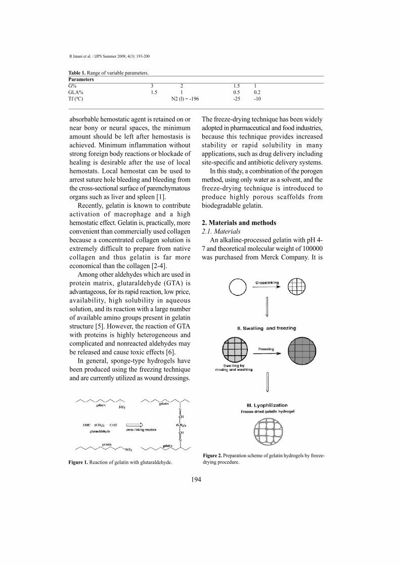
Among other aldehydes which are used inprotein matrix, glutaraldehyde (GTA) isadvantageous, for its rapid reaction, low price,availability, high solubility in aqueoussolution, and its reaction with a large numberof available amino groups present in gelatinstructure [5]. However, the reaction of GTAwith proteins is highly heterogeneous andcomplicated and nonreacted aldehydes maybe released and cause toxic effects [6].
In general, sponge-type hydrogels havebeen produced using the freezing techniqueand are currently utilized as wound dressings.
The freeze-drying technique has been widelyadopted in pharmaceutical and food industries,because this technique provides increasedstability or rapid solubility in manyapplications, such as drug delivery includingsite-specific and antibiotic delivery systems.
In this study, a combination of the porogenmethod, using only water as a solvent, and thefreeze-drying technique is introduced toproduce highly porous scaffolds frombiodegradable gelatin.
2. Materials and methods2.1. Materials
An alkaline-processed gelatin with pH 4-7 and theoretical molecular weight of 100000was purchased from Merck Company. It is
Figure 1. Reaction of gelatin with glutaraldehyde.Figure 2. Preparation scheme of gelatin hydrogels by freeze-drying procedure.
Table 1. Range of variable parameters.ParametersG% 3 2 1.5 1GLA% 1.5 1 0.5 0.2Tf (ºC) N2 (l) = -196 -25 -10

Biodegradable hemostat gelatin sponge
composed of glycine (27%), proline andhydroxyproline (25%), glutamic acid (10%),arginine (8%), alanine (9%), aspartic acid(6%) and other amino acids (15%).Glutaraldehyde was also Merck Co.
2.2. Preparation of gelatin sponge Porous, freeze-dried gelatin sponge was
prepared by three main procedures includingcrosslinking, swelling, and lyophilization.Preparation method is schematically illustratedin Figure 2.
Briefly, gelatin solution is prepared bysolving gelatin in double-distilled water at50 ºC. GTA aqueous solution was added to theaqueous solution of gelatin to give the finalconcentration. The solution mixture waspoured into a suitable mold. Molded gelatinshould be left for 2 h at room temperature forcrosslinking and then stand for 12 h at 4 ºCfor gelation.
Gelatin hydrogel was further washed thricewith double-distilled water for removing non-reacted residues of GTA. The fully hydratedhydrogel was frozen at different temperaturesfor 5 h. Then, the frozen hydrogel placed in
a Petri dish, lyophilized with a freeze-dryer(ALPHA 1-2 LD) for 24 h in order tocompletely dry. The frozen hydrogels shouldbe transferred from their freezing apparatusto the freeze-dryer with care to avoid meltingof the frozen samples.
We changed three parameters in preparingsamples: Concentration of gelatin,concentration of GTA in final product andfreezing temperature (Tf) as considered inTable 1.
Code of each sample involved these threeparameters, respectively (G%, GLA%, Tf).
For comparing and evaluation of syntheticsamples, we utilized a trade mark absorbablegelatin sponge (Curaspon™).
2.3. Characterizations of gelatin sponges2.3.1. Measurement of density of freeze-driedsponges
The density of sponge was calculated fromthe ratio of weight of the freeze-dried hydrogelto its volume. The density measurement wasperformed using samples with a volume of1×1×1 cm³ and the final densities wereobtained from the mean of three specimens.
195
Figure 3. SEM micrograph of gelatin sponge (×100) a: 1,0.5,N2 (cross); b: 1,0.5,N2 (surface); c:1,0.5,-10 (cross); d: 1,0.5,-25 (cross).

R Imani et al. / IJPS Summer 2008; 4(3): 193-200
196
2.3.2. Morphological observation of gelatinsponge
The morphology of sponges wereexamined using a scanning electronmicroscope (SEM) after coating the sampleswith a thin layer of gold under vacuum at 15kV. In addition, sponge's structure was viewedby optical microscope for better observationof pore structures and pore interconnections.
2.3.3. in vitro degradation of gelatin spongesSponge specimens (1cm³) were immersed
in phosphate buffer solution (10 ml, pH 7.4)for 24 h. the study was carried out at twoconditions: Room temperature (25±3 ºC) andincubator chamber (37 ºC) and results werecompared. After one day, the sponge wasfreeze dried. Then the change in weight ofsponges was monitored [7].Percent of degradation was measured by:%D = (Wf-Wi) / WiWf is final weight of sponge after freezedrying and Wi is initial weight of sponge.
2.3.4. Cytotoxicity analysis An established cell line of L-929 mouse
fibroblasts was used for evaluation ofcytotoxic potentials. Sponges in cut shapewere sterilized by immersing in 70% ethanoland then freeze dried for 24 h. Image of cellsafter 1 week represented cell viability incontact with gelatin crosslinked sponge.
2.3.5. Mechanical testsSamples for this test prepared in cylindrical
shape (2 cm height, 1 cm diameter). Stress-Strain data were recorded for compressionmode by rate of 1 mm/min by using HCT-25/400 Zwick/Roell. Then the curve wasplotted by MATLAB software. The Young'scompressing modules (Ec) was calculated bymeasuring the slope of the first linear part ofStress-Strain curve.
2.3.6. Hemostasis ability and hemolytic study Hemostasis ability by fragility of RBC
membranes in contact with absorbable gelatinsponge was investigated according to theASTM-F75600 [8]. Samples were cut intoappropriate pieces (3 cm²) and then transferredto polyethylene tubes. One ml of whole bloodwas added to tube and diluted by 7 mlphosphate buffer. The solutions were thenincubated at 37 ºC for 15 min. Followingthis, they were centrifuged at 800 rpm for 5min. The supernatant was then taken forhemolysis studies. Absorbance at 540 nmwas determined using Milton Roy Spectronic601 spectrophotometer.
Figure 4. Degradation of samples in room temperature(shorter column) and physiological condition.
Table 2. Average density of gelatin sponge.Sample code G% GLA% Tf(ºC) Average density (g / cm3)3,0.5,N2 3.0 0.5 N2(I) = -196 0.0602,0.5,N2 2.0 0.5 N2(I) = -196 0.0371.5,0.5,N2 1.5 0.5 N2(I) = -196 0.0311,0.5,N2 1.0 0.5 N2(I) = -196 0.0211.5,0.5,-25 1.5 0.5 -25 0.0291.5,0.5,-10 1.5 0.5 -10 0.026Trade - - - 0.030

Biodegradable hemostat gelatin sponge
3. Results and discussion3.1. Density measurement
The density and appearance of driedsponges are listed in Table 2. As the freezingtemperature was lowered, the density offreeze-dried sponges increased. Density offreeze-dried sponges obtained by freezing inN2 (liquid) was almost thrice as high as thatby freezing at -25 or -10 ºC. The resultsindicated that the density of samples isstrongly dependent on the freezingtemperature before freeze-drying [9]. Theaddition of GTA as cross linker agent changeddensity very slightly, but no direct relationshipbetween the amount of cross linker and thedensity was observed.
3.2. Morphology of sponges and porestructure
Gelatin sponges prepared by freeze-dryingafter freezing at -10 and -25 ºC had largepores which were visible noticeably, as shownin Figure 3(a, d). SEM observation of thesesponges demonstrated a network structure ofheterogeneous pores. However, homogeneoussmall polygonal pores were observed byfreeze-dried samples after freezing in N2 [l].
3.3. Degradation studyThe extent of crosslinking determines the
stability of biodegradable material and it canbe evaluated by measuring the time requiredfor complete solubility. The amount ofdissolved gelatin can be used in the
197
Figure 5. Microscopic examination of cell after 1 week exposure to gelatin sponge: a:1,1.5, N2 (×200); b: 1,1.5, N2 (×400);c: control (-) (×200); d: trade sample(×200).
Table 3. The physical properties of freeze-dried gelatin hydrogels following freezing at-10 and-25 ºC and in N2 [l].Freezing temperature -10(ºC) -25(ºC) N2(I) TradeDensity (g/cm3) 0.026 0.029 0.031 0.030Pore size (m) 410 370 60 240Mechanical properties Weak, brittle Brittle Elastic ElasticWall thickness Thin Thin Thick Thick

R Imani et al. / IJPS Summer 2008; 4(3): 193-200
198
quantification of matrix degradation [5].Degradation profiles of gelatin sponges aregiven in Figure 4.
The rate of weight loss decreased withincreasing degree of crosslinking of thesamples. This indicates that the higher degreeof crosslinking in gelatin sponges causesmore resistance to degradation in PBS solution[9].
The gelatin degradation is so sensitive totemperature. By increasing temperaturedegradation rate raises. Effect of pore structureis not significant and by increase of freezingtemperature degradation rises slightly.
3.4. Cytotoxicity analysisIn this study, we changed GTA
concentration in the range of 0.2-1.5 wt %.The microscopic examination does notillustrate any significant cytotoxicity in testedsamples compared to control (polystyrene
Petri dish) and trade sample. The results areshown in Figure 5.
3.5. Mechanical analysis The compression modules of gelatin
sponges (Ec) are given in Figure 6. Gelatinconcentration increase had very sharp effecton raising the compression module. Spongeswith 3% gelatin were hard and stiff. Bydecreasing gelatin concentration, spongesbecame soft and flexible.
On the other hand, the compression modulevalue did not demonstrate a significant changeupon varying the amount of GTA [5]. Freezingtemperature changed inner structure ofsponges and can affect mechanical behavior.
3.6. Hemolytic susceptibilityFigure 7 shows the results of hemolytic
experiments. The y-axis represents absorbancevalues at 540 nm [7]. Higher absorption valuerepresents more hemoglobin release and somore hemolytic ability of samples. As it isshown, the absorption ratios of all samples arehigher than control (+). Sample with 2%gelatin shows strong hemolysis that confirmsgelatin exhibit hemostatic effect by nature. Achange in the amount of cross linker has notsignificant effect on hemolytic ability. Openpores with inner connection allow blood entersponge rapidly at initial 30 min. Comparisonbetween samples (1, 0.5,-25) and (1, 0.5, N2)confirms this point.
Figure 6. Mechanical analysis of gelatin sponge.
Figure 7. Hemolytic behavior (15 min.) of samples.

Biodegradable hemostat gelatin sponge
4. ConclusionThe strength and stability of gelatin
sponges, used in surgery must satisfy theirintended biomedical needs. Crosslinking ofgelatin sponges with GTA provides a meansof easily tailoring a sponge biostability andmechanical properties. These techniques maybe used to increase ease of handling in general.Based on these results, intermediateconcentrations of GTA, specifically 0.5%increase gelatin sponge strength and stabilitywhile it has no cytotoxicity effect. Theseadvantages may contribute to improvedperformance in surgical application of thesponges.
References[1] Tomizawa Y. Clinical benefits and risk analysis
of topical hemostats: A review. J Artif Organs2006; 8: 137-42.
[2] Choi YS, Lee SB. Studies on gelatin-basedsponges. Part III. J Mater Sci: Mater in Med2001; 12: 67-73.
[3] Bigi A, Cojazzi G. Mechanical and thermalproperties of gelatin films at different degrees ofglutaraldehyde crosslinking. Biomaterials 2001;22: 763-68.
[4] Hong SR, Lee SJ. Study on gelatin-containingartificial skin IV. Biomaterials 2001; 26, 961-68.
[5] Ulbayram K, Aksu E. Cytotoxicity evaluation ofgelatin sponges prepared with different cross-linking agents. J Biomater Sci: Polym Ed 2002;13: 1203-19.
[6] Matsuda S, Iwata H, Y.Ikada NS. Bioadhesion ofgelatin films crosslinked with GA. J BiomedMater Res 1999; 45: 20-7.
[7] Vijayanand K, Deepak K. Interpreting Blood-biomaterial interactions from surface free energyand work of adhesion. Trends Biomater: ArtifOrgans 2005; 18: 73-84.
[8] ASTM.F756-00: standard practice for assessmentof hemolytic properties of materials.
[9] Choi YS, Hong SR. Study on gelatin-containingartificial skin: I. Biomaterials 1999; 20: 409-17.
199

R Imani et al. / IJPS Summer 2008; 4(3): 193-200
200
ONLINE SUBMISSION
www.ijps.ir