Synthesis, Characterization and Antimicrobial screening of some ...

1
SYNTHESIS AND ANTIMICROBIAL STUDIES OF
IRON (III) COMPLEXES OF CIPROFLOXACIN,
PENICILLINS AND ISATIN DERRIVATIVE
BY
EZE FABIAN IFEANYI
PG/ M.SC/07/43054
DEPARTMENT OF PHARMACEUTICAL AND
MEDICINAL CHEMISTRY
FACULTY OF PHARMACEUTICAL SCIENCES
UNIVERSITY OF NIGERIA, NSUKKA
MARCH, 2011
2
Synthesis and Antimicrobial Studies of Iron (III) Complexes
of Ciprofloxacin, Penicillins and Isatin Derivative
By
Eze Fabian Ifeanyi
PG/ M.Sc/07/43054
Department of Pharmaceutical and Medicinal Chemistry
Faculty of Pharmaceutical Sciences
University of Nigeria, Nsukka
March, 2011
3
Synthesis and Antimicrobial Studies of Iron (III) Complexes of
Ciprofloxacin, Penicillins and Isatin Derivative
By
Eze Fabian Ifeanyi
PG/ M.Sc/07/43054
A research project report submitted to the Department of Pharmaceutical and
Medicinal Chemistry, Faculty of Pharmaceutical Sciences, University of Nigeria,
Nsukka in partial fulfillment of the requirements for the award of Master of Science
degree (M.Sc) in Medicinal Chemistry.
Project Supervisor: Dr. U. Ajali
March, 2011
DEDICATION
To the sick and suffering
4
ACKNOWLEDGEMENT
I am grateful to the almighty God for seeing me through this tedious
research work. A lot of people had been God’s instrument in this work. This work
was supervised by Dr. U. Ajali. His friendly advices created the enabling
environment for the work. I thank him immensely. Dr. P.O. Ukoha of the Dept. of
Pure and Industrial Chemistry provided a lot of technical and material support. I

5
appreciate him a lot. I am indebted to Juhel (Pharm) Nig. Ltd. Enugu for donating
pure samples of Ciprofloxacin and Penicillins. Dr. C.J. Mbah, my H.O.D,
recommended me to Juhel for assistance. I thank him.
This work was sponsored by my mother, Mrs. V. Eze, and my sister,
Uchenna, may God bless them abundantly. Mr. G.C. Ebi, Dr. F.B.C. Okoye and
Pharm. E.O. Omeje made a lot of useful suggestions. My sister, Nkechi was a
source of motivation. She inspired me into the P.G. programme. I thank my
brothers, Emeka and Ikechukwu, my sisters; Chinonyelum and Chinedu for their
encouragement and prayers, and my colleagues; Sule, Amaka, Ifeyinwa, and
Rosy for keeping me company.
CERTIFICATION
The work embodied in this project report is an original investigation and
has not been submitted in part or full for any other diploma or degree of this or
any other university.
6
………………………………… ………………………….....
Dr. (Mrs.) N. J. Nwodo Dr. U. Ajali
Head of Department Supervisor
………………………………………………
External Examiner
ABSTRACT
Complexation is very relevant in pharmacy as a means of modifying the
pharmacological, toxicological and physico- chemical properties of drugs.In this
study, iron (III) complexes of ciprofloxacin, ampicillin, amoxicillin, and
cloxacillin were synthesized through systematic choice of solvents and reaction
conditions. A Schiff base derived from Isatin (Indole-1,2-dione) and
ethylenediamine, and its iron (III) complex were also synthesized. The aqueous
solubility profiles of the complexes were determined. The complexes showed
improved aqueous solubility when compared to those of the corresponding
ligands. Relative thermal and acid stabilities were determined
spectrophotometrically. Results showed that the complexes have enhanced
thermal and acid stabilities with respect to the pure ligands. Antimicrobial studies
7
carried out on the complexes showed that the complexes have decreased
antimicrobial activities against most of the microorganisms used. Ciprofloxacin
complex, however, showed almost the same activity as the corresponding ligand.
The iron (III) complex of the Schiff base derived from isatin and ethylenediamine
showed enhanced antibacterial activity against B.subtilis and S. aureus, but less
activity against P. aeruginosa, E. coli, and Salmonella typhi. Job’s method of
continuous variation and the approximate molecular weight of the complexes,
determined by measurement of boiling point elevation (ebullioscopy), suggested a
1:2 metal to ligand stoichiometry for most of the complexes). The structures of
the complexes were proposed based on the stoichiometry, infrared and
ebullioscopic data and assuming a coordination number of 6 or 4 for iron.
TABLE OF CONTENTS
Title page i
Dedication ii
Acknowledgement iii
Certification iv
Abstract v
CHAPTER ONE: GENERAL INTRODUCTION 1
1.1 Introduction 1
1.2 Complexes 3
1.2.1 Definition of Complex 3
1.2.2 Types of Complexes 5
1.2.3 Chemistry of complexes 8
1.2.3.1 Structure and bonding in metal ion (Coordination) Complexes 8
1.2.3.2 Methods of Studying the structure of Complexes 10
1.2.3.3 Theories and bonding in Transition metal complexes 11
1.2.3.4 IUPAC Nomenclature of Coordination Compounds 12
1.2.3.5 Isomerism in Transition metal Complexes 14
1.2.4 Reaction Conditions Favourable for Complex Formation 15
8
1.2.5 Complex Stability 17
1.2.5.1 Measurement of Complex Stability 17
1.2.6 Methods of Analysis and Determination of Complexes 20
1.3 Antibiotics 20
1.3.1 Definition of Antibiotics 21
1.3.2 Classes of Antibiotics 21
1.3.2.1β-lactam Antibiotics 21
1.3.2.2 Non- lactam Antibiotics 25
1.4 Medicinal Chemistry of the Essential Trace Elements 29
1.4.1 Iron 29
1.5 Aim of Study 30
CHAPTER TWO: MATERIALS AND METHODS 31
2.1 Equipment 31
2.2 Reagents and Solvents 31
2.3 Microorganisms 31
2.4 Synthesis of the Complexes 31
2.5 Ebullioscopic Determination of Molecular Weight of the Complexes 33
2.6 Job’s Method of Continuous Variation 33
2.7 Determination of some Physicochemical Parameters 34
2.7.1 Aqueous Solubility 34
2.7.2 Thermal and Acid Stabilities 34
2.8 Determination of melting Point
2.9 Spectral. Analysis
2.9.1 UV- Visible Spectral Analysis
2.9.2 Infrared Spectral Analysis
2.10 Antimicrobial Studies on the Complexes
2.10.1 Preparation of the Culture Media 2.10.2
Maintenance and Activation of the Microorganisms 2.10.3
Microbiological Sensitivity Screening Test
2.10.4 Determination of Minimum Inhibitory Concentration (MIC)
CHAPTER THREE: RESULTS 37
3.1
9
CHAPTER FOUR: DISCUSSION AND CONCLUSION
4.1 Possible Synthetic Reaction 54
4.2 Antimicrobial Results 57
4.3 Thermal and Acid Stability 57
4.4 Infrared Spectrophotometrical Data 58
4.5 Conclusion 60
References 61
LIST OF TABLES AND FIGURES
Table 1.1 Pharmaceutical Properties affected by complexation 1
Table 1.2 Drug-Metal Complexes and their Properties 2
Table 1.3 Classification of Molecular Complexes 6
Table 1.4 PH Ranges for Metal –EDTA Titration 16
Table 1.5 Some semi-synthetic Penicillins 22
Table 3.1 Percentage Yield of the Complexes 37
Table 3.2 approximate Molecular weight of the Complexes 37
Table 3.3 Aqueous Solubility 38
Table 3.4 Relative Thermal Stability of the Complexes 38
Table 3.5 Relative Acid Stability of the Complexes 38
Table 3.6 Result of sensitivity Screening 39
Table 3.7 Minimum Inhibitory Concentrations 39
Table 3.8 Result of Job’s Method of Continuous Variation for Ciprofloxacin Complex 40
Table 3.9 Result of Job’s Method of Continuous Variation for Cloxacillin Complex 40
Fig. 3.1 Job’s Plot for Ciprofloxacin Complex 41
Fig. 3.2 Job’s Plot for Cloxacillin Complex 42
Fig 3.3 UV- Visible Spectrum of Ciprofloxacin 43
Fig 3.4 UV-Visible Spectrum of Fe3+ complex of Ciprofloxacin 44
Fig 3.5 UV-Visible Spectrum of Amoxicillin Complex 45
Fig 3.6 UV-Visible Spectrum of Fe3+ Complex of Cloxacillin 46
Fig 3.7 UV-Visible Spectrum of Fe+ Complex of Isatin Schiff base 47 Fig
3.8 UV-Visible Spectrum of Ferric Chloride 48
Fig 3.9 Infrared Spectrum of Ciprofloxacin 49
Fig 3.10 Infrared Spectrum of Fe3+ Complex of Ciprofloxacin 50
Fig 3.11 Infrared Spectrum of Fe3+ Complex of Cloxacillin 51
10
Fig 3.12 Infrared Spectrum of Fe3+ Complex of Amoxicillin 52
Fig 3.13 infrared Spectrum of Fe3+ Complex of Isatin Schiff base 53
CHAPTER ONE
GENERAL INTRODUCTION
1.1 INTRODUCTION
Many drugs possess modified pharmacological, toxicological and physicochemical
properties when administered in the form of metal complexes. (Guangguo et al, 2003). It is
obvious that these complexes must possess some properties that are different from those of
their constituents; otherwise there would be no evidence for their existence. Among the
properties that may be altered upon complex formation are solubility, energy absorption,
conductance, partitioning behaviour and chemical reactivity. (Guangguo et al, 2003).
Some of the properties of drugs are so pertinent to dosage forms and drug delivery that
they are identified as pharmaceutical or biopharmaceutical properties. Complex formation may

11
affect these properties adversely or sometimes favourably. Many of these pharmaceutical
properties, affected by complexation, with corresponding examples of drug complexes are
given in table 1.1.
Table 1.1 Pharmaceutical Properties Affected by Complexation
Property Example (Connors, 2000)
1. Physical state Nitroglycerin-cyclodextrin
2. Volatility Iodine-PVP
3. Solid-state stability Vitamin A-cyclodextrin
4. Chemical stability Benzocaine-caffeine
5. Solubility Aspirin-caffeine
6. Dissolution rate Phenobarbital-cyclodextrin
7. Partition coefficient Benzoic acid-caffeine
8. Permeability Prednisone-dialkylamides
9. Absorption rate Salicylamide-caffeine
10. Bioavailability Digoxin-cyclodextrin
11. Biological activity Indomethacin-cyclodextrin
Table 1.2 Drug – metal Complexes and their Properties
Properties Drug-metal Complexes
Improved antibacterial activity, water Co(II), Ni(II), Cu(II) and Zn(II)
solubility and solid state stability complexes of 3-salicylidenehydrazono-2-
indolinone
Solubility, stability and light absorption Cu2+ complex of ciprofloxacin
(Guangguo, 2003)
Antifungal activity Co2 +-bisisatinthiocarbohydrazone (Satisha, 1999)

12
For many systems, it has been shown that the complex provides faster dissolution and greater
bioavailability than does the physical mixture. The processing characteristics (physical state,
stability, flowability, etc.) of the complex also may be better than those of the free drug (Connors,
2000).
Not all complexation is intentional or desirable, and some dosage form incompatibilities may
be the result of unwanted complexation reactions. For examples, some widely used polyethers
(Tweens, Carbowaxes or PEGS) can form precipitates with H- bond donors such as phenols and
carboxylic acids. A substance used widely in liquid dosage forms as a complexer of metal ions is
EDTA. The purpose of this application of complexation is to improve drug stability by inhibiting
reactions (usually oxidations) that are catalyzed by metal ions, the complexed form of the metal ion
being catalytically inactive. Citric acid (in the form of citrate anion) also is used for this purpose.
(Connors, 2000).
Drug complexation experiments could help medicinal chemists to predict some dosage
form incompatibilities, explain the mode of action of some drugs as well as devise new methods of
drug analysis. Formation of coordination complexes provides the basis for many analytical
methods for determination of metals in food, drug and biological systems (e.g. complexiometric
titration). Very low concentration of metal ions can also be determined spectrometrically by
complexation with a ligand that produces spectral change, (e.g. colorimetric analysis).The ferric
hydroxamate method for the detection and determination of carboxylic acid derivatives is another
important application of complexation in pharmaceutical analysis. Here, a carboxylic acid
derivative is reacted with hydroxyl amine to form the corresponding hydroxamic acid. An excess
of Fe (III) is added, and this forms a red-violent coordination complex with the hydroxamic acid
and the concentration of the complex determined spectrometrically (Sandell E.B, 1989).
Complexation is a useful means of studying the protein-binding of drugs. Some of the
constituents of the blood (e.g. HSA are capable of forming complexes with drugs and this has
important practical implication. Drug distribution and clearance are affected by its binding
characteristics. Complexes occur widely in biological systems, so the application of complex
formation processes in therapy is a reasonable approach to drug design. Among the most important
biological manifestations of complexation are many metal-ion coordination complexes whose
study constitutes a large part of bioinorganic chemistry. Examples of these complexes include:
haemoglobin (iron), cytochrome (iron), vitamin B12 (cobalt), carboxypeptidaseA (zinc) etc.
Molecular complexation in biological systems also occurs, as in DNA base pairing and stacking
interactions. Numerous antimicrobial and anti-neoplastic agents are believed to exert their action
13
by means of complex formation with DNA base pair (Intercalation), e.g. daunorubicin and
actinomycin D.
Many toxic effects of excessive metal-ion concentration can be treated by agents that
form strong coordination complexes, via chelation, thus aiding the excretion of the metal. Among
the metals whose toxicity can be treated by chelation therapy are lead, copper, cobalt, mercury,
nickel, iron and zinc. Coordination compounds could also release valuable trace elements needed
for maintenance of life when they are administered as drugs.
Complexation should, therefore, be given adequate attention in pharmaceutical research.
1.2 COMPLEXES
1.2.1 DEFINITION OF A COMPLEX
A complex is a chemical specie formed by the association of two or more interacting
molecules or ions (Connors, 2000). The term complex is also used to describe a substance
composed of two or more substances, each of which is capable of an independent existence,
bound by forces weaker than the regular chemical bonds. (Cotton et al, 1999)
A complex is also a species of definite substrate to ligand stoichiometry that can be formed in
equilibrium process in solution, and also may exist in the solid state. (Connors, 2000)
It should be noted that complexes are not formed with classic covalent bonds.
Some Basic Terms Associated with Complexes
Substrate: This is the reactant, usually an electron pair acceptor (Lewis acid) whose physical or
chemical properties are observed experimentally.
Ligand: The second interactant, usually an electron pair donor (Lewis base) which donates the pair of
electron used for bonding.
Chelate: (from chelos; Greek word for Crab) is a ring structure formed when the ligand has more than
one binding site and occupies more than one coordination position.
Chelated complexes are more stable than similar complexes with unidentate ligands as
dissociation of the complex involves breaking many bonds rather than one. The more rings that
are formed, the more stable the complex is. (Lee, 1996). Chelating agents with three, four and
six donor atoms are known and are termed tridentate, tetradentate and hexadentate ligands
respectively.
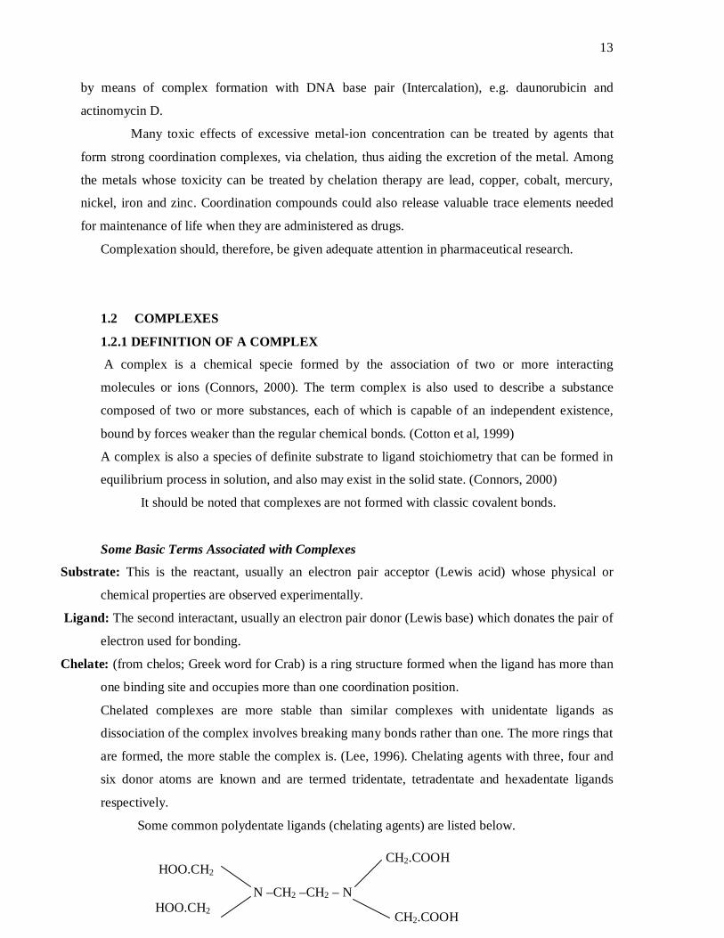
Some common polydentate ligands (chelating agents) are listed below.
HOO.CH2
HOO.CH2 N –CH2 –CH2 – N
CH2.COOH
CH2.COOH
14
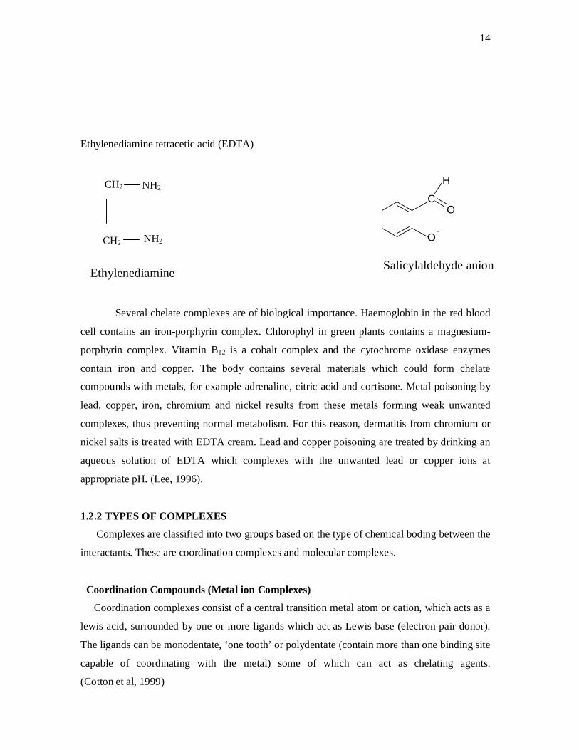
Ethylenediamine tetracetic acid (EDTA)
O-
O
H
C
Several chelate complexes are of biological importance. Haemoglobin in the red blood
cell contains an iron-porphyrin complex. Chlorophyl in green plants contains a magnesium-
porphyrin complex. Vitamin B12 is a cobalt complex and the cytochrome oxidase enzymes
contain iron and copper. The body contains several materials which could form chelate
compounds with metals, for example adrenaline, citric acid and cortisone. Metal poisoning by
lead, copper, iron, chromium and nickel results from these metals forming weak unwanted
complexes, thus preventing normal metabolism. For this reason, dermatitis from chromium or
nickel salts is treated with EDTA cream. Lead and copper poisoning are treated by drinking an
aqueous solution of EDTA which complexes with the unwanted lead or copper ions at
appropriate pH. (Lee, 1996).
1.2.2 TYPES OF COMPLEXES
Complexes are classified into two groups based on the type of chemical boding between the
interactants. These are coordination complexes and molecular complexes.
Coordination Compounds (Metal ion Complexes)
Coordination complexes consist of a central transition metal atom or cation, which acts as a
lewis acid, surrounded by one or more ligands which act as Lewis base (electron pair donor).
The ligands can be monodentate, ‘one tooth’ or polydentate (contain more than one binding site
capable of coordinating with the metal) some of which can act as chelating agents.
(Cotton et al, 1999)
Salicylaldehyde anion
CH2 NH2
NH2 CH2
Ethylenediamine

15
The ligands could be uncharged molecules e.g. H2O, NH3, CO, EDTA, ethylenediamine,
dipyridyl, O-phenanthroline, Ciprofloxacin, Penicillins, Isatin e.t.c or negatively charged
radical e.g. I–, Cl–, OH–, S2-, NO2–, NO2
-, CN– e.t.c. The number of coordinate bonds between
the metal ion and the ligand(s) is called coordination number. The ligands are bound to the
metal by coordinate covalent bond between the ligand electron pairs and the empty d-orbital of
the metal. These d-orbitals, according to ligand field theory, split into high and low energy
groups under the influence of the asymmetric (typically octahedral) electric field created by the
array of ligands. This accounts for the highly coloured nature of the complexes when (as
frequently happens) the d-orbital splitting energy difference corresponds to visible-light
wavelengths (Cotton et al, 1999).
Coordination compounds could arbitrarily be classified according to the nature of the
ligands into:
Classical (or Werner Complexes): Ligands bind to metal, almost exclusively, via their lone
pairs of electrons residing on the main group atoms of the ligand. Typical of such ligands are
H2O, NH3, Cl–, CN–, en–. Examples of such complexes are:
[Co (NH3)6]Cl3, (Fe (C2O4)3]K3, [Co (EDTA)]– e.t.c.
Organometallic Compounds: Ligands are organic (e.g. alkenes, alkynes, alkyls) as well as
organic-like ligand such as phosphines, hydride and CO. Example: (C5H5)Fe (CO)2 CH3,
Ferrocene e.t.c.
Bioinorganic compounds: Ligands are those provided by nature such as porphyrins. Example:
haemoglobin, cyanocobolamin (Cotton et al, 1999)
Molecular Complexes
Molecular complexes consist of an association of molecules held by weak, non-covalent
intermolecular forces. The non-covalent forces are of three broad types:
(a) The electrostatic forces among ions and molecules possessing permanent dipole moments.
(b) The induction (or polarization) forces between an ion and a non-polar molecule or a polar
molecule and a non-polar molecule.
(c) The dispersion (London) forces, which operate between all molecules.
There is no systematic classification of molecular complexes, nor has a system of
nomenclature been developed to describe them. Particular types, however, may be classified
in terms of the kinds of interactions involved in their formation, the kinds of interactants
involved or the kinds of complex formed, (Connors et al, 2000).
16
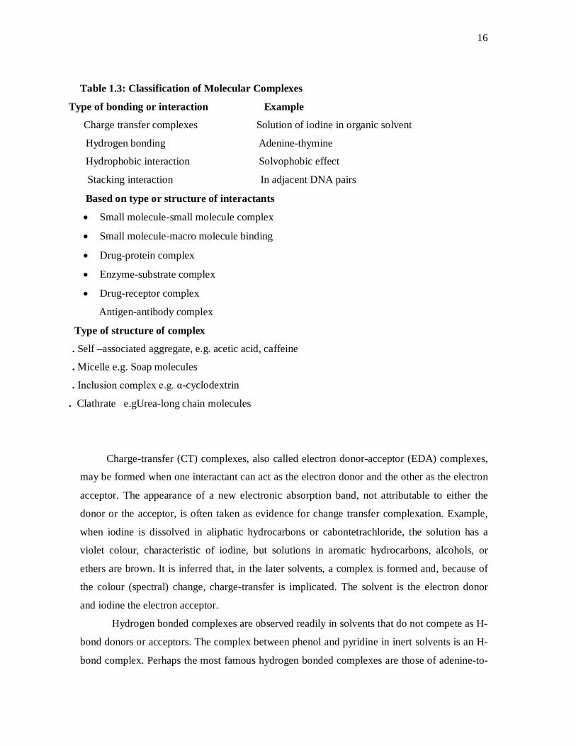
Table 1.3: Classification of Molecular Complexes
Type of bonding or interaction Example
Charge transfer complexes Solution of iodine in organic solvent
Hydrogen bonding Adenine-thymine
Hydrophobic interaction Solvophobic effect
Stacking interaction In adjacent DNA pairs
Based on type or structure of interactants
Small molecule-small molecule complex
Small molecule-macro molecule binding
Drug-protein complex
Enzyme-substrate complex
Drug-receptor complex
Antigen-antibody complex
Type of structure of complex
. Self –associated aggregate, e.g. acetic acid, caffeine
. Micelle e.g. Soap molecules
. Inclusion complex e.g. α-cyclodextrin
. Clathrate e.gUrea-long chain molecules
Charge-transfer (CT) complexes, also called electron donor-acceptor (EDA) complexes,
may be formed when one interactant can act as the electron donor and the other as the electron
acceptor. The appearance of a new electronic absorption band, not attributable to either the
donor or the acceptor, is often taken as evidence for change transfer complexation. Example,
when iodine is dissolved in aliphatic hydrocarbons or cabontetrachloride, the solution has a
violet colour, characteristic of iodine, but solutions in aromatic hydrocarbons, alcohols, or
ethers are brown. It is inferred that, in the later solvents, a complex is formed and, because of
the colour (spectral) change, charge-transfer is implicated. The solvent is the electron donor
and iodine the electron acceptor.
Hydrogen bonded complexes are observed readily in solvents that do not compete as H-
bond donors or acceptors. The complex between phenol and pyridine in inert solvents is an H-
bond complex. Perhaps the most famous hydrogen bonded complexes are those of adenine-to-

17
thymine of guanine–to-cytosine, which, as constituents of deoxyribonucleic acid, are
responsible for the double-helix structure of the DNA molecule.
When two planar molecules undergo a primarily hydrophobic association, the total
surface area of the complex exposed to the solvent can be minimized if molecules are in plane-
to-plane contact. This plane-to-plane orientation is called stacking interaction. The purine-
pyrimidine H-bonded base pairs in DNA are planar assemblies that undergo stacking
interactions with adjacent pairs.
Most complexes probably involve a combination of interactions.
Self association is a type of complexation in which a molecule forms complexes with others of
its own species. If S represents a molecule capable of self association, then S2 is its dimmer, S3
its trimer e.t.c. Benzene and caffeine form dimers in aqueous solution.
A micelle is a special form of self-aggregated complex in which the interactant is a surfactant, a
molecule possessing both a polar and non-polar portion.
Inclusion complexes are formed when a macrocyclic compound, possessing an intra-
molecular cavity of molecular dimensions, interacts with a small molecule that can enter the
cavity. The macrocyclic molecule is called the host and the small included molecule the guest.
Crown ethers present a non-polar external molecular surface, but the interior of the cavity is
relatively polar. As a consequence, polar guests such as ions can enter the cavity and, because
their polarity is masked by the surrounding host, exhibit unusual chemistry. For example,
KMnO4, which is not soluble in non-polar solvents, can be extracted into organic solvents from
water in the presence of crown ethers.
The cyclodextrins are macrocyclic hosts that are formed by the action of certain bacterial
enzymes on starch. They consist of α-D-glucose units joined with glycosidic (ether) linkages.
The interior of the cavity is lined with these glycosidic bonds and, therefore, is relatively non-
polar (i.e., relative to water), whereas the exterior of the molecule is quite polar because of the
large number of hydroxyl groups. The diameters of the cavities of the cyclodextrins are
approximately 5 Ǻ (for α –cyclodextrin), 6 to 7 Ǻ (for β) and 8 to 9 Ǻ (for ). Thus, small
guest molecules, or parts of molecules, may enter the host cavity to form inclusion complexes,
whose stabilities are in part the result of the hydrophobic effect. Many properties of the guest
molecule may be altered by inclusion in a cyclodextrin; these include volatility, solubility and
chemical stability. Numerous practical applications have been suggested (Mulliken, 1969).
The literature on molecular complexes frequently now uses the term molecular
recognition, which can be taken to mean a non-covalent interaction in which complementary
18
features of the two interactants result in significant specificity in the complex formation process
(Mulliken, 1969).
1.2.3 CHEMISTRY OF COMPLEXES
1.2.3.1 STRUCTURE AND BONDING IN METAL-ION COORDINATION
COMPLEXES
Alfred Werner’s coordination theory in 1893 was the first attempt to explain the
bonding in coordination complexes (Lee, 1996). He concluded that in complexes the metal
shows two different sorts of valency:
(a) Primary Valency: This is the number of charges on the complex ion. It is non-directional.
In compound, this charge is matched by the same number of charges from negative ions.
Example, the complex [Co (NH3)6]Cl3 actually exists as [Co(NH3)6]3+ and 3Cl–. There are three
ionic bonds, thus the primary valency is 3.
(b) Secondary Valency: The number of secondary valencies equals the number of ligand
atoms coordinated to the metal. This is called coordination number. It is directional. Each metal
has a characteristic number of secondary valencies. Thus in [Co (NH3)6]Cl3 the three Cl– are
hold by primary valencies and the six NH3 by secondary valencies.
Since secondary valencies are directional, a complex ion has a particular shape; e.g. the
complex ion [Co (NH3)6]3+ is octahedral. The most common coordination number in transition
metal complexes is 6 and the shape is usually octahedral. The coordination number of four is
also common and this gives rise to either tetrahedral or square planar complexes.
In the complexes [Co(NH3)6]Cl3, [Co(NH3)5Cl]Cl2, and [Co(NH3)4Cl2]Cl, Werner
established that the coordination number is 6 each and the primary valencies are three, two and
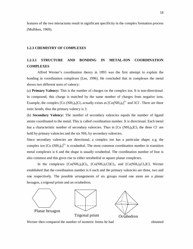
one respectively. The possible arrangements of six groups round one atom are a planar
hexagon, a trigonal prism and an octahedron.
Werner then compared the number of isomeric forms he had obtained
Planar hexagon Trigonal prism Octahedron
19
with the theoretical number for each of the possible shapes. His results strongly suggested that
these complexes have an octahedral shape. More recently the X-ray structures have been
determined and these establish that the shape is octahedral. With a bidentate ligand such as
ethylenediamine, two optically active isomers have been found.
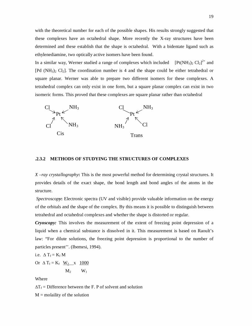
In a similar way, Werner studied a range of complexes which included [Pt(NH3)2 Cl2]2+ and
[Pd (NH3)2 Cl2]. The coordination number is 4 and the shape could be either tetrahedral or
square planar. Werner was able to prepare two different isomers for these complexes. A
tetrahedral complex can only exist in one form, but a square planar complex can exist in two
isomeric forms. This proved that these complexes are square planar rather than octahedral
.2.3.2 METHODS OF STUDYING THE STRUCTURES OF COMPLEXES
X –ray crystallography: This is the most powerful method for determining crystal structures. It
provides details of the exact shape, the bond length and bond angles of the atoms in the
structure.
Spectroscopy: Electronic spectra (UV and visible) provide valuable information on the energy
of the orbitals and the shape of the complex. By this means it is possible to distinguish between
tetrahedral and octahedral complexes and whether the shape is distorted or regular.
Cryoscopy: This involves the measurement of the extent of freezing point depression of a
liquid when a chemical substance is dissolved in it. This measurement is based on Raoult’s
law: “For dilute solutions, the freezing point depression is proportional to the number of
particles present’’. (Ibemesi, 1994).
i.e. ∆ Tf = Kf M
Or ∆ Tf = Kf W2 x 1000
M2 W1
Where
∆Tf = Difference between the F. P of solvent and solution
M = molaility of the solution
Cl Pt
NH3
Cl NH3 Cis
Cl Pt
NH3
NH3 Cl
Trans
20
W2 = weight of solute dissolved
W1 = weight of solvent
M2 = molecular weight of the solute
Kf = molal depression constant (unique for each solvent). (Ibemesi, 1994).
Cryoscopic measurements can be used to find if a molecule dissociates and how many ions
are formed. If a molecule dissociates into two ions it will give twice the expected depression
for a single particle, and so on. Information on the number of particles formed and that from the
total number of charges can be used together to establish the structure of a complex. Cryoscopy
is also used to determine molecular weight of a substance.
Electrical Conductivity: The electrical conductivity of a solution of an ionic material
depends on the concentration of the solute as well as the number of charges on the species
which are formed on dissolution. The total number of charges on the species formed when the
complex dissolves can be deduced by comparison of its molar conductivity with that of known
simple ionic materials. Conductivity is directly proportional to number of charges.
Measurement of Magnetic Moment: This provides information about the member of
unpaired electron spin present in a complex. From this, it is possible to decide how the
electrons are arranged and which orbital are occupied. Sometimes the structure of the complex
can be deduced from this. For example, the compound Ni (NH3)4 (NO3)2 2H2O might contain
four NH3 molecules coordinated to Ni2+ in a square planar [Ni (NH3)4]2+ ion and two molecules
of water of crystallization and have no unpaired electrons. Alternatively the water might be
coordinated to the metal, giving an octahedral [Ni(H2O)2(NH3)4]2+ complex with two unpaired
electrons. Both these complex ions exist and their structures can be deduced from magnetic
measurements.
Measurement of dipole Moments: This could be used to distinguish the cis and trans isomers
of a complex. For example, the complex ,[Pt (NH3)2 Cl2] is square planar and can exist as cis or
trans forms. The dipole moments from the various metal-ligand bonds cancel out in the trans
configuration. However, a finite dipole moment is obtained for the cis arrangement. This
method applies to non-ionic complexes only. (Lee, 1996).
1.2.3.3 THEORIES OF BONDING IN TRANSITION METAL COMPLEXES
Valence bond theory (developed by Pauling):
The theory holds that coordination compounds contain complex ions, in which ligands form
coordinate covalent bonds to the metal. Thus the ligands must have a lone pair of electrons, and
21
the metal must have an empty orbital of suitable energy available for bonding. The theory
considers which atomic orbitals on the metal are used for bonding from which the shape and
stability of the complex are predicted. The theory, however, fails to explain why these
complexes are coloured as well as why their magnetic properties vary with temperature.
Crystal field theory (Proposed by Bethe and Van Vleck).
This theory assumes that the attraction between the central metal and the ligands in a complex
is purely electrostatic. The following assumptions are made.
(i) Ligands are treated as point charges
(ii) There is no interaction between metal orbitals and ligand orbitals
(iii) The d-orbitals on the metal all have the same energy (degenerate) in the free
atom.However, when a complex is formed the ligands destroy the degeneracy.
The d-orbital is subdivided into the eg orbitals (dx2–y2 and dz2) and t2g orbitals (dxy, dxz and dyz).
In an octahedral complex, the metal is at the centre of the octahedron and the ligands at the six
corners. The lobes of the two eg orbitals point along the axes x, y and z while those of the three
t2g orbitals point in between the axes. It follows that the approach of six ligands along the axes
will increase the energy of the dx2 –y2 and dz2 orbitals much more than it increases the energy
of dxy, dxz and dyz orbitals. Thus, under the influence of an octahedral ligand filed, the d-orbitals
spilt into two groups of different energies. The energy difference is called d-orbital splitting
energy, ∆o. This energy difference most often corresponds to visible-light wavelengths and this
accounts for the coloured nature of the transition metal coordination complexes. An improved
and more complete model of the crystal filed theory, which incorporates molecular orbital
theory and make some allowance for covalency, is known as ligand field theory.
Molecular Orbital Theory
This theory fully allows for both covalent and ionic contributions to bonding. It has a
great advantage that it is easily extended to cover bonding. - Bonding helps to explain how
metals in low oxidation states [eg [Nio (CO)4] can form complexes even though there is no
change on the metal. The theory holds that the atomic orbitals of the ligands overlap with the
metal d-orbitals of right energy to form molecular orbitals.
1.2.3.4 IUPAC NOMENCLATURE OF COORDINATION COMPOUNDS
Rules:
1. The positive ion (cation, not central metal) is named first followed by the negative ion.
22
2. When naming a complex ion, the ligands are named before the central metal; they are named
alphabetically (numerical prefixes do not affect the order)
3. When writing the formular of complexes, the complex ion is enclosed in square bracket. The
metal is written first and the coordinated groups are listed in the order: Negative ligands,
neutral ligands, positive ligands.
4.Multiple occurring monodentate ligands receive a prefix according to the number of
occurrences: di-, tri-, tetra, penta-, e.t.c. Polydentate ligands, or ligands preceded by numerical
prefix eg ethylenediamine, dipyridyl e.t.c) receive bis-, tris-, tetrakis, pentakis -, e.t.c.
5. (a)Names of negative ligands end in –O. this replaces the final ‘e’ when the anion ends with-
‘ate’, eg. Sulphate –sulphato. It replaces ‘ide’: cyanide becomes cyano.
Some negative ligands:
CH3COO– acetato CN– cyano
-NO2 nitro OH– hydroxo
HS– mercapto –SCN thiocyanato
–ONO nitrito –NCS isothiocyanato
(b) Neutral ligands usually retain their names, with some of these exceptions:
NH3 ammine NO nitrosyl
H2O aqua or aquo CO carbonyl
(c) Positive ligands end in -ium e.g.
NH2NH2 - hydrazinuim
6. The oxidation state of the central metal is shown by Roman numeral in bracket immediately
following its name. If the complex is an anion, the central atom’s name will end in-
‘ate’ and its Latin name used if available (except Mercury). (Cotton et al, 1999).
7. If the complex contains two or more metal atoms, it is termed polynuclear. The bridging
ligands which link the two metal atoms together are linked by the prefix μ- . If there are two or
more bridging groups of the same kind, this is indicated by di - , tri- e.t.c. Bridging groups
are listed alphabetically with the other groups unless the symmetry of the molecule allows a
simpler name. If a bridging group bridges more than two metals, it is shown as 3 , 4 , 5 ,
or6
, to indicate how many atoms it is bonded to.
Examples:
[Co (NH3)5 Cl]SO4 Pentaamminechloro cobalt (III) sulphate.
[Cd (en)2 (CN)2 ] Dicyano bis (ethylenediamine) cadimium(II).
[CU NH3 Cl5]3– Aminepentachlorocuprate (II) ion

23
[Zn (NCS)4]2+ Tetrathiocyanato-N-zinc (II)
Na3 [Ag (S2O3)3]2 Sodium bis (thiosulphato) argentate (I)
Fe (C5H5)2 Bis (Cyclopentadienyl) iron (II)
[(NH3)5 Co.NH2. Co (NH3)5] (NO3)5 -amido bis [pentaamminecobalt (III)] nitrate.
[(Co)3 Fe (CO)3 Fe (CO)3] Tri -carbonyl-bis (tricarbonyl iron (O).
1.2.3.5 ISOMERISM IN TRANSITION METAL COMPLEXES
Isomerism is a phenomenon whereby two or more compounds have the same chemical
formular but different chemical structures. Because of the complicated formular of many
coordination compounds, the variety of bond types and the number of shapes possible, many
different types of isomerism occur.
Ionization Isomerism: This type of isomerism is due to the exchange of groups between the
complex ion and the ions outside it. Eg [Co (NH3)5 Br] SO4 and [Co(NH3)5SO4]Br. The former
is red-violet and gives a white precipitate with BaCl2 solution, while the latter is red in colour
and gives no precipitate with BaCl2, instead, it gives cream-coloured precipitate of Ag Br with
AgNO3, confirming the presence of free Br– ions.
Linkage Isomerism: Linkage isomerism occurs when the ligands contain more than one atom
which could donate an electron pair, e.g. –NO2 or –ONO, SCN- or –SCN e.t.c. The nitrito and
nitro complexes of cobalt constitute isomers.
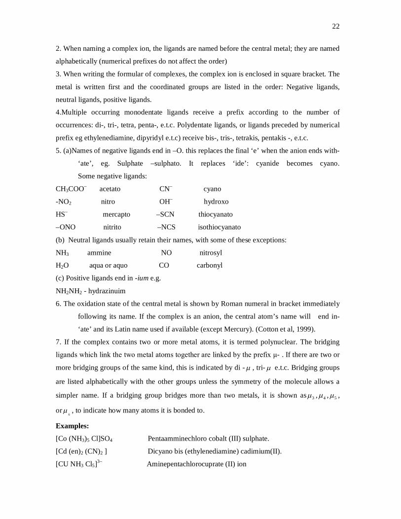
Geometric
Isomerism:
Square planar complexes such as [Pt (NH3)2Cl2] can be prepared in two forms: cis and
trans. If the complex is prepared by adding NH4OH to a solution of [PtCl4]2– ions, the complex
has a finite dipole moment and must therefore be cis. The complex prepared by treating
[Pt(NH3)4 ]2+ with HCl, however, has no dipole moment and must, therefore, be trans.
H2N Co
ONO
H2N NH3 NH3
NH3
H3N Co
NO2
H3N NH3 NH3
NH3
and
2+ 2+
Pentaammine nitrito cobalt (III) ion (red in colour, easily decomposed by acid
Pentaammine nitrito cobalt (III) yellow, stable to acid)
24
Optical Isomerism
Optical isomerism is common in octahedral complexes involving bidentate groups. For
e.g. [Co (en)2 Cl2]+ shows cis and trans forms (geometric isomerism). In addition the cis form
is optically active and exist in d and l forms, making a total of three isomers. Optical isomerism
also occurs in polynuclear complexes.
1.2.4 REACTION CONDITIONS FAVOURABLE FOR COMPLEX FORMATION
The reaction conditions, such as temperature and pH, favourable for complex formation
depend largely on the type of ligand, the charge on the metal ion and the type of metal (whether
the metal is in the first, second or third row of transition elements).
Ligands which cause only a small degree of crystal field splitting are termed weak field ligands
while those that cause large splitting are called strong field ligands. The spectrochemical series
of ligands is given below.
← Weak field ligands
I – <Br– < S2– < Cl– < NO3– < F– < OH– < EtOH < Oxalate <H2O <EDTA (NH3 and pyridine) <
ethylenediamine <dipyridyl < O– phenanthroline <NO2– < CN– < CO.
→ Strong field ligands.
The series is experimentally determined. The halides are in the order expected form
electrostatic effect. In other cases, a pattern of increasing donation is followed:
Halide donors < O donors < N donors < C donors. (Lee, 1996)
The magnitude of the crystal field stabilization energy of complexes increases as the charge on
the metal ion increases. It also increases by about 30% between adjacent members down a
group of transition elements. The thermodynamic stability, and hence ease of formation,
increases in that order. Complexes where the metal is in the (+ 3) oxidation states are generally
more stable than those where the metal is in the (+2) state:
trans
Pt
NH3
NH3
Cl
Cl Pt
NH3
Cl
Cl
NH3 cis
25
For a given ligand and metal the factors that influence complex formation include pH,
temperature, solvent, duration of reaction and continuous stirring. EDTA, for example, forms
stable complex with most metal ions in solution. The stability of the complex formed, however,
depends on the pH of the solution. Most divalent metals form stable EDTA complexes at pH >
7 while most trivalent metals form stable complexes at low pH, say 3. The application of this is
that by adjusting pH, one metal may be determined in the presence of the other in
complexiometric titration. The pH ranges for metal titration with EDTA is given below.
Table 1.4 pH ranges for Metal-EDTA titration
Fe3+ 1.5
Cu2+ 3.0
Zn2+ 4.0
Fe2+ 5.0
Ca2+ 8.0
Mg2+ 10.0
Temperature is another important factor that affects complex formation. If
complexation is exothermic, it would be favoured by low temperature and vice versa. Many
complexation reactions occur readily at room temperature, e.g. ciprofloxacin –Cu2+ (Guangguo,
et at, 2003), EDTA –Metals, Fe3+ – K2 C2 O4 . H2O (Szafran Z. et al, 1991) e.t.c. Schiff base
ligands require higher temperatures with reflux. e.g. in the synthesis of binuclear iron (III) –
iron (III) complexes with the tetradentate Schiff base, N, NI–bis (Salicylidene),
ethylenediamine and dicarboxylic acid bridging ligands, the reaction mixture is heated to
45–50oC under reflux with continuous stirring for 30 mins. (Zdenek et al, 1996). Similarly, in
the synthesis of metal complexes of 3 – salicylidenhydrazono-2-indolinone Schiff base, the
temperature is maintained between 60 – 65oC under reflux with continuous stirring for 2hrs.
(Sandra et al, 2003)
Solvent effects on complex-formation vary and can be complicated sometimes, but their
study may offer insight into the nature of the intermolecular interactions responsible for the
formation of the complex. A quantitative theory of solvent effects on complex formation has
been developed (Connors K. A. et al, 1992). The solvation contribution to the total free energy
of complex formation can be either stabilizing (if the complex is solvated more extensively
than the reactants) or destabilizing (if the reverse is applicable).
26
1.2.5 COMPLEX STABILITY
For the general complex formation equilibrium:
mS +nL Sm Ln
the overall binding constant, βmn is defined as βmn = [Sm Ln] where
[S]m[L]n
S = substrate, L = ligand, brackets denote molar concentrations. The binding constant is also
known as stability constant, formation constant or association constant. The reciprocal quality
is the dissociation constant. These constants depend on the identities of the substrate and
ligand. They also depend on the solvent and temperature (Connors et al, 2000). The binding
constant is an important measure of complex stability, and is related to the standard free energy
of complex formation by
∆GoII = – RT ln KII
where R = gas constant, T = absolute temperature, KII = binding constant for formation of
complex with I: I stoichiometry.
1.2.5.1 MEASUREMENT OF COMPLEX STABILITY
If a property of the substrate is altered upon its complexation with a ligand,
measurement of the property as a function of ligand concentration provides a means for
estimating the binding constant. The following methods have been devised.
Spectrometry: This method is based on the change in light absorption. Suppose the absorption
spectrum of the substrate is changed significantly upon binding, a wavelength at which a
substantial change in absorption occurs could be selected and, assuming that Beer’s law is
obeyed by the species, a particular chrornophore could be targeted and absorbance measured at
interval as a function of concentration.
Absorbance is additive. At a total substrate concentration absorbance is given as
Ao = s b St
In the presence of the ligand the absorbance is
AL = s b [S] + l b [L] + ll b [SL] where ll is the molar absorptivity of the complex (I: I
stoichiometry assumed).
This spectrometric method is applicable in the ultraviolet, visible and infrared regions.
Nuclear magnetic resonance spectrometry can as also be applied where a change in the
chemical shift is measured (Connors KA, 1987).

27
Chemical Reactivity: If the rate of a chemical reaction (such as hydrolysis) undergone by the
substrate is either increased or decreased by binding to the ligand, the stability constant can be
measured.
Consider the kinetic scheme:
Here R is a reagent that reacts with the substrate S, and complex SL, but does not form
complexes, P is the product and Ks, Kll are second order rate constants. The result of the
mathematical development is
Where qll = 1 – Kll/Kls and kls is the measured second order rate constant in a solution having
ligand concentration, [L].
Potentiometry: If the activity of an ion is changed upon complex formation, it may be
possible to make use of the measurement of electrical potential, E, according to Nernst
equation:
E= constant + (RT/nF) ln a
Where a is the ion activity, n is the no. of electrons in the redox process and F is the Faraday.
Potentiometry is the most widely used method for the study of metal ion coordination
complexes, for which the activity of the metal ion, ligand, or the hydrogen ion may be
measured (Hartley et al, 1980).
Solubility: Here, the total apparent solubility, St, of the substrate is measured as a function of
total ligand concentration, Lt. Because the system is prepared to contain excess (solid)
substrate, the free-substrate concentration is maintained constant at its intrinsic molar
solubility, So. Therefore the mass balance on substrate can be written thus:
St = So + [SL]
Combination of the above equation with Kll = [SL]
[S][L]
and Lt = [L] + SL
SL + R
KS
P
P Kll
S+R
1 + Kll [L] KS –– Kl
s qll Ku [L] = KS

28
yields St = So + Kll So Lt ... *
1 + KllSo
Equation * predicts that St is a linear function of Lt. The binding constant is obtained with
Kll = Slope
So (1 –slope)
There are other methods that, like the solubility method, involve a distribution between two
phases. The apparent partition coefficient of solutes between two immiscible solvent can be a
measure of complex formation. Several chromatographic methods are based on a similar
principle, the retention volume, or time, of a substrate being measured as a function of ligand
concentration.
Dialysis: This technique is applicable when one interactant, such as the substrate, is a very
large molecule and the other; the ligand is a small molecule. It is, therefore, used widely to
study the binding of drugs to proteins.
In dialysis, two compartments containing solvents are separated by a semi permeable
membrane. In one compartment (No1) the non-diffusible substrate is placed, and in the other
(No 2) the diffusible ligand is placed. The system then is allowed to come to equilibrium. At
equilibrium the free ligand concentration [L] is equal in the two compartments. The solutions in
the two compartments are analyzed for their total ligand concentration. Hence
[Lt]1 = [L]1 + [bound L]1
[Lt)2 = [L]2
and the equilibrium condition is [L]1 = [L]2.
Therefore i can be calculated for compartment No 1.
Using i = Lt – [L] (Connors et al, 2000)
St
i = average no of drug molecules bound per protein molecule at free-drug concentration [L]
Lt = total drug concentration
St = total protein concentration.
The experiment is repeated at different ligand concentrations to obtain i as a function of [L].
The data then are analyzed in terms of the model equation (Connors, 1987).
1.2.6 METHODS OF ANALYSIS AND DETERMINATION OF COMPLEXES
Most complex ions absorb light in the UV – visible region of the electromagnetic
spectrum. This property is usually exploited in both qualitative and quantitative analyses of
29
complexes. The various methods of characterization of complexes have been discussed in
section 1.2.3.2
Spectrometrical determination of the concentration of complexes in solution is based on
Beer-Lambert’s law: “When a monochromatic light is incident on dilute solutions, the
amount of light absorbed is proportional to the concentration of the absorbing species’’.
A = b c
Where A = absorbance, b= path length of the container
= molar absorptivity (dm3 mol–1 cm–1)
C = concentration (mol dm–3)
If the complex absorbs in the visible region of the spectrum, this is called colorimetric analysis.
Colorimetric analysis is based on colour matching with standard solutions. It is a valuable
method for complex analysis since most transition metal complexes are coloured.
1.3 ANTIBIOTICS
1.3.1 DEFINITION OF ANTIBIOTICS
An Antibiotic is any natural substance secreted by microorganism to ward off other
microorganisms. (Brown, 1996). Bacteria or moulds might secrete chemicals that interfere with
attacking microorganisms to harm, kill or slow them down.
Antibiotic is a chemical compound derived from or produced by a living organism or semi-
synthesized or produced as synthetic small analogue of naturally occurring compound, which is
capable, in small concentration, of inhibiting the life processes of microorganisms (Olaniyi A.
A., 2005). This is a more general definition.
In another view, an antibiotic is a drug that kills or slows the growth of bacteria. Antibiotics
constitute one class of antimicrobials. Antimicrobial is a larger group which also includes anti-
viral, anti-fungal, and anti-parasitic drugs. (Samuel U., 2002)
The first antibiotic was discovered by Alexander Fleming in 1928 in a significant
breakthrough for medical science. Fleming saw that the mould, penicillium, was inhibiting
bacteria growth.
1.3.2 CLASSES OF ANTIBIOTICS
Although there are several classification schemes for antibiotics; based on bacterial
spectrum (broad verses narrow), route of administration (injectable versus oral versus topical),

30
or type of activity (bactericidal vs. bacteriostatic), the most useful basis of classification is
chemical structure, Antibiotics within a structural class will generally have similar patterns of
effectiveness, toxicity and allergic potentials.
1.3.2.1 BETA LACTAM ANTIBIOTICS
Chemically, these antibiotics contain a β-lactam ring, i.e. a 4-membered cyclic amide.
HNO
They are divided into penicillins, cephalosporins, penems (eg clavulanic acid), carbapenems
(eg thienamycin) and monobactams.
THE PENICILLINS
The natural or biosynthetic penicillins are obtained by fermentation of the moulds,
penicillium notatum and penicillium chrysogenum. They are not effective against gram-
negative bacteria (rods). Chemical modifications have been carried out on 6-Aminopenicillanic
acid (6-APA), which is regarded as the parent compound of all penicillins, to produce a large
number of semi-synthetic penicillins with more desirable properties such as
(a) Improved acid stability, eg phenethicillin
(b) Stability to penicillinase (β-lactamase) enzyme, eg. Ampicillin;
(c) Broadened spectrum of activity, eg Ampicillin;
(d) Improved metabolic or pharmacological efficiency such as slow excretion, better tissue
diffusion and better oral absorption, eg. Cloxacillin, amoxicillin.
(e) Decreased allergenicity
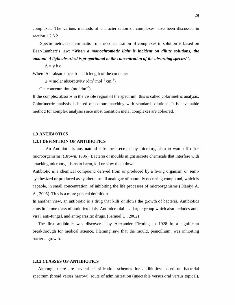
Penicillins are characterized by three fundamental structural features:
i. Fused β –lactam ring;
ii. A free carboxyl group;
iii. One or more substituted amide side chain.
The basic structure consists of thiazolidine ring connected to a β-lactam ring, to which is
attached a side chain R.
31
C O OH
R-C-NH-
CH3N
S
O
CH3
O 1
34
567
Basic structure of penicillin
Natural penicillins
When R is CH3 CH2CH = CH CH2 Penicillin F or I (2-Pentenyl Penicillin)
R = C6 H5 CH2– Penicillin G or II (Benzyl Penicillin)
R = HO C6 H4 CH2– Penicillin X or III (P-Hydroxy benzyl Penicillin)
R = CH3(CH2)6– Penicillin K or IV (n– heptyl penicillin)
R = C6H5OCH2– Penicillin V (Phenoxymethyl Penicillin)
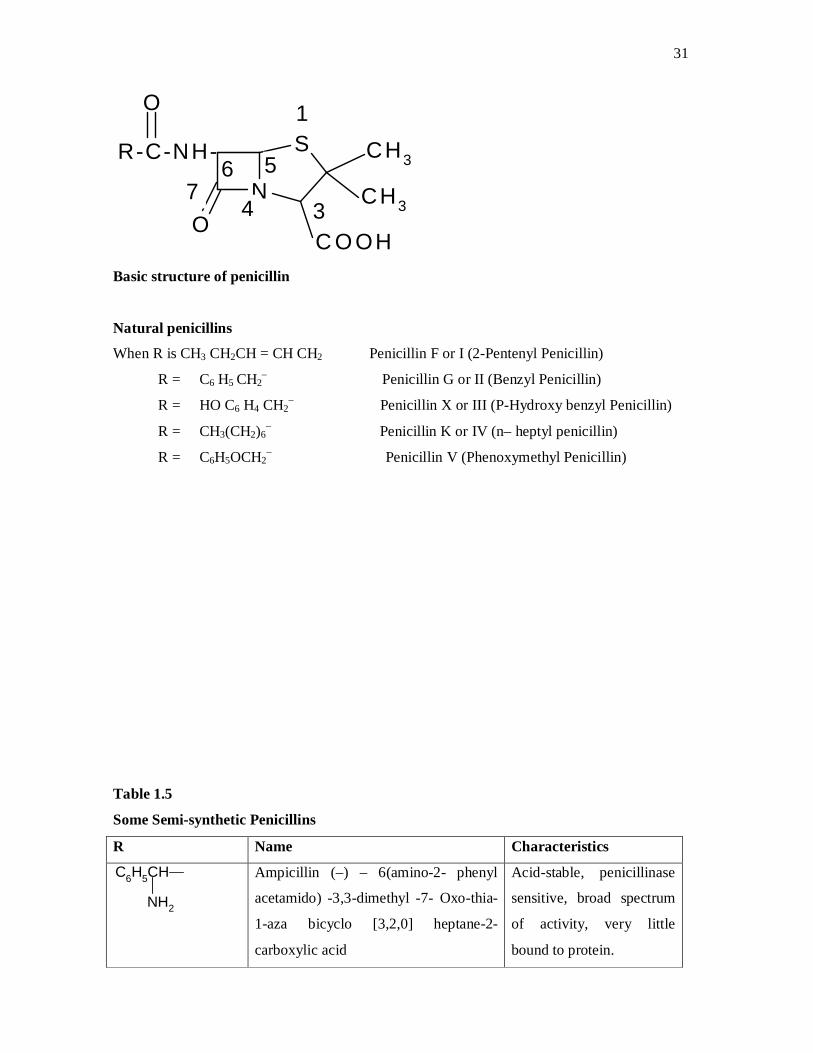
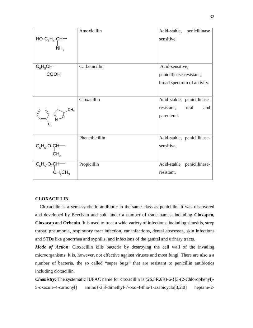
Table 1.5
Some Semi-synthetic Penicillins
R Name Characteristics
C6H5CH
NH2
Ampicillin (–) – 6(amino-2- phenyl
acetamido) -3,3-dimethyl -7- Oxo-thia-
1-aza bicyclo [3,2,0] heptane-2-
carboxylic acid
Acid-stable, penicillinase
sensitive, broad spectrum
of activity, very little
bound to protein.
32
HO-C6H4-CH
NH2
Amoxicillin Acid-stable, penicillinase
sensitive.
C6H5CH
COOH
Carbenicillin Acid-sensitive,
penicillinase-resistant,
broad spectrum of activity.
NO
CH3
Cl
Cloxacillin Acid-stable, penicillinase-
resistant, oral and
parenteral.
C6H5-O-CH
CH3
Phenethicillin Acid-stable, penicillinase-
sensitive,
CH2CH3
C6H5-O-CH
Propicillin Acid-stable penicillinase-
resistant.
CLOXACILLIN
Cloxacillin is a semi-synthetic antibiotic in the same class as penicillin. It was discovered
and developed by Beecham and sold under a number of trade names, including Cloxapen,
Cloxacap and Orbenin. It is used to treat a wide variety of infections, including sinusitis, strep
throat, pneumonia, respiratory tract infection, ear infections, dental abscesses, skin infections
and STDs like gonorrhea and syphilis, and infections of the genital and urinary tracts.
Mode of Action: Cloxacillin kills bacteria by destroying the cell wall of the invading
microorganisms. It is, however, not effective against viruses and most fungi. There are also a a
number of bacteria, the so called “super bugs” that are resistant to penicillin antibiotics
including cloxacillin.
Chemistry: The systematic IUPAC name for cloxacillin is (2S,5R,6R)-6-{[3-(2-Chlorophenyl)-
5-oxazole-4-carbonyl] amino}-3,3-dimethyl-7-oxo-4-thia-1-azabicyclo[3,2,0] heptane-2-

33
carboxylic acid. It has amolecular formular of C19H18ClN3O5S and molecular weight of 435.88
g mol-1.
CH3
O
CH3
ON
S
OCH
ON
Cl
CH3
H
O
NC
Cloxacillin
Pharmacokinetics: Cloxacillin is administered either orally or through intra-muscular route. It
has a bioavailability of 37- 90 % and protein binding of 95%. Its elimination half life is 30
minutes to 1 hour and excretion is renal and biliary.
AMOXICILLIN
Amoxicillin is one of the semi-synthetic penicillins discovered by Beecham scientists. It is
a moderate spectrum, bacteriolytic β-lactam antibiotic used to treat bacterial infections. It is
usually the drug of choice within the class because it is better absorbed, following oral
administration, than other β-lactam antibiotics. It is also a treatment for cystic acne.
Amoxicillin acts by inhibiting the synthesis of bacterial cell wall. It inhibits cross-linkage
between the linear peptidoglycan polymer chains that make up a major component of the cell
walls of both Gram-positive and Gram-negative bacteria.
Chemistry: The systematic IUPAC name for amoxicillin is (2S, 5R, 6R)-2-{[(2R)-2-amino-2-
(4-hydroxyphenyl)-acetyl] amino}-3, 3-dimethyl-7-oxo-4-thia-1-azabicyclo [3, 2, 0] heptane-2-
carboxylic acid. It has a formular of C16H19N3O5S and molecular weight of 365.4 g mol-1
CH3
CH3
CH-C-NH
NH2
ON
S
OO
CH
HO
O
Amoxicillin
34
Pharmacokinetics: Amoxicillin is well absorbed following oral administration with
bioavailability of 95%. Less than 30% of the drug is biotransformed in the liver. It has an
elimination half life of 61.3 minutes and its excretion is renal.
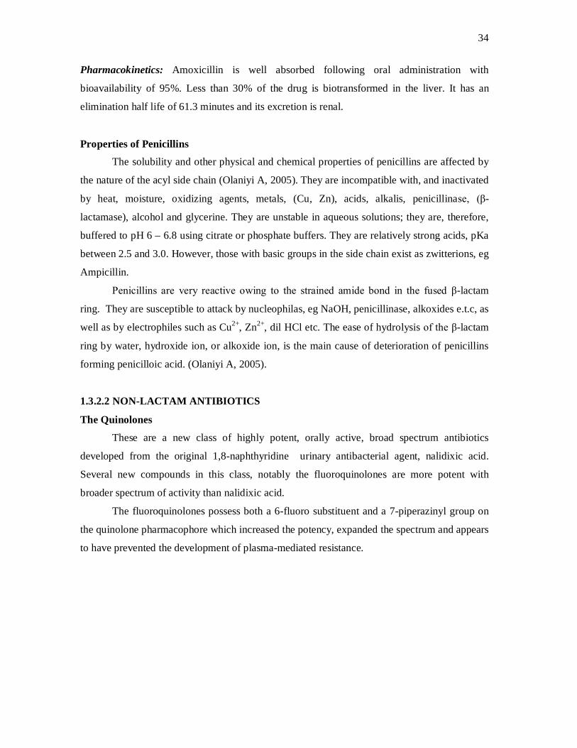
Properties of Penicillins
The solubility and other physical and chemical properties of penicillins are affected by
the nature of the acyl side chain (Olaniyi A, 2005). They are incompatible with, and inactivated
by heat, moisture, oxidizing agents, metals, (Cu, Zn), acids, alkalis, penicillinase, (β-
lactamase), alcohol and glycerine. They are unstable in aqueous solutions; they are, therefore,
buffered to pH 6 – 6.8 using citrate or phosphate buffers. They are relatively strong acids, pKa
between 2.5 and 3.0. However, those with basic groups in the side chain exist as zwitterions, eg
Ampicillin.
Penicillins are very reactive owing to the strained amide bond in the fused β-lactam
ring. They are susceptible to attack by nucleophilas, eg NaOH, penicillinase, alkoxides e.t.c, as
well as by electrophiles such as Cu2+, Zn2+, dil HCl etc. The ease of hydrolysis of the β-lactam
ring by water, hydroxide ion, or alkoxide ion, is the main cause of deterioration of penicillins
forming penicilloic acid. (Olaniyi A, 2005).
1.3.2.2 NON-LACTAM ANTIBIOTICS
The Quinolones
These are a new class of highly potent, orally active, broad spectrum antibiotics
developed from the original 1,8-naphthyridine urinary antibacterial agent, nalidixic acid.
Several new compounds in this class, notably the fluoroquinolones are more potent with
broader spectrum of activity than nalidixic acid.
The fluoroquinolones possess both a 6-fluoro substituent and a 7-piperazinyl group on
the quinolone pharmacophore which increased the potency, expanded the spectrum and appears
to have prevented the development of plasma-mediated resistance.
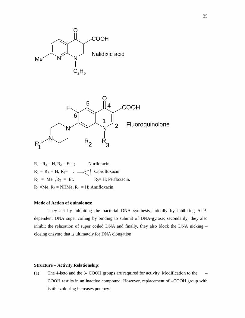
35
COOH
C2H5
Me N N
O
Nalidixic acid
COOH
N
O
NN
R RR
F
Fluoroquinolone12
3
45
6
21
R1 =R3 = H, R2 = Et ; Norfloracin
R1 = R3 = H, R2= ; Ciprofloxacin
R1 = Me ,R2 = Et, R3= H; Perfloxacin.
R1 =Me, R2 = NHMe, R3 = H; Amifloxacin.
Mode of Action of quinolones:
They act by inhibiting the bacterial DNA synthesis, initially by inhibiting ATP-
dependent DNA super coiling by binding to subunit of DNA-gyrase; secondarily, they also
inhibit the relaxation of super coiled DNA and finally, they also block the DNA nicking –
closing enzyme that is ultimately for DNA elongation.
Structure – Activity Relationship:
(a) The 4-keto and the 3- COOH groups are required for activity. Modification to the –
COOH results in an inactive compound. However, replacement of –COOH group with
isothiazolo ring increases potency.

36
(b) A C-6 fluorine atom increases potency as it increases lipophiilcity thus facilitating
penetration into tissues and cells.
(c) Substitution with a piperazinyl group at C -7 leads to potent antibiotic with broad
spectrum of activity.
Ciprofloxacin Ciprofloxacin is a member of the second generation fluoroquinolone class of synthetic
antimicrobial agent s that has broad spectrum of activity against both Gram-positive and Gram-
negative organisms. It was first patented in 1983 by A.G. Bayer and subsequently approved by
the United States Food and Drug Administration (FDA) in 1987. Intravenous formulation was
later introduced in 1991.
Chemistry: Ciprofloxacin is 1-cyclopropyl-6-fluoro-4-oxo-7-piperazin-1-yl quinoline-3-
carboxylic acid. It has a molecular formular of C17H18FN3O3 and molecular weight of 331.4
g mol-1 .It is a faintly yellowish to white crystalline substance.
N
OF COOH
NN
H
Ciprofloxacin
Mode of Action: Ciprofloxacin, like most fluoroquinolones, functions by inhibiting DNA
synthesis, initially by inhibiting ATP-dependent super coiling by binding to subunit A of DNA-
gyrase. Secondarily, they also inhibit the relaxation of the super coiled DNA, a reaction not
dependent on ATP. Finally, they also block the DNA nicking-closing enzyme that, in the
absence of drug interference, is ultimately responsible for DNA elongation. (Olaniyi, 2005)
Pharmacokinetics: The effects of 200 – 400 mg of ciprofloxacin given intravenously are linear;
drug accumulation does not occur when administered at 12 hour intervals. Bioavailability is
approximately 70-80 % with no significant first pass effect. Intravenous administration
produces a similar serum level as those achieved with 500 mg administered orally. I.V
administration over 60 minutes given every 8 hours produces similar serum level of the drug as
37
750 mg administered orally every 12 hours. Metabolism is hepatic including CYP1A2. The
elimination half life is 4 hours and excretion is renal.
Indications: The licensed uses of ciprofloxacin in adult population are as follows: typhoid fever
(enteric fever) caused by Salmonella typhi, acute uncomplicated cystitis in females, chronic
bacterial prostatitis, lower respiratory infections, acute sinusitis, skin and skin structure
infections, infectious diarrhea e.t.c. Ciprofloxacin is not recommended for the treatment of
tuberculosis.
Oral and I.V fluoroquinolones are not licensed by the United states F.D.A. for use in
children due to the risk of permanent injury to the musculoskeletal system.
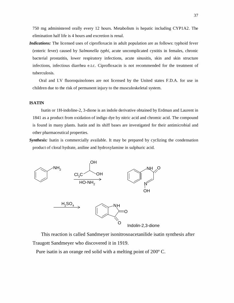
ISATIN
Isatin or 1H-indoline-2, 3-dione is an indole derivative obtained by Erdman and Laurent in
1841 as a product from oxidation of indigo dye by nitric acid and chromic acid. The compound
is found in many plants. Isatin and its shiff bases are investigated for their antimicrobial and
other pharmaceutical properties.
Synthesis: Isatin is commercially available. It may be prepared by cyclizing the condensation
product of cloral hydrate, aniline and hydroxylamine in sulphuric acid.
H2SO4
HO-NH2
OH
NHCl3C
NH2
OH
OH
N
O
NHO
O Indolin-2,3-dione This reaction is called Sandmeyer isonitrosoacetanilide isatin synthesis after
Traugott Sandmeyer who discovered it in 1919.
Pure isatin is an orange red solid with a melting point of 200º C.

38
1.4 MEDICINAL CHEMISTRY OF THE ESSENTIAL TRACE ELEMENTS
1.4.1 IRON
Biologically iron is the most important transition element. It is involved in
several different processes:
(a) As an oxygen carrier in the blood (haemoglobin)
(b) Oxygen storage in muscle tissue (myoglobin)
(c) As an electron carrier in animals, plants and bacteria (cytochromes) and for
electron transfer in plants and bacteria (ferredoxins).
(d) Storage and scavenging of iron in animals (ferretin and transferrin)
(e) As a content of enzymes like aldehyde oxidase, peroxidase and succinic
dehydrogenase (the aerobic oxidation of carbohydrates). (Lee, 1996).
Numerous iron (II) and iron (III) compounds, complexes and solutions
have been used as haematinics in the past. However because of their greater
gastrointestinal irritation and poor absorption, iron (III) compounds and their
preparations are used rarely today. Ferrous fumarate, ferrous gluconate, ferrous
sulphate (oral solution, syrup, and tablets) and dried ferrous sulphate are official
in the USP.
Iron dextran injection, a colloidal iron (III) hydroxide with partially
hydrolyzed dextran, and iron sorbitex injection, a complex of iron with sorbitol
and citric acid, are cited in the USP as injectable forms for patients with poor
gastrointestinal tolerance or poor absorption of iron.
A study carried out in Finland has cast doubt on the advisability of the
routine use of haematinics because men with higher levels of ferritin were found
to be more prone to heart attack. Interpretation of the results included speculation
about iron’s ability to give rise to free radicals after reaction with oxygen. The use
of haematinics without substantiated need is not advisable. (Clarence et al, 2000).
1.5 AIM OF STUDY

39
The aims of the current study are the following:
1. To prepare new Fe3+ complexes of ampicillin, amoxicillin, cloxacillin,
ciprofloxacin and a Schiff base derived from isatin and ethylenediamine, with
a view to improving the aqueous solubility, gastric acid stability and solid
dosage form stability of the drugs.
2. To investigate the antimicrobial properties of the iron (III) complexes of
these drugs and compare them with those of the free drugs (ligands).
3. To investigate the aqueous solubility, thermal and acid stability of these
complexes.
4. To carryout preliminary characterization of the iron (III) complexes of these
drugs and hence propose a structure for each complex.
CHAPTER TWO
MATERIALS AND METHODS
2.1 EQUIPMENT
Magnetic stirrer (Gallenkamp, England), Ultraviolet- visible spectrophotometer (Jenway
6305, Barlowood Sci. ltd, Dunmow), Infrared spectrophotometer (Shimadzu, ® Japan), pH
40
meter (Jenway, Dunmou), electronic weighing balance (Adventurer, OHAUS corp. China),
melting point apparatus (Electrothermal, ® England) were used.
2.2 REAGENTS AND SOLVENTS
Pure ciprofloxacin hydrochloride (Juhel Pharm. ltd, Enugu),amoxicillin powder
(Becham pham. Pakistan) cloxacillin (Neuchem pharm. Lagos) and ampicillin trihydrate (c/o
Neuchem pharm. Lagos) were obtained from Juhel pharm. Nig. ltd, Enugu & Neuchem (F&P)
ltd Lagos. Pure isatin powder and ethylenediamine were obtained from Dr. P.O. Ukoha’s
research lab. Dept. of Pure & Ind. Chem., UNN. Ferric chloride (Merck. Germany), analytical
grades of the following solvents (manufactured by Aldrich Chem. Ltd): absolute ethanol,
methanol and dioxan, and distilled water (Lion Table water Ltd UNN) were used as obtained.
2.3 MICROORGANISMS
The following microorganisms (clinical isolates): Staphylococcus aureus, Pseudomonas
aeruginosa, Bacillus subtilis, Escherichia coli, Salmonella typhi, Shigella spp, Aspergillus
niger and Candida albicans were used. They were maintained in the Department of
Pharmaceutics, University of Nigeria Nsukka.
2.4 SYNTHESIS OF THE COMPLEXES
Synthesis of Fe3+ Complex of Ciprofloxacin
Ciprofloxacin HCl (368 mg) was dissolved in a minimum quantity of distilled water. To this
solution was then added FeCl3 (81.25 mg) dissolved in absolute ethanol. The mixture was
stirred continuously with magnetic stirrer at room temperature for 3 hrs. The pH was adjusted
to 7.0-8.0 with dil. NaOH. The resulting red solution was transferred into an evaporating dish
and allowed to evaporate slowly at room temperature for two weeks. The reaction was
monitored spectrometrically. The red crystals formed were purified by recrystallizing in a
minimum quantity of ethanol, and weighed. (Guangguo et al, 2003)
Synthesis of Cloxacillin-Fe3+ Complex
Cloxacillin powder (435 mg) was dissolved in dioxane (100 ml). To this solution was added
an alcoholic FeCl3 solution containing 162.5 mg of FeCl3 in 100 ml of ethanol. The mixture
was stirred continuously for 4 hours at room temperature, at the end of which clay-brown
crystals separated from the solution. The mixture was filtered and the crystals washed
thoroughly with dioxane, dried in a desiccator and weighed. (Guangguo et al, 2003)

41
Synthesis of Amoxicillin-Fe3+ Complex
Amoxicillin powder (365 mg) was dissolved in methanol (100 ml) in a beaker. FeCl3 (162.5
mg) was put in another beaker containing ethanol (10 ml). The two solutions were mixed
together and stirred with a magnetic stirrer under reflux, maintained at 40oC for 4 hrs. The
resulting green solution, which was foaming, was transferred into an open beaker and allowed
to stand for 1 week. The green crystals obtained were washed thoroughly with small quantity of
ethanol, dried and weighed.
Synthesis of Ampicillin –Fe3+ Complex
Ampicillin trihidrate (403 mg) was dissolved in methanol (100 ml) in a beaker. FeCl3
(81.25 mg) was put in another beaker containing ethanol (10 ml). The two solutions were
mixed together and stirred with a magnetic stirrer under reflux, maintained at 40◦C for 4 hrs.
The resulting brown solution was transferred into an open beaker and allowed to stand for 1
week. The brown crystals obtained were washed thoroughly with small quantity of ethanol,
dried and weighed. (Guangguo et al, 2003)
Synthesis of Isatin-ethylenediamine Schiff base Ligand.
Isatin (147 mg) was dissolved in minimum quantity of absolute ethanol placed in an ice
bath. To this solution, 100% ethylenediamine (60 mg) maintained at 4oC was added and the
mixture stirred continuously for 2 hrs. The mixture was acidified with CH3COOH to pH 4 -
4.5. The temperature was maintained at 0-5oC throughout. Rusty brown product, separated after
two days, was filterd and washed with ethanol. The solid product was weighed after drying in a
desiccator.
Synthesis of Fe3+ Complex of the Isatin Schiff base
The Schiff base derived from isatin and ethylenediamine (190 mg) was dissolved in
minimum amount of absolute ethanol. To this refluxing solution, FeCl3 (82 mg) dissolved in
ethanol was added. The refluxing was continued for 2 hrs, at 60oC. The reddish brown product
was filtered, washed with ethanol and dried in a desiccator over CaCl2 .
These reactions were monitored spectrometrically and chromatographically.
2.5 EBULLIOSCOPIC DETERMINATION OF MOLECULAR WEIGHT OF THE
COMPLEXES
42
Each of the complexes (10 mg) was dissolved in absolute ethanol (50 ml) in a small
beaker. 2 ml of each of the solutions was put in a narrow fusion tube connected to a
thermometer by a thread, and heated in paraffin bath. The boiling point elevation in each case,
i.e. the difference between the boiling point of the solution and that of pure ethanol, was
recorded. The approximate molecular weight M2 of each solute was calculated using the
Raoult’s law:
M2 = Kb . W2 . 1000
∆Tb W1
Where Kb = Ebullioscopic constant (1.22 molal-1 for ethanol)
∆Tb = Boiling point elevation
W2 /W1 = concentration of the solute
2.6 JOB’S METHODS OF CONTINUOUS VARIATION
Standard solution (l mmole) each of ferric chloride and the ligands was prepared.
Different volumes (1ml, 2ml … 9ml) of the ferric chloride solution were delivered into nine
appropriately labeled test tubes using a clean burette. Different volumes of the ligands solution
were added to the test tube containing the ferric chloride solution such that the total volume of
each mixture is 10.00 ml. This keeps the total number of mole of reactants constant throughout
the series of mixtures but varies the mole fraction of each reactant from mixture to mixture.
Each of the solution mixture was shaken thoroughly, stirred in a small beaker with magnetic
stirrer for 30 mins and allowed to stand for 24 hrs to equilibrate. Samples were removed from
each test tube and placed in the cuvette of a UV-visible spectrophotometer. The absorbances
were read at the λ max of each complex. A graph of mole fraction of ferric ion versus
absorbance was plotted and analyzed carefully to determine the mole fraction of ferric ion
when there is the most significant change in absorbance.
The maximum change will occur when the mole fraction of the reactants is closest to the
actual stoichiometric ratio. Both the formular of the complex and stoichiometry were
determined using this approach.
2.7 DETERMINATION OF SOME PHYSICIOCHEMICAL PARAMETERS OF THE
COMPLEXES AND LIGANDS
2.7.1 AQUEOUS SOLUBILITY
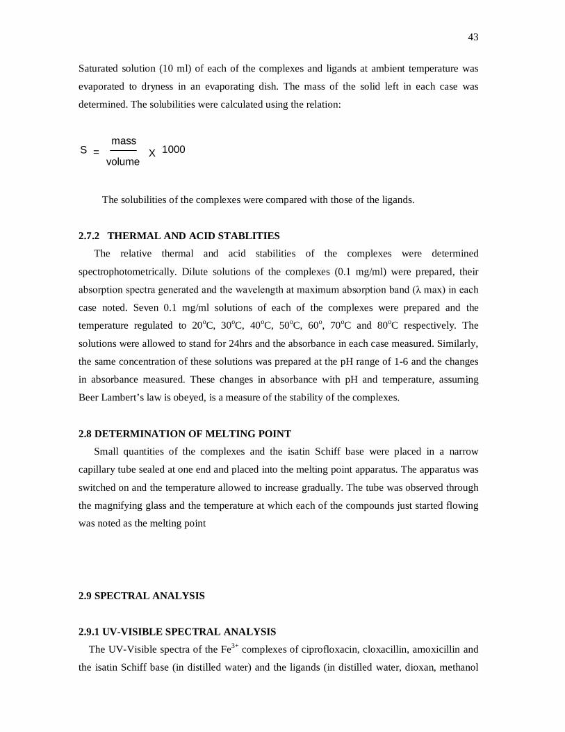
43
Saturated solution (10 ml) of each of the complexes and ligands at ambient temperature was
evaporated to dryness in an evaporating dish. The mass of the solid left in each case was
determined. The solubilities were calculated using the relation:
S =mass
volume1000X
The solubilities of the complexes were compared with those of the ligands.
2.7.2 THERMAL AND ACID STABLITIES
The relative thermal and acid stabilities of the complexes were determined
spectrophotometrically. Dilute solutions of the complexes (0.1 mg/ml) were prepared, their
absorption spectra generated and the wavelength at maximum absorption band (λ max) in each
case noted. Seven 0.1 mg/ml solutions of each of the complexes were prepared and the
temperature regulated to 20oC, 30oC, 40oC, 50oC, 60o, 70oC and 80oC respectively. The
solutions were allowed to stand for 24hrs and the absorbance in each case measured. Similarly,
the same concentration of these solutions was prepared at the pH range of 1-6 and the changes
in absorbance measured. These changes in absorbance with pH and temperature, assuming
Beer Lambert’s law is obeyed, is a measure of the stability of the complexes.
2.8 DETERMINATION OF MELTING POINT
Small quantities of the complexes and the isatin Schiff base were placed in a narrow
capillary tube sealed at one end and placed into the melting point apparatus. The apparatus was
switched on and the temperature allowed to increase gradually. The tube was observed through
the magnifying glass and the temperature at which each of the compounds just started flowing
was noted as the melting point
2.9 SPECTRAL ANALYSIS
2.9.1 UV-VISIBLE SPECTRAL ANALYSIS
The UV-Visible spectra of the Fe3+ complexes of ciprofloxacin, cloxacillin, amoxicillin and
the isatin Schiff base (in distilled water) and the ligands (in distilled water, dioxan, methanol

44
and ethanol respectively) were recorded in UV 6305 PC spectrophotometer (Jenway) using
1cm quartz cuvettes.
2.9.2 INFRARED SPECTRAL ANALYSIS
The IR spectra of the complexes and the ligands were determined at Sheda Science and
Tech. Complex, Abuja. The compounds were prepared in KBr disc and the spectra recorded in
FTIR (Shimadzu, ® Japan).
2.10 ANTIMICROBIAL STUDIES ON THE COMPLEXES
The ligands and complexes were assayed for antimicrobial activity by the agar diffusion
method. Solutions of the complexes and pure ciprofloxacin were made in distilled water and
those of the other ligands in DMSO.
2.10.1 PREPARATION OF THE CULTURE MEDIA
The culture media employed for the anti-microbial investigation were nutrient agar, for
bacteria, and Saboraud’s dextrose agar for fungi and yeast.
Nutrient agar formular
Beef extract 1.0 g NaCl 5.0 g
Peptone 5.0 g Distilled water 1000 ml
Agar 15.0 g Yeast extracts 2.0 g
Approximate pH 7.4
Saboraud’s Agar Formular
• Mycological peptone 10.0 g
• Dextrose 40.0 g
• Agar 15.0 g
• Distilled water 1000 ml
• Approx. pH 5.2
The various quantities of the needed ingredients were mixed together and dissolved in 1
dm3 of distilled water and allowed to stand for about 15 mins. The mixture was distributed in
bijou bottles and sterilized by autoclaving at 121oC for 15 minutes. The sterilized media were
maintained in a molten state until used.
2.10.2 MAINTENANCE AND ACTIVATION OF THE MICROORGANISMS
45
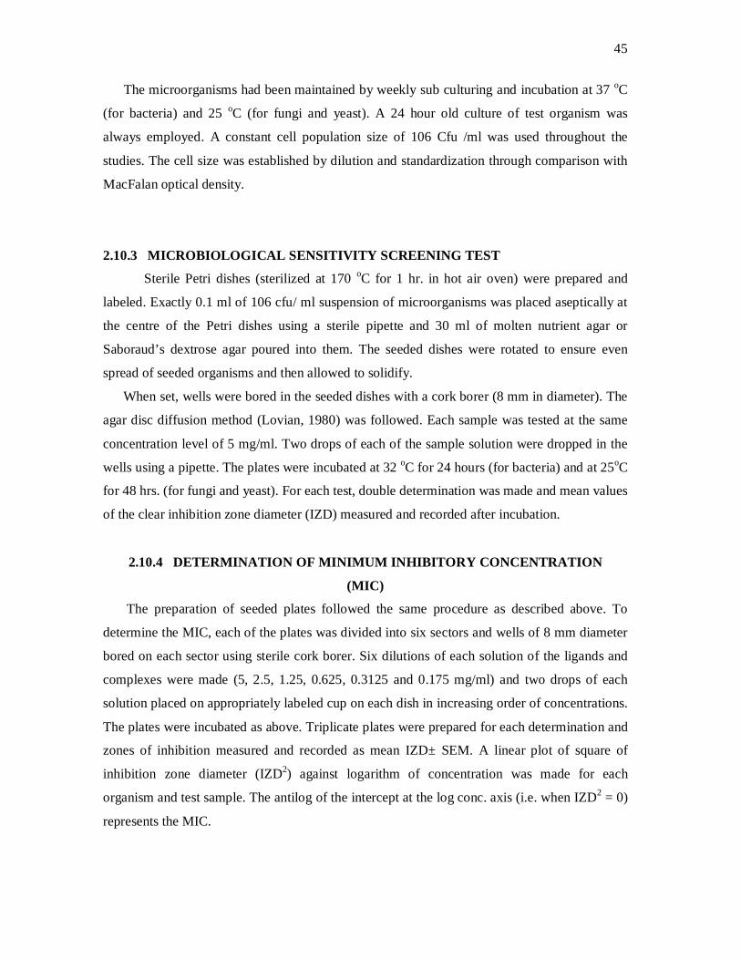
The microorganisms had been maintained by weekly sub culturing and incubation at 37 oC
(for bacteria) and 25 oC (for fungi and yeast). A 24 hour old culture of test organism was
always employed. A constant cell population size of 106 Cfu /ml was used throughout the
studies. The cell size was established by dilution and standardization through comparison with
MacFalan optical density.
2.10.3 MICROBIOLOGICAL SENSITIVITY SCREENING TEST
Sterile Petri dishes (sterilized at 170 oC for 1 hr. in hot air oven) were prepared and
labeled. Exactly 0.1 ml of 106 cfu/ ml suspension of microorganisms was placed aseptically at
the centre of the Petri dishes using a sterile pipette and 30 ml of molten nutrient agar or
Saboraud’s dextrose agar poured into them. The seeded dishes were rotated to ensure even
spread of seeded organisms and then allowed to solidify.
When set, wells were bored in the seeded dishes with a cork borer (8 mm in diameter). The
agar disc diffusion method (Lovian, 1980) was followed. Each sample was tested at the same
concentration level of 5 mg/ml. Two drops of each of the sample solution were dropped in the
wells using a pipette. The plates were incubated at 32 oC for 24 hours (for bacteria) and at 25oC
for 48 hrs. (for fungi and yeast). For each test, double determination was made and mean values
of the clear inhibition zone diameter (IZD) measured and recorded after incubation.
2.10.4 DETERMINATION OF MINIMUM INHIBITORY CONCENTRATION
(MIC)
The preparation of seeded plates followed the same procedure as described above. To
determine the MIC, each of the plates was divided into six sectors and wells of 8 mm diameter
bored on each sector using sterile cork borer. Six dilutions of each solution of the ligands and
complexes were made (5, 2.5, 1.25, 0.625, 0.3125 and 0.175 mg/ml) and two drops of each
solution placed on appropriately labeled cup on each dish in increasing order of concentrations.
The plates were incubated as above. Triplicate plates were prepared for each determination and
zones of inhibition measured and recorded as mean IZD± SEM. A linear plot of square of
inhibition zone diameter (IZD2) against logarithm of concentration was made for each
organism and test sample. The antilog of the intercept at the log conc. axis (i.e. when IZD2 = 0)
represents the MIC.

46
CHAPTER THREE
RESULTS
3.1 YIELD OF THE SYNTHESIS
The percentage yield, colour and the melting point of the products are listed in table 3.1
Table 3.1 PERCENTAGE YIELD OF THE COMPLEXES

47
Cpf-
Fe3+
Cloxa- Fe3+ Amoxil--
Fe3+
Ampi-
Fe3+
Isat-eth-
Fe3+
Isat-eth Schiff
base
Duration of reaction
3 hrs 4 hrs 4 hrs 4 hrs 2 hrs 2 days
Solvent Ethanol/ water
Dioxane Methanol Methanol Ethanol Ethanol
Initial Colour
Brown Rusty brown
Brown Brown Brown Orange
Final colour
Red Clay-brown
Green Brown Brown Brown
Melting point (°C)
172
150
142
46
225
210
Percentage Yield (%)
80
95
63.5
32
65
77
3.2 MOLECULAR WEIGHT AND STOICHIOMETRY OF THE COMPLEXES
The molecular weight and stoichiometry of the complexes are given in table 3.2. They were
determined through boiling point elevation and Job’s method of continuous variation
respectively.
Table 3.2 APPROXIMATE MOLECULAR WEIGHT OF THE COMPLEXES
Approx. Mol. Weight (g mol-1) Expected Metal- ligand
Stoichiometry
CPF-Fe3+ 891 1:2
Cloxa- Fe3+ 586 1:1
Amoxil- Fe3+ 532 1:1
Amp- Fe3+ 917 1:2
Isatin-eth. ligand 182 1:1 (isatin:ethylenediamine ratio)
Isat-eth.- Fe3+ complex 543 1:2
3.3 PHYSICOCHEMICAL PROPERTIES OF THE COMPLEXES AND LIGANDS
3.3.1 AQUEOUS STABILITY
The solubilities of the complexes and the ligands in water at room temperature are displayed
in table 3.3.
Table 3.3 AQUEOUS SOLUBILITY
Pure Ligand Fe3+ Complex

48
Compound Solubility (g dm-3) Solubility (g dm-3)
Ciprofloxacin < 1 >5
Cloxacillin negligible 1.8
Amoxicillin negligible 2.0
Ampicillin negligible 2.0
Isatin 0.5
Isatin-eth. Schiff base 0.8 2.2
3.3.2 RELATIVE THERMAL STABILITY
The absorbance of 0.1 mg/ml solutions of each of the ligands and their iron (III) complexes at
different temperatures are given in tables 3.4 and 3.5 respectively. These changes in absorbance
with temperature are a measure of the thermal stability of the compounds.
Table 3.4 ABSORBANCES OF THE COMPLEXES AT DIFFERENT TEMPERATURES
Complexes 20°C 30°C 40°C 50°C 60°C 70°C 80°C λmax
Cpf-Fe3+ 0.609 0.686 0.689 0.686 0.665 0.635 0.629 443 nm
Cloxa-Fe3+ 0.114 0.122 0.122 0.120 0.109 0.102 0.083 385 nm
Amoxil-Fe3+ 0.095 0.109 0.128 0.133 0.126 0.122 0.105 440 nm
Isat-eth-Fe3+ 0.525 0.516 0.522 0.514 0.511 0.407 0.223 495 nm
Table 3.5 ABSORBANCES OF THE LIGANDS AT DIFFERENT TEMPERATURES
Ligands 20°C 30°C 40°C 50°C 60°C 70°C 80°C λmax
Ciprofloxacin 0.348 0.362 0.311 0.240 0.170 0.104 0.081 315
Cloxacillin 0.721 0.727 0.683 0.455 0.337 0.190 0.184 290
Amoxicillin 0.661 0.664 0.643 0.428 0.369 0.312 0.126 254
Isat. Schff base 0.304 0.306 0.296 0.217 0.212 0.105 0.101 300
3.3.3 RELATIVE ACID STABILITY
The absorbance of 0.1 mg/ml solution of each of the ligands and their iron (III) complexes
at different pHs are given in tables 3.6 and 3.7 respectively. These changes in absorbance with
pHs are a measure of the acid stability of the compounds.
Table 3.6 ABSORBANCES OF THE COMPLEXES AT DIFFERENT pHs
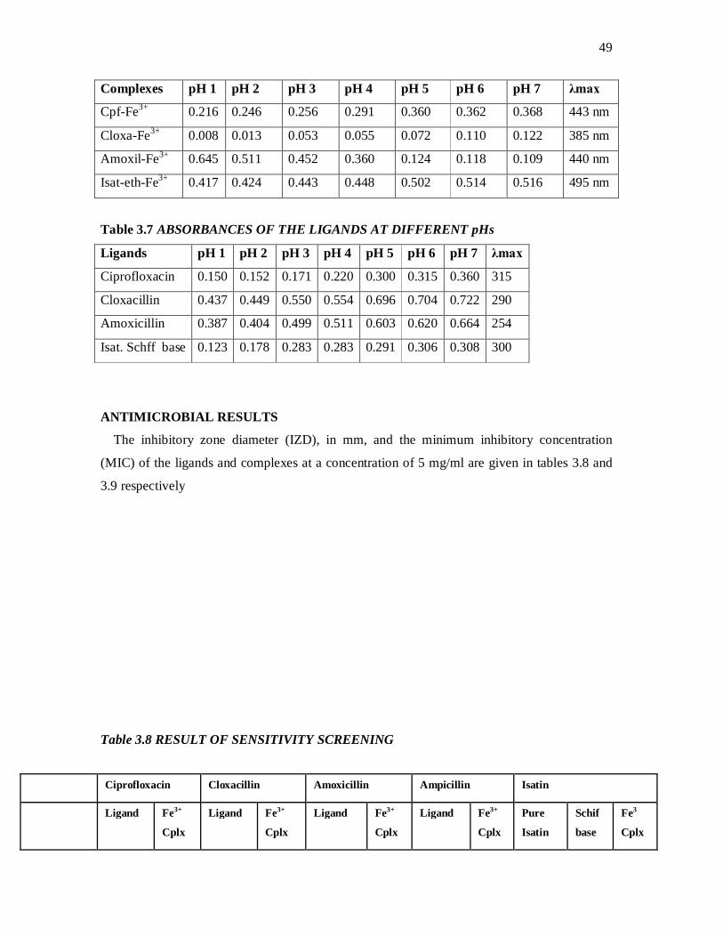
49
Complexes pH 1 pH 2 pH 3 pH 4 pH 5 pH 6 pH 7 λmax
Cpf-Fe3+ 0.216 0.246 0.256 0.291 0.360 0.362 0.368 443 nm
Cloxa-Fe3+ 0.008 0.013 0.053 0.055 0.072 0.110 0.122 385 nm
Amoxil-Fe3+ 0.645 0.511 0.452 0.360 0.124 0.118 0.109 440 nm
Isat-eth-Fe3+ 0.417 0.424 0.443 0.448 0.502 0.514 0.516 495 nm
Table 3.7 ABSORBANCES OF THE LIGANDS AT DIFFERENT pHs
Ligands pH 1 pH 2 pH 3 pH 4 pH 5 pH 6 pH 7 λmax
Ciprofloxacin 0.150 0.152 0.171 0.220 0.300 0.315 0.360 315
Cloxacillin 0.437 0.449 0.550 0.554 0.696 0.704 0.722 290
Amoxicillin 0.387 0.404 0.499 0.511 0.603 0.620 0.664 254
Isat. Schff base 0.123 0.178 0.283 0.283 0.291 0.306 0.308 300
ANTIMICROBIAL RESULTS
The inhibitory zone diameter (IZD), in mm, and the minimum inhibitory concentration
(MIC) of the ligands and complexes at a concentration of 5 mg/ml are given in tables 3.8 and
3.9 respectively
Table 3.8 RESULT OF SENSITIVITY SCREENING
Ciprofloxacin Cloxacillin Amoxicillin Ampicillin Isatin
Ligand Fe3+
Cplx
Ligand Fe3+
Cplx
Ligand Fe3+
Cplx
Ligand Fe3+
Cplx
Pure
Isatin
Schif
base
Fe3
Cplx
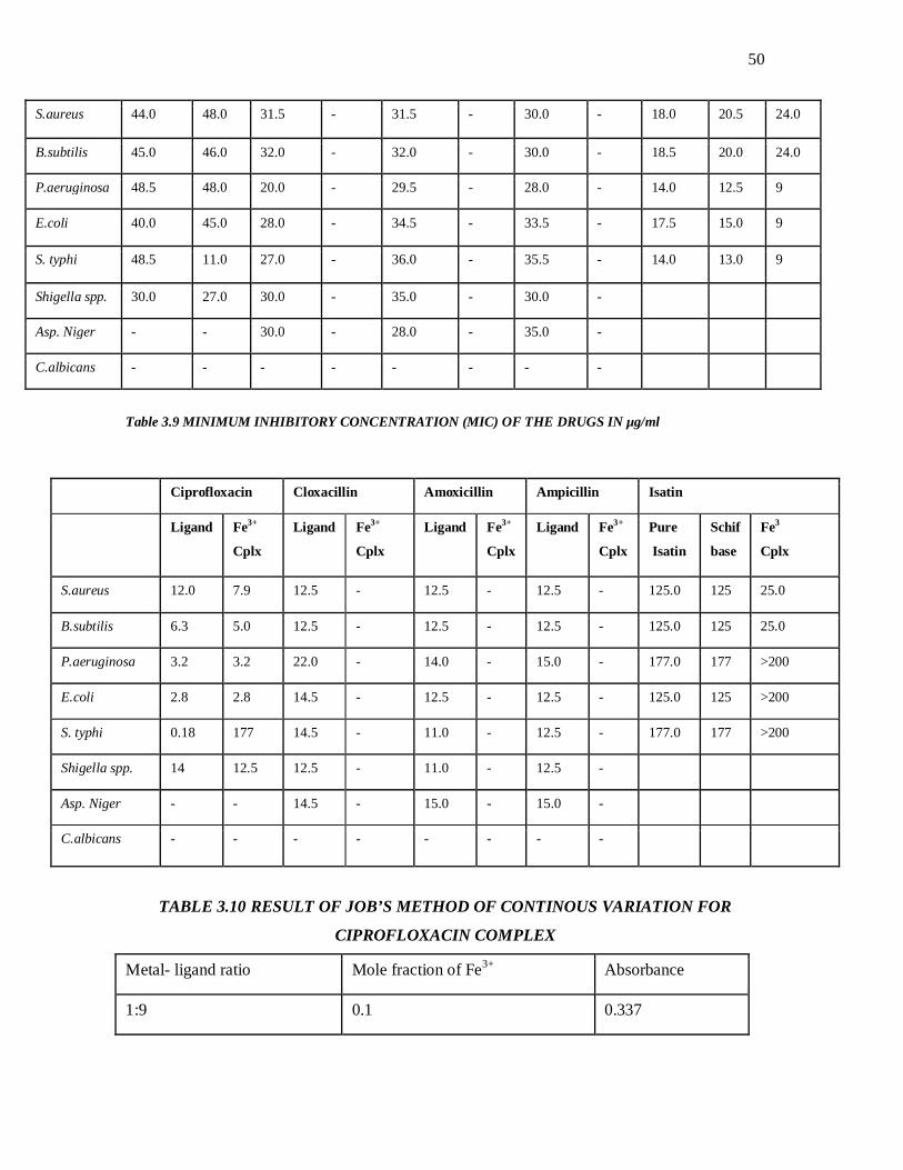
50
S.aureus 44.0 48.0 31.5 - 31.5 - 30.0 - 18.0 20.5 24.0
B.subtilis 45.0 46.0 32.0 - 32.0 - 30.0 - 18.5 20.0 24.0
P.aeruginosa 48.5 48.0 20.0 - 29.5 - 28.0 - 14.0 12.5 9
E.coli 40.0 45.0 28.0 - 34.5 - 33.5 - 17.5 15.0 9
S. typhi 48.5 11.0 27.0 - 36.0 - 35.5 - 14.0 13.0 9
Shigella spp. 30.0 27.0 30.0 - 35.0 - 30.0 -
Asp. Niger - - 30.0 - 28.0 - 35.0 -
C.albicans - - - - - - - -
Table 3.9 MINIMUM INHIBITORY CONCENTRATION (MIC) OF THE DRUGS IN µg/ml
TABLE 3.10 RESULT OF JOB’S METHOD OF CONTINOUS VARIATION FOR
CIPROFLOXACIN COMPLEX
Metal- ligand ratio Mole fraction of Fe3+ Absorbance
1:9 0.1 0.337
Ciprofloxacin Cloxacillin Amoxicillin Ampicillin Isatin
Ligand Fe3+
Cplx
Ligand Fe3+
Cplx
Ligand Fe3+
Cplx
Ligand Fe3+
Cplx
Pure
Isatin
Schif
base
Fe3
Cplx
S.aureus 12.0 7.9 12.5 - 12.5 - 12.5 - 125.0 125 25.0
B.subtilis 6.3 5.0 12.5 - 12.5 - 12.5 - 125.0 125 25.0
P.aeruginosa 3.2 3.2 22.0 - 14.0 - 15.0 - 177.0 177 >200
E.coli 2.8 2.8 14.5 - 12.5 - 12.5 - 125.0 125 >200
S. typhi 0.18 177 14.5 - 11.0 - 12.5 - 177.0 177 >200
Shigella spp. 14 12.5 12.5 - 11.0 - 12.5 -
Asp. Niger - - 14.5 - 15.0 - 15.0 -
C.albicans - - - - - - - -

51
2:8 0.2 0.692
3:7 0.3 0.943
4:6 0.4 0.882
5:5 0.5 0.742
6:4 0.6 0.600
7:3 0.7 0.453
8:2 0.8 0.280
9:1 0.9 0.157
Table 3.11 RESULT OF JOB’S METHOD OF CONTINOUS VARIATION FOR
CLOXACILLIN COMPLEX
Metal- ligand ratio Mole fraction of Fe3+ Absorbance
1:9 0.1 0.150
2:8 0.2 0.224
3:7 0.3 0.283
4:6 0.4 0.360
5:5 0.5 0.411
6:4 0.6 0.288
7:3 0.7 0.215
8:2 0.8 0.158
9:1 0.9 0.092
Fig 3.1 Job’s Plot for Ciprofloxacin Complex

52
The mole fraction of Iron (III) at the intersection of the two straight lines is 0.32. The mole ratio is 3.2: 6.8 which corresponds to 1: 2metal – ligand stoichiometry.
0
0.1
2.0.
0.3
0.4
0.5
0.6
0.7
0.8
0.9
1
0 0.2 0.4 0.6 0.8 1Mole Fraction of Fe3+
Absorbance

53
Fig 4.2 Job's Plot for Cloxacillin Complex
0
0.05
0.1
0.15
0.2
0.25
0.3
0.35
0.4
0.45
0 0.1 0.2 0.3 0.4 0.5 0.6 0.7 0.8 0.9 1
Mole Fraction of Iron (III)
Abs
orba
nce
Series1
The mole fraction of Iron (III) at the intersection of the two straight lines is
0.48.The mole ratio is 4.8 : 5.2 which corresponds to 1 : 1 metal – ligand
stoichiometry.
3.5 SPECTRAL DATA
The UV-Visible and infrared spectra of the drugs are displayed in figs. 3.3 – 3.13

54
Fig. 3.3 UV-VISIBLE SPECTRUM OF CIPROFLOXACIN

55
Fig.3.4 UV-Visible Spectrum of Fe3+ Complex of Ciprofloxacin
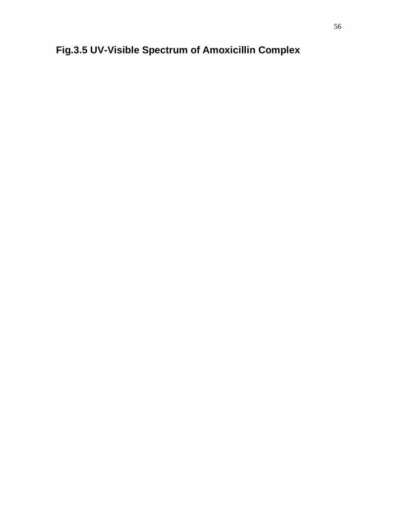
56
Fig.3.5 UV-Visible Spectrum of Amoxicillin Complex

57
Fig. 3.6 UV-Visible Spectrum of Fe3+ Complex of Cloxacillin

58
Fig. 3.7 UV-Visible Spectrum of Fe3+ Complex of Isatin Schiff Base

59
Fig. 3.8 UV-Visible Spectrum of Ferric Chloride

60
Fig. 3.9 Infrared Spectrum of Ciprofloxacin

61
Fig. 3.10 Infrared Spectrum of Fe3+ Complex of Ciprofloxacin

62
Fig. 3.11 Infrared Spectrum of Fe3+ Complex of Cloxacillin

63
Fig. 3.12 Infrared Spectrum of Fe3+ Complex of Amoxicillin

64
Fig. 3.13 Infrared Spectrum of Fe3+ Complex of Isatin Schiff Base
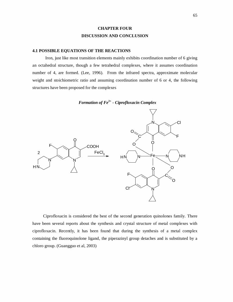
65
CHAPTER FOUR
DISCUSSION AND CONCLUSION
4.1 POSSIBLE EQUATIONS OF THE REACTIONS
Iron, just like most transition elements mainly exhibits coordination number of 6 giving
an octahedral structure, though a few tetrahedral complexes, where it assumes coordination
number of 4, are formed. (Lee, 1996). From the infrared spectra, approximate molecular
weight and stoichiometric ratio and assuming coordination number of 6 or 4, the following
structures have been proposed for the complexes
Formation of Fe3+ - Ciprofloxacin Complex
N
OFC
O
O
N
O
N N
F C
O
O
Fe N NN
OF COOH
NN
H
FeCl3
Cl
Cl
H H2
Ciprofloxacin is considered the best of the second generation quinolones family. There
have been several reports about the synthesis and crystal structure of metal complexes with
ciprofloxacin. Recently, it has been found that during the synthesis of a metal complex
containing the fluoroquinolone ligand, the piperazinyl group detaches and is substituted by a
chloro group. (Guangguo et al, 2003)

66
Proposed Structure for Fe3+ Complex of Amoxicillin
Cl
CH3
CH3
CH-C-NH
NH2O
ON
S
O
OC
Fe
2+O
The green colour of the complex suggests reduction of the iron (III) ions to iron (II).
Participation of a univalent negative ligand such as the chloro group in coordinate covalent
bonding with the metal (reductive substitution) has been suggested. This could account for
this observation. Participation of the phenolic oxygen is also suggested since the absorption
band for the OH group is lacking in the infrared spectrum of the complex.
Proposed Structure for Ampicillin Complex
CH3
CH3
CH-C-NH
NH2
N
S
OO
C
OO
Fe
3+
Steric crowding of bonds in the molecule and the use of the amide nitrogens for
coordination are suggested to have caused the poor yield of this complex

67
Formation of Isatin- ethylenediamine (Schiff base) Ligand
This is a Shiff reaction; reaction between a carbonyl group of the indolin-1, 2-dione
and the amino group of the ethylenediamine to form indolin-2- imine (Schiff base)
N
H
H2NCH2CH2NH2
O
O N
H
O
NCH2CH2NH2
+
Proposed Structure for Fe3+ Complex of the Isatin Schiff Base
N
H
O
NCH2CH2NH2
N
H
O
H2NH2CH2CN
Fe
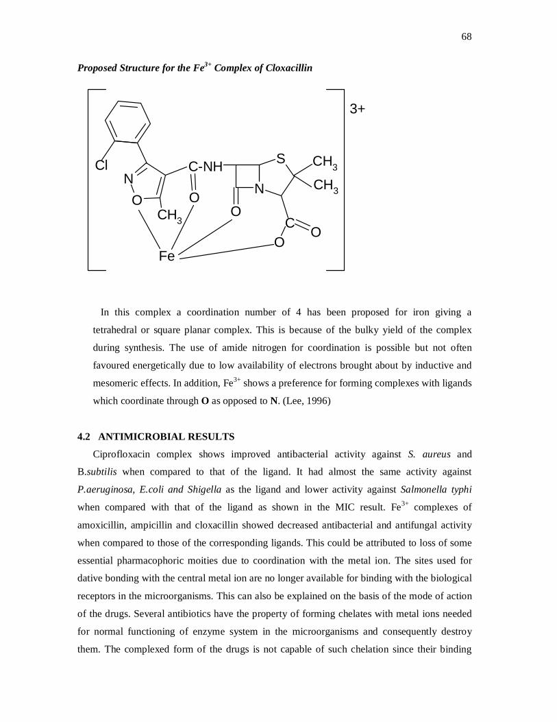
68
Proposed Structure for the Fe3+ Complex of Cloxacillin
CH3
OFe
Cl C-NH
NO
CH3
ON
S
OC
OCH3
3+
In this complex a coordination number of 4 has been proposed for iron giving a
tetrahedral or square planar complex. This is because of the bulky yield of the complex
during synthesis. The use of amide nitrogen for coordination is possible but not often
favoured energetically due to low availability of electrons brought about by inductive and
mesomeric effects. In addition, Fe3+ shows a preference for forming complexes with ligands
which coordinate through O as opposed to N. (Lee, 1996)
4.2 ANTIMICROBIAL RESULTS
Ciprofloxacin complex shows improved antibacterial activity against S. aureus and
B.subtilis when compared to that of the ligand. It had almost the same activity against
P.aeruginosa, E.coli and Shigella as the ligand and lower activity against Salmonella typhi
when compared with that of the ligand as shown in the MIC result. Fe3+ complexes of
amoxicillin, ampicillin and cloxacillin showed decreased antibacterial and antifungal activity
when compared to those of the corresponding ligands. This could be attributed to loss of some
essential pharmacophoric moities due to coordination with the metal ion. The sites used for
dative bonding with the central metal ion are no longer available for binding with the biological
receptors in the microorganisms. This can also be explained on the basis of the mode of action
of the drugs. Several antibiotics have the property of forming chelates with metal ions needed
for normal functioning of enzyme system in the microorganisms and consequently destroy
them. The complexed form of the drugs is not capable of such chelation since their binding
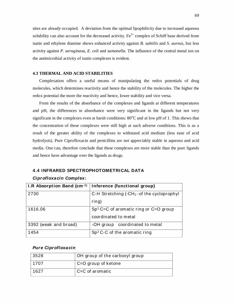
69
sites are already occupied. A deviation from the optimal lipophilicity due to increased aqueous
solubility can also account for the decreased activity. Fe3+ complex of Schiff base derived from
isatin and ethylene diamine shows enhanced activity against B. subtilis and S. aureus, but less
activity against P. aeruginasa, E. coli and samonella. The influence of the central metal ion on
the antimicrobial activity of isatin complexes is evident.
4.3 THERMAL AND ACID STABILITIES
Complexation offers a useful means of manipulating the redox potentials of drug
molecules, which determines reactivity and hence the stability of the molecules. The higher the
redox potential the more the reactivity and hence, lower stability and vice versa.
From the results of the absorbance of the complexes and ligands at different temperatures
and pH, the differences in absorbance were very significant in the ligands but not very
significant in the complexes even at harsh conditions: 80oC and at low pH of 1. This shows that
the concentration of these complexes were still high at such adverse conditions. This is as a
result of the greater ability of the complexes to withstand acid medium (less ease of acid
hydrolysis). Pure Ciprofloxacin and penicillins are not appreciably stable in aqueous and acid
media. One can, therefore conclude that these complexes are more stable than the pure ligands
and hence have advantage over the ligands as drugs.
4.4 INFRARED SPECTROPHOTOMETRICAL DATA Ciprofloxacin Complex:
I.R Absorption Band (cm-1) Inference (functional group)
2730 C-H Stretching (-CH2 -of the cycloprophyl
ring)
1616.06 Sp2 C=C of aromatic ring or C=O group
coordinated to metal
3392 (weak and broad) -OH group coordinated to metal
1454 Sp2 C-C of the aromatic ring
Pure Ciprofloxacin
3528 OH group of the carboxyl group
1707 C=O group of ketone
1627 C=C of aromatic

70
The disappearance of the strong absorption peak at 3528cm-1 (found in the
ligand but not in the complex) indicates the loss of the OH group of carboxyl
group to coordination with the metal. The band at 1707.6cm-1 due to the
ketone group, was not detected in the spectrum of the complex, indicating
that this moiety participates in coordinate bonding with the metal ion. The
corresponding band was recorded at 1616cm-1. The shift towards lower wave
number is consistent with this participation.
Isat- eth-Fe3+ Complex
Infrared Data (cm-1) Inference (Functional Group)
2923.57 C-H aliphatic
3399.90 (weak and broad) Bound –NH of ethylenediamine
1608 C=C of aromatic ring
The absence of any strong absorption band in the spectrum of this complex is an indication
that all the functional groups present are bonded to the metal ion. The ligand shows strong
absorption bands at about 1720cm-1 and above 3400cm-1 due to the carbonyl and amino groups
respectively.
Cloxacillin Complex
Infrared Data (cm-1) Inference (Functional Group)
1737.55 C=O group of acid or amide
1658.49 Sp2 C=C of aromatic
3000-2724 (very prominent peak) C-H aliphatic
1610.28 Sp2 C-C of aromatic

71
Amoxicillin –Fe3+ Complex
Infrared Data (cm-1) Inference (Functional Group)
2850 C-H stretching (-CH3)
1747 C=O of acid
1454 Sp2 C-C of aromatic ring
1376 Geminal dimethyl group on the thiazoline ring
840 Para disubstituted benzene ring
The absence of any strong absorption band beyond 3000 cm -1 indicates participation of the
amino group of the side chain and the -OH group of the phenolic group in dative bonding
with the metal.
72
4.5 CONCLUSION
Complexation improves the aqueous solubility, thermal and acid stabilities of ciprofloxacin,
penicillins and Isatin. This improvement in solubility can be exploited in parenteral
administration of these drugs. Loss of these drugs to deterioration during storage would be
minimized if they are stored in the form of their coordination complexes. Complexes of these
drugs, therefore, have advantage over the pure drugs due to this inherent stability. Oral
administration of drugs in their complex forms may reduce absorption in the lipid layers. This
is because the improved aqueous solubility would result in lower partition coefficient. Co-
administration of these drugs with iron tablets is therefore discouraged because complexation
may occur in vivo.
The structure – activity relationship of drugs could be predicted by complexation reactions.
If a particular biological activity of a drug is lost or diminished on complexation with metal
ions, it would be reasonable to suggest that one or more of the groups bonded to the metal is
necessary for activity. Further tests can be conducted to confirm the pharmacophoric moieties.
Most coordination compounds of Isatin (indolindione) are more effective antibacterial
drugs than isatin itself. The activity depends on the central metal ion of the complexes.
73
REFERENCES
Abraham, E. P. and P. B. Loder (1972); In: Cephalosposins and Penicillins; Chemistry and
Biology, E.H. Flynn (Ed.); New York: Academic Press: 484-503.
Afzal D, Ervin H.D, Moody A.E, Wohlers H.D, (2005); Coordination Chemistry. New York:
John Wiley, p.362.
Alberts B. et al. (1994); Molecular Biology of the Cell, 3rd Ed. New York: Garland Publishing
Inc. pp 430-437
Andrews L. J, Keefer R.M. (1964); Molecular Complexes in Organic Chemistry, San
Francisco: Holden-Day, p.221
Ayse E., Sema, O.Y., Mehmet A. (2007) Novel Isatin Schiff Base Cu (II) and Ni (II)
Complexes. X-ray Crystal Structure of Bis (3-(4-hexyl Phenylimino)- IH-indol-2 (3H)-
one)- dichloro copper (II) Complexes. Transition Met.Chem. 21, 56-63.
Bailar, J.C.(1946); Synthesis and Characterization of Co (II) Complex of Ampicillin. Journal
of Inorganic Synthesis, 2, 222-225.
Balkwill, F. and Rolph, M, (1995); Microbes, Bugs and Wonder Drugs: Potions to Penicillin,
Aspirin to Addiction. London: Portland Press Ltd. P. 187- 189.
Basole, F., Pearson R. G (1967); Mechanism of Inorganic Reactions, 2nd ed., New York:
Wiley. P. 107
Bridges J.W, Wilson A.G. (1978); Protein Binding of Drugs, Progress in Drug Metabolism,1,
p. 193.
Brown J. (1996);What the Heck is Antibiotic? The University of Kansas, Dept. of
Microbiology. http://falcon.cc.ukans.edu/─jbrown/penicillin.html.
Christensen, B. G. and R. W. Radcliffe (1976); Total Synthesis of β-lactam Antibiotics. In: Ann.
Rep. Med. Chem., 11, 271 -280.
Clarence A.D., Thomas M., (2000); Inorganic Pharmaceutical Chemistry’ In: Alfonso R.G
(Ed.) Remington: The Science and Practice of Pharmacy, Vol. 2, 20th ed New York:
Wiley Interscience, pp 361- 380.
Cohen J.L, Connors K. A (1970); Complexes in Biological systems, Journal of
Pharmaceutical Sciences, 59, P. 1271.
Connors K. A, Mulski M. J, Paulson A. (1992);Role of Solvent in organic Synthesis, American
Journal of Organic Chem, 57, P. 1794-1825.
Connors K. A. (1987); Binding Constants: The Measurement of Molecular Complex Stability.
New York: Wiley –Interscience. 172-178.
74
Connors K.A, Amidon G.L., Stella V.J. (1987); Chemical Stability of Pharmaceuticals, 2nd Ed.
New York: Wiley-Interscience, p. 100.
Connors K.A. (1982); A Textbook of Pharmaceutical Analysis, 3rd Ed. New York:Wiley-
Interscience. 189.
Connors K.A. (1997); Cyclodextrins, Chem. Rev, 97, p. 1325-1332.
Connors K.A. (2000) ‘Complexes Formation: In Alfonso R.G. (Ed.) Remington: The Science
and Practice of Pharmacy, Vol. 2, 20th ed. University of the Science in Philadelphia. 183-197
Corcoran, J. W. and F. E. Hahn (Eds); ‘Mechanism of Action of Antimicrobial and Antitumour
Agents’, Antibiotics, Vol. 3, New York: Springer-Verlag. 267-272.
Cotton F. A, Wilkinson G. (1980) Advanced Inorganic Chemistry, 4th Ed. New York: Wiley
Interscience, p. 619.
Cotton F.A, Geoffrey W., Carlos A.M. (1999). Advanced Inorganic Chemistry, London:
Blackwell Sci.,p. 1355.
De Vito, K., Weber J., Menbach A.E (2005) ‘Calculated Volume and Energy Profiles for
Water Exchange on t2g Rhodium (III) and Iridium (III) Hexaaquaions: Conclusive
Evidence for an Ia Mechanism. In: Inorganic Chemistry, 2005, 43, pp. 858-863.
Duchene D (Ed) (1987); Cyclodextrins and their Industrial Uses. Paris: Editions de Sante.
P.97
Durckheimer, W.F., Adam, G.F. (1988); ‘Recent Developments in the Field of Cephem
Antibiotics’. In: Advances in Drug Research,Vol. 17, pp. 61 -211, B. Testa (Ed.),
Lodon: Academic Press. pp 61-211
Elks, J. (Ed) (1977); Recent Advances in the Chemotherapy of β-lactam Antibiotics, Chichester:
The Chemical Society. Pp.111-119.
El-saild F.A, Ayad M.I, Issa R.M., Aly S.A. (2001) Synthesis and Characterization of Iron
(III), Cobalt (II), Nickel (II) and Copper (II) Complexes of 4- Formylazohydrazoaniline
Antipyrine. Polish J. Chem., 75, 773-783
Evans, R. M. (1965); The Chemistry of Antibiotics used in Medicine, Oxford: Pergamon Press.
P.300-325
Flynn, E. M (Ed.) (1972); Cephalosporins and Penicillins: Their Chemistry and Biology, New
York: Academic Press. p.495.

75
Gladwin M. and Trattler Bill (1995) Clinical Microbiology made ridiculously simple. Miami:
Med Master, Inc.p.124.
Glasby, J. S (Ed.) 1979; Encyclopadia of Antibiotics, New York: Wiley. P.87
Gottlieb, D. and Shaw P. D. (Eds) (1967); Antibiotics. Vols. 1 and 2, New York: Springer –
Verlag. 771-792
Guangguo Wu, Guoping W., Xuchun F., Longgman Z. (2003) ‘Synthesis, Crystal Structure,
Stacking Effect and Antibacterial Studies of a Novel Quaternary Copper (II) Complex
with Quinolone; In: Molecules, Vol 8, 287-296.
Gur’yanova E. N, Gol’dshtein I.P. Romm I. P.)1975); Donor-Acceptor Bond. New York:
Wiley.p. 444.
Hagan M. (1962); Clathrate Inclusion Compounds. New York: Reinhold. p.196.
Hanzlik R. P. (1976); Inorganic Aspects of Biological and Organic Chemistry. New York;
Academic press, P. 97.
Harris, D., Bertolucci M. (1989) Symmetry and Spectroscopy, New York: Dover
Publications.p.222.
Hartley F.R., Burgess C., Alcock R.M. (1980) Solution Equilibria. Chichester: Ellis Horwood,
p.289.
Higucli T. Bodin J.I, In: Higuchi T, Brochmann-Hansen E. (Eds.) (1961); Pharmaceutical
Analysis, New York: Interscience. p.186.
Hoover, J. R and Dunn G. L. (1979); In: Wolff, M.E. (Ed.), Burger ,s Medicinal Chemistry,
Part 11, 4th ed., New York: Wiley, pp. 83 -172.
Ibemesi J.A. (1994) Physical Chemistry for Tertiary Institutions, Enugu: Snaap Press ltd.,
p.100
Isrealachvili J. N. (1985); Intermolecular and Surface Forces, New York: Academic Press.
p.68
Jencks W. P. (1969); Catalysis in Chemistry and Enzymology, New York: McGraw-Hill,
p. 417.
Jones, M. M (1964); Elementary Coordination Chemistry; Englewood Cliffs, NJ: Prentice-
Hall, p. 33.
Kenneth T., (2000); Antibiotics, University of Wisconsin ─ Madison. Pp.118-124.
76
Lee, J. D. (1996); Concise Inorganic Chemistry, 5th Ed, London: Black well Science. Pp.754-
779.
Levy S., (1998); The Challenge of Antibiotic Resistance. Scientific American, 46-53.
Lovian, V. (1980) Antibiotics in Laboratory Medicines. Baltimore: Williams and Williams,
p.7
Manhas, M. S. and Bose A.K. (1969); Synthesis of Penicillin, Cephalosporins and Analogues,
New York: Dekker.p.302.
Marcus S, Sienko M.J. and Plane, R.A. (1988); Experimental General Chemistry; New York:
McGraw─ Hill. p.194.
Miessler G.L, Donald A.T (1999) Inorganic Chemistry London: BlackwellScience Pp. 624 .
Mitscher, L. A. (1978); ‘The Chemistry of the Tetracycline Antibiotic’ Medical Research
Series, Vol. 9, New York: Marcel –Dekker.p.101-122.
Morin, R. B. and Gorinan, M (Eds) (1982); Chemistry and Biology of β-lactam Antibiotics,
New York: Academic Press. Pp.66-69.
Mulliken R. S, Pearson W. B. (1969); Molecular Complexes. New York: Wiley Interscience.
p.120
Newall C. E. (1987) “Ceflazidine, an Injectable Antibiotic,” Chemistry in Britain, 68, pp. 976 -
979.
Newall, C. E. (1984); In: Recent Advances in the Chemistry of β-lactam Antibiotics, Special
publication No 52, A.G. Brown and S. M. Roberts (Eds), London: The Royal Society
of Chemistry. Pp. 200-211.
Okoye C.O.B. (2000) Fundamental Principles of Analytical Chemistry, Nsukka: FIJAC
Academic Press, p.78.
Olaniyi A. A., (2005); Essential Medicinal Chemistry 3rd Ed. Ibadan: Hope publications.
pp.483-526.
Pauling L. (1960); The Nature of Chemical Bond, 3rd ed. New York: Cornell University Press.
p.113.
Pesez M., Batros J. (1974) Colorimetric and Fluorimetric Analysis of Organic Compounds
and Drugs. New York: Dekker, p. 139.
Peter T.J. (1985); Drug- Protein complex, Advances in Protein Chemistry, 37, p. 161-193.
77
Pitha J., Szente L., Szejtli J. In: Bruck S.D (Ed). (1983), Controlled Drug Delivery, Vol. 1.
Boea Raton, F.L: CRC Press. p.174.
Pratt, W. B and R. Fekety (1986) The Antimicrobial Drugs, New York: Oxford University
Press. p.420.
Rosen, H. et al. (1999). Reduced Immunotoxicity and Preservation of Antibacterial Activity
in a Releasable Side-Chain Carbapenem Antibiotic, Science, 283, (5402): 703 ─ 706.
Rosen,T (1990) ‘Fluoroqiunoline Antibacterial Agents’. In: Progress in Medicinal Chemistry,
Vol. 27, pp. 235 -296.
Russell, A. D; ‘Design of Antimicrobial Chemotherapeutic Agents’ In: Introduction to
Principles of Drug Design, 2nd ed., Smith, J. and H. Williams (Eds) (1988), Bristol,
England: Wright. p.193.
Samuel U. (2002) What Are Antibiotics’ In: The Gale Group Inc., Gale Detroit, Gale
Encyclopedia of Medicine, London: Chapman and Hall, p. 220-225.
Sandel E.B (1959); Colorimetric Determination of Traces of Metals, 3rd ed., New York:
Interscience. pp.551-564.
Sandra S.K., Blaga C.R., Zioojin C., (2003);Synthesis and Characterization of Co (II), Ni (II),
Cu (II) and Zn (II) Complexes with 3-Salicydene hydrazono-2-indolinone. Journal of
Seberia Chemical Society, 68, 641-647.
Sathisha, M.P, Revankan, V.K., Pai K.S. Synthesis, Structure, Electrochemistry and Spectral
Characterization of Bis - Isatin Thiocarbohydrazone Metal Complexes and their Anti
tumor Activity Against Ehrlich Ascites Carccinoma in Swiss Albino Mice. Molecules,
8, 302- 312. http://www.pubmedcentral.nih.gov/artidderender.
Schaelchter M , Medoff G. and Eisenstein B.L (1993) ;Mechanism of Microbial Disease, 2nd
ed. Baltimore Maryland: Williams and Wilkins. p.117.
Smith, J. T. and C. S. Lewin (1990); The Quinolones, London: Academic Press. p.198.
78
Strger L. (1995) Biochemistry, 4th ed. W.H Freeman and Company, p.212.
Szafran Z., Pike R.M., Singh M.M. (1991) Micro scale Inorganic Chemistry: A Comprehensive
Laboratory Experience. New York: John Wiley and Sons, p.118.
Szejtli J. (1982); Cyclodextrins and their Inclusion Complexes. Budapest: Akademiai Kiado.
P.84
Tanford C. (1980) The Hydrophobic Effect, 2nd ed. New York: Wiley-Interscience. p.97.
Tsoucaris G. In: Duchene D. (Ed) (1981), Cyclodextrins and Their Industrial Uses. Paris:
Editions de Sante. p.191.
Watson J. D. (1970); Molecular Biology of the Gene, 2nd Ed, New York: WA Benjamin p.
132.
Williams, J. D and A. M. Geddes (Eds) (1976 -1984); Chemotherapy, 8, New York: Plenum
Press. 1112-1174.
Zdenek S., Frantisek B., (1996); Binuclear Ion (III) ─ion (III) Complexes with the
Tetradentate Schiff Base N,N’─ Bis (Salicylidene) Ethylenediamine and Dicarboxylic
Acids Transition. Met. Chem., 21, 49-51.
Zumdahl S.S (2005) Chemical Principles, 5th Ed, New York: Houghton Mifflin,
pp. 943-946.