Synthesis of an Antimicrobial Textile Coating
26
SYNTHESIS OF AN ANTIMICROBIAL TEXTILE COATING By William M. Morris Department of Chemistry and Biochemistry California Polytechnic State University San Luis Obispo, CA 2011
description
anti bacterial
Transcript of Synthesis of an Antimicrobial Textile Coating

SYNTHESIS OF AN ANTIMICROBIAL TEXTILE COATING
By
William M. Morris
Department of Chemistry and Biochemistry
California Polytechnic State University
San Luis Obispo, CA
2011
2
SYNTHESIS OF AN ANTIMICROBIAL TEXTILE COATING
William M. Morris
Date Submitted ________________________
Project Advisor’s Signature ________________________
Department Chair’s Signature ________________________
Chem 400 Fall 2010
Chem 461 Winter 2011

3
Acknowledgements.
I would like to acknowledge Dr. Patrick Fidopiastis for his assistance with antimicrobial
efficacy analysis involved in this project. I also want to give a very special thanks to Dr.
Christopher Kitts for allowing access to microbiology equipment and laboratories and Dr. Trevor
Harding for his help with SEM characterization. Most importantly I would like Dr. Raymond
Fernando to be recognized for his guidance as an advisor on this senior project.

4
TABLE OF CONTENTS
SECTION PAGE(S)
I. INTRODUCTION…………………………………………………………………....06-09
II. EXPERIMENTAL…………………………………………………………………....10-14
III. RESULTS AND DISCUSSION……………………………………………………...15-22
IV. CONCLUSION……………………………………………………………………....23-24
V. REFERENCES……………………………………………………………………….25-26
5
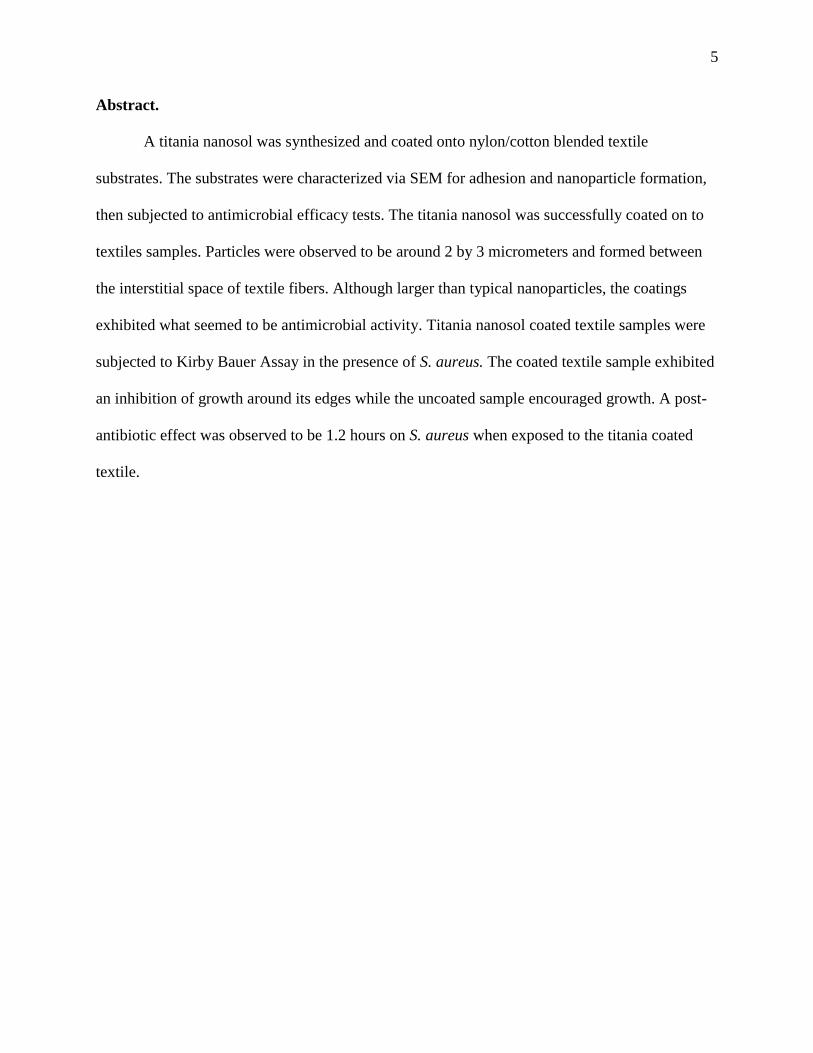
Abstract.
A titania nanosol was synthesized and coated onto nylon/cotton blended textile
substrates. The substrates were characterized via SEM for adhesion and nanoparticle formation,
then subjected to antimicrobial efficacy tests. The titania nanosol was successfully coated on to
textiles samples. Particles were observed to be around 2 by 3 micrometers and formed between
the interstitial space of textile fibers. Although larger than typical nanoparticles, the coatings
exhibited what seemed to be antimicrobial activity. Titania nanosol coated textile samples were
subjected to Kirby Bauer Assay in the presence of S. aureus. The coated textile sample exhibited
an inhibition of growth around its edges while the uncoated sample encouraged growth. A post-
antibiotic effect was observed to be 1.2 hours on S. aureus when exposed to the titania coated
textile.
6
Introduction.
Antimicrobial coatings have been developed for a variety of different applications to
reduce the proliferation of bacteria, fungi, and viruses on surfaces. Current scientific research
involving antimicrobial coatings for textiles is gaining attention in the healthcare industry due to
the increased risk of healthcare associated infections (HAIs) (Gouveia, 2010). Reducing the
number of pathogenic microorganisms in a patient’s environment is now a high priority in all
healthcare institutions. Antimicrobial coated surfaces can potentially reduce these troublesome
infections.
The goal of this project is to create an antimicrobial coating using nanoparticle titanium
dioxide to be applied to textile samples. A titanium dioxide coating will be characterized and
observed for antimicrobial activity. Titanium dioxide (also known as titania) was chosen as the
base for this coating because of its photocatalytic activity. The photocatalytic activity of
nanoparticle titania is useful for antimicrobial applications and is currently being investigated for
its ability to inhibit the growth of pathogenic microorganisms (Dastjerdi, 2010). Partly due to the
recent interest in reducing transmission of infectious disease, titania nanoparticles could be used
for surface treatments to deter growth of pathogenic microorganisms in the surrounding clinical
environment. Every year, 1.7 million HAIs cause almost 100,000 deaths in the United States
(Pollack, 2010). Those that don’t end in death can cause life-threatening illnesses. HAIs are
generally caused by transmission of microbes between surfaces, patients and employees. Patients
that are immune-suppressed are among the most susceptible to HAIs. In fact, any patient
admitted into a healthcare facility for a medical transplant, disease, or critical trauma is at risk
(Neely, 2000). One of the most common of these infections is Staphylococcus aureus, also
known as “Staph.” S. aureus is a facultative anaerobic gram positive coccus (spherical)
7
bacterium, which indicates its spherical shape and lack of an outer membrane. Staph colonizes in
both aerobic and anaerobic conditions and is a normal flora in about 30% of industrialized
countrie's populations (Page, 2007). Many strains of Staph have the ability to develop a
resistance to antibiotics. For example, methicillin resistant or multi-resistant Staphylococcus
aureus (MRSA) was found in 50 % of Staph infection cases in United States ICUs in 2007
(Rosenthal, 2009). Klebsiella pneumonia, Acinetobacter baumannii, Pseudomonas aeruginosa,
and Enterococcus have also developed antibiotic resistant strains that are difficult to control in
clinical environments (Rosenthal, 2009). Clostridium difficle has become one of the leading
causes of infection due to its spore forming bacterial cell wall. The dormant endospores of C.
diff. allow it to undergo harsh conditions from antibiotics and antimicrobial surfaces (Pant,
2011). One of the reasons that these infections might find their way to immune deficient patients
is contamination of the surrounding environment. For example, patients are in constant contact
with bed linen during their hospital stay. The transfer of infectious disease through linen,
although not highly recognized, is very common in clinical settings. Under certain temperatures
and humidity, textiles such as bed linen are a perfect place for microbes to grow. (Gouveia,
2010).
In most healthcare facilities, linens, scrubs and gowns are rented out by an industrial
launderer. During the transportation of linen from the industrial laundry plant, it may be handled
by up to eight different people before it is placed on the bed of a patient (according to healthcare
personnel at Sierra Vista Regional Medical Center in San Luis Obispo, CA). Environmental
personnel handling the linens may not be wearing protective gloves, allowing possible transfer
of pathogens from handler to patient. In order to strive for more sanitary conditions,
antimicrobial coatings and treatments of textiles can potentially be used to actively fight the

8
growth of microorganisms after they've been washed. By eliminating pathogens in a patient’s
direct environment, antimicrobial linen can potentially reduce the risk of infectious disease
transfer in the clinical environment.
Titanium dioxide is a semiconductor metal oxide that is used in a variety of coating
applications. It is capable of degrading organic matter through photocatalytic activity under
certain circumstances. Surface photocatalysis provides very harsh conditions for microbial
growth (Abidi, 2009). It was first discovered in the 1960s by Akira Fujishima. He observed the
formation of radicals by photocatalytic breakdown of water in the presence of TiO2 on Pt
electrodes under exposure to UV radiation (Fujishama, 2006). Photocatalysis of titania occurs
when light is absorbed by a TiO2 particle. The particle then creates electron holes which will
generate hydroxyl radicals. Therefore, O2 and H2O can be reduced to form peroxides. The
instability of most hydroxyl radicals and peroxide molecules allows them to decompose organic
molecules on contact (Abidi, 2009). The reactivity of a photocatalytic surface serves a variety of
applications. For example, immobilized TiO2 nanoparticles have been commonly used as
coatings for indoor odor removal, antimicrobial surfaces, self-cleaning surfaces, and water
treatment. (Fujishama, 2006). TiO2 containing wall paper and air fresheners are used because
they decompose ammonia, hydrogen sulfide, acetaldehyde, toluene, methyl mercaptan, and
nitrogen in the nearby area (Fujishama, 2006). In previous studies of titania coated wall tiles,
glass plates, and linens, photocatalytic TiO2 killed E. coli and even decomposed its dead cells
(6). It has been reported that nanoparticle titania is utilized as exterior architectural coatings and
has proved itself to be useful for its ability to decompose octadecane, glycerol, triolate, and PEG
under very weak UV illumination while exhibiting self-regeneration (Fujishama, 2006). This
application has the ability to destroy organic pollutants or soil buildup on many exterior surfaces

9
(Fujishama, 2006). Photocatalytic titania has been reported for its ability to treat toxic chemicals
in British river water without creating any toxic byproducts (Fujishama, 2006). In addition, its
use as a super-hydrophilic agent has been successfully used for anti-fogging films and self-
cleaning glass (Fujishama, 2006). Photocatalytic TiO2 is highly applicable due to its price,
chemical stability, and properties (Fernando, 2009).
In this study, a titania coating was synthesized in the form of a nanosol, and coated onto
hospital linen. The nanosol is a nanoparticle dispersion that consists of an inorganic metal oxide
dispersed in an organic matrix. This is often made feasible by the use of sol gel chemistry. A sol-
gel process is carried out through the combination of a low molecular weight organic precursor
and an inorganic precursor such as an inorganic alkoxide (Fernando, 2009). There are many uses
for titania nanosols. It has been reported that a sol gel process using a titanium alkoxide with
ethanol in acidic conditions has been successful in creating an anti-microbial and self-cleaning
coating on textiles (Wu, 2009; Daoud, 2005). Titania nanosols have been reported to have the
ability to be photo-activated which induces antimicrobial activity once coated onto textiles.
(Rahal, 2011; Dastjerdi, 2010). The efficiency of a titania nanosol surface treatment was
characterized with scanning electron microscopy (SEM), and antimicrobial resistance is
measured by both Kirby-Bauer diffusion-type assays (KBA), and Post-antibiotic Effect (PAE),
both of which are common for determining an antibiotics efficacy (Braga, 2004; Drew, 1972).

10
Experimental
Nanosol Preparation:
Titania nanosol was synthesized using 10 mL of titanium (IV) butoxide precursor
purchased from Sigma Aldrich. This inorganic alkoxide was dissolved in 25 mL of ethanol and
added drop wise to 200 mL of a .04 M nitric acid solution in a 500 mL round bottom flask while
being stirred at room temperature for 48 hours to ensure that the hydrolysis reaction takes place
to form titania crystals. This nanosol was set to stand at room temperature for an additional 72
hours before it could be used for the coating application. The TiO2 content was measured using a
TA Instruments Q-500 Thermogravimetric Analyzer (TGA). The dispersion was measured to
contain 0.66% solids.
Coating Application:
The titania nanosol was coated onto a freshly cleaned bed sheet using a dip coating
technique. A brand new hospital bed sheet, T-180 thread count, 65 % cotton, 35 % nylon, was
used. The bed sheet was sterilized by boiling for 10 minutes, then washed at 60°C with 50 mL
Tide® Original Scent liquid laundry detergent and 100 mL of deionized water using a 500 mL
beaker for 30 minutes to detach any dirt or impurities that might inhibit the coating from
adhering to the fabric. The fabric was then dipped in ethanol, washed with water to remove any
excess detergent, and dried overnight in a 60 °C oven. The bulk fabric was cut into 1 in2 pieces.
Three pieces were set aside and three were submersed in a titania nanosol for 2.5 minutes with
mixing. It was pressed between two pieces of filter paper to remove excess liquid and then
placed in a 60 °C oven for five minutes to cure the coating. Oven temperature must be increased
to 115 °C to facilitate attachment of titania to fiber as reported by Wu et al. (Wu, 2009) which
increases the formation of titanium dioxide crystals. Coated textiles were placed in a hot water
11
bath at 70 °C for 2 hours to remove any unbound titania particles from the textile to leave a
cleanly coated surface.
Surface Characterization:
Textile surfaces were characterized via SEM for adhesion of titania particulates. Linen
samples (coated and uncoated) were cut to 0.25 in2
pieces. In order to increase electrical
conductivity of linen, it was sputtered with a 50 Å thick layer of gold which increased the
focusing potential. Specimens were examined at approximately 500, 2000, and 10,000X
magnification. Energy Dispersive X-ray analysis (EDX) was carried out at the low and high
magnifications (500/10,000X) to determine the elemental composition of both coated and non-
coated substrates. Areas which were found to have high content of titania by EDX were
characterized.
Antimicrobial Activity
Antimicrobial activity was analyzed using two separate assays; KBA and PAE. A KBA is
used to determine the resistance of a bacterial or viral strain to a certain antibiotic or drug
(DeCross, 1993). Typically, a cotton disk is saturated in an antibiotic or detergent and is placed
on a growth medium such as tryptic-soy agar (TSA) containing a freshly coated liquid layer of a
diluted bacterial colony. The dish is then incubated at 35°C for 24 hours (temperature most
suitable for S. Aureus) and the “zone of inhibition” is measured. The area surrounding the disk
where no colonies have formed indicates the “zone of inhibition”. The diameter of this circular
area is significant of a bacteria’s resistance to a specific antibiotic. This diameter is strongly
dependent on the concentration of antibiotic, bacterial species and temperature of incubation
(Drew, 1972). In this study, 1 in2 titania coated fabric was tested using KBA. In addition, a PAE
is typically used to observe effectiveness of an antibiotic. PAE is measured by the recovery time
12
of a bacterial colony after it has been exposed to an antibiotic for a given amount of time. The
PAE is dependent on the exposure time, bacterial species, and concentration of the antibiotic
(Sharma, 2002). The PAE can be defined as the time it takes for a bacterial colony to recover to
full growing potential after it has been exposed to an antibiotic and can be measured using
absorption spectroscopy. By using absorption spectroscopy, the concentration of microorganism
or colony forming units (CFU) can be determined. The PAE can be calculated at the time a
bacterial colony has increased to its maximum growth potential or when the absorption on the
spectrometer increased by 1 OD600. An absorption increase of 1OD600 indicates an increase of 1
log10 colony forming units. As bacteria increased by 1 log CFU unit, it indicates that a bacterial
colony has returned to its maximum growth rate.
White textiles (same as above) were all tested for their ability to inhibit growth of S.
aureus on tryptic-soy agar (TSA) agar. S. aureus (ATCC 6538) was obtained from the Cal Poly
microbiology stockroom and analyzed for purity. It was streaked on a sheep blood agar plate and
incubated for 24 hours at approximately 35°C to detect purity. S. aureus is a beta-hemolytic
species that turns red agar yellow. Stock S. aureus was prepared by picking 4-5 typical colonies
with a sterile disposable 10 μL loop and re-suspending colony in 25 mL of tryptic-soy broth
(TSB) in a sterile Erlenmeyer flask. The flask was placed in a shaker-incubator for 2 hours at
35 °C and then adjusted to .09-.12 D.U. on a Spectronic 20 OD600 spectrometer which
corresponds to about 1x10⁸ CFU S. aureus. This inoculum was then used for PAE and KBA.
In preparation for the PAE assay, 1 mL of TSB was added to 5 separate test tubes. A 1 in2
piece of titania-coated white cloth was added to tube 1 and a non-coated piece of cloth was
added to tube 2. 1 mL of 25 ppb silver nitrate is added to tube 3. The inoculum described above
was diluted 1:10 with pre-warmed TSA and 1 mL was added to tubes 1-4. Tube 4 serves as the
13
inoculum control (only S. aureus and growth medium without fabric) and tube 5 serves as
medium sterility containing only the growth medium, (Table 1). The tubes were incubated for 1
hour and the inoculum was plated on TSA using a spread-plate method by pipetting 0.1mL
aliquots of 10-5
and 10-6
dilutions of inoculum to determine viable bacterial count. Tubes 1-6 are
incubated for 1 hour and then diluted 1:1000 with pre-warmed TSB and 250 μL aliquots were
pipetted into sterile micro well plates in duplicate. Growths curves were constructed taking
absorbance readings at 600 nm every 10 minutes for 12 hours on a Molecular Devices Spectra
Max Plus 384 micro plate reader.
Table 1: Contents of Tubes
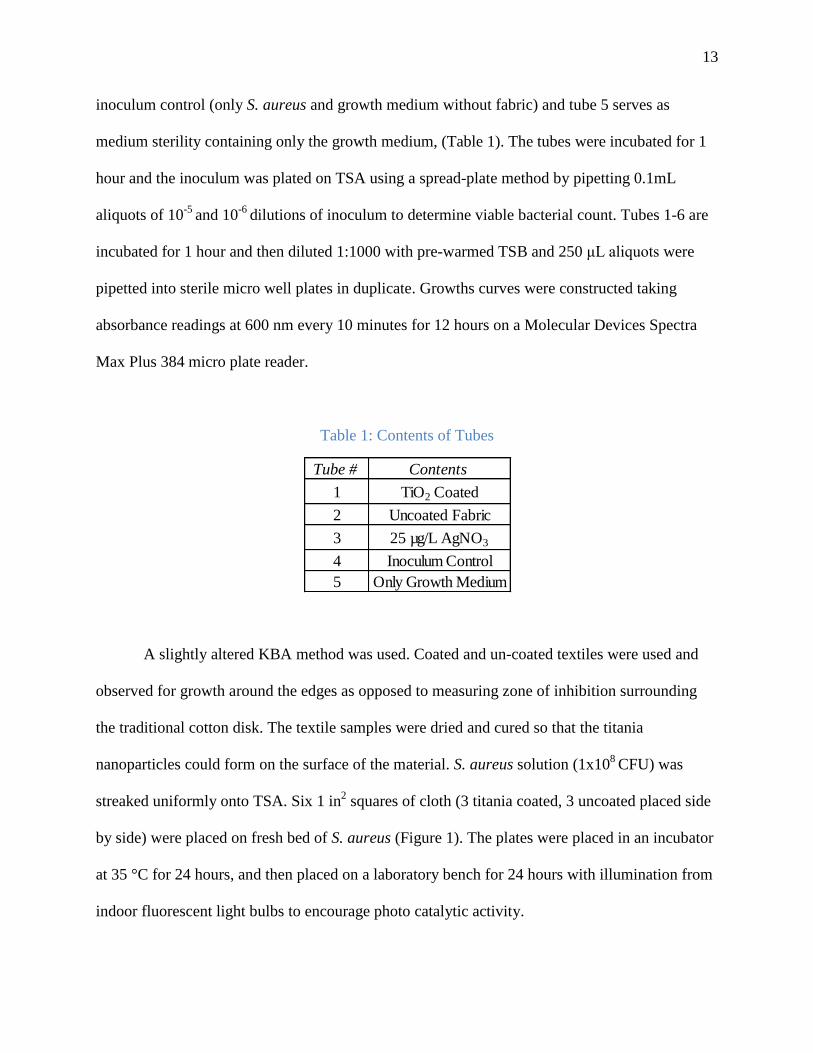
A slightly altered KBA method was used. Coated and un-coated textiles were used and
observed for growth around the edges as opposed to measuring zone of inhibition surrounding
the traditional cotton disk. The textile samples were dried and cured so that the titania
nanoparticles could form on the surface of the material. S. aureus solution (1x10⁸
CFU) was
streaked uniformly onto TSA. Six 1 in2 squares of cloth (3 titania coated, 3 uncoated placed side
by side) were placed on fresh bed of S. aureus (Figure 1). The plates were placed in an incubator
at 35 °C for 24 hours, and then placed on a laboratory bench for 24 hours with illumination from
indoor fluorescent light bulbs to encourage photo catalytic activity.
Tube # Contents
1 TiO2 Coated
2 Uncoated Fabric
3 25 µg/L AgNO3
4 Inoculum Control
5 Only Growth Medium
14
Figure 1: Diagram exhibits the orientation of titania coated vs. non-coated textiles on a bed of S.
aureus.
15
Results and Discussion:
Surface Characterization
Figures 2 and 3 display the SEM images of a coated vs. uncoated linen at 500 X
magnification. It is difficult to distinguish individual fibers in Figure 3 compared to easily
distinguished fibers in Figure 2. The difference in clarity is most likely be a result of the added
titanium dioxide particles. Figure 2 also exhibits a much greater depth of field than Figure 3.
This may indicate that there is less free space due to the presence of titania. This tightly packed
network makes it difficult to perceive depth in the image. It was also observed that the fabrics
were physically stiffer and brittle when coated compared to a pristine (uncoated) fabric. At low
magnifications, the EDS exhibited significant peaks for carbon and oxygen as expected. Carbon
and oxygen are the key elements of the polymer chains that make up cotton and nylon fibers. The
EDX displayed trace amounts of gold due to gold sputtering procedure used to enhance electrical
conductivity for SEM analysis. Titania coated fabric exhibited a large signal of titanium at
15.05 % weight as determined by EDX software.

16
Figure 2: SEM Image of uncoated white fabric at 500 X magnification.
Figure 3: SEM image of titania coated white fabric at 500 X magnification.

17
At higher magnifications (greater than 1500 X) particle formations are observed between
the fibers (Figure 5) rather than on the surface of the fibers. At approximately 2000 X
magnification there is little difference in depth of field between coated (Figure 5) and uncoated
(pristine) fabrics (Figure 4). The coating formed a branching network throughout the specimen in
Figure 5, while the non-coated fabric displayed very smooth surfaces on the fibers with free
space in between them.
Figure 4: SEM image of uncoated white fabric at 2000 X magnification.
18
Figure 5: SEM image of titania coated white fabric at 1500 X magnification.
Figures 6 and 7 display the SEM images of very high magnifications of coated and
pristine fabrics. Unfortunately, the SEM could not focus above 8000 X, before losing focus, but
was still comparable with the high magnification of the non-coated fabric in Figure 6 which
allowed focus as high as 12000 X. At this magnification, the non-coated fabric shows a very
clear depth of field, while the coated fabrics are very flat in their image. Titania formed
interstitially between various fibers of coated fabric (Figure 7). Titania particle sizes were
consistently between 5 and 10 µm. EDS characterized them to be titanium and oxygen indicating
that they are indeed titanium dioxide microparticles rather than nanoparticles. EDS also displays
the placement of titania particles in the fiber matrix. It is clear that the composition of the
interstitial area is largely made up by titanium and oxygen (32 and 46 % weight), which is likely

19
to be titanium dioxide (Figure 7). The fiber surfaces itself contains very little titanium (2.21 %
weight) compared to the interstitial space between fibers. The network of titania particles may
contribute to the observed stiffness of this textile.
Figure 6: SEM of Uncoated white fabric at 12000 X.
Figure 7: SEM Titania coated white fabric at 8000 X.
20
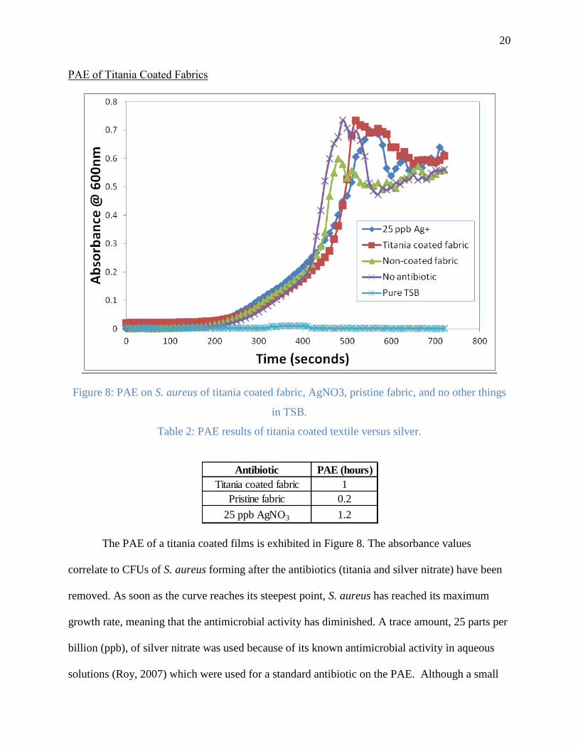
PAE of Titania Coated Fabrics
Figure 8: PAE on S. aureus of titania coated fabric, AgNO3, pristine fabric, and no other things
in TSB.
Table 2: PAE results of titania coated textile versus silver.
The PAE of a titania coated films is exhibited in Figure 8. The absorbance values
correlate to CFUs of S. aureus forming after the antibiotics (titania and silver nitrate) have been
removed. As soon as the curve reaches its steepest point, S. aureus has reached its maximum
growth rate, meaning that the antimicrobial activity has diminished. A trace amount, 25 parts per
billion (ppb), of silver nitrate was used because of its known antimicrobial activity in aqueous
solutions (Roy, 2007) which were used for a standard antibiotic on the PAE. Although a small
Antibiotic PAE (hours)
Titania coated fabric 1
Pristine fabric 0.2
25 ppb AgNO3 1.2

21
amount is used, AgNO3 was able to create a PAE of 1.2 hours in an ultra-dilute solution.
Considering that these antibiotics were only exposed to a S. aureus for 1 hour, a 1 hour lasting
effect is substantial compared to other antibiotics (Sharma, 2002). Titania coated textiles were
also able to suppress the bacterial growth by resulting in a PAE of 1 hour. The uncoated fabric
shows an inhibition of growth for 0.2 hours. This inhibition may be due to the fact that the
substrate is not the most applicable growth medium for S. aureus making it difficult for bacteria
to grow. It is possible that the 1 hour PAE observed in the titania coated samples is due to
photocatalytic antimicrobial activity. Typical PAEs may last between 1 and 3 hours but will not
completely describe the antimicrobial activity of a compound (Braga, 2004).
Kirby Bauer Assays
KBAs determine antimicrobial efficacy and may suggest photo catalytic activity of the
TiO2 coated fabric. The KBA is shown for six different fabrics. Three were coated with titania
(Figures 9d, 9e and 9f) while three were uncoated (9a, 9b, and 9c). Figure 9 displays the
inoculated medium TSA plate after textiles have been removed. From top to bottom was blue
nylon/cotton blended textile (9a and 9d), white nylon/cotton blend (9b and 9e), and yellow
carbon laced nylon textile (9c and 9f). This was done to measure the visual regrowth around the
edges of the cloth. All of the titania coated fabrics exhibited a low level of bacterial growth
around their edges (Figures 9d,9e, and 9f). Both untreated blue and white textiles encouraged
bacterial growth, but yellow did not. Unlike cotton/nylon blended fabrics, the yellow fabric was
composed of a mixture of nylon fibers laced with carbon. It is clear that S. aureus was unable to
grow around the edges and underneath this fabric sample whether or not it was coated in titania.
The activated carbon may contribute to antimicrobial behavior of uncoated textile. Overall, the
lack of growth around the white and blue coated fabrics compared to the non-coated fabrics

22
indicate that there is possible antimicrobial activity occurring. The plates may be exhibiting
photocatalytic activity due to indoor laboratory luminescence. The radical formation would
therefore inhibit microbial growth around the edges of the cloth.
Figure 9. Titania coated fabric areas are displayed on the right while pristine fabric areas are
displayed on the left side of the Petri dish. a) and d) are blue cotton/nylon blended fabric, b) and
e) are white cotton/nylon blended fabric, and c) and f) were carbon laced nylon.
23
Conclusion
A Titanium dioxide nanosol was prepared from a hydrolysis reaction of titanium (IV)
butoxide and ethanol. This nanosol was used as a coating for cotton and nylon blended fibers and
subjected to a series of tests. SEM characterization displayed formation of titania particle
formation in the interstitial spaces between fibers. The particles were determined to contain
mostly titanium and oxygen by EDS analysis, confirming that the particles were most likely pure
titanium dioxide particles. SEM instrument was unable to focus at high magnifications due to
lack of electrical conductivity. Although a gold sputtering technique was used to increase
electrical conductivity of sample, magnifications on the nano-scale were not obtained. Although
larger than expected in size, SEM showed successful adhesion of titania particles in between
threads of nylon/cotton textiles.
In biological analysis, titania coated textiles inhibited growth of S. aureus on and around
its surface, and produced post- antibiotic effects on S. aureus in TSB. The prevention of future
growth was exhibited in a PAE of 1 hour, meaning that it took S. aureus 1 hour to recover and
resume its maximum growth rate once the titania coated fabric was removed from its growth
medium. KBA displayed an inhibition of growth around the edges of titania coated textile, but
not around pristine substrates. In fact, S. aureus colonized favorably around pristine fabric on a
TSA growth medium. It's possible that textile samples were not completely sterilized in the pre-
treatment process. Linens were pre-treated by boiling in water for 10 minutes. It's possible that
the textile samples were contaminated with endosporic bacillus (able to withstand very high
temperatures and pressures) prior to the boiling process. It might be that excess growth around
the edges of the untreated nylon/cotton textile in KBA is just contamination from earlier contact.
In future experiments, it would be more aseptic to autoclave the textile samples for completely

24
sterile testing subjects to ensure that all spore forming organisms are killed. In addition to further
microbiological tests, future work might include the study of adhesion efficiency of titania
particles and antibacterial activity dependence on particle concentration. In addition to
experimental improvements, the health effect of a titania coated textile for human application
could be studied in future work. Before an application like this can be used in a clinical setting,
its health effects must be studied. Photocatalysis and its effect on the skin (for use of titania
textiles in clothing) needs to been investigated.
In conclusion, titanium (IV) butoxide was successfully used to synthesize a coating on
the cotton/nylon substrates. The coating exhibited possible antimicrobial ability, but further
research must be conducted to determine existence of photocatalytic activity. Because of titania's
cost and ease of use, it has potential in the field of antimicrobial coatings to reduce the spread of
infectious disease on surfaces. Although linen is not commonly known as a bacterial vector, it is
gaining much more attention in hospitals and other clinical settings as an infectious disease
carrier. Titanium dioxide’s ability to photo-degrade small compounds has already been utilized,
but it has far greater potential and may contribute to saving lives by reducing the risk of
infectious disease.
25
References
Abidi, Noureddine, Eric Hequet, and Luis Cabrales. Functionalization of a Cotton Fabric Surface
with Titania Nanosols: Applications fo Self-Cleaning and UV-Protection Properties. ACS
Applied Materials and Interfaces, 2009, 2141-2146.
Bogner, A., P.H. Jouneau, G. Thollet, D. Basset, and C. Gauthier. A history of scnaning electorn
microscopy developments: Towards "wet-STEM" imaging. Micron, 2007, 390-401.
Braga, P. C., M. Culici, and M. Dal Sasso. The post-antibiotic effects of rokitamycin on
susceptible and erthromyci-resistant stratins of Streptococcus pyogenes. International
Journal of Antimicrobial Agents. 2004, 24.
Daoud, Walid A., John H. Xin, Yi-He Zhang, and Kaihong Qi. Surface characterization of thin
titania films prepared at low temperatures. Journal of Non-Crystalline Solids. 2005, 351.
Dastjerdi, Roya, and Majid Montazer. A review on the applicaiton of inorganic nano-stucture
materials in the modicification of textiles: Focus on anti-microbial properties. Colloids
and Surfaces B: Biointerfaces. 2010, 79.
DeCross, Arthur J., Barry J. Marshall, Richard W. McCallum, Susie R. Hoffman, Leah J. Barrett,
and Richard L. Guerrant. Metronidazole Susceptibility testing for Heliobacter Pylori:
Comparison of Disk, Broth, and Agar Dilution Methods and Their Clinical Relevence.
Journal of Clinical Microbiology. 1993, 8, 31.
Fernando, Raymond H., and Li-Piin Sung. Nanotechnology Applications in Coatings. ACS,
2009, 1008.
Fujishama, Akira, and Xintong Zhang. Titanium dioxide photocatalysis: present situation and
future approaches. Comptes Rendus, 2006, 750-760.
Goldstein, Joseph; Newbury, Dale; Joy, David; Lyman, Charles; Echlin, Patrick; Lifshin, Eric;
Sawyer, Linda; Michael, Joseph. Scanning Electron Microscopy and X-ray
Microanalysis. 2003, 1.
Gouveia, I.C. Nanobiotechnology: A new strategy to develop non-toxic antimicrobial textiles for
healthcare applications. Journal of Biotechnology. 2010, 150-S.
Lawrence Drew, W., A. L. Barry, Richard O'Toole, and John C. Sherris. Reliability of the Kirby-
Bauer Disc Diffusion Method for detecting Methicillin-Resistant Strains of
Staphylococcus aureus. Applied Microbiology. 1972, 240-247.
Neely, Alice N., and Matthey P. Maley. Survival of Enterococci and Staphylococci on Hospital
Fabrics and Plastic. Journal of Clinical Microbiology. 2000, 724-726.

26
Page, Kristopher, Robert G. Palgrave, Ivan P Parkin, Michael Wilson, Shelley L.P. Savin, and
Alan V. Chadwick. Titania and silver-titania composite films on glass: potent
antimicrobial coatings. Journal of Materials Chemistry. 2007, 95-104.
Pant, Chaitanya, Thomas J. Sferra, Abhishek Deshpande, and Anil Minocha. Clinical approach
to sever Clostridium difficle infection: Update for the hospital practitioner. European
Journal of Internal Medicine. 2011.
Pollack, Andrew. Rising Threat fo Infections Unfazed by Antibiotics. The New York Times,
February 27, 2010: B1.
Rahal, R., T. Pigot., D. Foix, and S. Lacombe. Photocatalytic efficiency and self-cleaning
properties under visible light of cotton fabrics coated with sensitized TiO2. Applied
Catalysis B: Environmental. 2011, 104.
Rosenthal, Victor D., Dennis G Maki, Silom Jamulitrat, and Eduardo A. Medeiros. International
Nosocomial Infection Contro Consortium (INICC) report, data summary for 2003-2008,
issued June 2009. American Journal of Infection Control. 2009, 95-104.
Roy, R.; Hoover, M. R.; Bhalla, A. S.; Slawecki, T.; Dey, S.; Cao, W.; Li, J.; Bhaskar, S.
Ultradilute Ag-aquasols with extraordinary bactericidal properties: role of the system Ag-
O-H2O. Materials Research innovations. 2007, 11.
Sharma, K.K., H. Sangraula, and P.K. Mediratta. Some New Concepts in Antibacterial Drug
Therapy. Indian Journal of Pharmacology. 2002, 390-396.
Wu, Deyong; Long, Mingce; Zhou, Jiangya; Cai, Weimin; Zhu, Xiehao; Chen, Chao; Wu,
Yahui. Synthesis and characterization of self-cleaning cotton fabrics modified by TiO2
through facile appraoch. Surface & Coatings Technology. 2009, 3728-3733.