Synovial Fluid. Physiology Synovial fluid, often referred to as “joint fluid,” is a viscous...
20
-
Upload
allan-boyd -
Category
Documents
-
view
223 -
download
3
Transcript of Synovial Fluid. Physiology Synovial fluid, often referred to as “joint fluid,” is a viscous...
Physiology
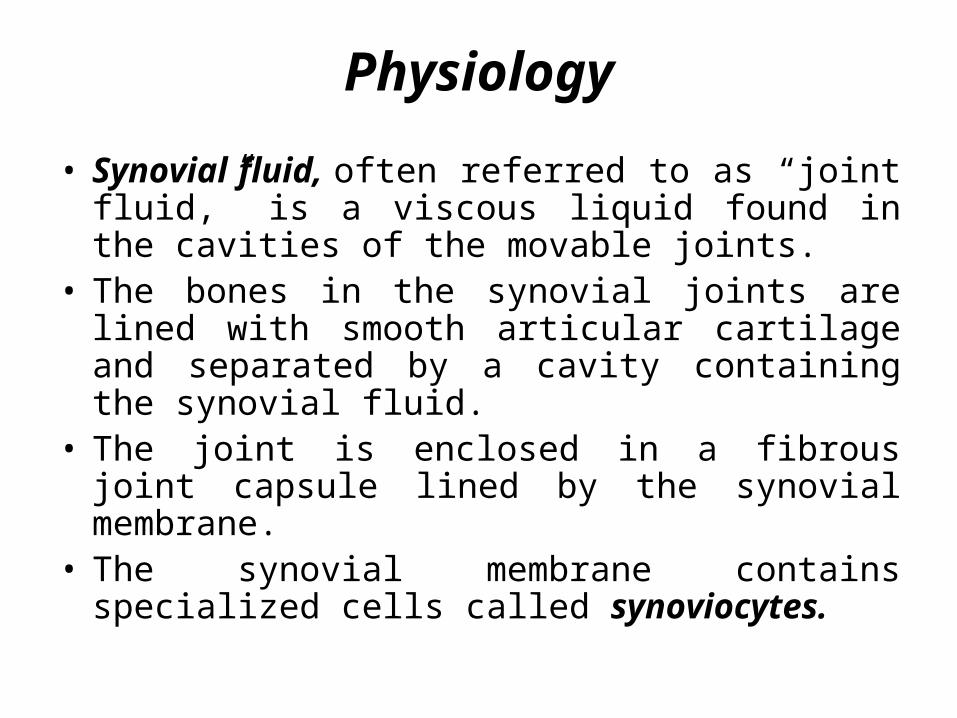
• Synovial fluid, often referred to as “joint fluid,” is a viscous liquid found in the cavities of the movable joints.
• The bones in the synovial joints are lined with smooth articular cartilage and separated by a cavity containing the synovial fluid.
• The joint is enclosed in a fibrous joint capsule lined by the synovial membrane.
• The synovial membrane contains specialized cells called synoviocytes.

• Synovial fluid is formed as an ultrafiltrate of plasma across the synovial membrane. The filtration is nonselective except for the exclusion of high molecular weight proteins.
• Therefore, the majority of the chemical constituents, although seldom of clinical significance, have concentrations similar to plasma values.
• The synoviocytes secrete a mucopolysaccharide containing hyaluronic acid and a small amount of protein (approximately one fourth of the plasma concentration) into the fluid.

Function1. The smooth articular cartilage and synovial fluid
reduce friction between the bones during joint movement.
2. To providing lubrication in the joints.
3. To providing nutrients to the articular cartilage.
4. Lessens the shock of joint compression that occurs during activities such as walking and jogging.
Why the test is performed
• The test can help diagnose the cause of pain, redness, or swelling in joints.
• Sometimes, removing the fluid can also help relieve (remove) joint pain.
• This test may be used when your doctor suspects:Bleeding in the joint after a joint injuryGout and other types of arthritis Infection in a joint

Specimen Collection and Handling
• Synovial fluid is collected by needle aspiration called arthrocentesis.
• The amount of fluid present varies with the size of the Joint. • The normal amount of fluid in the adult knee cavity is less than
3.5 mL, but can increase to greater than 25 mL with inflammation.
• The volume of fluid collected should be recorded.• Normal synovial fluid does not clot; however, fluid from a
diseased joint may contain fibrinogen and will clot.
Look
• When sufficient fluid is collected, it should be distributed into the following tubes based on the required tests:
1. A sterile heparinized tube for Gram stain and culture.
2. A heparin or ethylenediaminetetraacetic acid (EDTA) tube for cell counts.
3. A sodium fluoride tube for glucose analysis.
4. A nonanticoagulated tube for other tests.
• Powdered anticoagulants should not be used because they may produce artifacts that interfere with crystal analysis. All testing should be done as soon as possible to prevent cellular lysis and possible changes in crystals.


Color and Clarity• A report of the gross appearance is an essential part
of the synovial fluid analysis. Normal synovial fluid appears colorless to pale yellow.
• Turbidity is frequently associated with the presence of WBCs; however, synovial cell debris and fibrin also produce turbidity.
• The fluid may appear milky when crystals are present.

Viscosity• Viscosity of the synovial fluid comes from the
polymerization of the hyaluronic acid and is essential for the proper lubrication of the joints.
• Arthritis affects both the production of hyaluronate and its ability to polymerize.
• Several methods are available to measure the viscosity of the fluid.
• The simplest being to observe the ability of the fluid to form a string from the tip of a syringe, and can be done at the bedside. A string that measures 4 to 6 cm is considered normal.
Cell Counts• The total leukocyte count is the most frequently performed
cell count on synovial fluid. Red blood cell (RBC) counts are seldom requested.
• To prevent cellular disintegration, counts should be performed as soon as possible or the specimen should be refrigerated.
• Note: Very viscous fluid may need to be pretreated by adding a pinch of hyaluronidase to 0.5 mL of fluid or one drop of 0.05% hyaluronidase in phosphate buffer per milliliter of fluid and incubating at 37C for 5 minutes.
• WBC counts less than 200 cells/µL are considered normal and may reach 100,000 cells/µL or higher in severe infections.

Crystal Identification• Microscopic examination of synovial fluid for the
presence of crystals is an important diagnostic test in the evaluation of arthritis.
• Crystal formation in a joint frequently results in an acute, painful inflammation. It can also become a chronic condition.
• Causes of crystal formation include: metabolic disorders and decreased renal excretion that produce elevated blood levels of crystallizing chemicals, degeneration of cartilage and bone, and injection of medications, such as corticosteroids into a joint.

Types of Crystals
• The primary crystals seen in synovial fluid are monosodium urate (uric acid) (MSU) found in cases of gout and calcium pyrophosphate (CPPD) seen with pseudogout.
• The most frequent causes of gout are: Increased serum uric acid resulting from impaired
metabolism of purines. Increased consumption of high-purine-content foods,
alcohol, and fructose; Chemotherapy treatment of leukemias; Decreased renal excretion of uric acid are

• Pseudogout is most often associated with:Degenerative arthritis, producing cartilage calcificationEndocrine disorders that produce elevated serum calcium
levels.
Chemistry Tests• The most frequently requested test is the glucose
determination, as markedly decreased values are indicative of inflammatory.
• Because normal synovial fluid glucose values are based on the blood glucose level, simultaneous blood and synovial fluid samples should be obtained, preferably after the patient has fasted for 8 hours to allow equilibration between the two fluids.
• Under these conditions, normal synovial fluid glucose should not be more than 10 mg/dL lower than the blood value.
• To prevent falsely decreased values caused by glycolysis, specimens should be analyzed within 1 hour or preserved with sodium fluoride.
• Other chemistry tests that may be requested are the total protein and uric acid determinations. Because the large protein molecules are not filtered through the synovial membranes, normal synovial fluid contains less than 3 g/dL of protein (approximately one third of the serum value).
• Increased levels are found in inflammatory and hemorrhagic disorders;

Microbiologic Tests• An infection may occur as a secondary complication
of inflammation caused by trauma or through dissemination of a systemic infection; therefore, Gram stains and cultures are two of the most important tests
• Bacterial infections are most frequently seen; however, fungal, tubercular, and viral infections also can occur.
• In addition toStaphylococcus and Streptococcus, the common organisms that infect synovial fluid are the fastidious Haemophilus species and N. gonorrhoeae.

Possible Meanings of Abnormal Values
1. Gout2. Osteoarthritis3. Pseudogout4. Rheumatic fever5. Rheumatoid arthritis6. A Septic arthritis7. Systemic lupus erythematosus8. Traumatic arthritis9. Tuberculous arthritis