Survival strategies of the human respiratory tract ...
69
Survival strategies of the human respiratory tract pathogen Haemophilus influenzae Hallström, Teresia 2007 Link to publication Citation for published version (APA): Hallström, T. (2007). Survival strategies of the human respiratory tract pathogen Haemophilus influenzae. Medical Microbiology, Lund University. Total number of authors: 1 General rights Unless other specific re-use rights are stated the following general rights apply: Copyright and moral rights for the publications made accessible in the public portal are retained by the authors and/or other copyright owners and it is a condition of accessing publications that users recognise and abide by the legal requirements associated with these rights. • Users may download and print one copy of any publication from the public portal for the purpose of private study or research. • You may not further distribute the material or use it for any profit-making activity or commercial gain • You may freely distribute the URL identifying the publication in the public portal Read more about Creative commons licenses: https://creativecommons.org/licenses/ Take down policy If you believe that this document breaches copyright please contact us providing details, and we will remove access to the work immediately and investigate your claim. Download date: 06. Oct. 2021
Transcript of Survival strategies of the human respiratory tract ...

LUND UNIVERSITY
PO Box 117221 00 Lund+46 46-222 00 00
Survival strategies of the human respiratory tract pathogen Haemophilus influenzae
Hallström, Teresia
2007
Link to publication
Citation for published version (APA):Hallström, T. (2007). Survival strategies of the human respiratory tract pathogen Haemophilus influenzae.Medical Microbiology, Lund University.
Total number of authors:1
General rightsUnless other specific re-use rights are stated the following general rights apply:Copyright and moral rights for the publications made accessible in the public portal are retained by the authorsand/or other copyright owners and it is a condition of accessing publications that users recognise and abide by thelegal requirements associated with these rights. • Users may download and print one copy of any publication from the public portal for the purpose of private studyor research. • You may not further distribute the material or use it for any profit-making activity or commercial gain • You may freely distribute the URL identifying the publication in the public portal
Read more about Creative commons licenses: https://creativecommons.org/licenses/Take down policyIf you believe that this document breaches copyright please contact us providing details, and we will removeaccess to the work immediately and investigate your claim.
Download date: 06. Oct. 2021

Survival strategies of the human respiratory pathogen
Haemophilus influenzae
Teresia Hallström
Medicinsk Mikrobiologi
Institutionen för Laboratoriemedicin
Universitetssjukhuset MAS, Malmö
Lunds Universitet
Akademisk avhandling
som med vederbörligt tillstånd av Medicinska fakulten vid Lunds Universitet för avläggande
av doktorsexamen i medicinsk vetenskap kommer att offentligen försvaras i
Patologiska Institutionens föreläsningssal, Ingång 78, Universitetssjukhuset MAS,
Malmö, tisdagen den 15 maj 2007, kl. 14.00.
Fakultetsopponent: Prof. Mogens Kilian, Institute of Medical Microbiology and Immunology
Aarhus University, Denmark


3
Survival strategies of the human respiratory pathogen
Haemophilus influenzae
Teresia Hallström
Doctoral thesis
MALMÖ 2007
Medical Microbiology
Department of Laboratory Medicine
Lund University
Sweden

4
Teresia Hallström 2007
Printed by Media Tryck, Lund University, Sweden
ISBN 978-91-85559-60-2

5
Till mamma och pappa

6
flavae quoque facunt

7
CONTENTS
LIST OF PAPERS 8
ABSTRACT 9
ABBREVIATIONS 10
INTRODUCTION 11HAEMOPHILUS INFLUENZAE 11
General properties 11 Prevalence 11 Infections caused by H. influenzae 12
Pathogenesis and virulence factors 13
GRAM-NEGATIVE SECRETION MECHANISMS 20 General structure of autotransporters 21
THE IMMUNE SYSTEM 23 THE COMPLEMENT SYSTEM 23 Activation of the complement system 24 Regulation of the complement system 27 Functions of the complement system 31 Microbes and the complement system 32 Complement in the respiratory tract 34
H. influenzae and the complement system 35 IMMUNOGLOBULINS 36 Immunoglobulin D 37 Bacterial non-immune immunoglobulin binding 38 Gram-positive bacteria 38
Gram-negative bacteria 39 Importance of bacterial non-immune binding 39
THE PRESENT INVESTIGATION 41 AIMS 41 RESULTS AND DISCUSSION 42 CONCLUDING REMARKS 49
FUTURE PERSPECTIVES 51
SVENSK POPULÄRVETENSKAPLIG SAMMANFATTNING 52
ACKNOWLEDGEMENTS 54
REFERENCES 56 APPENDICES: Papers I-V 69

8
LIST OF PAPERS
This thesis is based on the following papers, which are referred in the text by their respective
Roman numerals:
I: Hallström, T., H. Jarva, K. Riesbeck, and A. Blom. 2007. Interaction with C4b-
binding protein contributes to nontypeable Haemophilus influenzae serum resistance.
The Journal of Immunology, in press.
II: Hallström, T., P. Zipfel, A. Blom, A. Forsgren, and K. Riesbeck. 2007. Haemophilus
influenzae interacts with human complement Factor H.
Manuscript
III: Hallström, T., E. Trajkovska, A. Forsgren, and K. Riesbeck. 2006. Haemophilus
influenzae surface fibrils contribute to serum resistance by interacting with vitronectin.
The Journal of Immunology 177(1): 430-436.
IV: Hallström, T., M. Mörgelin, A. Forsgren, and K. Riesbeck. 2007. Haemophilus
influenzae surface fibrils (Hsf) is a trimeric autotransporter that is both secreted and
tethered to the bacterial cell surface.
Submitted to Journal of Bacteriology
V: Samuelsson, M., T. Hallström, A. Forsgren, and K. Riesbeck. 2007. Characterisation
of the IgD-binding site of encapsulated Haemophilus influenzae serotype b.
The Journal of Immunology, in press.
Published papers are reproduced with the permission of the copyright holders. Paper I, III and V The
American Association of Immunologists, Inc.

9
ABSTRACT
Haemophilus influenzae is an important respiratory tract pathogen responsible for a variety of
infections in humans. Encapsulated H. influenzae belongs to one of six serotypes (a-f), of
which type b is the most virulent one causing serious and sometimes life-threatening diseases
(e.g., epiglottitis, septicaemia and meningitis). In contrast, non-typeable H. influenzae (NTHi)
accounts for the majority of local and upper and lower respiratory tract infections.
The pathogenesis of many microorganisms relies on the capacity of pathogens to avoid, resist
or neutralise the host defence including the complement system.
We demonstrate that H. influenzae interferes with both the classical/lectin and
alternative pathways of the complement system. NTHi binds C4BP, the inhibitor of the
classical pathway, and the majority of the H. influenzae tested bound factor H, the inhibitor of
the alternative pathway. Importantly, the capacity to bind C4BP and factor H appears to
render the bacteria more resistant to serum mediated killing. Furthermore, both C4BP and
factor H bound to the surface of H. influenzae retains its cofactor activity as determined by
analysis of C4b and/or C3b degradation.
In addition to interacting with the classical/lectin and alternative pathways, we
demonstrate that Haemophilus surface fibrils (Hsf), which is expressed by encapsulated H.
influenzae, binds vitronectin, a regulator of the terminal pathway of the complement system.
Mapping of the membrane bound Hsf with gold-labelled specific antibodies in
transmission electron microscopy (TEM) revealed a double-folded 100 nm long fibrillar
structure. Using a series of mutants, we showed that when the C-terminal translocator domain
was inactivated, Hsf was not translocated to the bacterial surface. Interestingly, we also show
that outer membrane vesicles (OMV) secreted by the bacteria carry Hsf, and that Hsf is
secreted into the extracellular milieu.
IgD-binding is another important feature of encapsulated H. influenzae type b.
By using a series of different IgD chimeric proteins, the site on the IgD molecule responsible
for the interaction with H. influenzae was characterised. The binding site was localised to the
CH1 region of IgD.
In summary, H. influenzae binds C4BP, factor H and vitronectin, which are
regulators of the complement system. The interaction between H. influenzae and these
regulators protects the bacteria and makes them more resistant to the bactericidal activity of
human serum. Finally, H. influenzae type b binds human IgD.

10
ABBREVATIONS
AOM acute otitis media
C4BP C4b-binding protein
CCP complement control protein
COPD chronic obstructive pulmonary disease
ECM extracellular matrix
FHL-1 factor H like protein 1
Hib Haemophilus influenzae type b
Hsf Haemophilus influenzae surface fibrils
Ig immunoglobulin
kDa kiloDalton
LOS lipooligosaccharide
LPS lipopolysaccharide
MAC membrane attack complex
MBL membrane bound lectin
MID Moraxella IgD-binding protein
mIg membrane-bound Ig
NHS normal human serum
NTHi non-typeable Haemophilus influenzae
OM outer membrane
OMP outer membrane protein
OMV outer membrane vesicle
RCA regulators of complement activation
SCR short consensus repeat
SDS-PAGE sodium dodecyl sulphate polyacrylamide gel electrophoresis
TEM transmission electron microscopy

11
INTRODUCTION
HAEMOPHILUS INFLUENZAE
General properties
Haemophilus is a member of the Pasteurellaceae family, which includes H. influenzae, H.
haemolyticus, H. parainfluenzae, H. parahaemolyticus, H. ducreyi, H. aphrophilus, H.
paraphrophilus, and H. segnis (168). Haemophilus influenzae was first discovered by Robert
Pfeiffer in 1892 (164). The bacteria were first considered to be the cause of influenza,
therefore the characteristic name. However, in 1933 Smith showed that influenza was caused
by a viral agent (206).
H. influenzae is a Gram-negative, rod-shaped, facultative anaerobic, human
specific pathogen, which requires media supplemented with the two growth-stimulating
factors contained in blood: haemin (X factor) and NAD (V factor). Thus, H. influenzae can be
cultured on chocolate agar where both factor V and X are available. H. influenzae grows in a
humid atmosphere with 5-10 % CO2 and at an optimal temperature of 35-37 °C (105). The
bacteria are oxidase positive, catalase positive, ferment glucose and can reduce nitrate.
Figure 1. H. influenzae attached to respiratory epithelium.
(This picture is taken from; http://sitemaker.umich.edu/medchem6/files/influenzae.jpg)
Prevalence
Approximately 10 % of the normal flora, in the respiratory tract, consists of different
Haemophilus species (105). H. influenzae is part of the normal flora and is commonly present

12
throughout the year in the nasopharynx of healthy children (85). However, the presence of H.
influenzae is higher in the wintertime compared to the summer season. The colonisation rates
of healthy individuals are approximately 40-80 % among children and adults and carriage of
H. influenzae in the respiratory tract decreases with age (119). Colonisation of one particular
strain may persist for weeks or months and during that time most individuals remain
asymptomatic (224). Respiratory viral infections, underlying lung disease (i.e., COPD,
bronchiectasis and cystic fibrosis), and exposure to cigarette smoke are factors that predispose
individuals to be infected by unencapsulated H. influenzae (209). Transmission of the bacteria
occurs by inhalation of aerosolised respiratory droplets or by direct contact with respiratory
secretions (30).
Infections caused by H. influenzae
H. influenzae can be divided into encapsulated strains and unencapsulated strains according to
the presence of a polysaccharide capsule (165). The polysaccharide capsule is the major
virulence factor of invasive H. influenzae strains. Encapsulated strains belong to one of six
serotypes (a to f), where type b (Hib) is the most virulent one (165, 226). The most serious
and sometimes life-threatening conditions caused by encapsulated H. influenzae are
septicaemia, meningitis and epiglottitis (28). Historically, Hib strains were the major cause of
bacterial meningitis in children. Invasive diseases caused by Hib have, however, decreased
dramatically since the introduction of the Hib vaccine. The present Hib vaccines consist of
type b capsular polysaccharide conjugated to one of several non-H. influenzae immunogenic
carrier proteins (104). The vaccines only protect against Hib and are not able to protect
against the other capsular types or unencapsulated H. influenzae strains. Thus, invasive
infections caused by non-type b encapsulated strains have recent years increased in frequency
(1, 161, 228). Moreover, the Hib vaccine is not commonly used in developing countries,
making the population susceptible to all types of H. influenzae infections.
In contrast, unencapsulated and hence non-typeable Haemophilus influenzae (NTHi)
commonly causes local disease in the upper and lower respiratory tract (e.g., bronchitis,
sinusitis and acute otitis media (AOM)). NTHi is after pneumococci the second most common
pathogen isolated from children with AOM (226). In addition, NTHi is also the main cause of
acute exacerbations in patients with chronic obstructive pulmonary disease (COPD) and
bronchiectasis (63, 232, 243). In contrast to encapsulated H. influenzae, NTHi strains are only

13
sometimes invasive and 25 % of all invasive NTHi occur in adults (51). The majority of these
patients have underlying medical problems (e.g., COPD, malignancy, and human deficiency
virus type 1 (HIV-1) infection) that predispose them to infection (30).
Pathogenesis and virulence factors
A crucial factor in the pathogenesis of both encapsulated and unencapsulated H. influenzae
involves the initial adherence to the mucosa in the respiratory tract (140). If the bacteria
manage to overcome the host clearance mechanisms including the mucociliary escalator and
local immunity, they may colonise and cause damage to epithelial cells and breakdown of
tight junctions (182, 244). Adherence to the epithelium is a mechanism to circumvent the
mucuciliary clearance, and penetration of the bacteria between host cells may facilitate
evasion of the immune system. Moreover, the bacteria can reach the basement membrane and
the extracellular matrix (ECM) and may penetrate into deeper tissue layers and consequently
into the circulation. H. influenzae expresses a number of surface structures that influence the
process of adherence and colonisation. If the microbe overcomes the local immune response
and colonises, this can result in a contiguous spread to other sites of the body. In some cases
bacteria can enter host cells where they survive intracellularly. Some of the relevant factors
involved in the virulence of H. influenzae are outlined in Table 1, shown in Figs. 2-3 and
described below.
Polysaccharide capsule
A capsule composed of repeating units of polyribose-ribitol phosphate (PRP) (39) surrounds
the encapsulated strains. This capsule is a well characterized virulence factor and it makes the
strains more resistant to phagocyte mediated killing (152). Antibodies raised against the
specific polysaccharides of the capsule are poorly immunogenic. To overcome this event, the
vaccine developed against Hib is based on covalently linkage of an immunogenic protein to
the capsular polysaccharide antigens (104). This elicits a strong immune response to the
capsular polysaccharides and induction of memory. H. influenzae expressing the type b
capsule have been shown to more likely cause meningitis and sepsis in humans compared to
the other capsular serotypes and NTHi (227). Studies with non-immune rats have
demonstrated that Hib strains expressing a capsule are more resistant to clearance from the
blood stream than their corresponding unencapsulated mutants (240).

14
Table 1. Relevant outer membrane proteins of H. influenzae
OMP Serotype Function(s) Size (kDa) Reference(s)
Pili All serotypes Adhesion Variable (229)
Hsf Encapsulated Adhesion/Serum resistance 245 (211, Paper III)
Hia NTHi Adhesion 115 (14)
HMW1 NTHi Adhesion 125 (213)
HMW2 NTHi Adhesion 120 (213)
Hap All serotypes Adhesion 155 (212)
IgA1 protease All serotypes Cleaves secretory IgA 169 (106, 128)
P2 All serotypes Porin activity and adhesion 40 (80)
P5 All serotypes Adhesion 27-35 (80)
Protein D All serotypes Adhesion and cilia toxin 42 (96, 97)
Tbp1 All serotypes Iron acquisition 95 (87)
Tbp2 All serotypes Iron acquisition 68-85 (87)
Pili
Pili are helical structures up to 450 nm long (215), which promote adherence to respiratory
mucus and bronchial epithelial cells (71, 117). They have mainly been characterised and
observed on the surface of encapsulated H. influenzae, but a subset of NTHi strains also
expresses pili that mediate binding to oropharyngeal cells (229). In addition, studies have
shown that pili expressed by Hib interact with ECM proteins such as fibronectin (233). Pili
have been sequenced and cloned from both Hib and NTHi and are encoded by a gene cluster
containing five genes designated hif A-hifE (68, 231). After colonisation in the nasopharynx,
H. influenzae loose the expression of pili suggesting a role for pili in the initial stage of
infection (11). It has been demonstrated that Hib isolates found in blood and cerebrospinal
fluid are non-piliated (131).
Fimbriae
Fimbriae are thin and non-hemagglutinating filaments expressed by NTHi strains (205). The
gene encoding for fimbriae shows homology with the gene encoding for OmpA of other
Gram-negative bacteria but not to the gene encoding for the subunit in pili. A fimbriae
deficient mutant was demonstrated to exhibit reduced adherence to oropharyngeal cells and
reduced virulence in a chinchilla model. Moreover, fimbriae enhanced the attachment of
NTHi to erythrocytes and respiratory tract mucus (15).

15
LOS
Lipopolysaccharide (LPS) is present in the outer membrane of Gram-negative bacteria and
belongs to a family of toxic glycolipids (139). LPS is a strong inducer of inflammatory
responses and a well known virulence factor, composed of an O-specific polysaccharide
chain, a core oligosaccharide and lipid A. In contrast to some other Gram-negative bacteria,
H. influenzae produces lipooligosaccharides (LOS), which lacks the O-chain (repetitive side
chain) of LPS (187). The oligosaccharides are highly variable from one molecule to another
(176). Bacteria releases LOS in the surrounding medium during growth and therefore LOS
has been suggested to play an important role in the pathogenicity of AOM caused by NTHi
(78). In addition, released LOS has been shown to be a more potent inducer of inflammation
compared to surface bound LOS.
Phosphorylcholine (ChoP) is a phase variable molecule linked to different
hexoses on the LOS molecule depending on the particular strain (239). Phase variation is a
phenomenon, which allows bacteria to turn on and off selected genes in order to regulate the
expression of different molecules. ChoP expression is associated with more efficient
colonisation of the nasopharynx in an experimental infant rat model. On the other hand,
ChoP+ organisms are more susceptible to the bactericidal activity of human serum compared
to ChoP- strains. Nevertheless, the ability of H. influenzae to vary the expression of ChoP
correlates with its ability to colonise and persist on mucosal surfaces (ChoP+) and to cause
invasive infections by evading the innate immunity (ChoP-).
IgA1 protease
IgA is the predominant immunoglobulin produced by mucosal tissues and is involved in the
human host defence, including inhibition of bacterial adherence and invasion, and inactivation
of bacterial toxins (107). IgA1 is the subclass that represents over 90 % of all IgA present in
the respiratory tract (26). To overcome local immune mechanisms including the presence of
secretory IgA1, H. influenzae expresses an extracellular endopeptidase called IgA1 protease
responsible for degrading the molecule (106, 128). IgA1 protease cleaves the IgA in the hinge
region, which separates the antigen binding Fab domains from the Fc portion resulting in
elimination of the agglutinating activity of both free and antigen bound IgA1 (166). Almost
all H. influenzae strains express IgA1 protease, but higher levels of protease activity have
been found in disease associated isolates compared with isolates from asymptomatic carriers
(234).

16
Figure 2. Schematic picture of some important virulence factors of encapsulated H.
influenzae.
Haemophilus surface fibrils (Hsf)
The major non-pilus adhesin in H. influenzae type b is a 245 kDa protein designated
Haemophilus surface fibrils (Hsf) (211). The hsf gene is highly conserved among
encapsulated H. influenzae strains and encodes an approximately 2,414 amino acids long
protein consisting of three repetitive domains with high sequence similarity (Fig. 3). Hsf is
found as short, thin surface fibrils at the bacterial surface and is associated with adherence to
epithelial cells. Hsf contains three binding regions with striking homology to the binding
domains of Hia responsible for interaction with epithelial cells (38), which is situated in the
N-terminal (BD2), the middle region (BD3) and the C-terminal (BD1) part of the molecule.
Two of these binding domains (BD2; Hsf 537-652 and BD1; Hsf 1904-2022) were shown to bind
Chang epithelial cells (38).
Figure 3. Schematic outline of Hsf. The hatched boxes indicate regions with sequence
similarities.

17
Haemophilus influenzae adhesin (Hia)
Haemophilus influenzae adhesin (Hia), a homologue to the Hsf protein, can be found in
approximately 25 % of all unencapsulated strains (14, 133). The hia gene, which is shorter
than the hsf gene, encodes for a protein with a size of 1,098 amino acids and harbours only
one domain that corresponds to the three repetitive domains in Hsf (211). However, Southern
blot analysis has revealed that hsf and hia are alleles of the same locus with 81 % similarity
and 72 % identity (211). The two proteins are most similar in the N-terminal and C-terminal
ends. Hia was not able to be visualised by transmission electron microscopy (TEM), whereas
Hsf was seen as thin surface fibers on the surface of the bacteria, maybe according to their
differences in size (14). Furthermore, it has been suggested that Hia and Hsf are ligands for
the same receptor on epithelial cells (38). It has been reported that Hia possesses two distinct
binding domains (BD1 and BD2), which stands for the binding of the same host cell receptor
on the epithelial cells but with different affinities (120). Furthermore, Hia belongs to the
autotransporter family and is characterised as a trimeric autortansporter (37). In contrast to
most known autotransporters, which undergo processing and is released into the extracellular
milieu, Hia remains uncleaved at the C-terminus and is fully cell associated (45, 84, 210).
High molecular weight proteins (HMW) 1 and 2
NTHi strains lacking the expression of Hia (75 %) express filamentous high-molecular weight
proteins called HMW1 and HMW2, which have been shown to bind respiratory epithelial
cells and macrophages (14, 151, 213). Studies have demonstrated that in some cell lines,
HMW1 mediates high level of adherence compared to HMW2 and vice versa (92). HMW1 is
a 125 kDa protein responsible for adherence to Chang epithelial cells and HMW2 (120 kDa)
mediates binding to oropharyngeal epithelial cells (92, 213). The HMW proteins are also
members of the autotransporter family (82).
Haemophilus adhesion and penetration protein (Hap)
Mutants lacking HMW1/2 or Hia remain capable of adherence to epithelial cells, suggesting
the presence of additional adhesins. Haemophilus adherence and penetration protein (Hap) is
an 155 kDa autotransporter protein with homology to the IgA1 protease, involved in adhesion
to epithelial cells (212). The protein was originally identified in NTHi, but is also present in
Hib (86, 212). In addition, a study by Rodriguez and coworkers showed that the hap gene is
found in all encapsulated H. influenzae types, suggesting that Hap plays a major role in
colonisation of H. influenzae in the nasopharynx (188). Furthermore, Hap mediates bacterial

18
aggregation and microcolony formation, and interacts with ECM proteins, including
fibronectin, laminin and collagen IV (58, 86).
Figure 4. Examples of some important virulence factors found in unencapsulated H.
influenzae (NTHi).
Other OMPs
The major outer membrane proteins of H. influenzae are P1-P6, of which P2 is the most
abundant one (170). P2 is highly variable and immunogenic and the variability allows the
bacteria to evade protective antibodies (59, 79). P2 is a porin capable of interacting with
mucin and this binding may facilitate the establishment of infection (183). P5 is another major
outer membrane protein, also showing antigenic variability and capability of binding mucin
(183). It has been suggested as an adhesin while strains lacking P5 expression showed
reduced adherence.
Protein D is a conserved 42 kDa lipoprotein found in Hib and NTHi strains and
was originally characterised as an IgD binding protein, but was later shown to be detected by
the IgD myeloma itself (96, 97, 198). Protein D has been suggested to be a promising vaccine
candidate since the protein is antigenic conserved in all H. influenzae strains (6). Studies have
shown that a mutant lacking Protein D expression was 100-fold less infectious compared to
the wild type NTHi strain in a rat AOM model, suggesting Protein D as a crucial virulence
factor (98).

19
Opacity-associated protein A (OapA) is a surface associated protein reported to
contribute to the adherence of H. influenzae to epithelial cells (173). The adherence to Chang
epithelial cells was shown to decrease three- to nine-fold with a mutant deficient in OapA
expression compared to the wild type counterpart. In addition, OapA is also responsible for
the transparent-colony phenotype and is required for efficient colonization of the nasopharynx
in an infant rat model (238).
H. influenzae requires iron for bacterial growth. Transferrin binding proteins 1
and 2 (Tbp1/2) are responsible for binding of human transferrin (87). The growth of H.
influenzae is severely impaired if either of the Tbp proteins is lacking (74).
Outer membrane vesicles (OMV)
H. influenzae releases outer membrane vesicles in vitro during growth in broth. Other Gram-
negative bacteria (e.g., Proteus mirabilis, Neisseria gonorrhoeae, Pseudomonas aeruginosa,
and Moraxella catarrhalis) release OMV from the cell surface during growth in various
environments, including liquid culture, solid culture, and biofilms (21, 118). Blebs are
thought to carry some periplasm together with OMP, porins, receptors and LPS from the outer
membrane layer. Many functions have been attributed to blebs in general, for example,
interaction with host cells deep in tissues that are not accessible by infecting bacteria,
activation of immune cells and induction of leukocyte migration, and finally escape of
immune detection during colonisation (5, 67, 163).
Figure 5. Outer membrane vesicles secreted from H. influenzae as seen in TEM. OMVs are
discharged from the surface of the bacterium (Hallström et al, unpublished). Dark spots
represent gold-labelled antibodies directed against Hsf.

20
GRAM-NEGATIVE SECRETION MECHANISMS
In order to survive and multiply within a host, bacterial pathogens have to execute an array of
complex functions. Virulence determinants are often proteins, which are either secreted to the
bacterial cell surface or released into the extracellular milieu. Gram-negative bacteria have
developed five major secretion systems, by which secreted virulence proteins pass through
their outer membranes (81-83).
The type I secretion system requires three accessory proteins. These proteins
comprise a channel, which spans the inner and outer membranes (52, 69). The secretion of
Escherichia coli hemolysin (HlyA) is one typical example of this system.
The type II secretion system involves 14 accessory proteins, which are encoded
on a single continuous operon (155). The macromolecular and multi-component structure
probably also spans the inner and the outer membrane. One example using the type II
pathway is pullulanase (PulA) of Kleibsella oxytoca and secretion of this protein is sec
dependent (177).
The type III secretion system involves proteins, which are assembled into an
oligomeric structure spanning the inner and outer membranes (32). The sec machinery is
required for translocation of some of the components of the secretion apparatus of the inner
membrane. The first identified proteins using this pathway are the Yop proteins of Yersinia
spp.
The type IV secretion system requires at least nine proteins (82). These proteins
are associated with both the inner and outer membranes and are localised in the periplasm and
cytoplasm. The secretion of Bordetella pertussis toxin (PT) is one of the best characterised
type IV systems (29).
The type V secretion system is probably the least complicated of the pathways,
including proteins secreted via the autotransporter system (Table 2) (82, 83). Proteins secreted
by this system, mediate their own translocation across the membrane without any accessory
proteins, and are called autotransporters. Since Hsf (Papers III and IV) is suggested to be an
autotransporter belonging to the type V secretion system (37, 38), this group is further
described in detail below.

21
Table 2. Examples of autotransporters (type V secretion pathway) and their functions.
Species Protein Function(s) Reference(s)
Escherichia coli Tsh
AIDA-1
Hemagglutinin/ haemoglobin binding
Adhesin
(156)
(17)
Haemophilus influenzae IgA1 protease
Hsf
Hia
Hap
Cleavage of IgA1
Adhesin/serum resistance
Adhesin
Adhesin
(171)
(211, Paper III)
(14)
(212)
Helicobacter pylori VacA
BabA
Toxin
Adhesin
(185)
(94)
Moraxella catarrhalis UspA1
UspA2
MID
Adhesin/ ECM binding
Serum resistance/ ECM binding
Adhesin/hemagglutination/
IgD binding
(121, 220, 222)
(121, 141, 220,
222)
(60)
Neisseria spp. IgA1 protease Cleavage of IgA1 (169)
Yersinia enterocolitica YadA Adhesin (191)
General structure of autotransporters
The autotransporters consist of three domains (Fig. 6). These include, (i) an N-terminal signal
sequence, targeting the protein through the inner membrane into the periplasm, (ii) a C-
terminal translocation unit responsible for membrane anchoring and transport of the protein to
the bacterial surface, and finally (iii) an internal passenger domain, which is surface exposed
and harbours different effector functions of the protein (84). The proteins are exported from
the cytoplasm to the periplasm, the signal peptide is cleaved off and the C-terminal inserts
into the outer membrane. It has been suggested that the passenger domain is translocated
through the C-terminal domain. Once the passenger domain is translocated to the surface of
the bacteria, it can be cleaved off and released into the extracellular milieu or remain in
contact with the bacterial surface (45). An autotransporter is not necessarily cleaved off but
can also remain intact. Hia is an example of this phenomenon; here the passenger domain is
only attached to the cell surface and not cleaved off (210).

22
Figure 6. Schematic outline of an autotransporter and the secretion pathway. Step 1: The -
domain is inserted into the outer membrane forming a -barrel pore. Step 2: The secretion is
lead through the pore by the linker region. Step 3: The passenger domain starts to fold and
emerges from the -barrel by a triggering signal from the autochaperone domain. Step 4: The
passenger domain is exposed on the cell surface and is either tethered to the surface or
released into the extracellular milieu.

23
THE IMMUNE SYSTEM
The immune system protects our body against invading microbes or infectious agents. It is a
complex network consisting of tissues, cells and molecules working together to eliminate
intruders. When a microbe enters the host it must overcome both mechanical and chemical
barriers, which block initial attempts to cause disease. However, some microbes are able to
damage the epithelium and invade the host, whereas others cause infections without having to
penetrate the host. The immune system consists of the innate and the adaptive (acquired)
parts, which are tightly linked together. The innate immune system includes the complement
system, lysozymes, and phagocytes. The adaptive immunity is distinguished from the innate
counterpart by having specificity for particular foreign antigens. In addition, the adaptive
immune system also involves the ability to distinguish between self and non-self. A
previously encountered antigen results in a more rapid and vigorous response due to the
immunological memory. The mediators of the adaptive immune system are lymphocytes,
which can be divided into B and T cells. The T cells are involved in cell-mediated immunity
whereas the B cells are involved in the humoral immunity producing antigen specific
antibodies (immunoglobulins).
THE COMPLEMENT SYSTEM
The complement system is the first line of defence and is an essential part of the innate
immune system. This system has three main functions; (i) defend the host against invaders by
opsonisation and lysis of microbes, (ii) bridge innate and adaptive immunity and, (iii) dispose
immune complexes and the products of inflammatory injury (237). It consists of more than 30
soluble and membrane bound proteins. Activation of complement leads to a cascade of
protein activation and deposition on the surface of the pathogen, resulting in formation of the
MAC and opsonisation of the pathogen followed by phagocytosis. Invading pathogens
activate complement either spontaneously due to differences in envelope/membrane
composition compared to host (alternative and lectin pathways) or through antibody binding
(classical pathway). All three pathways lead to the formation of C3 convertase and thereafter
they follow the same terminal pathway, which is a key step in producing an inflammatory
response. A schematic overview of the activation of the complement system is presented in
Fig. 7.

24
Activation of the complement system
The classical pathway
The classical pathway is activated when IgG or IgM interacts with an antigenic surface (36,
103). C1q is the first component of the classical pathway and is composed of six subunits,
each of which is comprised of a collagen-like tail at the N-terminus and a globular head at the
C-terminus (184). It circulates in complex with the catalytic subunit C1s-C1r-C1r-C1s
(C1qr2s2), and this complex is calcium-dependent (36). The classical pathway is initiated
when the Fc region of the antigen-bound IgG or IgM binds and activates the macromolecule
C1. C-reactive protein, nucleic acids and damaged cell membranes are other activators of the
classical pathway (35, 101, 145). The binding of C1q to the Igs results in a conformational
change of C1q, leading to proteolytic activation of C1r (46, 73). The proteolytic active C1r
then cleaves C1s, producing an active enzyme capable of interacting with and cleaving C4
and C2. C1s in the activated complex binds and cleaves C4 producing C4a, a small
anaphylatoxin, which is released and C4b, which binds covalently to the antigenic surface.
Attachment of the C4b molecule to antigenic surfaces is inefficient and most of the C4b is
quickly hydrolysed and consequently, inactivated in fluid phase. C4b bound to the surface of
the antigen acts as a receptor for C2 and the molecules are cleaved by C1s. The cleavage
products are C2b, which is released from the complex and C2a, which remains bound to C4b
forming the C3 convertase (C4b2a) of the classical pathway. The C3 convertase has the
ability to cleave C3 into C3a and C3b. C3a is released from the C3b molecule and has various
inflammatory promoting properties. C3b can bind covalently to the complement activating
surface and C4b2a forming the classical pathway C5 convertase, C4b2aC3b, which in turn
binds C5.
The lectin pathway
Binding of mannose binding lectin (MBL) is the initiation of the lectin pathway. In contrast to
the classical pathway, this pathway does not require antibodies to get activated. MBL binds
mannose-containing carbohydrates present on microbial surfaces. MBL is a large molecule
that is structurally related to C1q, which is associated with three proteases called MBL
associated protease 1, 2 and 3 (MASP-1/2/3) (34). MBL undergoes conformational change
when it binds mannose residues on the microbe, leading to activation of the MASPs.

25
Figure 7. An overview of the complement system.
Furthermore, this MBL-MASP complex cleaves C4 and C2, resulting in membrane bound
C4b2a (C3 convertase) and the activation of this pathway follows the same route as the
classical pathway.
The alternative pathway
C3 is the central molecule of the alternative pathway. It is constantly hydrolysed in the
circulation at a slow rate forming C3(H2O), which is a form of C3 with C3b-like properties

26
(159). The plasma protein factor B binds C3(H2O) and the resulting complex is then cleaved
by the enzymatic action of factor D (124). Factor D is a serine protease, which cleaves factor
B to Ba that is released, and Bb which forms the fluid phase C3 convertase (C3(H2O)Bb) of
this pathway (123). This fluid phase C3 convertase cleaves C3 to C3a and C3b, which can be
deposited on nearby surfaces (148). C3b is quickly hydrolysed, unless it finds a surface to
attach to. Once attached to a surface, C3b binds factor B in the presence of Mg2+.
Subsequently, this complex is cleaved by factor D resulting in a surface-bound C3 convertase,
C3bBb. This C3 convertase will cleave more C3 in an amplification loop. The activation of
the alternative pathway augments activation initiated by the classical pathway. This occurs
after classical pathway activation has lead to the deposition of C3b on surfaces.
C3b binds surfaces in a non-discriminative manner, while it can bind both non-
activating (self) surface, and activating (non-self) surfaces. However, amplification of the
activation usually occurs only on foreign surfaces while activating surfaces favour binding of
factor B and non-activating surfaces favours binding of the inhibitor factor H to surface bound
C3b (90).
The surface bound C3 convertase (C3bBb) is relatively unstable and easily
decays, but an increased stability and extended half-life is provided when properdin, a
positive regulator of complement system, binds the C3bBb (54, 55, 103). When additional
C3b is attached to C3bBb, the C5 convertase is formed and the substrate has changed to C5.
The terminal pathway
The C5 convertases, C4b2a3b of the classical/lectin pathways and C3bBbC3b of the
alternative pathway, binds and cleaves bound C5. This cleavage leads to a release of the
anaphylatoxin C5a (235) and the larger fragment C5b, which binds C6. This C5b6 complex is
capable of non-covalently interacting with biological membranes and binding of C7, the next
component of the complement cascade. The C5b67 insertion into the membrane is now
initiated and binding C8 further incorporates it into the membrane and causes some leakage
(219). This is followed by addition of up to 14 C9 monomers forming a pore in the
membrane. These channels, called membrane attack complex (MAC), are the endpoint of the
complement cascade. They reduce osmotic pressure and causes lysis of the target cell.

27
Regulation of the complement system
The complement system is tightly regulated, otherwise it could cause extensive tissue damage
in the host. Both soluble and membrane bound regulators keep the system in control and
protect our cells from unwanted damage. Regulators of complement activation (RCA), is a
group of proteins, including C4BP, factor H, FHL-1, CR1, CR2, MCP and DAF (110). The
regulators and their functions are summarised in Table 3 and described below.
Table 3. Soluble and membrane-bound regulators of complement
Regulator MW (kDa) Recognition Function
Soluble C1-inhibitor 76 C1r, C1s Dissociation of C1s and C1r from C1 complex,
binds to activated MASPs C4BP 570 C4b Inhibits the formation and accelerates the decay
of C3 convertase of the classical pathway, cofactor for factor I
Factor H 150 C3b Inhibits the formation and accelerates the decay of C3 convertase of the alternative pathway, cofactor for factor I
FHL-1 47 C3b Inhibits the formation and accelerates the decay of C3 convertase of the alternative pathway, cofactor for factor I
Factor I 88 C3b, C4b Degradation of C3b and C4b Properdin 53 Stabilises the C3 convertase of the alternative
pathway Vitronectin 65-75 C5b-7, C5b-9 Prevention of C5b-7 membrane insertion Clusterin 80 C5b-7, C5b-9 Prevention of C5b-7 membrane insertion Membrane bound CR1 (CD35) 200 C3b, iC3b,
C4b Inhibits the formation and accelerates the decay of C3/C5 convertases, cofactor for factor I
MCP 45-65 C3b, C4b Act as a cofactor for factor I DAF 70 C3b, C4b Decay accelerating activities for C3/C5
convertases CD59 (protectin) 18-23 C8, C9 Inhibits the incorporation of C9 into MAC HRF 65 C9 Prevents C9 to bind to C5b-8
Soluble regulators
C1 inhibitor
The C1 inhibitor is a 105 kDa regulator of the classical pathway. It is regulating the initial
step by acting as a substrate to the enzymatic C1r and C1s. When C1r and C1s cleaves the C1
28
inhibitor, it remains bound and inactivates the complex (248). In addition, the C1 inhibitor is
also able to block the lectin pathway by binding activated MASPs (132).
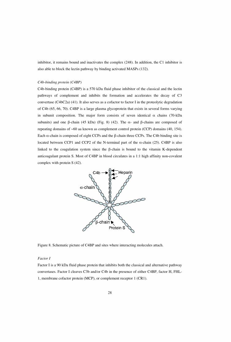
C4b-binding protein (C4BP)
C4b-binding protein (C4BP) is a 570 kDa fluid phase inhibitor of the classical and the lectin
pathways of complement and inhibits the formation and accelerates the decay of C3
convertase (C4bC2a) (41). It also serves as a cofactor to factor I in the proteolytic degradation
of C4b (65, 66, 70). C4BP is a large plasma glycoprotein that exists in several forms varying
in subunit composition. The major form consists of seven identical chains (70-kDa
subunits) and one -chain (45 kDa) (Fig. 8) (42). The - and -chains are composed of
repeating domains of ~60 aa known as complement control protein (CCP) domains (40, 154).
Each -chain is composed of eight CCPs and the -chain three CCPs. The C4b binding site is
located between CCP1 and CCP2 of the N-terminal part of the -chain (25). C4BP is also
linked to the coagulation system since the -chain is bound to the vitamin K-dependent
anticoagulant protein S. Most of C4BP in blood circulates in a 1:1 high affinity non-covalent
complex with protein S (42).
Figure 8. Schematic picture of C4BP and sites where interacting molecules attach.
Factor I
Factor I is a 90 kDa fluid phase protein that inhibits both the classical and alternative pathway
convertases. Factor I cleaves C3b and/or C4b in the presence of either C4BP, factor H, FHL-
1, membrane cofactor protein (MCP), or complement receptor 1 (CR1).

29
The factor H family
Factor H is a 150 kDa fluid phase protein that regulates the alternative pathway of the
complement system (189). It is a glycoprotein found in human plasma composed of 20
repetitive units of 60 amino acids, which is designated CCPs or short consensus repeats
(SCRs) (Fig. 9) (186). The alternative pathway is regulated by factor H via a binding of C3b,
accelerating the decay of the alternative pathway C3-convertase (C3bBb) and acting as a
cofactor for the factor I-mediated cleavage of C3b. Factor H regulates the complement system
both in fluid phase and on cell surfaces, C3b is inactivated by factor H in the fluid phase,
whereas inactivation of surface bound C3b by factor H is dependent on the chemical
composition of the surface to which C3b is bound. Factor H has three binding sites for C3b,
i.e., SCRs 1-4, SCRs 12-14, and SCRs 19-20 (202). Furthermore, factor H has an important
role in the discrimination between self (non-activating) and non-self (activating) surfaces.
Factor H has high affinity for C3b when the molecule is deposited on human cells (non-
activators), which are coated with sialic acid and glycosaminoglycans (135). This keep the
alternative pathway activation under control on self surfaces. However, the complement
activation proceeds if the C3b coated surface lacks the polyanions.
Figure 9. The factor H molecule. The C3b and heparin (dark blue) binding sites are indicated.
In addition to factor H, the factor H family consists of six multidomain and multifunctional
serum proteins (250). Factor H-like protein 1 (FHL-1) and five factor H-related proteins
(FHR1-5) are the additional members of this group (Fig. 10).
FHL-1 also acts as a regulator of the alternative pathway and is a product of
alternative splicing of the factor H gene on chromosome 1 (50). FHL-1 is a 42 kDa protein
composed of seven SCRs identical to the N-terminal SCRs of factor H, and four unique amino
acids at the C-terminal of the protein (249). The protein also displays cofactor and decay-
accelerating activity. The plasma concentration of FHL-1 is approximately 10-50 fold less
than the concentration of factor H.

30
Genes located in the RCA gene cluster encode the FHRs, and the proteins are all
composed of SCRs. There are five FHRs and their SCRs show structural similarities to factor
H and to each other (Fig. 10) (250). Despite the fact that FHRs are known to bind C3b and
heparin, the functions of the different FHRs are still unknown and are under extensive
investigation.
Figure 10. The factor H protein family. Each member is composed of SCRs and the SCR are
aligned vertically by their similarities. The dark blue SCRs indicate heparin-binding domains.
Modified from ref. 250.
Vitronectin
Vitronectin is a regulator of the terminal pathway by inhibiting MAC (200), It is found as a
single chain (75 kDa) and a truncated form of 65 and 10 kDa in human plasma. Vitronectin
binds the C5b-7 complex at its membrane-binding site and thereby inhibits the insertion of the
complex into the cell membrane and prevents cell lysis of the microbe (174). This complex is
still able to bind C8 and C9 to form SC5b-8 and SC5b-9 complexes but the latter is non-lytic.
Furthermore, vitronectin blocks the tubular formation by binding C5b-9 (167, 175).
In addition to the complement regulatory activities, vitronectin is involved in cell adhesion,
spreading and migration, coagulation and fibrinolysis (174, 200). Furthermore, it is present in
the extra-cellular matrix (ECM) as a 75 kDa protein.

31
Clusterin
In addition to vitronectin, clusterin is also an inhibitor of MAC of the terminal pathway (100).
It is a 70-80 kDa glycoprotein, which binds C5b67 and C5b678 complexes and thereby
prevents insertion into cell membranes (225).
Membrane-bound regulators
Complement receptor (CR1) is a receptor for C3b and C4b and is primarily found on
peripheral blood cells (53). CR1 acts as a cofactor for factor I-mediated cleavage of C3b and
C4b and accelerate the decay of C3/C5 convertases (93).
Membrane cofactor protein (MCP) is a glycoprotein that is expressed on all cell
types except erythrocytes (126). In addition to CR1, MCP acts as a cofactor for factor I-
mediated cleavage of C3b and C4b (201).
Decay accelerating factor (DAF) is a membrane bound glycoprotein, which is
found on most human cells (109). DAF binds to and dissociates both the classical (C4b2a)
and the alternative (C3bBb) C3 convertases (147).
CD 59 (protectin) and homologous restriction factor (HRF) are membrane
bound regulators of the terminal pathway. Protectin is present on practically all cell surfaces
and it inhibits MAC formation by binding the C5b-8 complex and thereby blocking the
incorporation of C9 (134). In addition, HRF prevents C9 to bind the C5b-8 complex (247).
Functions of the complement system
The best known function of the complement system is the ability to trigger a powerful,
coordinated repertoire of anti-microbial reactions, including inflammation, opsonisation and
cell lysis. Activation of the components in the complement cascade releases small cleavage
products, C3a, C4a and C5a. These are called anaphylatoxins and are powerful chemotactic
agents (64). They have different functions, including induction of smooth muscle contraction,
enhancing vascular permeability and causes release of histamine and other vasoactive
substances from basophils. In addition, C5a has been shown to be strongly chemotactic for
phagocytic cells.
C4b, C3b and cleavage products of C3b are components of the complement
system called opsonins, which are deposited on the surface of the target cell following
activation of the cascade. Recognition of these opsonins by phagocytes enhances

32
phagocytosis. The phagocytes express receptors for C3b and C4b (CR1) and iC3b (CR3 and
CR4) (56). Binding of the opsonised microbe results in phagocytosis and degradation of the
microbe.
The end product of the complement system is the formation of MAC. The
incorporation of MAC into the membrane of the pathogen causes lysis of the cell and,
consequently, death. This process is very important in the defence against Gram-negative
bacteria. Gram-positive bacteria have a thick peptidoglycan layer, which effectively resists the
formation of MAC.
Another function of complement is its involvement in the clearance of apoptotic
cells. C1q binds directly to surface blebs on the apoptotic cell, resulting in activation of the
classical pathway (145). This activation leads to C3b deposition and inactivation of C3b
(iC3b). CR3 on macrophages binds the iC3b and this leads to phagocytosis of the apoptotic
cell.
Microbes and the complement system
In order to be a successful pathogen, the microbes have to overcome the host defence. One
essential part of the innate immune system is the complement system. Bacteria have evolved
an array of strategies to survive the complement system and be able to colonise the host and
cause disease. The pathogenesis of many microorganisms therefore relies on the capacity of
pathogens to avoid, resist or neutralise the complement system (114, 193, 246). Some
pathogens are able to inactivate complement components, whereas other produce proteins or
other components that mimic complement inhibitors.
Group A Streptococci (GAS) express a 31 kDa protein called Streptococcal
inhibitor of complement (SIC), which mimics the action of the terminal inhibitors; vitronectin
and clusterin, by inhibiting the insertion of C5b-7 into the membrane (4). Borrelial CD59-like
protein is expressed by Borrelia burgdorferi and shares functional and antigenic similarities
with CD59 (protectin) (160). Both proteins inhibit formation of MAC and thereby prevent cell
lysis.
Another frequent strategy used by some pathogens is binding of complement
inhibitors such as C4BP, factor H and vitronectin, which protects them from complement
attacks. These regulators are captured on the bacterial surface in such way that they are still
complement regulatory active. M. catarrhalis, Neisseria gonorrhoeae and E. coli K1 are
bacteria, which express surface molecules (UspA1/A2, Por1A/1B and OmpA, respectively)

33
capable of binding C4BP and thereby protecting the bacteria against the classical and lectin
pathways (153, 179, 245). Another pathogen able to bind C4BP is Streptococcus pyogenes
(31). The interaction is mediated by the M protein and it has been correlated to phagocytosis
resistance. Many pathogens also bind the inhibitors of the alternative pathway; factor H
and/or FHL-1, in order to protect them from complement attacks by accelerating the decay of
alternative pathway C3 convertase and inactivation of C3b. In fact, a number of
microorganisms, including Streptococcus pneumoniae, S. pyogenes, B. burgdorferi, Neisseria
meningitidis and Candida albicans have been reported to bind factor H (91, 99, 137, 199,
236). Several microbes, e.g., group A streptococci, gonococci, B. pertussis and C. albicans
have also been shown to bind both C4BP and factor H (19, 20, 137, 180). When these
regulators are bound to the surface of the microbes, they maintain their complement
regulatory activity and protect the microbes against direct lysis. In general, the binding of
complement inhibitors may contribute to serum resistance and prevention of
opsonophagocytosis (181). Many M. catarrhalis strains are resistant to the bactericidal
activity of human serum. In addition to C4BP-binding (153), M. catarrhalis ubiquitous
surface protein A2 (UspA2) has been found to bind vitronectin, the inhibitor of the terminal
pathway, and this interaction significantly contributes to serum resistance (12). Furthermore,
Staphylococcus aureus, E. coli and -hemolytic streptococci are efficient binders of
vitronectin (33). In addition to rendering the bacteria more resistant to complement induced
lysis, the bacteria/vitronectin interaction also contributes to the attachment and invasion into
host cells. The vitronectin binding of Pseudomonas aeruginosa is involved in internalization
of the bacteria into human epithelial cells (122). Blocking this interaction with anti-vitronectin
antibodies inhibited the bacterial invasion. In addition, N. gonorrhoeae probably uses
vitronectin as a bridge for attachment and invasion of human cells (43).
Gram-positive bacteria are in general resistant to MAC induced cell lysis. They
are shielded by a thick cell wall comprised of peptidoglycan (102). In contrast, the majority of
Gram-negative bacteria are susceptible to the action of the terminal pathway (MAC formation
and cell lysis). Thus, the capsule of some of the species makes them more resistant to
complement mediated attacks and opsonophagocytosis than the unencapsulated counterparts.
The capacity to adhere to mucosal epithelium is crucial for the virulence of
many pathogens. Different regulators of the complement system have been shown to mediate
adherence and ingestion to epithelial and endothelial cells. The binding of C4BP by C.
albicans has been shown to mediate adherence to endothelial cells and the binding of S.

34
pyogenes to FHL-1 mediates and enhances the ingestion of the pathogen into epithelial cells
(136, 158).
Some pathogens expresses enzymes capable of cleaving complement
components and thereby avoid complement activation or restrict the inflammatory reaction. P.
aeruginosa and Helicobacter pylori are two species expressing enzymes capable of cleaving
C1q and/or C3 (89, 192).
Table 4. Interaction between pathogens and complement regulators.
Pathogen Protein Regulator SCRs required for binding Reference
B. burgdorferi BbCRASP-1 Factor H, FHL-1 SCRs 5-7, SCRs 19-20 (112, 113)
BbCRASP-2 Factor H, FHL-1 SCRs 6-7, SCRs 19-20 (112, 113)
BbCRASP-3/4/5 Factor H SCRs 19-20 (112, 113)
E. coli K1 OmpA C4BP SCR 3 (172)
M. catarrhalis UspA1/UspA2 C4BP SCRs 2, 5 or 7 (153)
N. gonorrhoeae Por1A C4BP SCR 1 (179)
Por1B C4BP SCR 1 (179)
S. pneumoniae Hic Factor H SCRs 8-11, 12-14 (99)
PspC Factor H SCRs 13-15 (47)
S. pyogenes Fba Factor H, FHL-1 SCR 7 (157, 158)
M protein Factor H. FHL-1 SCR 7 (111)
M protein C4BP SCRs 1-2 (24)
Complement in the respiratory tract
For many microorganisms, including H. influenzae, the primary interaction with the human
host is through colonisation of the mucosal surface of the respiratory tract. Therefore, it is of
highest interest for the host to exploit an efficient defence at these surfaces. The complement
system is classified as a part of serum, but there are several studies demonstrating the
presence of complement in various sites of the body. Reports of the presence of complement
components in the respiratory tract of healthy individuals are scarce. Functionally active C3,
C4 and factor B are complement components that have been detected in human saliva (10).
However, there are several studies indicating the importance of complement in the respiratory
tract during infections. During inflammation, the permeability of the mucosa increases and
plasma, including complement proteins, immunoglobulins and components of the coagulation

35
and fibrinolysis systems, enters the airway lumen (75, 76, 162). This process designated
plasma exudation, has been suggested to be the first line of the mucosal defence system.
Complement components, i.e., C3a and C5a, have been found in the mucosa of
the nose and lower airways in various respiratory tract challenges, including allergy and
influenza virus infection (9, 23). In patients with chronic otitis media with effusion (OME),
local complement activation in the middle ear mucosa was observed, including an intense
deposition of C3 (143). In addition, factor H, FHL-1 and FHR-1/2/3/4/5 are complement
components found in middle ear effusions of patients with OME (142). Marc and coworkers
found increased concentrations of C5a in patients with COPD, suggesting the involvement of
complement in the pathogenesis of the disease (130). Furthermore, highly elevated levels of
C3a has been observed in middle ear effusions, indicating ongoing complement activation
(144).
H. influenzae and the complement system
An important feature for a pathogen to cause invasive diseases is the ability to avoid and resist
the bactericidal activity of the complement system in human serum. A pathogen having the
capability to survive in human blood, also has the potential to spread to other sites of the
body. Hib is capable of activating both the classical/lectin and alternative pathways of the
complement system. By using capsular polysaccharide or encapsulated bacteria, complement-
dependent bactericidal antibodies were efficiently absorbed from serum indicating presence of
antibodies initiating the classical pathway (8). Both NTHi and Hib are capable of activating
the alternative pathway, suggesting the cell wall rather than the capsule as a target for the
alternative pathway (178). The concentration of polysaccharide seems to be important for the
bacterial survival, since an increased production of polysaccharide is associated with
increased resistance to lysis (217). Hib has been shown to be more resistant to the bactericidal
effect of complement. To analyze whether there are differences in resistance to the actions of
complement depending on the capsular serotype, Swift and coworkers used capsule deficient
mutants that were identical with respect to outer membrane proteins, LPS and antibiotic
susceptibility (218, 251). The capsule types a, b and e transformants were shown to be equally
resistant to the complement activity as compared to the Hib wild type.
In addition to encapsulated H. influenzae, NTHi has been found to be invasive
and resistant to the actions of human serum (242). In that study the serum resistant strains
delayed the C3 deposition on the cell surface, resulting in prevention of MAC accumulation.

36
Furthermore, a recent study demonstrated that serum resistance is facilitated in a NTHi strain
by a delay of C4b deposition (88). When the LOS biosynthetic gene lgtC was inactivated the
C4b deposition increased and the survival in serum and blood was reduced, suggesting an
involvement of LOS in the interaction with the complement system. As mentioned before, the
ChoP associated with LOS is involved in serum resistance of H. influenzae (239). ChoP+
strains were more susceptible to the bactericidal actions of human serum than the ChoP–
counterpart. It was shown that C-reactive protein mediated this susceptibility to serum by
binding directly to the ChoP+ strain and thereby activating the classical pathway. Furthermore,
the complement system was suggested to have a central role in the innate immune defence
against NTHi in experimental AOM (57). When depleting complement in chinchillas, two
otherwise avirulent siaB mutants (defective in their ability to sialyate LPS) caused EOM with
severity similar to their wild type counterparts. In addition, a higher deposition of C3 was
detected on the serum sensitive mutants compared to the more serum resistant wild types.
Moreover, to conquer the innate immune system, some pathogens collaborate in
a sophisticated way. In a recent study performed by our group, Tan et al. showed that OMV
produced by M. catarrhalis contributed to an increased survival of H. influenzae in human
serum (221). This observation may explain why these two pathogens are often found together
in the human respiratory tract. Finally, patients with complement components (including C2
and C3) deficiencies have been shown to be associated with increased susceptibility to Hib (7,
223).
IMMUNOGLOBULINS
Antibodies belong to a group of glycoproteins also known as immunoglobulins (Igs). Igs are
present in tissue fluids, human serum and on the surface of B cells and they are important
effector molecules of the adaptive immune system. Thus, their function is to recognise and
bind foreign antigens. Furthermore, they are also involved in the innate immunity by their
ability to initiate the classical pathway of the complement system. Five different Igs belong to
the family; IgG, IgA, IgM, IgD, and IgE. IgM and IgG are the Igs responsible for the primary
and secondary response to antigens, whereas IgA is the predominant Ig protecting mucosal
surfaces. IgE is involved in allergy and in the protection against parasitic worms. IgD is
further described in the next section.

37
The antibodies all differ in size, carbohydrate and amino acid composition, and function.
Nevertheless, all Igs consist of two Fab regions and one Fc part, responsible for antigen
recognition and mediation of effector function, respectively. The Igs are composed of two
heavy (H) chains and two light (L) chains hold together by disulphide bonds (Fig. 11). Two
disulphide bonds link the mid-region of the two H chains, and are designated the hinge region,
displaying a high flexibility. The outer parts of the H and L chains comprise the variable
regions containing the antigen-binding site. This variable region varies between the different
antibody isotypes and recognises and attaches specifically to a particular antigen. The
remainder of the H and L chain is called the constant (C) region and this region is almost the
same in all antibodies of the same Ig class. Moreover, the L chains exist in two forms named
lambda ( ) and kappa ( ).
Figure 11. Schematic picture of an Ig molecule.
Immunoglobulin D (IgD)
In 1965 Rowe and Fahey first discovered IgD by studying serum from a myeloma patient
(194). After a couple of years it was shown that IgD is expressed on mature B cells together
with IgM (230). In contrast, immature B cells and memory cells lack the expression of
membrane bound IgD (mIgD) (146). IgD and the other Igs have structural similarities.
However, IgD has an unusually long hinge region, which probably makes it susceptible to
proteolytic degradation and hence the short half-life in serum (77). Thus, IgD is found in
serum at such a low concentrations as 30 μg/ml compared to 12 mg/ml of IgG. The role of
soluble IgD still has to be established and IgD secreting plasma cells are relatively

38
uncommon. However, the number IgD secreting plasma cells have been shown to be
increased in nasal and salivary glands (27). Patients suffering from frequent tonsillitis has
higher levels of IgD secreting plasma cells and a spontaneous production of IgD by tonsils
cultured in vitro has been observed (125, 216). Furthermore, children with recurrent AOM
have high concentrations of IgD in the middle ear effusions and nasopharyngeal secretions
(207, 208). It has been shown that higher concentrations of serum IgD can be found in
patients suffering from COPD as compared to healthy individuals (138). IgD is also, as
mentioned earlier, together with IgM expressed on mature B cells and recognises and binds
foreign antigens. In the presence of T cells, this event leads to activation of the B cell. The
signal arisen from crosslinked mIgD is both stronger and more prolonged compared to that of
mIgM (108). To further investigate the role of IgD as a BCR, IgD knockout mice have been
generated. These mice showed independently of mIgD expression normal development of the
immune system and a normal antibody response (150, 190). However, these knockouts had a
certain delay in the affinity maturation in the early primary response. Thus, this maturation is
important in the defence against pathogens (190). In IgM-knockout mice, mIgD replaced
mIgM function and showed normal B cell development and maturation indicating similar
functions of IgD and IgM (127). Taken together, these findings strongly indicate that both
secreted and mIgD are important for the immune response against pathogens in the upper
respiratory tract.
Bacterial non-immune immunoglobulin binding
Immune binding is a process where an antibody binds its antigen through the variable region.
Several pathogens are able to bind immunoglobulins in a non-immune manner, i.e., the
binding does not involve the normal antigen binding sites of the antibody. Several surface
proteins responsible for the interactions have been identified and the first protein binding an
immunoglobulin in a non-immune fashion was protein A (SpA) from S. aureus (62). Non-
immune binding of Igs is a feature shared by several pathogens but is more prevalent among
the Gram-positive than the Gram-negative bacteria.
Non-immune immunoglobulin binding to Gram-positive bacteria
The first characterised bacterial Ig binding protein was isolated from S. aureus and is called
Protein A (SpA) (62). SpA binds the Fc region of IgG1, 2 and 4, and the binding region is
located between CH2 and CH3 (44, 116). In addition, a weak binding between SpA and the Fab

39
region in all Ig molecules has also been demonstrated (95). The capacity of SpA to bind IgG
is widely used as a purification method for IgG.
Group C and G streptococci express Protein G, a protein with affinity for all IgG
subclasses (22). Protein G has been shown to bind the Fc region of IgG with high affinity and
the Fab region (CH1 domain) with low affinity (49). It is able to bind both regions
simultaneously.
Protein L is a protein expressed by Peptostreptococcus magnus with affinity for
Ig -light chain of all Ig classes (2). Protein L was shown to bind the variable region of the -
light chain. This interaction has been shown not to interfere with the capacity of the
antibodies to bind antigens (149).
S. pyogenes is another Gram-positive pathogen capable of binding Igs,
including IgG and IgA. Protein H is the protein expressed by S. pyogenes responsible for the
interaction with IgG, whereas Protein Sir binds both IgA and IgG (3, 214).
Non-immune immunoglobulin binding to Gram-negative bacteria
In contrast to Gram-positive bacteria, non-immune Ig binding to Gram-negative bacteria is
much less frequent. However, there are a few examples of Gram-negative bacteria that bind
Igs in a non-immune manner. Haemophilus somnus and E. coli have been shown to bind
human IgG and IgA, respectively (197, 241). In addition, E. coli has also been demonstrated
to bind human IgG. Interestingly, both M. catarrhalis and H. influenzae bind IgD (61).
Moraxella IgD-binding protein (MID) was isolated from M. catarrhalis in our laboratory
(60). The MID-IgD interaction is located to the CH1 region of IgD (196). Protein D isolated
from H. influenzae, identified by Ruan and colleagues, was shown to bind an IgD myeloma
serum (195). However, later on it was shown that Protein D was merely detected by the IgD
myeloma used, suggesting another IgD binding protein in encapsulated H. influenzae (198).
Importance of bacterial non-immune binding
The reason why several bacterial pathogens have acquired the ability to bind Igs in a non-
immune fashion is not fully understood. There are some indications of involvement in the
pathogenesis. By binding IgG in a non-immune manner, Protein H inhibits complement
activation (18). Similarly, SpA interferes with the complement system by inhibiting the
classical pathway (129).

40
A B cell superantigen is an antigen capable of stimulating B cells independently
of their antigenic specificity (203). Some of the Ig binding proteins belongs to this family of
proteins. SpA and Protein L are two superantigens known to activate B cells (13, 115). MID
of M. catarrhalis stimulates B cell proliferation by binding mIgD, and is thus considered to be
a superantigen (72). The biological effects of these B cell superantigens are unclear. However,
the superantigens are capable of interacting with the specific humoral immune response by
activating B cells independently of their antigen specificity. A polyclonal response is induced
by the superantigens, resulting in production of unspecific antibodies that most likely are not
directed against the pathogen in question. The interaction of the superantigens with the B-cell
receptor may lead to anergy or apoptosis of the B cell. In fact, SpA has been shown to induce
apoptotic B cell death in a mouse model (204). However, the findings reported by Silverman
and collaborators have not been possible to reproduce with human tonsillar B cells in vitro
(16).

41
THE PRESENT INVESTIGATION
AIMS
The aims of the studies upon which this thesis is based were as follows:
• To investigate whether H. influenzae interacts with the classical pathway of the
complement system by binding C4BP and to analyse the biological importance.
• To examine whether H. influenzae interacts with the alternative pathway by binding
factor H and to analyse the functional importance.
• To analyse whether H. influenzae interacts with the terminal pathway by binding
vitronectin and to identify the key molecule important for the interaction.
• To characterise the structural features of Hsf and provide evidence that it belongs to
the autotransporter family.
• To identify the region of IgD responsible for the interaction with H. influenzae type b.

42
RESULTS AND DISCUSSION
H. influenzae interferes with the classical pathway by binding C4BP (Paper I)
In paper I, we describe an interaction between different NTHi isolates and the complement
inhibitor C4BP. Complement resistance is crucial for bacterial virulence and binding of
complement inhibitors such as C4BP and factor H is an efficient strategy used by several
serum resistant pathogens (114, 193). The complement resistance mechanisms of H.
influenzae have not yet been completely resolved. It has previously been shown that serum-
resistant NTHi strains prevent MAC accumulation by delaying the synthesis of C3b through
the classical pathway (242). By using both flow cytometry and a direct binding assay, it was
shown in Paper I that most of the NTHi strains tested bound C4BP, whereas the majority of
the typeable strains did not. The binding occurred also when serum was used as a source of
C4BP. There was a significant difference in C4BP binding between two NTHi strains (506
and 69) and this difference may explain why one C4BP-binding strain 506 was serum
resistant while the non C4BP-binding strain 69 was sensitive to killing by human serum.
When C4BP was depleted from serum, a statistically significant decrease in survival was seen
with NTHi strain 506, suggesting that binding to C4BP indeed protected the bacteria against
the complement system. Our findings are in contrast to what has been shown in a recent
study, in which no binding of H. influenzae to C4BP was found (88).
Several different experiments, including bactericidal assay, antibody and C3
deposition in flow cytometry were performed to ascertain the relevance of the classical
pathway. When inhibiting the classical pathway with Mg-EGTA, a significant increase in the
survival of the NTHi strain investigated was observed. By using flow cytometry, a significant
decrease in C3b deposition was also seen when the classical pathway was inhibited, which
further corroborated this hypothesis. Moreover, it is a well known fact that the classical
pathway is initiated when antibodies bind to bacteria. In our experiments, both IgG and IgM
were deposited on the surface of the bacteria, suggesting an initiation of the classical pathway.
The ability of H. influenzae to bind C4BP suggests that the species uses the
capacity of C4BP to inhibit complement-mediated attacks. A critical question was whether
C4BP was still active and able to exert its complement regulatory functions when bound to
the bacterial surface. To analyse this, cofactor assays were performed. C4BP bound to the
surface of H. influenzae maintained its activity to degrade C4b, and C3b in the presence of
factor I, which affects all three pathways of complement (Fig. 12). Consequently, such
degradation prevented C4b and C3b from participating in the opsonisation of the pathogen. In

43
addition, the surface bound C4BP also binds C4b and thereby inhibits C3 convertase
formation and accelerating its decay.
Figure 12. Bacterial evasion of complement by binding C4BP. C4BP bound to the surface of
NTHi retains its functions, including inhibition of C3 convertase formation and acceleration
of its decay. The binding domains CCP2 and CCP7 are indicated by dark green.
To further analyse the interaction between NTHi and C4BP, we wanted to localise the binding
site on the C4BP molecule. Binding experiments in whole cell ELISAs with rC4BP mutants,
each deficient in one CCP domain, suggested that CCP2 and CCP7 were involved in the
binding. Previously, many of microbial binding sites on C4BP have been localised to CCPs 1-
3. As this is the same region required for the binding of C4b, one could speculate that binding
to microbial ligands inhibited the regulatory function of C4BP. However, C4BP is a polymer
of 7 identical -chains, which allows simultaneous binding of several different ligands even if
they utilize overlapping binding site.
H. influenzae interferes with the alternative pathway by binding factor H (Paper II)
In this paper we investigated whether H. influenzae interacts with the alternative pathway by
binding the regulator factor H. A collection of different H. influenzae strains was incubated
with [125I]-labelled factor H followed by separation of unbound ligand in a direct binding
assay. Interestingly, the majority of the H. influenzae strains bound factor H. Our findings are
in contrast to what has been shown in a recent study, in which no binding of H. influenzae to

44
factor H was found (57). Amongst all the strains tested, H. influenzae types b (Hib), c and f
showed the highest factor H binding, whereas the other H. influenzae strains bound factor H
to a varying extent. The Hib strains used in this study showed strong factor H binding and
were chosen for further studies. When serum was used as a source of factor H, all the Hib
strains tested bound factor H and some of them bound FHL-1.
To evaluate the physiological significance of the interaction between factor H
and H. influenzae, a serum bactericidal assay was performed. The approach was to compare a
low factor H-binding and a high factor H-binding strain in terms of survival. The results
showed that the low-binding strain H. influenzae type d (Hid 555) was more sensitive to
killing by human serum than the high factor H-binding strain Hib 541, suggesting that the
bacteria use the capacity of factor H to inhibit complement-mediated attacks. After 7.5 min,
more than 50 % of Hib 541 survived as compared to Hid 555 that only showed an 11 %
survival. Thus, the H. influenzae-dependent interaction with factor H is important for bacterial
serum resistance.
Factor H regulates the alternative pathway activity by serving as a cofactor for
factor I in the degradation of C3b, which results in the appearance of the cleaving fragments
´68-kDa, 46 kDa, and 43 kDa. It is very important for the pathogen that the regulator
maintains its regulatory functions when bound to the surface of the bacteria. Functional
analysis showed that factor H maintained cofactor activity when bound to the surface of Hib
and promoted factor I-mediated C3b inactivation (Fig. 13). The cleavage of C3b was
determined by the detection of the three cleavage products ( ´68-kDa, 46 kDa, and 43 kDa)
after separation in SDS-PAGE.
Figure 13. Factor H bound to the surface of H. influenzae retains its functions, including
inactivation and acceleration of the decay of the C3 convertase of the alternative pathway.
The SCRs important for the interaction with Hib is coloured in blue.

45
To investigate which part of the factor H molecule involved in the interaction between Hib
and factor H, we used whole cell ELISAs to test the ability of different fragments spanning
the entire factor H molecule to bind Hib. Interestingly, two binding domains were identified
within the molecule; one binding site common to both factor H and FHL-1 was located in the
N-terminal, whereas one was located in the C-terminal SCRs 15-20 that only can be found in
factor H. Heparin inhibited the interaction between Hib 541 and factor H in the direct binding
assay using [125I]-factor H, suggesting involvement of the heparin binding domains. The
heparin binding domains of factor H is located in SCRs 7, 13 and 19-20 and in FHL-1 in SCR
7 (189).
Hsf binds vitronectin and thus interferes with the terminal pathway (Paper III)
Some pathogens have been shown to interact with vitronectin, the inhibitor of the MAC of the
terminal pathway (12, 33). To analyse whether Hib bound soluble vitronectin, flow cytometry
was performed. The results show a strong binding to vitronectin. We wanted to investigate
whether the outer membrane protein Haemophilus influenzae surface fibrils (Hsf) was
responsible for the interaction with vitronectin. To analyse this, H. influenzae type b strains
(RM804 and Eagan) were mutated by introduction of a kanamycin resistance gene cassette in
the gene encoding for Hsf. Resulting mutants were confirmed by PCR, flow cytometry and
Western blot. The two latter methods showed a lack of Hsf expression. Flow cytometry
analysis of the Hsf-deficient mutant revealed that Hsf is the major vitronectin binding protein
in encapsulated H. influenzae. These findings were in contrast to a previous study, where no
binding to soluble vitronectin could be detected (48). In addition, we showed H. influenzae-
dependent binding to immobilised vitronectin. To further confirm that Hsf bound vitronectin,
E. coli transformants expressing Hsf was constructed, and experiments with these
transformants demonstrated a Hsf-dependent binding to both soluble and immobilised
vitronectin.
To analyse the biological significance, the Hsf-deficient mutants and their
corresponding wild types were analysed in a serum bactericidal assay. The H. influenzae
devoid of Hsf had a markedly reduced survival as compared to the wild type when exposed to
human serum. The ability to bind vitronectin suggests that Hsf uses the capacity of vitronectin
to inhibit complement-mediated attacks.
The vitronectin molecule contains three heparin-binding domains: residues 82-
137, 175-219, and 348-376. To analyse which part of the vitronectin molecule involved in the

46
Hsf interaction, inhibition experiments with heparin were performed, using flow cytometry.
Heparin inhibited the binding between H. influenzae and vitronectin, and blocking
experiments using peptides encompassing vitronectin 341-370 suggested that the other two
regions (residues 82-137 and 175-219) were involved in the interaction.
Vitronectin is also a component of the ECM, and binding to exposed ECM
components may facilitate adherence, which is an essential step in bacterial pathogenesis.
Moreover, to investigate which part of Hsf that was involved in the vitronectin binding,
several recombinant fragments spanning the entire molecule were constructed. Hsf 608-1351 and
Hsf 1536-2414 displayed the highest binding to vitronectin in ELISA. These binding sites also
contain amino acids involved in the attachment to epithelial cells (38). As a consequence, the
Hsf molecule may be able to bind two vitronectin molecules and stabilising adherence despite
the physical forces in the respiratory tract, including the mucociliary escalator, sneezing, and
coughing.
Hsf is a double-folded autotransporter protein (Paper IV)
Outer membrane proteins of Gram-negative pathogenic bacteria comprise an important group
of virulence determinants highly specialised in a variety of functions most of which are
targeting the host. Hsf is an outer membrane protein of approximately 245 kDa belonging to
the autotransporter protein family. The aim of the present study was to in detail study how Hsf
is expressed at the bacterial surface and to further prove that it belongs to the autotransporter
family. Initially we analysed the Hsf expression in a collection of encapsulated H. influenzae
strains by using flow cytometry. The results showed a large variation in Hsf expression. The
majority of the H. influenzae type b strains expressed Hsf on the surface of the bacteria,
whereas the Hsf expression of types c and type d varied. Types a and f were negative for Hsf
when analysed by flow cytometry. The differences in Hsf expression suggested that phase
variation occurred, since all isolates contained the hsf gene. The presence of hsf was analysed
by Southern blot. The conservation of the gene indicates that the product plays an important
role in the life cycle of the bacteria (211). It is very important to analyse virulence factors by
the different serotypes. Thus, since the introduction of the vaccine against serotype b, the
other serotypes have been more frequent causes of invasive diseases (1, 161, 228).
Hsf has been suggested to be a trimeric autotransporter, but this suggestion was
mainly based on sequence alignment with the related outer membrane protein Hia (38). We
wanted to further evaluate Hsf as an autotransporter. Three mutants were constructed by
inserting antibiotic resistance cassettes in the N-terminal and/or the C-terminal ends of the

47
gene (Fig. 14). When the translocating C-terminal end of the hsf gene was inactivated, Hsf
could be detected in the outer membrane extractions as analysed by Western blots. However,
flow cytometry and transmission electron microscopy (TEM) showed a lack of expression on
the bacterial surface, suggesting an expression of the protein but inhibition of the membrane
insertion.
Figure 14. A Schematic drawing of the three mutated hsf genes with antibiotic resistance gene
cassettes indicated.
Three immuno-purified and gold-labelled antibodies were used in TEM to in detail examine
the ultrastructural features of Hsf. These antibodies were directed against the N-terminal, C-
terminal and the middle region of the Hsf molecule. The results showed a 100 nm, double-
folded structure (Fig. 15). Both the antibody directed against the N-terminal and the C-
terminal bound close to the membrane of the bacteria. In addition, an antibody used against
the middle part of the molecule bound at the very tip of the protein.
Both outer membrane vesicles (OMVs) and free Hsf was observed in the
surroundings of the bacteria when analysed by TEM and specific anti-Hsf antibodies. OMVs
(or blebs) are produced in vitro during growth in broth and are thought to carry some
periplasm together with OMP, porins, receptors and lipopolysaccharide (LPS) from the outer
membrane layer (21, 118). Blebs are suggested to have many functions, for example
interacting with host cells deep in tissues that are not accessible by infecting bacteria, activate
immune cells and induce leukocyte migration, and escape immune detection during
colonisation (5, 67, 163). Both Western blot and TEM showed a presence of Hsf on the blebs,
suggesting a role outside the bacteria. Furthermore, Hsf was both tethered to the surface of the
bacteria and was released into the extra-cellular milieu, also proven by both Western blot and
TEM. Most known autotransporters undergo processing and is released into the extracellular

48
milieu (45, 84) and so does Hsf, further supporting the hypothesis as an autotransporter
protein.
Figure 15. Schematic illustration of the Hsf protein on the bacterial surface. TEM showed that
Hsf has a 100 nm long, double-folded structure with one of the vitronectin binding domains
at the tip of the protein.
Haemophilus influenzae type b binds human IgD (Paper V)
Previous studies have shown that H. influenzae bind human IgD in a non-immune fashion (61,
198). The ligand responsible for this interaction has not yet been identified. A collection of
different NTHi and Hib isolates were analysed for IgD binding by using a whole-cell ELISA
and flow cytometry. The results revealed that 50 % of the Hib strains displayed IgD-binding
capacity, whereas the NTHi lacked this ability. These results confirmed previously published
data by Sasaki and coworkers (198). Two strains of the Hib collection were chosen for further
studies; one isolate demonstrating high IgD binding and one moderate IgD binding. ELISAs
were performed to characterise the binding region on the IgD molecule responsible for the
interaction with H. influenzae, using a series of recombinant IgD chimeras. A part of the IgD
molecule was replaced by part of an IgG molecule in these chimeras. Replacing amino acids
157-224, 167-206 or 185-224 of the IgD molecule by the corresponding IgG fragments,
resulted in an abolished binding by Hib. In contrast, the replacement of IgD amino acids 157-

49
197 with IgG did not affect the interaction. Thus, these results show that the CH1 region of the
IgD molecule, namely amino acids 198-224, is responsible for the interaction between Hib
and IgD. Flow cytometry analysis confirmed these results. M. catarrhalis is another
respiratory pathogen shown to bind IgD (60). Interestingly, MID is responsible for the
interaction with IgD and the binding site on the IgD molecule is located to the same region as
H. influenzae, i.e., the CH1 region (196). We have shown an interaction between H. influenzae
and IgD, and together with the previously reported M. catarrhalis-IgD interaction, these
observations suggests that these interactions may be involved in the pathogenesis of
respiratory pathogens.
CONCLUDING REMARKS
Microbes often interact with human complement regulatory proteins in order to conquer the
innate immune system. Complement resistance is crucial for bacterial virulence and binding
of complement inhibitors such as C4BP, factor H and vitronectin is an efficient strategy used
by several serum resistant pathogens. In Papers I-III we studied the interactions of H.
influenzae with the classical/lectin, the alternative and the terminal pathways of the
complement system. We demonstrated interactions with C4BP, factor H and vitronectin, all
regulators of the complement system. All these interactions contributed to increased survival
of H. influenzae in human serum and, consequently, most likely contributed to their
pathogenicity. Hsf was shown to be the protein responsible for the vitronectin-binding of Hib
(Paper III). Vitronectin is also a component of the ECM. Thus, the interaction between Hsf
and vitronectin, not only prevent complement induced cell lysis, but may also contribute to
bacterial colonization and spread of H. influenzae. We further characterised the structure of
Hsf and its properties as an autotransporter (Paper IV). Hsf has a double-folded structure
exposing one of the vitronectin-binding domain at the tip of the protein. Hsf was also present
at the surface of OMVs and secreted into the extracellular milieu. To in detail studying the
virulence mechanisms of microbes helps us to understand how the pathogens evade the host
immune defence and how our immune system works. This may give us tools to be able to
prevent infections. Binding complement regulators are important virulence mechanisms of H.
influenzae. The best way to block a cascade for a pathogen is to inhibit every subsequent step
in the cascade and the best evasion strategy is to interact with many of the attacking steps.
Finally, we have also been able to prove an interaction between H. influenzae
and human IgD using recombinant material and to further characterise the binding domain of

50
IgD involved (Paper V). Further studies need to be done to evaluate the functional importance
of this interaction.

51
FUTURE PERSPECTIVES
In the first two papers we show that H. influenzae interacts with both the classical and the
alternative pathway of the complement system. These interactions affect the survival of the
bacteria. In the first paper, we identified that NTHi binds the regulator C4BP and it would be
of high interest to find the ligand in NTHi responsible for the interaction. In addition, we
demonstrated an interaction of H. influenzae and the inhibitor of the alternative pathway,
factor H (Paper II). The protein responsible for the binding of factor H is also unknown and
would be very interesting to identify.
Hsf has been identified as the major vitronectin binding protein in H. influenzae
type b (Paper III) and this interaction contributes to serum resistance. Vitronectin is an
inhibitor of MAC in the terminal pathway of the complement cascade and it is also a
component in the ECM. Hsf is an adhesin and has been shown to interact with epithelial cells,
but the receptor for the interaction is not known. Vitronectin has been found on the surface of
some epithelial cells. Thus, investigations to evaluate the role of vitronectin in the Hsf-
dependent adhesion of H. influenzae would be interesting experiments.
Hsf has been suggested to be a trimeric autotransporter based on sequence
analysis (38). In paper IV we have mapped the ultrastructure of Hsf by using gold-labelled
antibodies and investigated whether Hsf is an autotransporter. By using scanning transmission
electron microscopy (STEM) combined with the molecular weight and the precise length of
the protein, the oligomeric structure of Hsf could be established. In addition, another proof of
Hsf as an autotransporter would be to identify the translocator domain of Hsf. H. influenzae
secretes OMVs expressing Hsf and additional experiments evaluating the biological
importance of this mechanism are needed.
The respiratory pathogens M. catarrhalis and H. influenzae bind IgD (61). An
interaction between recombinant IgD with a defined antigen binding site and H. influenzae
type b was identified (Paper V), and it would be of highest interest to find the protein
responsible for the interaction. The function of IgD is not fully understood and it would be
tempting to further evaluate the role of the H. influenzae-IgD interaction.

52
SVENSK POPULÄRVETENSKAPLIG SAMMANFATTNING
Haemophilus influenzae är en bakterie som orsakar flera olika sjukdomar hos människa. Den
kan delas in i typbara och icke-typbara beroende på förekomsten av en polysackaridkapsel.
De typbara kan delas in i 6 olika typer (a-f), där typ b är den som är mest virulent och ger
svårast sjukdomar. H. influenzae typ b (Hib) kan bla orsaka struplocksinflammation
(epiglottit) och hjärnhinneinflammation (meningit). Dock har antalet sjukdomsfall orsakade
av typ b sjunkit drastiskt sedan ett vaccin infördes på 1990-talet. De icke typbara saknar
kapseln och orsakar mindre svåra sjukdomar som tex bihåleinflammation (sinuit),
ögoninflammation (konjunktivit) och öroninflammation (media otit). Patienter med kronisk
obstruktiv lungsjukdom (KOL) och cystisk fibros blir ofta infekterade med icke typbara H.
influenzae. Denna icke typbara är efter Pneumokocker en vanlig orsak till öroninflammation
hos barn. Det finns ännu inget vaccin mot dessa bakterier. Det finns ett stort
forskningsintresse för att utreda hur och varför dessa bakterier överlever vårt immunförsvar
och kan orsaka sjukdom.
När en inkräktare kommer in i kroppen så möter den vårt immunförsvar. En av
de första delarna av detta försvar är en kaskad av proteiner som kallas komplementsystemet.
Aktivering av denna kaskad leder till att olika proteiner samverkar med varandra med målet
att oskadliggöra inkräktaren. Komplemensystemet kan delas in i tre vägar beroende på hur de
aktiveras. Den klassiska vägen aktiveras av antikropparna IgG och IgM, den alternativa av
bindning till mikrobens yta och lektinvägen av bindande till sockermolekyler på bakteriens
yta. Alla dessa vägar går ihop till den terminala vägen, som slutar i att en stor por bildas.
Denna por tar hål på bakteriens membran (lyserar) och dödar den. Detta system är strikt
kontrollerat av regulatorer så att det inte ger sig på och skadar våra egna celler. En av dessa
regulatorer heter C4BP (C4b-binding protein) och vi visar i artikel I att icke typbara H.
influenzae binder till denna regulator resulterade i längre överlevnad i humant serum. C4BP
hämmar den klassiska vägen och lektinvägen av komplementsystemet och C4BP bundet till
de icke typbara H. influenzaes yta är fortfarande funktionellt aktivt, dvs regulatorn kan
fortfarande hämma komplementsystemet och på så sätt skydda bakterien mot lysering.
Detta är bara två av vägarna som leder till döden för bakterien så därför var det
mycket viktigt att undersöka om den alternativa vägen också kunde blockeras. Vi såg att H.
influenzae, både typbara och icke typbara, band faktor H (en hämmare av den alternativa
vägen) (artikel II). Vi såg även här att faktor H fortfarande är aktiv (inhiberar alternativa

53
vägen) när den är bunden till bakteriens yta och att denna bindning gör så att den överlever
längre i serum om man jämför med en icke-bindande stam.
De tre vägarna i komplementsystemet går alla ihop till en gemensam väg som
kallas den terminala vägen. Denna vägen regleras också, bla av ett protein som heter
vitronektin. Vitronektin hämmar uppbyggnaden av poren som lyserar och dödar mikroben. I
artikel nummer III så visade vi att Hib binder till vitronektin och att proteinet som står för
bindningen heter Haemophilus influenzae surface fibrils (Hsf). Bindningen mellan Hsf och
vitronektin bidrog till att bakterien överlevde bättre i humant serum om man jämför med en
mutant som ej uttrycker Hsf. Mutanten dog mycket fortare än vildtypen. Vitronektin är även
en viktig komponent i extracellulär matrix och bindningen till vitronektin kan förmodligen
hjälpa H. influenzae när den ska adherera till epitelcellerna i början av en infektion.
Bakterier uttrycker olika proteiner på sin yta och dessa proteiner interagerar med
värden på olika sätt för att överleva. Det är viktigt att karaktärisera dessa proteiner för att få
mer kunskap om hur bakterien klarar av att starta en infektion och överleva vårt försvar. Om
man har kunskap om dess struktur och med vad den interagerar med hos värden har man
kommit en bra bit på vägen för att kunna tillverka bättre läkemedel som kan angripa
bakterien. Med hjälp av elektronmikroskopi och antikroppar riktade mot olika delar av
proteinet så karaktäriserade vi Hsf. Resultatet visar att Hsf är 100 nm långt, dubbelvikt och
sitter ankrat med den C-terminala delen i membranet. Vidare så klyvs proteinet (Hsf) också av
och utsöndras i den omgivande miljön.
Vi har visat att H. influenzae typ b binder humant immunoglobulin D (IgD) som
är en av fem olika typer av antikroppar vi har i kroppen. Dessa antikroppar är medlemmar av
vårt immunförsvar och de känner igen främmande ämnen (antigen) som kommer in i kroppen
och hjälper till att eliminera dem. IgD finns både membranbundet på humana B-celler och
lösligt i plasma men man vet inte riktigt funktionen hos lösligt IgD. H. influenzae binder CH1
regionen av IgD och det är samma region som en annan viktig luftvägspatogen, Moraxella
catarrhalis, binder till. Att två olika luftvägspatogener binder i samma område visar att det är
en viktig interaktion för bakterien. Vidare forskning krävs för att undersöka betydelsen av
denna interaktion.
Sammanfattningsvis så presenteras i denna avhandling hur H. influenzae
interagerar med komplementsystemet genom att binda tre av dess inhibitorer och därmed
förbättra sin överlevnad i humant serum. Vi karaktäriserar även proteinet Hsf och visar att H.
influenzae binder humant IgD.

54
ACKNOWLEDGEMENTS
I would like to express my deepest gratitude to everyone that have helped me or been
involved in my work during these years. In particular, I wish to thank:
Min handledare Kristian Riesbeck för att du har varit ett enormt stöd under mina år här. Du
har alltid tagit dig tid, stöttat och uppmuntrat mig. Din kunskap, engagemang och alltid lika
positiva inställning har verkligen sporrat mig genom denna utbildning. Du är verkligen en
inspirerande forskarförebild!
Professor Arne Forsgren för ditt engagemang och aldrig sinande intresse för vetenskapen.
Tack för att du alltid har sett möjligheterna i alla project!
Professor Anna Blom för all hjälp med komplementsystemet komplexa (men roliga) värld.
Har varit jätteroligt att samarbeta med dig.
Herrn Professor Peter Zipfel, Ich möchte mich recht herzlich bei Ihnen für unsere fruchtbaren
Zusammenarbeit mit Faktor H bedanken. Ich sehe mit Spannung eine weiteren
Zusammenarbeit entgegen.
Ett stort tack till Ulrika Brynnel som guidade mig i 2-D elektroforesens värld och som alltid
tagit sig tid när jag ringt och bett om hjälp. Även ett stort tack till Matthias Mörgelin och
Maria Baumgarten för all hjälp med EM och alla snygga bilder jag fått. Tack till Hanna
Jarva för samarbetet med C4BP.
Jag vill självklart också tacka alla som varit och är på labbet. Tack till Marta för att du alltid
ställt (täckt ) upp för mig när det behövts. Du har varit ett enormt stöd för mig både
labbmässigt och socialt under alla dessa år! Vet inte hur jag skulle klarat mig utan dig! Ett
stort tack till min student Anna Thorssell och till Maria Hylén Olsson, ni gjorde mitt sista år
jätteroligt med alla skratt! Ni är verkligen trevliga och omtänksamma och det uppskattar jag
verkligen! Tack till Martin, Elena, Thuan Tong, Taras, Johan, Barbara, Andrea och
Radinka. Tack till alla på femte våningen. Har varit roligt att lära känna er alla.
Gertrud Hansson för all administrativ hjälp.

55
Lars-Erik för all hjälp med jodering och alla roliga samtal.
Eva, Ulla, och Ulla på cytometrilabb, ni har alltid ställt upp och hjälpt till, stöttat vid problem
och stått ut med att jag ofta var sen. Sorry!
Gertrud, Margareta, Nasida och Anki på första våningen, ni är oumbärliga!
Lars-Göran för all hjälp med beställningar.
Birgitta Andersson för all hjälp med bakterierna.
Ett stort tack till mina vänner som varit och är i huset, speciellt till Therése Nordström som
stöttade mig och lärde upp mig i början av min doktorandtid. Du var ett enormt stöd både
innanför och utanför labbets väggar. Till Carro för att du är så glad och sprallig och alltid
livar upp mig! Till Tina för alla peppande samtal om allt på labbet och privat och för allt stöd
under skrivandet av denna avhandling. Till Stina (snäll men also gorgeous ) för alla roliga
pratstunder, fester och alla gånger vi dansat som galningar. Tack till Emily (the killer) för att
du tog dig an mig :) i början av min doktorandtid och för alla roliga fester och träningspass.
Även ett stort tack för att jag fick bo hos dig när jag var i Australien!
Tessan, ett stort tack till den bästa vän man kan önska sig!!! Du anar inte hur mycket jag
uppskattar dig! Under alla dessa år har du lyssnat och ställt upp för mig under allt mitt tjat om
labbet. Du är bäst! TACK! Vill även tacka Eva, Ulrika, Anna W, Marie B, Kiki,
Margareta, Jenny T, Annika, Helena och Björn för allt stöd och för att ni lyssnat på mig
när jag tragglat och pratat om och om igen om labbet. Ni betyder massor för mig!
Mamma och pappa för att ni alltid har stöttat mig i alla mina val i livet. Tack för att ni
outtröttligt lyssnar och stöttar mig i alla lägen! Ni är de bästa föräldrarna man kan önska sig!
Ett stort tack till min bror Jens, du betyder massor för mig! Ett speciellt tack för all hjälp med
avhandlingen . Varma kramar till min svägerska Hülya, kämpa på!

56
REFERENCES
1. Adderson, E. E., C. L. Byington, L. Spencer, A. Kimball, M. Hindiyeh, K. Carroll, S. Mottice, E. K. Korgenski, J. C. Christenson, and A. T. Pavia. 2001. Invasive serotype a Haemophilus influenzae infections with a virulence genotype resembling Haemophilus influenzae type b: emerging pathogen in the vaccine era? Pediatrics 108:E18.
2. Akerstrom, B., and L. Bjorck. 1989. Protein L: an immunoglobulin light chain-binding bacterial protein. Characterization of binding and physicochemical properties. J Biol Chem 264:19740-19746.
3. Akesson, P., J. Cooney, F. Kishimoto, and L. Bjorck. 1990. Protein H--a novel IgG binding bacterial protein. Mol Immunol 27:523-531.
4. Akesson, P., A. G. Sjoholm, and L. Bjorck. 1996. Protein SIC, a novel extracellular protein of Streptococcus pyogenes interfering with complement function. J Biol Chem 271:1081-1088.
5. Akira, S., K. Takeda, and T. Kaisho. 2001. Toll-like receptors: critical proteins linking innate and acquired immunity. Nat Immunol 2:675-680.
6. Akkoyunlu, M., M. Ruan, and A. Forsgren. 1991. Distribution of protein D, an immunoglobulin D-binding protein, in Haemophilus strains. Infect Immun 59:1231-1238.
7. Alper, C. A., N. Abramson, R. B. Johnston, Jr., J. H. Jandl, and F. S. Rosen. 1970. Increased susceptibility to infection associated with abnormalities of complement-mediated functions and of the third component of complement (C3). N Engl J Med 282:350-354.
8. Anderson, P., R. B. Johnston, Jr., and D. H. Smith. 1972. Human serum activities against Hemophilus influenzae, type b. J Clin Invest 51:31-38.
9. Andersson, M., L. Michel, J. B. Llull, and U. Pipkorn. 1994. Complement activation on the nasal mucosal surface--a feature of the immediate allergic reaction in the nose. Allergy 49:242-245.
10. Andoh, A., Y. Fujiyama, T. Kimura, H. Uchihara, H. Sakumoto, H. Okabe, and T. Bamba. 1997. Molecular characterization of complement components (C3, C4, and factor B) in human saliva. J Clin Immunol 17:404-407.
11. Apicella, M. A., M. Shero, K. C. Dudas, R. R. Stack, W. Klohs, L. J. LaScolea, T. F. Murphy, and J. M. Mylotte. 1984. Fimbriation of Haemophilus species isolated from the respiratory tract of adults. J Infect Dis 150:40-43.
12. Attia, A. S., S. Ram, P. A. Rice, and E. J. Hansen. 2006. Binding of vitronectin by the Moraxella catarrhalis UspA2 protein interferes with late stages of the complement cascade. Infect Immun 74:1597-1611.
13. Axcrona, K., L. Bjorck, and T. Leanderson. 1995. Multiple ligand interactions for bacterial immunoglobulin-binding proteins on human and murine cells of the hematopoetic lineage. Scand J Immunol 42:359-367.
14. Barenkamp, S. J., and J. W. St Geme, 3rd. 1996. Identification of a second family of high-molecular-weight adhesion proteins expressed by non-typable Haemophilus influenzae. Mol Microbiol 19:1215-1223.
15. Barsum, W., R. Wilson, R. C. Read, A. Rutman, H. C. Todd, N. Houdret, P. Roussel, and P. J. Cole. 1995. Interaction of fimbriated and nonfimbriated strains of unencapsulated Haemophilus influenzae with human respiratory tract mucus in vitro. Eur Respir J 8:709-714.
16. Bekeredjian-Ding, I., S. Inamura, T. Giese, H. Moll, S. Endres, A. Sing, U. Zahringer, and G. Hartmann. 2007. Staphylococcus aureus protein A triggers T cell-independent B cell proliferation by sensitizing B cells for TLR2 ligands. J Immunol 178:2803-2812.
17. Benz, I., and M. A. Schmidt. 1989. Cloning and expression of an adhesin (AIDA-I) involved in diffuse adherence of enteropathogenic Escherichia coli. Infect Immun 57:1506-1511.
18. Berge, A., B. M. Kihlberg, A. G. Sjoholm, and L. Bjorck. 1997. Streptococcal protein H forms soluble complement-activating complexes with IgG, but inhibits complement activationby IgG-coated targets. J Biol Chem 272:20774-20781.

57
19. Berggard, K., E. Johnsson, F. R. Mooi, and G. Lindahl. 1997. Bordetella pertussis binds the human complement regulator C4BP: role of filamentous hemagglutinin. Infect Immun 65:3638-3643.
20. Berggard, K., G. Lindahl, B. Dahlback, and A. M. Blom. 2001. Bordetella pertussis binds to human C4b-binding protein (C4BP) at a site similar to that used by the natural ligand C4b. Eur J Immunol 31:2771-2780.
21. Beveridge, T. J. 1999. Structures of gram-negative cell walls and their derived membrane vesicles. J Bacteriol 181:4725-4733.
22. Bjorck, L., and G. Kronvall. 1984. Purification and some properties of streptococcal protein G, a novel IgG-binding reagent. J Immunol 133:969-974.
23. Bjornson, A. B., M. A. Mellencamp, and G. M. Schiff. 1991. Complement is activated in the upper respiratory tract during influenza virus infection. Am Rev Respir Dis 143:1062-1066.
24. Blom, A. M., K. Berggard, J. H. Webb, G. Lindahl, B. O. Villoutreix, and B. Dahlback.2000. Human C4b-binding protein has overlapping, but not identical, binding sites for C4b and streptococcal M proteins. J Immunol 164:5328-5336.
25. Blom, A. M., J. Webb, B. O. Villoutreix, and B. Dahlback. 1999. A cluster of positively charged amino acids in the C4BP alpha-chain is crucial for C4b binding and factor I cofactor function. J Biol Chem 274:19237-19245.
26. Brandtzaeg, P. 1992. Humoral immune response patterns of human mucosae: induction and relation to bacterial respiratory tract infections. J Infect Dis 165 Suppl 1:S167-176.
27. Brandtzaeg, P., S. T. Gjeruldsen, F. Korsrud, K. Baklien, P. Berdal, and J. Ek. 1979. The human secretory immune system shows striking heterogeneity with regard to involvement of J chain-positive IgD immunocytes. J Immunol 122:503-510.
28. Broome, C. V. 1987. Epidemiology of Haemophilus influenzae type b infections in the United States. Pediatr Infect Dis J 6:779-782.
29. Burns, D. L. 1999. Biochemistry of type IV secretion. Curr Opin Microbiol 2:25-29. 30. Campos, J. M. 1995. Haemophilus, p. 556-565. In P. R. Murray (ed.), Manual of Clinical
Microbiology, vol. 6th. ASM Press, Washimgton, D.C. 31. Carlsson, F., K. Berggard, M. Stalhammar-Carlemalm, and G. Lindahl. 2003. Evasion of
phagocytosis through cooperation between two ligand-binding regions in Streptococcus pyogenes M protein. J Exp Med 198:1057-1068.
32. Cheng, L. W., and O. Schneewind. 2000. Type III machines of Gram-negative bacteria: delivering the goods. Trends Microbiol 8:214-220.
33. Chhatwal, G. S., K. T. Preissner, G. Muller-Berghaus, and H. Blobel. 1987. Specific binding of the human S protein (vitronectin) to streptococci, Staphylococcus aureus, and Escherichia coli. Infect Immun 55:1878-1883.
34. Childs, R. A., K. Drickamer, T. Kawasaki, S. Thiel, T. Mizuochi, and T. Feizi. 1989. Neoglycolipids as probes of oligosaccharide recognition by recombinant and natural mannose-binding proteins of the rat and man. Biochem J 262:131-138.
35. Claus, D. R., J. Siegel, K. Petras, A. P. Osmand, and H. Gewurz. 1977. Interactions of C-reactive protein with the first component of human complement. J Immunol 119:187-192.
36. Cooper, N. R. 1985. The classical complement pathway: activation and regulation of the first complement component. Adv Immunol 37:151-216.
37. Cotter, S. E., N. K. Surana, and J. W. St Geme, 3rd. 2005. Trimeric autotransporters: a distinct subfamily of autotransporter proteins. Trends Microbiol 13:199-205.
38. Cotter, S. E., H. J. Yeo, T. Juehne, and J. W. St Geme, 3rd. 2005. Architecture and adhesive activity of the Haemophilus influenzae Hsf adhesin. J Bacteriol 187:4656-4664.
39. Crisel, R. M., R. S. Baker, and D. E. Dorman. 1975. Capsular polymer of Haemophilus influenzae, type b. I. Structural characterization of the capsular polymer of strain Eagan. J Biol Chem 250:4926-4930.
40. Dahlback, B. 1991. Protein S and C4b-binding protein: components involved in the regulation of the protein C anticoagulant system. Thromb Haemost 66:49-61.
41. Dahlback, B. 1983. Purification of human C4b-binding protein and formation of its complex with vitamin K-dependent protein S. Biochem J 209:847-856.

58
42. Dahlback, B., and J. Stenflo. 1981. High molecular weight complex in human plasma between vitamin K-dependent protein S and complement component C4b-binding protein. Proc Natl Acad Sci U S A 78:2512-2516.
43. Dehio, M., O. G. Gomez-Duarte, C. Dehio, and T. F. Meyer. 1998. Vitronectin-dependent invasion of epithelial cells by Neisseria gonorrhoeae involves alpha(v) integrin receptors. FEBS Lett 424:84-88.
44. Deisenhofer, J. 1981. Crystallographic refinement and atomic models of a human Fc fragment and its complex with fragment B of protein A from Staphylococcus aureus at 2.9- and 2.8-A resolution. Biochemistry 20:2361-2370.
45. Desvaux, M., N. J. Parham, and I. R. Henderson. 2004. The autotransporter secretion system. Res Microbiol 155:53-60.
46. Dodds, A. W., R. B. Sim, R. R. Porter, and M. A. Kerr. 1978. Activation of the first component of human complement (C1) by antibody-antigen aggregates. Biochem J 175:383-390.
47. Duthy, T. G., R. J. Ormsby, E. Giannakis, A. D. Ogunniyi, U. H. Stroeher, J. C. Paton, and D. L. Gordon. 2002. The human complement regulator factor H binds pneumococcal surface protein PspC via short consensus repeats 13 to 15. Infect Immun 70:5604-5611.
48. Eberhard, T., and M. Ullberg. 2002. Interaction of vitronectin with Haemophilus influenzae. FEMS Immunol Med Microbiol 34:215-219.
49. Erntell, M., E. B. Myhre, U. Sjobring, and L. Bjorck. 1988. Streptococcal protein G has affinity for both Fab- and Fc-fragments of human IgG. Mol Immunol 25:121-126.
50. Estaller, C., W. Schwaeble, M. Dierich, and E. H. Weiss. 1991. Human complement factor H: two factor H proteins are derived from alternatively spliced transcripts. Eur J Immunol 21:799-802.
51. Farley, M. M., D. S. Stephens, P. S. Brachman, Jr., R. C. Harvey, J. D. Smith, and J. D. Wenger. 1992. Invasive Haemophilus influenzae disease in adults. A prospective, population-based surveillance. CDC Meningitis Surveillance Group. Ann Intern Med 116:806-812.
52. Fath, M. J., and R. Kolter. 1993. ABC transporters: bacterial exporters. Microbiol Rev 57:995-1017.
53. Fearon, D. T. 1979. Regulation of the amplification C3 convertase of human complement by an inhibitory protein isolated from human erythrocyte membrane. Proc Natl Acad Sci U S A 76:5867-5871.
54. Fearon, D. T., and K. F. Austen. 1975. Properdin: binding to C3b and stabilization of the C3b-dependent C3 convertase. J Exp Med 142:856-863.
55. Fearon, D. T., and K. F. Austen. 1975. Properdin: initiation of alternative complement pathway. Proc Natl Acad Sci U S A 72:3220-3224.
56. Fearon, D. T., and W. W. Wong. 1983. Complement ligand-receptor interactions that mediate biological responses. Annu Rev Immunol 1:243-271.
57. Figueira, M. A., S. Ram, R. Goldstein, D. W. Hood, E. R. Moxon, and S. I. Pelton. 2007. Role of complement in defense of the middle ear revealed by restoring the virulence of nontypeable Haemophilus influenzae siaB mutants. Infect Immun 75:325-333.
58. Fink, D. L., B. A. Green, and J. W. St Geme, 3rd. 2002. The Haemophilus influenzae Hap autotransporter binds to fibronectin, laminin, and collagen IV. Infect Immun 70:4902-4907.
59. Forbes, K. J., K. D. Bruce, A. Ball, and T. H. Pennington. 1992. Variation in length and sequence of porin (ompP2) alleles of non-capsulate Haemophilus influenzae. Mol Microbiol 6:2107-2112.
60. Forsgren, A., M. Brant, A. Mollenkvist, A. Muyombwe, H. Janson, N. Woin, and K. Riesbeck. 2001. Isolation and characterization of a novel IgD-binding protein from Moraxella catarrhalis. J Immunol 167:2112-2120.
61. Forsgren, A., and A. O. Grubb. 1979. Many bacterial species bind human IgD. J Immunol 122:1468-1472.
62. Forsgren, A., and J. Sjoquist. 1966. "Protein A" from S. aureus. I. Pseudo-immune reaction with human gamma-globulin. J Immunol 97:822-827.
63. Foxwell, A. R., J. M. Kyd, and A. W. Cripps. 1998. Nontypeable Haemophilus influenzae: pathogenesis and prevention. Microbiol Mol Biol Rev 62:294-308.

59
64. Frank, M. M., and L. F. Fries. 1991. The role of complement in inflammation and phagocytosis. Immunol Today 12:322-326.
65. Fujita, T., I. Gigli, and V. Nussenzweig. 1978. Human C4-binding protein. II. Role in proteolysis of C4b by C3b-inactivator. J Exp Med 148:1044-1051.
66. Fujita, T., and V. Nussenzweig. 1979. The role of C4-binding protein and beta 1H in proteolysis of C4b and C3b. J Exp Med 150:267-276.
67. Galdiero, M., A. Folgore, M. Molitierno, and R. Greco. 1999. Porins and lipopolysaccharide (LPS) from Salmonella typhimurium induce leucocyte transmigration through human endothelial cells in vitro. Clin Exp Immunol 116:453-461.
68. Geluk, F., P. P. Eijk, S. M. van Ham, H. M. Jansen, and L. van Alphen. 1998. The fimbria gene cluster of nonencapsulated Haemophilus influenzae. Infect Immun 66:406-417.
69. Gentschev, I., G. Dietrich, and W. Goebel. 2002. The E. coli alpha-hemolysin secretion system and its use in vaccine development. Trends Microbiol 10:39-45.
70. Gigli, I., T. Fujita, and V. Nussenzweig. 1979. Modulation of the classical pathway C3 convertase by plasma proteins C4 binding protein and C3b inactivator. Proc Natl Acad Sci U S A 76:6596-6600.
71. Gilsdorf, J. R., M. Tucci, and C. F. Marrs. 1996. Role of pili in Haemophilus influenzae adherence to, and internalization by, respiratory cells. Pediatr Res 39:343-348.
72. Gjorloff Wingren, A., R. Hadzic, A. Forsgren, and K. Riesbeck. 2002. The novel IgD binding protein from Moraxella catarrhalis induces human B lymphocyte activation and Ig secretion in the presence of Th2 cytokines. J Immunol 168:5582-5588.
73. Golan, M. D., R. Burger, and M. Loos. 1982. Conformational changes in C1q after binding to immune complexes: detection of neoantigens with monoclonal antibodies. J Immunol 129:445-447.
74. Gray-Owen, S. D., S. Loosmore, and A. B. Schryvers. 1995. Identification and characterization of genes encoding the human transferrin-binding proteins from Haemophilus influenzae. Infect Immun 63:1201-1210.
75. Greiff, L., M. Andersson, J. S. Erjefalt, C. G. Persson, and P. Wollmer. 2003. Airway microvascular extravasation and luminal entry of plasma. Clin Physiol Funct Imaging 23:301-306.
76. Greiff, L., I. Erjefalt, C. Svensson, P. Wollmer, U. Alkner, M. Andersson, and C. G. Persson. 1993. Plasma exudation and solute absorption across the airway mucosa. Clin Physiol 13:219-233.
77. Griffiths, R. W., and G. J. Gleich. 1972. Proteolytic degradation of IgD and its relation to molecular conformation. J Biol Chem 247:4543-4548.
78. Gu, X. X., C. M. Tsai, M. A. Apicella, and D. J. Lim. 1995. Quantitation and biological properties of released and cell-bound lipooligosaccharides from nontypeable Haemophilus influenzae. Infect Immun 63:4115-4120.
79. Hansen, E. J., D. A. Hart, J. L. McGehee, and G. B. Toews. 1988. Immune enhancement of pulmonary clearance of nontypable Haemophilus influenzae. Infect Immun 56:182-190.
80. Hardy, G. G., S. M. Tudor, and J. W. St Geme, 3rd. 2003. The pathogenesis of disease due to nontypeable Haemophilus influenzae. Methods Mol Med 71:1-28.
81. Henderson, I. R., R. Cappello, and J. P. Nataro. 2000. Autotransporter proteins, evolution and redefining protein secretion. Trends Microbiol 8:529-532.
82. Henderson, I. R., and J. P. Nataro. 2001. Virulence functions of autotransporter proteins. Infect Immun 69:1231-1243.
83. Henderson, I. R., F. Navarro-Garcia, M. Desvaux, R. C. Fernandez, and D. Ala'Aldeen.2004. Type V protein secretion pathway: the autotransporter story. Microbiol Mol Biol Rev 68:692-744.
84. Henderson, I. R., F. Navarro-Garcia, and J. P. Nataro. 1998. The great escape: structure and function of the autotransporter proteins. Trends Microbiol 6:370-378.
85. Hendley, J. O., F. G. Hayden, and B. Winther. 2005. Weekly point prevalence of Streptococcus pneumoniae, Hemophilus influenzae and Moraxella catarrhalis in the upper airways of normal young children: effect of respiratory illness and season. Apmis 113:213-220.

60
86. Hendrixson, D. R., and J. W. St Geme, 3rd. 1998. The Haemophilus influenzae Hap serine protease promotes adherence and microcolony formation, potentiated by a soluble host protein. Mol Cell 2:841-850.
87. Herrington, D. A., and P. F. Sparling. 1985. Haemophilus influenzae can use human transferrin as a sole source for required iron. Infect Immun 48:248-251.
88. Ho, D. K., S. Ram, K. L. Nelson, P. J. Bonthuis, and A. L. Smith. 2007. lgtC Expression Modulates Resistance to C4b Deposition on an Invasive Nontypeable Haemophilus influenzae. J Immunol 178:1002-1012.
89. Hong, Y. Q., and B. Ghebrehiwet. 1992. Effect of Pseudomonas aeruginosa elastase and alkaline protease on serum complement and isolated components C1q and C3. Clin Immunol Immunopathol 62:133-138.
90. Horstmann, R. D., M. K. Pangburn, and H. J. Muller-Eberhard. 1985. Species specificity of recognition by the alternative pathway of complement. J Immunol 134:1101-1104.
91. Horstmann, R. D., H. J. Sievertsen, J. Knobloch, and V. A. Fischetti. 1988. Antiphagocytic activity of streptococcal M protein: selective binding of complement control protein factor H. Proc Natl Acad Sci U S A 85:1657-1661.
92. Hultgren, S. J., S. Abraham, M. Caparon, P. Falk, J. W. St Geme, 3rd, and S. Normark.1993. Pilus and nonpilus bacterial adhesins: assembly and function in cell recognition. Cell 73:887-901.
93. Iida, K., and V. Nussenzweig. 1983. Functional properties of membrane-associated complement receptor CR1. J Immunol 130:1876-1880.
94. Ilver, D., A. Arnqvist, J. Ogren, I. M. Frick, D. Kersulyte, E. T. Incecik, D. E. Berg, A. Covacci, L. Engstrand, and T. Boren. 1998. Helicobacter pylori adhesin binding fucosylated histo-blood group antigens revealed by retagging. Science 279:373-377.
95. Inganas, M. 1981. Comparison of mechanisms of interaction between protein A from Staphylococcus aureus and human monoclonal IgG, IgA and IgM in relation to the classical FC gamma and the alternative F(ab')2 epsilon protein A interactions. Scand J Immunol 13:343-352.
96. Janson, H., L. O. Heden, and A. Forsgren. 1992. Protein D, the immunoglobulin D-binding protein of Haemophilus influenzae, is a lipoprotein. Infect Immun 60:1336-1342.
97. Janson, H., L. O. Heden, A. Grubb, M. R. Ruan, and A. Forsgren. 1991. Protein D, an immunoglobulin D-binding protein of Haemophilus influenzae: cloning, nucleotide sequence, and expression in Escherichia coli. Infect Immun 59:119-125.
98. Janson, H., A. Melhus, A. Hermansson, and A. Forsgren. 1994. Protein D, the glycerophosphodiester phosphodiesterase from Haemophilus influenzae with affinity for human immunoglobulin D, influences virulence in a rat otitis model. Infect Immun 62:4848-4854.
99. Jarva, H., R. Janulczyk, J. Hellwage, P. F. Zipfel, L. Bjorck, and S. Meri. 2002. Streptococcus pneumoniae evades complement attack and opsonophagocytosis by expressing the pspC locus-encoded Hic protein that binds to short consensus repeats 8-11 of factor H. J Immunol 168:1886-1894.
100. Jenne, D. E., and J. Tschopp. 1992. Clusterin: the intriguing guises of a widely expressed glycoprotein. Trends Biochem Sci 17:154-159.
101. Jiang, H., B. Cooper, F. A. Robey, and H. Gewurz. 1992. DNA binds and activates complement via residues 14-26 of the human C1q A chain. J Biol Chem 267:25597-25601.
102. Joiner, K., E. Brown, C. Hammer, K. Warren, and M. Frank. 1983. Studies on the mechanism of bacterial resistance to complement-mediated killing. III. C5b-9 deposits stably on rough and type 7 S. pneumoniae without causing bacterial killing. J Immunol 130:845-849.
103. Joiner, K. A., E. J. Brown, and M. M. Frank. 1984. Complement and bacteria: chemistry and biology in host defense. Annu Rev Immunol 2:461-491.
104. Kelly, D. F., E. R. Moxon, and A. J. Pollard. 2004. Haemophilus influenzae type b conjugate vaccines. Immunology 113:163-174.
105. Kilian, M. 2003. Haemophilus, p. 623-635. In P. R. Murray (ed.), Manual of Clinical Microbology, vol. 8th. ASM Press, Washington, D.C.

61
106. Kilian, M., J. Mestecky, and R. E. Schrohenloher. 1979. Pathogenic species of the genus Haemophilus and Streptococcus pneumoniae produce immunoglobulin A1 protease. Infect Immun 26:143-149.
107. Kilian, M., J. Reinholdt, H. Lomholt, K. Poulsen, and E. V. Frandsen. 1996. Biological significance of IgA1 proteases in bacterial colonization and pathogenesis: critical evaluation of experimental evidence. Apmis 104:321-338.
108. Kim, K. M., and M. Reth. 1995. The B cell antigen receptor of class IgD induces a stronger and more prolonged protein tyrosine phosphorylation than that of class IgM. J Exp Med 181:1005-1014.
109. Kinoshita, T., M. E. Medof, R. Silber, and V. Nussenzweig. 1985. Distribution of decay-accelerating factor in the peripheral blood of normal individuals and patients with paroxysmal nocturnal hemoglobinuria. J Exp Med 162:75-92.
110. Kirkitadze, M. D., and P. N. Barlow. 2001. Structure and flexibility of the multiple domain proteins that regulate complement activation. Immunol Rev 180:146-161.
111. Kotarsky, H., J. Hellwage, E. Johnsson, C. Skerka, H. G. Svensson, G. Lindahl, U. Sjobring, and P. F. Zipfel. 1998. Identification of a domain in human factor H and factor H-like protein-1 required for the interaction with streptococcal M proteins. J Immunol 160:3349-3354.
112. Kraiczy, P., C. Skerka, V. Brade, and P. F. Zipfel. 2001. Further characterization of complement regulator-acquiring surface proteins of Borrelia burgdorferi. Infect Immun 69:7800-7809.
113. Kraiczy, P., C. Skerka, M. Kirschfink, V. Brade, and P. F. Zipfel. 2001. Immune evasion of Borrelia burgdorferi by acquisition of human complement regulators FHL-1/reconectin and Factor H. Eur J Immunol 31:1674-1684.
114. Kraiczy, P., and R. Wurzner. 2006. Complement escape of human pathogenic bacteria by acquisition of complement regulators. Mol Immunol 43:31-44.
115. Kristiansen, S. V., V. Pascual, and P. E. Lipsky. 1994. Staphylococcal protein A induces biased production of Ig by VH3-expressing B lymphocytes. J Immunol 153:2974-2982.
116. Kronvall, G., and R. C. Williams, Jr. 1969. Differences in anti-protein A activity among IgG subgroups. J Immunol 103:828-833.
117. Kubiet, M., R. Ramphal, A. Weber, and A. Smith. 2000. Pilus-mediated adherence of Haemophilus influenzae to human respiratory mucins. Infect Immun 68:3362-3367.
118. Kuehn, M. J., and N. C. Kesty. 2005. Bacterial outer membrane vesicles and the host-pathogen interaction. Genes Dev 19:2645-2655.
119. Kuklinska, D., and M. Kilian. 1984. Relative proportions of Haemophilus species in the throat of healthy children and adults. Eur J Clin Microbiol 3:249-252.
120. Laarmann, S., D. Cutter, T. Juehne, S. J. Barenkamp, and J. W. St Geme. 2002. The Haemophilus influenzae Hia autotransporter harbours two adhesive pockets that reside in the passenger domain and recognize the same host cell receptor. Mol Microbiol 46:731-743.
121. Lafontaine, E. R., L. D. Cope, C. Aebi, J. L. Latimer, G. H. McCracken, Jr., and E. J. Hansen. 2000. The UspA1 protein and a second type of UspA2 protein mediate adherence of Moraxella catarrhalis to human epithelial cells in vitro. J Bacteriol 182:1364-1373.
122. Leroy-Dudal, J., H. Gagniere, E. Cossard, F. Carreiras, and P. Di Martino. 2004. Role of alphavbeta5 integrins and vitronectin in Pseudomonas aeruginosa PAK interaction with A549 respiratory cells. Microbes Infect 6:875-881.
123. Lesavre, P. H., T. E. Hugli, A. F. Esser, and H. J. Muller-Eberhard. 1979. The alternative pathway C3/C5 convertase: chemical basis of factor B activation. J Immunol 123:529-534.
124. Lesavre, P. H., and H. J. Muller-Eberhard. 1978. Mechanism of action of factor D of the alternative complement pathway. J Exp Med 148:1498-1509.
125. Litwin, S. D., and B. D. Zehr. 1987. In vitro studies on human IgD. I. Sources and characteristics of "externalized" IgD in tonsil lymphocyte cultures. Eur J Immunol 17:483-489.
126. Lublin, D. M., M. K. Liszewski, T. W. Post, M. A. Arce, M. M. Le Beau, M. B. Rebentisch, L. S. Lemons, T. Seya, and J. P. Atkinson. 1988. Molecular cloning and chromosomal localization of human membrane cofactor protein (MCP). Evidence for

62
inclusion in the multigene family of complement-regulatory proteins. J Exp Med 168:181-194.
127. Lutz, C., B. Ledermann, M. H. Kosco-Vilbois, A. F. Ochsenbein, R. M. Zinkernagel, G. Kohler, and F. Brombacher. 1998. IgD can largely substitute for loss of IgM function in B cells. Nature 393:797-801.
128. Male, C. 1979. Immunoglobulin A1 protease production by Haemophilus influenzae and Streptococcus pneumoniae. Infection and Immunity 26:254-261.
129. Malmheden Yman, I., G. Stalenheim, and J. Sjoquist. 1982. The effect of C4 on C1 binding and activation. Scand J Immunol 15:587-593.
130. Marc, M. M., P. Korosec, M. Kosnik, I. Kern, M. Flezar, S. Suskovic, and J. Sorli. 2004. Complement factors c3a, c4a, and c5a in chronic obstructive pulmonary disease and asthma. Am J Respir Cell Mol Biol 31:216-219.
131. Mason, E. O., Jr., S. L. Kaplan, B. L. Wiedermann, E. P. Norrod, and W. A. Stenback.1985. Frequency and properties of naturally occurring adherent piliated strains of Haemophilus influenzae type b. Infect Immun 49:98-103.
132. Matsushita, M., S. Thiel, J. C. Jensenius, I. Terai, and T. Fujita. 2000. Proteolytic activities of two types of mannose-binding lectin-associated serine protease. J Immunol 165:2637-2642.
133. Meng, G., N. K. Surana, J. W. St Geme, 3rd, and G. Waksman. 2006. Structure of the outer membrane translocator domain of the Haemophilus influenzae Hia trimeric autotransporter. Embo J 25:2297-2304.
134. Meri, S., B. P. Morgan, A. Davies, R. H. Daniels, M. G. Olavesen, H. Waldmann, and P. J. Lachmann. 1990. Human protectin (CD59), an 18,000-20,000 MW complement lysis restricting factor, inhibits C5b-8 catalysed insertion of C9 into lipid bilayers. Immunology 71:1-9.
135. Meri, S., and M. K. Pangburn. 1990. Discrimination between activators and nonactivators of the alternative pathway of complement: regulation via a sialic acid/polyanion binding site on factor H. Proc Natl Acad Sci U S A 87:3982-3986.
136. Meri, T., A. M. Blom, A. Hartmann, D. Lenk, S. Meri, and P. F. Zipfel. 2004. The hyphal and yeast forms of Candida albicans bind the complement regulator C4b-binding protein. Infect Immun 72:6633-6641.
137. Meri, T., A. Hartmann, D. Lenk, R. Eck, R. Wurzner, J. Hellwage, S. Meri, and P. F. Zipfel. 2002. The yeast Candida albicans binds complement regulators factor H and FHL-1. Infect Immun 70:5185-5192.
138. Miller, R. D., G. J. Gleich, K. P. Offord, and S. L. Dunnette. 1979. Immunoglobulin concentrations in serum and nasal secretions in chronic obstructive pulmonary disease. A matched-pair study. Am Rev Respir Dis 119:229-238.
139. Moran, A. P., M. M. Prendergast, and B. J. Appelmelk. 1996. Molecular mimicry of host structures by bacterial lipopolysaccharides and its contribution to disease. FEMS Immunol Med Microbiol 16:105-115.
140. Murphy, T. F., J. M. Bernstein, D. M. Dryja, A. A. Campagnari, and M. A. Apicella.1987. Outer membrane protein and lipooligosaccharide analysis of paired nasopharyngeal and middle ear isolates in otitis media due to nontypable Haemophilus influenzae: pathogenetic and epidemiological observations. J Infect Dis 156:723-731.
141. Möllenkvist, A., S. A. Müller, M. Mörgelin, T. Hallström, A. Forsgren, and K. Riesbeck.2007. A novel group of tetrameric autotransporters is defined by the Moraxella IgD binding protein MID/Hag. Submitted.
142. Narkio-Makela, M., J. Hellwage, O. Tahkokallio, and S. Meri. 2001. Complement-regulator factor H and related proteins in otitis media with effusion. Clin Immunol 100:118-126.
143. Narkio-Makela, M., J. Jero, and S. Meri. 1999. Complement activation and expression of membrane regulators in the middle ear mucosa in otitis media with effusion. Clin Exp Immunol 116:401-409.

63
144. Narkio-Makela, M., A. M. Teppo, and S. Meri. 2000. Complement C3 cleavage and cytokines interleukin-1beta and tumor necrosis factor-alpha in otitis media with effusion. Laryngoscope 110:1745-1749.
145. Nauta, A. J., L. A. Trouw, M. R. Daha, O. Tijsma, R. Nieuwland, W. J. Schwaeble, A. R. Gingras, A. Mantovani, E. C. Hack, and A. Roos. 2002. Direct binding of C1q to apoptotic cells and cell blebs induces complement activation. Eur J Immunol 32:1726-1736.
146. Nicholson, I. C., M. J. Brisco, and H. Zola. 1995. Memory B lymphocytes in human tonsil do not express surface IgD. J Immunol 154:1105-1113.
147. Nicholson-Weller, A., J. Burge, D. T. Fearon, P. F. Weller, and K. F. Austen. 1982. Isolation of a human erythrocyte membrane glycoprotein with decay-accelerating activity for C3 convertases of the complement system. J Immunol 129:184-189.
148. Nicol, P. A., and P. J. Lachmann. 1973. The alternate pathway of complement activation. The role of C3 and its inactivator (KAF). Immunology 24:259-275.
149. Nilson, B. H., A. Solomon, L. Bjorck, and B. Akerstrom. 1992. Protein L from Peptostreptococcus magnus binds to the kappa light chain variable domain. J Biol Chem 267:2234-2239.
150. Nitschke, L., M. H. Kosco, G. Kohler, and M. C. Lamers. 1993. Immunoglobulin D-deficient mice can mount normal immune responses to thymus-independent and -dependent antigens. Proc Natl Acad Sci U S A 90:1887-1891.
151. Noel, G. J., S. J. Barenkamp, J. W. St Geme, 3rd, W. N. Haining, and D. M. Mosser.1994. High-molecular-weight surface-exposed proteins of Haemophilus influenzae mediate binding to macrophages. J Infect Dis 169:425-429.
152. Noel, G. J., S. K. Hoiseth, and P. J. Edelson. 1992. Type b capsule inhibits ingestion of Haemophilus influenzae by murine macrophages: studies with isogenic encapsulated and unencapsulated strains. J Infect Dis 166:178-182.
153. Nordstrom, T., A. M. Blom, A. Forsgren, and K. Riesbeck. 2004. The emerging pathogen Moraxella catarrhalis interacts with complement inhibitor C4b binding protein through ubiquitous surface proteins A1 and A2. J Immunol 173:4598-4606.
154. Norman, D. G., P. N. Barlow, M. Baron, A. J. Day, R. B. Sim, and I. D. Campbell. 1991. Three-dimensional structure of a complement control protein module in solution. J Mol Biol 219:717-725.
155. Nunn, D. 1999. Bacterial type II protein export and pilus biogenesis: more than just homologies? Trends Cell Biol 9:402-408.
156. Otto, B. R., S. J. van Dooren, J. H. Nuijens, J. Luirink, and B. Oudega. 1998. Characterization of a hemoglobin protease secreted by the pathogenic Escherichia coli strain EB1. J Exp Med 188:1091-1103.
157. Pandiripally, V., E. Gregory, and D. Cue. 2002. Acquisition of regulators of complement activation by Streptococcus pyogenes serotype M1. Infect Immun 70:6206-6214.
158. Pandiripally, V., L. Wei, C. Skerka, P. F. Zipfel, and D. Cue. 2003. Recruitment of complement factor H-like protein 1 promotes intracellular invasion by group A streptococci. Infect Immun 71:7119-7128.
159. Pangburn, M. K., R. D. Schreiber, and H. J. Muller-Eberhard. 1981. Formation of the initial C3 convertase of the alternative complement pathway. Acquisition of C3b-like activities by spontaneous hydrolysis of the putative thioester in native C3. J Exp Med 154:856-867.
160. Pausa, M., V. Pellis, M. Cinco, P. G. Giulianini, G. Presani, S. Perticarari, R. Murgia, and F. Tedesco. 2003. Serum-resistant strains of Borrelia burgdorferi evade complement-mediated killing by expressing a CD59-like complement inhibitory molecule. J Immunol 170:3214-3222.
161. Perdue, D. G., L. R. Bulkow, B. G. Gellin, M. Davidson, K. M. Petersen, R. J. Singleton, and A. J. Parkinson. 2000. Invasive Haemophilus influenzae disease in Alaskan residents aged 10 years and older before and after infant vaccination programs. Jama 283:3089-3094.
162. Persson, C. G., I. Erjefalt, U. Alkner, C. Baumgarten, L. Greiff, B. Gustafsson, A. Luts, U. Pipkorn, F. Sundler, C. Svensson, and et al. 1991. Plasma exudation as a first line respiratory mucosal defence. Clin Exp Allergy 21:17-24.

64
163. Pettit, R. K., and R. C. Judd. 1992. The interaction of naturally elaborated blebs from serum-susceptible and serum-resistant strains of Neisseria gonorrhoeae with normal human serum. Mol Microbiol 6:729-734.
164. Pfeiffer, R. 1893. Die Äetiologie der Influenzae. Zeitschrift für Hygiene und Infectionskrankheiten:357-386.
165. Pittman, M. 1931. Variation and type specificity in the bacterial species Haemophilus influenzae. Journal of Exprimental Medicine 53:471-492.
166. Plaut, A. G., J. V. Gilbert, and R. Wistar, Jr. 1977. Loss of antibody activity in human immunoglobulin A exposed extracellular immunoglobulin A proteases of Neisseria gonorrhoeae and Streptococcus sanguis. Infect Immun 17:130-135.
167. Podack, E. R., K. T. Preissner, and H. J. Muller-Eberhard. 1984. Inhibition of C9 polymerization within the SC5b-9 complex of complement by S-protein. Acta Pathol Microbiol Immunol Scand Suppl 284:89-96.
168. Pohl, S. 1981. DNA relatedness in HPA. In Haemophilus, Pasteurella and Actinobacillus. Academic press, London.
169. Pohlner, J., R. Halter, K. Beyreuther, and T. F. Meyer. 1987. Gene structure and extracellular secretion of Neisseria gonorrhoeae IgA protease. Nature 325:458-462.
170. Poolman, J. T., L. Bakaletz, A. Cripps, P. A. Denoel, A. Forsgren, J. Kyd, and Y. Lobet.2000. Developing a nontypeable Haemophilus influenzae (NTHi) vaccine. Vaccine 19 Suppl 1:S108-115.
171. Poulsen, K., J. Brandt, J. P. Hjorth, H. C. Thogersen, and M. Kilian. 1989. Cloning and sequencing of the immunoglobulin A1 protease gene (iga) of Haemophilus influenzae serotype b. Infect Immun 57:3097-3105.
172. Prasadarao, N. V., A. M. Blom, B. O. Villoutreix, and L. C. Linsangan. 2002. A novel interaction of outer membrane protein A with C4b binding protein mediates serum resistance of Escherichia coli K1. J Immunol 169:6352-6360.
173. Prasadarao, N. V., E. Lysenko, C. A. Wass, K. S. Kim, and J. N. Weiser. 1999. Opacity-associated protein A contributes to the binding of Haemophilus influenzae to chang epithelial cells. Infect Immun 67:4153-4160.
174. Preissner, K. T. 1991. Structure and biological role of vitronectin. Annu Rev Cell Biol 7:275-310.
175. Preissner, K. T., E. R. Podack, and H. J. Muller-Eberhard. 1985. The membrane attack complex of complement: relation of C7 to the metastable membrane binding site of the intermediate complex C5b-7. J Immunol 135:445-451.
176. Preston, A., R. E. Mandrell, B. W. Gibson, and M. A. Apicella. 1996. The lipooligosaccharides of pathogenic gram-negative bacteria. Crit Rev Microbiol 22:139-180.
177. Pugsley, A. P., O. Francetic, K. Hardie, O. M. Possot, N. Sauvonnet, and A. Seydel. 1997. Pullulanase: model protein substrate for the general secretory pathway of gram-negative bacteria. Folia Microbiol (Praha) 42:184-192.
178. Quinn, P. H., F. J. Crosson, Jr., J. A. Winkelstein, and E. R. Moxon. 1977. Activation of the alternative complement pathway by Haemophilus influenzae type B. Infect Immun 16:400-402.
179. Ram, S., M. Cullinane, A. M. Blom, S. Gulati, D. P. McQuillen, B. G. Monks, C. O'Connell, R. Boden, C. Elkins, M. K. Pangburn, B. Dahlback, and P. A. Rice. 2001. Binding of C4b-binding protein to porin: a molecular mechanism of serum resistance of Neisseria gonorrhoeae. J Exp Med 193:281-295.
180. Ram, S., A. K. Sharma, S. D. Simpson, S. Gulati, D. P. McQuillen, M. K. Pangburn, and P. A. Rice. 1998. A novel sialic acid binding site on factor H mediates serum resistance of sialylated Neisseria gonorrhoeae. J Exp Med 187:743-752.
181. Rautemaa, R., and S. Meri. 1999. Complement-resistance mechanisms of bacteria. Microbes Infect 1:785-794.
182. Read, R. C., R. Wilson, A. Rutman, V. Lund, H. C. Todd, A. P. Brain, P. K. Jeffery, and P. J. Cole. 1991. Interaction of nontypable Haemophilus influenzae with human respiratory mucosa in vitro. J Infect Dis 163:549-558.
65
183. Reddy, M. S., J. M. Bernstein, T. F. Murphy, and H. S. Faden. 1996. Binding between outer membrane proteins of nontypeable Haemophilus influenzae and human nasopharyngeal mucin. Infect Immun 64:1477-1479.
184. Reid, K. B., and R. R. Porter. 1976. Subunit composition and structure of subcomponent C1q of the first component of human complement. Biochem J 155:19-23.
185. Reyrat, J. M., V. Pelicic, E. Papini, C. Montecucco, R. Rappuoli, and J. L. Telford. 1999. Towards deciphering the Helicobacter pylori cytotoxin. Mol Microbiol 34:197-204.
186. Ripoche, J., A. J. Day, T. J. Harris, and R. B. Sim. 1988. The complete amino acid sequence of human complement factor H. Biochem J 249:593-602.
187. Risberg, A., E. K. Schweda, and P. E. Jansson. 1997. Structural studies of the cell-envelope oligosaccharide from the lipopolysaccharide of Haemophilus influenzae strain RM.118-28. Eur J Biochem 243:701-707.
188. Rodriguez, C. A., V. Avadhanula, A. Buscher, A. L. Smith, J. W. St Geme, 3rd, and E. E. Adderson. 2003. Prevalence and distribution of adhesins in invasive non-type b encapsulated Haemophilus influenzae. Infect Immun 71:1635-1642.
189. Rodriguez de Cordoba, S., J. Esparza-Gordillo, E. Goicoechea de Jorge, M. Lopez-Trascasa, and P. Sanchez-Corral. 2004. The human complement factor H: functional roles, genetic variations and disease associations. Mol Immunol 41:355-367.
190. Roes, J., and K. Rajewsky. 1993. Immunoglobulin D (IgD)-deficient mice reveal an auxiliary receptor function for IgD in antigen-mediated recruitment of B cells. J Exp Med 177:45-55.
191. Roggenkamp, A., N. Ackermann, C. A. Jacobi, K. Truelzsch, H. Hoffmann, and J. Heesemann. 2003. Molecular analysis of transport and oligomerization of the Yersinia enterocolitica adhesin YadA. J Bacteriol 185:3735-3744.
192. Rokita, E., A. Makristathis, E. Presterl, M. L. Rotter, and A. M. Hirschl. 1998. Helicobacter pylori urease significantly reduces opsonization by human complement. J Infect Dis 178:1521-1525.
193. Rooijakkers, S. H., and J. A. van Strijp. 2007. Bacterial complement evasion. Mol Immunol 44:23-32.
194. Rowe, D. S., and J. L. Fahey. 1965. A New Class of Human Immunoglobulins. Ii. Normal Serum Igd. J Exp Med 121:185-199.
195. Ruan, M. R., M. Akkoyunlu, A. Grubb, and A. Forsgren. 1990. Protein D of Haemophilus influenzae. A novel bacterial surface protein with affinity for human IgD. J Immunol 145:3379-3384.
196. Samuelsson, M., J. Jendholm, S. Amisten, S. L. Morrison, A. Forsgren, and K. Riesbeck.2006. The IgD CH1 region contains the binding site for the human respiratory pathogen Moraxella catarrhalis IgD-binding protein MID. Eur J Immunol 36:2525-2534.
197. Sandt, C. H., Y. D. Wang, R. A. Wilson, and C. W. Hill. 1997. Escherichia coli strains with nonimmune immunoglobulin-binding activity. Infect Immun 65:4572-4579.
198. Sasaki, K., and R. S. Munson, Jr. 1993. Protein D of Haemophilus influenzae is not a universal immunoglobulin D-binding protein. Infect Immun 61:3026-3031.
199. Schneider, M. C., R. M. Exley, H. Chan, I. Feavers, Y. H. Kang, R. B. Sim, and C. M. Tang. 2006. Functional significance of factor H binding to Neisseria meningitidis. J Immunol 176:7566-7575.
200. Schvartz, I., D. Seger, and S. Shaltiel. 1999. Vitronectin. Int J Biochem Cell Biol 31:539-544.
201. Seya, T., and J. P. Atkinson. 1989. Functional properties of membrane cofactor protein of complement. Biochem J 264:581-588.
202. Sharma, A. K., and M. K. Pangburn. 1996. Identification of three physically and functionally distinct binding sites for C3b in human complement factor H by deletion mutagenesis. Proc Natl Acad Sci U S A 93:10996-11001.
203. Silverman, G. J. 1997. B-cell superantigens. Immunol Today 18:379-386. 204. Silverman, G. J., and C. S. Goodyear. 2002. A model B-cell superantigen and the
immunobiology of B lymphocytes. Clin Immunol 102:117-134.

66
205. Sirakova, T., P. E. Kolattukudy, D. Murwin, J. Billy, E. Leake, D. Lim, T. DeMaria, and L. Bakaletz. 1994. Role of fimbriae expressed by nontypeable Haemophilus influenzae in pathogenesis of and protection against otitis media and relatedness of the fimbrin subunit to outer membrane protein A. Infect Immun 62:2002-2020.
206. Smith W., C. H. A., and P.P Laidlaw. 1933. A virus obtained from influenzae patients. Lancet 2:66-68.
207. Sorensen, C. H. 1983. Quantitative aspects of IgD and secretory immunoglobulins in middle ear effusions. Int J Pediatr Otorhinolaryngol 6:247-253.
208. Sorensen, C. H., and P. L. Larsen. 1988. IgD in nasopharyngeal secretions and tonsils from otitis-prone children. Clin Exp Immunol 73:149-154.
209. St Geme, J. W., 3rd. 1993. Nontypeable Haemophilus influenzae disease: epidemiology, pathogenesis, and prospects for prevention. Infect Agents Dis 2:1-16.
210. St Geme, J. W., 3rd, and D. Cutter. 2000. The Haemophilus influenzae Hia adhesin is an autotransporter protein that remains uncleaved at the C terminus and fully cell associated. J Bacteriol 182:6005-6013.
211. St Geme, J. W., 3rd, D. Cutter, and S. J. Barenkamp. 1996. Characterization of the genetic locus encoding Haemophilus influenzae type b surface fibrils. J Bacteriol 178:6281-6287.
212. St Geme, J. W., 3rd, M. L. de la Morena, and S. Falkow. 1994. A Haemophilus influenzae IgA protease-like protein promotes intimate interaction with human epithelial cells. Mol Microbiol 14:217-233.
213. St Geme, J. W., 3rd, S. Falkow, and S. J. Barenkamp. 1993. High-molecular-weight proteins of nontypable Haemophilus influenzae mediate attachment to human epithelial cells. Proc Natl Acad Sci U S A 90:2875-2879.
214. Stenberg, L., P. W. O'Toole, J. Mestecky, and G. Lindahl. 1994. Molecular characterization of protein Sir, a streptococcal cell surface protein that binds both immunoglobulin A and immunoglobulin G. J Biol Chem 269:13458-13464.
215. Stull, T. L., P. M. Mendelman, J. E. Haas, M. A. Schoenborn, K. D. Mack, and A. L. Smith. 1984. Characterization of Haemophilus influenzae type b fimbriae. Infect Immun 46:787-796.
216. Surjan, L., Jr., P. Brandtzaeg, and P. Berdal. 1978. Immunoglobulin systems of human tonsils. II. Patients with chronic tonsillitis or tonsillar hyperplasia: quantification of Ig-producing cells, tonsillar morphometry and serum Ig concentrations. Clin Exp Immunol 31:382-390.
217. Sutton, A., R. Schneerson, S. Kendall-Morris, and J. B. Robbins. 1982. Differential complement resistance mediates virulence of Haemophilus influenzae type b. Infect Immun 35:95-104.
218. Swift, A. J., E. R. Moxon, A. Zwahlen, and J. A. Winkelstein. 1991. Complement-mediated serum activities against genetically defined capsular transformants of Haemophilus influenzae. Microb Pathog 10:261-269.
219. Tamura, N., A. Shimada, and S. Chang. 1972. Further evidence for immune cytolysis by antibody and the first eight components of complement in the absence of C9. Immunology 22:131-140.
220. Tan, T. T., A. Forsgren, and K. Riesbeck. 2006. The respiratory pathogen moraxella catarrhalis binds to laminin via ubiquitous surface proteins A1 and A2. J Infect Dis 194:493-497.
221. Tan, T. T., M. Mörgelin, A. Forsgren, and K. Riesbeck. 2007. Haemophilus influenzae survival during complement-mediated attacks is promoted by Moraxella catarrhalis outer membrane vesicles. Journal of infectious diseases In Press.
222. Tan, T. T., T. Nordstrom, A. Forsgren, and K. Riesbeck. 2005. The respiratory pathogen Moraxella catarrhalis adheres to epithelial cells by interacting with fibronectin through ubiquitous surface proteins A1 and A2. J Infect Dis 192:1029-1038.
223. Thong, Y. H., D. A. Simpson, and H. J. Muller-Eberhard. 1980. Homozygous deficiency of the second component of complement presenting with recurrent bacterial meningitis. Arch Dis Child 55:471-473.

67
224. Trottier, S., K. Stenberg, and C. Svanborg-Eden. 1989. Turnover of nontypable Haemophilus influenzae in the nasopharynges of healthy children. J Clin Microbiol 27:2175-2179.
225. Tschopp, J., A. Chonn, S. Hertig, and L. E. French. 1993. Clusterin, the human apolipoprotein and complement inhibitor, binds to complement C7, C8 beta, and the b domain of C9. J Immunol 151:2159-2165.
226. Turk, D. C. 1984. The pathogenicity of Haemophilus influenzae. J Med Microbiol 18:1-16. 227. Turk, D. C., and J. R. May. 1967. In Haemophilus influenzae: its clinical importance, p. 95-
105, Immunology. The English University Press, Ltd., London. 228. Urwin, G., J. A. Krohn, K. Deaver-Robinson, J. D. Wenger, and M. M. Farley. 1996.
Invasive disease due to Haemophilus influenzae serotype f: clinical and epidemiologic characteristics in the H. influenzae serotype b vaccine era. The Haemophilus influenzae Study Group. Clin Infect Dis 22:1069-1076.
229. van Alphen, L., L. Geelen-van den Broek, L. Blaas, M. van Ham, and J. Dankert. 1991. Blocking of fimbria-mediated adherence of Haemophilus influenzae by sialyl gangliosides. Infect Immun 59:4473-4477.
230. Van Boxel, J. A., W. E. Paul, W. D. Terry, and I. Green. 1972. Communications. IgD-bearing human lymphocytes. J Immunol 109:648-651.
231. van Ham, S. M., L. van Alphen, F. R. Mooi, and J. P. van Putten. 1994. The fimbrial gene cluster of Haemophilus influenzae type b. Mol Microbiol 13:673-684.
232. van Schilfgaarde, M., P. Eijk, A. Regelink, P. van Ulsen, V. Everts, J. Dankert, and L. van Alphen. 1999. Haemophilus influenzae localized in epithelial cell layers is shielded from antibiotics and antibody-mediated bactericidal activity. Microb Pathog 26:249-262.
233. Virkola, R., M. Brummer, H. Rauvala, L. van Alphen, and T. K. Korhonen. 2000. Interaction of fimbriae of Haemophilus influenzae type B with heparin-binding extracellular matrix proteins. Infect Immun 68:5696-5701.
234. Vitovski, S., K. T. Dunkin, A. J. Howard, and J. R. Sayers. 2002. Nontypeable Haemophilus influenzae in carriage and disease: a difference in IgA1 protease activity levels. Jama 287:1699-1705.
235. Vogt, W., G. Schmidt, B. Von Buttlar, and L. Dieminger. 1978. A new function of the activated third component of complement: binding to C5, an essential step for C5 activation. Immunology 34:29-40.
236. von Lackum, K., J. C. Miller, T. Bykowski, S. P. Riley, M. E. Woodman, V. Brade, P. Kraiczy, B. Stevenson, and R. Wallich. 2005. Borrelia burgdorferi regulates expression of complement regulator-acquiring surface protein 1 during the mammal-tick infection cycle. Infect Immun 73:7398-7405.
237. Walport, M. J. 2001. Complement. First of two parts. N Engl J Med 344:1058-1066. 238. Weiser, J. N., S. T. Chong, D. Greenberg, and W. Fong. 1995. Identification and
characterization of a cell envelope protein of Haemophilus influenzae contributing to phase variation in colony opacity and nasopharyngeal colonization. Mol Microbiol 17:555-564.
239. Weiser, J. N., N. Pan, K. L. McGowan, D. Musher, A. Martin, and J. Richards. 1998. Phosphorylcholine on the lipopolysaccharide of Haemophilus influenzae contributes to persistence in the respiratory tract and sensitivity to serum killing mediated by C-reactive protein. J Exp Med 187:631-640.
240. Weller, P. F., A. L. Smith, P. Anderson, and D. H. Smith. 1977. The role of encapsulation and host age in the clearance of Haemophilus influenzae bacteremia. J Infect Dis 135:34-41.
241. Widders, P. R., L. A. Dorrance, M. Yarnall, and L. B. Corbeil. 1989. Immunoglobulin-binding activity among pathogenic and carrier isolates of Haemophilus somnus. Infect Immun 57:639-642.
242. Williams, B. J., G. Morlin, N. Valentine, and A. L. Smith. 2001. Serum resistance in an invasive, nontypeable Haemophilus influenzae strain. Infect Immun 69:695-705.
243. Wilson, R. 1998. The role of infection in COPD. Chest 113:242S-248S. 244. Wilson, R., R. Read, and P. Cole. 1992. Interaction of Haemophilus influenzae with mucus,
cilia, and respiratory epithelium. J Infect Dis 165 Suppl 1:S100-102.

68
245. Wooster, D. G., R. Maruvada, A. M. Blom, and N. V. Prasadarao. 2006. Logarithmic phase Escherichia coli K1 efficiently avoids serum killing by promoting C4bp-mediated C3b and C4b degradation. Immunology 117:482-493.
246. Wurzner, R. 1999. Evasion of pathogens by avoiding recognition or eradication by complement, in part via molecular mimicry. Mol Immunol 36:249-260.
247. Zalman, L. S., and H. Muller-Eberhard. 1994. Homologous restriction factor: effect on complement C8 and C9 uptake and lysis. Mol Immunol 31:301-305.
248. Ziccardi, R. J. 1982. A new role for C-1-inhibitor in homeostasis: control of activation of the first component of human complement. J Immunol 128:2505-2508.
249. Zipfel, P. F., and C. Skerka. 1999. FHL-1/reconectin: a human complement and immune regulator with cell-adhesive function. Immunol Today 20:135-140.
250. Zipfel, P. F., C. Skerka, J. Hellwage, S. T. Jokiranta, S. Meri, V. Brade, P. Kraiczy, M. Noris, and G. Remuzzi. 2002. Factor H family proteins: on complement, microbes and human diseases. Biochem Soc Trans 30:971-978.
251. Zwahlen, A., J. S. Kroll, L. G. Rubin, and E. R. Moxon. 1989. The molecular basis of pathogenicity in Haemophilus influenzae: comparative virulence of genetically-related capsular transformants and correlation with changes at the capsulation locus cap. Microb Pathog 7:225-235.