Medical Nutrition Therapy for Gastrointestinal Tract Disorders.
Surgery, Nutrition and Gastrointestinal Function
in Critically Ill Infants
Heelkunde, voeding en maagdarmfunctie
bij ernstig zieke zuigelingen
Proefschrift
Marcel Johannes Ivo Jacques Albers

Copyright © M.J.I.J. Albers, Groningen, 2004 ISBN 90-9018036-2 Cover illustration: “De balans” by Ziyad Albers Printed by Stichting Drukkerij C. Regenboog, Groningen
Surgery, Nutrition and Gastrointestinal Function
in Critically Ill Infants
Heelkunde, voeding en maagdarmfunctie
bij ernstig zieke zuigelingen
Proefschrift
ter verkrijging van de graad van doctor aan de
Erasmus Universiteit Rotterdam
op gezag van de
Rector Magnificus
Prof.dr. S.W.J. Lamberts
en volgens besluit van het College voor Promoties.
De openbare verdediging zal plaatsvinden op
donderdag 29 april 2004 om 16.00 uur
door
Marcel Johannes Ivo Jacques Albers
geboren te Geldrop
Promotiecommissie
Promotoren: Prof.dr. D. Tibboel Prof.dr. F.W.J. Hazebroek
Overige leden: Prof.dr. H.J. Bonjer Prof.dr. H.A. Büller Prof.dr. P.B. Soeters
CONTENTS
Chapter 1 Introduction 7
Chapter 2 Intestinal permeability in newborns with necrotizing enterocolitis and controls: does the sugar absorption test provide guidelines for the time to (re-)introduce enteral nutrition?
17
Chapter 3 Introduction of enteral feeding in neonates on extracorporeal membrane oxygenation after evaluation of intestinal permeability changes
31
Chapter 4 The incidence of septic complications in newborns on extracorporeal membrane oxygenation is not affected by feeding route
43
Chapter 5 Male sex predisposes the surgical newborn to parenteral nutrition-associated cholestasis and to sepsis
55
Chapter 6 Clinical relevancy of nonurinary nitrogen excretion in newborns and infants after digestive tract surgery
65
Chapter 7 Glutamine supplementation of parenteral nutrition - a double-blind, randomised, controlled trial
79
Chapter 8 Summary, discussion, and future perspectives 97
Chapter 9 Samenvatting 109
Curriculum vitae 115
Dankwoord 117


1 Introduction

Introduction 9
Adequate nutrition is essential for survival, growth and development. Newborns and infants are in a stage of rapid somatic and developmental growth. High metabolic demands, limited nutritional reserves, and physiological immaturity put them at risk for developing both short- and long term consequences of malnutri-tion.1-5 Critical illness and major surgery elicit a stress response that augments the risk of malnutrition and the morbidity and mortality associated with malnutri-tion. A mild catabolic reaction accompanies all infections, and many other diseases, even when subclinical.6 The neuroendocrine and humoral changes induced by critical illness and major surgery, however, result in hypermetabolism and ca-tabolism, to the extent that outright protein-energy malnutrition may occur.7,8 Both acute and chronic protein-energy malnutrition are frequently seen in pediat-ric in-hospital patients and in pediatric intensive care patients.1,2,9-11 Patients un-der two years of age and patients with a surgical condition are at highest risk of malnutrition.1,2 Acute protein-energy malnutrition is associated with increased physiological in-stability and quantity of care, and increased mortality.8,12 Malnutrition affects the immune response and renders the critically ill child more susceptible to infec-tion.6,8,13 Malnutrition may also result in impaired wound healing and in clinically relevant dysfunction of the respiratory, cardiovascular and digestive system.8,14 Hypermetabolism and catabolism are considered key features of the response to critical illness and major surgery, crucial to the development of critical illness-re-lated protein-energy malnutrition. In children, like in adults, energy expenditure is proportionate to the severity of illness, surgery or trauma,15-20 though it does not increase to the same extent.17,19,21 Energy expenditure may even be lower than predicted,10,22-24 or than observed in normal children.18,20,25 Overfeeding the critically ill child therefore is very well possible.26 Like underfeeding, overfeeding can have detrimental effects: it may compromise immunity, increase energy ex-penditure, compromise the respiratory system and lead to lipogenesis and hepatic steatosis.8,13,26-28 Moreover, overfeeding cannot reverse catabolism until the stress response has resolved.26 The dynamics of the response to stress are age-related. In newborns and young infants, stress hormone levels, energy expenditure and nitrogen excretion may normalise within hours to days after major surgery.19,29-31 In view of the observa-tion that infants and patients with a surgical condition are at high risk to develop malnutrition,1,2,32 it seems likely that this subpopulation of pediatric patients may benefit from early and aggressive nutritional support.
Chapter 1 10
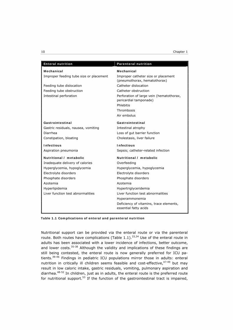
Nutritional support can be provided via the enteral route or via the parenteral route. Both routes have complications (Table 1.1).33,34 Use of the enteral route in adults has been associated with a lower incidence of infections, better outcome, and lower costs.35-38 Although the validity and implications of these findings are still being contested, the enteral route is now generally preferred for ICU pa-tients.39-46 Findings in pediatric ICU populations mirror those in adults: enteral nutrition in critically ill children seems feasible and cost-effective,47-49 but may result in low caloric intake, gastric residuals, vomiting, pulmonary aspiration and diarrhea.48-52 In children, just as in adults, the enteral route is the preferred route for nutritional support.53 If the function of the gastrointestinal tract is impaired,
Enteral nutrition Parenteral nutrition
Mechanical Mechanical
Improper feeding tube size or placement Improper catheter size or placement (pneumothorax, hematothorax)
Feeding tube dislocation Catheter dislocation
Feeding tube obstruction Catheter obstruction
Intestinal perforation Perforation of large vein (hematothorax, pericardial tamponade)
Phlebitis
Thrombosis
Air embolus
Gastrointestinal Gastrointestinal
Gastric residuals, nausea, vomiting Intestinal atrophy
Diarrhea Loss of gut barrier function
Constipation, bloating Cholestasis, liver failure
Infectious Infectious
Aspiration pneumonia Sepsis; catheter-related infection
Nutritional / metabolic Nutritional / metabolic
Inadequate delivery of calories Overfeeding
Hyperglycemia, hypoglycemia Hyperglycemia, hypoglycemia
Electrolyte disorders Electrolyte disorders
Phosphate disorders Phosphate disorders
Azotemia Azotemia
Hyperlipidemia Hypertriglyceridemia
Liver function test abnormalities Liver function test abnormalities
Hyperammonemia
Deficiency of vitamins, trace elements, essential fatty acids
Table 1.1 Complications of enteral and parenteral nutrition
Introduction 11
however, parenteral nutrition may -and probably should- be used as an adjunct to enteral nutrition. The indication for total parenteral nutrition has been nar-rowed down to a non-functioning gastrointestinal tract.53 Examples of impaired function or non-function of the gastrointestinal tract include intestinal obstruction, recent gastrointestinal surgery, necrotizing enterocolitis, and severe intestinal or global hypoxia-ischemia, such as may occur in midgut volvulus or critical illness.53 Gastrointestinal function is a complex issue. Barrier function, gut-related immu-nity, splanchnic blood flow and nutrient absorption are interrelated components of this function that may be impaired in the setting of critical illness and gastroin-testinal surgery.54 An intact barrier function of the gut prevents or limits bacterial translocation, i.e. the absorption of microorganisms and toxins. Intestinal hyper-permeability is seen in critical illness and after gastrointestinal surgery, and has been associated with increased morbidity and mortality.55-57 Apparent or true hy-perpermeability may be caused by intestinal or global ischemia.58 The exact rela-tion between intestinal hyperpermeability and increased bacterial translocation, however, and the clinical relevancy of bacterial translocation are still being unrav-eled.55-57,59,60 Be that as it may, increased bacterial translocation should in all likelihood be considered a sign of an altered balance of nonimmunologic and im-munologic host defenses on the one hand, and changing virulence of indigenous bacteria on the other hand.60 Moreover, gastrointestinal dysfunction in critical ill-ness is considered an instrumental early event of a sequence that may result in septic inflammatory response syndrome and multiple organ failure. In the case of poor gastrointestinal function, the choice how to provide nutritional support is delicate. Enteral nutrition, if started in an early phase of critical illness, is believed to preserve or restore gut barrier function and to attenuate the hypermetabolic stress reponse.61 In surgical infants receiving total parenteral nu-trition, even small quantities of enteral nutrition improve immune function.62 In premature newborns, progression from minimal enteral nutrition to full enteral nutrition in the first days of life appears to increase the incidence of necrotizing enterocolitis,63 whereas early reintroduction of enteral nutrition after necrotizing enterocolitis may reduce length of hospital stay.64 Parenteral nutrition is more likely to provide the target intake and thus to attenuate or reverse the progres-sion of malnutrition,51,52 but may impair immunity65,66 and cause or aggravate intestinal hyperpermeability67-69 and cholestasis.70
AIM OF THIS THESIS
This thesis describes studies on the interaction of nutrition and gastrointestinal function in newborns and infants who require surgical treatment for diseases en-tailing poor gastrointestinal function.
Chapter 1 12
The studies described in chapters 2 to 4 aim to delineate the indications for total parenteral versus enteral nutrition in the setting of critical illness: • In Chapter 2 we assess whether the sugar absorption test, a measure of in-
testinal permeability, may serve as a tool to time the (re-)introduction of en-teral nutrition in patients suffering from necrotizing enterocolitis.
• In Chapter 3 the same sugar absorption test is used to assess whether in-testinal permeability and absorptive capacity are affected by the route of feeding in newborns treated with extracorporeal membrane oxygenation.
• In Chapter 4 we assess whether the incidence of septic complications in newborns treated with extracorporeal membrane oxygenation is affected by the route of feeding.
The studies described in chapters 5 and 6 aim to critically appraise widely held notions about parenteral nutrition: • In Chapter 5 we assess the relation between septic events and parenteral
nutrition-associated cholestasis in surgical newborns with an intestinal anom-aly.
• In Chapter 6 we assess the clinical relevancy of nonurinary nitrogen excre-tion compared with urinary nitrogen excretion, in newborns and infants re-ceiving total parenteral nutrition after major digestive tract surgery.
The study described in chapter 7 aims to attenuate the adverse effects of par-enteral nutrition by improving the composition of the amino acid mixture: • In Chapter 7 we assess the effects of glutamine supplementation of par-
enteral nutrition in newborns and infants after major digestive tract surgery, with emphasis on intestinal permeability and nitrogen balance.
In Chapter 8 we summarize our findings and discuss them in a broader context. Chapter 9 is a summary in Dutch.
Introduction 13
REFERENCES 1. Pollack MM, Wiley JS, Holbrook PR. Early nutritional depletion in critically ill children.
Crit Care Med 1981;9:580-3.
2. Pollack MM, Wiley JS, Kanter R, Holbrook PR. Malnutrition in critically ill infants and children. JPEN J Parenter Enteral Nutr 1982;6:20-4.
3. Strupp BJ, Levitsky DA. Enduring cognitive effects of early malnutrition: a theoretical reappraisal. J Nutr 1995;125:2221S-32S.
4. Grantham-McGregor SM, Walker SP, Chang S. Nutritional deficiencies and later behav-ioural development. Proc Nutr Soc 2000;59:47-54.
5. Hsu A, Heshka S, Janumala I, et al. Larger mass of high-metabolic-rate organs does not explain higher resting energy expenditure in children. Am J Clin Nutr 2003;77:1506-11.
6. Scrimshaw NS, SanGiovanni JP. Synergism of nutrition, infection, and immunity: an overview. Am J Clin Nutr 1997;66:464S-77S.
7. Long CL, Schaffel N, Geiger JW, Schiller WR, Blakemore WS. Metabolic response to in-jury and illness: estimation of energy and protein needs from indirect calorimetry and nitrogen balance. JPEN J Parenter Enteral Nutr 1979;3:452-6.
8. Pollack MM. Nutritional support of children in the intensive care unit. In: Suskind RM, Lewinter-Suskind L, editors. Textbook of pediatric nutrition. 2nd ed. New York: Raven Press; 1993. p. 207-23.
9. Hendricks KM, Duggan C, Gallagher L, et al. Malnutrition in hospitalized pediatric pa-tients. Current prevalence. Arch Pediatr Adolesc Med 1995;149:1118-22.
10. Coss-Bu JA, Jefferson LS, Walding D, David Y, Smith EO, Klish WJ. Resting energy ex-penditure in children in a pediatric intensive care unit: comparison of Harris-Benedict and Talbot predictions with indirect calorimetry values. Am J Clin Nutr 1998;67:74-80.
11. Briassoulis G, Zavras N, Hatzis T. Malnutrition, nutritional indices, and early enteral feeding in critically ill children. Nutrition 2001;17:548-57.
12. Pollack MM, Ruttimann UE, Wiley JS. Nutritional depletions in critically ill children: asso-ciations with physiologic instability and increased quantity of care. JPEN J Parenter En-teral Nutr 1985;9:309-13.
13. Chandra RK. Nutrition and the immune system: an introduction. Am J Clin Nutr 1997;66:460S-3S.
14. Reynolds JV, O'Farrelly C, Feighery C, et al. Impaired gut barrier function in malnour-ished patients. Br J Surg 1996;83:1288-91.
15. Winthrop AL, Wesson DE, Pencharz PB, Jacobs DG, Heim T, Filler RM. Injury severity, whole body protein turnover, and energy expenditure in pediatric trauma. J Pediatr Surg 1987;22:534-7.
16. Phillips R, Ott L, Young B, Walsh J. Nutritional support and measured energy expendi-ture of the child and adolescent with head injury. J Neurosurg 1987;67:846-51.
17. Tilden SJ, Watkins S, Tong TK, Jeevanandam M. Measured energy expenditure in pedi-atric intensive care patients. Am J Dis Child 1989;143:490-2.
18. Steinhorn DM, Green TP. Severity of illness correlates with alterations in energy me-tabolism in the pediatric intensive care unit. Crit Care Med 1991;19:1503-9.
19. Jones MO, Pierro A, Hammond P, Lloyd DA. The metabolic response to operative stress in infants. J Pediatr Surg 1993;28:1258-62; discussion 62-3.
20. Chwals WJ, Letton RW, Jamie A, Charles B. Stratification of injury severity using energy expenditure response in surgical infants. J Pediatr Surg 1995;30:1161-4.
21. Groner JI, Brown MF, Stallings VA, Ziegler MM, O'Neill JA, Jr. Resting energy expendi-ture in children following major operative procedures. J Pediatr Surg 1989;24:825-7; discussion 7-8.
Chapter 1 14
22. Chwals WJ, Lally KP, Woolley MM, Mahour GH. Measured energy expenditure in critically ill infants and young children. J Surg Res 1988;44:467-72.
23. Letton RW, Chwals WJ, Jamie A, Charles B. Early postoperative alterations in infant en-ergy use increase the risk of overfeeding. J Pediatr Surg 1995;30:988-92; discussion 92-3.
24. Selby AM, McCauley JC, Schell DN, O'Connell A, Gillis J, Gaskin KJ. Indirect calorimetry in mechanically ventilated children: a new technique that overcomes the problem of en-dotracheal tube leak. Crit Care Med 1995;23:365-70.
25. Gebara BM, Gelmini M, Sarnaik A. Oxygen consumption, energy expenditure, and sub-strate utilization after cardiac surgery in children. Crit Care Med 1992;20:1550-4.
26. Chwals WJ. Overfeeding the critically ill child: fact or fantasy? New Horiz 1994;2:147-55.
27. Dimand RJ. Parenteral nutrition in the critically ill infant and child. In: Baker Jr. RD, Baker SS, Davis AM, editors. Pediatric parenteral nutrition. New York: Chapman & Hall; 1997. p. 273-300.
28. Shew SB, Keshen TH, Jahoor F, Jaksic T. The determinants of protein catabolism in neonates on extracorporeal membrane oxygenation. J Pediatr Surg 1999;34:1086-90.
29. Duffy B, Pencharz P. The effects of surgery on the nitrogen metabolism of parenterally fed human neonates. Pediatr Res 1986;20:32-5.
30. Shanbhogue RL, Jackson M, Lloyd DA. Operation does not increase resting energy ex-penditure in the neonate. J Pediatr Surg 1991;26:578-80.
31. Bouwmeester NJ, Anand KJ, van Dijk M, Hop WC, Boomsma F, Tibboel D. Hormonal and metabolic stress responses after major surgery in children aged 0-3 years: a double-blind, randomized trial comparing the effects of continuous versus intermittent mor-phine. Br J Anaesth 2001;87:390-9.
32. Georgieff MK. Nutrition. In: Avery GB, Fletcher MA, MacDonald MG, editors. Neonatol-ogy - Pathophysiology and management of the newborn. 5th ed. Philadelphia: Lippin-cott Williams & Wilkins; 1999. p. 363-94.
33. Davis A. Indications and techniques for enteral feeds. In: Baker SB, Baker Jr. RD, Davis A, editors. Pediatric enteral nutrition. New York: Chapman & Hall; 1994. p. 67-94.
34. Davis AM. Initiation, monitoring, and complications of pediatric parenteral nutrition. In: Baker Jr. RD, Baker SS, Davis AM, editors. Pediatric parenteral nutrition. New York: Chapman & Hall; 1997. p. 212-37.
35. Lipman TO. Grains or veins: is enteral nutrition really better than parenteral nutrition? A look at the evidence. JPEN J Parenter Enteral Nutr 1998;22:167-82.
36. Finck C. Enteral versus parenteral nutrition in the critically ill. Nutrition 2000;16:393-4.
37. Bozzetti F, Braga M, Gianotti L, Gavazzi C, Mariani L. Postoperative enteral versus par-enteral nutrition in malnourished patients with gastrointestinal cancer: a randomised multicentre trial. Lancet 2001;358:1487-92.
38. Marik PE, Pinsky M. Death by parenteral nutrition. Intensive Care Med 2003;29:867-9.
39. Adam S, Batson S. A study of problems associated with the delivery of enteral feed in critically ill patients in five ICUs in the UK. Intensive Care Med 1997;23:261-6.
40. MacFie J. Enteral versus parenteral nutrition: the significance of bacterial translocation and gut-barrier function. Nutrition 2000;16:606-11.
41. De Jonghe B, Appere-De-Vechi C, Fournier M, et al. A prospective survey of nutritional support practices in intensive care unit patients: what is prescribed? What is delivered? Crit Care Med 2001;29:8-12.
42. Woodcock NP, Zeigler D, Palmer MD, Buckley P, Mitchell CJ, MacFie J. Enteral versus parenteral nutrition: a pragmatic study. Nutrition 2001;17:1-12.
Introduction 15
43. Maykel JA, Bistrian BR. Is enteral feeding for everyone? Crit Care Med 2002;30:714-6.
44. Woodcock N, MacFie J. Optimal nutrition support (and the demise of the enteral versus parenteral controversy). Nutrition 2002;18:523-4.
45. Preiser JC, Chiolero R, Wernerman J. Nutritional papers in ICU patients: what lies be-tween the lines? Intensive Care Med 2003;29:156-66.
46. Varga P, Griffiths R, Chiolero R, et al. Is parenteral nutrition guilty? Intensive Care Med 2003;29:1861-4.
47. Chellis MJ, Sanders SV, Webster H, Dean JM, Jackson D. Early enteral feeding in the pediatric intensive care unit. JPEN J Parenter Enteral Nutr 1996;20:71-3.
48. Panadero E, López-Herce J, Caro L, et al. Transpyloric enteral feeding in critically ill children. J Pediatr Gastroenterol Nutr 1998;26:43-8.
49. Briassoulis GC, Zavras NJ, Hatzis MT. Effectiveness and safety of a protocol for promo-tion of early intragastric feeding in critically ill children. Pediatr Crit Care Med 2001;2:113-21.
50. Horn D, Chaboyer W. Gastric feeding in critically ill children: a randomized controlled trial. Am J Crit Care 2003;12:461-8.
51. Rogers EJ, Gilbertson HR, Heine RG, Henning R. Barriers to adequate nutrition in criti-cally ill children. Nutrition 2003;19:865-8.
52. Taylor RM, Preedy VR, Baker AJ, Grimble G. Nutritional support in critically ill children. Clin Nutr 2003;22:365-9.
53. Baker SS. Indications for parenteral nutrition. In: Baker Jr. RD, Baker SS, Davis AM, editors. Pediatric parenteral nutrition. New York: Chapman & Hall; 1997. p. 18-30.
54. Rombeau JL, Takala J, editors. Gut dysfunction in critical illness. Berlin: Springer-Ver-lag; 1996.
55. Doig CJ, Sutherland LR, Sandham JD, Fick GH, Verhoef M, Meddings JB. Increased in-testinal permeability is associated with the development of multiple organ dysfunction syndrome in critically ill ICU patients. Am J Respir Crit Care Med 1998;158:444-51.
56. Kanwar S, Windsor AC, Welsh F, Barclay GR, Guillou PJ, Reynolds JV. Lack of correlation between failure of gut barrier function and septic complications after major upper gas-trointestinal surgery. Ann Surg 2000;231:88-95.
57. Thomson AB, Drozdowski L, Iordache C, et al. Small bowel review: Normal physiology, part 2. Dig Dis Sci 2003;48:1565-81.
58. Bijlsma PB, Peeters RA, Groot JA, Dekker PR, Taminiau JA, van der Meer R. Differential in vivo and in vitro intestinal permeability to lactulose and mannitol in animals and hu-mans: a hypothesis. Gastroenterology 1995;108:687-96.
59. Fink MP. Interpreting dual-sugar absorption studies in critically ill patients: what are the implications of apparent increases in intestinal permeability to hydrophilic solutes? In-tensive Care Med 1997;23:489-92.
60. Alverdy JC, Laughlin RS, Wu L. Influence of the critically ill state on host-pathogen in-teractions within the intestine: gut-derived sepsis redefined. Crit Care Med 2003;31:598-607.
61. van Haren FMP, van der Hoeven JG. Early enteral nutrition in the intensive care unit. In: Vincent JL, editor. Yearbook of intensive care and emergency medicine 2002. Berlin: Springer-Verlag; 2002. p. 481-91.
62. Okada Y, Klein N, van Saene HK, Pierro A. Small volumes of enteral feedings normalise immune function in infants receiving parenteral nutrition. J Pediatr Surg 1998;33:16-9.
63. Berseth CL, Bisquera JA, Paje VU. Prolonging small feeding volumes early in life de-creases the incidence of necrotizing enterocolitis in very low birth weight infants. Pedi-atrics 2003;111:529-34.
Chapter 1 16
64. Bohnhorst B, Müller S, Dördelmann M, Peter CS, Petersen C, Poets CF. Early feeding after necrotizing enterocolitis in preterm infants. J Pediatr 2003;143:484-7.
65. Cruccetti A, Pierro A, Uronen H, Klein N. Surgical infants on total parenteral nutrition have impaired cytokine responses to microbial challenge. J Pediatr Surg 2003;38:138-42; discussion -42.
66. Shulman RJ, Phillips S. Parenteral nutrition in infants and children. J Pediatr Gastroen-terol Nutr 2003;36:587-607.
67. van der Hulst RRWJ, van Kreel BK, von Meyenfeldt MF, et al. Glutamine and the preser-vation of gut integrity. Lancet 1993;341:1363-5.
68. Hadfield RJ, Sinclair DG, Houldsworth PE, Evans TW. Effects of enteral and parenteral nutrition on gut mucosal permeability in the critically ill. Am J Respir Crit Care Med 1995;152:1545-8.
69. Cunningham-Rundles S, Lin DH. Nutrition and the immune system of the gut. Nutrition 1998;14:573-9.
70. Teitelbaum DH, Tracy T. Parenteral nutrition-associated cholestasis. Semin Pediatr Surg 2001;10:72-80.
2 Intestinal permeability in newborns with
necrotizing enterocolitis and controls:
does the sugar absorption test provide guidelines
for the time to (re-)introduce enteral nutrition?
Marjolein Piena-Spoel Marcel J.I.J. Albers Joop ten Kate Dick Tibboel J Pediatr Surg 2001;36:587-92
Chapter 2 18
ABSTRACT
BACKGROUND: In necrotizing enterocolitis (NEC), (sub)mucosal edema, hemor-rhage, ulceration and/or necrosis will disturb intestinal integrity, as reflected by an increased intestinal permeability. In many clinics, enteral substrate is there-fore withheld for a variable period up to three weeks. We used the sugar absorp-tion test to measure changes of intestinal permeability in surgically treated NEC patients and surgical controls, in order to evaluate the usefulness of this test in timing the (re-)introduction of enteral feeding in NEC patients as intestinal integrity recovers. METHODS: Changes in intestinal permeability to lactulose and rhamnose were pro-spectively evaluated in thirteen children with necrotizing enterocolitis and ten op-erated control patients. The patients were given 1 mL/kg body weight lactu-lose/rhamnose solution at different time intervals after admission. The lactu-lose/rhamnose ratio was determined by gaschromatography in 4-hour urine sam-ples. RESULTS: The L/R ratios in NEC patients were increased for prolonged periods of time with a tendency to decrease in the third week after the start of NEC. How-ever, in some cases, the increased L/R ratios even exceeded the 3-week period of starvation. High peaks in the L/R ratio were seen in patients suffering from bowel perforation or sepsis. Compared to NEC patients, L/R ratios of control patients were increased only in the first days after surgery and normalized more rapidly. The results of the L/R tests in this study corroborated the clinical condition of the patients. CONCLUSION: In a group of seriously ill newborns with advanced stages of NEC, the sugar absorption test shows individual variability in the recovery of intestinal permeability. An individual approach in restarting enteral nutrition seems to be justified; however, the optimal timepoint to restart enteral nutrition cannot be determined by the sugar absorption test alone. Combining parameters of intesti-nal integrity and function could enable a more accurate determination of this op-timal timepoint.
L/R ratio & NEC 19
INTRODUCTION
Necrotizing enterocolitis (NEC) is a life-threatening condition, in over 90% of cases affecting premature neonates. NEC is a worldwide problem with an inci-dence of 1 to 3 cases in 1,000 live births.1 During the past two decades its inci-dence has increased, largely because more infants of lower gestational age sur-vive.2 In the United States, its mortality rate is 13.1 deaths in 100,000 live births, or 20% to 40% of all cases.3 NEC is a disease of the immature mucosal barrier. Three major factors are claimed to be involved in the pathogenesis: enteral nutri-tion, intestinal ischemia, and colonization of the gut by pathogenic bacteria. Focal or diffuse mucosal ulceration, submucosal edema, hemorrhage, and necrosis in-crease the intestinal permeability.4 Initial treatment usually consists of broad-spectrum antibiotics and bowel starvation, supported by total parenteral nutrition for a period up to 3 weeks. Pneumoperitoneum and/or persistent acidosis with continuing deterioration of the clinical condition are indications for surgery.5 Ob-jective criteria for the optimal timing of (re-)introduction of enteral nutrition in these patients are not available. Studies in critically ill adults document that im-pairment of the mucosal barrier function, together with overgrowth of pathogenic bacteria within the gastrointestinal tract, enhances translocation of bacteria and endotoxins. Consequently, various inflammatory mediators are activated, result-ing in systemic inflammatory response syndrome, sepsis, and multiple organ fail-ure.6-9 Early (re-)introduction of enteral nutrition in the critically ill (“minimal en-teral feeding”) helps to preserve an intact intestinal mucosal barrier function du-ring critical illness.10,11 The sugar absorption test (SAT) is a noninvasive and reliable test to evaluate ab-normal intestinal permeability under different conditions such as coeliac disease, cystic fibrosis and Crohn’s disease. It enables the physician to monitor the condi-tion of the diseased bowel, both in children and adults.12 The SAT measures uri-nary excretion of orally administered inert markers with high renal clearance rates. In this study we measured changes in intestinal permeability to lactulose, rhamnose and xylose to evaluate the usefulness of this test in timing the (re-)in-troduction of enteral feeding in NEC patients as intestinal integrity recovers. Most patients were operated in the acute phase of NEC. In order to evaluate the effect of abdominal surgery versus intestinal disease and surgery, patients undergoing surgery with bowel handling such as in case of diaphragmatic hernia, malrotation and esophageal atresia, served as controls.
MATERIALS AND METHODS
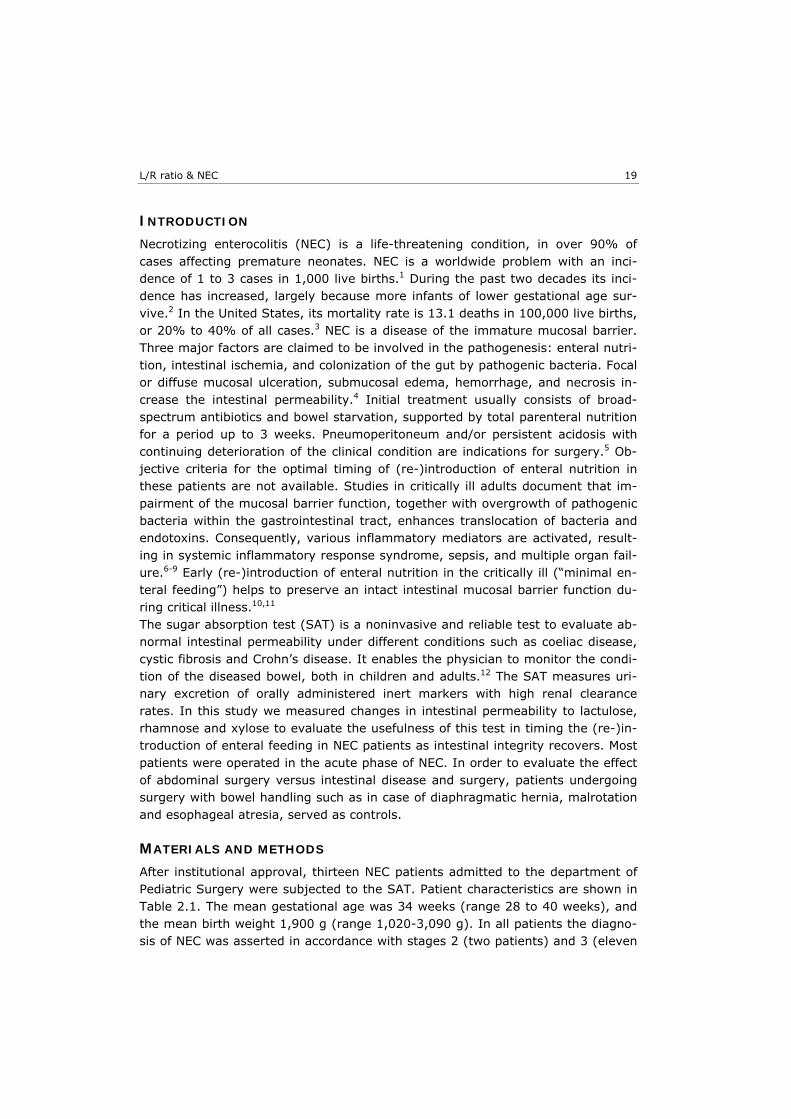
After institutional approval, thirteen NEC patients admitted to the department of Pediatric Surgery were subjected to the SAT. Patient characteristics are shown in Table 2.1. The mean gestational age was 34 weeks (range 28 to 40 weeks), and the mean birth weight 1,900 g (range 1,020-3,090 g). In all patients the diagno-sis of NEC was asserted in accordance with stages 2 (two patients) and 3 (eleven
Ino
tro
pic
s
no
no
yes
no
yes
no
no
no
no
no
yes
no
no
Ven
tila
tio
n
no
8 d
44 d
13 d
5 d
no
4 d
25 d
7 d
no
17 d
5 d
30 d
Po
stn
ata
l ag
e
at
surg
ery
8 d
5 d
5 d
5 d
5 d
8 d
8 d
5 w
k
16 d
7 d
8 d
7 d
19 d
Su
rgic
al
pro
ced
ure
subto
tal co
lect
om
y, ile
ost
om
y
subto
tal co
lect
om
y, c
olo
stom
y
subto
tal co
lect
om
y, c
olo
stom
y
subto
tal co
lect
om
y, ile
ost
om
y
conse
rvat
ive
lapar
oto
my
without
rese
ctio
n
subto
tal co
lect
om
y, ile
ost
om
y
ileost
om
y
rese
ctio
n 1
cm
ile
um
, ile
ost
om
y
subto
tal co
lect
om
y, ile
ost
om
y
subto
tal co
lect
om
y, r
esec
tion
10 c
m ile
um
, ile
ost
om
y
rese
ctio
n 5
cm
ile
um
, ile
ost
om
y
subto
tal co
lect
om
y, r
esec
tion
20 c
m ile
um
, ile
ost
om
y
NEC
st
ag
e
3
3
3
3
3
2
3
2
3
3
3
3
3
Bir
th w
eig
ht
(g
) 2655
2235
1020
2010
1050
3240
1330
1090
3090
2300
1600
2020
1120
1905
Gest
ati
on
al
ag
e (
wk)
35
40
31
36
29
38
35
31
38
35
34
34
28
34
Pati
en
t
1
2
3
4
5
6
7
8
9
10
11
12
13
Mea
n
Tab
le 2
.1 C
hara
cteri
stic
s o
f N
EC
pati
en
ts
L/R ratio & NEC 21
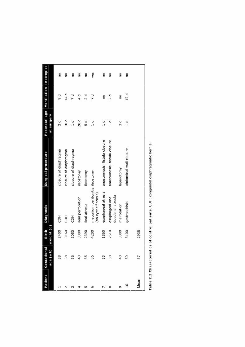
patients) of Bell’s classification.13 In eleven patients surgery was performed be-cause the bowel perforated in the course of NEC or because the patient’s condi-tion deteriorated rapidly. Eight patients suffered from bowel perforation within 5 days after the start of NEC. In one patient (No. 9) the bowel perforated on day 10 after the start of NEC. After resection of the necrotic bowel part(s), an ileostomy was placed in eight patients and a colostomy in two patients. In one patient a laparotomy was performed without resecting bowel parts, and two patients were treated conservatively. In one patient (No. 8) multiple intestinal strictures were diagnosed and managed surgically 5 weeks after start of NEC. As part of the sup-portive therapy following the diagnosis of NEC, all patients were starved. Accord-ing to our protocol starvation lasted for 3 weeks and during this period all pa-tients received total parenteral nutrition. Broad-spectrum antibiotics were admin-istered to all patients. Ten patients were mechanically ventilated following admis-sion. In three patients persistent hypotension necessitated treatment with inotropic medication. Patients 8 through 11 and 13 were starved a few days longer than the other NEC patients because their clinical condition was still com-promised at the end of the third week (three patients were recovering from sepsis and two patients suffered from abdominal distension). In the same period gut permeability was measured in ten control patients who had undergone surgery including bowel handling. Patient characteristics of the control patients are shown in Table 2.2. The mean gestational age was 37 weeks (range 33-40 weeks), the mean birth weight 2,900 g (range 1,860-4,200 g). All patients were fed parenterally. Eight patients were mechanically ventilated. In one patient low systemic bloodpressure was treated with inotropic medication. In three patients, who suffered from a congenital diaphragmatic hernia, the dia-phragm was closed. An ileostomy was placed in three other patients. In two pa-tients an esophageal atresia with a tracheoesophageal fistula was corrected. In one patient a malrotation, resulting in intestinal obstruction, was diagnosed and managed surgically, and in one patient with gastroschisis the abdominal wall defect was closed. The sugar absorption test solution was prepared by the hospital pharmacy. One hundred fourty milligrams of glucose, 140 mg of rhamnose and 70 mg of xylose were dissolved in 50 mL demineralized water. A total of 8.6 g of lactulose was added, and demineralized water was added up to 100 mL. The osmolarity was measured in 15 samples and averaged 429 mosmol/L (range 410-452 mosmol/L). In each patient, 2 to 7 lactulose/rhamnose (L/R) tests were performed during the acute phase of NEC and after surgery. Testing at predetermined time points fol-lowing the start of NEC (with 2- to3-day intervals) could not be realized in all cases because some patients were not admitted until surgery was indicated, and referred from the NICU of other hospitals. As a consequence they could not be measured at the start of NEC. Moreover, extremely ill patients were not tested because of hemodynamic or respiratory instability. As a result, fewer measure-ments were performed in NEC patients in the first days after surgery. The accu-
Ino
tro
pic
s
no
no
no
no
no
yes
no
no
no
no
Ven
tila
tio
n
9 d
14 d
7 d
4 d
2 d
7 d
no
2 d
no
17 d
Po
stn
ata
l ag
e
at
surg
ery
3 d
10 d
1 d
20 d
5 d
1 d
1 d
1 d
3 d
1 d
Su
rgic
al
pro
ced
ure
closu
re o
f dia
phra
gm
a
closu
re o
f dia
phra
gm
a
closu
re o
f dia
phra
gm
a
ileost
om
y
ileost
om
y
ileost
om
y
anas
tom
osi
s, f
istu
la c
losu
re
anas
tom
osi
s, f
istu
la c
losu
re
lapar
oto
my
abdom
inal
wal
l cl
osu
re
Dia
gn
osi
s
CD
H
CD
H
CD
H
ileal
per
fora
tion
ileal
atr
esia
mec
oniu
m p
eritonitis
(n
o c
ystic
fibro
sis)
esophag
eal at
resi
a
esophag
eal an
d
duoden
al a
tres
ia
mal
rota
tion
gas
trosc
his
is
Bir
th
weig
ht
(g)
2400
3160
3050
3380
2390
4200
1860
2510
3300
3100
2935
Gest
ati
on
al
ag
e (
wk)
38
38
36
40
35
36
33
38
40
39
37
Pati
en
t
1
2
3
4
5
6
7
8
9
10
Mea
n
Tab
le 2
.2 C
hara
cteri
stic
s o
f co
ntr
ol p
ati
en
ts. CD
H:
congen
ital
dia
phra
gm
atic
her
nia
.
L/R ratio & NEC 23
rate collection of a 4-hour urine sample was dif-ficult, especially in female newborns, and the test had to be repeated in some cases. Patients were given 1 mL/kg body weight L/R solution via a nasogastric tube. All urine passed in the next 4 hours was collected.14 The urine volume was measured and 1 mL was frozen at –80 oC. A volume corresponding with 0.66 µmol creatinine of the urine sample was dried in addi-tion of 100 µL mannitol and silylated with 40 µL N,O- bis(trimethylsilyl)acetamide, 40 µL trimeth-ylchloorsilane, and 80 µL pyridine (1 hour at 70 oC). After prepurification, 1-2 µL was analyzed by capillary gas chromatography (CP-Sil-5 col-umn, 25 m x 0.22 mm, oven program 100 to 260 oC with 7 oC/min) and detected with a flame ionization detector.15 The concentrations of lac-tulose, rhamnose and xylose were expressed in millimole per mole creatinine and as a percent-age of the ingested dose. Intestinal permeability is increased if the L/R (lactulose-%/rhamnose-%) ratio is above 0.05.14,16 Comparisons were carried out using the Mann-Whitney U test; significance was established at P <0.05.
RESULTS
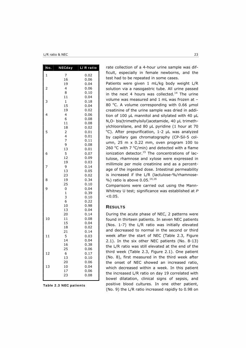
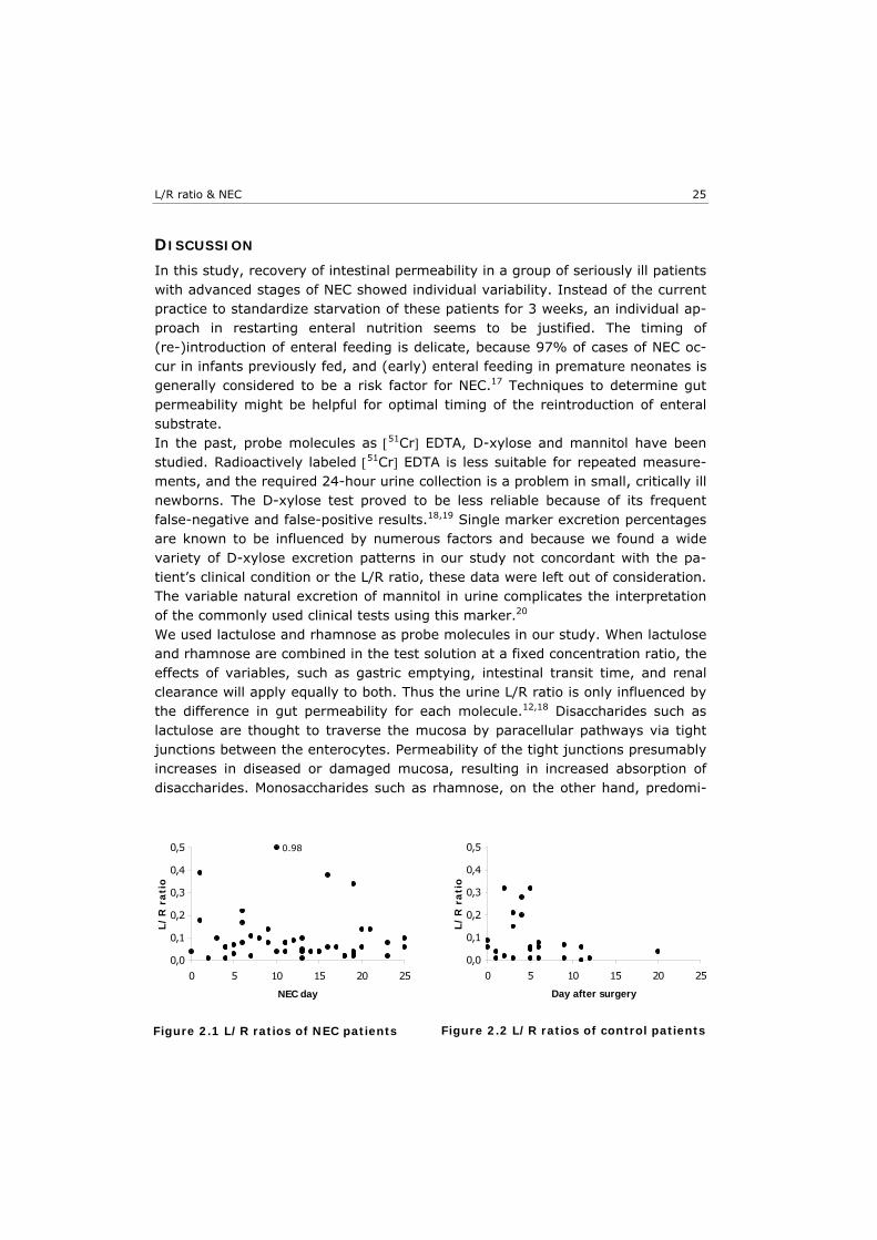
During the acute phase of NEC, 2 patterns were found in thirteen patients. In seven NEC patients (Nos. 1-7) the L/R ratio was initially elevated and decreased to normal in the second or third week after the start of NEC (Table 2.3, Figure 2.1). In the six other NEC patients (No. 8-13) the L/R ratio was still elevated at the end of the third week (Table 2.3, Figure 2.1). One patient (No. 8), first measured in the third week after the onset of NEC showed an increased ratio, which decreased within a week. In this patient the increased L/R ratio on day 19 correlated with bowel dilatation, clinical signs of sepsis, and positive blood cultures. In one other patient, (No. 9) the L/R ratio increased rapidly to 0.98 on
No. NECday L/R ratio
1 7 0.02 16 0.06 19 0.04 2 4 0.06 8 0.10 11 0.04 3 1 0.18 15 0.04 19 0.02 4 4 0.06 6 0.08 11 0.08 18 0.02 5 2 0.01 4 0.01 7 0.11 9 0.08 13 0.01 6 5 0.07 12 0.09 19 0.03 7 9 0.14 13 0.05 23 0.02 8 19 0.34 25 0.10 9 0 0.04 1 0.39 3 0.10 6 0.22 10 0.98 13 0.04 20 0.14 10 11 0.08 15 0.04 18 0.02 21 0.14 11 5 0.03 14 0.04 16 0.38 25 0.06 12 6 0.17 13 0.10 20 0.06 13 10 0.04 17 0.06 23 0.08
Table 2.3 NEC patients
Chapter 2 24
the tenth day after start of NEC, which coincided with a clinical deterioration of the patient’s con-dition due to perforation of the bowel. One other patient (No. 11) had normal L/R ratios in the first 2 weeks after the start of NEC, but on day 16 this ratio increased to 0.38. This patient also suffered from bowel dilatation and clinical signs of sepsis, although blood cultures remained negative. In two other patients (Nos. 10 and 13) the L/R ratios increased unexpected at the end of the third week. Both suffered from mild ab-dominal distension, without any other signs of recurrent NEC or sepsis, which spontaneously disappeared within 2 days. In one patient (No. 12) the increased L/R ratio of 0.17 on day 6 slowly decreased to 0.06 on day 20. No signifi-cant differences in gestational age, birth weight, NEC stage, days on the ventilator, and need of vasopressive medication were observed between the group with a normal and the group with an increased L/R ratio at the end of the third week (P >0.3). In all patients with a normal L/R ratio at the end of the third week, only the colon was involved as part of the disease process, whereas in five of six patients in the group with an in-creased ratio at the end of the third week the
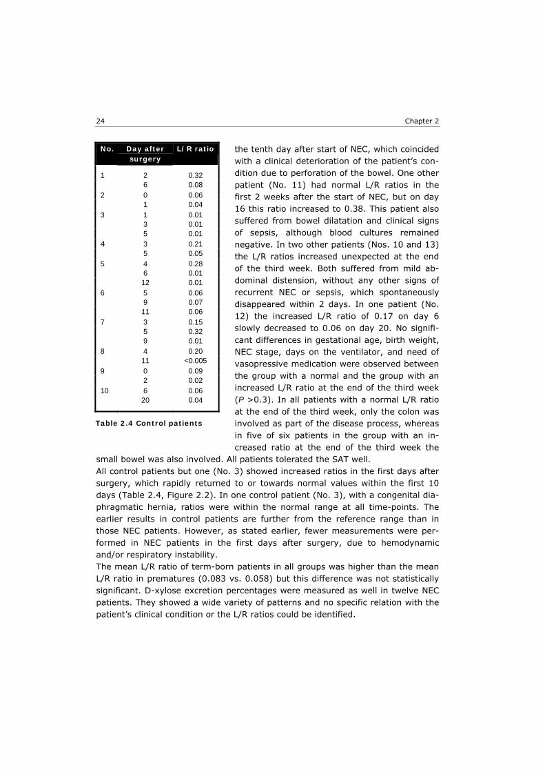
small bowel was also involved. All patients tolerated the SAT well. All control patients but one (No. 3) showed increased ratios in the first days after surgery, which rapidly returned to or towards normal values within the first 10 days (Table 2.4, Figure 2.2). In one control patient (No. 3), with a congenital dia-phragmatic hernia, ratios were within the normal range at all time-points. The earlier results in control patients are further from the reference range than in those NEC patients. However, as stated earlier, fewer measurements were per-formed in NEC patients in the first days after surgery, due to hemodynamic and/or respiratory instability. The mean L/R ratio of term-born patients in all groups was higher than the mean L/R ratio in prematures (0.083 vs. 0.058) but this difference was not statistically significant. D-xylose excretion percentages were measured as well in twelve NEC patients. They showed a wide variety of patterns and no specific relation with the patient’s clinical condition or the L/R ratios could be identified.
No. Day after surgery
L/R ratio
1 2 0.32 6 0.08 2 0 0.06 1 0.04 3 1 0.01 3 0.01 5 0.01 4 3 0.21 5 0.05 5 4 0.28 6 0.01 12 0.01 6 5 0.06 9 0.07 11 0.06 7 3 0.15 5 0.32 9 0.01 8 4 0.20 11 <0.005 9 0 0.09 2 0.02 10 6 0.06 20 0.04
Table 2.4 Control patients
L/R ratio & NEC 25
DISCUSSION
In this study, recovery of intestinal permeability in a group of seriously ill patients with advanced stages of NEC showed individual variability. Instead of the current practice to standardize starvation of these patients for 3 weeks, an individual ap-proach in restarting enteral nutrition seems to be justified. The timing of (re-)introduction of enteral feeding is delicate, because 97% of cases of NEC oc-cur in infants previously fed, and (early) enteral feeding in premature neonates is generally considered to be a risk factor for NEC.17 Techniques to determine gut permeability might be helpful for optimal timing of the reintroduction of enteral substrate. In the past, probe molecules as [51Cr] EDTA, D-xylose and mannitol have been studied. Radioactively labeled [51Cr] EDTA is less suitable for repeated measure-ments, and the required 24-hour urine collection is a problem in small, critically ill newborns. The D-xylose test proved to be less reliable because of its frequent false-negative and false-positive results.18,19 Single marker excretion percentages are known to be influenced by numerous factors and because we found a wide variety of D-xylose excretion patterns in our study not concordant with the pa-tient’s clinical condition or the L/R ratio, these data were left out of consideration. The variable natural excretion of mannitol in urine complicates the interpretation of the commonly used clinical tests using this marker.20 We used lactulose and rhamnose as probe molecules in our study. When lactulose and rhamnose are combined in the test solution at a fixed concentration ratio, the effects of variables, such as gastric emptying, intestinal transit time, and renal clearance will apply equally to both. Thus the urine L/R ratio is only influenced by the difference in gut permeability for each molecule.12,18 Disaccharides such as lactulose are thought to traverse the mucosa by paracellular pathways via tight junctions between the enterocytes. Permeability of the tight junctions presumably increases in diseased or damaged mucosa, resulting in increased absorption of disaccharides. Monosaccharides such as rhamnose, on the other hand, predomi-
Figure 2.1 L/R ratios of NEC patients
0,0
0,1
0,2
0,3
0,4
0,5
0 5 10 15 20 25
Day after surgery
L/
R r
ati
o
Figure 2.2 L/R ratios of control patients
0,0
0,1
0,2
0,3
0,4
0,5
0 5 10 15 20 25
NEC day
L/
R r
ati
o
0.98
Chapter 2 26
nantly pass by transcellular pathways through aqueous pores into the enterocyte membrane that are too small to permit the passage of lactulose. In villous atro-phy the intestinal surface area decreases, resulting in decreased absorption of monosaccharides.20 In 1995, Bijlsma et al proposed an alternative model to explain clinically obtained dual-sugar absorption data.21 They hypothesized that the hyperosmolarity of vil-lus tips is the result of the countercurrent exchanger features of the villus mi-crovasculature in humans. The high urine recovery of monosaccharides is caused by solvent drag through pores that allow the passage of monosaccharides but not of disaccharides. Lactulose recovery may represent paracellular passive diffusion over the mucosal barrier as a whole, whereas monosaccharide recovery mainly depends on water absorption in the upper part of the villus. Because intestinal ischemia and necrosis are instrumental in the pathogenesis of NEC, the tissue osmolarity of the villus tips will be affected resulting in a decreased solvent drag and, thus, monosaccharide absorption. According to this hypothesis, the disac-charide to monosaccharide ratio is primarily a standard for the normal metabolite absorption of villus epithelial cells and for normal villus flow rather than a pa-rameter for gut permeability.21,22 Although improvement of the ratios is explained differently in the above-mentioned theory, the SAT may also serve as a parame-ter for monitoring the condition of the diseased bowel. Beach et al showed increased permeability during the first week of life in neo-nates of gestational age 31 to 36 weeks.14 Prematures of 26 to 29 weeks of ges-tation showed a “mature” pattern of permeability at birth followed by a period of enhanced permeability after 3 to 4 weeks of life. Weaver et al showed that new-borns born before 34 weeks’ gestation exhibited a higher intestinal permeability than more mature newborns and that all preterm babies showed an appreciable decline in lactulose absorption during the first week of oral feeding.23 Babies of 34 to 37 weeks’ gestation achieved a mature intestinal permeability within 4 days after starting oral feedings. More recently, Shulman et al found increasing intesti-nal permeability in preterm infants of 26 to 30 weeks of gestational age in the first 28 days of life, which declined afterward.24 In contrast to our data, all above-mentioned studies were performed in enterally fed patients. Although the mean gestational ages of NEC and control patients differed by 3 weeks in our study (34 vs. 37 weeks gestational age), there was no obvious relation between gestational age and increased permeability. Differences in L/R ratios between term and pre-term born babies were not statistically significant and were primarily related to the degree of illness. We concluded that increased intestinal permeability for pro-longed periods of time, as found in patients 8 through 13, is more likely to be caused by the primary disease process than by the surgical intervention. The results of the L/R tests in this study corroborated the clinical condition of the patients; increased L/R ratios in the second or third week after start of NEC were found only in those patients suffering from sepsis, bowel perforation, or bowel dilatation.
L/R ratio & NEC 27
We do realize that the SAT is a small-bowel permeability test, especially suitable for patients with advanced stages of NEC. In these severely ill patients, surgery is often inevitable, and only then the affected bowel parts can be identified accu-rately. Further work is necessary to compare the results of the SAT with other parame-ters of bowel function and integrity, eventually leading to more individualized management of NEC patients. In some NEC patients enteral nutrition might be introduced earlier, which can favor the gastrointestinal immunological function and reduce total parenteral nutrition-related morbidity.
ACKNOWLEDGEMENTS
The authors thank M.J.B. Vaessen (Department of Clinical Chemistry, Atrium Medical Center, Heerlen) for excellent technical assistance, and Prof Dr H.A. Büller (Department of Pediatrics, Sophia Children’s Hospital, Rotterdam) for his helpful suggestions in preparing the manuscript.
Chapter 2 28
REFERENCES 1. Kosloske AM. Epidemiology of necrotizing enterocolitis. Acta Paediatr Suppl
1994;396:2-7.
2. Israel EJ. Necrotizing enterocolitis. In: Walker WA, Durie PR, editors. Pediatric gastro-intestinal disease: pathophysiology, diagnosis, management. Philadelphia: BC Decker Inc.; 1991. p. 639-46.
3. Holman RC, Stehr-Green JK, Zelasky MT. Necrotizing enterocolitis mortality in the United States, 1979-85. Am J Public Health 1989;79:987-9.
4. Israel EJ. Neonatal necrotizing enterocolitis, a disease of the immature intestinal mu-cosal barrier. Acta Paediatr Suppl 1994;396:27-32.
5. Neu J. Necrotizing enterocolitis: the search for a unifying pathogenic theory leading to prevention. Pediatr Clin North Am 1996;43:409-32.
6. Carrico CJ, Meakins JL, Marshall JC, Fry D, Maier RV. Multiple-organ-failure syndrome. Arch Surg 1986;121:196-208.
7. Hadfield RJ, Sinclair DG, Houldsworth PE, Evans TW. Effects of enteral and parenteral nutrition on gut mucosal permeability in the critically ill. Am J Respir Crit Care Med 1995;152:1545-8.
8. Deitch EA. The role of intestinal barrier failure and bacterial translocation in the devel-opment of systemic infection and multiple organ failure. Arch Surg 1990;125:403-4.
9. Marshall JC, Christou NV, Meakins JL. The gastrointestinal tract. The "undrained ab-scess" of multiple organ failure. Ann Surg 1993;218:111-9.
10. Heyland DK, Cook DJ, Guyatt GH. Enteral nutrition in the critically ill patient: a critical review of the evidence. Intensive Care Med 1993;19:435-42.
11. Zaloga GP, Black KW, Prielipp R. Effect of rate of enteral nutrient supply on gut mass. JPEN J Parenter Enteral Nutr 1992;16:39-42.
12. van Elburg RM, Uil JJ, Kokke FT, et al. Repeatability of the sugar-absorption test, using lactulose and mannitol, for measuring intestinal permeability for sugars. J Pediatr Gas-troenterol Nutr 1995;20:184-8.
13. Bell MJ, Ternberg JL, Feigin RD, et al. Neonatal necrotizing enterocolitis. Therapeutic decisions based upon clinical staging. Ann Surg 1978;187:1-7.
14. Beach RC, Menzies IS, Clayden GS, Scopes JW. Gastrointestinal permeability changes in the preterm neonate. Arch Dis Child 1982;57:141-5.
15. Jansen G, Muskiet FA, Schierbeek H, Berger R, van der Slik W. Capillary gas chroma-tographic profiling of urinary, plasma and erythrocyte sugars and polyols as their trimethylsilyl derivatives, preceded by a simple and rapid prepurification method. Clin Chim Acta 1986;157:277-93.
16. Miki K, Butler R, Moore D, Davidson G. Rapid and simultaneous quantification of rham-nose, mannitol, and lactulose in urine by HPLC for estimating intestinal permeability in pediatric practice. Clin Chem 1996;42:71-5.
17. Navarro J. Neonatal necrotizing enterocolitis. In: Navarro J, Schmitz J, editors. Paediat-ric gastroenterology. Oxford: Oxford University Press; 1992. p. 161-7.
18. Menzies IS. Transmucosal passage of inert molecules in health and disease. In: Skad-hauge E, Heintze K, editors. Intestinal absorption and secretion; 1983; Titisee, Ger-many: MTP Press; 1983. p. 527-43.
19. Lifschitz CH, Shulman RJ. Intestinal permeability tests: are they clinically useful? J Pe-diatr Gastroenterol Nutr 1990;10:283-7.
20. Travis S, Menzies I. Intestinal permeability: functional assessment and significance. Clin Sci (Lond) 1992;82:471-88.
L/R ratio & NEC 29
21. Bijlsma PB, Peeters RA, Groot JA, Dekker PR, Taminiau JA, Van Der Meer R. Differential in vivo and in vitro intestinal permeability to lactulose and mannitol in animals and hu-mans: a hypothesis. Gastroenterology 1995;108:687-96.
22. Fink MP. Interpreting dual-sugar absorption studies in critically ill patients: what are the implications of apparent increases in intestinal permeability to hydrophilic solutes? In-tensive Care Med 1997;23:489-92.
23. Weaver LT, Laker MF, Nelson R. Intestinal permeability in the newborn. Arch Dis Child 1984;59:236-41.
24. Shulman RJ, Schanler RJ, Lau C, Heitkemper M, Ou CN, Smith EO. Early feeding, ante-natal glucocorticoids, and human milk decrease intestinal permeability in preterm in-fants. Pediatr Res 1998;44:519-23.

3 Introduction of enteral feeding in neonates on
extracorporeal membrane oxygenation after
evaluation of intestinal permeability changes
Marjolein Piena Marcel J.I.J. Albers Paul M.M. van Haard Saskia Gischler Dick Tibboel J Pediatr Surg 1998;33:30-4
Chapter 3 32
ABSTRACT
BACKGROUND/PURPOSE: Neonates meeting criteria for extracorporeal membrane oxygenation (ECMO) often suffer from variable periods of hypoxia. During ECMO, starvation of the gut is common practice in many centers as splanchnic ischemia results in loss of intestinal integrity, which in turn predisposes for bacterial trans-location and sepsis and eventually necrotizing enterocolitis (NEC) and multiorgan failure. However, minimal enteral feeding is thought to be of benefit in the criti-cally ill. Data on intestinal integrity in newborns on ECMO and the effects of en-teral nutrition are not available. This study prospectively evaluates the changes in small intestinal integrity in 16 neonatal ECMO patients. METHODS: With 2-day intervals, excretion percentages of lactulose/L-rhamnose (nonmediated diffusion), D-xylose (passive), and 3-O-methyl-D-glucose (active carrier-mediated transport) were measured by gas-liquid chromotography in a 4-hour urine sample. After obtaining baseline data in nine patients, enteral feeding was started in the next seven patients between the third and the ninth day of ECMO. RESULTS: Thirteen patients had increased lactulose/L-rhamnose ratios (>0.05) consistent with increased intestinal permeablity. In three patients the lactulose/L-rhamnose ratios were within the normal range. D-xylose excretion percentages were normal (or slightly increased) in 11 patients consistent with normal (or in-creased) passive carrier-mediated transport. 3-O-methyl-D-glucose excretion percentages were decreased (<10%) in all but one patient, consistent with de-creased active carrier-mediated transport. After introduction of enteral nutrition no significant changes of these parameters were seen. CONCLUSIONS: The authors conclude that intestinal integrity is compromised in neonates on ECMO and that introduction of enteral nutrition does not result in further deterioration. This conclusion does not support the practice of withholding enteral nutrition in critically ill newborns supported by ECMO.
L/R ratio & ECMO 33
INTRODUCTION
Venoarterial extracorporeal membrane oxygenation (ECMO) is used to provide partial heart-lung bypass for neonates with life-threatening, reversible cardiopul-monary failure unresponsive to more conventional therapies. Conditions most commonly treated with ECMO are meconium aspiration syndrome, persistent pulmonary hypertension, sepsis, pneumonia, severe asphyxia, or congenital dia-phragmatic hernia. In most centers, patients meet ECMO criteria when they reach the 80th percentile for expected mortality.1-3 Starvation of the gut supported by total parenteral nutrition is common practice in ECMO patients in many centers for different reasons. First, severe respiratory insufficiency, asphyxia, and/or prolonged periods of hypoxia with the need for vasopressive therapy are often present in neonates meeting ECMO criteria. These may result in splanchnic ischemia with progressive functional and histological changes associated with loss of mucosal barrier function.4 In newborns, intestinal ischemia predisposes for bacterial translocation and sepsis and eventually for the development of necrotizing enterocolitis.5,6 Second, during venoarterial ECMO, blood is being drained by gravity from the right atrium to a roller pump, wich propels it through the membrane oxygenator and a heat exchanger before it is returned to the body. The hemodynamic alterations in patients on ECMO may ad-versely affect splanchnic circulation.7 Valid data about intestinal permeability and absorptive capacity changes in ECMO patients are not available. Recently, simple tests have been proposed for the evaluation of gut permeability, both in adults and children, including newborns.8,9 The sugar absorption test (SAT) measures urinary excretion of inert markers with high renal clearance rates after oral administration. The SAT is a practical and noninvasive method for as-sessing intestinal permeability and absorption of neutral molecules. The aim of this study is to evaluate the changes in intestinal integriy during ECMO and before and after the start of (minimal) enteral nutrition using the SAT. The lactulose/L-rhamnose (L/R) ratio (nonmediated transport) was used as a pa-rameter for intestinal permeability in combination with D-xylose (passive) and 3-O-methyl-D-glucose (active carrier mediated transport) excretion percentages as parameters for intestinal absorption capacity for saccharides.
MATERIALS AND METHODS
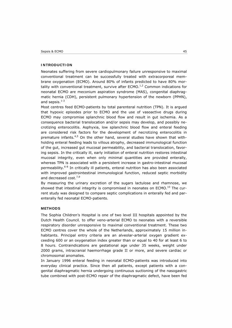
Patients
From January 1996 through February 1997, sixteen neonates were admitted to the department of Pediatric Surgery of the Sophia Children’s Hospital in Rotter-dam for ECMO. Patient characteristics are shown in Tables 3.1 and 3.2. The mean gestational age was 40 weeks (range, 36 to 43 weeks), and the mean birth weight was 3,300 g (range, 2,160 to 4,500 g). In all patients ECMO treatment was started within the first week after birth (median, 1 day after birth: range, 12
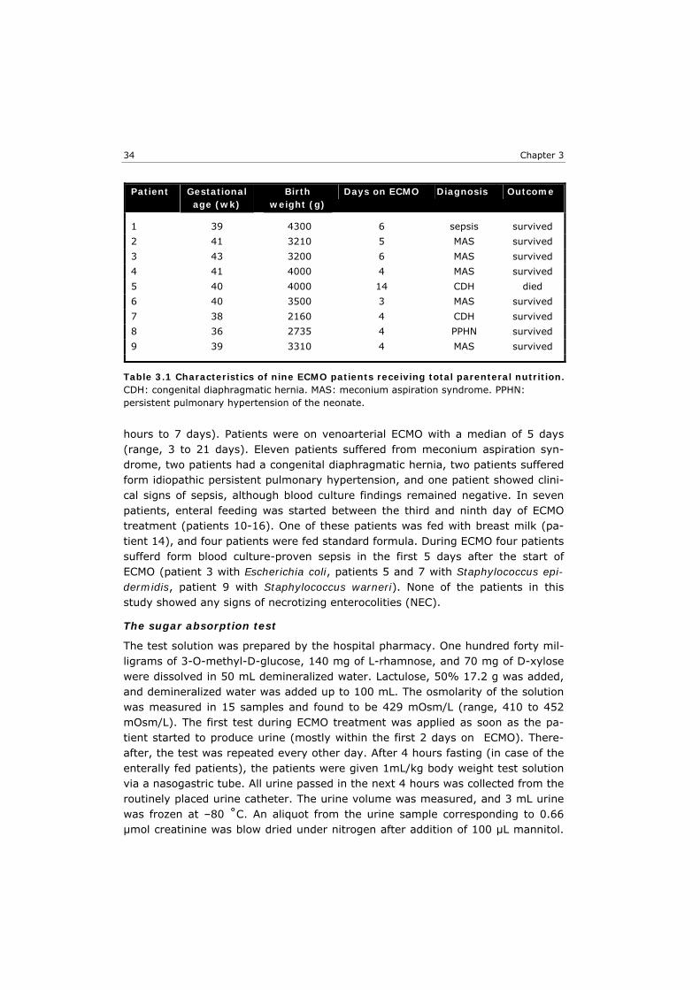
Chapter 3 34
hours to 7 days). Patients were on venoarterial ECMO with a median of 5 days (range, 3 to 21 days). Eleven patients suffered from meconium aspiration syn-drome, two patients had a congenital diaphragmatic hernia, two patients suffered form idiopathic persistent pulmonary hypertension, and one patient showed clini-cal signs of sepsis, although blood culture findings remained negative. In seven patients, enteral feeding was started between the third and ninth day of ECMO treatment (patients 10-16). One of these patients was fed with breast milk (pa-tient 14), and four patients were fed standard formula. During ECMO four patients sufferd form blood culture-proven sepsis in the first 5 days after the start of ECMO (patient 3 with Escherichia coli, patients 5 and 7 with Staphylococcus epi-dermidis, patient 9 with Staphylococcus warneri). None of the patients in this study showed any signs of necrotizing enterocolities (NEC).
The sugar absorption test
The test solution was prepared by the hospital pharmacy. One hundred forty mil-ligrams of 3-O-methyl-D-glucose, 140 mg of L-rhamnose, and 70 mg of D-xylose were dissolved in 50 mL demineralized water. Lactulose, 50% 17.2 g was added, and demineralized water was added up to 100 mL. The osmolarity of the solution was measured in 15 samples and found to be 429 mOsm/L (range, 410 to 452 mOsm/L). The first test during ECMO treatment was applied as soon as the pa-tient started to produce urine (mostly within the first 2 days on ECMO). There-after, the test was repeated every other day. After 4 hours fasting (in case of the enterally fed patients), the patients were given 1mL/kg body weight test solution via a nasogastric tube. All urine passed in the next 4 hours was collected from the routinely placed urine catheter. The urine volume was measured, and 3 mL urine was frozen at –80 ˚C. An aliquot from the urine sample corresponding to 0.66 µmol creatinine was blow dried under nitrogen after addition of 100 µL mannitol.
Patient Gestational age (wk)
Birth weight (g)
Days on ECMO Diagnosis Outcome
1 39 4300 6 sepsis survived
2 41 3210 5 MAS survived
3 43 3200 6 MAS survived
4 41 4000 4 MAS survived
5 40 4000 14 CDH died
6 40 3500 3 MAS survived
7 38 2160 4 CDH survived
8 36 2735 4 PPHN survived
9 39 3310 4 MAS survived
Table 3.1 Characteristics of nine ECMO patients receiving total parenteral nutrition. CDH: congenital diaphragmatic hernia. MAS: meconium aspiration syndrome. PPHN: persistent pulmonary hypertension of the neonate.
L/R ratio & ECMO 35
The residue was persilylated in 40 µL N,O-bis(trimethyl)acetamide, 40 µL trimethylchloorsilane, and 80 µL pyridine during 1 hour at 70 ˚C.10 After cooling to room temperature, 1 to 2 µL of the solution was analysed by capillary gas-liq-uid chromatography (CP-Sil-5-CB column, 25 m x 0.22 mm, 0.12 µm film thick-ness, oven programme 100 to 245 ˚C, rate, 3 ˚C/min) and detection with a flame ionization detector. The concentrations of lactulose, L-rhamnose, D-xylose, and 3-O-methyl-D-glucose were expressed in mg/L and as a percentage of the ingested dose. Intestinal permeability is considered to be normal if the L/R ratio (lactu-lose%/rhamnose%) is below 0.05.11,12 Intestinal absorptive capacity for saccha-rides is considered to be normal when excretion percentages of D-xylose (passive carrier-mediated transport) and 3-O-methyl-D-glucose (active carrier-mediated transport) are in the 10% to 30% range.7,13 In four patients, the first SAT was performed when they were still oliguric (urine production <0.5 mL/kg/h), and the urine sample was too small to determine the D-xylose (in two cases) and/or the 3-O-methyl-D-glucose (in all four cases) ex-cretion percentages.
RESULTS
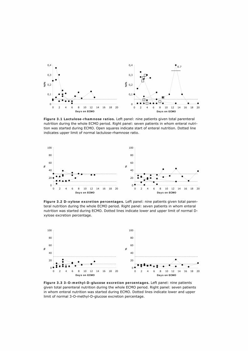
L/R ratio
The L/R ratio was increased (>0.05) in 28 of 48 measurements. In 5 of 28 in-creased ratios the lactulose excretion percentage was increased (>1%). In 26 of 28 increased ratios the L-rhamnose excretion percentage was decreased (<10%). In thirteen patients, the L/R ratios were increased during ECMO treatment. In three patients, (patients 1, 6, 11) all L/R ratios were within the normal range. In four other patients (patients 3, 5, 7, 9) L/R ratio peaked on day 3, 7, and 1, re-spectively, coinciding with sepsis. In patient 7, who suffered from sepsis during the whole ECMO period, the second L/R ratio peak on day 8 coincided with a de-
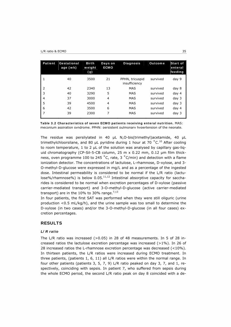
Patient Gestational age (wk)
Birth weight
(g)
Days on ECMO
Diagnosis Outcome Start of enteral feeding
1 40 3500 21 PPHN, tricuspid
insufficiency survived day 9
2 42 2340 13 MAS survived day 8
3 40 3290 5 MAS survived day 4
4 37 3000 4 MAS survived day 3
5 39 4500 4 MAS survived day 3
6 42 3500 6 MAS survived day 4
7 39 2300 7 MAS survived day 3
Table 3.2 Characteristics of seven ECMO patients receiving enteral nutrition. MAS: meconium aspiration syndrome. PPHN: persistent pulmonary hypertension of the neonate.
0
0,1
0,2
0,3
0,4
0 2 4 6 8 10 12 14 16 18 20
Days on ECMO
%/%
0
0,1
0,2
0,3
0,4
0 2 4 6 8 10 12 14 16 18 20
Days on ECMO
%/%
0
20
40
60
80
100
0 2 4 6 8 10 12 14 16 18 20
Days on ECMO
%
0
20
40
60
80
100
0 2 4 6 8 10 12 14 16 18 20
Days on ECMO
%
0
20
40
60
80
100
0 2 4 6 8 10 12 14 16 18 20
Days on ECMO
%
0
20
40
60
80
100
0 2 4 6 8 10 12 14 16 18 20
Days on ECMO
%
Figure 3.1 Lactulose-rhamnose ratios. Left panel: nine patients given total parenteral nutrition during the whole ECMO period. Right panel: seven patients in whom enteral nutri-tion was started during ECMO. Open squares indicate start of enteral nutrition. Dotted line indicates upper limit of normal lactulose-rhamnose ratio.
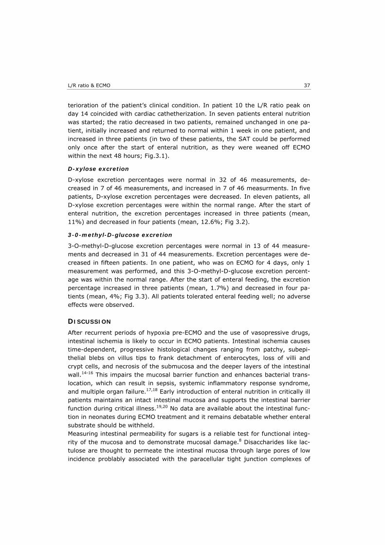
Figure 3.2 D-xylose excretion percentages. Left panel: nine patients given total paren-teral nutrition during the whole ECMO period. Right panel: seven patients in whom enteral nutrition was started during ECMO. Dotted lines indicate lower and upper limit of normal D-xylose excretion percentage.
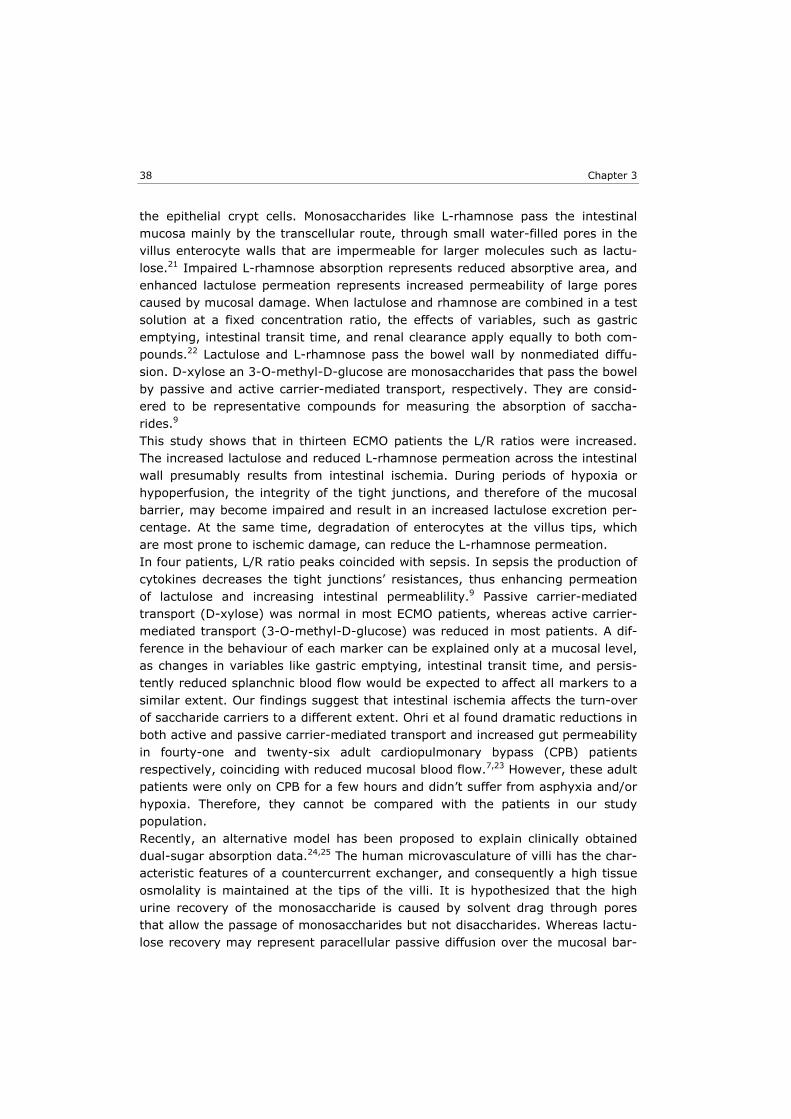
Figure 3.3 3-O-methyl-D-glucose excretion percentages. Left panel: nine patients given total parenteral nutrition during the whole ECMO period. Right panel: seven patients in whom enteral nutrition was started during ECMO. Dotted lines indicate lower and upper limit of normal 3-O-methyl-D-glucose excretion percentage.
0.7
L/R ratio & ECMO 37
terioration of the patient’s clinical condition. In patient 10 the L/R ratio peak on day 14 coincided with cardiac cathetherization. In seven patients enteral nutrition was started; the ratio decreased in two patients, remained unchanged in one pa-tient, initially increased and returned to normal within 1 week in one patient, and increased in three patients (in two of these patients, the SAT could be performed only once after the start of enteral nutrition, as they were weaned off ECMO within the next 48 hours; Fig.3.1).
D-xylose excretion
D-xylose excretion percentages were normal in 32 of 46 measurements, de-creased in 7 of 46 measurements, and increased in 7 of 46 measurments. In five patients, D-xylose excretion percentages were decreased. In eleven patients, all D-xylose excretion percentages were within the normal range. After the start of enteral nutrition, the excretion percentages increased in three patients (mean, 11%) and decreased in four patients (mean, 12.6%; Fig 3.2).
3-0-methyl-D-glucose excretion
3-O-methyl-D-glucose excretion percentages were normal in 13 of 44 measure-ments and decreased in 31 of 44 measurements. Excretion percentages were de-creased in fifteen patients. In one patient, who was on ECMO for 4 days, only 1 measurement was performed, and this 3-O-methyl-D-glucose excretion percent-age was within the normal range. After the start of enteral feeding, the excretion percentage increased in three patients (mean, 1.7%) and decreased in four pa-tients (mean, 4%; Fig 3.3). All patients tolerated enteral feeding well; no adverse effects were observed.
DISCUSSION
After recurrent periods of hypoxia pre-ECMO and the use of vasopressive drugs, intestinal ischemia is likely to occur in ECMO patients. Intestinal ischemia causes time-dependent, progressive histological changes ranging from patchy, subepi-thelial blebs on villus tips to frank detachment of enterocytes, loss of villi and crypt cells, and necrosis of the submucosa and the deeper layers of the intestinal wall.14-16 This impairs the mucosal barrier function and enhances bacterial trans-location, which can result in sepsis, systemic inflammatory response syndrome, and multiple organ failure.17,18 Early introduction of enteral nutrition in critically ill patients maintains an intact intestinal mucosa and supports the intestinal barrier function during critical illness.19,20 No data are available about the intestinal func-tion in neonates during ECMO treatment and it remains debatable whether enteral substrate should be withheld. Measuring intestinal permeability for sugars is a reliable test for functional integ-rity of the mucosa and to demonstrate mucosal damage.8 Disaccharides like lac-tulose are thought to permeate the intestinal mucosa through large pores of low incidence problably associated with the paracellular tight junction complexes of
Chapter 3 38
the epithelial crypt cells. Monosaccharides like L-rhamnose pass the intestinal mucosa mainly by the transcellular route, through small water-filled pores in the villus enterocyte walls that are impermeable for larger molecules such as lactu-lose.21 Impaired L-rhamnose absorption represents reduced absorptive area, and enhanced lactulose permeation represents increased permeability of large pores caused by mucosal damage. When lactulose and rhamnose are combined in a test solution at a fixed concentration ratio, the effects of variables, such as gastric emptying, intestinal transit time, and renal clearance apply equally to both com-pounds.22 Lactulose and L-rhamnose pass the bowel wall by nonmediated diffu-sion. D-xylose an 3-O-methyl-D-glucose are monosaccharides that pass the bowel by passive and active carrier-mediated transport, respectively. They are consid-ered to be representative compounds for measuring the absorption of saccha-rides.9 This study shows that in thirteen ECMO patients the L/R ratios were increased. The increased lactulose and reduced L-rhamnose permeation across the intestinal wall presumably results from intestinal ischemia. During periods of hypoxia or hypoperfusion, the integrity of the tight junctions, and therefore of the mucosal barrier, may become impaired and result in an increased lactulose excretion per-centage. At the same time, degradation of enterocytes at the villus tips, which are most prone to ischemic damage, can reduce the L-rhamnose permeation. In four patients, L/R ratio peaks coincided with sepsis. In sepsis the production of cytokines decreases the tight junctions’ resistances, thus enhancing permeation of lactulose and increasing intestinal permeablility.9 Passive carrier-mediated transport (D-xylose) was normal in most ECMO patients, whereas active carrier-mediated transport (3-O-methyl-D-glucose) was reduced in most patients. A dif-ference in the behaviour of each marker can be explained only at a mucosal level, as changes in variables like gastric emptying, intestinal transit time, and persis-tently reduced splanchnic blood flow would be expected to affect all markers to a similar extent. Our findings suggest that intestinal ischemia affects the turn-over of saccharide carriers to a different extent. Ohri et al found dramatic reductions in both active and passive carrier-mediated transport and increased gut permeability in fourty-one and twenty-six adult cardiopulmonary bypass (CPB) patients respectively, coinciding with reduced mucosal blood flow.7,23 However, these adult patients were only on CPB for a few hours and didn’t suffer from asphyxia and/or hypoxia. Therefore, they cannot be compared with the patients in our study population. Recently, an alternative model has been proposed to explain clinically obtained dual-sugar absorption data.24,25 The human microvasculature of villi has the char-acteristic features of a countercurrent exchanger, and consequently a high tissue osmolality is maintained at the tips of the villi. It is hypothesized that the high urine recovery of the monosaccharide is caused by solvent drag through pores that allow the passage of monosaccharides but not disaccharides. Whereas lactu-lose recovery may represent paracellular passive diffusion over the mucosal bar-
L/R ratio & ECMO 39
rier as a whole, monosaccharide recovery depends mainly on water absorption in the upper part of the villus. Changes in intestinal perfusion can cause changes in tissue osmolality at the tips of the villi, and the efficiency of this mechanism will be reduce in states of intestinal ischemia. According to this hypothesis, the L/R ratio is primarily a standard for the normal villus blood flow and for the normal functioning of epithelial villus cells rather than a parameter for gut permeability. Most increased L/R ratios in this study were caused by a decreased L-rhamnose excretion, and according to this theory, the intestinal ischemia in ECMO patients may have caused changes in the tissue osmolality of villus tips, which in turn may have affected solvent drag and carrier-mediated absorption of monosaccharides (to a different extent). In this study the introduction of enteral feeding in ECMO patients after determina-tion of the L/R ratio and D-xylose and 3-O-methyl-D-glucose excretion percent-ages did not cause a significant deterioration of these parameters. These findings do not support the practice of withholding enteral nutrition in critically ill new-borns supported by ECMO.
Chapter 3 40
REFERENCES 1. Bernbaum J, Schwartz IP, Gerdes M, D'Agostino JA, Coburn CE, Polin RA. Survivors of
extracorporeal membrane oxygenation at 1 year of age: the relationship of primary di-agnosis with health and neurodevelopmental sequelae. Pediatrics 1995;95:907-13.
2. Field DJ, Pearson GA. Neonatal extra corporeal membrane oxygenation (ECMO). J Peri-nat Med 1994;22:565-9.
3. Pearson GA, Firmin RK, Sosnowski A, Field D. Neonatal extracorporeal membrane oxy-genation. Br J Hosp Med 1992;47:646-53.
4. Desai TR, Sisley AC, Brown S, Gewertz BL. Defining the critical limit of oxygen extrac-tion in the human small intestine. J Vasc Surg 1996;23:832-7; discussion 8.
5. Crissinger KD. Regulation of hemodynamics and oxygenation in developing intestine: insight into the pathogenesis of necrotizing enterocolitis. Acta Paediatr Suppl 1994;396:8-10.
6. Navarro J. Neonatal necrotizing enterocolitis. In: Navarro J, Schmitz J, editors. Paediat-ric gastroenterology. Oxford: Oxford University Press; 1992. p. 161-7.
7. Ohri SK, Bjarnason I, Pathi V, et al. Cardiopulmonary bypass impairs small intestinal transport and increases gut permeability. Ann Thorac Surg 1993;55:1080-6.
8. van Elburg RM, Uil JJ, Kokke FT, et al. Repeatability of the sugar-absorption test, using lactulose and mannitol, for measuring intestinal permeability for sugars. J Pediatr Gas-troenterol Nutr 1995;20:184-8.
9. Travis S, Menzies I. Intestinal permeability: functional assessment and significance. Clin Sci (Lond) 1992;82:471-88.
10. Jansen G, Muskiet FA, Schierbeek H, Berger R, van der Slik W. Capillary gas chroma-tographic profiling of urinary, plasma and erythrocyte sugars and polyols as their trimethylsilyl derivatives, preceded by a simple and rapid prepurification method. Clin Chim Acta 1986;157:277-93.
11. Miki K, Butler R, Moore D, Davidson G. Rapid and simultaneous quantification of rham-nose, mannitol, and lactulose in urine by HPLC for estimating intestinal permeability in pediatric practice. Clin Chem 1996;42:71-5.
12. Beach RC, Menzies IS, Clayden GS, Scopes JW. Gastrointestinal permeability changes in the preterm neonate. Arch Dis Child 1982;57:141-5.
13. van Elburg RM, Uil JJ, de Monchy JG, Heymans HS. Intestinal permeability in pediatric gastroenterology. Scand J Gastroenterol Suppl 1992;194:19-24.
14. Reilly PM, Bulkley GB. The splanchnic haemodynamic response to circulatory shock. In: Peters TJ, editor. International conference on inflammation in the gastrointestinal tract; 1990; London: Corners Publications; 1990. p. 145-66.
15. Haglund U, Bulkley GB, Granger DN. On the pathophysiology of intestinal ischemic in-jury. Clinical review. Acta Chir Scand 1987;153:321-4.
16. Haglund U, Hulten L, Ahren C, Lundgren O. Mucosal lesions in the human small intes-tine in shock. Gut 1975;16:979-84.
17. Hadfield RJ, Sinclair DG, Houldsworth PE, Evans TW. Effects of enteral and parenteral nutrition on gut mucosal permeability in the critically ill. Am J Respir Crit Care Med 1995;152:1545-8.
18. Mythen MG, Webb AR. The role of gut mucosal hypoperfusion in the pathogenesis of post-operative organ dysfunction. Intensive Care Med 1994;20:203-9.
19. Heyland DK, Cook DJ, Guyatt GH. Enteral nutrition in the critically ill patient: a critical review of the evidence. Intensive Care Med 1993;19:435-42.
20. Zaloga GP, Black KW, Prielipp R. Effect of rate of enteral nutrient supply on gut mass. JPEN J Parenter Enteral Nutr 1992;16:39-42.
L/R ratio & ECMO 41
21. Menzies IS. Transmucosal passage of inert molecules in health and disease. In: Skad-hauge E, Heintze K, editors. Intestinal absorption and secretion; 1983; Titisee, Ger-many: MTP Press; 1983. p. 527-43.
22. Bjarnason I, MacPherson A, Hollander D. Intestinal permeability: an overview. Gastro-enterology 1995;108:1566-81.
23. Ohri SK, Somasundaram S, Koak Y, et al. The effect of intestinal hypoperfusion on in-testinal absorption and permeability during cardiopulmonary bypass. Gastroenterology 1994;106:318-23.
24. Bijlsma PB, Peeters RA, Groot JA, Dekker PR, Taminiau JA, van Der Meer R. Differential in vivo and in vitro intestinal permeability to lactulose and mannitol in animals and hu-mans: a hypothesis. Gastroenterology 1995;108:687-96.
25. Fink MP. Interpreting dual-sugar absorption studies in critically ill patients: what are the implications of apparent increases in intestinal permeability to hydrophilic solutes? In-tensive Care Med 1997;23:489-92.

4 The incidence of septic complications in newborns
on extracorporeal membrane oxygenation is not
affected by feeding route
Heiman F.L. Wertheim Marcel J.I.J. Albers Marjolein Piena-Spoel Dick Tibboel J Pediatr Surg 2001;36:1485-9
Chapter 4 44
ABSTRACT
PURPOSE: The aim of this study was to compare the effects of enteral and total parenteral feeding on septic complications in neonates on extracorporeal mem-brane oxygenation (ECMO). METHODS: Ninety-six neonates were on ECMO between January 1992 and February 1998. Matching for diagnosis and exclusion of neonates with sepsis prior to ECMO or undergoing surgery on ECMO left sixteen enterally fed neonates (cases) and thirty-five parenterally fed neonates (controls) for analysis. Septic complications were scored using the criteria of the Society of Critical Care Medicine and the American College of Chest Physicians adapted to children. RESULTS: Both groups were comparable with respect to gestational age, sex and age at initiation of ECMO. The frequency of septic complications did not differ between cases and controls: no complications 75% vs. 69%, systemic inflamma-tory response syndrome 13 vs. 6%, bacteraemia 6 vs. 14%, sepsis 6 vs. 11%. There were no complications associated with enteral feeding. The ECMO-run was significantly longer in the case group (median 161 vs. 111 hours, P = 0.01) and mortality was lower in the case group (0 versus 14%, P = 0.17). CONCLUSIONS: Enteral nutrition does not affect the risk of sepsis in neonates on ECMO when compared to total parenteral nutrition. Enteral nutrition is well toler-ated and not associated with adverse effects.
Sepsis & ECMO
45
INTRODUCTION
Neonates suffering from severe cardiopulmonary failure unresponsive to maximal conventional treatment can be successfully treated with extracorporeal mem-brane oxygenation (ECMO). Around 80% of infants predicted to have 80% mor-tality with conventional treatment, survive after ECMO.1,2 Common indications for neonatal ECMO are meconium aspiration syndrome (MAS), congenital diaphrag-matic hernia (CDH), persistent pulmonary hypertension of the newborn (PPHN), and sepsis.1-3 Most centres feed ECMO-patients by total parenteral nutrition (TPN). It is argued that hypoxic episodes prior to ECMO and the use of vasoactive drugs during ECMO may compromise splanchnic blood flow and result in gut ischemia. As a consequence bacterial translocation and/or sepsis may develop, and possibly ne-crotizing enterocolitis. Asphyxia, low splanchnic blood flow and enteral feeding are considered risk factors for the development of necrotizing enterocolitis in premature infants.4,5 On the other hand, several studies have shown that with-holding enteral feeding leads to villous atrophy, decreased immunological function of the gut, increased gut mucosal permeability, and bacterial translocation, favor-ing sepsis. In the critically ill, early initiation of enteral nutrition restores intestinal mucosal integrity, even when only minimal quantities are provided enterally, whereas TPN is associated with a persistent increase in gastro-intestinal mucosal permeability.6-8 In critically ill patients, enteral nutrition has also been associated with improved gastrointestinal immunological function, reduced septic morbidity and decreased cost.7,9 By measuring the urinary excretion of the sugars lactulose and rhamnose, we showed that intestinal integrity is compromised in neonates on ECMO.10 The cur-rent study was designed to compare septic complications in enterally fed and par-enterally fed neonatal ECMO-patients.
METHODS
The Sophia Children’s Hospital is one of two level III hospitals appointed by the Dutch Health Council, to offer veno-arterial ECMO to neonates with a reversible respiratory disorder unresponsive to maximal conventional treatment. These two ECMO centres cover the whole of the Netherlands, approximately 15 million in-habitants. Principal entry criteria are an alveolar-arterial oxygen gradient ex-ceeding 600 or an oxygenation index greater than or equal to 40 for at least 6 to 8 hours. Contraindications are gestational age under 35 weeks, weight under 2000 grams, intracranial haemorrhage grade II or more, and severe cardiac or chromosomal anomalies. In January 1996 enteral feeding in neonatal ECMO-patients was introduced into everyday clinical practice. Since then all patients, except patients with a con-genital diaphragmatic hernia undergoing continuous suctioning of the nasogastric tube combined with post-ECMO repair of the diaphragmatic defect, have been fed
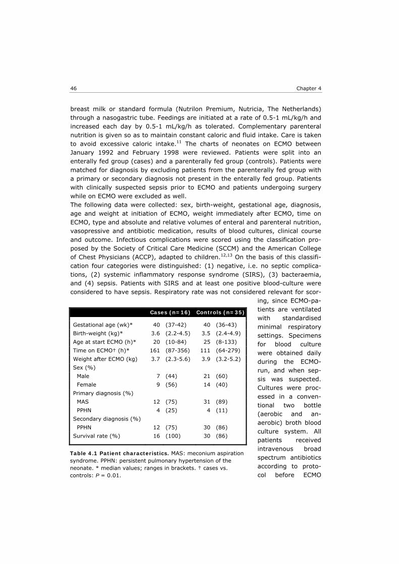
Chapter 4 46
breast milk or standard formula (Nutrilon Premium, Nutricia, The Netherlands) through a nasogastric tube. Feedings are initiated at a rate of 0.5-1 mL/kg/h and increased each day by 0.5-1 mL/kg/h as tolerated. Complementary parenteral nutrition is given so as to maintain constant caloric and fluid intake. Care is taken to avoid excessive caloric intake.11 The charts of neonates on ECMO between January 1992 and February 1998 were reviewed. Patients were split into an enterally fed group (cases) and a parenterally fed group (controls). Patients were matched for diagnosis by excluding patients from the parenterally fed group with a primary or secondary diagnosis not present in the enterally fed group. Patients with clinically suspected sepsis prior to ECMO and patients undergoing surgery while on ECMO were excluded as well. The following data were collected: sex, birth-weight, gestational age, diagnosis, age and weight at initiation of ECMO, weight immediately after ECMO, time on ECMO, type and absolute and relative volumes of enteral and parenteral nutrition, vasopressive and antibiotic medication, results of blood cultures, clinical course and outcome. Infectious complications were scored using the classification pro-posed by the Society of Critical Care Medicine (SCCM) and the American College of Chest Physicians (ACCP), adapted to children.12,13 On the basis of this classifi-cation four categories were distinguished: (1) negative, i.e. no septic complica-tions, (2) systemic inflammatory response syndrome (SIRS), (3) bacteraemia, and (4) sepsis. Patients with SIRS and at least one positive blood-culture were considered to have sepsis. Respiratory rate was not considered relevant for scor-
ing, since ECMO-pa-tients are ventilated with standardised minimal respiratory settings. Specimens for blood culture were obtained daily during the ECMO-run, and when sep-sis was suspected. Cultures were proc-essed in a conven-tional two bottle (aerobic and an-aerobic) broth blood culture system. All patients received intravenous broad spectrum antibiotics according to proto-col before ECMO
Cases (n=16) Controls (n=35)
Gestational age (wk)* 40 (37-42) 40 (36-43)
Birth-weight (kg)* 3.6 (2.2-4.5) 3.5 (2.4-4.9)
Age at start ECMO (h)* 20 (10-84) 25 (8-133)
Time on ECMO† (h)* 161 (87-356) 111 (64-279)
Weight after ECMO (kg) 3.7 (2.3-5.6) 3.9 (3.2-5.2)
Sex (%)
Male 7 (44) 21 (60)
Female 9 (56) 14 (40)
Primary diagnosis (%)
MAS 12 (75) 31 (89)
PPHN 4 (25) 4 (11)
Secondary diagnosis (%)
PPHN 12 (75) 30 (86)
Survival rate (%) 16 (100) 30 (86)
Table 4.1 Patient characteristics. MAS: meconium aspiration syndrome. PPHN: persistent pulmonary hypertension of the neonate. * median values; ranges in brackets. † cases vs. controls: P = 0.01.
Sepsis & ECMO
47
was initiated. Antibiotics were either geared to the micro-organism cul-tured or discontinued when the cul-tures remained negative and clinical signs of sepsis were absent. Data were analysed with the Fisher’s exact test and Mann-Whitney rank sum test where appropriate using a statistical computer program (SPSS 7.5 for Windows). Differences were considered statistically significant when P <0.05.
RESULTS
In the study period, 96 neonates were treated with ECMO. To match for diagno-sis, neonates from the parenterally fed group with the following diagnoses were excluded: congenital diaphragmatic hernia (n=17), pneumonia (n=4), blood as-piration (n=2), lung hypoplasia (n=2), and other (n=4). After matching the fol-lowing patients were excluded: five infants from the enterally fed group and nine infants from the parenterally fed group because of septic complications prior to ECMO, one infant because of surgery on ECMO and one infant who went off ECMO before nutrition had been started. Thus, sixteen cases and thirty-five controls were available for analysis. Gestational age, birth weight, sex, age and weight at initiation of the ECMO-run did not differ between cases and controls. The ECMO-run was significantly longer in the case group (median 161 versus 111 h, P = 0.01) (Table 4.1). All patients received vasoactive drugs, except for 2 controls (Table 4.2). Mortality was 0% in the case group versus 14% in the control group (P = 0.17). In two controls ECMO was discontinued after 3 and 4 days because of brain death. One patient with MAS died of relapsing pulmonary hypertension 10 days after decannulation. One patient with PPHN died 13 days after decannulation, shortly after operative cor-rection of an atrial and ventricular septum defect and patent arterial duct. Autopsy findings were consistent with interstitial pneumonia, acute respiratory distress syndrome and pulmonary venous congestion. One patient with MAS died 8 days after decannulation of massive intractable gastrointestinal bleeding, 7 days after being transferred back to the referring hospital. Broad spectrum antibiotics were given to all neonates, most frequently amoxicillin in combination with cefotaxim (cases: 88%; controls: 89%). Other antibiotic combinations were penicillin and tobramycin (cases 12%; controls 3%), amoxicil-lin and tobramycin (cases 0%; controls 5%), and amoxicillin and gentamicin (cases 0%; controls 3%). Enteral feedings were introduced 30 to 138 hours (me-dian 67 h) after the initiation of ECMO. Highest intakes of enteral feeding ranged
Cases (n) Controls (n)
Dopamine 16 28
Dobutamine 11 18
Norepinephrin 10 7
Epoprostenol 6 10
NO-inhalation 8 6
Isoprel 1 3
Nitroprusside 0 1
none 0 2
Table 4.2 Use of inotropic and vasoactive drugs
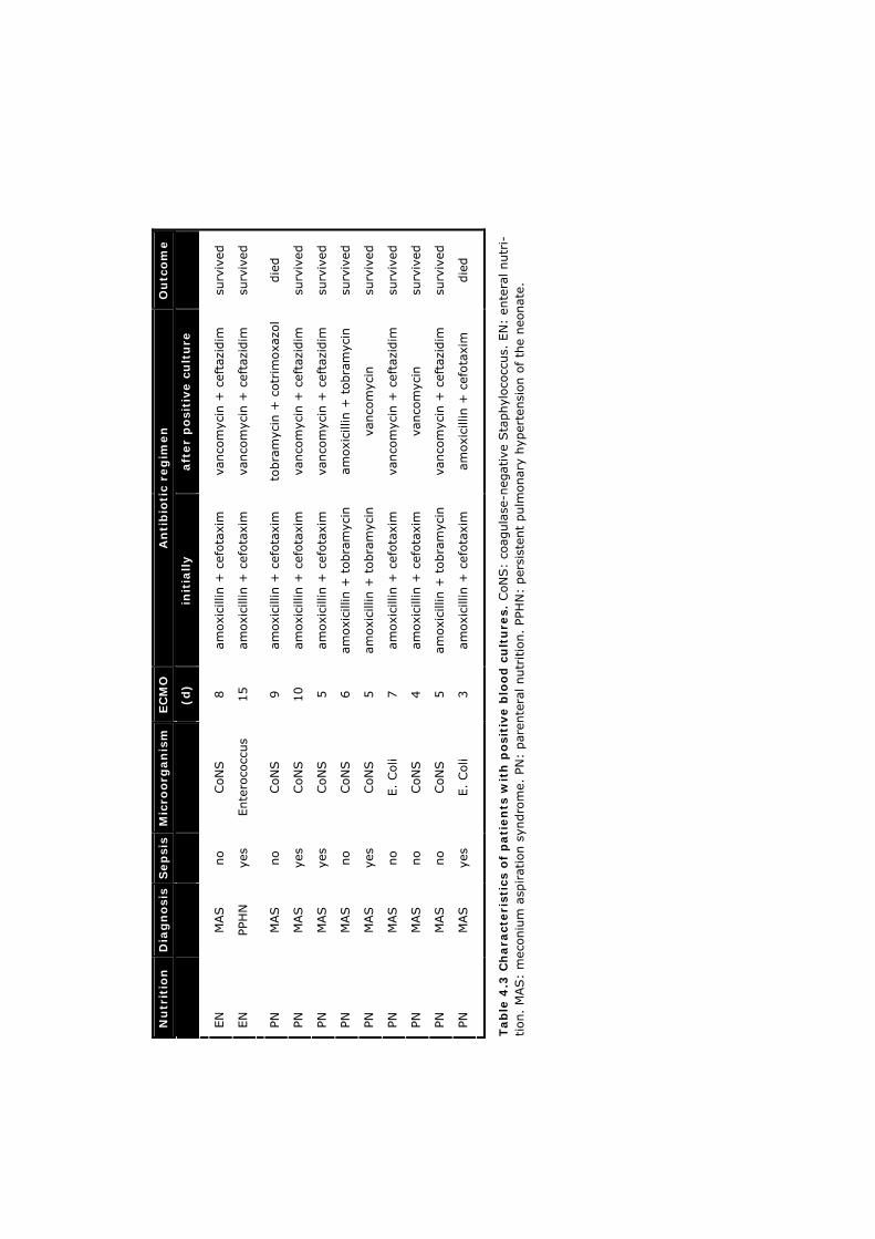
Ou
tco
me
su
rviv
ed
surv
ived
die
d
surv
ived
surv
ived
surv
ived
surv
ived
surv
ived
surv
ived
surv
ived
die
d
aft
er
po
siti
ve c
ult
ure
va
nco
myc
in +
cef
tazi
dim
vanco
myc
in +
cef
tazi
dim
to
bra
myc
in +
cotr
imoxa
zol
vanco
myc
in +
cef
tazi
dim
vanco
myc
in +
cef
tazi
dim
amoxi
cilli
n +
tobra
myc
in
vanco
myc
in
vanco
myc
in +
cef
tazi
dim
vanco
myc
in
vanco
myc
in +
cef
tazi
dim
amoxi
cilli
n +
cef
ota
xim
An
tib
ioti
c re
gim
en
init
iall
y
am
oxi
cilli
n +
cef
ota
xim
amoxi
cilli
n +
cef
ota
xim
am
oxi
cilli
n +
cef
ota
xim
amoxi
cilli
n +
cef
ota
xim
amoxi
cilli
n +
cef
ota
xim
amoxi
cilli
n +
tobra
myc
in
amoxi
cilli
n +
tobra
myc
in
amoxi
cilli
n +
cef
ota
xim
amoxi
cilli
n +
cef
ota
xim
amoxi
cilli
n +
tobra
myc
in
amoxi
cilli
n +
cef
ota
xim
EC
MO
(d) 8
15
9
10
5
6
5
7
4
5
3
Mic
roo
rgan
ism
CoN
S
Ente
roco
ccus
CoN
S
CoN
S
CoN
S
CoN
S
CoN
S
E.
Coli
CoN
S
CoN
S
E.
Coli
Sep
sis
no
yes
no
yes
yes
no
yes
no
no
no
yes
Dia
gn
osi
s
M
AS
PPH
N
M
AS
MAS
MAS
MAS
MAS
MAS
MAS
MAS
MAS
Nu
trit
ion
EN
EN
PN
PN
PN
PN
PN
PN
PN
PN
PN
Tab
le 4
.3 C
hara
cteri
stic
s o
f p
ati
en
ts w
ith
po
siti
ve b
loo
d c
ult
ure
s. C
oN
S:
coag
ula
se-n
egative
Sta
phyl
oco
ccus.
EN
: en
tera
l nutr
i-tion.
MAS:
mec
oniu
m a
spirat
ion s
yndro
me.
PN
: par
ente
ral nutr
itio
n.
PPH
N:
per
sist
ent
pulm
onary
hyp
erte
nsi
on o
f th
e neo
nate
.
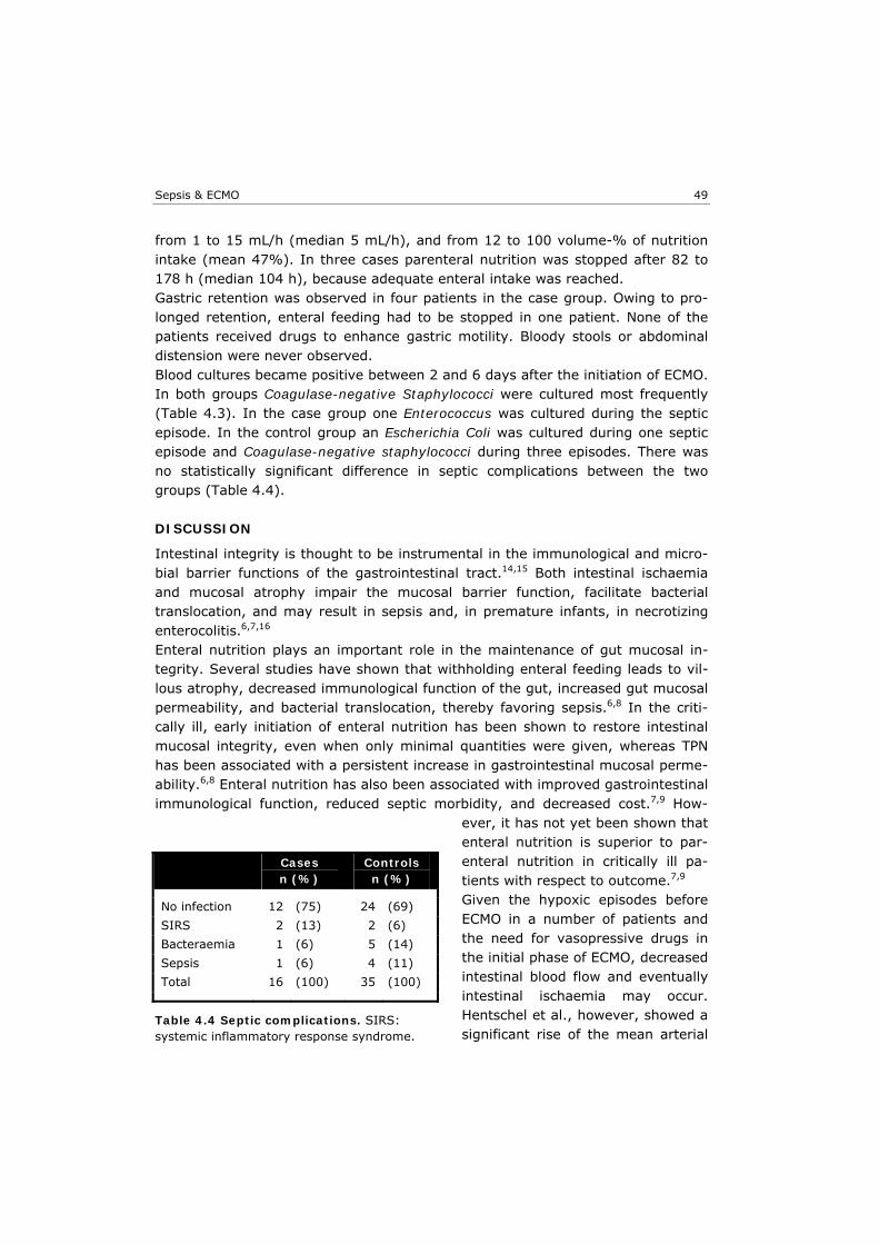
Sepsis & ECMO
49
from 1 to 15 mL/h (median 5 mL/h), and from 12 to 100 volume-% of nutrition intake (mean 47%). In three cases parenteral nutrition was stopped after 82 to 178 h (median 104 h), because adequate enteral intake was reached. Gastric retention was observed in four patients in the case group. Owing to pro-longed retention, enteral feeding had to be stopped in one patient. None of the patients received drugs to enhance gastric motility. Bloody stools or abdominal distension were never observed. Blood cultures became positive between 2 and 6 days after the initiation of ECMO. In both groups Coagulase-negative Staphylococci were cultured most frequently (Table 4.3). In the case group one Enterococcus was cultured during the septic episode. In the control group an Escherichia Coli was cultured during one septic episode and Coagulase-negative staphylococci during three episodes. There was no statistically significant difference in septic complications between the two groups (Table 4.4).
DISCUSSION
Intestinal integrity is thought to be instrumental in the immunological and micro-bial barrier functions of the gastrointestinal tract.14,15 Both intestinal ischaemia and mucosal atrophy impair the mucosal barrier function, facilitate bacterial translocation, and may result in sepsis and, in premature infants, in necrotizing enterocolitis.6,7,16 Enteral nutrition plays an important role in the maintenance of gut mucosal in-tegrity. Several studies have shown that withholding enteral feeding leads to vil-lous atrophy, decreased immunological function of the gut, increased gut mucosal permeability, and bacterial translocation, thereby favoring sepsis.6,8 In the criti-cally ill, early initiation of enteral nutrition has been shown to restore intestinal mucosal integrity, even when only minimal quantities were given, whereas TPN has been associated with a persistent increase in gastrointestinal mucosal perme-ability.6,8 Enteral nutrition has also been associated with improved gastrointestinal immunological function, reduced septic morbidity, and decreased cost.7,9 How-
ever, it has not yet been shown that enteral nutrition is superior to par-enteral nutrition in critically ill pa-tients with respect to outcome.7,9 Given the hypoxic episodes before ECMO in a number of patients and the need for vasopressive drugs in the initial phase of ECMO, decreased intestinal blood flow and eventually intestinal ischaemia may occur. Hentschel et al., however, showed a significant rise of the mean arterial
Cases n (%)
Controls n (%)
No infection 12 (75) 24 (69)
SIRS 2 (13) 2 (6)
Bacteraemia 1 (6) 5 (14)
Sepsis 1 (6) 4 (11)
Total 16 (100) 35 (100)
Table 4.4 Septic complications. SIRS: systemic inflammatory response syndrome.
Chapter 4 50
pressure in the superior mesenteric artery in preterm infants when dopamine or dobutamine was administered.17 Others have shown an increase in intestinal blood flow after feeding.18,19 This suggests that vasopressors and enteral nutrition may have a beneficial effect on the splanchnic blood supply and gut mucosal in-tegrity. Two studies have been published reporting the effects of enteral nutrition in ECMO patients. Our group, using urinary lactulose/rhamnose excretion to pro-spectively assess small intestinal integrity in 16 consecutive neonatal ECMO pa-tients, found intestinal integrity to be compromised in neonates on ECMO. Intro-duction of enteral nutrition did not result in further deterioration.10 Pettignano et al conducted a chart review of 27 nonneonatal ECMO patients.20 The adequacy, tolerance, and complications of enteral nutrition were compared with those of parenteral nutrition. The investigators concluded that enteral nutrition can be administered safely. In the current study, the number of septic complications in neonates on ECMO was not affected by the type of nutritional support. Enteral nutrition was well tol-erated and was not associated with any adverse effects. Our findings agree with those of Pettignano et al20 and extend his findings to the neonatal age. In both studies the number of infections in which gut mucosal integrity and bacterial translocation may have played a role was low. In the current study, Gram-nega-tive bacteria were cultured three times, i.e. an equal incidence of 6% in both groups. Coagulase-negative staphylococci were the microorganisms cultured most frequently. Some may be the result of contamination, but we assume that in cases of sepsis these cultures were true-positive.21 To our knowledge, bactere-mias with Coagulase-negative staphylococci have never been related to bacterial translocation. Studies that evaluate sepsis incidence in neonatal intensive care units show an increase of septic episodes and bacteremias due to Coagulase-negative staphylococci, formerly considered nonpathogenic.21-24 This increase is thought to result from intensified use of vascular access techniques, such as ECMO cannulas and central venous catheters, by-passing the first defence against infection, the skin.23 Several factors may explain that patients fed enterally were on ECMO longer than those fed parenterally. First, in 2 patients on parenteral nutrition ECMO was dis-continued early because of brain death due to severe asphyxia before ECMO. Second, the proportion of PPHN-patients in the enteral group was higher than that in the parenteral group. Of PPHN patients approximately 83% survive vs. 93% of MAS patients, and PPHN-patients generally need more time on ECMO than MAS-patients.1 A third explanatory factor may be the increasing complexity of cases over the years, associated with a decrease in survival.2 Kössel et al very recently pointed out that ECMO criteria for neonates pretreated with high fre-quency oscillation and/or inhaled nitric oxide should be less strict than the ones commonly used.25 In the Netherlands, the use of high frequency oscillation and inhaled nitric oxide in the referring NICUs has increased gradually since 1996.
Sepsis & ECMO
51
This makes the trend towards higher survival in the more recently treated enteral group observed in our study all the more interesting. Cases of necrotizing entero-colitis were not observed, as might be expected in view of the small numbers of patients and their gestational age. The implications of the present study are limited by its design and patient num-bers. In some patients enteral feedings were introduced fairly late in the course of ECMO, thereby potentially obscuring differences between feeding routes. Al-though the ECMO protocol remained the same throughout the whole study period, minor changes of clinical practice may have resulted in differences between the controls -treated before 1996- and the cases -treated after 1995-, not related to the feeding route. Of 96 newborns on ECMO, fourty-five (47%) were excluded. Our findings, therefore, do not necessarily apply to all newborns on ECMO. The limitations imposed by patient numbers are best illustrated by taking our re-sults and using them for a power analysis. Episodes of bacteraemia and sepsis, that is, episodes in which an infectious agent was identified, occurred in 12% of cases and 25% of controls (Table 4.4). A study aiming to demonstrate a reduc-tion in infection rate from 25% to 12%, with alpha set at 0.05 and beta at 0.20 (power 0.80), would require 125 patients in both groups. Even larger patient groups would be needed in order to identify differences with respect to gut-re-lated infectious complications. Alternatively it might be argued that -under these conditions at least- bacterial translocation does not play the role it has formerly been attributed. In view of the number of neonates treated with ECMO worldwide, our results may serve as an incentive to perform prospective randomized studies that compare benefits of enteral and parenteral nutrition in neonates on ECMO.
Chapter 4 52
REFERENCES 1. Stolar CJ, Snedecor SM, Bartlett RH. Extracorporeal membrane oxygenation and neo-
natal respiratory failure: experience from the extracorporeal life support organization. J Pediatr Surg 1991;26:563-71.
2. Zwischenberger JB, Nguyen TT, Upp JR, Jr., et al. Complications of neonatal extracor-poreal membrane oxygenation. Collective experience from the Extracorporeal Life Sup-port Organization. J Thorac Cardiovasc Surg 1994;107:838-48; discussion 48-49.
3. Anonymous. ECLS registry report - International summary July 1999. Ann Arbor (MI): Extracorporeal Life Support Organization; 1999 july 1999.
4. Berseth CL. Minimal enteral feedings. Clin Perinatol 1995;22:195-205.
5. Crissinger KD. Regulation of hemodynamics and oxygenation in developing intestine: insight into the pathogenesis of necrotizing enterocolitis. Acta Paediatr Suppl 1994;396:8-10.
6. Hadfield RJ, Sinclair DG, Houldsworth PE, Evans TW. Effects of enteral and parenteral nutrition on gut mucosal permeability in the critically ill. Am J Respir Crit Care Med 1995;152:1545-8.
7. Heyland DK, Cook DJ, Guyatt GH. Enteral nutrition in the critically ill patient: a critical review of the evidence. Intensive Care Med 1993;19:435-42.
8. Okada Y, Klein N, van Saene HK, Pierro A. Small volumes of enteral feedings normalise immune function in infants receiving parenteral nutrition. J Pediatr Surg 1998;33:16-9.
9. Lipman TO. Grains or veins: is enteral nutrition really better than parenteral nutrition? A look at the evidence. JPEN J Parenter Enteral Nutr 1998;22:167-82.
10. Piena M, Albers MJ, Van Haard PM, Gischler S, Tibboel D. Introduction of enteral feeding in neonates on extracorporeal membrane oxygenation after evaluation of intestinal permeability changes. J Pediatr Surg 1998;33:30-4.
11. Shew SB, Keshen TH, Jahoor F, Jaksic T. The determinants of protein catabolism in neonates on extracorporeal membrane oxygenation. J Pediatr Surg 1999;34:1086-90.
12. Hayden WR. Sepsis terminology in pediatrics. J Pediatr 1994;124:657-8.
13. Anonymous. American College of Chest Physicians/Society of Critical Care Medicine Consensus Conference: definitions for sepsis and organ failure and guidelines for the use of innovative therapies in sepsis. Crit Care Med 1992;20:864-74.
14. Fink MP. Intestinal mucosal hyperpermeability in critical illness. In: Rombeau JL, Takala J, editors. Gut dysfunction in critical illness. Berlin Heidelberg New York: Springer-Ver-lag; 1996. p. 12-24.
15. Wells CL, Erlandsen SL. Bacterial translocation: intestinal epithelial permeability. In: Rombeau JL, Takala J, editors. Gut dysfunction in critical illness. Berlin Heidelberg New York: Springer-Verlag; 1996. p. 131-49.
16. Sanderson IR. The physicochemical environment of the neonatal intestine. Am J Clin Nutr 1999;69:1028S-34S.
17. Hentschel R, Hensel D, Brune T, Rabe H, Jorch G. Impact on blood pressure and intesti-nal perfusion of dobutamine or dopamine in hypotensive preterm infants. Biol Neonate 1995;68:318-24.
18. Gladman G, Sims DG, Chiswick ML. Gastrointestinal blood flow velocity after the first feed. Arch Dis Child 1991;66:17-20.
19. Maruyama K, Koizumi T, Tomomasa T, Morikawa A. Intestinal blood-flow velocity in uncomplicated preterm infants during the early neonatal period. Pediatr Radiol 1999;29:472-7.
Sepsis & ECMO
53
20. Pettignano R, Heard M, Davis R, Labuz M, Hart M. Total enteral nutrition versus total parenteral nutrition during pediatric extracorporeal membrane oxygenation. Crit Care Med 1998;26:358-63.
21. Baltimore RS. Is it real or is it a contaminant? A guide to the interpretation of blood culture results. Am J Dis Child 1987;141:241-2.
22. Freeman J, Platt R, Sidebottom DG, Leclair JM, Epstein MF, Goldmann DA. Coagulase-negative staphylococcal bacteremia in the changing neonatal intensive care unit popu-lation. Is there an epidemic? JAMA 1987;258:2548-52.
23. Freeman J, Epstein MF, Smith NE, Platt R, Sidebottom DG, Goldmann DA. Extra hospital stay and antibiotic usage with nosocomial coagulase-negative staphylococcal bactere-mia in two neonatal intensive care unit populations. Am J Dis Child 1990;144:324-9.
24. Gladstone IM, Ehrenkranz RA, Edberg SC, Baltimore RS. A ten-year review of neonatal sepsis and comparison with the previous fifty-year experience. Pediatr Infect Dis J 1990;9:819-25.
25. Kössel H, Bauer K, Kewitz G, Karaca S, Versmold H. Do we need new indications for ECMO in neonates pretreated with high-frequency ventilation and/or inhaled nitric ox-ide. Intensive Care Med 2000;26:1489-95.


5 Male sex predisposes the surgical newborn to
parenteral nutrition-associated cholestasis and to
sepsis
Marcel J.I.J. Albers Dana-Anne H. de Gast-Bakker Nicolette A.M. van Dam Gerard C. Madern Dick Tibboel Arch Surg 2002;137:789-93
Chapter 5 56
ABSTRACT
HYPOTHESIS: sepsis is an epiphenomenon of parenteral nutrition associated cho-lestasis (PNAC) and not a causative factor; sepsis incidence is not affected by presence or absence of PNAC. DESIGN: observational cohort study. SETTING: pediatric surgery department in a tertiary referral children’s hospital. PATIENTS: newborns receiving PN for at least 7 days following intestinal surgery. MAIN OUTCOME MEASURES: PNAC criteria were: PN for at least 14 consecutive days, conjugated bilirubin level greater than 26 micromol/L, conjugated bilirubin frac-tion greater than 50%, and absence of an other identifiable cause of cholestasis. The identification of septic events was based on Centers of Disease Control and Prevention criteria. RESULTS: The patients (26 with PNAC and 72 without PNAC) were well comparable with respect to underlying disease, gestational age, birth weight and age at the start of PN. Time receiving PN and length of hospital stay were significantly longer in patients with PNAC (P <0.001). PNAC was associated with male sex (P = 0.03; odds ratio 2.8, 95% confidence interval 1.1-7.1). Overall sepsis incidence was low (9 per 1000 hospital days). Sepsis incidence tended to be higher in patients with PNAC than in patients without PNAC (11.8 vs. 7.1 per 1000 days; P =0.08), but was significantly higher in male than in female patients (12.2 vs. 5.6 per 1000 days; P =0.01). Most septic events was caused by coagulase-negative staphylo-cocci. CONCLUSION: sepsis is an epiphenomenon of PNAC rather than a causative factor. Moreover, male sex predisposes the newborn surgical patient to PNAC and to sepsis.
Sex, sepsis & cholestasis
57
INTRODUCTION
Parenteral nutrition-associated cholestasis (PNAC) is a potentially life-threatening complication of parenteral nutrition (PN) seen in 7.4% to 84% of parenterally fed infants.1 Its aetiology is multifactorial; underlying disease, gestational age, birth weight, interval between birth and start of PN, PN duration, sepsis, remaining small bowel length and number of operative procedures have all been found to influence PNAC incidence.2-16 The identification of risk factors has been hampered by their interaction and the small size and heterogeneity of the populations de-scribed in the majority of studies. The role of sepsis has remained especially con-troversial. Although several authors suggest every attempt should be made to prevent sepsis because they believe it to be an important causative factor for PNAC,5,8,12,17,18 others have argued that the role of sepsis has been overempha-sized or even that sepsis is an epiphenomenon of PNAC.2,8,19,20 To assess the relation between sepsis and PNAC, we studied a homogeneous co-hort of surgical newborns with a congenital or an acquired intestinal anomaly. We hypothesized that sepsis is an epiphenomenon of PNAC and that sepsis incidence would not be affected by presence or absence of PNAC.
MATERIAL AND METHODS
Clinical setting
The Department of Pediatric Surgery forms part of a university tertiary referral children’s hospital serving a population of approximately 4.5 million. The pediatric surgical in-patient clinic consists of a 35-bed medium care unit and a 14-bed in-tensive care and high care unit, annually admitting between 1600 and 1800 pa-tients. Most are newborns and infants admitted because of congenital or neona-tally acquired anomalies. The study period was from January 1, 1991, through December 31, 1996. During the study period the PN regimen and the antibiotic policy did not change. When PN was administered for more than a few days, as in our study population, a central venous catheter (Broviac catheter) was surgically introduced under general anesthesia.
Parenteral nutrition
Parenteral nutrition was provided according to hospital guidelines. Total PN pro-vided 2.5 gram/kg per day of amino acids (Aminovenös N Paediatric; Fresenius AG, Bad Homburg, Germany) to newborns and 2.0 gram/kg per day of amino ac-ids to older infants. Carbohydrates (Dextrose, Fresenius AG) provided approxi-mately 65% and fat (Intralipid 20%; Pharmacia AB, Stockholm, Sweden) ap-proximately 35% of non-protein calories. Enteral nutrition was introduced and advanced, and PN tapered as quickly as possible, based on stool output and weight gain. During tapering, the relative volumes of amino acids, lipids and car-bohydrates in PN were kept within narrow limits.
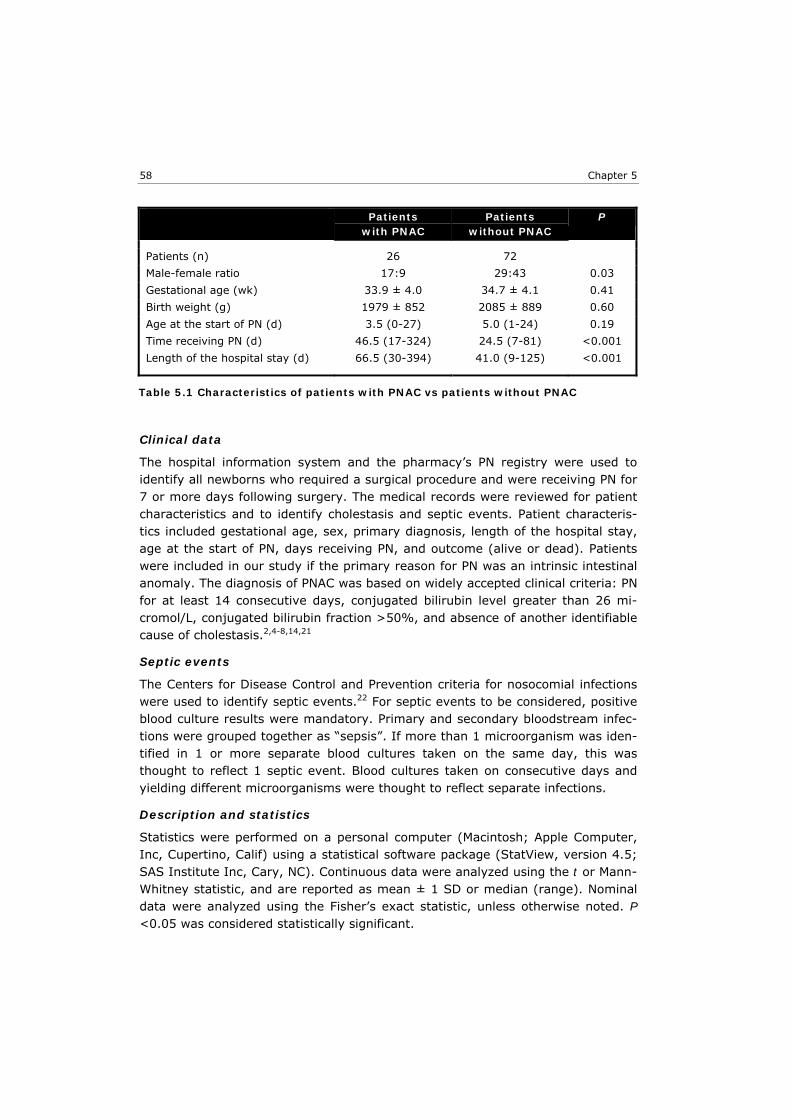
Chapter 5 58
Clinical data
The hospital information system and the pharmacy’s PN registry were used to identify all newborns who required a surgical procedure and were receiving PN for 7 or more days following surgery. The medical records were reviewed for patient characteristics and to identify cholestasis and septic events. Patient characteris-tics included gestational age, sex, primary diagnosis, length of the hospital stay, age at the start of PN, days receiving PN, and outcome (alive or dead). Patients were included in our study if the primary reason for PN was an intrinsic intestinal anomaly. The diagnosis of PNAC was based on widely accepted clinical criteria: PN for at least 14 consecutive days, conjugated bilirubin level greater than 26 mi-cromol/L, conjugated bilirubin fraction >50%, and absence of another identifiable cause of cholestasis.2,4-8,14,21
Septic events
The Centers for Disease Control and Prevention criteria for nosocomial infections were used to identify septic events.22 For septic events to be considered, positive blood culture results were mandatory. Primary and secondary bloodstream infec-tions were grouped together as “sepsis”. If more than 1 microorganism was iden-tified in 1 or more separate blood cultures taken on the same day, this was thought to reflect 1 septic event. Blood cultures taken on consecutive days and yielding different microorganisms were thought to reflect separate infections.
Description and statistics
Statistics were performed on a personal computer (Macintosh; Apple Computer, Inc, Cupertino, Calif) using a statistical software package (StatView, version 4.5; SAS Institute Inc, Cary, NC). Continuous data were analyzed using the t or Mann-Whitney statistic, and are reported as mean ± 1 SD or median (range). Nominal data were analyzed using the Fisher’s exact statistic, unless otherwise noted. P <0.05 was considered statistically significant.
Patients with PNAC
Patients without PNAC
P
Patients (n) 26 72
Male-female ratio 17:9 29:43 0.03
Gestational age (wk) 33.9 ± 4.0 34.7 ± 4.1 0.41
Birth weight (g) 1979 ± 852 2085 ± 889 0.60
Age at the start of PN (d) 3.5 (0-27) 5.0 (1-24) 0.19
Time receiving PN (d) 46.5 (17-324) 24.5 (7-81) <0.001
Length of the hospital stay (d) 66.5 (30-394) 41.0 (9-125) <0.001
Table 5.1 Characteristics of patients with PNAC vs patients without PNAC
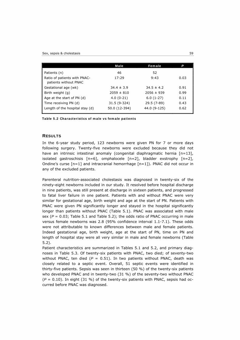
Sex, sepsis & cholestasis
59
RESULTS
In the 6-year study period, 123 newborns were given PN for 7 or more days following surgery. Twenty-five newborns were excluded because they did not have an intrinsic intestinal anomaly (congenital diaphragmatic hernia [n=13], isolated gastroschisis [n=6], omphalocele [n=2], bladder exstrophy [n=2], Ondine’s curse [n=1] and intracranial hemorrhage [n=1]). PNAC did not occur in any of the excluded patients. Parenteral nutrition-associated cholestasis was diagnosed in twenty-six of the ninety-eight newborns included in our study. It resolved before hospital discharge in nine patients, was still present at discharge in sixteen patients, and progressed to fatal liver failure in one patient. Patients with and without PNAC were very similar for gestational age, birth weight and age at the start of PN. Patients with PNAC were given PN significantly longer and stayed in the hospital significantly longer than patients without PNAC (Table 5.1). PNAC was associated with male sex (P = 0.03; Table 5.1 and Table 5.2); the odds ratio of PNAC occurring in male versus female newborns was 2.8 (95% confidence interval 1.1-7.1). These odds were not attributable to known differences between male and female patients. Indeed gestational age, birth weight, age at the start of PN, time on PN and length of hospital stay were all very similar in male and female newborns (Table 5.2). Patient characteristics are summarized in Tables 5.1 and 5.2, and primary diag-noses in Table 5.3. Of twenty-six patients with PNAC, two died; of seventy-two without PNAC, ten died (P = 0.51). In two patients without PNAC, death was closely related to a septic event. Overall, 51 septic events were identified in thirty-five patients. Sepsis was seen in thirteen (50 %) of the twenty-six patients who developed PNAC and in twenty-two (31 %) of the seventy-two without PNAC (P = 0.10). In eight (31 %) of the twenty-six patients with PNAC, sepsis had oc-curred before PNAC was diagnosed.
Male Female P Patients (n) 46 52
Ratio of patients with PNAC-patients without PNAC
17:29 9:43 0.03
Gestational age (wk) 34.4 ± 3.9 34.5 ± 4.2 0.91
Birth weight (g) 2059 ± 810 2056 ± 939 0.99
Age at the start of PN (d) 4.0 (0-21) 6.0 (1-27) 0.11
Time receiving PN (d) 31.5 (9-324) 29.5 (7-89) 0.43
Length of the hospital stay (d) 50.0 (12-394) 44.0 (9-125) 0.62
Table 5.2 Characteristics of male vs female patients
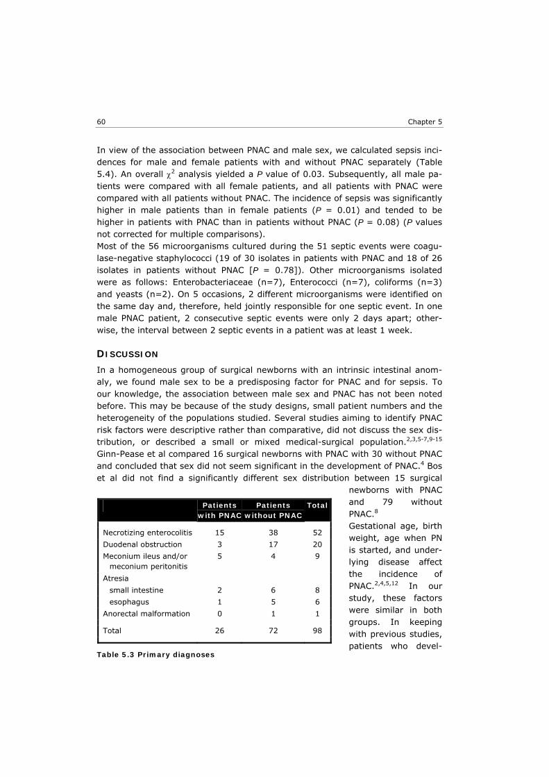
Chapter 5 60
In view of the association between PNAC and male sex, we calculated sepsis inci-dences for male and female patients with and without PNAC separately (Table 5.4). An overall χ2 analysis yielded a P value of 0.03. Subsequently, all male pa-tients were compared with all female patients, and all patients with PNAC were compared with all patients without PNAC. The incidence of sepsis was significantly higher in male patients than in female patients (P = 0.01) and tended to be higher in patients with PNAC than in patients without PNAC (P = 0.08) (P values not corrected for multiple comparisons). Most of the 56 microorganisms cultured during the 51 septic events were coagu-lase-negative staphylococci (19 of 30 isolates in patients with PNAC and 18 of 26 isolates in patients without PNAC [P = 0.78]). Other microorganisms isolated were as follows: Enterobacteriaceae (n=7), Enterococci (n=7), coliforms (n=3) and yeasts (n=2). On 5 occasions, 2 different microorganisms were identified on the same day and, therefore, held jointly responsible for one septic event. In one male PNAC patient, 2 consecutive septic events were only 2 days apart; other-wise, the interval between 2 septic events in a patient was at least 1 week.
DISCUSSION
In a homogeneous group of surgical newborns with an intrinsic intestinal anom-aly, we found male sex to be a predisposing factor for PNAC and for sepsis. To our knowledge, the association between male sex and PNAC has not been noted before. This may be because of the study designs, small patient numbers and the heterogeneity of the populations studied. Several studies aiming to identify PNAC risk factors were descriptive rather than comparative, did not discuss the sex dis-tribution, or described a small or mixed medical-surgical population.2,3,5-7,9-15 Ginn-Pease et al compared 16 surgical newborns with PNAC with 30 without PNAC and concluded that sex did not seem significant in the development of PNAC.4 Bos et al did not find a significantly different sex distribution between 15 surgical
newborns with PNAC and 79 without PNAC.8 Gestational age, birth weight, age when PN is started, and under-lying disease affect the incidence of PNAC.2,4,5,12 In our study, these factors were similar in both groups. In keeping with previous studies, patients who devel-
Patients with PNAC
Patients without PNAC
Total
Necrotizing enterocolitis 15 38 52
Duodenal obstruction 3 17 20
Meconium ileus and/or meconium peritonitis
5 4 9
Atresia
small intestine 2 6 8
esophagus 1 5 6
Anorectal malformation 0 1 1
Total 26 72 98
Table 5.3 Primary diagnoses
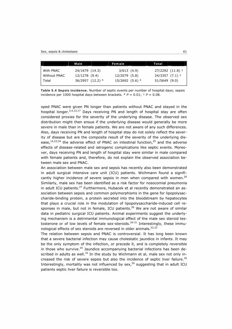
Sex, sepsis & cholestasis
61
oped PNAC were given PN longer than patients without PNAC and stayed in the hospital longer.2,4,12,17 Days receiving PN and length of hospital stay are often considered proxies for the severity of the underlying disease. The observed sex distribution might then ensue if the underlying disease would generally be more severe in male than in female patients. We are not aware of any such differences. Also, days receiving PN and length of hospital stay do not solely reflect the sever-ity of disease but are the composite result of the severity of the underlying dis-ease,14,23,24 the adverse effect of PNAC on intestinal function,25 and the adverse effects of disease-related and iatrogenic complications like septic events. Moreo-ver, days receiving PN and length of hospital stay were similar in male compared with female patients and, therefore, do not explain the observed association be-tween male sex and PNAC. An association between male sex and sepsis has recently also been demonstrated in adult surgical intensive care unit (ICU) patients. Wichmann found a signifi-cantly higher incidence of severe sepsis in men when compared with women.26 Similarly, male sex has been identified as a risk factor for nosocomial pneumonia in adult ICU patients.27 Furthermore, Hubacek et al recently demonstrated an as-sociation between sepsis and common polymorphisms in the gene for lipopolysac-charide-binding protein, a protein secreted into the bloodstream by hepatocytes that plays a crucial role in the modulation of lipopolysaccharide-induced cell re-sponses in male, but not in female, ICU patients.28 We are not aware of similar data in pediatric surgical ICU patients. Animal experiments suggest the underly-ing mechanism is a detrimental immunological effect of the male sex steroid tes-tosterone or of low levels of female sex-steroids.29-31 Interestingly, these immu-nological effects of sex steroids are reversed in older animals.32,33 The relation between sepsis and PNAC is controversial. It has long been known that a severe bacterial infection may cause cholestatic jaundice in infants. It may be the only symptom of the infection, or precede it, and is completely reversible in those who survive.34 Jaundice accompanying bacterial infections has been de-scribed in adults as well.35 In the study by Wichmann et al, male sex not only in-creased the risk of severe sepsis but also the incidence of septic liver failure.26 Interestingly, mortality was not influenced by sex,26 suggesting that in adult ICU patients septic liver failure is reversible too.
Male Female Total With PNAC 24/1679 (14.3) 3/613 (4.9) 27/2292 (11.8) †
Without PNAC 12/1278 (9.4) 12/2079 (5.8) 24/3357 (7.1) †
Total 36/2957 (12.2) * 15/2692 (5.6) * 51/5649 (9.0)
Table 5.4 Sepsis incidence. Number of septic events per number of hospital days; sepsis incidence per 1000 hospital days between brackets. * P = 0.01; † P = 0.08.
Chapter 5 62
Several,5,8,12,17,18 but not all,19,36 studies have shown that sepsis is seen in a larger proportion of patients with PNAC than in patients without PNAC, and con-cluded that sepsis is a risk factor for PNAC. Most of these studies, however, did not account for differences in sex ratio or exposure time, i.e., length of stay, even though greater length of stay had previously been associated with PNAC.2,4 In effect, these studies were sepsis prevalence studies. When male and female pa-tients are taken together, sepsis prevalence in our patients with PNAC was 50%, implying that PNAC occurred and progressed in 1 of 2 patients without the aid of a septic event. In only 1 of 3 did sepsis occur before PNAC was diagnosed. Sepsis incidence in patients with PNAC tended to be higher than that in patients without PNAC, but remained low (11.8 per 1000 hospital days, i.e., approximately 1 event in 3 months, versus 7.1 per 1000 hospital days). These incidences resemble those found by Sondheimer in 42 newborns on long term PN after neonatal intes-tinal resection (approximately 10-12 per 1000 hospital days in patients with PNAC and 4 per 1000 hospital days in patients without PNAC).14 The overall sepsis inci-dence in our study (9 per 1000 hospital days) was comparable to those recently found in surgical newborns37 and surgical patients of all pediatric ages (7.3 per 1000 days in both studies).17 In the study by Sondheimer et al, the age at first infection was much lower in infants with PNAC than infants without PNAC and cholestasis developed relatively shortly after the first infection in 90% of the pa-tients.14 Unfortunately, Sondheimer et al did not describe the sex distribution of their patients. Our data suggest that observed differences between patients with PNAC and patients without PNAC may depend on the number of male patients with PNAC. Whereas many researchers find sepsis an important risk factor for the initiation or progression of PNAC, some have taken a different view2,19 or argued that an in-creased sepsis incidence is a consequence rather than a cause of PNAC.8,20 In our study, sepsis incidence was not increased in female patients with PNAC, and sep-sis incidence was higher in male than in female patients, irrespective of the ab-sence or presence of PNAC. Moreover, most septic events were caused by coagu-lase-negative staphylococci. These infections are thought to result from vascular access techniques and, to our knowledge, have never been linked to intestinal pathological features.38 All these observations support the view that sepsis is an epiphenomenon of PNAC rather than a causative factor. Our observation that male sex predisposes the surgical newborn to PNAC and to sepsis may explain the association between sepsis and cholestasis found in other studies. If our findings are corroborated, they may have implications both for our understanding of the mechanism of disease and for the design of studies aiming to prevent or treat parenteral nutrition associated cholestasis.
Sex, sepsis & cholestasis
63
REFERENCES 1. Kelly DA. Liver complications of pediatric parenteral nutrition - epidemiology. Nutrition
1998;14:153-7.
2. Pereira GR, Sherman MS, DiGiacomo J, Ziegler M, Roth K, Jacobowski D. Hyperalimen-tation-induced cholestasis. Increased incidence and severity in premature infants. Am J Dis Child 1981;135:842-5.
3. Hodes JE, Grosfeld JL, Weber TR, Schreiner RL, Fitzgerald JF, Mirkin LD. Hepatic failure in infants on total parenteral nutrition (TPN): clinical and histopathologic observations. J Pediatr Surg 1982;17:463-8.
4. Ginn-Pease ME, Pantalos D, King DR. TPN-associated hyperbilirubinemia: a common problem in newborn surgical patients. J Pediatr Surg 1985;20:436-9.
5. Bell RL, Ferry GD, Smith EO, et al. Total parenteral nutrition-related cholestasis in in-fants. JPEN J Parenter Enteral Nutr 1986;10:356-9.
6. Drongowski RA, Coran AG. An analysis of factors contributing to the development of total parenteral nutrition-induced cholestasis. JPEN J Parenter Enteral Nutr 1989;13: 586-9.
7. Spurr SG, Grylack LJ, Mehta NR. Hyperalimentation-associated neonatal cholestasis: effect of oral gentamicin. JPEN J Parenter Enteral Nutr 1989;13:633-6.
8. Bos AP, Tibboel D, Hazebroek FW, Bergmeijer JH, van Kalsbeek EJ, Molenaar JC. Total parenteral nutrition associated cholestasis: a predisposing factor for sepsis in surgical neonates? Eur J Pediatr 1990;149:351-3.
9. Nousia-Arvanitakis S, Angelopoulou-Sakadami N, Metroliou K. Complications associated with total parenteral nutrition in infants with short bowel syndrome. Hepatogastroen-terology 1992;39:169-72.
10. Moss RL, Das JB, Raffensperger JG. Total parenteral nutrition-associated cholestasis: clinical and histopathologic correlation. J Pediatr Surg 1993;28:1270-4; discussion 4-5.
11. Jacquemin E, Maurage C, Borderon JC, Gold F, Laugier J, Rolland JC. Early cholestasis in premature infants receiving total parenteral nutrition: a possible consequence of shock and hypoxia. Eur J Pediatr Surg 1995;5:259-61.
12. Beath SV, Davies P, Papadopoulou A, et al. Parenteral nutrition-related cholestasis in postsurgical neonates: multivariate analysis of risk factors. J Pediatr Surg 1996;31:604-6.
13. Moss RL, Das JB, Raffensperger JG. Necrotizing enterocolitis and total parenteral nutri-tion-associated cholestasis. Nutrition 1996;12:340-3.
14. Sondheimer JM, Asturias E, Cadnapaphornchai M. Infection and cholestasis in neonates with intestinal resection and long-term parenteral nutrition. J Pediatr Gastroenterol Nutr 1998;27:131-7.
15. Mayr JM, Schober PH, Weissensteiner U, Hollwarth ME. Morbidity and mortality of the short-bowel syndrome. Eur J Pediatr Surg 1999;9:231-5.
16. Moss RL, Amii LA. New approaches to understanding the etiology and treatment of total parenteral nutrition-associated cholestasis. Semin Pediatr Surg 1999;8:140-7.
17. Yeung CY, Lee HC, Huang FY, Wang CS. Sepsis during total parenteral nutrition: ex-ploration of risk factors and determination of the effectiveness of peripherally inserted central venous catheters. Pediatr Infect Dis J 1998;17:135-42.
18. Amii LA, Moss RL. Nutritional support of the pediatric surgical patient. Curr Opin Pediatr 1999;11:237-40.
19. Beale EF, Nelson RM, Bucciarelli RL, Donnelly WH, Eitzman DV. Intrahepatic cholestasis associated with parenteral nutrition in premature infants. Pediatrics 1979;64:342-7.
Chapter 5 64
20. Sondheimer JM, Cadnapaphornchai M, Sontag M, Zerbe GO. Predicting the duration of dependence on parenteral nutrition after neonatal intestinal resection. J Pediatr 1998; 132:80-4.
21. Teitelbaum DH, Han-Markey T, Schumacher RE. Treatment of parenteral nutrition-asso-ciated cholestasis with cholecystokinin-octapeptide. J Pediatr Surg 1995;30:1082-5.
22. Garner JS, Jarvis WR, Emori TG, Horan TC, Hughes JM. CDC definitions for nosocomial infections, 1988 . Am J Infect Control 1988;16:128-40; erratum: Am J Infect Control 1988;16(4):177.
23. Carbonnel F, Cosnes J, Chevret S, et al. The role of anatomic factors in nutritional autonomy after extensive small bowel resection. JPEN J Parenter Enteral Nutr 1996;20: 275-80.
24. Kaufman SS, Loseke CA, Lupo JV, et al. Influence of bacterial overgrowth and intestinal inflammation on duration of parenteral nutrition in children with short bowel syndrome. J Pediatr 1997;131:356-61.
25. Hofmann AF. Defective biliary secretion during total parenteral nutrition: probable mechanisms and possible solutions. J Pediatr Gastroenterol Nutr 1995;20:376-90.
26. Wichmann MW, Inthorn D, Andress HJ, Schildberg FW. Incidence and mortality of se-vere sepsis in surgical intensive care patients: the influence of patient gender on dis-ease process and outcome. Intensive Care Med 2000;26:167-72.
27. Kropec A, Schulgen G, Just H, Geiger K, Schumacher M, Daschner F. Scoring system for nosocomial pneumonia in ICUs. Intensive Care Med 1996;22:1155-61.
28. Hubacek JA, Stuber F, Frohlich D, et al. Gene variants of the bactericidal/permeability increasing protein and lipopolysaccharide binding protein in sepsis patients: gender-specific genetic predisposition to sepsis. Crit Care Med 2001;29:557-61.
29. Wichmann MW, Ayala A, Chaudry IH. Male sex steroids are responsible for depressing macrophage immune function after trauma-hemorrhage. Am J Physiol 1997;273: C1335-40.
30. Angele MK, Schwacha MG, Ayala A, Chaudry IH. Effect of gender and sex hormones on immune responses following shock. Shock 2000;14:81-90.
31. Knöferl MW, Diodato MD, Angele MK, et al. Do female sex steroids adversely or benefi-cially affect the depressed immune responses in males after trauma-hemorrhage? Arch Surg 2000;135:425-33.
32. Kahlke V, Angele MK, Schwacha MG, et al. Reversal of sexual dimorphism in splenic T lymphocyte responses after trauma-hemorrhage with aging. Am J Physiol Cell Physiol 2000;278:C509-16.
33. Kahlke V, Angele MK, Ayala A, et al. Immune dysfunction following trauma-haemor-rhage: influence of gender and age. Cytokine 2000;12:69-77.
34. Hamilton JR, Sass-Kortsak A. Jaundice associated with severe bacterial infection in young infants. J Pediatr 1963;63:121-32.
35. Jaundice due to bacterial infection. Gastroenterology 1979;77:362-74.
36. Vileisis RA, Inwood RJ, Hunt CE. Prospective controlled study of parenteral nutrition-associated cholestatic jaundice: effect of protein intake. J Pediatr 1980;96:893-7.
37. Pierro A, van Saene HK, Donnell SC, et al. Microbial translocation in neonates and in-fants receiving long-term parenteral nutrition. Arch Surg 1996;131:176-9.
38. Freeman J, Epstein MF, Smith NE, Platt R, Sidebottom DG, Goldmann DA. Extra hospital stay and antibiotic usage with nosocomial coagulase-negative staphylococcal bactere-mia in two neonatal intensive care unit populations. Am J Dis Child 1990;144:324-9.
6 Clinical relevancy of nonurinary nitrogen excretion
in newborns and infants after digestive tract
surgery
Marcel J.I.J. Albers Ewout W. Steyerberg Trinet Rietveld Dick Tibboel JPEN J Parenter Enteral Nutr 2003;27:327-32
Chapter 6
66
ABSTRACT
BACKGROUND: Whether the contribution of nonurinary nitrogen excretion (N2NU) to total nitrogen excretion (N2TOT) is clinically relevant has not been tested in chil-dren in an intensive care unit. Particularly after digestive tract surgery, fecal ni-trogen losses, and losses via nasogastric tubes, enterostomies and wound drains may be large. METHODS: We prospectively measured urinary nitrogen excretion (N2U) and N2NU 4 to 6 days after digestive tract surgery in 78 newborns and infants who were given total parenteral nutrition. RESULTS: Two hundred seven collections of excreta, each representing one 24-hour period, were obtained. Median N2NU was 15 mg/kg/24 hours (range, 0.4-153), median N2U 153 mg/kg/24 hours (range, 57-558), median N2TOT 179 mg/kg/24 hours (range, 72-577), and the median ratio of N2NU and N2U 9.9% (range, 0.2-110). The observed variations could not be attributed to differences in the severity of the underlying disease or the surgical stress. The mean differ-ence between N2TOT and N2U was 21 mg/kg/24 hours (95% prediction interval –20 - +63). Use of a linear regression equation that predicts N2TOT according to N2U and the weights of other excreta eliminated bias and improved precision (95% prediction interval –34 - +34 mg/kg/24 hours). For individual measure-ments, however, considerable imprecision remained. CONCLUSION: In newborns and infants, receiving parenteral nutrition 4 to 6 days after digestive tract surgery, N2NU is variable and not to be neglected. The only way to accurately assess total nitrogen excretion in individual patients is by measuring the nitrogen content of all excreta.
Urinary & nonurinary nitrogen excretion
67
INTRODUCTION
Catabolism and anabolism are fundamental aspects of illness and reconvales-cence, often defined as whole-body nitrogen loss and whole-body nitrogen reten-tion, or as a negative nitrogen balance and a positive nitrogen balance.1,2 Nitro-gen balance studies therefore are one of the cornerstones of clinical nutrition re-search. When assessing whole-body nitrogen excretion, urinary nitrogen excre-tion (N2U) usually is measured or derived from urinary urea excretion. Nitrogen excretion via other routes (skin, stools, wounds, drains) is often assumed to be of minor importance or predictable.3-6 In children admitted to an intensive care unit (ICU), the value of urinary urea excretion as a predictor of N2U has been ques-tioned.6-9 Whether nonurinary nitrogen excretion (N2NU) is of little importance when compared to N2U has not been tested in such children. N2NU may be par-ticularly large after digestive tract surgery, when body fluids may be lost through nasogastric tubes, enterostomies, wound drains, and feces. We measured N2NU and N2U in newborns and infants in our pediatric surgical ICU who were receiving parenteral nutrition after digestive tract surgery and assessed the contribution of N2NU to total nitrogen excretion (N2TOT).
MATERIAL AND METHODS
Clinical setting
Our pediatric surgical ICU is part of a tertiary referral university children’s hospi-tal serving approximately 4.5 million inhabitants. At the time of the study, the ICU consisted of 2 essentially identical 7-bed units. Annually 600 to 700 patients were admitted for perioperative intensive and high care. The majority were new-borns and infants admitted because of a congenital anomaly that required sur-gery. The second most frequent reason for admission was postoperative intensive care in all pediatric age groups.
Patients
From January 1997 through December 1999, 80 newborns, infants, and children, admitted to our ICU after digestive tract surgery, entered a prospective trial that studied the effects of isonitrogenous enrichment of parenteral nutrition with glu-tamine. As part of this trial, nitrogen excretion was measured on day 4, 5 and 6 after surgery (day 0). The trial was approved by the institutional review board. Inclusion criteria were: gestational age > 30 weeks and age ≤ 1 month or gestational age ≥ 35 weeks and age ≤ 2 years, not expected to tolerate enteral nutrition for at least 4 days following digestive tract surgery, and written and signed informed parental con-sent. Exclusion criteria were: simultaneous participation in another trial or previ-ous participation in the current trial, preexistent renal or hepatic dysfunction that precluded the use of standard parenteral nutrition, an inborn error of metabolism,
Chapter 6
68
an immunodeficiency or a disease that entailed impaired growth (other than dys-maturity), the use of corticosteroid drugs, life expectancy <6 months (according to index diagnosis). The Pediatric Risk of Mortality II score (PRISM), obtained on the day of admission to the ICU, was used to assess the severity of illness.10 The Surgical Stress Score (SSS) was used as a proxy for the severity of the postop-erative stress response.11
Parenteral nutrition
Total parenteral nutrition was started on day 2 after surgery. Depending on age and weight, it provided 85 to 100 kcal/kg/24 hours and 1.5 to 2.5 gram/kg/24 hours of amino acids (Vaminolact®, Pharmacia, Stockholm, Sweden). Carbohy-drates (dextrose, Fresenius, Bad Homburg, Germany) provided approximately 65% of non-protein calories and fat (Intralipid® 20%, Pharmacia) approximately 35%. If the patient’s condition and the surgical procedure would allow it, tapering of parenteral nutrition and reintroduction of enteral nutrition was started halfway through day 6.
Excreta collection and laboratory analysis
All excreta, with the exception of bronchopulmonary secretions and one patient’s decubitus wound exudate, were collected on day 4, 5 and 6 after surgery. For the collection of urine, a transurethral catheter was used, if present. In girls, if such a catheter was not being used already, it was inserted; for boys, appropriately sized urine bags were then used. Urine collection bags were emptied, and the collected volume was measured at least once every 4 hours. Leakage of urine that could not be quantified rendered that interval’s collection invalid (cf Appen-dix). Collected urine was stored in a refrigerator in containers preacidified with sulfuric acid, until the morning of the next day, when it was transferred to the laboratory. Fluids produced through nasogastric or nasoduodenal tube drainage, feces, and enterostomy and wound drain output were also collected and stored in a refrigerator until transferal to the laboratory. Collection of these excreta was considered invalid if leakage was observed. In the laboratory, all collections were weighed and homogenized. Urine collections were further acidified with sulfuric acid to a pH of 2 to 4. An aliquot of every col-lection was then stored at –20 °C until thawing and analysis. The total nitrogen content was determined by a continuous flow elemental analyzer (Carlo Erba NC-1500; Interscience BV, The Netherlands). In brief, triplicate samples are weighed in tin sample containers, freeze-dried and combusted at 1020 °C. Copper reduces the formed nitrogen oxides to elemental nitrogen gas. The nitrogen gas flows through a thermal conductivity detector that generates an electrical signal pro-portional to the concentration of nitrogen. This is an automation of the Dumas combustion method.12
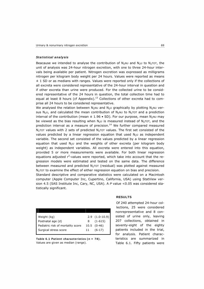
Urinary & nonurinary nitrogen excretion
69
Statistical analysis
Beacause we intended to analyse the contribution of N2NU and N2U to N2TOT, the unit of analysis was 24-hour nitrogen excretion, with one to three 24-hour inter-vals being available per patient. Nitrogen excretion was expressed as milligrams nitrogen per kilogram body weight per 24 hours. Values were reported as means ± 1 SD or as medians with ranges. Values were reported only if the collections of all excreta were considered representative of the 24-hour interval in question and if other excreta than urine were produced. For the collected urine to be consid-ered representative of the 24 hours in question, the total collection time had to equal at least 8 hours (cf Appendix).13 Collections of other excreta had to com-prise all 24 hours to be considered representative. We analysed the relation between N2NU and N2U graphically by plotting N2NU ver-sus N2U, and calculated the mean contribution of N2NU to N2TOT and a prediction interval of the contribution (mean ± 1.96 • SD). For our purpose, mean N2NU may be viewed as the bias resulting when N2U is measured instead of N2TOT, and the prediction interval as a measure of precision.14 We further compared measured N2TOT values with 2 sets of predicted N2TOT values. The first set consisted of the values predicted by a linear regression equation that used N2U as independent variable. The second set consisted of the values predicted by a linear regression equation that used N2U and the weights of other excreta (per kilogram body weight) as independent variables. All excreta were entered into this equation, provided 5 or more measurements were available. For both linear regression equations adjusted r2-values were reported, which take into account that the re-gression models were estimated and tested on the same data. The difference between measured and predicted N2TOT (residual) was plotted against measured N2TOT to examine the effect of either regression equation on bias and precision. Standard descriptive and comparative statistics were calculated on a Macintosh computer (Apple Computer Inc, Cupertino, California, USA) using StatView ver-sion 4.5 (SAS Institute Inc, Cary, NC, USA). A P value <0.05 was considered sta-tistically significant.
RESULTS
Of 240 attempted 24-hour col-lections, 25 were considered nonrepresentative and 8 con-sisted of urine only, leaving 207 collections, obtained in seventy-eight of the eighty patients included in the trial, for analysis. Patient charac-teristics are summarized in Table 6.1. Fifty patients were
Weight (kg) 2.9 (1.0-10.9)
Postnatal age (d) 8 (1-615)
Pediatric risk of mortality score 10.5 (0-46)
Surgical stress score 11 (6-17)
Table 6.1 Patient characteristics (n = 78). Values are given as median (range).
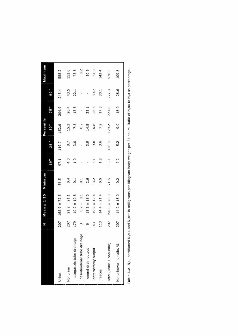
Maxim
um
558.2
152.6
73.8
0.2
50.6
54.0
142.4
576.5
109.8
90
th
248.4
43.5
22.1
-
-
39.7
30.1
277.3
28.6
75
th
204.9
26.4
13.5
-
23.1
26.5
17.3
223.6
18.0
50
th
152.6
15.3
7.5
0.2
14.8
16.8
7.2
179.2
9.9
25
th
119.7
8.7
3.0
-
3.9
9.8
3.6
136.8
5.2
Perc
en
tile
10
th
97.1
4.0
1.0
-
-
6.1
1.8
111.1
2.2
Min
imu
m
56.5
0.4
0.1
0.1
2.6
3.2
0.5
71.5
0.2
Mean
± 1
SD
168.9
± 7
2.5
21.2
± 2
1.1
10.2
± 1
0.8
0.2
±
0.1
18.3
± 1
8.0
19.2
± 1
2.4
14.4
± 2
1.4
190.0
± 7
6.0
14.2
± 1
5.0
N
207
207
179
3
6
43
113
207
207
Urine
Nonurine
nas
ogas
tric
tube
dra
inag
e
nas
oduoden
al t
ube
dra
inag
e
wound d
rain
outp
ut
ente
rost
om
y outp
ut
faec
es
Tota
l (u
rine
+ n
onurine)
Nonurine/
urine
ratio,
%
Tab
le 6
.2. N
2U,
par
titioned
N2N
U,
and N
2TO
T in m
illig
ram
s per
kilo
gra
m b
ody
wei
ght
per
24 h
ours
. Rat
io o
f N
2N
U t
o N
2U a
s per
centa
ge.
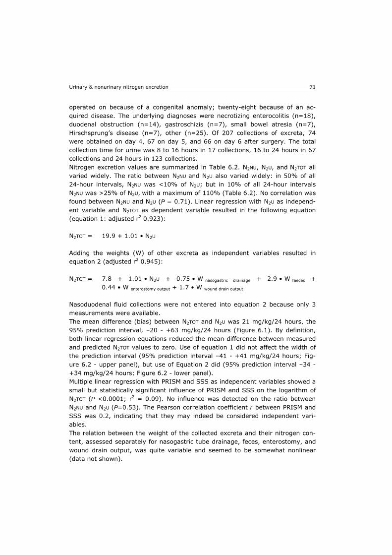
Urinary & nonurinary nitrogen excretion
71
operated on because of a congenital anomaly; twenty-eight because of an ac-quired disease. The underlying diagnoses were necrotizing enterocolitis (n=18), duodenal obstruction (n=14), gastroschizis (n=7), small bowel atresia (n=7), Hirschsprung’s disease (n=7), other (n=25). Of 207 collections of excreta, 74 were obtained on day 4, 67 on day 5, and 66 on day 6 after surgery. The total collection time for urine was 8 to 16 hours in 17 collections, 16 to 24 hours in 67 collections and 24 hours in 123 collections. Nitrogen excretion values are summarized in Table 6.2. N2NU, N2U, and N2TOT all varied widely. The ratio between N2NU and N2U also varied widely: in 50% of all 24-hour intervals, N2NU was <10% of N2U; but in 10% of all 24-hour intervals N2NU was >25% of N2U, with a maximum of 110% (Table 6.2). No correlation was found between N2NU and N2U (P = 0.71). Linear regression with N2U as independ-ent variable and N2TOT as dependent variable resulted in the following equation (equation 1: adjusted r2 0.923): N2TOT = 19.9 + 1.01 • N2U Adding the weights (W) of other excreta as independent variables resulted in equation 2 (adjusted r2 0.945): N2TOT = 7.8 + 1.01 • N2U + 0.75 • W nasogastric drainage + 2.9 • W faeces +
0.44 • W enterostomy output + 1.7 • W wound drain output Nasoduodenal fluid collections were not entered into equation 2 because only 3 measurements were available. The mean difference (bias) between N2TOT and N2U was 21 mg/kg/24 hours, the 95% prediction interval, –20 - +63 mg/kg/24 hours (Figure 6.1). By definition, both linear regression equations reduced the mean difference between measured and predicted N2TOT values to zero. Use of equation 1 did not affect the width of the prediction interval (95% prediction interval –41 - +41 mg/kg/24 hours; Fig-ure 6.2 - upper panel), but use of Equation 2 did (95% prediction interval –34 - +34 mg/kg/24 hours; Figure 6.2 - lower panel). Multiple linear regression with PRISM and SSS as independent variables showed a small but statistically significant influence of PRISM and SSS on the logarithm of N2TOT (P <0.0001; r2 = 0.09). No influence was detected on the ratio between N2NU and N2U (P=0.53). The Pearson correlation coefficient r between PRISM and SSS was 0.2, indicating that they may indeed be considered independent vari-ables. The relation between the weight of the collected excreta and their nitrogen con-tent, assessed separately for nasogastric tube drainage, feces, enterostomy, and wound drain output, was quite variable and seemed to be somewhat nonlinear (data not shown).
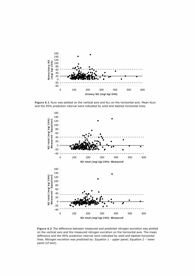
Figure 6.1 N2NU was plotted on the vertical axis and N2U on the horizontal axis. Mean N2NU and the 95% prediction interval were indicated by solid and dashed horizontal lines.
Figure 6.2 The difference between measured and predicted nitrogen excretion was plotted on the vertical axis and the measured nitrogen excretion on the horizontal axis. The mean difference and the 95% prediction interval were indicated by solid and dashed horizontal lines. Nitrogen excretion was predicted by: Equation 1 - upper panel; Equation 2 – lower panel (cf text).
-40-20
020406080
100120140160
0 100 200 300 400 500 600
Urinary N2 (mg/kg/24h)
No
nu
rin
ary
N2
(mg
/kg
/2
4h
)
-40
-20
0
20
40
60
80
100
120
140
160
0 100 200 300 400 500 600
N2 total (mg/kg/24h): Measured
N2
tota
l (m
g/
kg
/2
4h
):M
easu
red
- E
qu
ati
on
1
-40
-20
0
20
40
60
80
100
120
140
160
0 100 200 300 400 500 600
N2 total (mg/kg/24h): Measured
N2
tota
l (m
g/
kg
/2
4h
):M
easu
red
- E
qu
ati
on
2
Urinary & nonurinary nitrogen excretion
73
DISCUSSION
In a population of newborns and infants receiving parenteral nutrition 4 to 6 days after digestive tract surgery, we have shown N2NU and N2U, and their ratio, to vary widely. No correlation was found between N2NU and N2U. The observed variations in total nitrogen excretion and in the ratio between N2NU and N2U could only for a small part be attributed to the observed variations in the severity of the surgical stress or the underlying disease. Nitrogen intake minus nitrogen excretion constitutes the whole-body nitrogen balance. Especially if a patient is being fed parenterally, nitrogen intake can easily be calculated. Measuring nitrogen excretion, however, is an arduous task. In newborns and infants, acquiring accurate nitrogen excretion data is particularly difficult. The relatively small volumes of the excreta may result in relatively large effects of sampling and weighing errors. Critical illness, not having been toilet trained, and the use of urine bags may cause significant sampling errors and leakage. Although we preferred the use of transurethral catheters over urine bags, we felt we could not justify catheterization in boys solely for the purpose of this study because this procedure may cause urethral trauma.15 To avoid the leakage associated with the use of urine bags and long collection intervals, we modified the approach proposed by Boehm et al.13 In a population of low birth-weight infants, they showed that a continuous 6-hour collection period suffices for metabolic monitoring purposes. Because our protocol did not insist on continuity of urine collection, we arbitrarily decided 8 hours to be the lower time limit of a representative urine collection. The majority of collection intervals (92%), how-ever, comprised 16 to 24 hours. Because other excreta are not produced continu-ously, these collections had to comprise the full 24-hour interval. Despite our efforts, sampling and weighing errors, particularly of urine or faeces, may have occurred. Such errors, though inherent to the clinical setting, would likely affect our precision but not necessarily bias our results. In this setting, vali-dation of data is of paramount importance. However, the wide variation seen in daily nitrogen excretion, and the paucity of similar studies in comparable patient populations, make validation of our findings difficult. Still, other studies have found similarly wide variations of the daily N2U in surgical newborns8 and infants3, pediatric ICU patients5, and healthy children.16 Chaloupecky et al studied thirty-seven infants on the first day after cardiopulmonary bypass surgery and found N2U values averaging 235 ± 83 mg/kg/24 hours.3 Helms et al found N2U in eight preterm newborns to be 209 ± 99 mg/kg/24 hours on day 1 to 3 after digestive tract surgery, and 96 ± 49 mg/kg/24 hours on day 7.8 In our study, N2U meas-ured on day 4 to 6 after digestive tract surgery, was 169 ± 73 mg/kg/24 hours. Moreover, we found a small but significant influence of both SSS and PRISM on total nitrogen excretion. Our findings are in keeping with those of Chaloupecky et al and Helms et al and with the notion that nitrogen excretion in infants, as in
Chapter 6
74
older children and adults, is proportional to the stress of surgery and to the stress imposed by the underlying disease, indicating that our findings are valid.1,17-19 The limitations of the scoring systems should, however, be noted. SSS was origi-nally designed by Anand and Ainsley-Green to grade the surgical stress of several types of surgery, including cardiopulmonary bypass surgery.11 When using SSS solely for digestive tract surgery, its range of potential values and its discrimina-tive power are limited. Also, the effect of surgical stress on nitrogen excretion may have worn off by day 4 to 6 after surgery. Jones et al and Bouwmeester et al have shown that resting energy expenditure and (nor)epinephrine levels in new-born patients normalize within 24 hours after surgery.20,21 Nitrogen excretion in surgical newborns may parallel resting energy expenditure as it does in adults.1 PRISM II has been validated for use in the Netherlands, but its performance as a predictor of mortality in surgical patients was not as good as that in nonsurgical patients.22,23 PRISM II should be obtained on the day of first admission to the ICU, which occasionally preceded inclusion into this study by several days or even weeks. More importantly, PRISM predicts mortality risk and as such does not al-ways reflect the severity of illness.24 These limitations, and the wide intrinsic variation in nitrogen excretion discussed earlier, may explain why SSS and PRISM had only small effects on total nitrogen excretion. We are not aware of any data on N2NU in populations comparable to the one in our study. As a rule, nonurinary nitrogen loss is neglected or accounted for by a formula that converts urinary urea or urinary nitrogen excretion to total nitrogen excretion.3-6 In one textbook on paediatric intensive care, fecal nitrogen loss is said to equal 20% of urinary excretion, but a reference is not given, precluding identification of the population the original data apply to.2 In a study by Ziegler et al in 123 normal children aged 1 to 11 years, fecal nitrogen excretion averaged 15% of N2U.16 In our study, fecal nitrogen excretion was 10 ± 15 % of urinary excretion (data not shown), and nitrogen excretion through all excreta other than urine equaled 14 ± 15 % of urinary excretion. It should be noted that our pa-tients were fed parenterally, which would likely yield a lower stool output when compared with a general pediatric ICU population.6 Measuring nitrogen excretion in newborns and infants is difficult and laborious, as was mentioned earlier. For this reason, we, and others before us, have tried to find equations that derive N2TOT from easily-obtainable parameters. Because uri-nary urea content does not reliably predict urinary nitrogen content in critically ill children6-9 and because urine generally is the main route of nitrogen excretion, we used measured N2U as the basis of our equations. On average, measured N2U underestimated N2TOT by 21 mg/kg/24 hours (Figure 6.1). Adding this fixed amount, or a fixed percentage (in our study, 14%; Table 6.2) to measured (uri-nary) nitrogen excretion is the approach others have followed in different set-tings.2,4,25 Whereas this approach reduces bias, it does not improve precision. Similarly, using a linear regression equation will, by definition, eliminate bias, but not necessarily improve precision, as is illustrated by equation 1 (Figure 6.2 – up-
Urinary & nonurinary nitrogen excretion
75
per panel). Equation 2, on the other hand, by incorporating the weights of ex-creta, did improve precision (Figure 6.2 – lower panel). We chose to use these weights because they are easy to obtain and often are obtained as part of stan-dard patient care. Although equation 2 may suffice for group comparisons, con-siderable imprecision remained for individual patient assessment, which could only be avoided by measuring the nitrogen content of all excreta. Also, when de-ciding whether to use equation 2, the clinician should bear in mind that the re-sulting loss of precision is added to that of sampling and weighing of excreta. Ideally, external validation should be performed first to assess whether the re-gression coefficients apply to other settings and case mixes. In conclusion, N2NU is variable and not to be neglected when assessing total ni-trogen excretion in newborns and infants receiving parenteral nutrition shortly after digestive tract surgery. Although relatively simple predictive equations may yield reasonable estimates, the only way to accurately assess total nitrogen ex-cretion is by measuring the nitrogen content of all excreta.
APPENDIX: URINE COLLECTION
Urine collection bags were emptied and the collected volume (VCU) was measured at least once every 4 hours. At the time of emptying, the tared diapers were reweighed to check for quantifiable urine leakage and the bedding was inspected for (nonquantifiable) leakage. The presence of any nonquantifiable leakage ren-dered the corresponding interval’s urine collection invalid. That interval’s collected urine would then be discarded. If all leakage was quantifiable, the weight of quantifiable urine leakage was converted to volume (VQUL) using a conversion factor of 1 g/mL. Leakage was assumed to have occurred at random. Urine, col-lected in the interval between emptyings (I), thus represents the fraction of that interval defined by
I • VCU/(VCU+VQUL). The total volume of urine collected over 24 hours represents a total collection time equal to the sum of the fractions of the intervals
∑ [ I • VCU/(VCU+VQUL)]. Twenty-four hour N2U was calculated by multiplying the nitrogen content of the collected urine by
24 / ∑ [ I • VCU/(VCU+VQUL)].
Chapter 6
76
REFERENCES 1. Long CL, Schaffel N, Geiger JW, Schiller WR, Blakemore WS. Metabolic response to in-
jury and illness: estimation of energy and protein needs from indirect calorimetry and nitrogen balance. JPEN J Parenter Enteral Nutr 1979;3:452-6.
2. Deutschman CS. Nutrition and metabolism in the critically ill child. In: Rogers MC, edi-tor. Textbook of pediatric intensive care. 2 ed. Baltimore: Williams & Wilkins; 1992. p. 1109-31.
3. Chaloupecky V, Hucín B, Tláskal T, et al. Nitrogen balance, 3-methylhistidine excretion, and plasma amino acid profile in infants after cardiac operations for congenital heart defects: the effect of early nutritional support. J Thorac Cardiovasc Surg 1997;114:1053-60.
4. Maldonado J, Faus MJ, Bayes R, Molina JA, Gil A. Apparent nitrogen balance and 3-methylhistidine urinary excretion in intravenously fed children with trauma and infec-tion. Eur J Clin Nutr 1988;42:93-100.
5. Mickell JJ. Urea nitrogen excretion in critically ill children. Pediatrics 1982;70:949-55.
6. Prelack K, Dwyer J, Yu YM, Sheridan RL, Tompkins RG. Urinary urea nitrogen is impre-cise as a predictor of protein balance in burned children. J Am Diet Assoc 1997;97:489-95.
7. Boehm KA, Helms RA, Storm MC. Assessing the validity of adjusted urinary urea nitro-gen as an estimate of total urinary nitrogen in three pediatric populations. JPEN J Par-enter Enteral Nutr 1994;18:172-6.
8. Helms RA, Mowatt-Larssen CA, Boehm KA, et al. Urinary nitrogen constituents in the postsurgical preterm neonate receiving parenteral nutrition. JPEN J Parenter Enteral Nutr 1993;17:68-72.
9. Patterson BW, Nguyen T, Pierre E, Herndon DN, Wolfe RR. Urea and protein metabolism in burned children: effect of dietary protein intake. Metabolism 1997;46:573-8.
10. Pollack MM, Ruttimann UE, Getson PR. Pediatric risk of mortality (PRISM) score. Crit Care Med 1988;16:1110-6.
11. Anand KJ, Aynsley-Green A. Measuring the severity of surgical stress in newborn in-fants. J Pediatr Surg 1988;23:297-305.
12. Fiedler R, Proksch G. The determination of nitrogen-15 by emission and mass spec-trometry in biochemical analysis: a review. Anal Chim Acta 1975;78:1-62.
13. Boehm G, Wiener M, Schmidt C, Ungethüm A, Ungethüm B, Moro G. Usefulness of short-term urine collection in the nutritional monitoring of low birthweight infants. Acta Paediatr 1998;87:339-43.
14. Altman DG. Practical statistics for medical research. London New York: Chapman and Hall; 1991.
15. Elder JS. Urologic disorders in infants and children. In: Behrman RE, Kliegman RM, Jen-son HB, editors. Nelson Textbook of Pediatrics. 16 ed. Philadelphia: W.B. Saunders Company; 2000. p. 1619-58.
16. Ziegler EE, O'Donnell AM, Stearns G, Nelson SE, Burmeister LF, Fomon SJ. Nitrogen balance studies with normal children. Am J Clin Nutr 1977;30:939-46.
17. Pollack MM. Nutritional support of children in the intensive care unit. In: Suskind RM, Lewinter-Suskind L, editors. Textbook of pediatric nutrition. 2nd ed. New York: Raven Press; 1993. p. 207-23.
18. Shew SB, Keshen TH, Jahoor F, Jaksic T. The determinants of protein catabolism in neonates on extracorporeal membrane oxygenation. J Pediatr Surg 1999;34:1086-90.
19. Steinhorn DM, Green TP. Severity of illness correlates with alterations in energy me-tabolism in the pediatric intensive care unit. Crit Care Med 1991;19:1503-9.
Urinary & nonurinary nitrogen excretion
77
20. Jones MO, Pierro A, Hammond P, Lloyd DA. The metabolic response to operative stress in infants. J Pediatr Surg 1993;28:1258-62; discussion 62-63.
21. Bouwmeester NJ, Anand KJ, van Dijk M, Hop WC, Boomsma F, Tibboel D. Hormonal and metabolic stress responses after major surgery in children aged 0-3 years: a double-blind, randomized trial comparing the effects of continuous versus intermittent mor-phine. Br J Anaesth 2001;87:390-9.
22. Gemke RJ, Bonsel GJ, van Vught AJ. Effectiveness and efficiency of a Dutch pediatric intensive care unit: validity and application of the Pediatric Risk of Mortality score. Crit Care Med 1994;22:1477-84.
23. Gemke RJ, Bonsel GJ. Comparative assessment of pediatric intensive care: a national multicenter study. Pediatric Intensive Care Assessment of Outcome (PICASSO) Study Group. Crit Care Med 1995;23:238-45.
24. Shann F. Are we doing a good job: PRISM, PIM and all that. Intensive Care Med 2002;28:105-7.
25. Mackenzie TA, Clark NG, Bistrian BR, Flatt JP, Hallowell EM, Blackburn GL. A simple method for estimating nitrogen balance in hospitalized patients: a review and support-ing data for a previously proposed technique. J Am Coll Nutr 1985;4:575-81.

7 Glutamine supplementation of parenteral nutrition
- a double-blind, randomised, controlled trial
Marcel J.I.J. Albers Ewout W. Steyerberg Frans W.J. Hazebroek Marjan Mourik Gerard J.J.M. Borsboom Trinet Rietveld Jan G.M. Huijmans Dick Tibboel Submitted
Chapter 7
80
ABSTRACT
OBJECTIVE: To assess the effect of isocaloric isonitrogenous parenteral glutamine supplementation on intestinal permeability and nitrogen loss in newborns and in-fants after major digestive tract surgery. SUMMARY BACKGROUND DATA: Glutamine supplementation in critically ill and surgical adults may normalise intestinal permeability, attenuate nitrogen loss, improve survival and lower the incidence of nosocomial infections. Previous studies in criti-cally ill children were limited to very low birthweight infants and had equivocal results. METHODS: 80 newborns and infants were included in a double-blind, randomized trial, comparing standard parenteral nutrition (sPN; n=39) to glutamine-supple-mented parenteral nutrition (GlnPN; glutamine target-intake 0.4 g•kg-1•day-1; n=41), starting on day 2 after major digestive tract surgery. Primary endpoints were intestinal permeability, as assessed by the urinary excretion ratio of lactu-lose and rhamnose (week 1 through 4); nitrogen balance (day 4 through 6), and urinary 3-methylhistidine excretion (day 5). Secondary endpoints were mortality, length of stay in the ICU and the hospital, number of septic episodes, and usage of antibiotics and ICU resources. RESULTS: Glutamine intake plateaued at 90% of the target on day 4. No differ-ences were found between patients assigned sPN and patients assigned GlnPN regarding any of the endpoints. Glutamine supplementation was not associated with adverse effects. CONCLUSIONS: In newborns and infants after major digestive tract surgery, we did not identify beneficial effects of isonitrogenous, isocaloric glutamine supplemen-tation of parenteral nutrition. Glutamine supplementation in these patients therefore is not warranted.
Glutamine & parenteral nutrition
81
INTRODUCTION
Glutamine is the most abundant free amino acid of the body and has many es-sential metabolic functions. During critical illness, demands are increased and may not be met by endogenous supply and dietary intake.1 In critically ill adults, placebo-controlled randomized studies have shown that glutamine supplementa-tion normalises intestinal permeability, attenuates nitrogen losses, and enhances immunological defenses against infection.1-7 Related clinical effects include im-proved survival, a lower incidence of infections, shorter hospital stay, and lower ICU and hospital costs.1,3,5,8,9 Placebo-controlled randomized studies in critically ill children have so far been limited to very low birth weight infants.10-13 Lacey et al studied 78 preterm infants, and concluded that parenteral glutamine reduced the time on total parenteral nutrition and on the mechanical ventilator, and acceler-ated the transition to full enteral feeding in a subgroup of infants weighing less than 800 grams.10 Neu et al studied 67 very low birth weight infants and con-cluded that enteral glutamine lowered the incidence of sepsis, improved the toler-ance to enteral feeding, and lowered hospital costs.11,14 A subsequent multi-cen-ter trial in 649 very low birth weight infants, however, failed to show an effect of enteral glutamine on the incidence of blood-culture proven nosocomial sepsis.13 We hypothesized that glutamine supplementation might be of benefit to newborns and infants who needed intensive care and parenteral nutrition after major diges-tive tract surgery. The primary endpoints of this study are the effect of isocaloric isonitrogenous parenteral glutamine supplementation on intestinal permeability and nitrogen loss in this population.
MATERIALS AND METHODS
Study outline
The study protocol was approved by the institutional review board. The study was designed as a double-blind randomised controlled trial. In newborns and infants who needed intensive care after digestive tract surgery, we compared standard parenteral nutrition (sPN) with isonitrogenous, isocaloric, glutamine-supple-mented parenteral nutrition (GlnPN). The primary endpoints of the study were intestinal permeability, as measured by sugar absorption tests; nitrogen balance; and urinary 3-methylhistidine (3MH) excretion. Secondary endpoints were mor-tality and morbidity. Measures of morbidity assessed were: length of ICU stay and length of hospital stay; number of septic episodes; usage of antibiotics; usage of ICU resources, as assessed by the therapeutic intervention scoring system – 76 (TISS); and renal and hepatic damage, as assessed by renal and liver function tests.
Chapter 7
82
Patients
Our pediatric surgical ICU is part of a tertiary referral university children’s hospi-tal. Provided informed parental consent was obtained, patients admitted to our ICU were enrolled if they fullfilled all of the inclusion criteria and none of the ex-clusion criteria. The inclusion criteria were: gestational age >30 weeks and age ≤2 years; not expected to tolerate enteral nutrition for at least 4 days following digestive tract surgery. Exclusion criteria were: simultaneous participation in an-other trial or previous participation in the current trial; preexistent renal or he-patic dysfunction that precluded the use of standard parenteral nutrition; an in-born error of metabolism; an immunodeficiency or a disease that entailed im-paired growth (other than dysmaturity); the use of corticosteroid drugs; preexis-tent life expectancy less than 6 months. Baseline data included demographic data, index diagnosis, operative procedure and the Pediatric Risk of Mortality II score (PRISM) and Surgical Stress Score (SSS). PRISM, obtained on the day of admission to the ICU, was used to assess the severity of illness,15 and SSS to assess the severity of the post-operative stress response.16
Stratification, randomization, blinding
The patients were divided into four strata according to age: I - gestational age ≥30 weeks and post-conceptional age <37 weeks; II - post-conceptional age ≥37 weeks and age ≤0.5 year; III - age >0.5 and ≤1 years; IV - age >1 and ≤2 years. We used a computer-generated randomisation schedule to assign patients sPN or GlnPN. Patients were randomized blockwise in groups of four. The ran-domisation schedule was made available only to the pharmacist that supervised the processing of parenteral nutrition. Glutamine-enriched and standard nutrition bags were labeled identically. All physicians and nurses involved in the patients' care were blinded to nutrition assignments.
Parenteral nutrition
On a daily basis, parenteral nutrition was custom-made for each patient. A two-bag system was used, with one bag containing amino acids (Vaminolact®, Phar-macia, Sweden), carbohydrates (Dextrose, Fresenius, Bad Homburg, Germany), minerals, trace elements, carnitene, water-soluble vitamins and water; and the other bag containing lipids (Intralipid® 20%, Pharmacia, Stockholm, Sweden) and fat-soluble vitamins. Total parenteral nutrition was started on day 2 after surgery (designated day 0), according to hospital guidelines, stating that amino acid and lipid intake start at approximately 50% of the recommended intake in the first 24 hours of parenteral feeding and reach the recommended intake in the following 24 hours. Depending on weight, recommended intakes were 1.5-2.5 gram/kg/24h of amino acids and 85-100 kcal/kg/24h, with carbohydrates providing approxi-mately 65% of non-protein calories and fat approximately 35%. Provided the pa-
Glutamine & parenteral nutrition
83
tient’s condition and the surgical procedure would allow it, tapering of parenteral nutrition and reintroduction of enteral nutrition was started on or after day 6. Parenteral nutrition was withheld once patients received more than 75-80% of the recommended intake via the enteral route. If patients needed to revert to parenteral feeding, they were assigned the same parenteral nutrition (sPN or GlnPN) as before. If patients were still being fed parenterally at day 31, the study nutrition would be replaced by standard parenteral nutrition. L-glutamine was purchased as a 2.5% solution in sterile water from Oxford Nutri-tion (Oxford, UK) and stored at –20 ˚C. Isonitrogenous isocaloric supplementa-tion was achieved by substituting predefined volumes of amino acids and water with L-glutamine. Based on a study by Allen 17, we aimed to administer 0.4 gram of glutamine per kilogram body weight per day to patients fully dependent on parenteral nutrition. Parenteral glutamine intake and non-protein caloric intake were calculated in the same fashion as nitrogen intake (see below).
Sugar absorption tests
A sugar absorption test was performed on the fifth day of the 1st through 4th week after surgery, unless the patient had already been discharged from the hospital. The test solution was prepared by the hospital pharmacy. One hundred and forty milligrams of glucose, 140 mg of rhamnose, and 70 mg of xylose were dissolved in 50 mL demineralized water, 8.6 g of lactulose was added, and demineralized water was added up to a total volume of 100 mL. Patients were given 1 mL of this solution per kilogram body weight, via nasogastric tube. Urine, passed in the next 4 hours, was collected; if the collection failed, the test was repeated once within the next 24 hours. One mL was frozen at –80 ˚C until thawing and analysis. We have described the details of the analytical procedure elsewhere.18 The sugars’ urinary excretions were expressed as a percentage of the dose ingested and the lactulose-rhamnose ratio as the ratio of these percentages.
Nitrogen balance
Nitrogen balance data were acquired on day 4, 5 and 6. The nitrogen excretion data were also used to study the contribution of nonurinary nitrogen loss to total nitrogen loss, and have recently been published.19 All excreta, with the exception of bronchopulmonary secretions and one patient’s decubitus wound exudate, were collected, stored and analysed.19 Nitrogen intake was calculated as the product of the volumes and the nitrogen contents of the administered amino acid solutions. The nitrogen content was calculated for each solution separately, based on the product information and the patients’ parenteral nutrition datasheets. Ni-trogen balance was calculated as the difference between nitrogen intake and ni-trogen excretion and expressed as milligrams nitrogen per kilogram body weight per 24 hours.
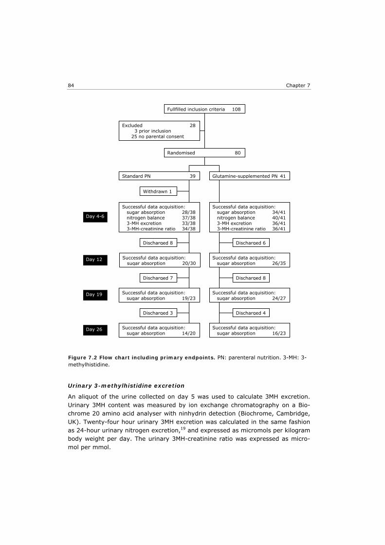
Chapter 7
84
Urinary 3-methylhistidine excretion
An aliquot of the urine collected on day 5 was used to calculate 3MH excretion. Urinary 3MH content was measured by ion exchange chromatography on a Bio-chrome 20 amino acid analyser with ninhydrin detection (Biochrome, Cambridge, UK). Twenty-four hour urinary 3MH excretion was calculated in the same fashion as 24-hour urinary nitrogen excretion,19 and expressed as micromols per kilogram body weight per day. The urinary 3MH-creatinine ratio was expressed as micro-mol per mmol.
Fullfilled inclusion criteria 108
Randomised 80
Excluded 28 3 prior inclusion 25 no parental consent
Withdrawn 1
Discharged 8
Discharged 7
Discharged 3 Discharged 4
Discharged 6
Discharged 8
Successful data acquisition: sugar absorption 28/38 nitrogen balance 37/38 3-MH excretion 33/38 3-MH-creatinine ratio 34/38
Successful data acquisition: sugar absorption 34/41 nitrogen balance 40/41 3-MH excretion 36/41 3-MH-creatinine ratio 36/41
Successful data acquisition: sugar absorption 20/30
Successful data acquisition: sugar absorption 26/35
Successful data acquisition: sugar absorption 19/23
Successful data acquisition: sugar absorption 24/27
Successful data acquisition: sugar absorption 14/20
Successful data acquisition: sugar absorption 16/23
Standard PN 39 Glutamine-supplemented PN 41
Day 4-6
Day 12
Day 19
Day 26
Figure 7.2 Flow chart including primary endpoints. PN: parenteral nutrition. 3-MH: 3-methylhistidine.
Glutamine & parenteral nutrition
85
Mortality, length of stay in the ICU and in the hospital
A distinction was made between 31-day mortality, i.e. mortality during the inter-val that the study nutrition was used as parenteral nutrition; and all-cause in-hospital mortality. Length of stay in the ICU and in the hospital was expressed in days, and calculated as the difference between the date of discharge and the start of surgery.
Septic episodes, antibiotic usage
The Centers for Disease Control and Prevention criteria for nosocomial infections were used to identify septic events.20 For septic events to be taken into account, positive blood cultures were mandatory. Primary and secondary bloodstream in-fections were grouped together as ‘sepsis’. Standard 48-hour perioperative anti-biotic prophylaxis consisted of amoxicillin and cefotaxime or cefoxitin and to-bramycin, depending on age. Conversion to a therapeutic regimen was based on the underlying disease and the intraoperative findings. Empirical therapy for nosocomial infections was directed by hospital guidelines. During the study period the antibiotic policy did not change. As part of standard patient care, medication prescription data were entered into the hospital information system by the hos-pital pharmacy. We retrieved this data to calculate the number of antibiotic doses prescribed to each trial patient on each day, from day 0 through day 31.
TISS
As part of standard ICU patient care, TISS data were prospectively entered into the hospital information system by the ICU nurses, during each night shift.21 We retrieved the TISS data for each patient, from day 0 through day 31. A TISS score of 0 (zero) was assigned each day a patient was not in the ICU. As a result of a problem with electronic storage of TISS data in the hospital information sys-tem, not all TISS data were retrievable. We did not attempt to reconstruct miss-ing data, but analysed the data that were available.
Renal and liver function tests
Serum urea and creatinine were determined on day 4 through 6, and on day 7 of the 2nd through 4th week after surgery; total and direct bilirubin, aspartate and alanine aminotransferases, gamma-glutamyltransferase, alkaline phosphatase, lactate dehydrogenase and ammonia were determined on day 7 of the 1st through 4th week. Blood samples were not taken if the patient did not receive any par-enteral nutrition.
Statistical analyses
Normal lactulose-rhamnose ratios in newborns and infants are approximately 0.05 ± 0.02 (mean ± 1 SD).22 With two-sided tests, α = 0.05 and β = 0.20, we calculated that inclusion of 80 patients should allow us to detect a between-group difference of 0.015. At the time the study was planned, nitrogen balance and 3MH
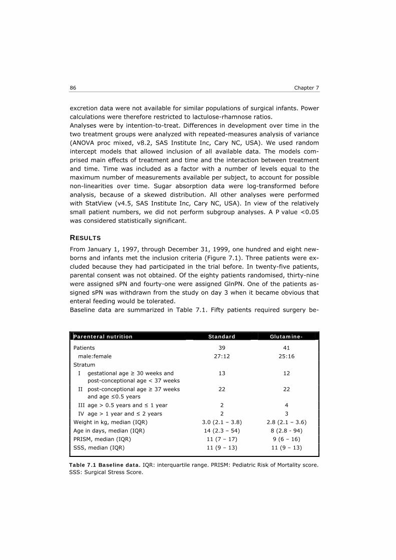
Chapter 7
86
excretion data were not available for similar populations of surgical infants. Power calculations were therefore restricted to lactulose-rhamnose ratios. Analyses were by intention-to-treat. Differences in development over time in the two treatment groups were analyzed with repeated-measures analysis of variance (ANOVA proc mixed, v8.2, SAS Institute Inc, Cary NC, USA). We used random intercept models that allowed inclusion of all available data. The models com-prised main effects of treatment and time and the interaction between treatment and time. Time was included as a factor with a number of levels equal to the maximum number of measurements available per subject, to account for possible non-linearities over time. Sugar absorption data were log-transformed before analysis, because of a skewed distribution. All other analyses were performed with StatView (v4.5, SAS Institute Inc, Cary NC, USA). In view of the relatively small patient numbers, we did not perform subgroup analyses. A P value <0.05 was considered statistically significant.
RESULTS
From January 1, 1997, through December 31, 1999, one hundred and eight new-borns and infants met the inclusion criteria (Figure 7.1). Three patients were ex-cluded because they had participated in the trial before. In twenty-five patients, parental consent was not obtained. Of the eighty patients randomised, thirty-nine were assigned sPN and fourty-one were assigned GlnPN. One of the patients as-signed sPN was withdrawn from the study on day 3 when it became obvious that enteral feeding would be tolerated. Baseline data are summarized in Table 7.1. Fifty patients required surgery be-
Parenteral nutrition Standard Glutamine- Patients 39 41
male:female 27:12 25:16
Stratum
I gestational age ≥ 30 weeks and post-conceptional age < 37 weeks
13 12
II post-conceptional age ≥ 37 weeks and age ≤0.5 years
22 22
III age > 0.5 years and ≤ 1 year 2 4
IV age > 1 year and ≤ 2 years 2 3
Weight in kg, median (IQR) 3.0 (2.1 – 3.8) 2.8 (2.1 – 3.6)
Age in days, median (IQR) 14 (2.3 – 54) 8 (2.8 - 94)
PRISM, median (IQR) 11 (7 – 17) 9 (6 – 16)
SSS, median (IQR) 11 (9 – 13) 11 (9 – 13)
Table 7.1 Baseline data. IQR: interquartile range. PRISM: Pediatric Risk of Mortality score. SSS: Surgical Stress Score.
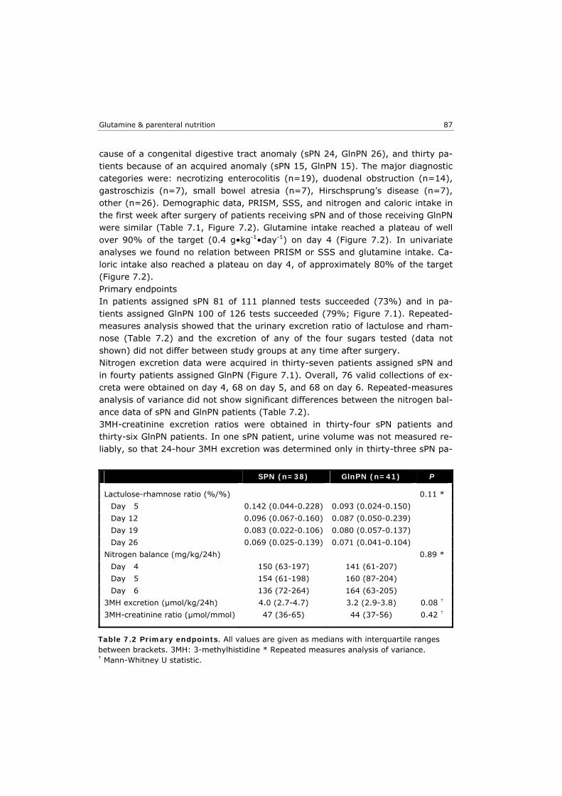
Glutamine & parenteral nutrition
87
cause of a congenital digestive tract anomaly (sPN 24, GlnPN 26), and thirty pa-tients because of an acquired anomaly (sPN 15, GlnPN 15). The major diagnostic categories were: necrotizing enterocolitis (n=19), duodenal obstruction (n=14), gastroschizis (n=7), small bowel atresia (n=7), Hirschsprung’s disease (n=7), other (n=26). Demographic data, PRISM, SSS, and nitrogen and caloric intake in the first week after surgery of patients receiving sPN and of those receiving GlnPN were similar (Table 7.1, Figure 7.2). Glutamine intake reached a plateau of well over 90% of the target (0.4 g•kg-1•day-1) on day 4 (Figure 7.2). In univariate analyses we found no relation between PRISM or SSS and glutamine intake. Ca-loric intake also reached a plateau on day 4, of approximately 80% of the target (Figure 7.2). Primary endpoints In patients assigned sPN 81 of 111 planned tests succeeded (73%) and in pa-tients assigned GlnPN 100 of 126 tests succeeded (79%; Figure 7.1). Repeated-measures analysis showed that the urinary excretion ratio of lactulose and rham-nose (Table 7.2) and the excretion of any of the four sugars tested (data not shown) did not differ between study groups at any time after surgery. Nitrogen excretion data were acquired in thirty-seven patients assigned sPN and in fourty patients assigned GlnPN (Figure 7.1). Overall, 76 valid collections of ex-creta were obtained on day 4, 68 on day 5, and 68 on day 6. Repeated-measures analysis of variance did not show significant differences between the nitrogen bal-ance data of sPN and GlnPN patients (Table 7.2). 3MH-creatinine excretion ratios were obtained in thirty-four sPN patients and thirty-six GlnPN patients. In one sPN patient, urine volume was not measured re-liably, so that 24-hour 3MH excretion was determined only in thirty-three sPN pa-
SPN (n=38) GlnPN (n=41) P
Lactulose-rhamnose ratio (%/%) 0.11 *
Day 5 0.142 (0.044-0.228) 0.093 (0.024-0.150)
Day 12 0.096 (0.067-0.160) 0.087 (0.050-0.239)
Day 19 0.083 (0.022-0.106) 0.080 (0.057-0.137)
Day 26 0.069 (0.025-0.139) 0.071 (0.041-0.104)
Nitrogen balance (mg/kg/24h) 0.89 *
Day 4 150 (63-197) 141 (61-207)
Day 5 154 (61-198) 160 (87-204)
Day 6 136 (72-264) 164 (63-205)
3MH excretion (µmol/kg/24h) 4.0 (2.7-4.7) 3.2 (2.9-3.8) 0.08 †
3MH-creatinine ratio (µmol/mmol) 47 (36-65) 44 (37-56) 0.42 †
Table 7.2 Primary endpoints. All values are given as medians with interquartile ranges between brackets. 3MH: 3-methylhistidine * Repeated measures analysis of variance. † Mann-Whitney U statistic.
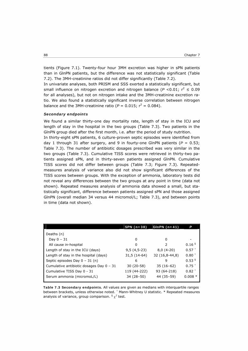
Chapter 7
88
tients (Figure 7.1). Twenty-four hour 3MH excretion was higher in sPN patients than in GlnPN patients, but the difference was not statistically significant (Table 7.2). The 3MH-creatinine ratios did not differ significantly (Table 7.2). In univariate analyses, both PRISM and SSS exerted a statistically significant, but small influence on nitrogen excretion and nitrogen balance (P <0.01; r2 ≤ 0.09 for all analyses), but not on nitrogen intake and the 3MH-creatinine excretion ra-tio. We also found a statistically significant inverse correlation between nitrogen balance and the 3MH-creatinine ratio (P = 0.015; r2 = 0.084).
Secondary endpoints
We found a similar thirty-one day mortality rate, length of stay in the ICU and length of stay in the hospital in the two groups (Table 7.3). Two patients in the GlnPN group died after the first month, i.e. after the period of study nutrition. In thirty-eight sPN patients, 6 culture-proven septic episodes were identified from day 1 through 31 after surgery, and 9 in fourty-one GlnPN patients (P = 0.53; Table 7.3). The number of antibiotic dosages prescribed was very similar in the two groups (Table 7.3). Cumulative TISS scores were retrieved in thirty-two pa-tients assigned sPN, and in thirty-seven patients assigned GlnPN. Cumulative TISS scores did not differ between groups (Table 7.3; Figure 7.3). Repeated-measures analysis of variance also did not show significant differences of the TISS scores between groups. With the exception of ammonia, laboratory tests did not reveal any differences between the two groups at any point in time (data not shown). Repeated measures analysis of ammonia data showed a small, but sta-tistically significant, difference between patients assigned sPN and those assigned GlnPN (overall median 34 versus 44 micromol/L; Table 7.3), and between points in time (data not shown).
SPN (n=38) GlnPN (n=41) P Deaths (n)
Day 0 – 31 0 0 -
All cause in-hospital 0 2 0.16 §
Length of stay in the ICU (days) 9,5 (4,5-23) 8,0 (4-20) 0.57 †
Length of stay in the hospital (days) 31,5 (14-64) 32 (16,8-44,8) 0.80 †
Septic episodes Day 0 – 31 (n) 6 9 0.53 §
Cumulative antibiotic dosages Day 0 – 31 30 (20-58) 35 (16–62) 0.75 †
Cumulative TISS Day 0 - 31 119 (44-222) 93 (64-218) 0.82 †
Serum ammonia (micromoL/L) 34 (28–50) 44 (35–59) 0.008 *
Table 7.3 Secondary endpoints. All values are given as medians with interquartile ranges between brackets, unless otherwise noted. † Mann-Whitney U statistic. * Repeated measures analysis of variance, group comparison. § χ2 test.
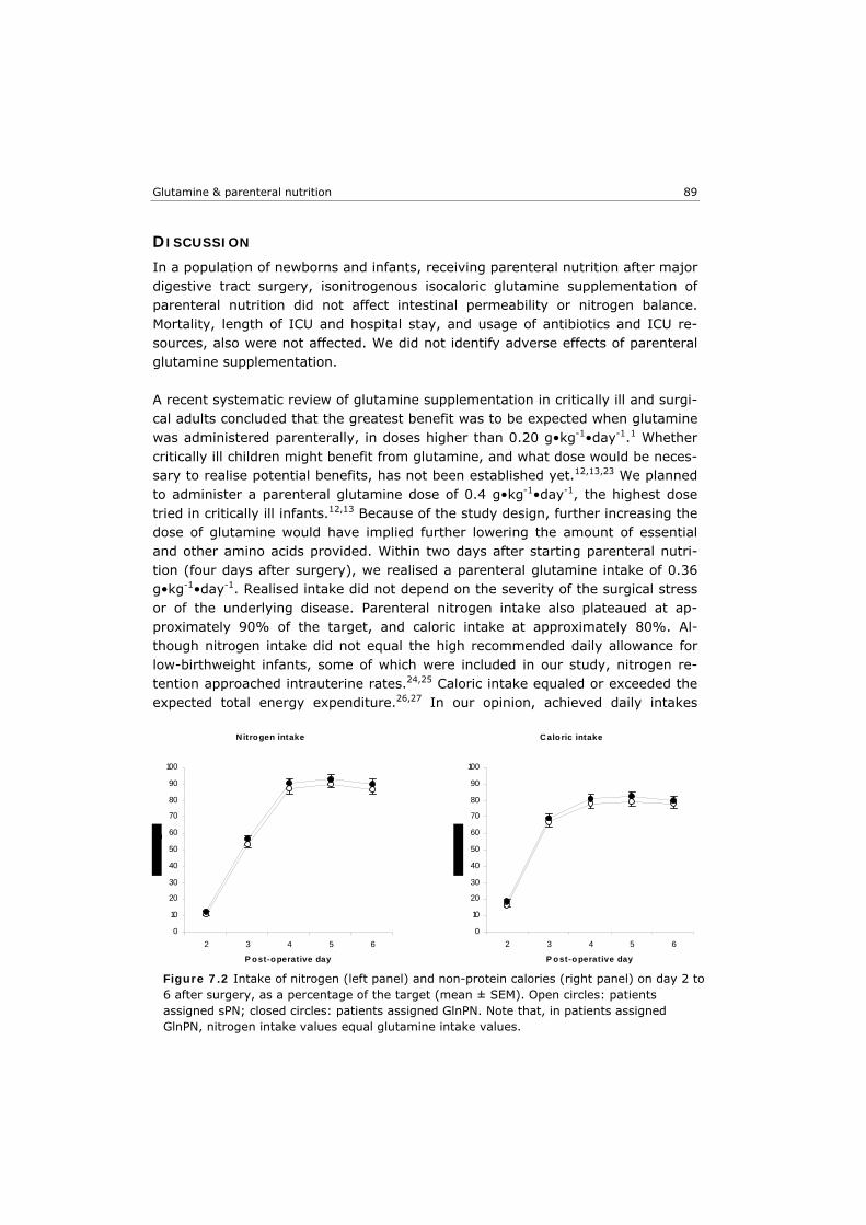
Glutamine & parenteral nutrition
89
DISCUSSION
In a population of newborns and infants, receiving parenteral nutrition after major digestive tract surgery, isonitrogenous isocaloric glutamine supplementation of parenteral nutrition did not affect intestinal permeability or nitrogen balance. Mortality, length of ICU and hospital stay, and usage of antibiotics and ICU re-sources, also were not affected. We did not identify adverse effects of parenteral glutamine supplementation. A recent systematic review of glutamine supplementation in critically ill and surgi-cal adults concluded that the greatest benefit was to be expected when glutamine was administered parenterally, in doses higher than 0.20 g•kg-1•day-1.1 Whether critically ill children might benefit from glutamine, and what dose would be neces-sary to realise potential benefits, has not been established yet.12,13,23 We planned to administer a parenteral glutamine dose of 0.4 g•kg-1•day-1, the highest dose tried in critically ill infants.12,13 Because of the study design, further increasing the dose of glutamine would have implied further lowering the amount of essential and other amino acids provided. Within two days after starting parenteral nutri-tion (four days after surgery), we realised a parenteral glutamine intake of 0.36 g•kg-1•day-1. Realised intake did not depend on the severity of the surgical stress or of the underlying disease. Parenteral nitrogen intake also plateaued at ap-proximately 90% of the target, and caloric intake at approximately 80%. Al-though nitrogen intake did not equal the high recommended daily allowance for low-birthweight infants, some of which were included in our study, nitrogen re-tention approached intrauterine rates.24,25 Caloric intake equaled or exceeded the expected total energy expenditure.26,27 In our opinion, achieved daily intakes
Nitrogen intake
0
10
20
30
40
50
60
70
80
90
100
2 3 4 5 6
P ost-operative day
Calo ric intake
0
10
20
30
40
50
60
70
80
90
100
2 3 4 5 6
P ost-operative day
Figure 7.2 Intake of nitrogen (left panel) and non-protein calories (right panel) on day 2 to 6 after surgery, as a percentage of the target (mean ± SEM). Open circles: patients assigned sPN; closed circles: patients assigned GlnPN. Note that, in patients assigned GlnPN, nitrogen intake values equal glutamine intake values.
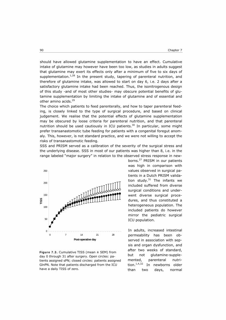
Chapter 7
90
should have allowed glutamine supplementation to have an effect. Cumulative intake of glutamine may however have been too low, as studies in adults suggest that glutamine may exert its effects only after a minimum of five to six days of supplementation.1,28 In the present study, tapering of parenteral nutrition, and therefore of glutamine intake, was allowed to start on day 6, i.e. 2 days after a satisfactory glutamine intake had been reached. Thus, the isonitrogenous design of this study -and of most other studies- may obscure potential benefits of glu-tamine supplementation by limiting the intake of glutamine and of essential and other amino acids.29 The choice which patients to feed parenterally, and how to taper parenteral feed-ing, is closely linked to the type of surgical procedure, and based on clinical judgement. We realise that the potential effects of glutamine supplementation may be obscured by loose criteria for parenteral nutrition, and that parenteral nutrition should be used cautiously in ICU patients.30 In particular, some might prefer transanastomotic tube feeding for patients with a congenital foregut anom-aly. This, however, is not standard practice, and we were not willing to accept the risks of transanastomotic feeding. SSS and PRISM served as a calibration of the severity of the surgical stress and the underlying disease. SSS in most of our patients was higher than 8, i.e. in the range labeled “major surgery” in relation to the observed stress response in new-
borns.27 PRISM in our patients was high in comparison with values observed in surgical pa-tients in a Dutch PRISM valida-tion study.31 The infants we included suffered from diverse surgical conditions and under-went diverse surgical proce-dures, and thus constituted a heterogeneous population. The included patients do however mirror the pediatric surgical ICU population.
In adults, increased intestinal permeability has been ob-served in association with sep-sis and organ dysfunction, and after two weeks of standard, but not glutamine-supple-mented, parenteral nutri-tion.1,4,32 In newborns older than two days, normal
0
50
100
150
200
250
0 7 14 21 28
Post-operative day
TISS
Figure 7.3. Cumulative TISS (mean ± SEM) from day 0 through 31 after surgery. Open circles: pa-tients assigned sPN; closed circles: patients assigned GlnPN. Note that patients discharged from the ICU have a daily TISS of zero.
Glutamine & parenteral nutrition
91
intestinal permeability is not related to gestational age or birth weight.33 We have previously reported increased intestinal permeability in critically ill preterm infants operated because of necrotizing enterocolitis.18 Permeability normalized in several of these infants, even though total parenteral nutrition was continued for three weeks.18 In newborns on extracorporeal membrane oxygenation, we found intestinal permeability to be increased, irrespective of whether they received parenteral or enteral nutrition.34 In the present study, lactulose-rhamnose ratios remained elevated for up to four weeks in the majority of patients. Also, intestinal permeability in patients who received total parenteral nutrition was not statistically different from that in patients who received enteral nutrition (data not shown). These findings suggest that not parenteral nutrition per se, but rather the underlying disease, alone or in combination with surgery, causes increased intestinal permeability. Resting energy expenditure and (nor)epinephrine levels in newborn patients in-crease in response to the stress of surgery, but normalize again within 24 hours.27,35 Preterm infants may retain nitrogen in the first seventy-two hours after surgery.36 In the present study, the severity of the surgical stress and the under-lying disease had limited effects on nitrogen excretion and nitrogen balance, and most patients retained adequate amounts of nitrogen on day 4 to 6.24 These findings add to the evidence that the stress response in young infants may be short-lived. Nevertheless, the urinary 3MH-creatinine ratio and 3MH excretion were increased, suggesting increased breakdown of muscle protein.37 Increased breakdown of muscle protein in the face of nitrogen retention may indicate re-routing of nitrogen fluxes for wound healing, or for the defense against infec-tion.38 Alternatively, persistently increased breakdown of muscle protein might be accompanied by increased synthesis and thus reflect increased turnover, and not loss of muscle protein. In adults with systemic inflammatory response syndrome, enteral glutamine sup-plementation reduced the number of nosocomial infections, even though nitrogen balance and intestinal permeability were not different.9 In very-low-birthweight-children however, the rate of suspected and culture-proven nosocomial sepsis was not affected by enteral glutamine supplementation.13 In our population, parenteral glutamine supplementation did not change the incidence of culture-proven sepsis. We also analysed antibiotic usage and cumulative TISS scores, because we argued that a reduction of the rate or severity of any infection, not just sepsis, would be clinically relevant if it resulted in reduced usage of antibiotics or resources. We found no difference in antibiotic usage between groups, and no difference in TISS. TISS does not assess resource usage outside the ICU. Nonetheless, we did not demonstrate an effect of glutamine supple-mentation on ICU resource usage.
Chapter 7
92
Studies in critically ill adults and children suggest that glutamine supplementation is safe.1,10,11,13,39-41 We found no difference in 31-day mortality between patients assigned sPN or GlnPN. Two in-hospital deaths occurred in the group assigned GlnPN, but only after the study nutrition had been replaced with standard par-enteral nutrition. Glutamine supplementation is unlikely to have played a role in these deaths. It should be noted that our study was not designed to detect differ-ences in mortality. As mortality in pediatric surgical intensive care populations generally is very low, this would have required a very large trial.31 Serum ammo-nia was higher in glutamine-supplemented infants, but the difference was not clinically relevant (Table 7.3). In view of the promising results in adults, more research into the effects of glu-tamine supplementation in infants and children seems justified.23,29 Currently however, our findings suggest that parenteral glutamine supplementation for sur-gical infants is not warranted.
ACKNOWLEDGEMENTS
This study was funded by an educational grant from the Erasmus Medical Center’s Clinical Research Board (Commissie Patient-gebonden Onderzoek) and by a grant from Pharmacia & Upjohn BV, Woerden, The Netherlands. The authors wish to thank the staff of the pediatric surgical intensive care unit and the hospital phar-macy for their help in carrying out the study.
Glutamine & parenteral nutrition
93
REFERENCES 1. Novak F, Heyland DK, Avenell A, Drover JW, Su X. Glutamine supplementation in seri-
ous illness: a systematic review of the evidence. Crit Care Med 2002;30:2022-9.
2. Stehle P, Zander J, Mertes N, et al. Effect of parenteral glutamine peptide supplements on muscle glutamine loss and nitrogen balance after major surgery. Lancet 1989;1: 231-3.
3. Ziegler TR, Young LS, Benfell K, et al. Clinical and metabolic efficacy of glutamine-sup-plemented parenteral nutrition after bone marrow transplantation. Ann Intern Med 1992;116:821-8.
4. van der Hulst RRWJ, van Kreel BK, von Meyenfeldt MF, et al. Glutamine and the pres-ervation of gut integrity. Lancet 1993;341:1363-5.
5. Karwowska KA, Dworacki G, Trybus M, Zeromski J, Szulc R. Influence of glutamine-en-riched parenteral nutrition on nitrogen balance and immunologic status in patients un-dergoing elective aortic aneurysm repair. Nutrition 2001;17:475-8.
6. Boelens PG, Houdijk AP, Fonk JC, et al. Glutamine-enriched enteral nutrition increases HLA-DR expression on monocytes of trauma patients. J Nutr 2002;132:2580-6.
7. Piccirillo N, De Matteis S, Laurenti L, et al. Glutamine-enriched parenteral nutrition after autologous peripheral blood stem cell transplantation: effects on immune reconstitution and mucositis. Haematologica 2003;88:192-200.
8. Schloerb PR, Amare M. Total parenteral nutrition with glutamine in bone marrow trans-plantation and other clinical applications (a randomized, double-blind study). JPEN J Parenter Enteral Nutr 1993;17:407-13.
9. Conejero R, Bonet A, Grau T, et al. Effect of a glutamine-enriched enteral diet on intes-tinal permeability and infectious morbidity at 28 days in critically ill patients with sys-temic inflammatory response syndrome: a randomized, single-blind, prospective, multi-center study. Nutrition 2002;18:716-21.
10. Lacey JM, Crouch JB, Benfell K, et al. The effects of glutamine-supplemented parenteral nutrition in premature infants. JPEN J Parenter Enteral Nutr 1996;20:74-80.
11. Neu J, Roig JC, Meetze WH, et al. Enteral glutamine supplementation for very low birth weight infants decreases morbidity. J Pediatr 1997;131:691-9.
12. Tubman TRJ, Thompson SW. Glutamine supplementation for prevention of morbidity in preterm infants (Cochrane review). In: The Cochrane Library. Oxford: Update software; 2003.
13. Vaughn P, Thomas P, Clark R, Neu J. Enteral glutamine supplementation and morbidity in low birth weight infants. J Pediatr 2003;142:662-8.
14. Dallas MJ, Bowling D, Roig JC, Auestad N, Neu J. Enteral glutamine supplementation for very-low-birth-weight infants decreases hospital costs. JPEN J Parenter Enteral Nutr 1998;22:352-6.
15. Pollack MM, Ruttimann UE, Getson PR. Pediatric risk of mortality (PRISM) score. Crit Care Med 1988;16:1110-6.
16. Anand KJ, Aynsley-Green A. Measuring the severity of surgical stress in newborn in-fants. J Pediatr Surg 1988;23:297-305.
17. Allen SJ, Pierro A, Cope L, et al. Glutamine-supplemented parenteral nutrition in a child with short bowel syndrome. J Pediatr Gastroenterol Nutr 1993;17:329-32.
18. Piena-Spoel M, Albers MJ, Ten Kate J, Tibboel D. Intestinal permeability in newborns with necrotizing enterocolitis and controls: Does the sugar absorption test provide guidelines for the time to (re-)introduce enteral nutrition? J Pediatr Surg 2001;36:587-92.
Chapter 7
94
19. Albers MJ, Steyerberg EW, Rietveld T, Tibboel D. Clinical relevancy of non-urinary ni-trogen excretion in newborns and infants after digestive tract surgery. JPEN J Parent Enteral Nutr 2003;27:327-32.
20. Garner JS, Jarvis WR, Emori TG, Horan TC, Hughes JM. CDC definitions for nosocomial infections, 1988 [published erratum appears in Am J Infect Control 1988 Aug;16(4):177]. Am J Infect Control 1988;16:128-40.
21. Keene AR, Cullen DJ. Therapeutic Intervention Scoring System: update 1983. Crit Care Med 1983;11:1-3.
22. Beach RC, Menzies IS, Clayden GS, Scopes JW. Gastrointestinal permeability changes in the preterm neonate. Arch Dis Child 1982;57:141-5.
23. Ball PA, Hardy G. Glutamine in pediatrics: where next? Nutrition 2002;18:451-4.
24. Anderson MS, Hay Jr WW. Intrauterine growth restriction and the small-for-gestational-age infant. In: Avery GB, Fletcher MA, MacDonald MG, editors. Neonatology - Patho-physiology and management of the newborn. 5th ed. Philadelphia: Lippincott Williams & Wilkins; 1999. p. 411-44.
25. Georgieff MK. Nutrition. In: Avery GB, Fletcher MA, MacDonald MG, editors. Neonatol-ogy - Pathophysiology and management of the newborn. 5th ed. Philadelphia: Lippin-cott Williams & Wilkins; 1999. p. 363-94.
26. Pierro A, Carnielli V, Filler RM, Kicak L, Smith J, Heim TF. Partition of energy metabo-lism in the surgical newborn. J Pediatr Surg 1991;26:581-6.
27. Jones MO, Pierro A, Hammond P, Lloyd DA. The metabolic response to operative stress in infants. J Pediatr Surg 1993;28:1258-62; discussion 62-63.
28. van Leeuwen PA. A minimum of 5 days of feeding. Nutrition 2002;18:715.
29. Neu J. Glutamine supplements in premature infants: why and how. J Pediatr Gastro-enterol Nutr 2003;37:533-5.
30. Varga P, Griffiths R, Chiolero R, et al. Is parenteral nutrition guilty? Intensive Care Med 2003;29:1861-4.
31. Gemke RJ, Bonsel GJ, van Vught AJ. Effectiveness and efficiency of a Dutch pediatric intensive care unit: validity and application of the Pediatric Risk of Mortality score. Crit Care Med 1994;22:1477-84.
32. Johnston JD, Harvey CJ, Menzies IS, Treacher DF. Gastrointestinal permeability and absorptive capacity in sepsis. Crit Care Med 1996;24:1144-9.
33. van Elburg RM, Fetter WP, Bunkers CM, Heymans HS. Intestinal permeability in relation to birth weight and gestational and postnatal age. Arch Dis Child Fetal Neonatal Ed 2003;88:F52-5.
34. Piena M, Albers MJ, Van Haard PM, Gischler S, Tibboel D. Introduction of enteral feeding in neonates on extracorporeal membrane oxygenation after evaluation of intestinal permeability changes. J Pediatr Surg 1998;33:30-4.
35. Bouwmeester NJ, Anand KJ, van Dijk M, Hop WC, Boomsma F, Tibboel D. Hormonal and metabolic stress responses after major surgery in children aged 0-3 years: a double-blind, randomized trial comparing the effects of continuous versus intermittent mor-phine. Br J Anaesth 2001;87:390-9.
36. Duffy B, Pencharz P. The effects of surgery on the nitrogen metabolism of parenterally fed human neonates. Pediatr Res 1986;20:32-5.
37. Parvy PR, Bardet JI, Rabier DM, Kamoun PP. Age-related reference values for free amino acids in first morning urine specimens. Clin Chem 1988;34:2092-5.
38. Patterson BW, Nguyen T, Pierre E, Herndon DN, Wolfe RR. Urea and protein metabolism in burned children: effect of dietary protein intake. Metabolism 1997;46:573-8.
Glutamine & parenteral nutrition
95
39. Thompson SW, McClure BG, Tubman TR. A randomized, controlled trial of parenteral glutamine in ill, very low birth-weight neonates. J Pediatr Gastroenterol Nutr 2003;37:550-3.
40. Coghlin Dickson TM, Wong RM, offrin RS, et al. Effect of oral glutamine supplementation during bone marrow transplantation. JPEN J Parenter Enteral Nutr 2000;24:61-6.
41. Long CL, Nelson KM, DiRienzo DB, et al. Glutamine supplementation of enteral nutri-tion: impact on whole body protein kinetics and glucose metabolism in critically ill pa-tients. JPEN J Parenter Enteral Nutr 1995;19:470-6.


8 Summary, discussion, and future perspectives

Summary, discussion & future perspectives
99
SUMMARY
In the setting of critical illness, major surgery and poor gastrointestinal function, the choice how to provide nutritional support is delicate. This thesis describes studies on nutritional support in newborns and infants who required surgical treatment for diseases entailing poor gastrointestinal function. In Chapter 2 we compared the urinary excretion ratios of enterally administered lactulose and rhamnose -a measure of intestinal permeability- obtained in thir-teen preterm newborns who underwent surgery because of necrotizing enteroco-litis, with those obtained in ten newborns who underwent surgery for other rea-sons. In patients with necrotizing enterocolitis, intestinal hyperpermeability gen-erally persisted throughout the first week after surgery. In some patients, intesti-nal permeability started to normalise in the second to third week after surgery, whereas in others hyperpermeability persisted throughout the third week. In-creases of intestinal permeability occurring in the second or third week after sur-gery coincided with episodes of sepsis and bowel obstruction. In control patients, intestinal permeability started to normalise in the first week after surgery. We concluded that intestinal hyperpermeability persisting beyond the first week after surgery should be attributed to the underlying disease, i.e. to necrotizing entero-colitis. Customising the (re)introduction of enteral nutrition seemed justified, but should not be based solely upon the lactulose-rhamnose ratio. In Chapter 3 we used the same measure of intestinal permeability, combined with measures of carrier-mediated transport, in newborns treated with extracor-poreal membrane oxygenation. We compared seven patients who received enteral nutrition with nine patients who received total parenteral nutrition. In thirteen of these sixteen patients, intestinal permeability was increased. Urinary D-xylose excretion, a measure of passive carrier-mediated transport, was normal in both groups of patients. Urinary 3-O-methyl-D-glucose excretion, a measure of active carrier-mediated transport, was decreased in both groups of patients and did not differ between the two groups. Neither the lactulose-rhamnose excretion ratios, nor any of the other parameters, changed after introduction of enteral nutrition. We concluded that intestinal permeability and transport were compromised in all patients, irrespective of feeding route, and that the introduction of enteral nutri-tion did not result in further deterioration. Our findings did not support the clinical practice of withholding enteral nutrition from patients treated with extracorporeal membrane oxygenation. In Chapter 4 we assessed the effect of the route of feeding on the incidence of septic complications in newborns treated with extracorporeal membrane oxy-genation. The frequency of septic complications observed in sixteen patients re-ceiving enteral nutrition did not differ from that observed in thirty-five patients receiving total parenteral nutrition, even though extracorporeal membrane oxy-genation lasted longer in the group of patients receiving enteral nutrition. Of 11 septic complications in which a microorganism was identified, 8 were caused by
Chapter 8
100
coagulase-negative staphylococci. Therefore, they were considered not to be re-lated to intestinal integrity, but to the use of central venous catheters and arterial and venous cannulas. We did not identify adverse effects of enteral nutrition. We once more concluded that our findings did not support the withholding of enteral nutrition from newborns treated with extracorporeal membrane oxygenation. In Chapter 5 we described a cohort of newborns with an intrinsic intestinal anomaly who received parenteral nutrition for at least 7 days following surgery. We compared the incidence of sepsis in twenty-six patients who developed par-enteral nutrition-associated cholestasis with that in seventy-two patients who didn’t. Although the incidence of sepsis was somewhat higher in patients who did develop cholestasis than in patients who didn’t, overall sepsis incidence was low (9 per 1000 hospital days). Of the 56 microorganisms that were cultured, 37 were coagulase-negative staphylococci. As noted earlier, infections caused by these microorganisms are thought to be related to vascular access techniques, and not to intestinal pathology. Parenteral nutrition-associated cholestasis oc-curred and progressed without a septic event in thirteen out of twenty-six pa-tients. Both sepsis and cholestasis occurred more frequently in male than in fe-male newborns. We concluded that sepsis is an epiphenomenon of parenteral nu-trition-associated cholestasis rather than a causative factor. In Chapter 6 we compared the contribution of nonurinary nitrogen excretion and urinary nitrogen excretion to total nitrogen excretion in seventy-eight newborns and infants who were receiving total parenteral nutrition 4 to 6 days after gas-trointestinal surgery. Both nonurinary and urinary nitrogen excretion, and their ratio, varied widely. Relatively simple predictive equations yielded reasonable es-timates of nonurinary nitrogen excretion, but considerable imprecision remained in individual measurements. We concluded that nonurinary nitrogen excretion should not be neglected, and that total nitrogen excretion could only be accu-rately assessed in the individual patient by measuring the nitrogen content of all excreta. In Chapter 7 we described the effects of isonitrogenous isocaloric glutamine sup-plementation of parenteral nutrition in newborns and infants who underwent ma-jor gastrointestinal surgery. In a double-blind randomized controlled trial, we compared fourty-one patients who received glutamine-supplemented parenteral nutrition with thirty-nine patients who received standard parenteral nutrition. In most patients, intestinal permeability was increased after surgery and remained increased during the study period. Glutamine supplementation did not have a measurable effect on intestinal permeability, nitrogen balance, the incidence of septic events, and usage of antibiotics and ICU resources, nor did it affect mor-tality and length of ICU and hospital stay. We did not identify any beneficial effect of glutamine supplementation of total parenteral nutrition, and concluded it was not warranted in these patients.
Summary, discussion & future perspectives
101
DISCUSSION
There is growing consensus that critically ill medical and surgical patients may benefit from nutritional support and that the gastrointestinal tract should be used for nutritional support whenever possible.1-8 The focus of nutritional research in critically ill adults has shifted to the potential benefits of early compared with de-layed enteral nutrition.7,9 Likewise, in pediatric ICU patients, how best to provide enteral nutrition now seems to be the main topic of interest.10-14 Nonetheless, one meta-analysis in surgical and critically ill adults and two recent randomised pro-spective trials in adults, one of them including patients with severe sepsis, sug-gest parenteral nutrition may be superior to enteral nutrition in selected groups of patients.5,15,16 In one of these trials, 41% of 562 adults were deemed to have a functioning gastrointestinal tract, and were therefore given enteral nutrition by the attending physican. Permanent or temporary intestinal failure was diagnosed in 48% of patients, who were then given total parenteral nutrition. In 11% of patients, there was doubt as to the adequacy of gastrointestinal function; the authors concluded that this subgroup should also be given parenteral nutrition.5 Fewer than 6% of the subgroup of non-surgical patients required total parenteral nutrition, versus 55% of postoperative patients.5 In a recent retrospective study of ninety-five pediatric ICU patients, seventeen of whom had undergone gastro-intestinal surgery (orthotopic liver transplantation), fifty-six patients were given nutritional support: fourty-six patients (82%) received enteral nutrition, seven patients (13%) received total parenteral nutrition, and three patients (5%) re-ceived a combination of parenteral and enteral nutrition.8 These data illustrate that enteral nutrition may not always be superior to parenteral nutrition, and that a substantial number of patients are being given parenteral nutrition. Also of note, the decision that gastrointestinal function was “adequate”, and that par-enteral nutrition was not required, was based solely on clinical experience.5 Poor function and non-function of the gastrointestinal tract constitute indications for parenteral nutrition.1 Within the context of critical illness, gastrointestinal function has been ill-defined. Emphasis has been put on the barrier function of the gastrointestinal tract, because of the association of increased intestinal per-meability with severity of disease, ‘gut-derived’ sepsis and multiple organ fail-ure.17,18 Intestinal permeability itself, however, is a complex notion, and hyper-permeability to molecular probes, such as lactulose and rhamnose, is not neces-sarily associated with increased permeability to bacteria or bacterial prod-ucts.7,19,20 The reverse may also hold true: after the second day of life, intestinal permeability to molecular probes in enterally fed preterm neonates did not differ from that of healthy term neonates,21 but advancing enteral feedings in preterm neonates was found to increase the incidence of necrotizing enterocolitis when compared to minimal enteral feeding.22 The findings we presented in Chapter 2 through 4 of this thesis lend further support to the idea that intestinal perme-
Chapter 8
102
ability is multifaceted. In Chapter 2 we showed that intestinal hyperpermeability may persist throughout the third week of necrotizing enterocolitis. Yet, restarting enteral feeding after a median of 4 days from the onset of necrotizing enterocoli-tis has been associated with significant benefits and no apparent adverse effects, when compared to restarting after 10 days.23 In Chapter 3 and Chapter 4 we showed that enteral nutrition was well tolerated by a group of critically ill term neonates treated with extracorporeal membrane oxygenation, and not associated with adverse effects, even though intestinal permeability to molecular probes was increased and these neonates were at risk for intestinal ischemia. Irrespective of feeding route, active carrier-mediated transport over the intestinal wall was also affected, whereas passive carrier-mediated transport in these patients was nor-mal, again attesting to the complexity of gastrointestinal permeability. It is also apparent from these data that markers of intestinal permeability like the ones used in Chapter 2 and Chapter 3 are of little or no help in deciding whether or not to provide enteral nutrition to critically ill patients. Enteral nutrition had no measurable effect on intestinal permeability in the criti-cally ill neonates described in Chapter 3 and Chapter 4, but has been shown to normalise intestinal permeability to molecular probes in other ICU patients.7 Moreover, as was shown in parenterally fed surgical infants, small volumes of enteral nutriton may suffice to normalise immune function, and by doing so, im-prove gut barrier function.24 In postoperative cardiac surgery patients with circu-latory compromise, enteral nutrition may increase cardiac index and splanchnic blood flow.25 These and other effects of the presence of nutrients in the gut lumen may be regarded as what constitutes the difference between parenteral nutrition and total parenteral nutrition. Many of the complications of parenteral nutrition, however, occur in patients who receive a combination of enteral and parenteral nutrition. These complications therefore are related to the composition of the parenteral mixture, to the way it is administered, to the disease or condition for which parenteral nutrition support is required, or to a combination of the aforementioned. The composition of par-enteral nutrition has traditionally been determined on the basis of recommended daily allowances of macronutrients and micronutrients, and potential toxicity of individual constituents.26 More recently, the concept of immunonutrition or phar-maconutrition has been introduced to delineate non-nutritive effects of specific nutrients. The awareness that certain nutrients, when supplied to the right pa-tients at the right time and in the right, frequently supraphysiological, doses, may favourably affect the immune response, has resulted in several prospective trials that explored the potential benefits of these nutrients.27 Promising results in adult patients undergoing surgery28,29 have been offset by the finding that enteral immunonutrition containing arginine increases mortality in critically ill adults.16,28 Glutamine has been added to enteral as well as parenteral mixtures, and has been associated with beneficial effects.29 A recent meta-analysis concluded that a
Summary, discussion & future perspectives
103
benefit from glutamine may exist for surgical and critically ill adults, but that these two groups should be studied separately to decide whether these effects are clinically relevant.29 The findings we presented in Chapter 7 should be viewed in this light. Whereas we did not identify beneficial effects of parenteral glutamine supplementation in surgical infants, further research is needed to discriminate potential effects on the response to surgery from effects on the underlying dis-ease. The study described in Chapter 7 serves to illustrate another issue pertinent to the complications of parenteral nutrition. In keeping with other studies, use of the parenteral route made it possible to realise a nitrogen and caloric intake close to the predefined target. Enteral nutrition, on the other hand, frequently results in intakes far below target.3,5,6,8,14 Target intakes in our study and in many other studies, were based on recommended daily allowances for healthy individuals, and were not adapted to critical illness or individual requirements, because the relevant guidelines are not available. Measurements of energy expenditure in children after gastrointestinal and cardiopulmonary bypass surgery and in pedia-tric ICU patients and children with sepsis and systemic inflammatory response syndrome, have yielded values lower than or equal to those observed in normal children.30-34 Furthermore, considerable variability of energy expenditure exists within these populations.31,33-35 In Chapter 6 we found nonurinary, urinary and total nitrogen excretion to vary widely in surgical infants receiving total parenteral nutrition. These findings corroborate earlier studies in children that found postoperative nitrogen balance to vary widely, and lend further support to the notion that nutritional requirements show large variations between individuals.36,37 From these joint observations it follows that overfeeding is likely to occur in criti-cally ill children and is even more likely to occur if nutritional support is given via the parenteral route. Symptoms of overfeeding critically ill patients include hy-perglycemia.38 Hyperglycemia is part of the metabolic response to critical illness and major surgery, but is also more common in adult ICU patients receiving par-enteral nutrition than in those receiving enteral nutrition.39 In adult diabetics, postoperative hyperglycemia has been associated with a five-fold increased rate of nosocomial infections,40 and in surgical ICU patients, strict control of blood glu-cose levels with insulin dramatically reduced ICU and in-hospital mortality, largely because of a lower incidence of sepsis-induced multiple organ failure.41 Although in one trial lowering the amount of calories provided as parenteral nutriton did not reduce the incidence of hyperglycemia and infections,42 some have argued that trials demonstrating beneficial effects of enteral nutrition in ICU patients were poorly-controlled and in fact demonstrated the deleterious effects of over-feeding and hyperglycemia in patients receiving parenteral nutrition.43 Several studies in pediatric ICU patients have documented symptoms that may be indica-tive of overfeeding, notably high respiratory quotients obtained by indirect calo-
Chapter 8
104
rimetry.31,34,35 However, these data have not been analysed with the aim to pro-vide insight in the incidence and severity of overfeeding or hyperglycemia in pedi-atric ICU patients in relation to the route of nutrition. One of the main reasons analyses like these have not been done may be the lack of readily applicable, sensitive techniques to assess the nutritional status of critically ill children and short-term changes of this status.44,45 Sepsis is a potentially life-threatening complication for all patients receiving par-enteral nutrition. In the setting of critical illness (Chapter 4) as well as in the setting of long-term parenteral nutrition (Chapter 5), we found the majority of septic events to be caused by coagulase-negative staphylococci. Bloodstream in-fections by coagulase-negative staphylococi are closely related to the use of cen-tral venous catheters.46,47 Catheter-related bloodstream infections, in turn, are among the leading causes of nosocomial infections in intensive care units.46,48,49 Infection-prevention guidelines and catheter insertion and maintenance protocols have been shown to lower the rate of catheter-related infections in critically ill patients and in patients who receive parenteral nutrition.46,48,50 Our findings un-derscore the importance of strict adherence to these guidelines and protocols. Parenteral nutrition-associated cholestasis may over time develop into terminal liver failure and thereby is a potentially life-threatening complication for patients who remain dependent on parenteral nutrition for months or years. The relation of sepsis and parenteral nutrition-associated cholestasis has been discussed in Chapter 5. The finding that sex affects the incidence of sepsis in infants receiving parenteral nutrition, however, merits discussion in a broader context. As dis-cussed in Chapter 5, male sex has been identified as a risk factor for sepsis and nosocomial pneumonia in adult ICU patients.51,52 In adults, the age-related sepsis mortality rates for men are slightly higher than for women, but these differences are explained by differences in comorbidity and site of infection.53 A recent survey of severe pediatric sepsis found the incidence and the mortality rate of sepsis to be higher in male than in female infants.54 In children aged 1 to 9 years, the inci-dence of sepsis was higher in boys than girls, but mortality rates were similar.54 These findings should be taken into account when interpreting the findings of studies on nutrition and gastrointestinal function in the setting of critical illness and major surgery; particularly when the numbers of patients included are small, which often is the case, sex and age distribution inequalities may heavily influ-ence the results. One of the first patients to benefit from parenteral nutrition was an infant with an intestinal anomaly.55 Today, parenteral nutrition still is of vital importance to the care of infants with a poor or non-functioning gastrointestinal tract. Over the years, the use of parenteral nutrition has become increasingly safe, and new in-sights and vascular access techniques are likely to make it even safer. Still, the
Summary, discussion & future perspectives
105
complications associated with the use of parenteral nutrition are well-defined and potentially life-threatening. The use of enteral nutrition, though generally consid-ered feasible, safe and less costly, is not without complications either. Particularly in the setting of poor gastrointestinal function, major surgery and critical illness, the decision how to provide nutritional support may be difficult. Gastrointestinal function has so far proved too complex to identify easily applicable, sensitive markers that help the clinician make this decision. For lack of randomised trials on nutritional support in surgical infants, clinical experience remains the basis of decision-making. Clinical experience, however, has not always been a good coun-sellor, and much more fundamental and clinical research are needed to arrive at nutritional support strategies for surgical infants that meet standards of efficacy, safety and cost-effectiveness.
FUTURE PERSPECTIVES
The complexity of the interaction of nutrition and gastrointestinal function, and the potential of nutritional support to improve the outcome of critical illness, jus-tify the allocation of resources to further knowledge in this field. Fundamental re-search will have to clarify the significance of poor gastrointestinal function in criti-cal illness and its relation with ‘gut-derived’ sepsis and multiple organ failure, to identify immunonutritive effects of individual nutrients, including trophic effects on the gastrointestinal tract, and to clarify the mechanisms of the interaction of sepsis, sex and cholestasis. Of particular importance to the setting of pediatric critical illness and pediatric surgery, however, is the initiation of clinical research that seeks to translate fundamental insights into clinically applicable strategies for nutrition support. The research on the nonnutritive effects of glutamine may serve to illustrate this point. An abundance of animal experiments and several prospective randomised trials in critically ill adult patients suggest that glutamine may exert beneficial effects in selected patients, but the evidence is judged insuf-ficient to include glutamine supplementation in adult guidelines for nutritional support.29 Promising results of enteral glutamine supplementation in prospective, but small, and not very well controlled, trials in low birthweight infants have sub-sequently been refuted by one large, well controlled multi-center trial.56 The studies on intestinal permeability that we presented in this thesis illustrate the drawbacks of using surrogate endpoints instead of hard endpoints, like mortality, sepsis incidence, resource usage and length of ICU and hospital stay. In view of the necessary homogeneity of the study population, the limited numbers of pa-tients with a specific disease, the variability of the natural course of the diseases of interest, and the relatively low incidence of relevant hard endpoints in the pe-diatric population, designing and executing the multi-center prospective trials needed to test the notions discussed earlier and to improve nutritional support will truly be a challenge.
Chapter 8
106
REFERENCES 1. Baker SS. Indications for parenteral nutrition. In: Baker Jr. RD, Baker SS, Davis AM,
editors. Pediatric parenteral nutrition. New York: Chapman & Hall; 1997. p. 18-30.
2. Kalfarentzos F, Kehagias J, Mead N, Kokkinis K, Gogos CA. Enteral nutrition is superior to parenteral nutrition in severe acute pancreatitis: results of a randomized prospective trial. Br J Surg 1997;84:1665-9.
3. Braga M, Gianotti L, Gentilini O, Parisi V, Salis C, Di Carlo V. Early postoperative enteral nutrition improves gut oxygenation and reduces costs compared with total parenteral nutrition. Crit Care Med 2001;29:242-8.
4. Lewis SJ, Egger M, Sylvester PA, Thomas S. Early enteral feeding versus "nil by mouth" after gastrointestinal surgery: systematic review and meta-analysis of controlled trials. BMJ 2001;323:773.
5. Woodcock NP, Zeigler D, Palmer MD, Buckley P, Mitchell CJ, MacFie J. Enteral versus parenteral nutrition: a pragmatic study. Nutrition 2001;17:1-12.
6. Abou-Assi S, Craig K, O'Keefe SJ. Hypocaloric jejunal feeding is better than total par-enteral nutrition in acute pancreatitis: results of a randomized comparative study. Am J Gastroenterol 2002;97:2255-62.
7. van Haren FMP, van der Hoeven JG. Early enteral nutrition in the intensive care unit. In: Vincent JL, editor. Yearbook of intensive care and emergency medicine 2002. Berlin: Springer-Verlag; 2002. p. 481-91.
8. Taylor RM, Preedy VR, Baker AJ, Grimble G. Nutritional support in critically ill children. Clin Nutr 2003;22:365-9.
9. Marik PE, Zaloga GP. Early enteral nutrition in acutely ill patients: a systematic review. Crit Care Med 2001;29:2264-70.
10. Chellis MJ, Sanders SV, Webster H, Dean JM, Jackson D. Early enteral feeding in the pediatric intensive care unit. JPEN J Parenter Enteral Nutr 1996;20:71-3.
11. Panadero E, López-Herce J, Caro L, et al. Transpyloric enteral feeding in critically ill children. J Pediatr Gastroenterol Nutr 1998;26:43-8.
12. Briassoulis GC, Zavras NJ, Hatzis MT. Effectiveness and safety of a protocol for promo-tion of early intragastric feeding in critically ill children. Pediatr Crit Care Med 2001;2:113-21.
13. Horn D, Chaboyer W. Gastric feeding in critically ill children: a randomized controlled trial. Am J Crit Care 2003;12:461-8.
14. Rogers EJ, Gilbertson HR, Heine RG, Henning R. Barriers to adequate nutrition in criti-cally ill children. Nutrition 2003;19:865-8.
15. Heyland DK, MacDonald S, Keefe L, Drover JW. Total parenteral nutrition in the critically ill patient: a meta- analysis [see comments]. JAMA 1998;280:2013-9.
16. Bertolini G, Iapichino G, Radrizzani D, et al. Early enteral immunonutrition in patients with severe sepsis: results of an interim analysis of a randomized multicentre clinical trial. Intensive Care Med 2003;29:834-40.
17. Rombeau JL, Takala J, editors. Gut dysfunction in critical illness. Berlin: Springer-Ver-lag; 1996.
18. Doig CJ, Sutherland LR, Sandham JD, Fick GH, Verhoef M, Meddings JB. Increased in-testinal permeability is associated with the development of multiple organ dysfunction syndrome in critically ill ICU patients. Am J Respir Crit Care Med 1998;158:444-51.
19. Fink MP. Interpreting dual-sugar absorption studies in critically ill patients: what are the implications of apparent increases in intestinal permeability to hydrophilic solutes? In-tensive Care Med 1997;23:489-92.
Summary, discussion & future perspectives
107
20. Alverdy JC, Laughlin RS, Wu L. Influence of the critically ill state on host-pathogen in-teractions within the intestine: gut-derived sepsis redefined. Crit Care Med 2003;31:598-607.
21. van Elburg RM, Fetter WP, Bunkers CM, Heymans HS. Intestinal permeability in relation to birth weight and gestational and postnatal age. Arch Dis Child Fetal Neonatal Ed 2003;88:F52-5.
22. Berseth CL, Bisquera JA, Paje VU. Prolonging small feeding volumes early in life de-creases the incidence of necrotizing enterocolitis in very low birth weight infants. Pedi-atrics 2003;111:529-34.
23. Bohnhorst B, Müller S, Dördelmann M, Peter CS, Petersen C, Poets CF. Early feeding after necrotizing enterocolitis in preterm infants. J Pediatr 2003;143:484-7.
24. Okada Y, Klein N, van Saene HK, Pierro A. Small volumes of enteral feedings normalise immune function in infants receiving parenteral nutrition. J Pediatr Surg 1998;33:16-9.
25. Revelly JP, Tappy L, Berger MM, Gersbach P, Cayeux C, Chiolero R. Early metabolic and splanchnic responses to enteral nutrition in postoperative cardiac surgery patients with circulatory compromise. Intensive Care Med 2001;27:540-7.
26. Shulman RJ, Phillips S. Parenteral nutrition in infants and children. J Pediatr Gastroen-terol Nutr 2003;36:587-607.
27. Jolliet P, Pichard C. Immunonutrition in the critically ill. Intensive Care Med 1999;25:631-3.
28. Heyland DK, Novak F, Drover JW, Jain M, Su X, Suchner U. Should immunonutrition become routine in critically ill patients? A systematic review of the evidence. JAMA 2001;286:944-53.
29. Novak F, Heyland DK, Avenell A, Drover JW, Su X. Glutamine supplementation in seri-ous illness: a systematic review of the evidence. Crit Care Med 2002;30:2022-9.
30. Steinhorn DM, Green TP. Severity of illness correlates with alterations in energy me-tabolism in the pediatric intensive care unit. Crit Care Med 1991;19:1503-9.
31. Letton RW, Chwals WJ, Jamie A, Charles B. Early postoperative alterations in infant en-ergy use increase the risk of overfeeding. J Pediatr Surg 1995;30:988-92; discussion 92-3.
32. Gebara BM, Gelmini M, Sarnaik A. Oxygen consumption, energy expenditure, and sub-strate utilization after cardiac surgery in children. Crit Care Med 1992;20:1550-4.
33. Jaksic T, Shew SB, Keshen TH, Dzakovic A, Jahoor F. Do critically ill surgical neonates have increased energy expenditure? J Pediatr Surg 2001;36:63-7.
34. Turi RA, Petros AJ, Eaton S, et al. Energy metabolism of infants and children with sys-temic inflammatory response syndrome and sepsis. Ann Surg 2001;233:581-7.
35. Verhoeven JJ, Hazelzet JA, van der Voort E, Joosten KFM. Comparison of measured and predicted energy expenditure in mechanically ventilated children. Intensive Care Med 1998;24:464-8.
36. Chaloupecky V, Hucín B, Tláskal T, et al. Nitrogen balance, 3-methylhistidine excretion, and plasma amino acid profile in infants after cardiac operations for congenital heart defects: the effect of early nutritional support. J Thorac Cardiovasc Surg 1997;114: 1053-60.
37. Duffy B, Pencharz P. The effects of surgery on the nitrogen metabolism of parenterally fed human neonates. Pediatr Res 1986;20:32-5.
38. Klein CJ, Stanek GS, Wiles CE, 3rd. Overfeeding macronutrients to critically ill adults: metabolic complications. J Am Diet Assoc 1998;98:795-806.
39. Murray MJ. Total parenteral nutrition: can we decrease infectious complications? Crit Care Med 2000;28:3756-7.

Chapter 8
108
40. Pomposelli JJ, Baxter JK, 3rd, Babineau TJ, et al. Early postoperative glucose control predicts nosocomial infection rate in diabetic patients. JPEN J Parenter Enteral Nutr 1998;22:77-81.
41. van den Berghe G, Wouters P, Weekers F, et al. Intensive insulin therapy in the surgical intensive care unit. N Engl J Med 2001;345:1359-67.
42. McCowen KC, Friel C, Sternberg J, et al. Hypocaloric total parenteral nutrition: effec-tiveness in prevention of hyperglycemia and infectious complications--a randomized clinical trial. Crit Care Med 2000;28:3606-11.
43. Hoffer LJ. Protein and energy provision in critical illness. Am J Clin Nutr 2003;78:906-11.
44. Figueroa-Colon R. Clinical and laboratory assessment of the malnourished child. In: Suskind RM, Lewinter-Suskind L, editors. Textbook of pediatric nutrition. 2nd ed. New York: Raven Press; 1993. p. 191-205.
45. Ravasco P, Camilo ME, Gouveia-Oliveira A, Adam S, Brum G. A critical approach to nu-tritional assessment in critically ill patients. Clin Nutr 2002;21:73-7.
46. Saint S, Savel RH, Matthay MA. Enhancing the safety of critically ill patients by reducing urinary and central venous catheter-related infections. Am J Respir Crit Care Med 2002;165:1475-9.
47. Yogaraj JS, Elward AM, Fraser VJ. Rate, risk factors, and outcomes of nosocomial pri-mary bloodstream infection in pediatric intensive care unit patients. Pediatrics 2002;110:481-5.
48. Eggimann P, Harbarth S, Constantin MN, Touveneau S, Chevrolet JC, Pittet D. Impact of a prevention strategy targeted at vascular-access care on incidence of infections ac-quired in intensive care. Lancet 2000;355:1864-8.
49. Odetola FO, Moler FW, Dechert RE, VanDerElzen K, Chenoweth C. Nosocomial catheter-related bloodstream infections in a pediatric intensive care unit: risk and rates associ-ated with various intravascular technologies. Pediatr Crit Care Med 2003;4:432-6.
50. Othersen HB, Jr., Hebra A, Chessman KH, Tagge EP. Central lines in parenteral nutri-tion. In: Baker Jr. RD, Baker SS, Davis AM, editors. Pediatric parenteral nutrition. New York: Chapman & Hall; 1997. p. 255-72.
51. Kropec A, Schulgen G, Just H, Geiger K, Schumacher M, Daschner F. Scoring system for nosocomial pneumonia in ICUs. Intensive Care Med 1996;22:1155-61.
52. Wichmann MW, Inthorn D, Andress HJ, Schildberg FW. Incidence and mortality of se-vere sepsis in surgical intensive care patients: the influence of patient gender on dis-ease process and outcome. Intensive Care Med 2000;26:167-72.
53. Angus DC, Linde-Zwirble WT, Lidicker J, Clermont G, Carcillo J, Pinsky MR. Epidemiol-ogy of severe sepsis in the United States: analysis of incidence, outcome, and associ-ated costs of care. Crit Care Med 2001;29:1303-10.
54. Watson RS, Carcillo JA, Linde-Zwirble WT, Clermont G, Lidicker J, Angus DC. The epi-demiology of severe sepsis in children in the United States. Am J Respir Crit Care Med 2003;167:695-701.
55. Wilmore DW, Dudrick SJ. Growth and development of an infant receiving all nutrients exclusively by vein. JAMA 1968;203:860-4.
56. Vaughn P, Thomas P, Clark R, Neu J. Enteral glutamine supplementation and morbidity in low birth weight infants. J Pediatr 2003;142:662-8.

9 Samenvatting

Samenvatting
111
In de context van ernstige ziekte, grote chirurgische ingrepen en een slechte functie van het maagdarmkanaal is het kiezen van een voedingswijze een delicate aangelegenheid. Dit proefschrift bevat studies naar de voedingswijze van pas-geborenen en zuigelingen die een chirurgische behandeling ondergingen van aan-doeningen die gepaard gingen met een slechte functie van het maagdarmkanaal. In Hoofdstuk 2 wordt de verhouding van de urine-uitscheidingen van enteraal toegediend lactulose en rhamnose –een maat voor darmpermeabiliteit– van der-tien premature pasgeborenen die chirurgisch behandeld werden vanwege een ne-crotiserende enterocolitis, vergeleken met die van tien pasgeborenen die om an-dere redenen chirurgische behandeling ondergingen. Meestal bleef bij patiënten met necrotiserende enterocolitis gedurende de eerste week na operatieve behan-deling de darmpermeabiliteit verhoogd. In sommige patiënten normaliseerde de darmpermeabiliteit in de tweede of derde week, terwijl de darmpermeabiliteit bij anderen ook gedurende de derde week na chirurgie verhoogd bleef. Een verho-ging van de darmpermeabiliteit die optrad in de tweede of derde week na chirur-gie viel steeds samen met een episode van sepsis of darmobstructie. De darmper-meabiliteit van controle-patiënten normaliseerde in de eerste week na de opera-tieve ingreep. Wij maakten hieruit op dat het voortduren van verhoogde darmper-meabiliteit na de eerste postoperatieve week moet worden toegeschreven aan de onderliggende ziekte, in casu necrotiserende enterocolitis. Voorts lijkt maatwerk bij de herintroductie van enterale voeding aangewezen, maar zou men zich hierbij niet alleen moeten laten leiden door lactose-rhamnoseratio’s. In Hoofdstuk 3 gebruikten we dezelfde test voor darmpermeabiliteit, in combi-natie met tests voor “carrier-mediated” transport, bij pasgeborenen die behan-deld werden met extracorporele membraanoxygenatie. We vergeleken zeven pati-enten die enteraal gevoed werden met negen patiënten die volledig parenteraal gevoed werden. In dertien van deze zestien patiënten was de permeabiliteit van de darm toegenomen. De urine-uitscheiding van D-xylose, een maat voor passief “carrier-mediated” transport, was in beide groepen patiënten normaal. De urine-uitscheiding van 3-O-methyl-D-glucose, een maat voor actief “carrrier-mediated” transport, was vergelijkbaar laag in beide groepen patiënten. Noch de excretiera-tio van lactulose-rhamnose, noch enige andere parameter veranderde na intro-ductie van enterale voeding. Wij maakten hieruit op dat de permeabiliteit van de darm en het transport over de darmwand in alle patiënten aangetast waren, on-geacht de voedingswijze, en dat het starten van enterale voeding niet leidde tot een verdere verslechtering. Onze bevindingen bieden geen steun voor het ont-houden van enterale voeding aan patiënten die met extracorporele membraan-oxygenatie behandeld worden. In de studie beschreven in Hoofdstuk 4 onderzochten we het effect van de voe-dingswijze op de incidentie van septische complicaties bij pasgeborenen die be-handeld werden met extracorporele membraanoxygenatie. Hoewel de extracorpo-rele membraanoxygenatie langer duurde in de groep enteraal gevoede patiënten, verschilde de frequentie van septische complicaties in deze zestien patiënten niet
Chapter 9
112
van die in de vijfendertig patiënten die alleen parenteraal gevoed werden. Van de 11 septische complicaties waarvan een microbiële verwekker gevonden werd, werden er 8 veroorzaakt door een coagulase-negatieve stafylokok. Deze episodes werden dan ook niet toegeschreven aan een verstoring van de darmintegriteit, maar aan het gebruik van centraal-veneuze catheters en arteriële en veneuze ca-nules. Nadelige effecten van enterale voeding werden niet gevonden. Opnieuw werden geen argumenten gevonden om enterale voeding te onthouden aan pas-geborenen die behandeld worden met extracorporele membraanoxygenatie. In Hoofdstuk 5 wordt een cohort pasgeborenen met een intrinsieke anomalie van het maagdarmkanaal beschreven die gedurende minimaal zeven dagen na een operatieve behandeling parenteraal gevoed werden. We vergeleken de inci-dentie van sepsis in zesentwintig patiënten die parenterale voeding-geassocieerde cholestase ontwikkelden met de sepsis-incidentie in tweeënzeventig patiënten die geen cholestase ontwikkelden. De incidentie van sepsis was weliswaar iets hoger in de patiënten die cholestase ontwikkelden, maar gemiddeld over alle patiënten was de sepsis-incidentie laag (9 episoden per 1000 ziekenhuisdagen). Zevenen-dertig van de 56 gekweekte microörganismen waren coagulase-negatieve stafylo-kokken. Zoals hierboven al werd opgemerkt, worden infecties met dit microörga-nisme toegeschreven aan het gebruik van toegangstechnieken voor het vaatbed, en niet aan darmpathologie. Parenterale voeding-geassocieerde cholestase ont-stond en ontwikkelde zich zonder enige septische episode in dertien van de zes-entwintig patiënten. Zowel sepsis als cholestase werden vaker bij mannelijke dan bij vrouwelijke pasgeborenen gezien. Wij concludeerden hieruit dat sepsis een epifenomeen is van parenterale voeding-geassocieerde cholestase, en geen cau-sale factor. In Hoofdstuk 6 vergeleken wij de stikstofuitscheiding niet-via-urine met de stik-stofuitscheiding via urine bij achtenzeventig pasgeborenen en zuigelingen die op de vierde tot de zesde dag na maag-darmchirurgie totaal parenteraal gevoed werden. Zowel de stikstofuitscheiding niet-via-urine als die via urine, én de ver-houding tussen deze twee, varieerde sterk. Hoewel relatief eenvoudige voorspel-lende vergelijkingen een redelijke schatting opleverden van het stikstofverlies niet-via-urine, bleef in individuele gevallen aanzienlijke imprecisie bestaan. Wij trokken de conclusie dat het stikstofverlies niet-via-urine niet veronachtzaamd mag worden en dat het meten van het stikstofverlies via álle excreta de enige manier is om tot een juiste inschatting van het totale stikstofverlies te komen. In Hoofdstuk 7 worden de effecten beschreven van isonitrogene isocalorische glutamine-suppletie van parenterale voeding bij pasgeborenen en zuigelingen die een grote operatie aan het maagdarmkanaal ondergingen. In een dubbelblinde gerandomiseerde gecontroleerde trial vergeleken wij eenenveertig patiënten die glutamine-gesuppleerde parenterale voeding kregen met negenendertig patiënten die gewone parenterale voeding kregen. In het merendeel van de patiënten was de darmpermeabiliteit na de operatieve ingreep verhoogd en bleef deze verhoogd gedurende de studieperiode. Glutamine-suppletie had geen meetbaar effect op de
Samenvatting
113
darmpermeabiliteit, de stikstofbalans, de incidentie van septische episoden en het gebruik van antibiotica of IC-middelen. Evenmin werd een effect gevonden op mortaliteit en duur van IC- of ziekenhuisopname. Wij concludeerden dat glutaminesuppletie in deze categorie patiënten niet aangewezen is.

CURRICULUM VITAE MARCEL ALBERS
1959 geboren te Geldrop 1977 eindexamen gymnasium-ß: Lyceum Augustinianum, Eindhoven 1985 doctoraalexamen geneeskunde: Rijksuniversiteit Leiden 1987 artsexamen: Rijksuniversiteit Leiden 1988-1992 opleiding tot kinderarts: Academisch Ziekenhuis Leiden (opleider:
prof.dr L.J. Dooren; interim-opleider: prof.dr. J.M.J.J. Vossen) en Juliana Kinderziekenhuis, ‘s Gravenhage (opleider: dr. A.J. v.d. Heijden)
1992-1993 afronding opleiding tot kinderarts, opleiding tot kinderarts-inten-sivist: Wilhelmina Kinderziekenhuis, Utrecht (opleider subspecia-lisme: dr. A.J. v. Vught)
1993-2000 kinderarts-intensivist: Intensive Care Chirurgie, Afdeling Kinder-heelkunde, Sophia Kinderziekenhuis, Rotterdam. Het onderzoek, waarvan de resultaten beschreven worden in dit proefschrift, werd in deze periode verricht onder leiding van het hoofd van de IC, prof.dr. D. Tibboel, samen met het hoofd van de afdeling, prof.dr. F.W.J. Hazebroek. Bij het onderzoek waren daarnaast betrokken: de afdeling Inwendige Geneeskunde van het Academisch Ziekenhuis Rotterdam en de afdelingen Maatschappelijke Gezond-heidszorg en Klinische Genetica van de Erasmus Universiteit Rot-terdam
2000-heden medisch hoofd Kinder IC: Beatrix Kinderkliniek, Academisch Ziekenhuis Groningen
2003-heden opleider subspecialisme kindergeneeskunde - intensive care: Beatrix Kinderkliniek, Academisch Ziekenhuis Groningen

DANKWOORD
Velen hebben direkt of indirekt een bijdrage geleverd aan de totstandkoming van dit proefschrift. Zonder hun aller bijdrage zou er niets te verdedigen zijn. Hen allen ben ik dan ook zeer dankbaar: voor de bereidheid hun kinderen, hoe ziek ook, te laten deelnemen aan wetenschappelijk onderzoek; voor de professio-nele zorg voor de patiënten in de IC en de verpleegafdeling; voor inspiratie, idee-en, suggesties en discussies; voor het verzamelen en bewerken van materialen en voor het verzamelen en analyseren van data; voor gedrevenheid en collegiali-teit; voor het waarnemen en ondersteunen van mijn functies; of voor langdurige vriendschap. Allen zouden met naam en toenaam bedankt mogen worden; sommigen mogen niet ongenoemd blijven: Vanwege perfect voorwerk: mijn ouders. Vanwege briljante regie: Dick Tibboel en Frans Hazebroek. Vanwege grenzenloze, onvoorwaardelijke steun: Majidah. En vanwege hun sprankelende levenskracht: Ziyad en Maysoon.
Bestandsnaam: Proefschrift M Albers Map: C:\Documents and Settings\regenboog\Bureaublad Sjabloon: C:\Documents and Settings\regenboog\Application
Data\Microsoft\Sjablonen\Normal.dot Titel: Nutrition and gastrointestinal function in surgical infants Onderwerp: Auteur: Marcel JIJ Albers Trefwoorden: Opmerkingen: Aanmaakdatum: 8-4-2004 1:34 Wijzigingsnummer: 2 Laatst opgeslagen op: 8-4-2004 1:34 Laatst opgeslagen door: regenboog Totale bewerkingstijd: 28 minuten Laatst afgedrukt op: 13-4-2004 9:57 Vanaf laatste volledige afdruk Aantal pagina's: 117 Aantal woorden: 34.127 (ong.) Aantal tekens: 187.702 (ong.)