Surface Anatomy (Part 2) - Orbital Region • Nasal ... Locate and identify anatomical landmarks...
63
Surface Anatomy (Part 2) By the end of the session, students should be able to: 1) Locate and identify the regions of the head and neck: – REGIONS OF HEAD • Frontal Region • Parietal and Occipital Regions • Temporal and Auricular Regions • Orbital Region • Nasal Region • Infraorbital, Zygomatic, and Buccal Regions • Oral Region • Mental Region – REGIONS OF NECK 2) Locate and identify anatomical landmarks on a diagram and on a patient. 3) Discuss normal anatomical variation and how it applies to different structures of the head and neck. 1 Oral Exam Video: http://www.dentistry.umn.edu/dentalce/oral-cancer- video/index.htm
-
Upload
duongkhanh -
Category
Documents
-
view
215 -
download
1
Transcript of Surface Anatomy (Part 2) - Orbital Region • Nasal ... Locate and identify anatomical landmarks...

Surface Anatomy (Part 2)
By the end of the session, students should be able to: 1) Locate and identify the regions of the head and neck:
– REGIONS OF HEAD
• Frontal Region
• Parietal and Occipital Regions
• Temporal and Auricular Regions
• Orbital Region
• Nasal Region
• Infraorbital, Zygomatic, and Buccal Regions
• Oral Region
• Mental Region
– REGIONS OF NECK
2) Locate and identify anatomical landmarks on a diagram and on a patient.
3) Discuss normal anatomical variation and how it applies to different structures of the head and neck.
1
Oral Exam Video: http://www.dentistry.umn.edu/dentalce/oral-cancer-
video/index.htm
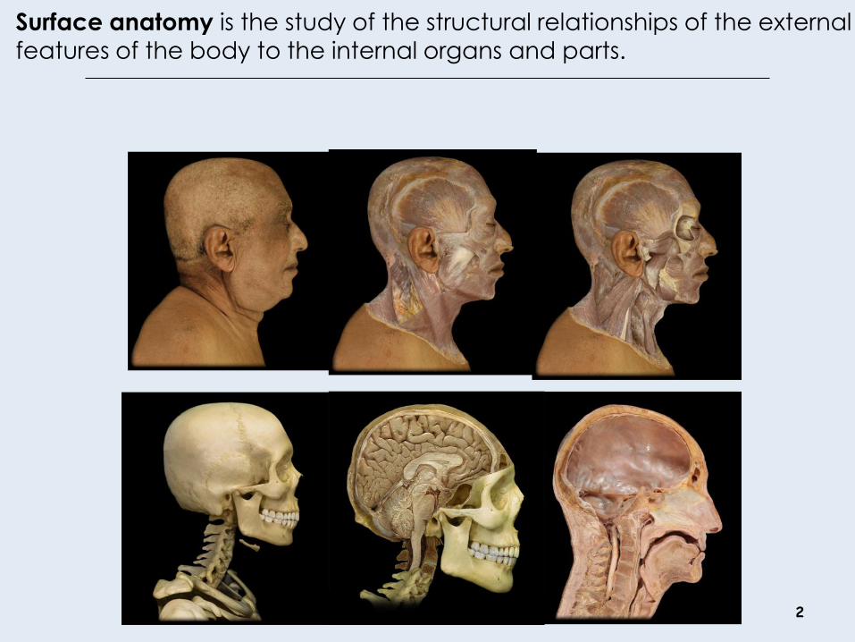
Surface anatomy is the study of the structural relationships of the external
features of the body to the internal organs and parts.
2

Health and Disease
3
http://www.nidcr.nih.gov/imagegallery/oralhealth/OralCancerE
xam.htm http://commons.wikimedia.org/wiki/File:Goiter.JPG
http://www.healthsym.com/causes-and-treatment-
for-jaundice.html
http://oralcancerfoundation.org/dental/slide_show.
htm
Surface Anatomy Overview
4
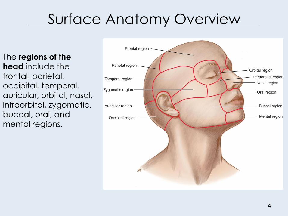
The regions of the
head include the
frontal, parietal,
occipital, temporal,
auricular, orbital, nasal,
infraorbital, zygomatic,
buccal, oral, and
mental regions.

5
Frontal Region
Parietal Region
Temporal Region
Zygomatic Region
Occipital Region
Orbital Region
Infraorbital Region Nasal Region
Oral Region
Buccal Region
Mental Region
1
2
3
4
5
6
7 8
9
10
11
Regions of the Head

6
External Acoustic Meatus, Tragus
Angle of the Mandible
Glabella
Frontal Eminence
Supraorbital Ridge
1
2
3
4
5
Anatomical Landmarks
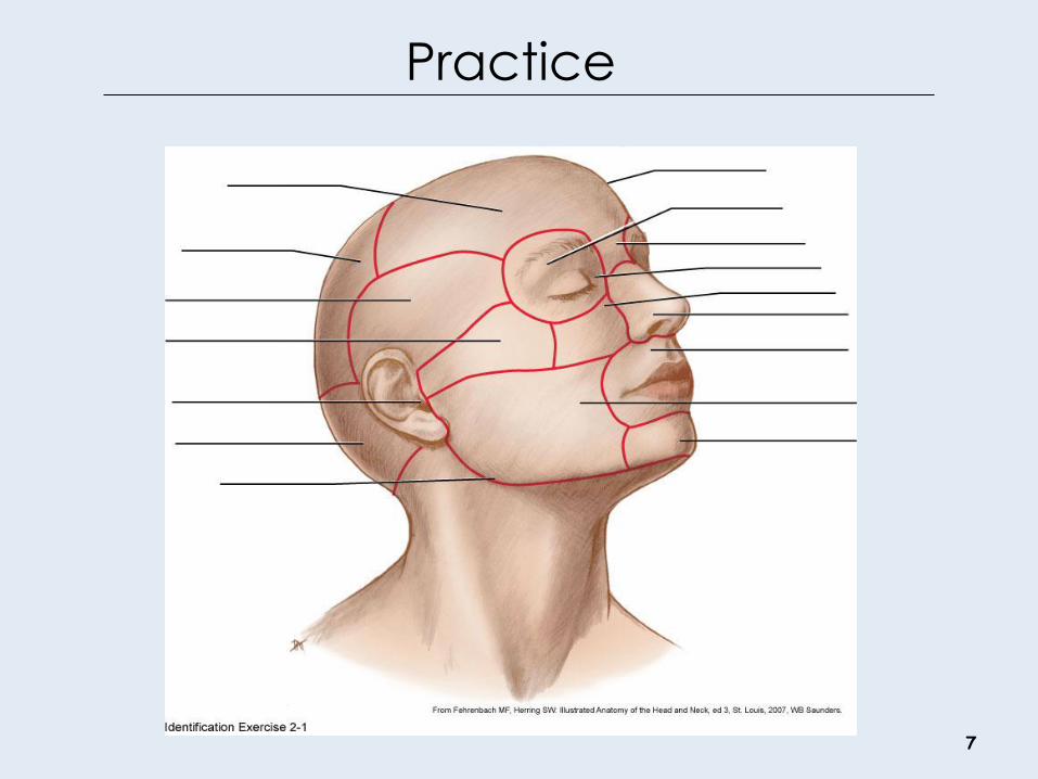
Practice
7
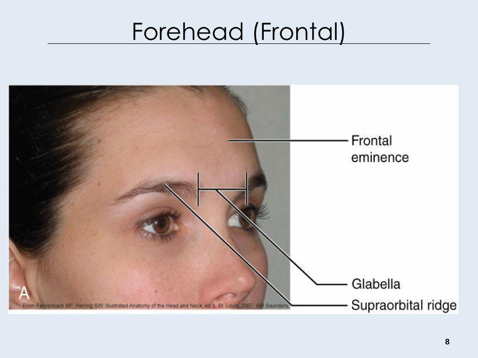
Forehead (Frontal)
8
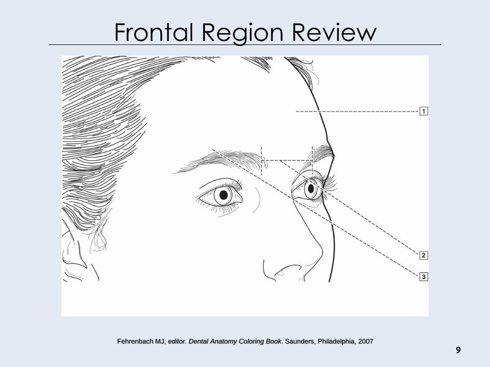
Frontal Region Review
9 Fehrenbach MJ, editor. Dental Anatomy Coloring Book. Saunders, Philadelphia, 2007

Parietal and Occipital Regions
10
Both the parietal region and occipital region of the head
are covered by the scalp.

11
Temporal Region
External ear most prominent,
includes:
• Auricle
• External acoustic meatus
• Helix
• Lobule
• Tragus
• Antitragus
• Intertragic notch

Ear Structure
12 Drake RL, et al. Gray’s Anatomy for Students, ed 2,
Churchill Livingson, 2010
Otitis Externa: inflammation or infection of the external ear or
“swimmer's ear.” Usually bacterial in origin, with the pathogens that include Pseudomonas aeruginosa and Staphylococcus aureus. Patient may present with itchiness, a sensation of having
the ear blocked, and pain. Otitis Media: pulling or rubbing
the ears because of ear pain,
fever, fussiness, or irritability, fluid
leaking from the ear, changes in
appetite or sleeping patterns, and
trouble hearing. Usually bacterial
infection

Auricular Region Review
13 Fehrenbach MJ, editor. Dental Anatomy Coloring Book. Saunders, Philadelphia, 2007
14
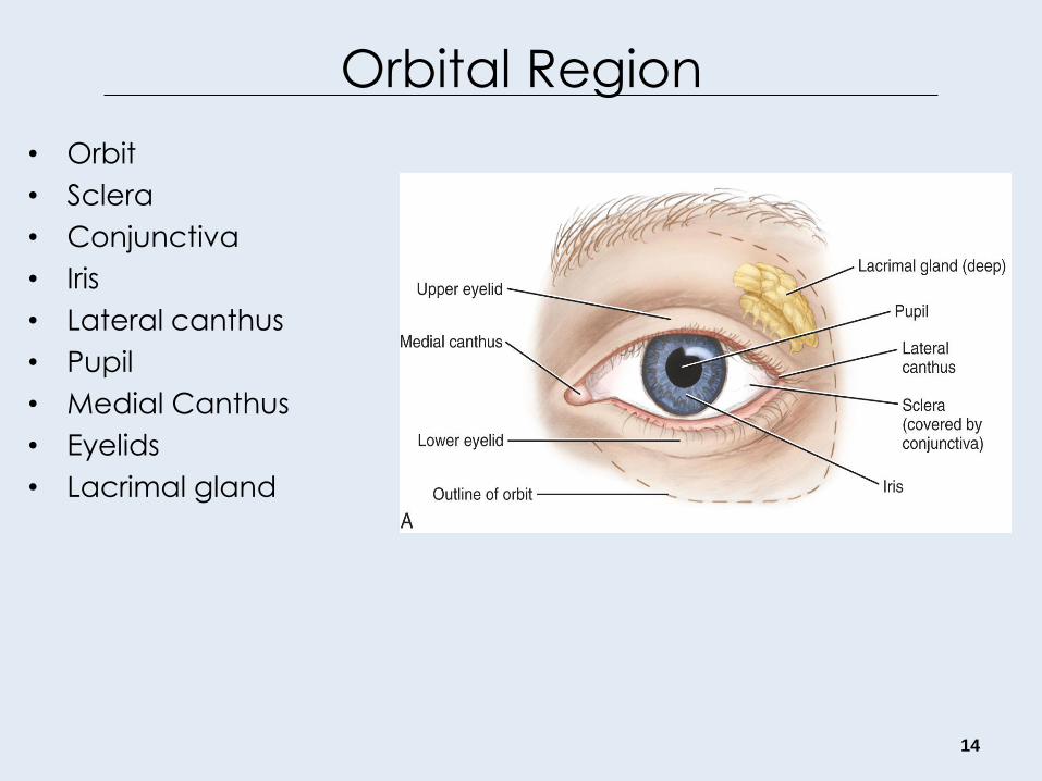
Orbital Region
• Orbit
• Sclera
• Conjunctiva
• Iris
• Lateral canthus
• Pupil
• Medial Canthus
• Eyelids
• Lacrimal gland
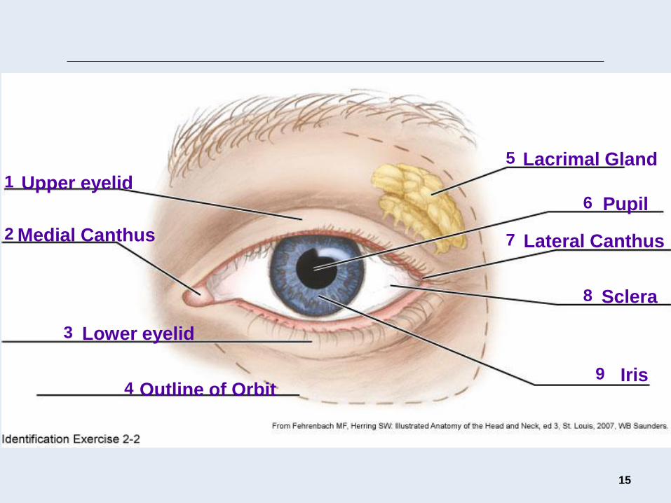
15
Upper eyelid
Medial Canthus
Lower eyelid
Outline of Orbit
Lacrimal Gland
Pupil
Lateral Canthus
Sclera
Iris
1
2
3
4
5
6
7
8
9
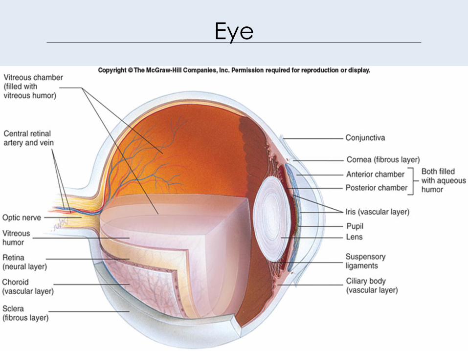
Eye
16
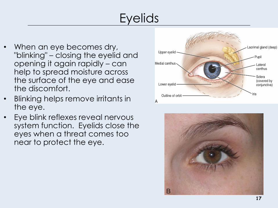
Eyelids
17
• When an eye becomes dry, "blinking" – closing the eyelid and opening it again rapidly – can help to spread moisture across the surface of the eye and ease the discomfort.
• Blinking helps remove irritants in the eye.
• Eye blink reflexes reveal nervous system function. Eyelids close the eyes when a threat comes too near to protect the eye.

Pupils
18
• Pupillary reflexes reveal information
about the nervous system. – In normal room light, a healthy pupil has a
diameter of about 3 to 4 mm.
– In bright light = 1.5 mm.
– In dim light, enlarged to about 8 mm.
• Pupil constricts to view something
close.
• Some drugs cause pupil constriction
(miosis), such as alcohol.
• Some drugs cause pupil dilation
(mydriasis) e.g. psychedelics (LSD).
http://library.med.utah.edu/kw/animations/hyperbr
ain/parasymp_reflex/reflex.html
http://library.med.utah.edu/kw/hyperb
rain/movies/ch7/orbit_autonomic.htm
http://www.ebmedicine.net/topics.php?paction=showTopicSeg&topic_i
d=69&seg_id=1320

Disease: Conjunctivitis and Cataracts
A cataract disrupts the
organization of the connective
tissue that forms the lens,
changing the optical properties
the opacity of the lens.
Conjunctivitis is an inflammation
of the epithelial lining of the eye
(the conjunctiva).
19 National Eye Institute, NIH
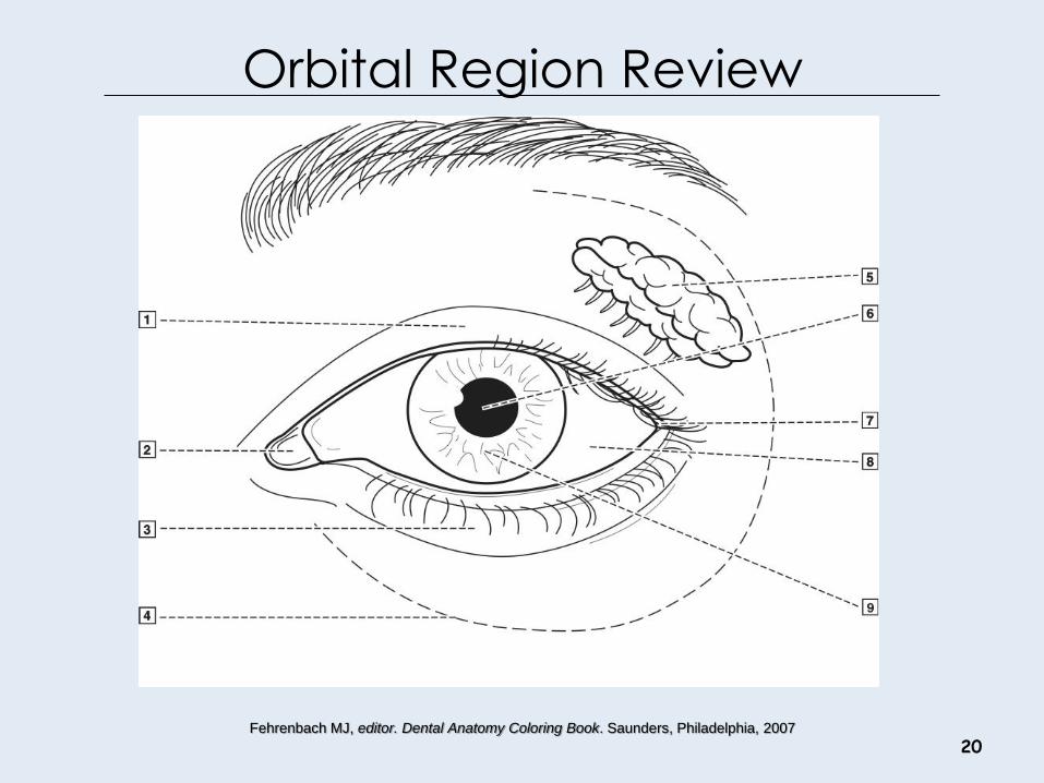
Orbital Region Review
20 Fehrenbach MJ, editor. Dental Anatomy Coloring Book. Saunders, Philadelphia, 2007
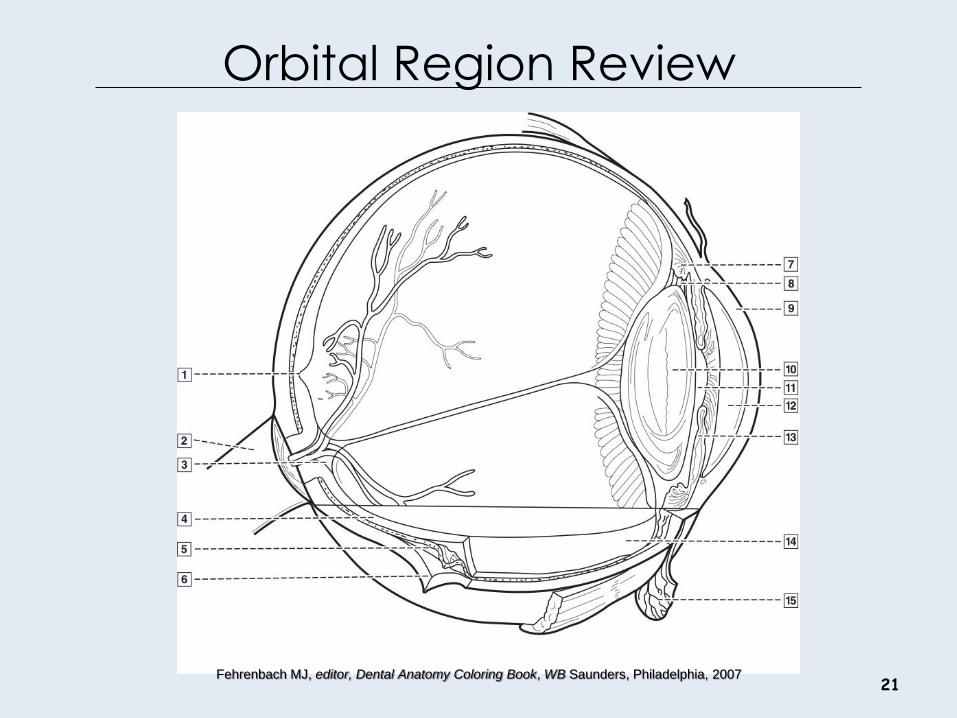
Orbital Region Review
21 Fehrenbach MJ, editor, Dental Anatomy Coloring Book, WB Saunders, Philadelphia, 2007

Nasal Region
• The root of the nose is located between the eyes.
• Inferior to the glabella is a midpoint landmark of the nasal region that corresponds with the junction between the underlying bones, the nasion.
• Inferior to the nasion is the bony structure that forms the bridge of the nose.
• At the other end is the tip or apex of the nose.
• Inferior to the apex on each side of the nose is a nostril or naris (plural, nares).
• The nares are separated by the midline nasal septum.
• The nares are bounded laterally on each side by a winglike cartilaginous structure, the ala (plural, alae) of the nose.
22
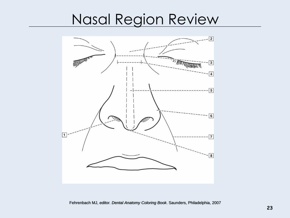
Nasal Region Review
23 Fehrenbach MJ, editor. Dental Anatomy Coloring Book. Saunders, Philadelphia, 2007
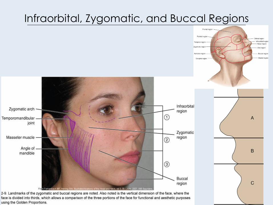
Infraorbital, Zygomatic, and Buccal Regions
24
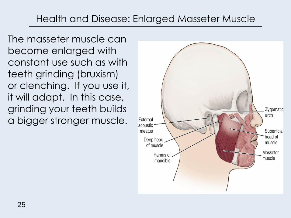
Health and Disease: Enlarged Masseter Muscle
The masseter muscle can
become enlarged with
constant use such as with
teeth grinding (bruxism)
or clenching. If you use it,
it will adapt. In this case,
grinding your teeth builds
a bigger stronger muscle.
25
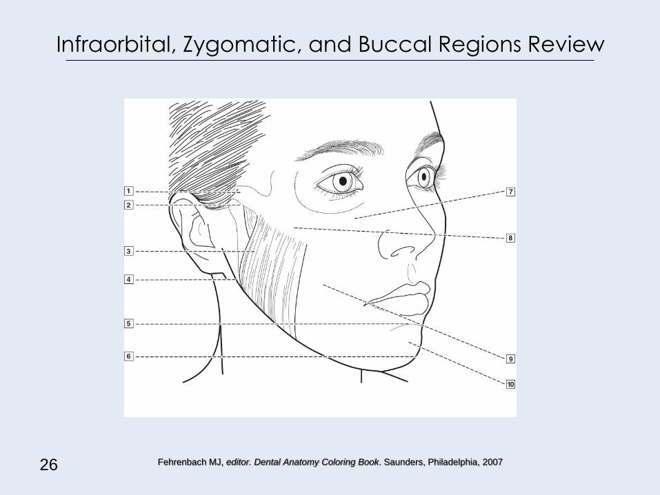
Infraorbital, Zygomatic, and Buccal Regions Review
26 Fehrenbach MJ, editor. Dental Anatomy Coloring Book. Saunders, Philadelphia, 2007
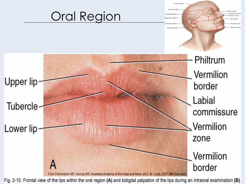
Oral Region
27
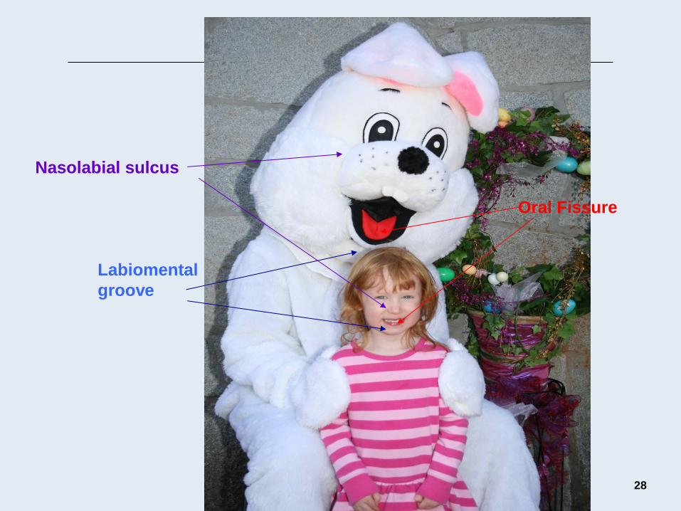
28
Labiomental
groove
Oral Fissure
Nasolabial sulcus
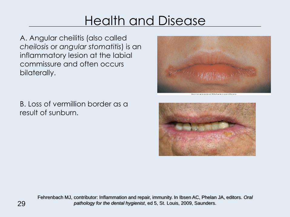
Health and Disease A. Angular cheilitis (also called
cheilosis or angular stomatitis) is an
inflammatory lesion at the labial
commissure and often occurs
bilaterally.
B. Loss of vermillion border as a
result of sunburn.
29 Fehrenbach MJ, contributor: Inflammation and repair, immunity. In Ibsen AC, Phelan JA, editors. Oral
pathology for the dental hygienist, ed 5, St. Louis, 2009, Saunders.

Lip Anatomy Review
30 Fehrenbach MJ, editor. Dental Anatomy Coloring Book. Saunders, Philadelphia, 2007

31
Oral Cavity
Structures:
– Maxilla
– Mandible
– Mucosa
– Labial mucosa
– Buccal mucosa
– Buccal fat pad
– Parotid papilla
– Maxillary tuberosity
– Vestibules
– Alveolar mucosa
– Mucobuccal fold
– Labial frenum
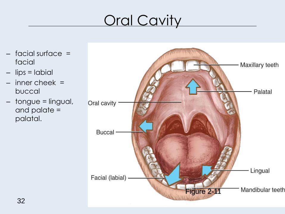
Oral Cavity
32
Figure 2-11
– facial surface =
facial
– lips = labial
– inner cheek =
buccal
– tongue = lingual,
and palate =
palatal.
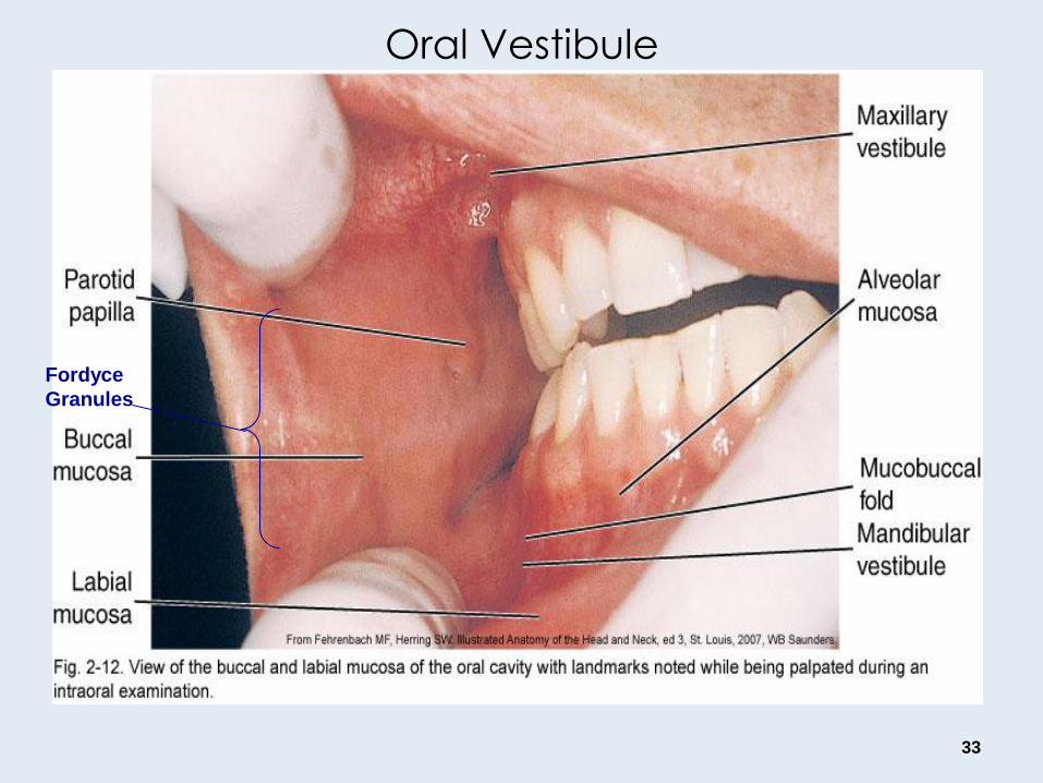
Oral Vestibule
33
Fordyce
Granules
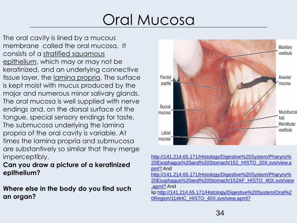
Oral Mucosa
34
http://141.214.65.171/Histology/Digestive%20System/Pharynx%
20Esophagus%20and%20Stomach/152_HISTO_20X.svs/view.a
pml? And
http://141.214.65.171/Histology/Digestive%20System/Pharynx%
20Esophagus%20and%20Stomach/152AF_HISTO_40X.svs/view
.apml? And
lip:http://141.214.65.171/Histology/Digestive%20System/Oral%2
0Region/114triC_HISTO_40X.svs/view.apml?
The oral cavity is lined by a mucous
membrane called the oral mucosa. It
consists of a stratified squamous
epithelium, which may or may not be
keratinized, and an underlying connective
tissue layer, the lamina propria. The surface
is kept moist with mucus produced by the
major and numerous minor salivary glands.
The oral mucosa is well supplied with nerve
endings and, on the dorsal surface of the
tongue, special sensory endings for taste.
The submucosa underlying the lamina
propria of the oral cavity is variable. At
times the lamina propria and submucosa
are substantively so similar that they merge
imperceptibly.
Can you draw a picture of a keratinized
epithelium?
Where else in the body do you find such
an organ?

Gingiva
35
Incisive
Fossa
Canine Eminence
Canine Fossa
and gingival
mucosa
Gingival margin Interdental papilla
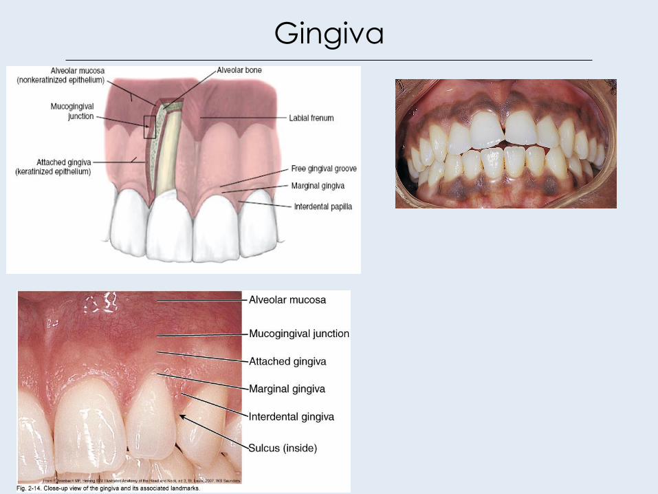
Gingiva
36

Variations of the oral cavity
Linea alba is a white ridge of
raised callused tissue that
extends horizontally at the level
where the maxillary and
mandibular teeth come
together and occlude.
Fordyce spots (or granules) are
often on the surface of the labial
and buccal mucosa is a normal
variation. They are small, yellowish
elevations due to deeper deposits of
sebum from trapped or misplaced
sebaceous gland tissue, usually
associated with hair follicles.
Cheek biting leads to mucosal
irritation.
37

Teeth and Jaws
38
Bath-Balogh M and Fehrenbach MJ.
Illustrated Dental Embryology, Histology,
Anatomy, ed 3. Saunders, Philadelphia,
2011.
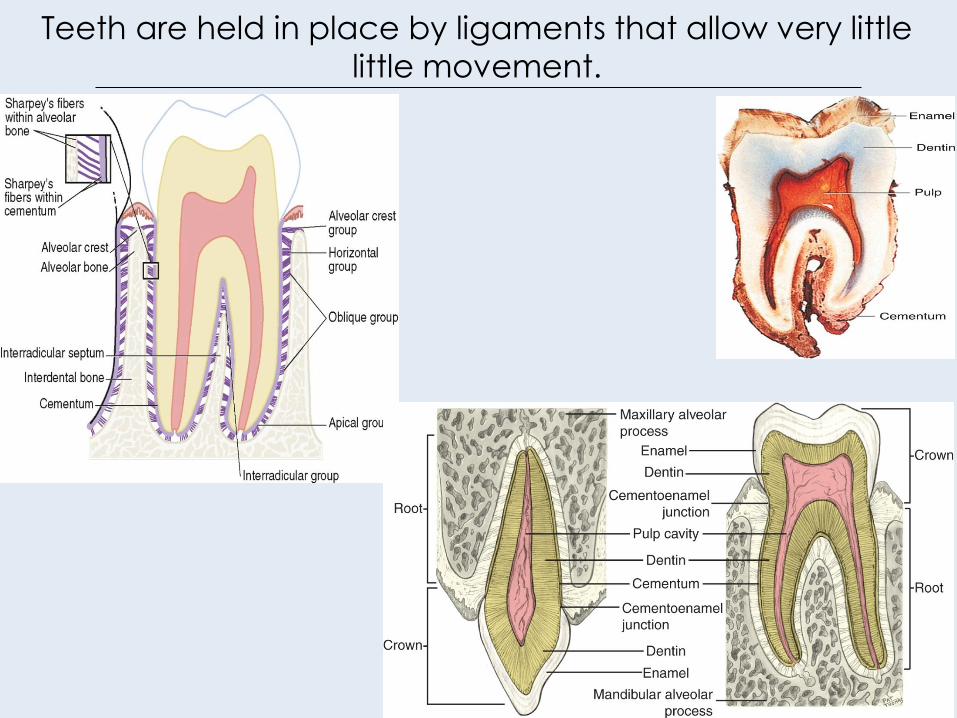
Teeth are held in place by ligaments that allow very little
little movement.
39

Oral Cavity Review
40
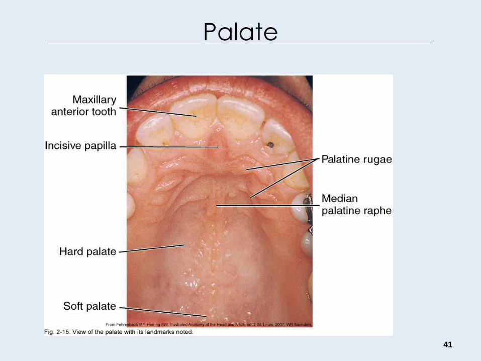
Palate
41
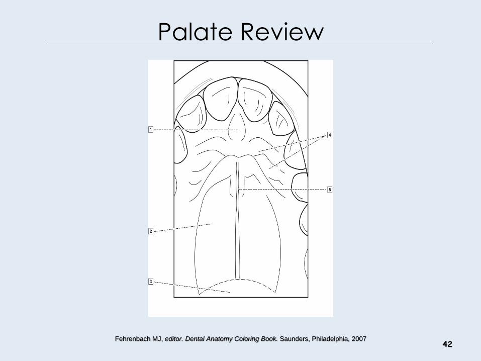
Palate Review
42 Fehrenbach MJ, editor. Dental Anatomy Coloring Book. Saunders, Philadelphia, 2007

Oral Cavity
43

Palate and Jaws Review
44
Fehrenbach MJ, editor. Dental Anatomy Coloring Book. Saunders, Philadelphia, 2007
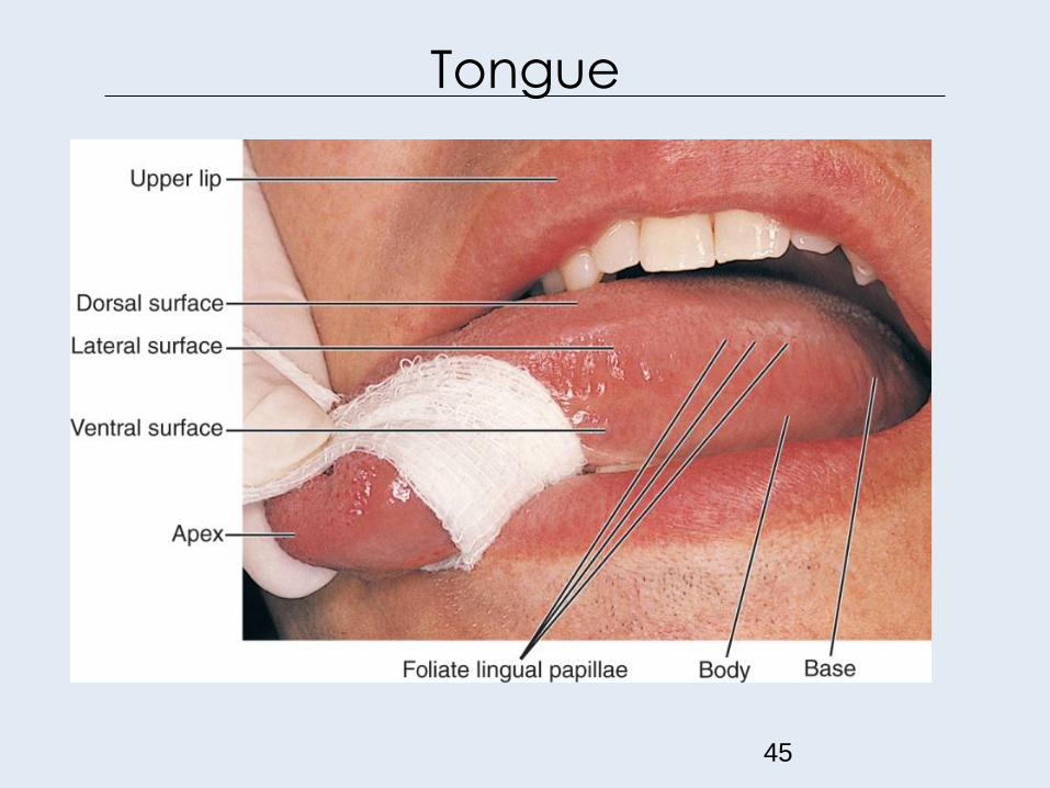
Tongue
45
Tongue
• The apex is the highly mobile, pointed anterior part of the tongue.
• Posterior to the apex lies the body of the tongue, which has dorsal (superior) and ventral (inferior) surfaces.
• The base is the most posterior part of the tongue and is not very mobile; a terminal V- shaped sulcus, or groove separates the body from the base of the tongue.
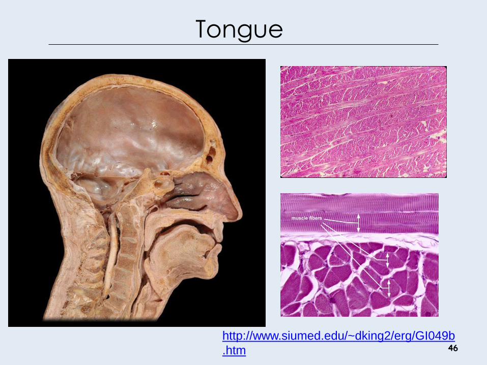
46
http://www.siumed.edu/~dking2/erg/GI049b
.htm

Tongue: Dorsal Surface
47

Tongue Papillae • The dorsal surface of the
tongue also has many lingual
papillae.
• The slender, threadlike lingual
papillae are the filiform
lingual papillae.
• The red mushroom-shaped
dots are the fungiform lingual
papillae (contain taste buds).
48 http://www.siumed.edu/~dking2/erg/GI064b.htm
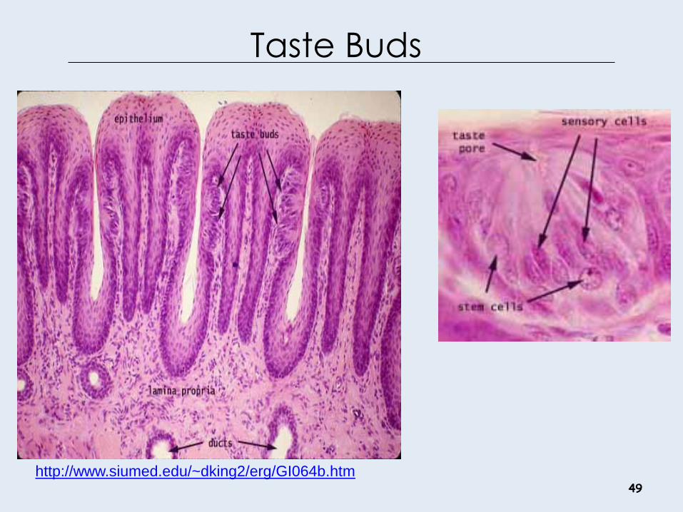
Taste Buds
49
http://www.siumed.edu/~dking2/erg/GI064b.htm
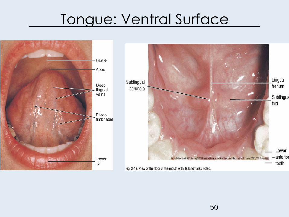
Tongue: Ventral Surface
50
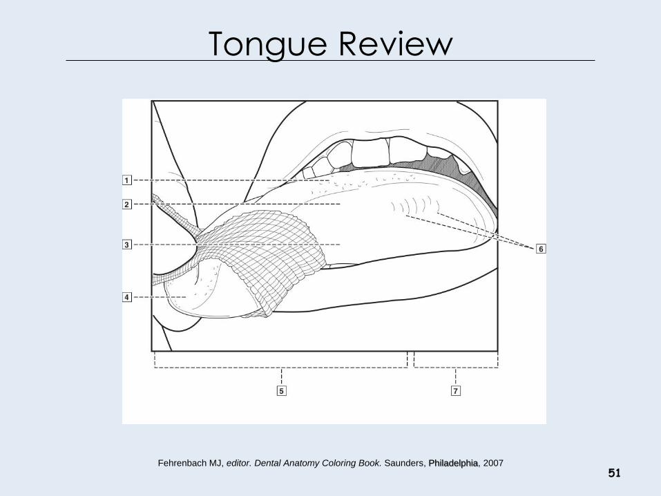
Tongue Review
51 Fehrenbach MJ, editor. Dental Anatomy Coloring Book. Saunders, Philadelphia, 2007

Tongue Review
52

Floor of the Mouth Review
53
Fehrenbach MJ, editor. Dental Anatomy Coloring Book. Saunders, Philadelphia, 2007

Pharynx
54
• The oral cavity also provides the entrance into the throat or pharynx.
• The pharynx consists of three parts: nasopharynx, oropharynx, and laryngopharynx.

Oropharynx
55

Pharynx Review
56

Mental Region
57
• The mental
protuberance is
the prominence
of the chin.

Regions of Head Review
58 Fehrenbach MJ, editor. Dental Anatomy Coloring Book. Saunders, Philadelphia, 2007
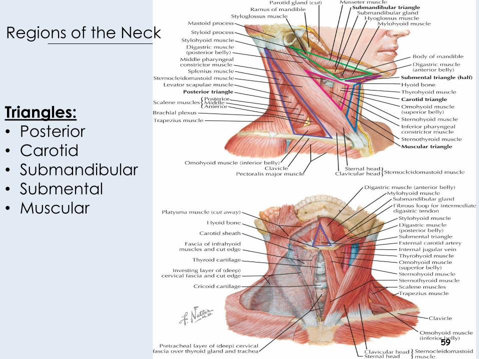
Regions of the Neck
59
Triangles:
• Posterior
• Carotid
• Submandibular
• Submental
• Muscular

Regions of Neck
60
• Thyroid cartilage
• Trachea
• Hyoid
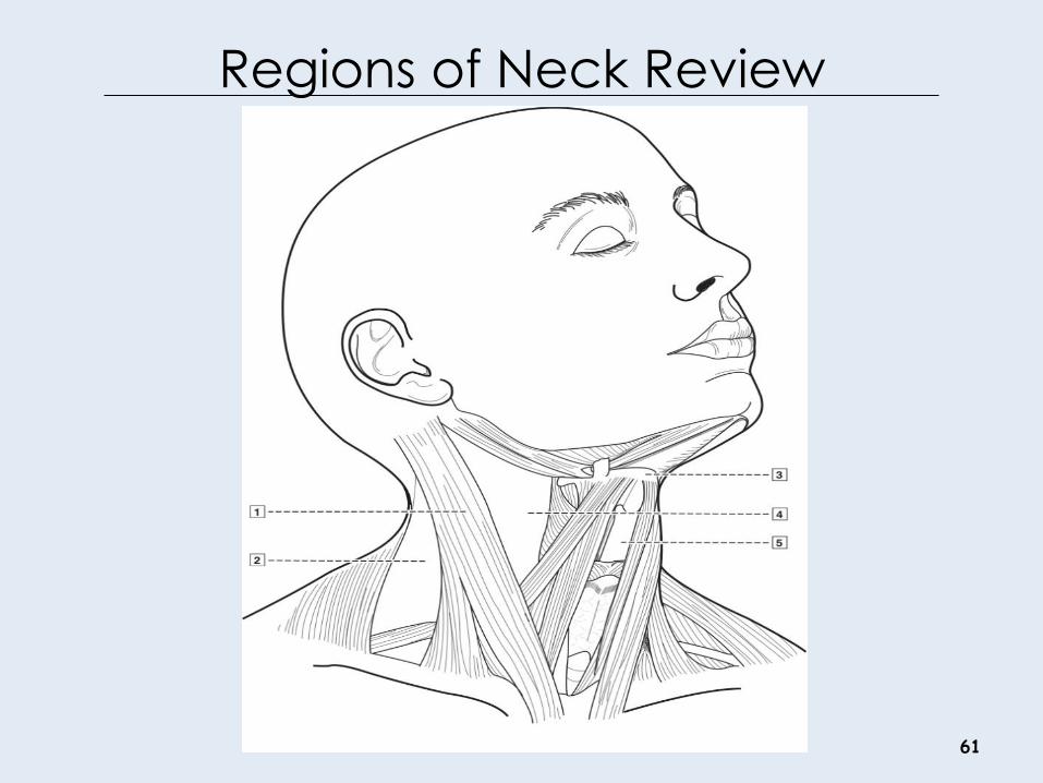
Regions of Neck Review
61 Fehrenbach MJ, editor. Dental Anatomy Coloring Book. Saunders, Philadelphia, 2007
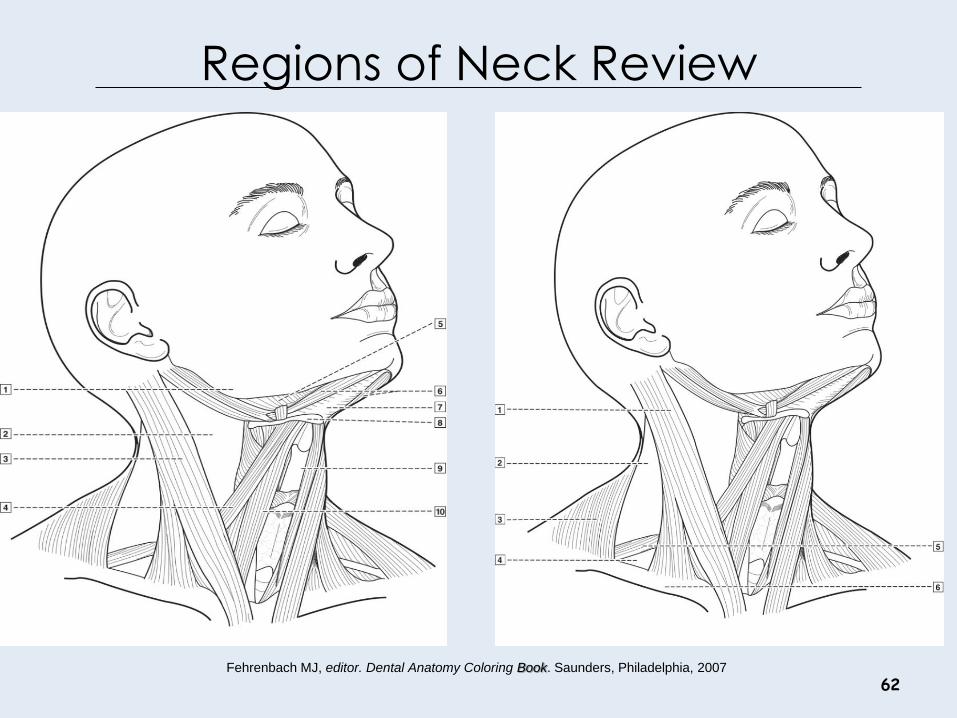
Regions of Neck Review
62 Fehrenbach MJ, editor. Dental Anatomy Coloring Book. Saunders, Philadelphia, 2007

Extra and Intra Oral Exam
63 Oral Exam Video: http://www.dentistry.umn.edu/dentalce/oral-cancer-video/index.htm