Supplementary Materi 96566a
6
www.BioTechniques.com 351 Vol. 48 | No. 6 | 2010 Benchmarks Standard molecular techniques All primers used in the study (Supple- mentary Table S3) were purchased from IDT (Coralville, IA, USA). DNA for PCR amplification of luxA, luxB, luxC, luxD, and luxE genes was from Photorhabdus lumine- scens (ATCC number 29999; Manassas, VA, USA) . Escherichia coli K12 DNA for PCR amplification of the lacZ and gusA genes was purified by standard methods (1). pCA24N (2) vector was used as a DNA template to PCR-amplify the gfp gene. DNA fragments and PCR mixtures were analyzed on 0.8% Seakem LE agarose gels (Lonza Rockland, Rockland, ME, USA) using 1 kb DNA ladder (New England BioLabs, Ipswich, MA, USA) as molecular size markers. Restriction enzymes and DNA modification enzymes were from New England BioLabs, and reactions were carried out under the recommended condi- tions. All other chemicals used in the study were of molecular biology grade. When necessary, DNA fragments were purified using agarose gels followed by purification using QIAquick-gel extraction kits from QIAGEN (Valencia, CA, USA). DNA sequencing was performed by Macrogen (Rockville, MD, USA); BioBricks were assembled as directed in the BioBrick Assembly Manual (New England BioLabs). Bacterial strains, plasmids, growth con- ditions and transformation procedures. All strains and plasmids used in the study are listed in Supplementary Table S2. Chemi- cally competent E. coli cells were prepared according to Inoue et al. (3). For each trans- formation, 1 μL overlap extension PCR reaction (Figure 1C) was used to transform 20 µL competent cells. e transformants (250 µL) were spread on Luria Bertani medium (LB) agar plates containing appro- priate antibiotic and 100 µM isopropyl β-D-1-thiogalactopyranoside (IPTG; if necessary). e constructs produced in this study conform to the BioBricks standard [Knight, T. Draſt Standard for Biobrick Biological Parts (OpenWetWare, MIT, Cambridge, MA, USA, 2007). http:// hdl.handle.net/1721.1/45138 ], but we emphasize that the cloning technique described here can be applied to any insert and vector without regard to restriction sites. A custom BioBrick accepting vector (pIMBB) was created by cloning a synthetic multiple cloning site (EcoRI-NotI- XbaI- SphI-ribosome binding site-NcoI-EcoRV- HindIII-SpeI-NotI-PstI) into the DraIII and Afl III sites of pSL1180 (Amersham Pharmacia Biotech, Piscataway, NJ, USA). is plasmid retains its ColE1 origin and β-lactamase gene and has restriction sites EcoRI, NotI, XbaI, SpeI, or PstI present only in the multiple cloning site, which is crucial for BioBricks assembly. Desired BioBricks cloned into this plasmid can subsequently be combined with others (see BioBrick Assembly Manual at http:// ginkgobioworks.com/support for details. A series of reporter BioBricks ( gfp, gusA, lacZ, luxA, luxB, luxC, luxD, and luxE) was produced by PCR amplification with appropriate primers (Supplementary Table S3) and subsequent cloning into the pIMBB vector. Each BioBrick was then combined with the ribosomal binding site (rbs) BioBrick (oligos 213–214; Supple- mentay Table S3). Aſter that, rbs-fused luxA, luxB, luxC, luxD and luxE BioBricks were combined to create a luxAluxBlux- CluxDluxE BioBrick (synthetic LUX operon). Reporter BioBricks were then separately combined with a BioBrick containing the T5-promoter and two lac operators (oligos 221–222; Supplementary Table S3) to enable regulated expression in the presence of the lacI repressor. Overlap extension PCR cloning Phusion DNA polymerase (New England BioLabs) (or Expand Long Template enzyme mix, Roche, Basel, Switzerland) and chimeric primers (5′ end, vector- specific sequences; 3′ end, insert-specific sequences) were used to PCR-amplify the inserts (Figure 1A). In particular, primers 278 and 279 were used to PCR-amplify gfp, gusA, lacZ, and LUX operon from pIMBB for cloning into pQBAV3Cam vector. Primers 278 and 280 were used to PCR-amplify gfp from pIMBB-gfp for cloning into pQBAV2ACam. Each PCR was subjected to a temperature regimen similar to the following: initial denatur- ation at 100°C for 2 min, denaturation at 94°C for 30 s, annealing at 60°C for 30 s, extension at 68°C for 90 s/kb for 30–32 cycles, with a final extension of 68°C for 10 min. All PCR-amplified inserts were gel-purified prior to use as megaprimers in (see Figure 1B). Primer design for overlap extension PCR follows 3 easy steps presented in Supple- mentary Figure S1. First, design appro- priate primers A and B to PCR-amplify the insert using web-based tools [e.g., Primer3 (http://primer3.sourceforge.net), Primer Design (www.bioinformatics.org/ jambw/5/2/index.html), or Primer-Blast (www.ncbi.nlm.nih.gov/tools/primer- blast)]. Second, select the desirable insert points on the plasmid; they can be in close proximity to each other or, preferably, 50 to several hundred base pairs apart. Now select 30–40 bp upstream of the leſt point of insert on the top strand of the plasmid. Copy this sequence and estimate its Tm using an online tool (Oligo Calcu- lator, www.idtdna.com/analyzer/Appli- cations/OligoAnalyzer/Default.aspx). If Tm parameter (60-65°C; see main text) is satisfied, save the sequence as primer C. Select 30–40 bp downstream of the right point of insert on the bottom strand of the plasmid. Copy the reverse sequence and analyze its Tm using an online tool (Oligo Calculator); If the Tm parameter is satisfied, save the sequence as primer D. Attach the sequence of primer C to the 5’ end of the primer A. Attach the sequence of primer D to the 5’ end of the primer B. Overlap extension PCR (Figure 1B) requires a combination of high GC content in the vector-specific part of the chimeric primers (5′ end) and relatively low (as compared with the primers designed Tm) annealing temperature in the thermocycler. Supplementary Material For: Overlap extension PCR cloning: a simple and reliable way to create recombinant plasmids Anton V. Bryksin and Ichiro Matsumura Department of Biochemistry, Center for Fundamental and Applied Molecular Evolution, Emory University, Atlanta, Georgia, USA BioTechniques 48:463-465 ( June 2010) doi 10.2144/000113418 Keywords: overlap extension PCR cloning; recombinant vector; Phusion; restriction enzyme ligation independent
-
Upload
adamos1945 -
Category
Documents
-
view
10 -
download
1
description
good article
Transcript of Supplementary Materi 96566a

www.BioTechniques.com351Vol. 48 | No. 6 | 2010
Benchmarks
Standard molecular techniquesAll primers used in the study (Supple-mentary Table S3) were purchased from IDT (Coralville, IA, USA). DNA for PCR amplification of luxA, luxB, luxC, luxD, and luxE genes was from Photorhabdus lumine-scens (ATCC number 29999; Manassas, VA, USA) . Escherichia coli K12 DNA for PCR amplification of the lacZ and gusA genes was purified by standard methods (1). pCA24N (2) vector was used as a DNA template to PCR-amplify the g fp gene. DNA fragments and PCR mixtures were analyzed on 0.8% Seakem LE agarose gels (Lonza Rockland, Rockland, ME, USA) using 1 kb DNA ladder (New England BioLabs, Ipswich, MA, USA) as molecular size markers. Restriction enzymes and DNA modification enzymes were from New England BioLabs, and reactions were carried out under the recommended condi-tions. All other chemicals used in the study were of molecular biology grade. When necessary, DNA fragments were purified using agarose gels followed by purification using QIAquick-gel extraction kits from QIAGEN (Valencia, CA, USA). DNA sequencing was performed by Macrogen (Rockville, MD, USA); BioBricks were assembled as directed in the BioBrick Assembly Manual (New England BioLabs).
Bacterial strains, plasmids, growth con-ditions and transformation procedures.All strains and plasmids used in the study are listed in Supplementary Table S2. Chemi-cally competent E. coli cells were prepared according to Inoue et al. (3). For each trans-formation, 1 μL overlap extension PCR reaction (Figure 1C) was used to transform 20 µL competent cells. The transformants (250 µL) were spread on Luria Bertani medium (LB) agar plates containing appro-
priate antibiotic and 100 µM isopropyl β-D-1-thiogalactopyranoside (IPTG; if necessary). The constructs produced in this study conform to the BioBricks standard [Knight, T. Draft Standard for Biobrick Biological Parts (OpenWetWare, MIT, Cambridge, MA, USA, 2007). http://hdl.handle.net/1721.1/45138 ], but we emphasize that the cloning technique described here can be applied to any insert and vector without regard to restriction sites.
A custom BioBrick accepting vector (pIMBB) was created by cloning a synthetic multiple cloning site (EcoRI-NotI-XbaI-SphI-ribosome binding site-NcoI-EcoRV-HindIII-SpeI-NotI-PstI) into the DraIII and AflIII sites of pSL1180 (Amersham Pharmacia Biotech, Piscataway, NJ, USA). This plasmid retains its ColE1 origin and β-lactamase gene and has restriction sites EcoRI, NotI, XbaI, SpeI, or PstI present only in the multiple cloning site, which is crucial for BioBricks assembly. Desired BioBricks cloned into this plasmid can subsequently be combined with others (see BioBrick Assembly Manual at http://ginkgobioworks.com/support for details. A series of reporter BioBricks (gfp, gusA, lacZ, luxA, luxB, luxC, luxD, and luxE) was produced by PCR amplification with appropriate primers (Supplementary Table S3) and subsequent cloning into the pIMBB vector. Each BioBrick was then combined with the ribosomal binding site (rbs) BioBrick (oligos 213–214; Supple-mentay Table S3). After that, rbs-fused luxA, luxB, luxC, luxD and luxE BioBricks were combined to create a luxAluxBlux-CluxDluxE BioBrick (synthetic LUX operon). Reporter BioBricks were then separately combined with a BioBrick containing the T5-promoter and two lac operators (oligos 221–222; Supplementary
Table S3) to enable regulated expression in the presence of the lacI repressor.
Overlap extension PCR cloningPhusion DNA polymerase (New England BioLabs) (or Expand Long Template enzyme mix, Roche, Basel, Switzerland) and chimeric primers (5′ end, vector-specific sequences; 3′ end, insert-specific sequences) were used to PCR-amplify the inserts (Figure 1A). In particular, primers 278 and 279 were used to PCR-amplify g fp, gusA, lacZ, and LUX operon from pIMBB for cloning into pQBAV3Cam vector. Primers 278 and 280 were used to PCR-amplify gfp from pIMBB-gfp for cloning into pQBAV2ACam. Each PCR was subjected to a temperature regimen similar to the following: initial denatur-ation at 100°C for 2 min, denaturation at 94°C for 30 s, annealing at 60°C for 30 s, extension at 68°C for 90 s/kb for 30–32 cycles, with a final extension of 68°C for 10 min. All PCR-amplified inserts were gel-purified prior to use as megaprimers in (see Figure 1B).
Primer design for overlap extension PCR follows 3 easy steps presented in Supple-mentary Figure S1. First, design appro-priate primers A and B to PCR-amplify the insert using web-based tools [e.g., Primer3 (http://primer3.sourceforge.net), Primer Design (www.bioinformatics.org/jambw/5/2/index.html), or Primer-Blast (www.ncbi.nlm.nih.gov/tools/primer-blast)]. Second, select the desirable insert points on the plasmid; they can be in close proximity to each other or, preferably, 50 to several hundred base pairs apart. Now select 30–40 bp upstream of the left point of insert on the top strand of the plasmid. Copy this sequence and estimate its Tm using an online tool (Oligo Calcu-lator, www.idtdna.com/analyzer/Appli-cations/OligoAnalyzer/Default.aspx). If Tm parameter (60-65°C; see main text) is satisfied, save the sequence as primer C. Select 30–40 bp downstream of the right point of insert on the bottom strand of the plasmid. Copy the reverse sequence and analyze its Tm using an online tool (Oligo Calculator); If the Tm parameter is satisfied, save the sequence as primer D. Attach the sequence of primer C to the 5’ end of the primer A. Attach the sequence of primer D to the 5’ end of the primer B.
Overlap extension PCR (Figure 1B) requires a combination of high GC content in the vector-specific part of the chimeric primers (5′ end) and relatively low (as compared with the primers designed Tm) annealing temperature in the thermocycler.
Supplementary Material For:
Overlap extension PCR cloning: a simple and reliable way to create recombinant plasmidsAnton V. Bryksin and Ichiro MatsumuraDepartment of Biochemistry, Center for Fundamental and Applied Molecular Evolution, Emory University, Atlanta, Georgia, USA
BioTechniques 48:463-465 ( June 2010) doi 10.2144/000113418 Keywords: overlap extension PCR cloning; recombinant vector; Phusion; restriction enzyme ligation independent

www.BioTechniques.com352Vol. 48 | No. 6 | 2010
Benchmarks
Supplementary Figure S1. Making the primers for overlap extension PCR cloning. (1) Design appropriate primers A and B to PCR-amplify the insert using web-based tools (e.g., Primer3, Primer Design, or Primer-Blast). (2). Select the desirable insert points on the plasmid; they could be in close proximity to each other or, preferably, 50 to several hundred bp apart. Then select 30–40 bp upstream of the left point of insert on the direct strand of the plasmid; copy this sequence. Analyze Tm using online tool (Oligo Calculator); if Tm parameter is satisfied, save the sequence of the primer C. Select 30–40 bp downstream of the right point of insert on the direct strand of the plasmid; copy the sequence. Analyze Tm using online tool (Oligo Calculator); if Tm parameter is satisfied, make reverse complement of the sequence using online tool (Reverse complement) and save the sequence of the primer D. (3) Attach the sequence of the primer C to the 5′ end of the primer A; attach the sequence of the primer D to the 5’ end of the primer B.
1
2
3

www.BioTechniques.com353Vol. 48 | No. 6 | 2010
Benchmarks
We recommend a molar excess of insert over plasmid template. The concentration of megaprimers (PCR product of Figure 1A) is much lower than the concentration of synthetic primers in regular PCRs; they could be shorter than expected and thus have lower melting temperature. Acceptor plasmid template (3–30 ng) was mixed with 250× molar excess of insert DNA (from Figure 1A) in a 10-µL total volume containing Phusion DNA polymerase reaction mixture containing dNTPs, buffer, and enzyme. The insert and vector underwent denaturation (98°C for 30 s), annealing (60°C for 30 s), and polymerase-catalyzed extension (98°C for 1.5 min per kb according to the length of the longest piece) for 5–30 cycles. We normally added
an extra 10-min extension step in the end of the program. For average-sized vectors and inserts, the total reaction time was <2 h. The same PCR parameters and volumes were used for overlap PCR cloning reactions with other polymerases and polymerase mixtures [KOD (Merck, San Diego, CA, USA); Expand Long Template mix (Roche); Deep Vent (New England BioLabs); and Pfu (Stratagene, La Jolla, CA, USA)], except that the buffer system was substituted with one recommended by each respective manufacturer.
The DpnI endonuclease works well in Phusion HF buffer. We typically add 10 units of the enzyme directly to the PCR tube right after the final extension is done and incubate the reaction for an hour at
37°C (see Figure 1C). Restriction endonu-clease DpnI targets methylated DNA sequences and can thereby cleave the DNA template isolated from most E. coli strains, but not the PCR product (4).
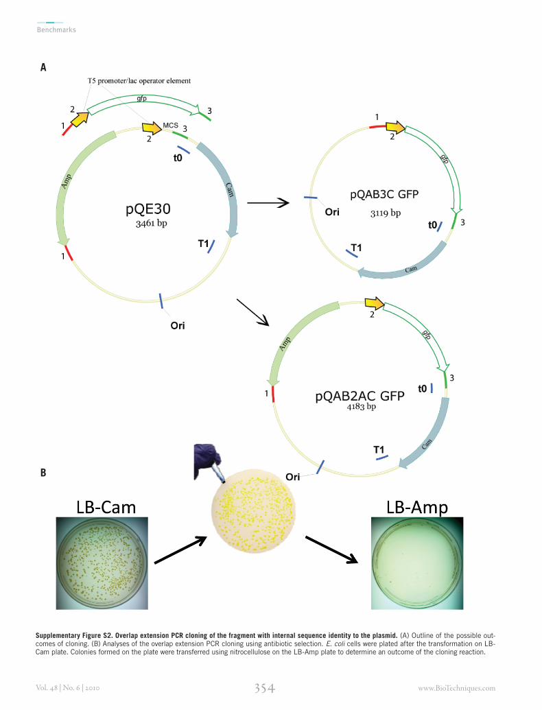
Additional resultsRecombinant plasmids often have repeated elements such as promoters, ribosomal binding sites, terminators, and scar sites. We wondered whether overlap extension PCR cloning was sensitive to the presence of internal sequences in the target plasmid identical to those in the insert. Chimeric primers were used to PCR-amplify a g fp gene fused to the T5 promotor/lac operator. A similar T5 promotor/lac operator sequence was present in the
Supplementary Table S2. Bacterial strains and plasmids used in the study
Name Description Reference or source
Strains
Escherichia coli
AG1 F- recA1 endA1 gyrA96 thi-1 hsdR17(rk- mk+) supE44 relA1 “NBRP (NIG, Japan): E.coli”
Photorhabdus luminescens ATCC number 29999
Plasmids
pQE30 Expression Vector; T5lacO; ColE1 replicon; AmprCamr Qiagen
pCA24N Expression Vector; T5lacO; ColE1 replicon; Camr (2)
IMBB-pSL1180-gfp Ampr This study
pIMBB BioBrick accepting vector; ColE1 replicon; Ampr This study
pIMBB-gfp Ampr This study
pIMBB-gusA Ampr This study
pIMBB-lacZ Ampr This study
pIMBB-luxABCDE Ampr This study
pQBAV3Cam-gfp Camr This study
pQBAV3Cam-gusA Camr This study
pQBAV3Cam-lacZ Camr This study
pQBAV3Cam-luxABCDE Camr This study
pQBAV2ACam-gfp AmprCamr This study
Supplementary Table S1. Comparison of the performance of different PCR systems in overlap extension PCR cloning
DNA polymerase Processivity Number of colonies expressing GFP/plate
Number of white colonies/plate
KOD DNA polymerase >300 bases (5) 14 0
Phusion DNA polymerase >360 bases (6) 417 8
Expand Long Template DNA polymerase mix
50–60 bases for Taq DNA polymerase (5)
12 2
Deep Vent DNA polymerase ? 0 0
Pfu DNA polymerase 15–20 bases (5) 9 1
Taq DNA polymerase 50–60 bases (6) Not tested Not tested
Phusion DNA polymerase was used to PCR-amplify green fluorescent protein (gfp) gene from the pIMBB-gfp plasmid. The PCR products were gel-purified and used in the overlap extension PCR reaction with pQE30 vector. Three nanograms of pQE30 vector was mixed with 500 ng insert in the total reaction volume of 10 μL, and subjected to 25 cycles of PCR with different PCR systems: KOD, Phusion, Expand Long Template mix, DeepVent and Pfu. The original plasmid was destroyed in restriction digests with DpnI, and the overlap extension PCR products were used to transform competent E. coli cells.
www.BioTechniques.com354Vol. 48 | No. 6 | 2010
Benchmarks
Supplementary Figure S2. Overlap extension PCR cloning of the fragment with internal sequence identity to the plasmid. (A) Outline of the possible out-comes of cloning. (B) Analyses of the overlap extension PCR cloning using antibiotic selection. E. coli cells were plated after the transformation on LB-Cam plate. Colonies formed on the plate were transferred using nitrocellulose on the LB-Amp plate to determine an outcome of the cloning reaction.
A
B

www.BioTechniques.com355Vol. 48 | No. 6 | 2010
Benchmarks
Supplementary Table S3. Oligonucleotides and PCR primers used in the study
Primer Sequence Additional information
201 SP/luxA/Bba_CluxA GGAATTCGCGGCCGCTTCTAGATGAAATTTGGAAACTTTTTGCTTACATACCAPCR amplification of luxA gene of Photorhabdus luminescens
202 ASP/luxA/Bba_CluxA CTGCAGCGGCCGCTACTAGTATTATTAATATAATAGCGAACGTTGTTTTTCTTTAAGAPCR amplification of luxA gene of Photorhabdus luminescens
203 SP/luxB/Bba_CluxB GGAATTCGCGGCCGCTTCTAGATGAAATTTGGATTGTTCTTCCTTAACTTCPCR amplification of luxB gene of Photorhabdus luminescens
204 ASP/luxB/Bba_CluxB CTGCAGCGGCCGCTACTAGTATTATTAGGTATATTCCATGTGGTACTTCTTAATAPCR amplification of luxB gene of Photorhabdus luminescens
205 SP/luxC/Bba_CluxC GGAATTCGCGGCCGCTTCTAGATGACTAAAAAAATTTCATTCATTATTAACGGCCPCR amplification of luxC gene of Photorhabdus luminescens
206 ASP/luxC/Bba_CluxC CTGCAGCGGCCGCTACTAGTATTATTATGGGACAAATACAAGGAACTTATCTTCTTCPCR amplification of luxC gene of Photorhabdus luminescens
207 SP/luxD/Bba_CluxD GGAATTCGCGGCCGCTTCTAGATGGAAAATGAATCAAAATATAAAACCATCGPCR amplification of luxD gene of Photorhabdus luminescens
208 ASP/luxD/Bba_CluxD CTGCAGCGGCCGCTACTAGTATTATTAAGACAGAGAAATTGCTTGATTTTCAATCPCR amplification of luxD gene of Photorhabdus luminescens
209 SP/luxE/Bba_CluxE GGAATTCGCGGCCGCTTCTAGATGACTTCATATGTTGATAAACAAGAAATTACAGCPCR amplification of luxE gene of Photorhabdus luminescens
210 ASP/luxE/Bba_CluxE CTGCAGCGGCCGCTACTAGTATTATTAACTATCAAACGCTTCGGTTAAGCTTAPCR amplification of luxE gene of Photorhabdus luminescens
213 INS_S/RBS/BBa_B0034 AATTCGCGGCCGCTTCTAGAGAAAGAGGAGAAATA Ribosomal binding site in BioBrick format
214 INS_AS/RBS/BBa_B0034 CTAGTATTTCTCCTCTTTCTCTAGAAGCGGCCGCG Ribosomal binding site in BioBrick format
221 INS_S/T52lacO/BBa_R0AB2AATTCGCGGCCGCTTCTAGAGGAAATCATAAAAAATTTATTTGCTTTGTGAGCGGATAA-CAATTATAATAGATTCAATTGTGAGCGGATAACAATTA
T5promoter/lac operator in BioBrick format
222 INS_AS/T52lacO/BBa_R0AB2CTAGTAATTGTTATCCGCTCACAATTGAATCTATTATAATTGTTATCCGCTCA-CAAAGCAAATAAATTTTTTATGATTTCCTCTAGAAGCGGCCGCG
(T5promoter/lac operator in BioBrick format
252 SP/GFP/1 GGAATTCGCGGCCGCTTCTAGATGCGTAAAGGAGAAGAACTTTTCACTGGAGTTGTCCC PCR amplification of gfp gene from pCA24N vector
253 ASP/GFP/736CGACTGCAGCGGCCGCTACTAGTATTATTATTTGTATAGTTCATCCATGCCATGTG-TAATCC
PCR amplification of gfp gene from pCA24N vector
1363ASP/LacZ wt/3075 CCAGCTGCAGCGGCCGCTACTAGTATTATTATTTTTGACACCAGACCAACTGGTAATGPCR amplification of lacZ gene from K12 E. coli genomic DNA
1364SP/LacZ wt/1 GCCGCTTCTAGATGACCATGATTACGGATTCACTGGCPCR amplification of lacZ gene from K12 E. coli genomic DNA
1365ASP/UidAwt/1812 CCAGCTGCAGCGGCCGCTACTAGTATTATTATTGTTTGCCTCCCTGCTGCGPCR amplification of gusA gene from K12 E. coli genomic DNA
1366SP/UidAwt/1 GCCGCTTCTAGATGTTACGTCCTGTAGAAACCCCAACCCGPCR amplification of gusA gene from K12 E. coli genomic DNA
278 INS/IMBB/pQE30CTGGATCTATCAACAGGAGTCCAAGCTCAGCTAATTGGCCTTTTGCTGGCCTTTT-GCTCACATG*
Overlap extenstion PCR cloning primer
279 INS/IMBB/pQE30SCAATCTAAAGTATATATGAGTAAACTTGGTCTGACAGATCAGGGCGATGGCCCAC-TACGTGG*
Overlap extenstion PCR cloning primer
280 INS/IMBB/pQE30LCCTATAAAAATAGGCGTATCACGAGGCCCTTTCGTCTTCATCAGGGCGATGGCCCAC-TACGTGG*
Overlap extenstion PCR cloning primer
281 INS/pQE30/noXbaI CAAATCCGCCCTCCAGAGCTGCCTCGCGC To remove XbaI restriction site from pQE30
282 INS/pQE30/noXbaI GCGCGAGGCAGCTCTGGAGGGCGGATTTG To remove XbaI restriction site from pQE30
* 5’ addition to the primer complimentary to the vector has been underlined

www.BioTechniques.com356Vol. 48 | No. 6 | 2010
Benchmarks
pQE30 vector; the insert had three regions identical to those on the plasmid (Supple-mentary Figure S2A). The primers also contained sequences similar to two regions on the plasmid separated by an Amp resis-tance marker. Successful overlap extension PCR cloning should therefore insert the T5 promoter/gfp cassette and eliminate the Amp marker from the pQE30 vector. Most of the colonies that appeared on the LB-agar plate supplemented with chloramphenicol displayed the “green” phenotype (Supplementary Figure S2B). Replica plating of the plate on LB-Amp resulted in only two surviving colonies (out of >500) that did not have the green phenotype (Supplementary Figure S2B), which probably reflected carryover of the original plasmid.
References1. Sambrook, J. and D.W. Russell. 2001.
Molecular cloning: a laboratory manual. Cold Spring Harbor Laboratory Press, Cold Spring Harbor, N.Y.
2. Kitagawa, M., T. Ara, M. Arifuzzaman, T. Ioka-Nakamichi, E. Inamoto, H. Toyonaga, and H. Mori. 2005. Complete set of ORF clones of Escherichia coli ASKA library (a complete set of E. coli K-12 ORF archive): unique resources for biological research. DNA Res. 12:291-299.
3. Inoue, H., H. Nojima, and H. Okayama. 1990. High efficiency transformation of Escherichia coli with plasmids. Gene 96:23-28.
4. Gomez-Eichelmann, M.C. and K.G. Lark. 1977. Endo R DpnI restriction of Escherichia coli DNA synthesized in vitro. Evidence that the ends of Okazaki pieces are determined by template deoxynucleotide sequence. J. Mol. Biol. 117:621-635.
5. Benson, L .M., A.P. Null, and D.C. Muddiman. 2003. Advantages of Thermo-coccus kodakaraenis (KOD) DNA polymerase for PCR-mass spectrometry based analyses. J. Am. Soc. Mass Spectrom. 14:601-604.
6. Wang, Y., D.E. Prosen, L. Mei, J.C. Sullivan, M. Finney, and P.B. Vander Horn. 2004. A novel strategy to engineer DNA polymerases for enhanced processivity and improved performance in vitro. Nucleic Acids Res. 32:1197-1207.