Comparative Study of Malondialdehyde, Superoxide Dismutase ...
Upload
manuela-voineaCategory
view
213download
0
www.elsevier.com/locate/ejphar
European Journal of Pharmacology 484 (2004) 111–118
Superoxide dismutase entrapped-liposomes restore the
impaired endothelium-dependent relaxation of
resistance arteries in experimental diabetes
Manuela Voinea, Adriana Georgescu, Adrian Manea, Elena Dragomir,Ileana Manduteanu, Doina Popov, Maya Simionescu*
Institute of Cellular Biology and Pathology ‘‘Nicolae Simionescu’’8, B.P. Hasdeu St., PO Box 35-14, Bucharest 79691, Romania
Received 3 July 2003; received in revised form 28 October 2003; accepted 4 November 2003
Abstract
Diabetes is associated with impaired endothelium-dependent relaxation. We questioned whether administration of superoxide dismutase
(superoxide: superoxide oxidoreductase, EC 1.15.1.1) entrapped in long-circulating liposomes improves the vascular reactivity of the
resistance arteries. Using the myograph technique, the vasodilation in response to acetylcholine was measured in mesenteric resistance
arteries isolated from diabetic or normal hamsters treated for 3 days with superoxide dismutase entrapped in liposomes, with the same
concentrations of free superoxide dismutase and plain liposomes, or untreated. Superoxide dismutase activity and nitric oxide (NO) levels
were assayed by spectrophotometry, superoxide dismutase levels by Western blot and the role of NE-nitro-L-arginine ethylester (L-NAME) on
vasodilation by the myograph technique. Our data revealed that: (i) superoxide dismutase entrapped in liposomes restored to a great extent
the endothelium-dependent relaxation of diabetic hamster resistance arteries; (ii) in superoxide dismutase entrapped in liposomes-treated
diabetic animals, the activity and the level of superoxide dismutase in arterial homogenates as well as the serum nitrite levels were
significantly higher than those in untreated hamsters or hamsters treated with free superoxide dismutase and plain liposomes: (iii) L-NAME
inhibited the response of arteries to acetylcholine in superoxide dismutase entrapped in liposomes-treated diabetic hamsters. These results
suggest that superoxide dismutase entrapped in liposomes is effective in scavenging superoxide anions, increases nitric oxide bioactivity and
improves the vasorelaxation of resistance arteries in diabetic hamsters.
D 2003 Elsevier B.V. All rights reserved.
Keywords: Artery; Diabetes; Liposome; Superoxide dismutase; Vasodilation
1. Introduction impaired ability of endothelium to mediate vasodilation
Notable alterations in endothelium-dependent relaxation
have been observed in the vessels of diabetic patients
(Johnstone et al., 1993) as well as in laboratory animals
with experimental diabetes (Diederich et al., 1994). Al-
though the nature of this feature is not fully understood, a
major role is attributed to the oxidant stress induced by
hyperglycemia (Tesfamariam, 1994; Cai and Harrison,
2000). Produced in excess, reactive oxygen species, espe-
cially superoxide anion (O2�), seem to be involved in the
0014-2999/$ - see front matter D 2003 Elsevier B.V. All rights reserved.
doi:10.1016/j.ejphar.2003.11.004
* Corresponding author. Tel.: +40-1-411-0860; fax: +40-1-411-1143.
E-mail addresses: [email protected] (M. Voinea),
[email protected] (M. Simionescu).
either by decreasing nitric oxide (NO) production and/or
its bioactivity (for review see Pieper, 1998). The O2� and NO
react rapidly and generate peroxynitrite (ONOO�), a potent
oxidant and potential mediator of vascular tissue injury
(Beckman et al., 1990; Mazzon et al., 2002). Moreover, it
has been reported that hyperglycemia promotes the glyca-
tion of superoxide dismutase, leading to its inactivation and
reduction in antioxidant defense (Arai et al., 1987).
An approach to decrease oxidative stress is to use anti-
oxidants as therapeutic agents (Ting et al., 1996; Mayhan and
Patel, 1998). Given the important role of O2�, superoxide
dismutase, a scavenger enzyme, may have the ability to
restore, at least in part, endothelial function. However, due to
its rapid elimination from the circulation (half-life < 10 min)
the therapeutic use of superoxide dismutase is limited.
M. Voinea et al. / European Journal of P112
Consequently, attempts to increase its therapeutic index by
designing other delivery strategies have been made. Among
these, coupling of polyethylene glycol to superoxide dis-
mutase (Mugge et al., 1991) or the use of lecithinized Cu2 +/
Zn2 +-superoxide dismutase (Yunoki et al., 1997) has been
shown to improve superoxide dismutase efficiency by
extending its time in the circulation. Another alternative is
to trap superoxide dismutase in liposomes, which has the
advantage that superoxide dismutase is protected from inac-
tivation and its life-span in the circulation is extended. There
are studies that show that liposome-incorporated superoxide
dismutase is internalized by cultured endothelial cells and
that the cells are protected against extracellular oxidative
stress (Beckman et al., 1986; Nakae et al., 1990a). Data from
in vivo experiments indicate that encapsulation of super-
oxide dismutase in liposomes may extend their circulation in
the blood stream. Moreover, in animal models parenteral
administration of superoxide dismutase-entrapped liposomes
provides protection against toxic hepatic necrosis (Nakae et
al., 1990b), adjuvant-induced arthritis (Corvo et al., 1999,
2002), oxidative injury of the brain (Yusa et al., 1984), and
cerebral ischemia/reperfusion injury (Imaizumi et al., 1990).
Also, in a clinical trial, the administration of Cu2 +/Zn2 +-
superoxide dismutase encapsulated in liposomes was effec-
tive in reducing late radiation-induced tissue fibrosis (Dela-
nian et al., 1994).
In the present study, we questioned whether superoxide
dismutase encapsulated in sterically stabilized liposomes
improves the diminished vascular response of resistance
arteries, a common feature of diabetes. We provide evidence
that in vivo administration of superoxide dismutase entrap-
ped in liposomes restores the endothelium-dependent relax-
ation of small arteries from diabetic hamsters, functioning as
a superoxide anion scavenger and thus contributing to an
increase in NO bioactivity.
2. Materials and methods
2.1. Reagents
Reagents were obtained from the following sources: egg
phosphatidylcholine, cholesterol, 1,2-distearoyl-sn-glycero-
3-phosphoethanolamine-N-[poly(ethylene glycol)-2000]
(PEG2000-PE) from Avanti Polar Lipids (Alabaster, AL,
USA); mouse anti-bovine superoxide dismutase from Bio-
trend Khemikalien, (Koln, Germany); the enzyme kit for
glucose assay from Dialab (Vienna, Austria); the enhanced
chemiluminiscence system (ECL), Super Signal West pico
chemiluminiscent substrate from Pierce (Rockford, USA);
streptozotocin, bovine Cu2 +/Zn2 + superoxide dismutase
(superoxide:superoxide oxidoreductase; EC 1.15.1.1), ace-
tylcholine, noradrenaline (bitartrate), sodium nitroprusside,
NE-nitro-L-arginine ethylester (L-NAME), Aspergillus nitrate
reductase, as well as all other chemicals from Sigma-Aldrich
Chemie (Germany).
2.2. Preparation of liposome-entrapped superoxide
dismutase
Unilamellar sterically stabilized liposomes were prepared
essentially as previously described after extruding multi-
lamellar vesicles through a polycarbonate membrane (Pater-
nostre et al., 1996). Briefly, a mixture of phospholipids in
chloroform was dried in a rotary evaporator under reduced
pressure; the ratio of lipids was egg phosphatidylcholine:
cholesterol: PEG2000-PE (65: 30: 5 mol%). The superoxide
dismutase was added in phosphate-buffered saline (PBS) so
as to achieve a final lipid concentration of 25 Amol/ml. After
a brief vortexing, the preparation was hydrated overnight in
a rotating wheel. The resulting multilamellar vesicles were
extruded 20 times through a 100-nm polycarbonate mem-
brane, using a Mini-Extruder (Avanti Polar Lipids, Alabas-
ter, AL, USA). The final liposome vesicles had an average
diameter of 115 nm, as determined on electron micrographs
after negative staining and transmission electron microsco-
py. The efficiency of superoxide dismutase encapsulation
was f 25% of the initial superoxide dismutase added; free
superoxide dismutase was separated from liposome-encap-
sulated superoxide dismutase using a Sepharose 4B column.
The final superoxide dismutase concentration in liposomes
was 750U/25 Amol/ml.
2.3. Experimental protocol
Forty male Golden Syrian hamsters, 115–135 g in body
weight, were divided into two groups: (i) in 30 animals,
diabetes was induced by one i.p. injection of 50 mg strepto-
zotocin/kg body weight; (ii) 10 age-matched were used as
control animals. After 8 weeks from the induction of diabetes,
both the diabetic and control hamsters were subdivided into
three groups: (a) hamsters (under slight ether anesthesia)
injected daily with superoxide dismutase entrapped in li-
posomes at the concentration of 1500U SOD/50 Amol lipo-
some/kg body weight, via venous retro-orbital plexus for 3
days; (b) hamsters injected with free SOD and plain li-
posomes at the same dose and time as above and (c) control
hamsters received no treatment; all hamsters were killed after
3 days. The experiments were approved by the Ethics
Committee from our institute and were conducted in accor-
dance with the Guide for the Care and Use of Laboratory
Animals published by the US National Institutes of Health
(NIH publication no 85-23, revised 1996).
2.4. Biochemical assays
At the time of death, animals had been fasted for 18
hours. Blood was collected from the venous plexus of the
eye and used for biochemical assays.
2.4.1. Glucose assay
At the beginning of the experiment and at 8 weeks after
the induction of diabetes, plasma glucose levels were
harmacology 484 (2004) 111–118
M. Voinea et al. / European Journal of Pharmacology 484 (2004) 111–118 113
determined by an enzymatic method according to the
manufacturer’s protocol (Dialab, Viena, Austria).
2.4.2. Nitric oxide determination
Detection of inorganic nitrite (the stable end metabolite
of NO) was performed in animal sera using NADPH
oxidation by Aspergillus nitrate reductase, followed by
Griess reaction (Smarason et al., 1997).
2.4.3. Superoxide dismutase activity assay
For each experimental condition used, the mesenteric bed
was isolated and stored at� 70 jC. Frozen fragments were
homogenized on ice and resuspended in lysis buffer con-
taining 20 mM Tris, 150 mM NaCl, 2 mM ethylenediami-
netetraacetic acid (EDTA), 1% Triton X-100, 0.5% sodium
deoxycholate, 1 mM sodium orthovanadate, 0.25 mM
phenylmethyl sulfonylfluoride and 1 Ag/Al proteases inhi-
bitors (aprotinin, leupeptin, pepstatin). After centrifugation
at 10000� g for 10 min at 4 jC, the supernatant was
collected and superoxide dismutase activity was assayed
by inhibition of NADH oxidation mediated by MnCl2,
monitored by spectrophotometry at 340 nm as described
(Paoletti and Mocali, 1990). The reaction was initiated by
addition of 1 mM h-mercaptoethanol in a mixture consisting
of 100 mM triethanolamine-diethanolamine-HCl buffer, pH:
7.4, 0.3 mM NADH, 100 mM/50 mM EDTA-MnCl2 and 10
Al of each sample. The SOD activity was expressed as units/
mg total protein, where one unit is defined as the amount of
enzyme able to inhibit by 50% the rate of NADH oxidation
of the control (reaction mixture without sample).
2.4.4. Determination of superoxide dismutase levels by
Western blot
Homogenates of mesenteric arteries were prepared as
described above. Total protein concentration was deter-
mined with the Amido Black technique, using bovine serum
albumin as standard (Schaffuer and Weissmann, 1973).
Each sample was resuspended in sample buffer (20 mM
dithiothreitol, 6% sodium dodecyl sulfate, 0.25 M Tris pH:
6.8, 10% glycerol, 5% h mercaptoethanol, 0.001% brom-
phenol blue) and after boiling was subjected to sodium
dodecyl sulfate polyacrylamide gel (SDS-PAGE) electro-
phoresis (15%) and then transferred to nitrocellulose mem-
branes. After incubation with the blocking buffer (supplied
by Pierce) supplemented with 0.05% polyoxyethyenesorbi-
tan monolaurate (Tween-20), the blot was incubated with
anti-bovine SOD (1:1000). Immunodetection was per-
formed with an enhanced chemiluminescence (ECL) system
using horseradish peroxidase-conjugated secondary anti-
body according to the manufacturer’s protocol (Pierce,
Rockford, USA).
2.5. Vascular reactivity assay
The hamsters were killed by cervical dislocation, and
after laparotomy the mesenteric vascular bed was carefully
dissected, and small segments (1–2 mm length) of resis-
tance arteries were removed and mounted in a small vessel
myograph chamber (model 410 A, JP Trading, Denmark)
filled with HEPES salt solution containing HEPES (5
mM); NaCl (140 mM); KCl (4.6 mM); Mg2SO4 (1.17
mM); CaCl2 (2.5 mM), and glucose (10 mM), pH 7.4.
Throughout the experiment the buffer was maintained at
37 jC with continuous oxygen supply. Each artery was set
to a ‘‘normalized lumen diameter’’ estimated to be 90% of
that it would have when relaxed (in situ) and subjected to
a transmural pressure of 100 mm Hg (Mulvany and
Halpern, 1977). To this purpose, the artery was stepwise
distended, and the force developed in the wall was
continuously monitored (in mN) on the interface of the
myograph. The values corresponding to the internal cir-
cumference-active tension relation were introduced into a
computer BASIC program (a gift from Prof. Mulvany,
Aarhus University, Denmark) that gives the values of the
‘‘normalized’’ lumen diameter of the artery. The mesenteric
resistance arteries used in the experiments had ‘‘normal-
ized’’ lumen diameters of f 225F 7 Am. Then, a ‘‘stan-
dard start procedure’’ was carried out, which consisted of
three contractions (3 min each) in response to noradrena-
line (10 AM) and KCl (123.7 mM) and two contractions in
response to noradrenaline (10 AM) and NaCl (140 mM),
followed by 5 min rest in HEPES salt solution, until a
stable, reproducible response was obtained. After the
‘‘standard start procedure’’ the arteries produced a maxi-
mum active force equivalent to a pressure of 100 mm Hg
and were ready to use for the experiments. In preliminary
experiments, we established the concentration of noradre-
naline that gives a similar state of precontraction for
diabetic and control groups. The data showed that at low
noradrenaline concentrations (10� 8 to 10� 6 M), the con-
traction was increased in diabetic hamsters in comparison
with control hamsters while in the range of 3� 10� 6–
10� 4 M (the plateau zone) no statistically significant
modifications of the contractile tension were recorded for
the two groups. Thus, 10� 5 M noradrenaline was used for
precontraction, a concentration that did not interfere with
the magnitude of the relaxation elicited by acetylcholine or
sodium nitroprusside. After precontraction with noradre-
naline, the arteries were exposed to a cumulative concen-
tration of acetylcholine (10� 8–10� 4 M) to evaluate the
endothelium-dependent vasorelaxation or to sodium nitro-
prusside (10� 8–10� 4 M) to determine the endothelium-
independent relaxation. The vascular response was mea-
sured at 2-min intervals. To estimate the role of NO in
acetylcholine relaxation, similar experiments were per-
formed in the presence of 10� 4 M L-NAME (a nitric
oxide synthase inhibitor) for 15 min.
2.6. Expression of results and statistics
The vasodilator responses to acetylcholine and sodium
nitroprusside were calculated as percentage of the force (in
M. Voinea et al. / European Journal of Pharmacology 484 (2004) 111–118114
mN) developed during the precontraction in response to
10� 5 M noradrenaline. The IC50 was determined from the
concentration–response curves for agonist-induced relaxa-
tion and represents the molar concentration of agonist that
produced 50% of the maximum response (Graves and
Poston, 1993). The sensitivity to acetylcholine and sodium
nitroprusside is expressed as pD2 (pD2 =� log EC50). All
other results are given as meansF S.E.M. The statistical
analysis was performed by One-Way ANalysis Of VAri-
ance (ANOVA) and differences were considered signifi-
cant when P < 0.05.
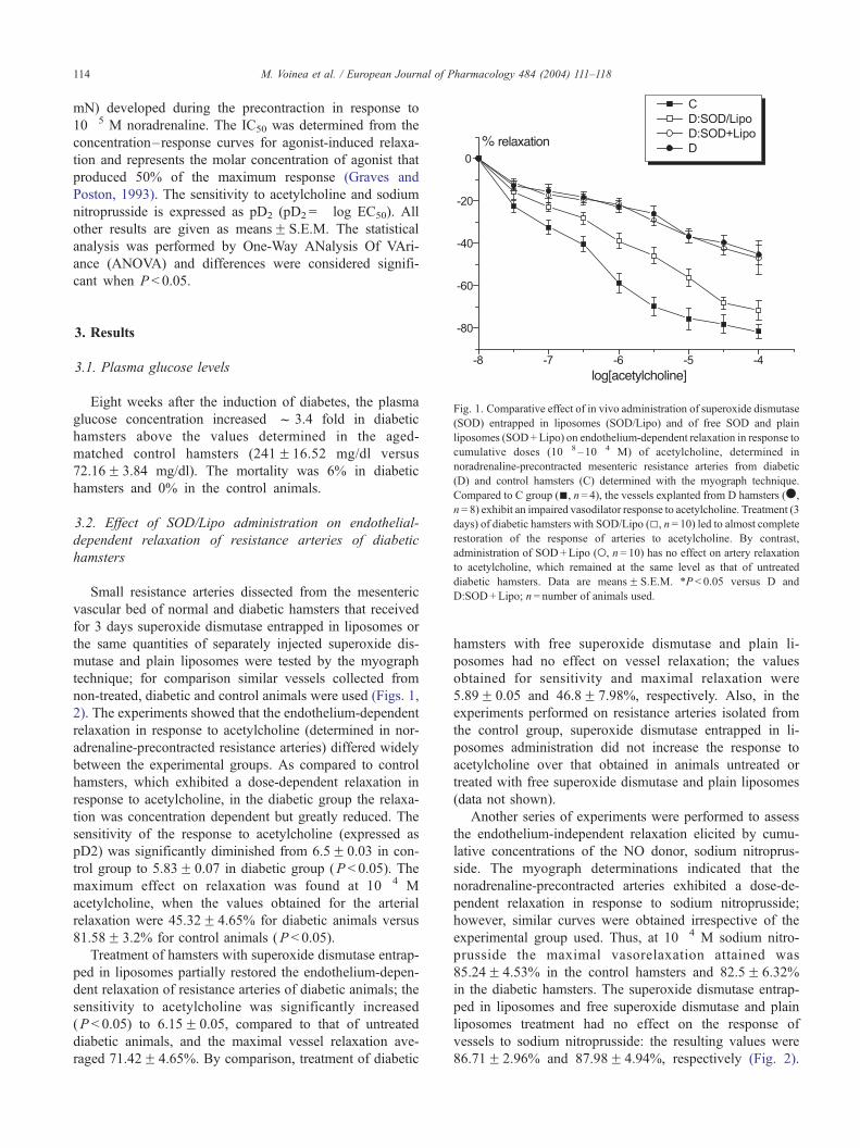
Fig. 1. Comparative effect of in vivo administration of superoxide dismutase
(SOD) entrapped in liposomes (SOD/Lipo) and of free SOD and plain
liposomes (SOD+Lipo) on endothelium-dependent relaxation in response to
cumulative doses (10� 8 –10� 4 M) of acetylcholine, determined in
noradrenaline-precontracted mesenteric resistance arteries from diabetic
(D) and control hamsters (C) determined with the myograph technique.
Compared to C group (n, n= 4), the vessels explanted from D hamsters (.,
n= 8) exhibit an impaired vasodilator response to acetylcholine. Treatment (3
days) of diabetic hamsters with SOD/Lipo (5, n= 10) led to almost complete
restoration of the response of arteries to acetylcholine. By contrast,
administration of SOD+Lipo (o, n= 10) has no effect on artery relaxation
to acetylcholine, which remained at the same level as that of untreated
diabetic hamsters. Data are meansF S.E.M. *P < 0.05 versus D and
D:SOD+Lipo; n= number of animals used.
3. Results
3.1. Plasma glucose levels
Eight weeks after the induction of diabetes, the plasma
glucose concentration increased f 3.4 fold in diabetic
hamsters above the values determined in the aged-
matched control hamsters (241F16.52 mg/dl versus
72.16F 3.84 mg/dl). The mortality was 6% in diabetic
hamsters and 0% in the control animals.
3.2. Effect of SOD/Lipo administration on endothelial-
dependent relaxation of resistance arteries of diabetic
hamsters
Small resistance arteries dissected from the mesenteric
vascular bed of normal and diabetic hamsters that received
for 3 days superoxide dismutase entrapped in liposomes or
the same quantities of separately injected superoxide dis-
mutase and plain liposomes were tested by the myograph
technique; for comparison similar vessels collected from
non-treated, diabetic and control animals were used (Figs. 1,
2). The experiments showed that the endothelium-dependent
relaxation in response to acetylcholine (determined in nor-
adrenaline-precontracted resistance arteries) differed widely
between the experimental groups. As compared to control
hamsters, which exhibited a dose-dependent relaxation in
response to acetylcholine, in the diabetic group the relaxa-
tion was concentration dependent but greatly reduced. The
sensitivity of the response to acetylcholine (expressed as
pD2) was significantly diminished from 6.5F 0.03 in con-
trol group to 5.83F 0.07 in diabetic group (P < 0.05). The
maximum effect on relaxation was found at 10� 4 M
acetylcholine, when the values obtained for the arterial
relaxation were 45.32F 4.65% for diabetic animals versus
81.58F 3.2% for control animals (P < 0.05).
Treatment of hamsters with superoxide dismutase entrap-
ped in liposomes partially restored the endothelium-depen-
dent relaxation of resistance arteries of diabetic animals; the
sensitivity to acetylcholine was significantly increased
(P < 0.05) to 6.15F 0.05, compared to that of untreated
diabetic animals, and the maximal vessel relaxation ave-
raged 71.42F 4.65%. By comparison, treatment of diabetic
hamsters with free superoxide dismutase and plain li-
posomes had no effect on vessel relaxation; the values
obtained for sensitivity and maximal relaxation were
5.89F 0.05 and 46.8F 7.98%, respectively. Also, in the
experiments performed on resistance arteries isolated from
the control group, superoxide dismutase entrapped in li-
posomes administration did not increase the response to
acetylcholine over that obtained in animals untreated or
treated with free superoxide dismutase and plain liposomes
(data not shown).
Another series of experiments were performed to assess
the endothelium-independent relaxation elicited by cumu-
lative concentrations of the NO donor, sodium nitroprus-
side. The myograph determinations indicated that the
noradrenaline-precontracted arteries exhibited a dose-de-
pendent relaxation in response to sodium nitroprusside;
however, similar curves were obtained irrespective of the
experimental group used. Thus, at 10� 4 M sodium nitro-
prusside the maximal vasorelaxation attained was
85.24F 4.53% in the control hamsters and 82.5F 6.32%
in the diabetic hamsters. The superoxide dismutase entrap-
ped in liposomes and free superoxide dismutase and plain
liposomes treatment had no effect on the response of
vessels to sodium nitroprusside: the resulting values were
86.71F 2.96% and 87.98F 4.94%, respectively (Fig. 2).
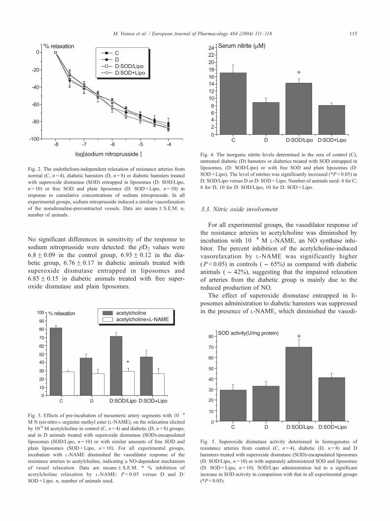
Fig. 4. The inorganic nitrite levels determined in the sera of control (C),
untreated diabetic (D) hamsters or diabetics treated with SOD entrapped in
liposomes, (D: SOD/Lipo) or with free SOD and plain liposomes (D:
SOD+Lipo). The level of nitrites was significantly increased (*P < 0.05) in
D: SOD/Lipo versus D or D: SOD+Lipo. Number of animals used: 4 for C;
8 for D; 10 for D: SOD/Lipo; 10 for D: SOD+Lipo.
Fig. 2. The endothelium-independent relaxation of resistance arteries from
normal (C, n= 4), diabetic hamsters (D, n= 8) or diabetic hamsters treated
with superoxide dismutase (SOD) entrapped in liposomes (D: SOD/Lipo,
n= 10) or free SOD and plain liposomes (D: SOD+Lipo, n= 10) in
response to cumulative concentrations of sodium nitroprusside. In all
experimental groups, sodium nitroprusside induced a similar vasorelaxation
of the noradrenaline-precontracted vessels. Data are meansF S.E.M. n,
number of animals.
M. Voinea et al. / European Journal of Pharmacology 484 (2004) 111–118 115
No significant differences in sensitivity of the response to
sodium nitroprusside were detected: the pD2 values were
6.8F 0.09 in the control group, 6.93F 0.12 in the dia-
betic group, 6.76F 0.17 in diabetic animals treated with
superoxide dismutase entrapped in liposomes and
6.85F 0.15 in diabetic animals treated with free super-
oxide dismutase and plain liposomes.
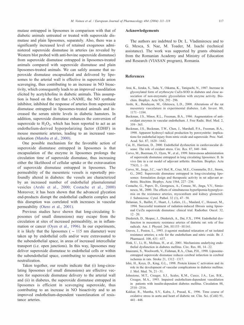
Fig. 3. Effects of pre-incubation of mesenteric artery segments with 10� 4
M N (N)-nitro-L-arginine methyl ester (L-NAME), on the relaxation elicited
by 10-4 M acetylcholine in control (C, n= 4) and diabetic (D, n= 8) groups,
and in D animals treated with superoxide dismutase (SOD)-encapsulated
liposomes (SOD/Lipo, n= 10) or with similar amounts of free SOD and
plain liposomes (SOD +Lipo, n = 10). For all experimental groups,
incubation with L-NAME diminished the vasodilator response of the
resistance arteries to acetylcholine, indicating a NO-dependent mechanism
of vessel relaxation. Data are meansF S.E.M. * % inhibition of
acetylcholine relaxation by L-NAME: P < 0.05 versus D and D:
SOD+Lipo. n, number of animals used.
3.3. Nitric oxide involvement
For all experimental groups, the vasodilator response of
the resistance arteries to acetylcholine was diminished by
incubation with 10� 4 M L-NAME, an NO synthase inhi-
bitor. The percent inhibition of the acetylcholine-induced
vasorelaxation by L-NAME was significantly higher
(P < 0.05) in controls (f 65%) as compared with diabetic
animals (f 42%), suggesting that the impaired relaxation
of arteries from the diabetic group is mainly due to the
reduced production of NO.
The effect of superoxide dismutase entrapped in li-
posomes administration to diabetic hamsters was suppressed
in the presence of L-NAME, which diminished the vasodi-
Fig. 5. Superoxide dismutase activity determined in homogenates of
resistance arteries from control (C, n= 4), diabetic (D, n= 8) and D
hamsters treated with superoxide dismutase (SOD)-encapsulated liposomes
(D: SOD/Lipo, n= 10) or with separately administered SOD and liposomes
(D: SOD+Lipo, n= 10). SOD/Lipo administration led to a significant
increase in SOD activity in comparison with that in all experimental groups
(*P< 0.05).
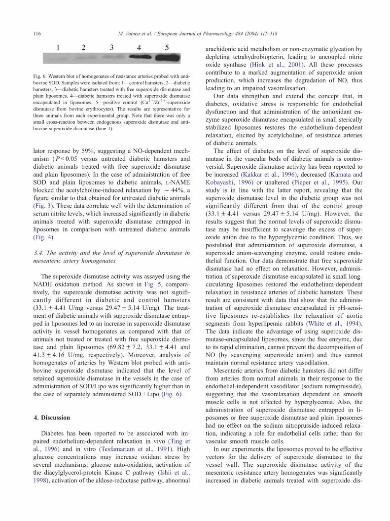
Fig. 6. Western blot of homogenates of resistance arteries probed with anti-
bovine SOD. Samples were isolated from: 1—control hamsters, 2—diabetic
hamsters, 3—diabetic hamsters treated with free superoxide dismutase and
plain liposomes, 4—diabetic hamsters treated with superoxide dismutase
encapsulated in liposomes, 5—positive control (Cu2 +/Zn2 +-superoxide
dismutase from bovine erythrocytes). The results are representative for
three animals from each experimental group. Note that there was only a
small cross-reaction between endogenous superoxide dismutase and anti-
bovine superoxide dismutase (lane 1).
M. Voinea et al. / European Journal of Pharmacology 484 (2004) 111–118116
lator response by 59%, suggesting a NO-dependent mech-
anism (P < 0.05 versus untreated diabetic hamsters and
diabetic animals treated with free superoxide dismutase
and plain liposomes). In the case of administration of free
SOD and plain liposomes to diabetic animals, L-NAME
blocked the acetylcholine-induced relaxation by f 44%, a
figure similar to that obtained for untreated diabetic animals
(Fig. 3). These data correlate well with the determination of
serum nitrite levels, which increased significantly in diabetic
animals treated with superoxide dismutase entrapped in
liposomes in comparison with untreated diabetic animals
(Fig. 4).
3.4. The activity and the level of superoxide dismutase in
mesenteric artery homogenates
The superoxide dismutase activity was assayed using the
NADH oxidation method. As shown in Fig. 5, compara-
tively, the superoxide dismutase activity was not signifi-
cantly different in diabetic and control hamsters
(33.1F 4.41 U/mg versus 29.47F 5.14 U/mg). The treat-
ment of diabetic animals with superoxide dismutase entrap-
ped in liposomes led to an increase in superoxide dismutase
activity in vessel homogenates as compared with that of
animals not treated or treated with free superoxide dismu-
tase and plain liposomes (69.82F 7.2, 33.1F 4.41 and
41.3F 4.16 U/mg, respectively). Moreover, analysis of
homogenates of arteries by Western blot probed with anti-
bovine superoxide dismutase indicated that the level of
retained superoxide dismutase in the vessels in the case of
administration of SOD/Lipo was significantly higher than in
the case of separately administered SOD+Lipo (Fig. 6).
4. Discussion
Diabetes has been reported to be associated with im-
paired endothelium-dependent relaxation in vivo (Ting et
al., 1996) and in vitro (Tesfamariam et al., 1991). High
glucose concentrations may increase oxidant stress by
several mechanisms: glucose auto-oxidation, activation of
the diacylglycerol-protein Kinase C pathway (Ishii et al.,
1998), activation of the aldose-reductase pathway, abnormal
arachidonic acid metabolism or non-enzymatic glycation by
depleting tetrahydrobiopterin, leading to uncoupled nitric
oxide synthase (Hink et al., 2001). All these processes
contribute to a marked augmentation of superoxide anion
production, which increases the degradation of NO, thus
leading to an impaired vasorelaxation.
Our data strengthen and extend the concept that, in
diabetes, oxidative stress is responsible for endothelial
dysfunction and that administration of the antioxidant en-
zyme superoxide dismutase encapsulated in small sterically
stabilized liposomes restores the endothelium-dependent
relaxation, elicited by acetylcholine, of resistance arteries
of diabetic animals.
The effect of diabetes on the level of superoxide dis-
mutase in the vascular beds of diabetic animals is contro-
versial. Superoxide dismutase activity has been reported to
be increased (Kakkar et al., 1996), decreased (Kamata and
Kobayashi, 1996) or unaltered (Pieper et al., 1995). Our
study is in line with the latter report, revealing that the
superoxide dismutase level in the diabetic group was not
significantly different from that of the control group
(33.1F 4.41 versus 29.47F 5.14 U/mg). However, the
results suggest that the normal levels of superoxide dismu-
tase may be insufficient to scavenge the excess of super-
oxide anion due to the hyperglycemic condition. Thus, we
postulated that administration of superoxide dismutase, a
superoxide anion-scavenging enzyme, could restore endo-
thelial function. Our data demonstrate that free superoxide
dismutase had no effect on relaxation. However, adminis-
tration of superoxide dismutase encapsulated in small long-
circulating liposomes restored the endothelium-dependent
relaxation in resistance arteries of diabetic hamsters. These
result are consistent with data that show that the adminis-
tration of superoxide dismutase encapsulated in pH-sensi-
tive liposomes re-establishes the relaxation of aortic
segments from hyperlipemic rabbits (White et al., 1994).
The data indicate the advantage of using superoxide dis-
mutase-encapsulated liposomes, since the free enzyme, due
to its rapid elimination, cannot prevent the decomposition of
NO (by scavenging superoxide anion) and thus cannot
maintain normal resistance artery vasodilation.
Mesenteric arteries from diabetic hamsters did not differ
from arteries from normal animals in their response to the
endothelial-independent vasodilator (sodium nitroprusside),
suggesting that the vasorelaxation dependent on smooth
muscle cells is not affected by hyperglycemia. Also, the
administration of superoxide dismutase entrapped in li-
posomes or free superoxide dismutase and plain liposomes
had no effect on the sodium nitroprusside-induced relaxa-
tion, indicating a role for endothelial cells rather than for
vascular smooth muscle cells.
In our experiments, the liposomes proved to be effective
vectors for the delivery of superoxide dismutase to the
vessel wall. The superoxide dismutase activity of the
mesenteric resistance artery homogenates was significantly
increased in diabetic animals treated with superoxide dis-
M. Voinea et al. / European Journal of Pharmacology 484 (2004) 111–118 117
mutase entrapped in liposomes in comparison with that of
diabetic animals untreated or treated with superoxide dis-
mutase and plain liposomes, separately. Also, there was a
significantly increased level of retained exogenous admi-
nistered superoxide dismutase in arteries (as revealed by
Western blot probed with anti-bovine superoxide dismutase)
from superoxide dismutase entrapped in liposomes-treated
animals compared with superoxide dismutase and plain
liposomes-treated animals. We can safely assume that su-
peroxide dismutase encapsulated and delivered by lipo-
somes to the arterial wall is effective in superoxide anion
scavenging, thus contributing to an increase in NO bioac-
tivity, which consequently leads to an improved vasodilation
elicited by acetylcholine in diabetic animals. This assump-
tion is based on the fact that L-NAME, the NO synthase
inhibitor, inhibited the response of arteries from superoxide
dismutase entrapped in liposomes-treated animals and in-
creased the serum nitrite levels in diabetic hamsters. In
addition, superoxide dismutase enhances the conversion of
superoxide to H2O2, which has been reported to act as an
endothelium-derived hyperpolarizing factor (EDHF) in
mouse mesenteric arteries, leading to an increased vaso-
relaxation (Matoba et al., 2000).
One possible mechanism for the favorable action of
superoxide dismutase entrapped in liposomes is that
encapsulation of the enzyme in liposomes prolongs the
circulation time of superoxide dismutase, thus increasing
either the likelihood of cellular uptake or the extravasation
of superoxide dismutase entrapped in liposomes. The
permeability of the mesenteric vessels is reportedly pro-
foundly altered in diabetes: the vessels are characterized
by an increased number of endothelial plasmalemmal
vesicles (Arshi et al., 2000; Costache et al., 2000)
Moreover, it has been shown that the advanced glycation
end-products disrupt the endothelial cadherin complex and
this disruption was correlated with increases in vascular
permeability (Otero et al., 2001).
Previous studies have shown that long-circulating li-
posomes (of small dimensions) may escape from the
circulation at sites of increased permeability, as in inflam-
mation or cancer (Oyen et al., 1996). In our experiments,
it is likely that the liposomes (f 115 nm diameter) were
taken up by endothelial cells and/or were extravasated to
the subendothelial space, in areas of increased intercellular
transport (i.e. open junctions). In this way, liposomes may
deliver superoxide dismutase to endothelial cells or within
the subendothelial space, contributing to superoxide anion
neutralization.
Taken together, our results indicate that (i) long-circu-
lating liposomes (of small dimensions) are effective vec-
tors for superoxide dismutase delivery to the arterial wall
and (ii) in diabetes, the superoxide dismutase entrapped in
liposomes is efficient in scavenging superoxide, thus
contributing to an increase in NO bioactivity and to an
improved endothelium-dependent vasorelaxation of resis-
tance arteries.
Acknowledgements
The authors are indebted to Dr. L. Vladimirescu and to
G. Mesca, S. Nae, M. Toader, M. Isachi (technical
assistance). The work was supported by grants obtained
from the Romanian Academy and Ministry of Education
and Research (VIASAN program), Romania.
References
Arai, K., Iizuka, S., Tada, Y., Oikama, K., Taniguchi, N., 1987. Increase in
glycosylated form of erythrocyte CuZn-SOD in diabetes and close as-
sociation of non-enzymatic glycosilation with enzyme activity. Bio-
chim. Biophys. Acta 924, 292–296.
Arshi, K., Bendayan, M., Ghitescu, L.D., 2000. Alterations of the rat
mesentery vasculature in experimental diabetes. Lab. Invest. 80,
1171–1184.
Beckman, J.S., Minor, R.L., Freeman, B.A., 1986. Augmentation of anti-
oxidant enzymes in vascular endothelium. J. Free Radic. Biol. Med. 2,
359–365.
Beckman, J.S., Beckman, T.W., Chen, J., Marshall, P.A., Freeman, B.A.,
1990. Apparent hydroxyl radical production by peroxynitrite: implica-
tions for endothelial injury from nitric oxide and superoxide. Proc. Natl.
Acad. Sci. 87, 1620–1642.
Cai, H., Harrison, D., 2000. Endothelial dysfunction in cardiovascular di-
sease. The role of oxidant stress. Circ. Res. 87, 840–844.
Corvo, M., Boerman, O., Oyen, W., et al., 1999. Intravenous administration
of superoxide dismutase entrapped in long circulating liposomes: II. In
vivo fate in a rat model of adjuvant arthritis. Biochim. Biophys. Acta
77633, 1–10.
Corvo, M., Jorge, J.C., van’t Hof, R., Cruz, M.E., Crommelin, D.J., Storm,
G., 2002. Superoxide dismutase entrapped in long-circulating lipo-
somes: formulation design and therapeutic activity in rat adjuvant ar-
thritis. Biochim. Biophys. Acta 1564, 227–236.
Costache, G., Popov, D., Georgescu, A., Cenuse, M., Jinga, V.V., Simio-
nescu, M., 2000. The effects of simultaneous hyperlipemia-hyperglyce-
mia on the resistance arteries, myocardium and kidney glomeruli.
J. Submicrosc. Cytol. Pathol. 32 (1), 47–58.
Delanian, S., Baillet, F., Huart, J., Lefaix, J.L., Maulard, C., Housset, M.,
1994. Successful treatment of radiation-induced fibrosis using liposo-
mal Cu/Zn superoxide dismutase: clinical trial. Radiother. Oncol. 32,
12–20.
Diederich, D., Skopec, J., Diederich, A., Dai, F.X., 1994. Endothelial dys-
function in mesenteric resistance arteries of diabetic rat: role of free
radicals. Am. J. Physiol. 266, H1153–H1161.
Graves, J., Poston, L., 1993. h-agonist mediated relaxation of rat isolated
resistance arteries; a role for the endothelium and nitric oxide. Br. J.
Pharmacol. 108, 631–637.
Hink, U., Li, H., Mollnau, H., et al., 2001. Mechanisms underlying endo-
thelial dysfunction in diabetes mellitus. Circ. Res. 88, 14–22.
Imaizumi, S., Woolworth, V., Fishman, R.A., Chan, P.H., 1990. Liposome-
entrapped superoxide dismutase reduces cerebral infarction in cerebral
ischemia in rats. Stroke 21, 1312–1317.
Ishii, H., Koya, D., King, G.L., 1998. Protein kinase C activation and its
role in the development of vascular complications in diabetes mellitus.
J. Mol. Med. 76, 21–31.
Johnstone, M.T., Creager, S.J., Scales, K.M., Cusco, J.A., Lee, B.K.,
Creager, M.A., 1993. Impaired endothelium-dependent vasodilation
in patients with insulin-dependent diabetes mellitus. Circulation 88,
2510–2516.
Kakkar, R., Mantha, S.V., Kalra, J., Prasad, K., 1996. Time course of
oxidative stress in aorta and heart of diabetic rat. Clin. Sci. (Cohl) 91,
441–448.
M. Voinea et al. / European Journal of Pharmacology 484 (2004) 111–118118
Kamata, K., Kobayashi, T., 1996. Changes in superoxide dismutase mRNA
expression by streptozotocin-induced diabetes. Br. J. Pharmacol. 119,
583–589.
Matoba, T., Shimokawa, H., Nakashima, M., et al., 2000. Hydrogen pe-
roxide is an endothelium-derived hyperpolarizing factor in mice. J. Clin.
Invest. 106, 1521–1530.
Mayhan, W.G., Patel, K.P., 1998. Treatment with dimethylthiourea pre-
vents impaired dilatation of the basilar artery during diabetes mellitus.
Am. J. Physiol, Heart Circ. Physiol. 274, H1895–H1901.
Mazzon, E., De Sarro, A., Caputi, A.P., Cuzzocrea, S., 2002. Role of tight
junction derangement in the endothelial dysfunction elicited by exoge-
nous and endogenous peroxynitrite and poly(ADP-ribose) synthetase.
Shock 18, 434–439.
Mugge, A., Elwell, J.H., Peterson, T.E., Hofmeyer, T.G., Heistad, D.D.,
Harrison, D.G., 1991. Chronic treatment of polyethylene glycolated
superoxide dismutase partially restores endothelium-dependent vascular
relaxations in cholesterol-fed rabbits. Circ. Res. 69, 1293–1300.
Mulvany, M.J., Halpern, W., 1977. Contractile properties of small arterial
resistance vessels in spontaneously hypertensive and normotensive rats.
Circ. Res. 41, 19–26.
Nakae, D., Yamamoto, K., Yoshiji, H., et al., 1990a. Liposome-encapsu-
lated superoxide dismutase prevents liver necrosis induced by acetami-
nophen. Am. J. Pathol. 136, 787–795.
Nakae, D., Yoshiji, H., Amanuma, T., Kinugasa, J., Farber, J., Konishi, Y.,
1990b. Endocytosis-independent uptake of liposome-encapsulated
superoxide dismutase prevents the killing of cultured hepatocytes by
tert-butyl hydroperoxide. Arch. Biochem. Biophys. 279, 315–319.
Otero, K., Martinez, F., Beltran, A., et al., 2001. Albumin-derived ad-
vanced glycation end-products trigger the disruption of the vascular
endothelial cadherin complex in cultured human and murine endothelial
cells. Biochem. J. 359, 567–574.
Oyen, W.J., Boerman, O.C., van der Laken, C.J., Claessens, R.A., van der
Meer, J.W., Corstens, F.H., 1996. The uptake mechanisms of inflamma-
tion-and infection-localizing agents. Eur. J. Nucl. Med. 23, 459–465.
Paoletti, F., Mocali, A., 1990. Determination of superoxide dismutase ac-
tivity by purely chemical system based on NAD(P)H oxidation. Me-
thods Enzymol. 186, 209–220.
Paternostre, M., Ollivon, M., Bolard, J., 1996. Description of four selected
techniques of liposome preparation. In: Prasad, R. (Ed.), Manual on
Membrane Lipids. Springer, Berlin, pp. 218–226.
Pieper, G.M., 1998. Review of alterations in endothelial nitric oxide pro-
duction in diabetes; protective role of arginine on endothelial dysfunc-
tion. Hypertension 31, 1047–1060.
Pieper, G.M., Jordan, M., Dondlinger, L.A., Adams, M.B., Roza, A.M.,
1995. Peroxidative stress in diabetic blood vessels. Diabetes 44,
884–889.
Schaffuer, W., Weissmann, C., 1973. A rapid, sensitive and specific method
for the determination of protein in dilute solution. Anal. Biochem. 56,
502–514.
Smarason, A.K., Allman, K.G., Young, D., Redman, C.W.G., 1997. Ele-
vated levels of serum nitrate, a stable end product of nitric oxide, in
women with pre-eclampsia. Br. J. Obstet. Gynaecol. 104, 538–543.
Tesfamariam, B., 1994. Free radicals in diabetic endothelial cell dysfunc-
tion. Free Radic. Biol. Med. 16, 383–392.
Tesfamariam, B., Brown, M.L., Cohen, R.A., 1991. Elevated glucose im-
pairs endothelium-dependent relaxation by activating protein kinase C.
J. Clin. Invest. 87, 1643–1648.
Ting, H.H., Timimi, F.K., Boles, K.S., Creager, S.J., Ganz, P., Creager,
M.A., 1996. Vitamin C improves endothelium-dependent vasodilation
in patients with non-insulin-dependent diabetes mellitus. J. Clin. Invest.
97, 22–28.
White, C.R., Brock, T.A., Chang, L.Y., et al., 1994. Superoxide and pero-
xinitrite in atherosclerosis. Proc. Natl. Acad. Sci. 91, 1044–1048.
Yunoki, M., Kawauchi, M., Ukita, N., et al., 1997. Effects of lecithinized
superoxide dismutase on traumatic brain injury in rats. J. Neurotrauma
14, 739–746.
Yusa, T., Crapo, J., Freeman, B.A., 1984. Liposome-mediated augmenta-
tion of brain SOD and catalase inhibits CNS O2 toxicity. J. Appl.
Physiol. 57, 1674–1681.