Neural Prolotherapy REGIONAL ANATOMY UPPER EXTREMITIES Lee Wolfer, MD, MS.
Upload
hoangthuanCategory
view
216download
0
Slide 1
Summary of Completed Research in Prolotherapy
and BioregenerativeInjection
K. Dean Reeves, M.D.Clinical Associate Professor
University of Kansas
"This work is provided under the
Creative Commons Attribution 3.0 Unported License."
This is an summary of prolotherapy research that is considered high quality, grade I to II level.
(This will be explained shortly.)

Slide 2
Sections of Talk
• Definitions
The slides will be explained as we proceed through. The first topic will be to define treatments
so we know what research applies to each treatment.

Slide 3
Nomenclature
Application of lotion to restore normal function in pain producing sensory nerves.
Caudal dextrose Injection
Subcutaneous injection to restore function
in pain producing sensory nerves.
Injection to repair connective tissue
(Lig/Tend/Cart) (Not including biologics)
Injection of biologics to repair connective
tissue.
BRI : Biologic Repair
Injection
Prolotherapy (Prolo)
PSI (Perineural
Subcutaneous Injection)
Caudal Dex: Caudal
Dextrose Injection
PDI (Perineural Deep
Injection) Also called
hydrodissection
PTA (Perineural Topical
Application)
Deeper nerve stretch, typically with
ultrasound guidance.
Other terms NPT or PIT
The bulk of research so far is focused on the on the areas in red that you see on the slide. These
two areas are biologic repair injection and prolotherapy.
Note that perineural injection was introduced as neural prolotherapy or NPT for short. However,
it is uncertain at this time if perineural injections causes proliferation so a more generic term
such as perineural injection is preferred. Note that in Australia and New Zealand the term
perineural injection therapy (PIT is becoming more commonly used). This may be the term that
will become most widely accepted.

Slide 4
Def: Biologic and Biologic Repair Injection (BRI)
• Biologic: A preparation that is synthesized from living
organisms or their products and used medically as a diagnostic, preventive, or therapeutic agent.
www.thefreedictionary/biologic
• B.R.I.: Injection of biologics to repair connective tissue. – Mechanism proposed is by stimulation of a repair cascade
– Various methods are utilized. IE: Whole blood, PRP, stem
cells.
– Emphasis is on direct regenerative/proliferative effects on
connective tissue.
BRI (Biologic Repair Injection) is the “Injection of biologics to repair connective tissue”. A
“biologic” is something from a living organism. For purposes of BRI in humans, and to avoid
rejection, the biologic is from the person themselves. Sometimes is it as simple as withdrawing
blood and injecting it into another part of the body. There are other biologics that are removed
and modified before re-injection. An example of this is drawing blood, spinning it in a
centrifuge to separate blood cells from plasma, and re-injecting the plasma with concentrated
platelets (platelet rich plasma) into an area of the body that needs repair. Stem cells are also
biologics and can be taken from bone marrow or fat cells of patients, modified in special ways,
and re-injected.
Connective tissue includes ligament, tendon, and cartilage. The primary emphasis of BRI is
direct repair of “connecting tissues”. These include primarily ligaments (bone to bone
connections), tendons (muscle to bone connections) and cartilage (the covering layer over
joints). The repair process, once begun, takes months for full completion after any single
treatment, although sometimes the healing cycles are overlapped in an attempt to speed up the
effect.

Slide 5
Def: Prolotherapy
• Prolotherapy: Injection to repair connective tissue (Not including biologics)
Prolotherapy is “Injection to repair connective tissue.” (Identical purpose to biologic repair
injection but, by definition, without use of biologics.) Thus, solutions used for prolotherapy do
not contain anything from the person being injected. The most common solution is dextrose
and others in common use that are added to dextrose include diluted sodium morrhuate (a salt
of morrhuic acid which is more inflammatory than dextrose), and diluted phenol (a six carbon
atom solution which is also more inflammatory and is also called carbolic acid). The dilution is
usually 9 to 1 for sodium morrhuate and 99 to 1 for phenol.
Slide 6
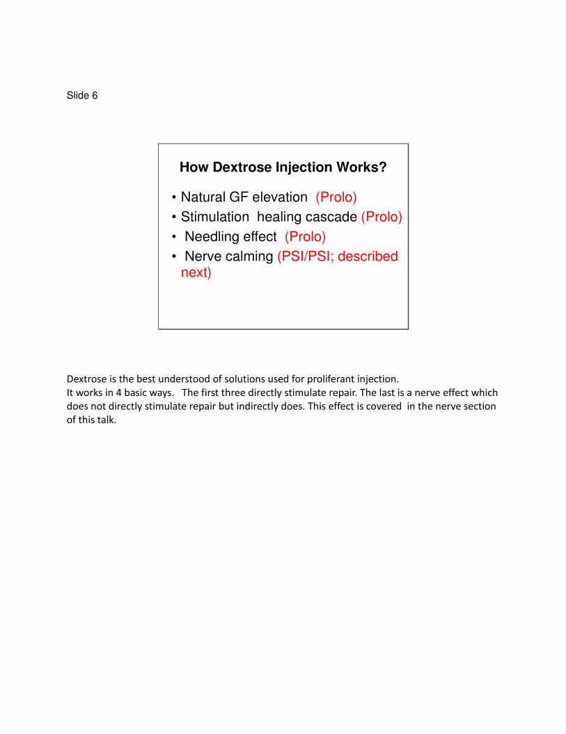
How Dextrose Injection Works?
• Natural GF elevation (Prolo)
• Stimulation healing cascade (Prolo)
• Needling effect (Prolo)
• Nerve calming (PSI/PSI; described next)
Dextrose is the best understood of solutions used for proliferant injection.
It works in 4 basic ways. The first three directly stimulate repair. The last is a nerve effect which
does not directly stimulate repair but indirectly does. This effect is covered in the nerve section
of this talk.

Slide 7
Concept: Time Frame Of Pain and
Functional Benefit Effects Research:
Ex Dextrose• Nerve Calming: 5 -20 seconds – 2 days
• Dextrose Noninflammatory Effect on GFs: 20
min – 10 days (Pain relief reason unclear)
• Hyperosmolar/irritative or Needling Effect:
20 min -10 days (Pain relief reason unclear)
• Tissue Maturation Over time: 10 days – 3
months (Repair with less insufficiency related
pain stimulation)
If you are going to research, whatever you are studying may have several effects whether you
are trying to measure either pain or functional changes as a result of the treatment.
Because our muscles are inhibited by nerves that are not functioning right, pain improvements
also cause improvements in both pain and function.
The speed of nerve calming effect explains why patients often feel better immediately,
especially if a technique is used that does not create immediate inflammation is gentler. The
downregulation effect on nerves begins in 5-20 seconds and lasts up to 2 days.
When perineural injection is done alone, the effect typically lasts 4-48 hours, and after several
treatments lasting benefit is usually noted.
However, there are overlapping effects from deeper injection that can continuing improvement
in some subjects/patients. Growth factors DNA activity changes occur within 20 minutes as a
result of dextrose exposure and changes are likely as fast with inflammatory proliferant
injection.
Thus, the conclusions of a study and data gathered may vary a great deal, depending on the
time of information gathering. IE: The epidural dextrose study. (Results to be available shortly

with immediate effects on nerve calming studied by immediate follow-up data gathering to 48
hours)

Slide 8
Natural GF Elevation Effect of
Dextrose > .4 %
Restoration of function in soft tissue is accomplished by altering the balance of growth factors
(GF) and disrepair factors (DF). A growth factor is a complex protein (polypeptide) that changes
the function of our DNA. This is much like a key being placed in a lock that turns on the “engine”
of the cell almost instantaneously. These GFs are produced in our own cells and dextrose, in as
little as 0.5% concentrations stimulations production GFs.

Slide 9
GFs that Dextrose Elevates
This slide lists some of the key GFs that stimulate repair in ligament/tendon (Lig/Tend) and
cartilage. The effect of dextrose exposure on these GF levels is shown in red.
Dextrose also has effects on complex proteins that nerves produce (neuropeptides = nerve
proteins) that are discussed elsewhere and are also quite important and the subject of research
at this time.
Murphy et al 1 found that elevation of glucose from 0.1% (a normal extracellular level) to 0.45%
(level found in diabetics intermittently) results in production of as many as 15 different proteins,
including key growth factors for soft tissue (IE connective tissue growth factor (CTGF) and
transforming growth factor beta (TGFβ). These effects occur within 20 minutes of cellular
exposure to 0.45% Dextrose. Figure 7 shows growth factor elevations that result from
elevation of dextrose level to 0.45%.2
1. Murphy M, Godson C, Cannon S, Kato S, Mackenzie HS, Martin F, Brady HR: Suppression
subtractive hybridization identifies high glucose levels as a stimulus for expression of connective
tissue growth factor and other genes in human mesangial cells. J Biol Chem 1999;274(9):5830-
5834.
2. Reeves KD, Topol GA, Fullerton BD: Evidence-based regenerative injection therapy
(prolotherapy) in sports medicine. In Seidenberg PH, Beutler PI. (Eds): The Sports Medicine
Resource Manual, Philadelphia, Saunders (Elsevier), 2008, pp 611-619.


Slide 10
GFs Alter DNA to Stimulate Repair
How GFs work is illustrated in a simple way in the figures above. The figure to the left shows a
gene for healing covered by other DNA. When the GF attaches to its receptor on the cell surface
it leads to an immediate change in the DNA structure, exposing key genes for healing.

Slide 11
Dextrose Levels > 10% Stimulate the Inflammatory
Cascade
• Osmotic effect ���� Cells shrink ����Stress with leakage of lipids ����
temporary inflammation
• Other concentrated solutions have similar effect but dextrose has several ways it works other than just osmotic
Simple osmosis, taught in biology, is the principle of water flowing from a low to high
concentration. When a cell is surrounded by a high concentration, it loses water and shrinks.
(crenates). The can stress the cell and cause it to release lipids form the cell membranes or to
produce GFs as a reaction to a preceived threat. This creates a temporary inflammation.
This is identical to how we normally heal but tissues around the cell have not been stretched or
damaged so the healing can proceed favorably without having to undue real damage such as
that of an actual injury. Without this process we could not heal even from a simple cut.
The key is temporary inflammation. Chronic inflammation is not good. Temporary
inflammation is. This is similar to the fact that acute dextrose elevation around a cell stimulate
healing but chronic dextrose elevation, such as in diabetics, does not stimulate healthy changes.

Slide 12
The Needle Itself Stimulates
Repair• Cell membrane disruption ���� Lipid
release
• Small blood vessel disruption ����Bleeding with platelet and blood effects
• Reason why prolotherapy studies with injection control have been dismissed despite good results. These are injection, not placebo, controlled studies.

Slide 13
Nerve Calming Effect of Dextrose
When dextrose injection if given, immediate improvement is often noted.
This is a cross over effect on nerves which is described in the next slides.
Immediate improvement must mean that nerves in the areas of injection are affected favorably,
and that the same solution that repairs ligament or tendon must also do something favorable
for nerves. This is not prolotherapy. It is called perineural injection therapy and will be
described in the next slides.

Slide 14
Why Does Healing Take Time?
• Growth of new tissue to line up along the
weakened rope to strengthen it.
• Dehydration and tightening of the loose rope.
• Together these make and tissue more
capable of normal activity and take several
months, perhaps as much as a year.
• This is why short studies with limited follow-
up are not good ways to study prolotherapy
effects.

Slide 15
Def: Perineural Subcutaneous
Injection (PSI)
• Subcutaneous near-nerve injection to restore function to peptidergic sensory nerves.
– Mechanism proposed is by downregulation of the TRPV1 receptor. Initial research just completed is suggestive of this.
– Injectants for this include primarily dextrose of mannitol currently but other solutions may be utilized as discoveries occur)
– Emphasis is on a direct, primarily immediate, effect on peptidergic nerves.
PSI, by definition, is injection under the skin (subcutaneous) of solutions to restore function to
nerves that cause pain. There are several types of sensory (sensation carrying) nerves that are
capable of transmitting pain. Only certain sensory nerves are able to produce proteins. They
are called “peptidergic” nerves, since peptide is another name for protein and ergic means
“producing”. These special nerves are found in virtually all parts of the body and they produce
either healthy or damaging proteins. There is a control on the surface of the cell that
determines whether the nerve produces healthy or damaging proteins. That control is called a
“receptor”. The receptor has been called the capsaicin (red-pepper) receptor because it is the
same receptor that senses red pepper on the tongue. It has been given a more specific name
and is now called the TRPV1 receptor. It is only found on the surface of protein-producing
(peptidergic) nerve cells.
If the TRPV1 receptor is calm the nerves produce healthy proteins. If the TRPV1 receptor is
overactive (also called up-regulated) it will produce damaging proteins. These damaging
proteins include pain-producing proteins such as substance P and degeneration-producing
proteins such CGRP (calcitonin gene related peptide) and NO (nitric oxide). There is a law
(Hilton’s Law) that says that sensory nerves that supply the skin over a joint also supply that
joint and ligaments and tendons around that joint. Thus when these nerves are producing
damaging proteins, they can travel into nearby joints, ligaments and tendons, causing pain and
damage. Sensory nerves can conduct signals and transport proteins both directions so that

anywhere along the “tree” of that nerve can be affected by proteins that that part of the nerve
produces.
The primary way that perineural injection is thought to work is by calming (down-regulating)
the TRPV1 receptor on the surface of the nerve cell.

Slide 16
Def: PDI (Perineural Deep
Injection) (Hydrodissection )
• Stretching a deeper sensory or combined sensory/ motor nerve under guidance.
– Mechanism proposed is by untethering/restoration of normal motion to a peripheral nerve.
– If dextrose is utilized (Dextrose hydrodissection), an additional mechanism is restoration of nerve function by a direct effect of dextrose.
– If lidocaine is utilized (Lidocaine hydrodissection), an additional mechanism is nerve block
– If steroid is utilized (Steroid hydrodissection), an additional mechanism is a local effect on arachadonicacid inflammation pathway.
PDI (Perineural Deep Injection) is stretching of a deeper nerve under guidance. Our muscles are
surrounded by thin but strong layers of tissue that are a bit like “Saran” wrap. These tissue
layers are called fascia. They also separate our body into layers (Fascial layers). Sensory nerves
have to penetrate these layers and can sometimes get trapped or irritated when they do so.
Animal studies have shown that, when a nerve is touched all around, even without squeezing it,
that nerve can become very irritable and swell, making it even harder for it to fit through the
small holes in the fascia. When a injection of a solution is given around a nerve, the liquid and
pull apart (dissect) the layers of fascia, freeing the nerve. The solutions used for injection
always have some water in them, and the term commonly used for stretching apart by fluid
injection is “hydrodissection” (hydro means water). Several solutions can be used. The effective
of dextrose appears to be by calming the TRPV1 receptor. If anesthetic is used it would be a
nerve block, as lidocaine actually stops nerve transmission for a period of time. If steroid is
used, the injection will have a direct anti-inflammatory effect. For purposes of study discussion
we will focus on the purpose of restoring nerve function since anesthetic and steroid effects
generally are helpful only briefly, and seldom curative in effect.
Slide 17
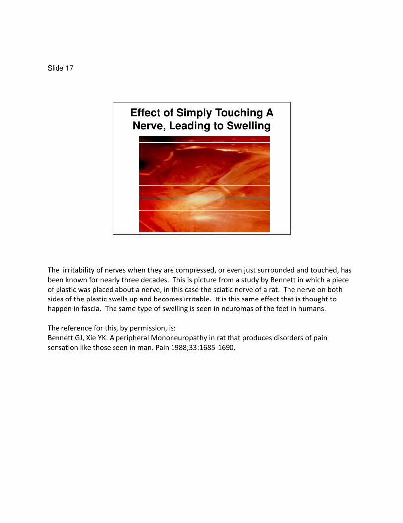
Effect of Simply Touching A
Nerve, Leading to Swelling
The irritability of nerves when they are compressed, or even just surrounded and touched, has
been known for nearly three decades. This is picture from a study by Bennett in which a piece
of plastic was placed about a nerve, in this case the sciatic nerve of a rat. The nerve on both
sides of the plastic swells up and becomes irritable. It is this same effect that is thought to
happen in fascia. The same type of swelling is seen in neuromas of the feet in humans.
The reference for this, by permission, is:
Bennett GJ, Xie YK. A peripheral Mononeuropathy in rat that produces disorders of pain
sensation like those seen in man. Pain 1988;33:1685-1690.

Slide 18
Prolotherapy and Perineural
Injections are Complementary
• Perineural injection helps nerves produce proteins that encourage repair.
• Deeper injection with prolotherapy helps calm nerves in the area where repair is stimulated which can help nerves function better elsewhere.
The goal of prolotherapy is repair of connective tissue and the goal of perineural injection is to
restore normal function in nerve. These two treatments have a complementary effect because:
• Through perineural injection nerves begin producing healthy instead of damaging proteins,
which can favorably affect deeper structures, making it easier for them to heal.
• When deeper structures are injected in prolotherapy, nerves in those structures calm,
helping other nerves in the nerve tree to begin functioning more normally, further reducing
the levels of painful protein production by the nerves.

Slide 19
Def: Caudal Dextrose Injection
• Guided or unguided injection of
dextrose into the caudal epidural space.
• Mechanism is proposed to be partly a hydrodissection effect and a direct nerve effect of dextrose, but this has not been determined.
There is a sac of fluid outside the spinal-cord-containing (dural) sac. It is called the epidural
space. Steroids are commonly injected there. Dextrose is now under investigation as an
alternative for epidural injection, with very favorable initial responses. Completion of the first
study is anticipated within the next year. This is by a modified short needle approach very low
in the back over the lower part of the sacrum bone using a thin needle at a depth of only about
1 inch.

Slide 20
Definition: PTA (Perineural Topical Application)
• Application of lotion to restore normal function in pain producing sensory nerves.
• Mechanism is proposed to be via downregulation of the TRPV1 receptor, but this has not been determined.
• Penetrating agents or other methods of delivery or solutions may be utilized. IE: Ultrasound Delivery of TransdermalDextrose/Vitamin D
Yet another method of treatment being used more is the application of dextrose directly on the
skin in the form of a cream. Dextrose is a small molecule, and, with the help of oil or other
“penetrators” in cream, is thought to be able to penetrate the skin. Since dextrose affects
nerves favorably by injection, if it can penetrate the skin, topical dextrose can affect nerves
favorably. Other applicants are under investigation.

Slide 21
Sections of Talk
• Levels of Evidence
Next we will consider levels of evidence in research

Slide 22
U.S. Preventative Service Task Force Basic Classification of Evidence
• Level I: Well designed RCT (s) with clinical and statistically significant evidence.
• Level II: Well designed:
II-1 Controlled trials without randomization.
Controlled treatment comparison studies.
II-2 Cohort (delayed Rx) studies from more
than one center.
II-3 Uncontrolled trial with dramatic result.
Uncontrolled trial with blindable objective
outcome measure.
Level III. Substantially flawed RCTs or other controlled studies. Single well designed cohort or case control study.
The highest level of evidence is a randomized controlled trial. (A RCT). This is a level I study.
Depending on the size of the study, successful data capture, design quality, and other
characteristics, subratings such as Ia or Ib may be given.
The next level of study, which can be powerful, particularly if more than one study is published
in a given area, is level II. This can include studies in which some patients are treated
immediately and others delayed (delayed treatment study). It also includes studies comparing
a treatment that is being studied with one that is already known to have benefit to see how the
new treatment compares with the old treatment. In addition, trials in which consecutive (one
after another) patients are enrolled can reach level II if they have a dramatic result or if they
have an important result that is measurable objectively. (For example, an X-ray).

Slide 23
DEXTROSE PROLOTHERAPY
and PSI RESEARCH
Using the grading method we talked about, lets look at what evidence we have thus far for
dextrose use in prolotherapy and perineural injection. (Only 1 article for the latter; see Achilles
tendon study by Dr. Lyftogt) We need to remember that we can have a lot of evidence for
something, enough that it is clearly not experimental, but still not force change to occur in
insurance coverage. We will see that is the case.

Slide 24
When Will Prolotherapy Be
Covered by Insurance?
• When doctors start using it routinely to avoid accusations of malpractice.
• When Insurers are forced to cover it due to fear of lawsuit. (Except government insurance which may not be sue-able.)
• When enough largely self-funded studies accumulate
Prolotherapy has been studied the most of techniques mentioned today. It is often asked
“When is prolotherapy going to be covered by insurance”. The short answer is that, because
studies are largely self funded with no pharmaceutical company financial support, it will take a
while for enough research to build up. Another issue is that there has been considerable
“political” resistance to prolotherapy. This had made it apparent that the only way to get
prolotherapy covered by insurance is to “force it” by having so much evidence that it becomes
quite unethical not to offer prolotherapy. When that occurs, insurers will be forced to cover
certain types of prolotherapy. However, be aware that there are many different treatment
approaches and prolotherapy will not be “overall approved”. Instead it will approved area by
area and condition by condition as evidence accumulates to force that.

Slide 25
U.S. Preventative Services Taskforce Recommendations for When Doctors
Should Discuss a Treatment.
• Good evidence Benefit > Risk
Level I evidence and minimal risk)
• Fair evidence Benefit > Risk
Level II evidence and minimal risk)
Note that U.S. guidelines indicate that doctors have an ethical obligation to mention treatments
to patients that have level I or II evidence and minimal risk, if they know about them. Multiple
reviews have been published showing that prolotherapy is as safe as any other injection
technique. I.E. Rabago D, Slattengren A, Zgierska A. Prolotherapy in Primary Care Practice. Prim
Care. 2010 March ; 37(1): 65–80. The following slides make it clear the there is level I or II
evidence for prolotherapy in treatment of several different conditions.

Slide 26
Although Further Research is Needed, Prolotherapy is NOT
Experimental • It is taught as an acceptable method
procedure by one or more approved post
graduate programs for the healing arts?
(Univ Wisconsin, specialty college in
AOA), and
• It is based upon sufficient learned
publications supporting the safety and
efficacy? (Level II or higher in multiple
areas)
For a treatment to not be considered experimental it needs to meet two basic criteria.
Prolotherapy meets these criteria easily. It is taught by approved post graduate programs and
there is level II or higher research in multiple areas.

Slide 27
Summary of Published Prolotherapy
Research
• Dextrose Prolotherapy has 4 areas of
level I evidence (Knee OA, OSD, Finger
OA, and Lateral Epicondylosis, and 5
additional areas of level II evidence (SI
joint pain, Low Back Pain, Achilles
Tendinosis, Groin Pain, and ACL laxity.
This slide lists 4 areas of level I evidence and 5 additional areas of level II evidence for
prolotherapy using dextrose. Thus, according to the U.S. Preventative Services Task Force,
doctors should be discussing this treatment. The following slides will describe that evidence.

Slide 28
Good Size
Sig. Clinically
Sig. Statistically
Adequate F-UP
Data Capture
Accepted Tool
Simple
Inexpensive
Min. invasive
Study Characteristics That Force Change
• Change will be forced if TWO
studies are published in Pub
Med journals that are good
size, show both clinical and
statistically significant benefit,
have adequate follow-up, good
data capture, and use accepted
tools, PARTICULARLY if they
use simple, inexpensive and
minimally invasive methods
which are practical for a
primary care practitioner.
There are certain characteristics that very high quality studies have. This is quite a challenge for
largely non funded studies. In order to do so the methods need to be quite simple whenever
possible to improve affordability. A look at this list on the right reveals some obvious
characteristics such as a study being good size (which usually means 20 or more in each group).
The outcome of a study needs to not only be significant according to statistics, but also must be
significant in terms of its amount of benefit to the patients. (IE: Did it make a big difference for
the patient’s quality of life?) A good study needs to follow patients long enough to be sure the
benefit will hold and that “capture” of data needs to be good so that one is sure that selective
data was not gathered. Studies also use measures of improvement that are called
measurement tools. Some are better than others and it is preferable to use a well accepted
measurement tool. Several keys often not considered include simplicity and minimal discomfort
or “invasiveness” so that primary physicians will want to do the technique. (Practical P.C.
[practical for primary care use])
If a good quality study can be repeated that is ideal and should force change depending on the
amount of “resistance to change” there is.

Slide 29
• K nee OA : Dextrose injeciton is more effective in improving
function than either saline injection or at-home exercise.
• Knee OA: Dextrose injection is more effective for pain reduction
and functional improvement than exercise alone.
• Knee OA: Dextrose injection improved knee ROM and
subjective swelling more than lidocaine injection. Improvements
increased over 1 year follow-up.
• OSD: Dextrose injection is more effective than lidocaine or usual
care in symptoms elimination in OSD.
• Hand OA: Dextrose injection is more effective than lidocaine
injection in pain reduction and range of motion improvement.
• Tennis Elbow: Dextrose/NaMorr is more effective than saline in
improving pain and strength.
Dextrose Prolotherapy: Areas of Level I Evidence
This is a summary of level I evidence on use of dextrose prolotherapy.

Slide 30
• Tennis Elbow: Dextrose or sodium morrhate are more effecive than delayed treatment.
• SI Joint: Dextrose injection is more effective than steroid injection in treating chronic SI
joint pain.
• Chronic Low Back Pain: Both dextrose and saline injection result in sustainable and
significant improvements in pain and disability in chronic low back pain patients.
• Compared to a well studied and effective treatment (ELE) of Achilles tendinosis: Both
dextrose PSI and combination treatment result in faster improvement in symptoms but no
significant different in eventual outcome.
• Dextrose injection in groin pain results in higher full sport return than any therapy study
and as much as expensive surgical options.
• Dextrose injection in knee osteoarthritis results in substantial long term functional
improvement. (Twice the MCID)
• Dextrose intraarticular injection reduces pain, swelling and ACL laxity by objective
machine measure progressively to 36 months in knee OA patients with KT-1000
documented ACL laxity.
• Dextrose injection in Hypoechoic regions in Achilles tendinosis results in impressive pain
reduction accompanied by objective changes in non blinded ultrasound measurements.
Areas of Level II Evidence
This is a summary of level II evidence on use of dextrose prolotherapy.

Slide 31
RCT: Signif. Diff. I Knee OA 2013, Knee OA 2012 , OSD
2011.
RCT: Signif Diff. Small I Finger OA 2000, Lat Elbow 2008
RCT: Signif Diff. Design Limits I Knee OA 2000
RCT: Signif Diff. Rx Compar. II Ster vs Dex SI
RCT: Signif Diff. Delay vs Immed II Lat Elbow 2013
RCT: Non Sig. Diff. Active control II Back Pain 2003, Achilles Tendinosis
2009.(PSI)
Controlled (NonRand): Sig. Diff II
Delayed Rx (NonRand): Sig Diff II
Consecutive: Marked Change II Groin Pain 2008, Knee OA 2012
Single Arm
Consecutive: Objective Measure II ACL 2003, Achilles Tendinosis 2010
RCT/Consecutive Patient:
Trends seen but size issues
III Multiple
This is a table that shows the current dextrose prolotherapy studies completed and the level of
evidence that they represent.

Slide 32
Knee OA 2013 (Dextrose vs
Saline vs In-Home Exercise
• Rabago D, Patterson JJ, Mundt M,
Kijowski R, Grettie J, Segal NA,
Zgierska A Dextrose prolotherapy
for knee osteoarthritis: a
randomized controlled trial. Ann
Fam Med. 2013 May-Jun;11(3):229-
37.
The first level I study considered was published in the Annals of Family Medicine in 2013. This
was a well designed randomized controlled study double blinded between dextrose injection
and saline injection and also with a random assignment to in-home exercise. There is a free
PDF of the whole article at http://www.annfammed.org/content/11/3/229.full?sid=cf599129-
aa40-4ec3-8776-4ef8b83034fe
Here is a news article on the paper which is easy reading
http://www.medicaldaily.com/articles/15890/20130524/sugar-water-injections-knee-pain-
arthritic-knees-prolotherapy.htm
Here is the abstract: (The following two slides will summarize)
PURPOSE Knee osteoarthritis is a common, debilitating chronic disease. Prolotherapy is an
injection therapy for chronic musculoskeletal pain. We conducted a 3-arm, blinded (injector,
assessor, injection group participants), randomized controlled trial to assess the efficacy of
prolotherapy for knee osteoarthritis. METHODS Ninety adults with at least 3 months of painful
knee osteoarthritis were randomized to blinded injection (dextrose prolotherapy or saline) or
at-home exercise. Extra- and intra-articular injections were done at 1, 5, and 9 weeks with as-
needed additional treatments at weeks 13 and 17. Exercise participants received an exercise
manual and in-person instruction. Outcome measures included a composite score on the
Western Ontario McMaster University Osteoarthritis Index (WOMAC; 100 points); knee pain
scale (KPS; individual knee), post-procedure opioid medication use, and participant satisfaction.
Intention-to-treat analysis using analysis of variance was used. RESULTS No baseline differences
existed between groups. All groups reported improved composite WOMAC scores compared
with baseline status (P <.01) at 52 weeks. Adjusted for sex, age, and body mass index, WOMAC
scores for patients receiving dextrose prolotherapy improved more (P <.05) at 52 weeks than
did scores for patients receiving saline and exercise (score change: 15.3 ± 3.5 vs 7.6 ± 3.4, and
8.2 ± 3.3 points, respectively) and exceeded the WOMAC-based minimal clinically important
difference. Individual knee pain scores also improved more in the prolotherapy group (P = .05).
Use of prescribed postprocedure opioid medication resulted in rapid diminution of injection-
related pain. Satisfaction with prolotherapy was high. There were no adverse events.
CONCLUSIONS Prolotherapy resulted in clinically meaningful sustained improvement of pain,
function, and stiffness scores for knee osteoarthritis compared with blinded saline injections
and at-home exercises.

Slide 33
77% did at home
exercises as directed
NSAIDs discouraged.
Composite WOMAC / WOMAC Osteoarthritis Index + Knee Pain Scale (KPS) at 0,5, 9, 12 , 26 and 52 weeks
5 ml IA, ½ to 1 ml in multiple areas, (diagram) @ 1, 5, 9 weeks and PRN 13 and 17 weeks.
OA Knee Diagnosis, Chronicity, No Prior Surgery = 90 assigned
Home-Based Exercise
30 31
Dextrose 25%/15% Saline Injection
Personal Instruction + Manual. 10 exercises with
10-15 reps on increasing schedule. Phone contact
and monthly mail-in logs encouraged compliance.
29
Relative Knee Rest for 2-3 days and progressive activity gradually over 1 month
Optional 5 mg Hydrocodone 30 min Prior
Dextrose Saline Exercise
Mean 3.8 Injection
Treatments
Mean 4 Injection
Treatments
General Method 2013 Knee RCT
Subject were randomly assigned to either at-home exercise or blinded injection of either
dextrose prolotherapy or saline injeciton. Injections were both extra and intraarticular. Next
Subjects either received dextrose injection or a home based program exercise program.
Those that received injection were given the option of receiving a single pain pill prior to
injection. Then they were injected with either dextrose or saline in a method described in the
next slide. Injections were given 3 times at weekly intervals than then as needed with relative
rest for 2-3 days afterwards. Those assigned randomly to home-based exercise were given a
manual with 10 exercises, which were demonstrated in person. They then were contacted
intemittently for encouragement and to answer questions about the exercises. Monthly mail in
logs were utilized through the first 6 months to encourage and document compliance further.
Subjects were informed that this was standard of care treatment and that they would likely be
candidates for a follow-up study that would involve dextrose injection to further encourage
compliance with exercises.
Those that received dextrose injection received 6 5 ml of 25% dextrose in the knee via a knee-
bent approach without ultrasound guidance and 15% dextrose into collateral ligaments in each
side. This was given every 4 weeks up to 4 treatments)
The primary measuring tool was the WOMAC (Western Ontario and McMaster Universities
Osteoarthritis Index) which has a 24 items with 5 points each. Five items are for pain, 2 are
for stiffness and 17 are function related.
A Knee pain scale was also used.
For simplicity we will consider the WOMAC results, since the results from the two main scales
parallel each other.

Slide 34
6 ml of 25% dextrose was injected in the knee via an inferomedialapproach. (Solid circle) 15% dextrose was then injected in 0.5 ml aliquots in up to 3 areas using skin slide and redirection with each of up to 15 separate sites for a maximum of 22.5 ml. Areas emphasized included 1: Medial collateral ligament origin and
insertion. 2. Pes anserine attachment.
3. Tibial tuberosity/patellar ligament insertion.
4. Medial and lateral coronary ligaments. 5. Superior patella at quads insertion,
medial patella at retinacularattachments, and inferior patella and
patellar ligament origin, and 6. Lateral collateral ligament origin and
insertion
INJECTION METHOD
1
1
3 2
4
5
4
6
6
This study involved much more injections at one time than the previous single injection study
earlier. This is a depiction of areas of injection. This would be expected to address more
mechanical structures of importance outside the knee and potentially nerve structures as well
since many of the injections were of superficial structures which would be the equivalent of
perineural subsutaneous injection. Overall, this study involved contact with deep structure with
intention of repair and would be considered prolotherapy.

Slide 35
0
5
10
15
0 1Rx/5wk 2Rx/9 wk 4Rx/6Mo 4 Rx/1Yr
Dextrose
Saline
Exercise
MCID
WOMAC IMPROVEMENT OVER TIME Significant Difference by 2 Rx
This graph shows changes over 1 year in the well-accepted WOMAC score. The improvement in
the dextrose group was 24%, and the improvement in excess of 15 exceeds the level that clearly
indicates a clinically significant improvement (MCID = minimal clinically important difference).
The solid black line represents the results in knees treated with dextrose. The dash line is the
saline group result and the dotted line is the exercise group result.
Note that exercise is the standard of care. However the standard of care is not necessarily
effective care. This study suggests that dextrose prolotherapy may improve upon standard care
of knee osteoarthritis for certain patients. This is not a large study but reinforces results in
other studies to be summarized below.
usual care, so what this means is that

Slide 36
0
5
10
15
0 1Rx/5wk 2Rx/9 wk 4Rx/6Mo 4 Rx/1Yr
Dextrose
Saline
Exercise
MCID
WOMAC PAIN IMPROVEMENT OVER TIME Significant Difference by 2 Rx
This graph shows changes over 1 year in the pain subscale of the WOMAC. The important thing
to notice is that pain improvements were clearly diverging in the dextrose treated subjects even
after only 1 treatment. It is also important to notice that the improvement was measured at 5
weeks which is too early for useful repair to occur. This suggests that there is an effect of
dextrose other than repair. This would likely be an effect via decreasing nerve sensitivity in the
region. (See information on perineural injection seen above)
Slide 37
REC 2013 Knee OA Crossover (Rabago et al)
Good Size Moderate to somewhat small size
Sig Clinically Yes.
Sig Statistically Yes.
Adequate F-UP Yes
Data Capture Yes
Accepted Tool Excellent tool choice
Simple No. Multiple area of injection.
Inexpensive Yes in materials. Moderate in terms of time
Min invasive Yes in terms of surgery. Moderate in # injections
Grade Ia-
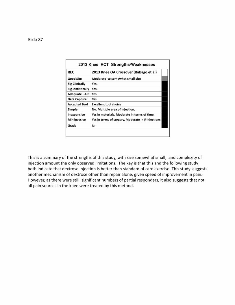
2013 Knee RCT Strengths/Weaknesses
This is a summary of the strengths of this study, with size somewhat small, and complexity of
injection amount the only observed limitations. The key is that this and the following study
both indicate that dextrose injection is better than standard of care exercise. This study suggests
another mechanism of dextrose other than repair alone, given speed of improvement in pain.
However, as there were still significant numbers of partial responders, it also suggests that not
all pain sources in the knee were treated by this method.

Slide 38
Knee OA 2012 (Dextrose vsExercise Crossover )
• Dumais R, Benoit C, Dumais A, Babin L,
Bordage R, de Arcos C, Allard J, Bélanger
M. Effect of Regenerative Injection
Therapy on Function and Pain in Patients
with Knee Osteoarthritis: A Randomized
Crossover Study. Pain Med. 2012 Jul 3.
doi: 10.1111/j.1526-4637.2012.01422.x.
[Epub ahead of print]
The next level I study considered was published in the Journal of Alternative and
Complementary Medicine and had a clear design of randomization, with randomized exercise
control and a crossover design. There is no free pdf.
Here is the abstract: (The following two slides will summarize)
OBJECTIVE:
We assessed the effectiveness of regenerative injection therapy (RIT) to relieve pain and restore
function in patients with knee osteoarthritis.
DESIGN:
Crossover study where participants were randomly assigned to receive exercise therapy for 32
weeks in combination with RIT on weeks 0, 4, 8, and 12 or RIT on weeks 20, 24, 28, and 32.
PATIENTS:
Thirty-six patients with chronic knee osteoarthritis.
INTERVENTIONS:
RIT, which is made up of injections of 1 cc of 15% dextrose 0.6% lidocaine in the collateral
ligaments and a 5 cc injection of 20% dextrose 0.5% lidocaine inside the knee joint.
OUTCOME MEASURES:
The primary outcome was the Western Ontario and McMaster Universities Osteoarthritis Index
of severity of osteoarthrosis symptoms (WOMAC) score (range: 0-96).
RESULTS:

Following 16 weeks of follow-up, the participants assigned to RIT presented a significant
reduction of their osteoarthritis symptoms (mean ± standard deviation: -21.8 ± 12.5, P < 0.001).
WOMAC scores in this group did not change further during the last 16 weeks of follow-up, when
the participants received exercise therapy only (-1.2 ± 10.7, P = 0.65). WOMAC scores in the first
16 weeks did not change significantly among the participants receiving exercise therapy only
during this period (-6.1±13.9, P=0.11). There was a significant decrease in this groups' WOMAC
scores during the last 16 weeks when the participants received RIT (-9.3±11.4, P=0.006). After
36 weeks, WOMAC scores improved in both groups by 47.3% and 36.2%. The improvement
attributable to RIT alone corresponds to a 11.9-point (or 29.5%) decrease in WOMAC scores.
CONCLUSIONS:
The use of RIT is associated with a marked reduction in symptoms, which was sustained for over
24 weeks

Slide 39
Focus on Changes from 0 to 16 weeks and 20 to 36 weeks
Follow-up every 4 weeks in
both groups. No NSAIDs.
WOMAC 3.1, Brief Pain Inventory, Wong-Baker Faces,
NRS, Timed Up and Go Test. Measurers blinded.
Dextrose 25% 5 ml IA, 15% Col.
Lig @ 0,4, 8, 12 weeks†
OA Knee Diagnosis, Chronicity, No Prior Surgery = #45/60 screened
Home-Based Exercise
Program* Without Injection
Home based Exercise program
Without Further Injection
Dextrose 25% 5 ml IA, 15% Col.
Lig @ 20, 24, 28, 32 weeeks.
21 24
Subjects either received dextrose injection or a home based program exercise program.
Those that received dextrose injection received 5 ml of 25% dextrose in the knee via a knee-
bent approach without ultrasound guidance and 15% dextrose into collateral ligaments in each
side. This was given every 4 weeks up to 4 treatments)
Those assigned randomly to a home based exercise program received four strengthening
exercises: (Isometric quads, leg extension exercises with quadriceps roll, straight leg raise, and
sitting end range knee extension; three sets of 10 reps daily) This was via instruction by a senior
physiotherapist, who reviewed exercises every 4 weeks. The same physical therapist was used
throughout the study.
The primary measuring tool was the WOMAC (Western Ontario and McMaster Universities
Osteoarthritis Index)w which has a 24 items with 5 points each. Five items are for pain, 2
are for stiffness and 17 are function related.
A Brief Pain Inventory (Short Form) was also used, as well as other secondary measures.
Everyone stayed in their groups for 12 weeks (4 dextrose injections). They then were crossed
over to start the other treatment at 20 weeks.

Slide 40
WOMAC Total Score Change Over Time
0 4 8 12 16 20 24 28 32 36
WEEKS
0
-5
-10
-15
-20
-25
BETTERIMPRO
EXERCISE
DEXTROSE EXERCISE
DEXTROSE
The WOMAC score dropped quickly (improved) in those receiving dextrose, and held
improvement during the crossover into exercise.
The exercise group improved minimally and then improved rapidly after dextrose injection
began such that differences between the groups became much less after three injections of
dextrose.

Slide 41
16 week
Between
Group
Difference
(P value)
36 week
Difference
Overall
Crossover
Design
Test
WOMAC
total
.002 .046 <.001
WOMAC
pain
.01 .28 .02
WOMAC
stiffness
.02 .01 .002
WOMAC
function
.0044 .06 .001
This is the significance of differences on the major measure (WOMAC). The second column
shows that most measures were no longer significantly different after each group had the
opportunity to receive dextrose. The third column takes into account the information from
both portions of the crossover, showing clearly significant differences between the treatments.

Slide 42
Data Collection Weaknesses
• Group one: 1 dropped out from injection
group due to no benefit and 2 due to
complete benefit. 18/21 analyzed.
• Group two: 3 did not participate once
assigned to exercise group , 2 were lost to
folllow-up and 1 had pain after injections and
dropped out. (18/24 analyzed)
• Overall these are unlikely to have affected
conclusions or significance other than by
reducing power in analysis.
There was some weakness in capture of data but these should not have affected outcome or
conclusions significantly

Slide 43
REC 2012 Knee OA Crossover (Dumais et al)
Good Size Moderate Size
Sig Clinically Very much so.
Sig Statistically Yes, unequivocally.
Adequate F-UP Limited
Data Capture Limited
Accepted Tool Excellent tool choice
Simple Yes
Inexpensive Yes
Min invasive Yes
Grade Ib (Single study)
Knee Crossover Strengths/Weaknesses
This is a summary of the strengths of this study, with size somewhat small, limited time of
followup to less than 1 year and some limitation of data capture. Since exercise has clearly been
shown to be beneficial in knee osteoarthritis, and prolotherapy with dextrose was clinically
significantly better than exercise, this is a strong support for prolotherapy.
This treatment would be practical for primary care doctors to perform since it involved injection
inside the knee and over the collateral ligaments on each side.

Slide 44
Osgood-Schlatter Disease
• Topol GA, Podesta LA, Reeves
KD, Raya MF, Fullerton BD, Yeh
H. Hyperosmolar Dextrose
Injection for Recalcitrant
Osgood-Schlatter Disease.
Pediatrics 2011;128(5):e1121-
e1128.
The next study was published in the best journal in Pediatrics in late 2011 and was also a level I
randomized controlled study. This is an open access study, so is available on line without charge.
A full PDF is available by going to the following internet site:
http://pediatrics.aappublications.org/content/128/5/e1121.long
Slide 45
Patellar Tendon
Kneecap
(Patella)
“Bump”
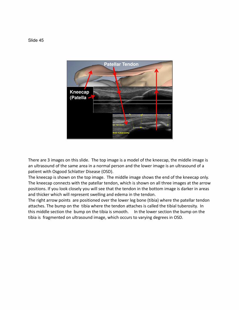
There are 3 images on this slide. The top image is a model of the kneecap, the middle image is
an ultrasound of the same area in a normal person and the lower image is an ultrasound of a
patient with Osgood Schlatter Disease (OSD).
The kneecap is shown on the top image. The middle image shows the end of the kneecap only.
The kneecap connects with the patellar tendon, which is shown on all three images at the arrow
positions. If you look closely you will see that the tendon in the bottom image is darker in areas
and thicker which will represent swelling and edema in the tendon.
The right arrow points are positioned over the lower leg bone (tibia) where the patellar tendon
attaches. The bump on the tibia where the tendon attaches is called the tibial tuberosity. In
this middle section the bump on the tibia is smooth. In the lower section the bump on the
tibia is fragmented on ultrasound image, which occurs to varying degrees in OSD.

Slide 46
This is a larger image of the subject with OSD. The yellow lines represent the direction of tiny
needle injections used in the study. Ultrasound was not used for injection in this study, but is
used here just to show the anatomy and to illustrate that injections were shallow, only a little
more than 1 centimeter in depth. . This ultrasound image is included to show the direction of
the small (allergy size) needles that were used in the study, and the shallow depth, which is
only a little more than 1 centimeter for the deepest injection.

Slide 47
Here are those same areas of injection shown on the ultrasound, but from a straight on view of
the knee.
Slide 48
694 Screened
Randomized
Lidocaine injection monthly for 3 months
Usual Care for 3 months
Dextrose Injection monthly for 3 months
OSD Criteria MetImproved With Additional Therapy10
2
Refused Enrollment
66
19
18
17
54
3 month data collection, unblinding, and optional dextrose injection for 1 year ����1 year data
19 17
18
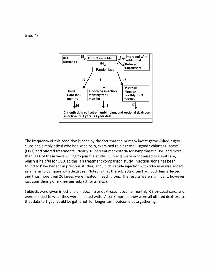
The frequency of this condition is seen by the fact that the primary investigator visited rugby
clubs and simply asked who had knee pain, examined to diagnose Osgood Schlatter Disease
(OSD) and offered treatments. Nearly 10 percent met criteria for symptomatic OSD and more
than 80% of these were willing to join the study. Subjects were randomized to usual care,
which is helpful for OSD, so this is a treatment comparison study. Injection alone has been
found to have benefit in previous studies, and, in this study injection with lidocaine was added
as an arm to compare with dextrose. Noted is that the subjects often had both legs affected
and thus more then 20 knees were treated in each group. The results were significant, however,
just considering one knee per subject for analysis.
Subjects were given injections of lidocaine or dextrose/lidocaine monthly X 3 or usual care, and
were blinded to what they were injected with. After 3 months they were all offered dextrose so
that data to 1 year could be gathered for longer term outcome data gathering.

Slide 49
0: No pain or stiffness
1: STIFF/SORE after sport2: Stiff/sore before and after sport3: Pain during sport
4. SPORT ALTERED5. Painful self care.
6. Self care interference.7. SLEEP ALTERED
NPPS: Nirschl Pain Phase Scale
A sports measurement scale was used which clearly shows when complete resolution of
symptoms occurs (no stiffness or soreness), since some athletes do not realize that stiffness is
not normal.
The spread of scores from stiff (1) to altered sport (4) to altered sleep (7) is shown. The key was
alteration of sport and that was the requirement for admission into the study.

Slide 50
0
1
2
3
4
5
0 3 6 12
Dex (n=21)
Lido Only (n=13)
Lido-Dex (n=9)
NPPS
S CORE
This graph shows changes over 12 months in the NPPS score. The solid black line represents
the results in knees treated with dextrose. The solid blue line is lidocaine and the blue and then
black line depicts results in knees that were treated with lidocaine and then received dextrose.
The knees receiving dextrose rapidly dropped in levels of pain and functional limits .

Slide 51
0
1
2
3
4
5
0 3 6 12
Dex (n=21)
Usual Only (n=14)
Usual-Dex (n=8)
Dex (n=21)Usual Only (n=14)Usual-Dex (n=8)
NPPS
S CORE
This graph is similar but shows results comparing usual care to dextrose. The differences
between dextrose and usual care were even more.

Slide 52
ANOVA Tukey
Dextrose vs
Exercise
<.00001 <.00001
Dextrose vs
LIdocaine
.004 .017
Lidocaine vs
Exercise
.024 .046
OSD: Significance of Differences Between Groups. ANOVA and two “Post Hoc” Tests
When there are more than two groups that are being compared, errors can occur. These are
compensated for by what is called a ‘Post Hoc Multiple Comparison Tests. The most accurate is
probably the Tukey.
Prolotherapy study results comparing dextrose to other injection have sometimes not shown a
significant difference and have been ignored. This study clearly shows that lidocaine injection is
not a placebo control and could easily confound results from studies only comparing two forms
of injection. The inclusion of a usual care group helps illustrate the beneficial effect of injection
alone, although dextrose injection was superior to lidocaine injection.

Slide 53
This is from January 7, 2012. Avg 24/3000 articles
“POEMs” = "Patient-Oriented Evidence that Matters”.
Review by Essential Evidence Plus
Less than 1% of articles reviewed in major journals by a research organization called “Essential
Evicence Plus” are chosen to be reviewed.
This review of the article was by a PhD from Tufts University who rates the article in the I range
(Ib-), clearly indicating that dextrose injection is effective in youths with OSD. Note that the
only error here is the statement about scarring with higher concentration dextrose, which has
never been shown to occur. Because OSD has changes in both cartilage and tendon, these
results are consistent with potential benefit for both cartilage and tendon.

Slide 54
Key Features RCT: Osgood Schlatter Study 2011
Good Size Moderate Size
Sig Clinically Very much so.
Sig Statistically Yes, unequivocally.
Adequate F-UP 2 years, more than enough.
Data Capture Perfect
Accepted Tool NPPS not well studied and no MCID.
Simple Yes
Inexpensive Yes
Min invasive Yes but any injection difficult in this age group.
Practical PC Several injections may be excessive.
Grade Ib (Single study)
OSD 2011 Strengths/Weaknesses
This study meets the criteria to change the way medicine is performed mostly but the
measurement tool was not the most accepted version. (Although it showed dramatic
improvements in both pain and function and has been used in other studies.) More importantly,
despite use of a small needle, pediatricians may be resultant to use injection to treat and pain
condition in adolescents. The small needle injection, however, was tolerated very well in the
clinical study.

Slide 55
Finger OA 2000
• Reeves KD, Hassanein K: Randomized prospective placebo-controlled double-blind study of dextrose prolotherapy for osteoarthritic thumbs and fingers (DIP, PIP and trapeziometacarpalJoints) : Evidence of clinical efficacy. Jnl Alt Compl Med 2000;6(4):311-320.
The next study was the first randomized control trial on use of dextrose prolotherapy in finger
osteoarthritis.
To view a full PDF go to the following site:
http://drreeves.com/sites/default/files/Finger%20Arthritis%20Study.pdf

Slide 56
Kelgreen Criteria for hand OA met plus pain > 3 months
.075 Lidocaine inj. at 0, 2 and 4 months.
Dex10%/ .075 Lido injection at 0, 2 and 4 months
13 14
6 month data, then open label Dextrose 10% PRN
11
2
Lost for medical reasons (progressive CHF and
severe depression
11 113 dropped out (1 much better and 2 not improved)
3
1 year data collection on 22 patients. Intention to treat.
14
Patients were accepted if they met X-ray criteria for finger arthritis and had pain more then 3
months in their fingers. They were injected with either 10% dextrose or low concentration
lidocaine. After 6 months they were all given the option to receive dextrose injection. There
were two dropouts due to no improvement in the lidocaine group.

Slide 57
% Impr. Movement Pain After 3
Injections 42
15
Dextrose
Lidocaine
(P = .027)
The dextrose group was favored in improvement of pain with grip, finger movement, and at
reset. However, the difference in movement pain was the most impressive, reaching statistical
significance with 42% versus 15% improvement. (A P value of < 0.5 is significant) This means
that this result would occur by chance only 1/20 times.

Slide 58
Impr. Finger Flexion in Degrees
Dextrose +8
Lidocaine -8.7
(P = .003)
The result on flexibility of the fingers is shown here. Those injected with dextrose improved by
8 degrees in range of motion and those injected with lidocaine lost more than 8 degrees of
range of motion. After 6 months those joints injected with lidocaine received dextrose injection
and range of motion improved similarly to the dextrose treated joints.

Slide 59
Key Features REC: Finger OA Dextrose Inj. (2000 JACM)
Good Size Small size
Sig Clinically Yes. Difference exceeded MCID of 2.
Sig Statistically Yes, for grip pain and ROM despite small size.
Adequate F-UP 6 months blind and 1 year total.
Data Capture Good. Only medical dropout to 6 months.
Accepted Tool VAS Pain but no functional hand measure.
Simple Yes
Inexpensive Yes
Min invasive Yes, but method used was uncomfortable.
Practical PC Injections not tolerated enough for P.C. use
Grade Ib- (Small RCT)
Finger OA 2000 Strengths/Weaknesses
Finger injection into the joint is uncomfortable even with a tiny needle and limit use of this
method, along with resistance to prolotherapy in general. However, there are new methods of
injection, which appear to be equally effective and minimally uncomfortable. This was a small
study and used pain and range rather than a standard arthritis scale. However, it showed that
injection with dextrose was clearly beneficial and more so than lidocaine injection in patients
with symptomatic hand osteoarthritis.
The ideal treatment for finger arthritis would not involve injection at all. Note that studies on
use of cream application for arthritis are in formulation at this time.

Slide 60
Extensor Tendinosis • Scarpone M, Rabago DP,
Zgierska A, Arbogast G,
Snell E: The efficacy of prolotherapy for lateral
epicondylosis: a pilot study.
Clin J Sport Med 2008;18(3):248-5
This study was published in the Clinical Journal of Sports Medicine and was on “tennis elbow”,
also called “extensor tendinitis” or “extensor tendinosis” . Tennis elbow is more accurately
termed a tendinosis because the primary change is degenerative, not inflammatory. That
explains why anti-inflammatory (i.e. steroid) injections typically improve pain only temporarily
and less so when repeated.
For a full PDF equivalent view go to: http://www.ncbi.nlm.nih.gov/pmc/articles/PMC2751593/
Slide 61
10.7% Dex/14.7% NaMorr Three 0.5 ml portions
@ 0, 4 & 8 wks
Normal Saline Same Volume @ 0, 4 & 8 wks
26 met criteria for Criteria of 6 months pain, failure of rest, P.T., NSAIDs and two Steroid injections.
10
Grip strength by dynamometer,
extension strength by BTE,
and pain score NRS at 8, 16 and 52 weeks
10 (One moved after 16 weeks)
NSAIDs discouraged. No peppering 10
9
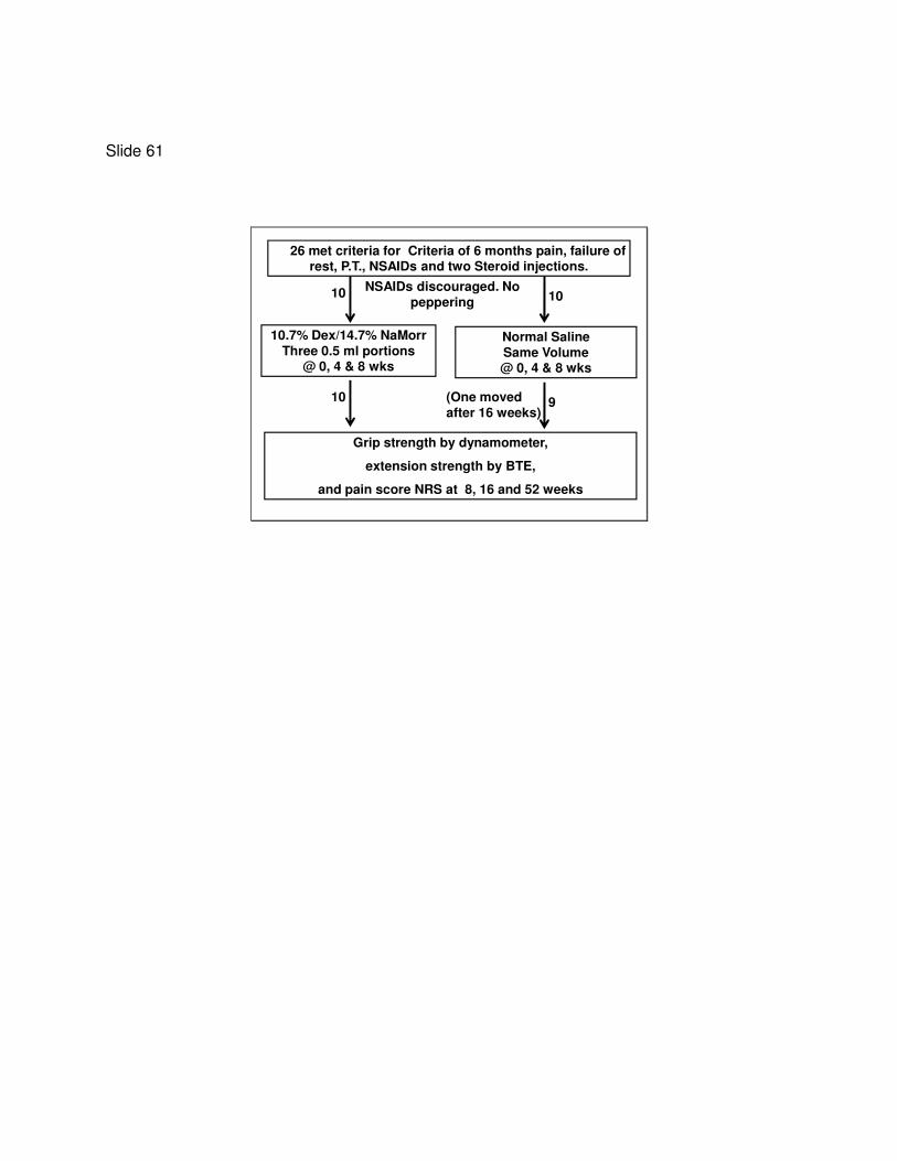
26 patients met criteria for inclusion but only 20 decided to undergo treatment. Grip strength,
wrist power and pain was measured at approximately 2 and 4 months and 1 year. Half the
group received normal saline injections and half received 10.7% dextrose/14.7% sodium
morrhuate. Each subject received 3 injection sessions. (0, 4 and 8 weeks).

Slide 62
Pain Scores From 0 to Months
Dextrose+ NaMorr Better. (P < .001)
0
2
4
6
Dex +NaMorr Saline
0 2 Mo 4 mo
The 2 month data (red) showed no significant difference between groups but by 4 months the
differences between groups reached statistical significance with a reduction in pain of 90% in
those subjects receiving dextrose + sodium morrhuate versus saline.
This illustrates again the importance of adequate follow-up to distinguish between temporary
effects of steroid and longer terms effects of proliferant injection.

Slide 63
Extensor Strength From 0 to 4 Months
Dex/NaMorr Better. (P < .01)
0
10
20
30
40
Dex + NaMorr Saline
0 2 Mo 4 mo (P < .01)
The ability to lift up something at the wrist (while palm is down), also called extensor strength,
showed a substantial difference between groups favoring the dextrose/sodium morrhuate
group.

Slide 64
Key Features RCT: Lat Epi Dex/NaMorr (2008 CJSM)
Good Size Small size
Sig Clinically Yes. Very impressive difference
Sig Statistically Yes, for pain/ext stren. Note for grip.
Adequate F-UP 1 yr phone but results not documented.
Data Capture Good. Only medical dropout to 6 months.
Accepted Tool VAS Pain but no functional hand measure.
Simple Yes
Inexpensive Yes
Min invasive Moderate. NaMorr pain with injection.
Practical PC NaMorr use will not encourage P.C. use
Grade Ib- (Small RCT + Blinding ?)
Lat Epi. 2008 Strengths/Weaknesses
The weaknesses of the tennis study are related to its size and the use of a more inflammatory
agent (sodium morrhuate) along with dextrose, which primary doctors may be more reluctant
to use. Nevertheless, results were impressive in this randomized controlled trial.
Slide 65
Knee OA Intraarticular RCT 2000
• Reeves KD, Hassanein K.
Randomized Prospective Double-
Blind Placebo-Controlled Study of
Dextrose Prolotherapy for Knee
Osteoarthritis With or Without ACL
laxity. Alt Ther Hlth Med
2000;6(2):68-80
The next randomized control trial was on knee arthritis. It was the first randomized control trial
of prolotherapy in knee arthritis and was published in the journal Alternative Therapies in
Health and Medicine. It involved a simple injection in the knee and no other pain sources about
the knee were treated. Thus this method may have left untreated several pain sources, but is
easily repeated due to its simplicity.
For a pdf of the article go to:
http://www.drreeves.com/sites/default/files/Knee%20Arthritis%20Study.pdf

Slide 66
0, 2 and 4 month injections. ROM, pain data at 6 months. Then open label dextrose Q 2-3 months PRN.
10% Dex/.075% Lido/.9BA .075% Lido in BW/.9BA
77 Patients Enrolled
34 Subjects
ROM, Pain, blinded X-Ray data at 1 year
3 unrelated medical dropouts and 1 efficacy dropout to 1 year
58 Knees
2 unrelated medical dropouts and 3 efficacy dropouts to 1 year.
38 39
34 Subjects 53 Knees
77 subjects enrolled in the knee study. 38 received dextrose injection with lidocaine and 39
received lidocaine only. The dextrose level was only 10% so it did not use inflammation as its
mechanism of action. Part of the purpose of this study was to show that dextrose has a
beneficial effect upon joint injection, separate from inflammation.
“BA” in this study is benzyl alcohol. This was used to prevent infection. Benzyl alcohol may
interfere with growth and repair and thus may have affected outcome adversely. However it
and the lidocaine concentration (.075%) was the same in each group. The only difference is that
dextrose 10% was included in the injectant to those randomly and blindly assigned to the
dextrose group.
In the study approximately 10% of participants did not complete 1 year follow up with most of
them due to medical issues unrelated to injection.
These were subjects with advanced knee arthritis and all candidates for knee replacement.

Slide 67
All TKA Candidates and Many
Bone on Bone
• No significant differences overall
between groups.
• Dextrose group tended to be more
severe. More dextrose knees were
stage IV (25 versus 15 knees by
skier’s view.
Although these subjects due to pain and stage of arthritis were candidates fore knee
replacement, the group receiving dextrose had more knees that were void of cartilage on a
skier’s view, (more accurate determination of lack of cartilage than a standing view), and
buckling episodes appeared to be more in the dextrose group, (the lidocaine group had very
little buckling at study onset). However, these differences did not reach statistical significance.

Slide 68
Multivariate Results: Knee OA:
Single Intraarticular
• Multivariate analysis of paired observations between 0 and 6 months for pain, swelling, buckling episodes, and knee flexion range revealed significantly more benefit from the dextrose injection (P=.015)
Overall the group receiving dextrose did better statistically between 0 and 6 months. Because
multivariate analysis was significant we are able to look at individual areas of differences in the
variables.

Slide 69
% Improvement in Walking Pain and
Subjective Swelling at 6 and 12
3525
4044
18
63
6 Mo Dex 6 Mo Lido 12 Mo Dex
Pain Swelling
Subjective Swelling Significantly
Different P= .004
The 6 months result for walking pain favored the dextrose group, but not significantly.
Subjective swelling in the knee however, improved 44% in the dextrose group versus 18% in the
lidocaine group.
Also notable is that the dextrose group, when followed to one year, continued their pattern of
improvement with further improvement in pain, swelling, and buckling. These patient were
treated as needed after the first 6 months.

Slide 70
Flexion Improvement Degrees:
Blinded 6 Mo., and Open 12 Mo.
13.2
7.7
0
5
10
15
6 Mo Dex 6 Mo L/B.A.
P = .005
The improvement in range of motion was highly significant in favor of the group receiving
dextrose. Buckling episodes improved 67% in the dextrose group and the change could not be
determined accurately in the lidocaine group since they were not buckling to begin with.
A substantial improvement in knee flexibility and a reduction in buckling tendency are both
important because research indicates that buckling tendency and loss of knee flexibility are two
common indications for knee replacement.
Yakhdani HR, Bafghi HA, Meijer OG, Bruijn SM, van den Dikkenberg N, Stibbe AB, van Royen BJ,
van Dieën JH. Stability and variability of knee kinematics during gait in knee osteoarthritis
before and after replacement surgery. Clin Biomech 2010; 25(3):230-6.

Slide 71
Key Features RCT: Single Inj. OA Change (2000 ATHM)
Good Size Yes
Sig Clinically Yes. Pain, ROM, buckling progressively better.
Sig Statistically Swelling and range but not pain to 6 months.
Adequate F-UP Good follow up to 1 year.
Data Capture Good.
Accepted Tool VAS Pain and gonio. range but no WOMAC
Simple Yes
Inexpensive Yes
Min invasive Yes, low volume single injection method.
Practical PC Quite practical for primary care.
Grade II-1 Suspect L-BA was not a placebo control.
Knee OA 2000 Strengths Weakneses
The knee study did not show statistically significant improvement in all variables but was a large
sized randomized control trial showing significant improvement in several key areas of knee
performance. The study is flawed and not quite level I due to no use of no well accepted
functional tools.
It is also important to note that results may have been reduced by use of a needle a bit too
short to injection through the fat pad. (1-1/4 inch needle was used and the fat pad can be more
thick than that) However, it is a simple, reproducible and minimally invasive method, and led to
progressive benefit with intermittent use to one year in this group with advanced arthritis.
The fact that both groups improved in pain does not negate the fact that range and stiffness
improved much more in the dextrose group. Lack of a non injection group is a limitation, as the
non dextrose group may have been an active treatment via a hypoosmolar or other effect
Note that knee buckling and pain are the two biggest reasons for knee replacement, and
dextrose affected both in a progressive manner.
This study showed that that even a single injection method using non-inflammatory dextrose
resulted in clinical benefit.
Slide 72
Steroid vs Dextrose SI Injection
• Kim WM, Lee HG, Won Jeong C, Kim
CM, Yoon MH. A randomized
controlled trial of intra-articular
prolotherapy versus steroid
injection for sacroiliac joint pain. J
Altern Complement Med Dec 2010,
16(12) p1285-90.
A randomized controlled study of the use of prolotherapy on the SI joint compared dextrose to
steroid injection. As a treatment comparison trial it was blinded and randomized and would be
a high level II study. .
A full pdf is available at the following internet site:
http://harborfm.pbworks.com/w/file/fetch/63256359/prolotherapy%20for%20SI%20jt%20pain.
Here is the abstract:
Kim WM; Lee HG; Won Jeong C; Kim CM; Yoon MH. ARTICLE TITLE: A randomized controlled
trial of intra-articular prolotherapy versus steroid injection for sacroiliac joint pain [In Process
Citation] ARTICLE SOURCE: J Altern Complement Med (United States), Dec 2010, 16(12) p1285-
90
AUTHOR'S ADDRESS: Department of Anesthesiology and Pain Medicine, Chonnam National
University Hospital , Gwang-Ju, Korea.
ABSTRACT: Abstract Objectives: Controversy exists regarding the efficacy of ligament
prolotherapy in alleviating sacroiliac joint pain. The inconsistent success rates reported in
previous studies may be attributed to variability in patient selection and techniques between
studies. It was hypothesized that intra-articular prolotherapy for patients with a positive
response to diagnostic block may mitigate the drawbacks of ligament prolotherapy. The purpose
of this study was to evaluate the efficacy and long-term effectiveness of intra-articular
prolotherapy in relieving sacroiliac joint pain, compared with intra-articular steroid injection.
Design: This was a prospective, randomized, controlled trial. Settings/location: The study was
conducted at an outpatient pain medicine clinic at Chonnam National University Hospital in
Gwang-ju, Korea. Subjects: The study included patients with sacroiliac joint pain, confirmed by
(yen)50% improvement in response to local anesthetic block, lasting 3 months or longer, and
who failed medical treatment. Interventions: The treatment involved intra-articular dextrose
water prolotherapy or triamcinolone acetonide injection using fluoroscopic guidance, with a
biweekly schedule and maximum of three injections. Outcome measures: Pain and disability
scores were assessed at baseline, 2 weeks, and monthly after completion of treatment. Results:
The numbers of recruited patients were 23 and 25 for the prolotherapy and steroid groups,
respectively. The pain and disability scores were significantly improved from baseline in both
groups at the 2-week follow-up, with no significant difference between them. The cumulative
incidence of (yen)50% pain relief at 15 months was 58.7% (95% confidence interval [CI] 37.9%-
79.5%) in the prolotherapy group and 10.2% (95% CI 6.7%-27.1%) in the steroid group, as
determined by Kaplan-Meier analysis; there was a statistically significant difference between
the groups (log-rank pv <v0.005). Conclusions: Intra-articular prolotherapy provided significant
relief of sacroiliac joint pain, and its effects lasted longer than those of steroid injections.
Further studies are needed to confirm the safety of the procedure and to validate an
appropriate injection protocol.

Slide 73
50% Imp with SI Inj with 2.5 ml L-bupiv
Pain > 3 mo below PSIS or + Gaenslen’s or Patrick maneu.
Randomized
40 mg Triam in .125 L-bupiv @ 0, 2 & 4
weeks PRN 10% Pain
25% Dex in .125 L-bupiv @ 0, 2 & 4
weeks PRN 10% pain
26 24
50
Followup to 15 months for Pain and Oswestry Disability Index (ODI)
FluoroGuidance
1 Dropout (pain) 25 1 Lost to Fup23
It is not easy to determine if pain is coming from the sacroiliac joint. In order to be more
confident that their patients did have pain from the SI joint, the authors used X-ray guidance to
inject anesthetic into the joint. If the patient’s pain improved 50% or more then it was
considered that they had pain in large part from the SI joint. The pain also had to be chronic so
it would not likely go away on its own. (More than 3 months)
Subjects were then randomized to receive steroid injection (trimcinolone) or 25% dextrose
injection, also under X-Ray guidance, into the SI joint. 2.5 ml of either solution was injected.
The data “capture” was good with minimal patient dropout, 1 in each group, and follow up was
for a total of 15 months.

Slide 74
2 Week % Improvement in Pain/Disability
77 75
66
0
10
20
30
40
50
60
70
80
90
100
Pain Disability
Dex
Triam
67
Quick improvement was seen in each group in both measures of pain and disability, as this two
week data shows. Pain improved by 3/4 in each group and disability measures by 2/3.
It is important to note that the speed of improvement cannot be due to repair of the SI
ligament, as that takes several months. This speed of improvement indicates a nerve
mechanism.
Steroid treats inflammation that is prostaglandin based. Dextrose injection treats inflammation
that is nerve based. They are two different pathways.

Slide 75
% with ≥ 50% Pain Relief
100
63.658.7 58.7
27.2
10.2 10.2
0
10
20
30
40
50
60
70
80
90
100
2 Weeks 6 Mo 10 Mo 15 Mo
Dex/A
Triam/A
P < .001
The long term data is shown in this slide. The authors considered “substantial benefit” to be
more than a 50% level of pain relief and the percentage of patients that maintained that level of
pain relief was followed. Although the 2 week data looked good for both groups, later data
showed highly significant differences between groups. The number of subjects with more than
50% pain relief in the steroid-injection group was only 10% at 15 months compared to nearly
60% in the dextrose group. This suggests a longer term effect of dextrose on the tissue, and
that process is thought to involve repair as described earlier. Thus is takes several months for it
to be observed.

Slide 76
Key Features RCT:SI Steroid vs Dextrose: JACM 2010
Good Size Moderate Size
Sig Clinically Very much so.
Sig Statistically Yes, unequivocally.
Adequate F-UP 15 months
Data Capture Very good
Accepted Tool Yes ODI and
Simple Yes
Inexpensive No. Requires Fluoro. But single injection
Min invasive Moderate. But limited by specificity.
Practical PC Fluoro removes it from this possibility
Grade Ib (Single study, peer review limits in JACM)
SI 2010 Strengths/Weaknesses
If one considers the sacroilaic (SI) joint study, it rates high in all elements except it is not truly
inexpensive, requiring X-ray guidance. Due to the use of the special X-ray (fluoroscopy) it will
not be practical for primary doctors to use which will limit spread of this technique.

Slide 77
Tennis Elbow: Dextrose or NaM-
Dextrose versus “Wait and See”
• Rabago D, Lee KS, Ryan M, Chourasia AO,
Sesto ME, Zgierska A, Kijowski R, Grettie J,
Wilson J, Miller D. Hypertonic Dextrose and
Morrhuate Sodium Injections (Prolotherapy)
for Lateral Epicondylosis (Tennis Elbow):
Results of a Single-blind, Pilot-Level,
Randomized Controlled Trial. Am J Phys Med
Rehabil. 2013 Jan 3.
This study, published in 2013, is a single blind study of dextrose versus sodium-morrhate
dextrose for tennis elbow (Lateral epicondylosis) versus wait and see with randomized
allocation to the three groups.
It was announced as a study with 4 arms, including use of platelet rich plasma but that arm
apparently did not get completed. PDF has been requested for review.
PDF will not be free. Here is the abstract:
Rabago.david.2013.wpd
Rabago D, Lee KS, Ryan M, Chourasia AO, Sesto ME, Zgierska A, Kijowski R, Grettie J, Wilson J,
Miller D. Hypertonic Dextrose and Morrhuate Sodium Injections (Prolotherapy) for Lateral
Epicondylosis (Tennis Elbow): Results of a Single-blind, Pilot-Level, Randomized Controlled Trial.
Am J Phys Med Rehabil. 2013 Jan 3. [Epub ahead of print]
Source
From the Department of Family Medicine (DR, AZ, JG, JW, DM), Department of Radiology (KSL,
RK), and Department of Orthopedics and Rehabilitation (MES), University of Wisconsin School of
Medicine and Public Health, Madison; Centre for Musculoskeletal Research & School of
Physiotherapy and Exercise Science, Griffith Health Institute, Griffith University, Gold Coast,
Australia (MR); and Department of Biomedical Engineering, University of Wisconsin-Madison
(AOC).

Abstract
OBJECTIVE:
Chronic lateral epicondylosis is common, debilitating, and often refractory. Prolotherapy (PrT) is
an injection therapy for tendinopathy. The efficacy of two PrT solutions for chronic lateral
epicondylosis was evaluated.
DESIGN:
This study is a three-arm randomized controlled trial. Twenty-six adults (32 elbows) with chronic
lateral epicondylosis for 3 mos or longer were randomized to ultrasound-guided PrT with
dextrose solution, ultrasound-guided PrT with dextrose-morrhuate sodium solution, or watchful
waiting ("wait and see"). The primary outcome was the Patient-Rated Tennis Elbow Evaluation
(100 points) at 4, 8, and 16 wks (all groups) and at 32 wks (PrT groups). The secondary outcomes
included pain-free grip strength and magnetic resonance imaging severity score.
RESULTS:
The participants receiving PrT with dextrose and PrT with dextrose-morrhuate reported
improved Patient-Rated Tennis Elbow Evaluation composite and subscale scores at 4, 8, and/or
16 wks compared with those in the wait-and-see group (P < 0.05). At 16 wks, compared with
baseline, the PrT with dextrose and PrT with dextrose-morrhuate groups reported improved
composite Patient-Rated Tennis Elbow Evaluation scores by a mean (SE) of 18.7 (9.6; 41.1%) and
17.5 (11.6; 53.5%) points, respectively. The grip strength of the participants receiving PrT with
dextrose exceeded that of the PrT with dextrose-morrhuate and the wait and see at 8 and 16
wks (P < 0.05). There were no differences in magnetic resonance imaging scores. Satisfaction
was high; there were no adverse events.
CONCLUSIONS:
PrT resulted in safe, significant improvement of elbow pain and function compared with
baseline status and follow-up data and the wait-and-see control group. This pilot study suggests
the need for a definitive trial.
PMID: 23291605 [PubMed - as supplied by publisher]

Slide 78
1.4 % NaM 21.5% Dex, .6% Lido @
0,4,8 weeks
22% Dex.45% Lido @ 0, 4, 8 weeks.
26 adults (32 elbows randomized ) 3 months or longer symptoms. Failure of 2 or 3 of NSAIDS, physical
therapy or corticosteroid injections).
9 subjects10 elbows
Patient rated tennis elbow evaluation (PRTEE) Time Frame: 0, 16 and 32 weeks
Also Grip Strength and Ultrasound PRE/POST
Delayed Treatment = Wait and see
8 subjects10 elbows
10 subjects12 elbows
11 subjects 9 subjects 11 subjects
2 subjects dropped form the dextrose group. 1 for unrelated medical urgency and 1 due to
advice of a specialist. 1 stopped from the dextrose/morrhute group due to medical urgency and
1 from the wait and see group due to deciding against participation after starting.

Slide 79
Dex vs Dex/NaM vs Wait and See
Results to 16 weeks
• Both prolotherapy groups did better than the wait
and see group. P < .05
• IE: (16 week): PRTEE Improved 18.7 in Dex, 17.5 in
Dex/NaM and 9.3 In wait and see groups. ( P <
.05). Grip strength better in Dex than both (P <
.05)
• These improvement exceed the MCID and further
improvements were noted to 32 weeks, although
wait and see groups were not followed after 16
weeks.
Results: The improvements in both prolotherapy groups was significantly better than in the wait
and see control group at 4, 8 and 16 weeks. (P < .05) The differences were clinically important.
For example, the Patient Rated Tennis Elbow Evaluation has been evaluated for what is a
clinically important difference. A 7 point improvement (22%) is a little better an a 11 point
improvement (37%) is much better.
In this study at 16 weeks the dextrose group improved 18.7 points, the dextrose/sodium
morrhuate group improved 17.5 points, and the wait and see group improved 9.3 points. Grip
was better in the dextrose group than in both other groups.
The reference for MCID was:
https://uhra.herts.ac.uk/dspace/bitstream/2299/6682/1/Measuring_PRTEE_clinically_Important
_Change_submission_revised.pdf
Thesis on : Measuring clinically Important Change with the Patient-rated Tennis Elbow
Evaluation (PRTEE)
Short title: Measuring change with the PRTEE

Slide 80
Patient Rated Tennis Elbow Evaluation
0
10
20
30
40
50
60
0 wk 4 wk 8 wk 16 wk 32 wk
Dex/NAM Dextrose Wait&See
This is the data in graphic form. Compared to the MCID of 11 or 37% the Dextrose group
improved by 45% at 16 weeks and 57% by 32 weeks. The Dextrose/Sodium morrhuate group
improved by 54% at 16 weeks and 75% by 32 weeks.
It is of interest that dextrose was the only group that improved in grip pain despite rather equal
improvements in function between dextrose and dextrose/sodium morrhuate otherwise.
Here it appears that the wait and see group was worse than the other 2 groups and the
dextrose/sodium group the best, but apparently this trend did not reach statistical significance.
The participants receiving PrT-DM reported more severe and persistent injection related pain
taking up to 3 wks to resolve.

Slide 81
Key Features RCT: Lat Epi Dex v DexNaM vs Wait
Good Size Small size
Sig Clinically Yes for valid measurement tool
Sig Statistically Yes for both PRTEE and for grip.
Adequate F-UP Short followup 16 weeks.
Data Capture Good.
Accepted Tool PRTEE and grip strength
Simple Ultrasound guided injection
Inexpensive Moderate with ultrasound used
Min invasive Yes.
Practical PC NaMorr use will not encourage P.C. use
Grade Ib- (Small RCT + Blinding ?)
Lat Epi. 2013 Strengths/Weaknesses
Well designed study with a critical wait and see arm to show that both injection arms are active
in comparison to usual care which actually is the closest to wait and see after failure of usual
care.

Slide 82
BACK PAIN 2003
• Yelland MJ, Glasziou PP, Bogduk N, Schluter PJ, McKernon M: Prolotherapy injections, saline injections, and exercises for chronic low-back pain: A randomized trial. Spine 2003;29(1):9-16.
The only blind and randomized study of back pain studying a single intervention was published
in the journal Spine.
For a PDF of this article go to: http://www.cebp.nl/media/m1107.pdf

Slide 83
20% Dextrose/.2% Lido Normal Saline
110 Consented to Participate
99% followup to 6 months
Limited injection method. Max 5 injections each side. (Dhillon method). Rx every 2 weeks X 6 then PRN at 4, 6,
12 and 24 month fup.
96% followup to 1 year
80% follow up to 2 years
110 patients consented to participate in the study, and they were assigned to either 20%
dextrose in .2% lidocaine or to normal saline. It is very important to notice that the injection
method was limited to only 5 injections on each side, which would not be considered a
comprehensive approach.
However this study was excellent in terms of data capture to 1 year and had good data capture
to 2 years.

Slide 84
% Impr VAS Pain and Roland-Morris
0
20
40
60
Pain
Dex
Pain
Sal
Diab
Dex
Disab
Sal
1 Year
2 Years
The important thing to notice about this slide is the strong evidence of ability to improve pain
and functional limitations and to hold benefit to a 2 year point of follow-up which would be
comparable or better than long term follow-up data post back surgery.

Slide 85
Key Features RCT: 20% Glu vs Saline 2003 Spine
Good Size Yes
Sig Clinically Yes. Substantial disab impr to 2 years. Su
Sig Statistically No.
Adequate F-UP Good follow up to 2 year.
Data Capture Good.
Accepted Tool VAS Pain and Roland Morris
Simple Method simple but likely incomplete
Inexpensive No. 6 injections at 2 weeks a lot of time/etc.
Min invasive Limited injections each session
Practical PC Impractical treatment frequency for P.C.
Grade II-1 No real control arm.
Back Pain 2003 Strengths/Weaknesses
The improvement in functional limits and pain in both groups indicated that both treatments
methods were beneficial. However this study is dismissed by those that overlook the substantial
improvement over 2 years. Instead, they focus on thinking that saline injection is a placebo and
that dextrose is not better than placebo treatment.
This study reminds us how important it is to have a usual care treatment group so that the
result of the study are not dismissed.

Slide 86
Achilles Tendonosis
• Yelland MJ, Sweeting KR, Lyftogt JA,
Ng SK, Scuffham PA, Evans KA.
Prolotherapy injections and eccentric
loading exercises for painful Achilles
tendinosis: a randomised trial. Br J
Sports Med (England), Apr 2011,
45(5) p421-8.
The 2009 Achilles Tendon study published in the British Journal of Sports Medicine and
compared usual care with dextrose injection. This was not typical prolotherapy as neither the
Achilles tendon nor its attachment were injected directly. This treatment was likely a cross
between regular prolotherapy and PSI (Perineural Subcutaneous Injection). The injection
frequency (weekly) is that more typically seen with PSI
No free PDF is available. Here is the abstract:
Br J Sports Med. 2011 Apr;45(5):421-8. doi: 10.1136/bjsm.2009.057968. Epub 2009 Jun 22.
Prolotherapy injections and eccentric loading exercises for painful Achilles tendinosis: a
randomised trial.
Yelland MJ, Sweeting KR, Lyftogt JA, Ng SK, Scuffham PA, Evans KA.
Source
Griffith University, Logan and Gold Coast, Australia. [email protected]
Abstract
OBJECTIVE:
To compare the effectiveness and cost-effectiveness of eccentric loading exercises (ELE) with
prolotherapy injections used singly and in combination for painful Achilles tendinosis.
DESIGN:
A single-blinded randomised clinical trial. The primary outcome measure was the VISA-A
questionnaire with a minimum clinically important change (MCIC) of 20 points.
SETTING:

Five Australian primary care centres.
PARTICIPANTS:
43 patients with painful mid-portion Achilles tendinosis commenced and 40 completed
treatment protocols.
INTERVENTIONS:
Participants were randomised to a 12-week program of ELE (n=15), or prolotherapy injections of
hypertonic glucose with lignocaine alongside the affected tendon (n=14) or combined treatment
(n=14).
MAIN OUTCOME MEASUREMENTS:
VISA-A, pain, stiffness and limitation of activity scores; treatment costs.
RESULTS:
At 12 months, proportions achieving the MCIC for VISA-A were 73% for ELE, 79% for
prolotherapy and 86% for combined treatment. Mean (95% CI) increases in VISA-A scores at 12
months were 23.7 (15.6 to 31.9) for ELE, 27.5 (12.8 to 42.2) for prolotherapy and 41.1 (29.3 to
52.9) for combined treatment. At 6 weeks and 12 months, these increases were significantly less
for ELE than for combined treatment.
TRIAL REGISTRATION NUMBER:
ACTRN: 12606000179538.

Slide 87
ELE 3 sets 15 reps knee straight and 3 sets with knee bent = 90 reps done BID. 3 discontinued treatmemt (MI, too much time, other injury
20% Dextrose /.1 lido/.1 ropivicaine 0.5 ml small needle injection up to 5 ml along and behind Achilles in tender areas (6 diff centers
Mid Substance Achilles Tendinosis: 145 interviewed, 60 examined, 50 with clinical tendinosis, 43 showed
tendinosis on HRUS
Data at 6 weeks, 3 mo, 6 mo and 12 mo
Both
15 14 14
12 14 14
This study was of patients with Achilles Tendinosis affecting the mid substance of the Achilles
rather than its insertion. The mid substance patients are those who have a bump on the
Achilles where it is abnormally large. (It accumulates fluid and swells in the area it is affected. It
is important to be aware that eccentric lengthening exercises are clearly helpful, with several
studies showing that eccentric lengthening is a helpful and thus “active” treatment. Thus there
was not placebo group here. Instead this study design compared one beneficial treatment (ELE)
to another that is being studied (Dextrose injection). One group received both treatments at the
same time.

Slide 88
ELE Twice
Daily 12 weeks
20% Dextrose PSI weekly 4-12 sessions
Mid Substance Achilles Tendinosis
Data at 6 weeks, 3 mo, 6 mo and 12 mo
BOTH
15 14 14
12 14 14
This study was of patients with Achilles Tendinosis affecting the mid substance of the Achilles
rather than insertional. These are the patients with a bump on the Achilles where it is
abnormally large. It is important to be aware that eccentric lengthening exercises are clearly
helpful so that usual care is actually effective. Therefore, one again that there was not a true
placebo group. Rather this study design compared one beneficial treatment (ELE) to another
that is being studied (Dextrose injection) , and to both together.

Slide 89
All Treatments Were Active: % with
>20 Increase in VISA-A by 1 Yr.
0
20
40
60
80
100
3 Mo 6 Mo 1 YR
ELE NPT BOTHPSI
An improvement of more than 20 on the VISA-A scale is clinically significant. Both ELE and
Improvement in the main functional scale, the VISA-A scale was significantly faster in those
receiving dextrose injection or both treatments but the overall amount of increase in VISA-A
scale was similar enough in all groups not to reach statistical significance.

Slide 90
Trend for PSI to Be Faster and Both Better in
Pain and Stiffness But Insufficient Study Size
0
20
40
60
80
100
ELE PSI BOTH
6 Mo Pain
1 Yr Pain
6 mo Stiff
1Yr Stiff
There was a trend to more improvement in pain and stiffness in the group receiving
peritendinous injection of dextrose. However this was not statistically significant. Given the
magnitude of difference, however, this suggests that the study size was likely too small. Funds
to repeat the study in a larger version have not been available.

Slide 91
According to VISA-A Score Significant
Difference Between ELE and Both (ELE + PSI)
23.727.5
41.1
0
10
20
30
40
50
ELE PSI BOTH
There was a trend to more improvement in pain and stiffness in the group receiving
peritendinous injection of dextrose. However this was not statistically significant. Given the
magnitude of difference, however, this suggests that the study size was likely too small. Funds
to repeat the study in a larger version have not been available.

Slide 92
Key Features RCT: ELE vs LT vs BOTH 2009 BJSM
Good Size Small
Sig Clinically VISA-A clearly shows signif change in all groups
Sig Statistically No.
Adequate F-UP Good follow up to 1 year.
Data Capture Good.
Accepted Tool VISA-A and pain/stiffness/activ. limitation
Simple Simple but less than optimum for LyT
Inexpensive More cost effective for combined treatment.
Min invasive Compared to pain of ELE, LyT likely less
Practical PC Treatment frequency an issue for P.C.
Grade II-1 No real control arm.
Achilles Tendinosis 2009 Strengths/Weaknesses
This study showed that subcutaneous injection performed equally to known effective physical
therapy for Achilles tendinosis and that both did better. It was too small to show much else.

Slide 93
Groin Pain Study
• Topol GA, Reeves KD. Regenerative
injection of elite athletes with
career-altering chronic groin pain
who fail conservative treatment: a
consecutive case series. Am J Phys
Med Rehabil. 2008;87(11):890-902.
This groin pain study was not randomized but was consecutive patient, good size, with very little
dropout, long follow-up, and clearly treated athletes who had failed usual care and who had no
alternatives but surgery.
No free PDF is available. Here is the abstract
Am J Phys Med Rehabil. 2008 Nov;87(11):890-902. doi: 10.1097/PHM.0b013e31818377b6.
Regenerative injection of elite athletes with career-altering chronic groin pain who fail
conservative treatment: a consecutive case series.
Topol GA, Reeves KD.
Source
Physical Medicine and Rehabilitation Service, Jaime Slullitel Rosario Orthopedic and Trauma
Institute, Argentina.
Abstract
OBJECTIVE:
To obtain multisport and long-term outcome data from the use of regenerative injection
therapy on career-threatened athletes.
DESIGN:
Consecutive enrollment of elite performance-limited athletes with chronic groin/abdominal pain
who failed a conservative treatment trial. The treatment consisted of monthly injections of
12.5% dextrose in 0.5% lidocaine in abdominal and adductor attachments on the pubis.

Injection of the nociceptive source was confirmed by repetition of resistive testing 5 mins after
injection.
RESULTS:
Seventy-five athletes were enrolled. Seventy-two athletes (39 rugby, 29 soccer, and 4 other)
completed the minimum two-treatment protocol. Their data revealed a mean groin pain history
of 11 (3-60) mos. Average number of treatments received was 3 (1-6). Individual paired t tests
for Visual Analog Scale (VAS) of pain with sport (VAS Pain) and Nirschl pain phase scale
measured at 0 and an average of 26 (6-73) mos indicated VAS Pain improvement of 82% (P < 10)
and Nirschl pain phase scale improvement of 78% (P < 10). Six athletes did not improve
following regenerative injection therapy treatment, and the remaining 66 returned to
unrestricted sport. Return to unrestricted sport occurred in an average of 3 (1-5) mos.
CONCLUSIONS:
Athletes returned to full elite-level performance in a timely and sustainable manner after
regenerative injection therapy using dextrose.

Slide 94
155 screened for elite, impaired, Rx failure, 3 mo pain, exam reproduction and 90% pain elimination with injection.
75 Candidates Disqualified due to other Rx prior to 2
Rx trial.
3
72
Minimum 2 Rx Trial Received
6 Still Impaired. 66 Full Sport
75 were enrolled and only 3 were disqualified due to receiving other treatment prior to
completion of a 2 treatment trial. The keys to this study were the exam specificity, achieving in
two ways. First, reproduction of exact pain by either squeezing legs together or doing a partial
setup. Second, complete relief of pain with those same maneuvers after injection to further
confirm diagnosis and confirm completeness of injection.

Slide 95
0: No pain or stiffness
1: STIFF/SORE after sport2: Stiff/sore before and after sport3: Pain during sport
4. SPORT ALTERED5. Painful self care.
6. Self care interference.7. SLEEP ALTERED
NPPS: Nirschl Pain Phase Scale
A sports measurement scale was used which clearly shows when complete resolution of
symptoms occurs (no stiffness or soreness), since some athletes do not realize that stiffness is
not normal.
The spread of scores from stiff (1) to altered sport (4) to altered sleep (7) is shown. The key was
alteration of sport and that was the requirement for admission into the study.

Slide 96
.
0
1
2
3
4
5
6
7
Start Post 26 Mo
6.5
1 1.2
5.1
0.9 1.1
VAS for Sport Pain Nirschl Pain Phase Scale
All participants were required to have at least altered sport (phase 4). Thus initial levels of the
sports scale (NPS) averaged more than 4.
With a mean of 3 injections, the 6 month follow-up is shown in the middle bars. Follow-up to
mean of 26 months showed the results were sustained with additional treatment in only three
athletes in these vigorous elite level athletes. (Predominantly rugby athletes)

Slide 97
NPPS Distribution Before/After Rx
0
10
20
30
40
50
60
70
80
0 1 2 3 4 5 6 7
After Before# SUBJECTS
An examination of function scores before (blue) and after (red) treatment showed that 72/72
athletes were sport impaired by their pain pre treatment and only 6/72 were sport impaired by
pain after treatments.

Slide 98
Groin Pain Rx OptionsType Verrall
(Therapy)
Topol
(Dextrose)
VanVeen
(Surgery)
Size 27 72 55
Subjects All comers All comers Abdominal
Involvement
Previous Rx
Failure
Not required Required Not
documented
Pain Duration 5(2-11 mo) 11(3-60 mo) >3 mo
Full Sport 74% (12-18
mo)
92% by 3 mo 91% by 3 mo
Full Level Play Not confirmed 92% by 3 mo 92% by 3 mo
Follow-up 2 years 26(6-73 mo) 2 yrs
Comparing outcome in this study to the best therapy outcome study available and the best
surgical outcome study available showed clearly superior results in speed and percentage
return to full sport in the dextrose and surgically treated patients and no clear difference, other
than an expected marked difference in costs, between dextrose injection and surgery.
The references for the studies compared are:
Verrall GM, Slavotinek JP, Fon GT, Barnes PG: Outcome
of conservative management of athletic
chronic groin injury diagnosed as pubic bone stress
injury. Am J Sports Med 2007;35:467–74
van Veen RN, de Baat P, Heijboer MP, et al.: Successful
endoscopic treatment of chronic groin pain
in athletes. Surg Endosc 2007;21:189–93

Slide 99
Key Features CP: Groin Pain Study: AJPM&R 2008
Good Size Yes
Sig Clinically Yes. Very impressive difference
Sig Statistically No control.
Adequate F-UP Exceptional. Mean 26 (6-73 mo)
Data Capture Exceptional. 100% data capture.
Accepted Tool NPPS not well studied, but specific for sport
Simple Yes
Inexpensive Yes
Min invasive Small needle injection min-mod invasive
Practical PC Technique not for beginners
Grade II-3 (CP study with exceptional results)
Groin Pain 2008: Strengths/Weaknesses
The groin pain study by its good size, dramatic outcome, excellent data capture and long-term
follow-up is easily a level II study.

Slide 100
Knee OA 2012: Single Arm Dextrose
• Rabago D, Zgierska A, Fortney L,
Kijowski R, Mundt M, Ryan M,
Grettie J, Patterson JJ. Hypertonic
dextrose injections (prolotherapy)
for knee osteoarthritis: results of a
single-arm uncontrolled study with
1-year follow-up. J Altern
Complement Med. 2012
Apr;18(4):408-14.
This knee study on dextrose injection was published in 2012.
No free PDF is available. Here is the abstract:
Altern Complement Med. 2012 Apr;18(4):408-14. doi: 10.1089/acm.2011.0030.
Hypertonic dextrose injections (prolotherapy) for knee osteoarthritis: results of a single-arm
uncontrolled study with 1-year follow-up.
Rabago D, Zgierska A, Fortney L, Kijowski R, Mundt M, Ryan M, Grettie J, Patterson JJ.
Source
Department of Family Medicine, University of Wisconsin School of Medicine and Public Health,
Madison, WI 53715, USA. [email protected]
Abstract
OBJECTIVE:
The objective of this study was to determine whether prolotherapy, an injection-based
complementary treatment for chronic musculoskeletal conditions, improves pain, stiffness, and
function in adults with symptomatic knee osteoarthritis (KOA) compared to baseline status.
DESIGN:
This was a prospective, uncontrolled study with 1-year follow-up.
SETTING:
The study was conducted in an outpatient setting.
PARTICIPANTS:

Adults with at least 3 months of symptomatic KOA, recruited from clinical and community
settings, participated in the study.
INTERVENTIONS:
Participants received extra-articular injections of 15% dextrose and intra-articular prolotherapy
injections of 25% dextrose at 1, 5, and 9 weeks, with as-needed treatments at weeks 13 and 17.
OUTCOME MEASURES:
Primary outcome measure was the validated Western Ontario McMaster University
Osteoarthritis Index (WOMAC). Secondary outcome measure was the validated Knee Pain Scale
(KPS). Tertiary outcome measure was procedure-related pain severity and participant
satisfaction.
RESULTS:
Thirty-six (36) participants (60 ± 8.7 years old, 21 female) with moderate-to-severe KOA received
an average of 4.3 ± 0.7 prolotherapy injection sessions over a 17-week treatment period and
reported progressively improved scores during the 52-week study on WOMAC and KPS
measures. Participants reported overall WOMAC score improvement 4 weeks after the first
injection session (7.6 ± 2.4 points, 17.2%), and continued to improve through the 52-week
follow-up (15.9 ± 2.5 points, p<0.001, 36.1%). KPS scores improved in both injected (p<0.001)
and uninjected knees (p<0.05). Prescribed low-dose opioid analgesia effectively treated
procedure-related pain. Satisfaction was high and there were no adverse events. Female gender,
age 46-65 years old, and body-mass index of 25 kg/m(2) or less were associated with greater
improvement on the WOMAC instrument.
CONCLUSIONS:
In adults with moderate to severe KOA, dextrose prolotherapy may result in safe, significant,
sustained improvement of knee pain, function, and stiffness scores. Randomized
multidisciplinary effectiveness trials including evaluation of potential disease modification are
warranted to further assess the effects of prolotherapy for KOA.

Slide 101
Dextrose 25% 6 ml IA, 15% multiple other extraarticular locations. @ 1, 5, 9 weeks and as needed at 13 and 17 weeks
OA Knee Diagnosis (Mod to severe) Minimum 3 Month Symptoms.
WOMAC and KPS (Knee Pain Scale) at 4 and 52 weeks
36 (38 enrolled, 1 early dropout for herniated
disc and 1 before Rx for scheduling conflict)
No later dropouts indicated

Slide 102
6 ml of 25% dextrose was injected in the knee via an inferomedialapproach. (Solid circle) 15% dextrose was then injected in 0.5 ml aliquots in up to 3 areas using skin slide and redirection with each of up to 15 separate sites for a maximum of 22.5 ml. Areas emphasized included 1: Medial collateral ligament origin and
insertion. 2. Pes anserine attachment.
3. Tibial tuberosity/patellar ligament insertion.
4. Medial and lateral coronary ligaments. 5. Superior patella at quads insertion,
medial patella at retinacularattachments, and inferior patella and
patellar ligament origin, and 6. Lateral collateral ligament origin and
insertion
INJECTION METHOD
1
1
3 2
4
5
4
6
6
The method here was the same as that of the randomized controlled study described earlier.
This is a depiction of areas of injection. This would be expected to address more mechanical
structures of importance outside the knee and potentially nerve structures as well since many
of the injections were of superficial structures which would be the equivalent of perineural
subsutaneous injection. Overall, this study involved contact with deep structure with intention
of repair and would be considered prolotherapy.

Slide 103
Knee OA 2012 Single Arm Results
• WOMAC change 17.2% at first 4
week follow-up. (1 injection)
and 36% at 52 week follow-up.
(Average 4.3 injections)
• Note MCID (Minimal Clinically
Important Difference) for total
WOMAC in Knee OA
approximates 16.
•
The benefits in both pain and functional improvements were substantial in this study. For
example by 4 weeks improvements already exceeded the minimal clinically important difference
(MCID) for patients with knee arthritis and, by 1 year more than doubled that improvement
with a mean number of injections of 4.3

Slide 104
Key Features Knee OA 2012
Good Size Yes – 36 subjects
Sig Clinically Twice MCID difference.
Sig Statistically No control.
Adequate F-UP Good follow-up to 1 year.
Data Capture Excellent data capture
Accepted Tool WOMAC and KPS
Simple Yes
Inexpensive Yes
Min invasive Multiple injections about the knee
Practical PC Technique not for beginners
Grade II-3 (CP study with exceptional results)
Knee OA 2012 Single Arm: Strengths/Weaknesses
Given the degree of improvement, excellent data capture and follow-up to 1 year, this would be
level II-3 study. However the technique involvement multiple injections in a method clearly not
for beginners. (Not easily imitatable)
Slide 105
ACL Study
• Reeves KD, Hassanein K.
Long-term effects of dextrose
prolotherapy for anterior
cruciate ligament laxity.
Altern Ther Health Med, May-
Jun 2003, 9(3) p58-62.
A study on the subjects with damage of the anterior cruciate ligament (ACL) of the knee was
published in 2003
For a PDF of this study go to:
http://drreeves.com/sites/default/files/ACL%20Laxity%20Study.pdf

Slide 106
18 seen for knee pain and found to have side to side difference on
KT-1000 of 2 mm or more on anterior drawer testing. (85% sensitive
and specific for ACL laxity [or rupture] (13 met criteria for OA)
Injections at 0, 2 and 4 months and PRN to 3 years
216
6/16 < 2 mm at 6 months
9/16 < 2 mm at 12 months
2 dropouts before 6
month: 1 for
disseminated cancer
and 1 for w/c bound with difficult travel
36 month symptom data
obtained on 14/16
10/16 < 2 mm at 36 months.
2
2 more dropouts: 1
too good to come in.
(No OA pain). 1 for health reasons.
16
18
18 patients were enrolled, with 2 dropouts before a year for reasons not connected with the
treatment.
Several very important features of this study are worth mentioning.
• These were older subjects who did not want surgery. Most had arthritis of the knee as well
and most would quality for knee replacement.
• The treatment only involved injection of the knee. It did not require any special training in
trying to directly inject the ligament (or what was left of it)
• The KT-1000 (joint looseness measuring device) is objective and well-studied. It will
successfully demonstrate changes in looseness of the knee. However was objective and
was performed in a blinded manner in that the performer did not know which knee was
affected. However, the KT-1000 can not determine if the ACL ligament is completely torn
and a number of the 16 subjects may have had completely torn ACL ligaments.
• Both knees were treated in those who had pain in both knees since some subjects were
enrolled in a knee osteoarthritis study as well, and, if both knees were tightened, the ability
to tell a difference with treatment could have been prevented
Despite the challenges of a simple single injection method, advanced arthritis in many, potential
complete tears in the ACL, treating both knees instead of just one in these patients who also had
arthritis, of the 16 subjects who received full treatment (minimum of 3 injections at 2 month
intervals), 9 were no longer loose by 1 year.


Slide 107
0
20
40
60
80
100
0 6Mo 12Mo 36Mo
Pain
Laxity
% Improvement in Pain and Machine
Measured ACL Laxity Over 3 Years
Improvements in pain and looseness (as measured by machine) were progressive over 36
months. Note these patients were receiving as needed injection with dextrose due to the
presence of osteoarthritis related pain as well.

Slide 108
Key Features CP: ACL Laxity Alt Ther Health Med 2003
Good Size Small size
Sig Clinically Yes, and progressive over time.
Sig Statistically No group to compare with.
Adequate F-UP 36 months
Data Capture Fair to Good. 14/18 to 3 years (Medical in 2)
Accepted Tool KT-1000 well studied but no MRI available
Simple Yes
Inexpensive Yes
Min invasive Yes. Single injection.
Practical PC Yes. Likely to pick this up if repeated.
Grade II-C
ACL Study Has Not Yet Inspired a Follow-up
This study was small, and although level II due to the use of objective measure, should be
repeated in a larger size and with MRI confirmation of whether the ACL ligament was still intact
at the start of the study.

Slide 109
Achilles Study (Ryan): 2010• Ryan M, Wong A, Taunton J.
Favorable outcomes after
sonographically guided
intratendinous injection of
hyperosmolar dextrose for chronic
insertional and midportion achilles
tendinosis. Am J Roentgenol. 2010
Apr;194(4):1047-53.
An Achilles tendon study that was also level II due to objective measures, and used 25%
dextrose for injection, in consecutive subjects with pre and post ultrasound examinations, was
published in the American Journal of Roentgenology.
For a PDF of this study go to: http://www.ajronline.org/doi/pdf/10.2214/AJR.09.3255

Slide 110
99 subjects who had generally failed conservative Rx
25% Dextrose/1% lidocaine injected in hypoechoic areas every 5-6 weeks for mean until no improvement or plateau for a mean of 5(1-13) injections. Reimaging
Surgery Followup Data Available
269
Non-contactable
28
Recontact attempt 12 months after last subject injected
Post Treatment Data at the time of last clinic followup @ a mean time of 28 weeks (5-73 weeks)
99 subjects were enrolled and 25% dextrose was injected in areas of observed abnormality in
the Achilles tendon. An average of 5 injection sessions were given. 4 month follow-up was
100% and long-term follow-up was fair to good.

Slide 111
% Impr. In Pain at 4 Months and at
Long-Term (> 1 Yr) Follow-up
5642
81 81
0
20
40
60
80
100
Midsubstance Insertional
The % improvement in pain increased from 4 months to more than 1 year indicated clearly
sustainable improvement.

Slide 112
Tendinosis Severity Distribution
change in 86 Mid-Substance Cases.
0
10
20
30
40
50
60
0 1 2 3
Pre Post#
OF
TENDONS
INCREASING SEVERITY ����
The tendon changes were rated in severity before (blue) and after (red) treatment. The severity
of changes decreased from 0 to 4 months. The long term data follow-up was for pain only.

Slide 113
Ultrasound Data
Midportion Tendinosis:Size of hypoechoic region (mm2 ± SD)
81.60 ± 108.7 � 52.1 ± 87.1 (P < .05)
Insertional Tendinosis Intratendinous tear size (mm ± SD)
5.3 ± 4.5 � 1.6 ± 2.8a (P < .05)
Changes were significant by ultrasound, although it should be noted that the measure,
although objective, was not measured in a blind fashion.
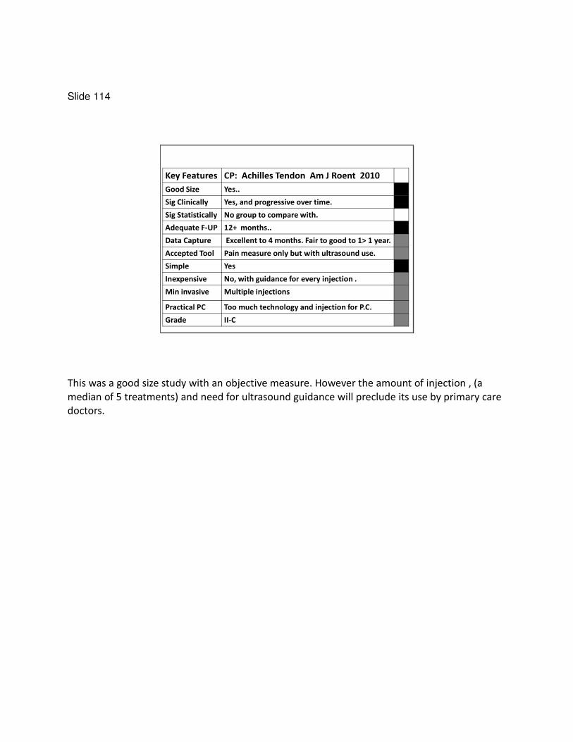
Slide 114
Key Features CP: Achilles Tendon Am J Roent 2010
Good Size Yes..
Sig Clinically Yes, and progressive over time.
Sig Statistically No group to compare with.
Adequate F-UP 12+ months..
Data Capture Excellent to 4 months. Fair to good to 1> 1 year.
Accepted Tool Pain measure only but with ultrasound use.
Simple Yes
Inexpensive No, with guidance for every injection .
Min invasive Multiple injections
Practical PC Too much technology and injection for P.C.
Grade II-C
This was a good size study with an objective measure. However the amount of injection , (a
median of 5 treatments) and need for ultrasound guidance will preclude its use by primary care
doctors.

Slide 115
BIOLOGIC REPAIR INJECTION:
PRP
Now we will consider some evidence of the benefit of platelet rich plasma. Unfortunately, little
information is available to compare to dextrose to platelet rich plasma because of cost factors
and no interest in companies to support such a comparison. That will come with time. For now,
a few articles are of particular importance.

Slide 116
Improving Study Quality PRP
• PRP vs Shock Wave Jumper’s Knee Randomized (Success)
• PRP vs Steroid vs Saline Lat Epicondy RCT blinded (Design Issues)
• PRP vs Saline RCT Knee OA blinded (Success)
• PRP vs HA in Knee OA (Mixed Success)
But Systemic GF effect issue
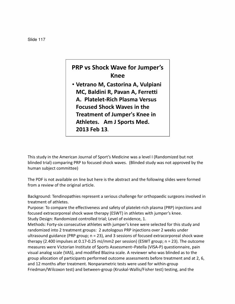
Slide 117
PRP vs Shock Wave for Jumper’s
Knee
• Vetrano M, Castorina A, Vulpiani
MC, Baldini R, Pavan A, Ferretti
A. Platelet-Rich Plasma Versus
Focused Shock Waves in the
Treatment of Jumper's Knee in
Athletes. Am J Sports Med.
2013 Feb 13.
This study in the American Journal of Sport’s Medicine was a level I (Randomized but not
blinded trial) comparing PRP to focused shock waves. (Blinded study was not approved by the
human subject committee)
The PDF is not available on line but here is the abstract and the following slides were formed
from a review of the original article.
Background: Tendinopathies represent a serious challenge for orthopaedic surgeons involved in
treatment of athletes.
Purpose: To compare the effectiveness and safety of platelet-rich plasma (PRP) injections and
focused extracorporeal shock wave therapy (ESWT) in athletes with jumper’s knee.
Study Design: Randomized controlled trial; Level of evidence, 1.
Methods: Forty-six consecutive athletes with jumper’s knee were selected for this study and
randomized into 2 treatment groups: 2 autologous PRP injections over 2 weeks under
ultrasound guidance (PRP group; n = 23), and 3 sessions of focused extracorporeal shock wave
therapy (2.400 impulses at 0.17-0.25 mJ/mm2 per session) (ESWT group; n = 23). The outcome
measures were Victorian Institute of Sports Assessment–Patella (VISA-P) questionnaire, pain
visual analog scale (VAS), and modified Blazina scale. A reviewer who was blinded as to the
group allocation of participants performed outcome assessments before treatment and at 2, 6,
and 12 months after treatment. Nonparametric tests were used for within-group
Friedman/Wilcoxon test) and between-group (Kruskal-Wallis/Fisher test) testing, and the

significance level was set at .05. Results: The 2 groups were homogeneous in terms of age, sex,
level of sports participation, and pretreatment clinical status.
Patients in both groups showed statistically significant improvement of symptoms at all follow-
up assessments. The VISA-P, VAS, and modified Blazina scale scores showed no significant
differences between groups at 2-month follow-up (P = .635, .360, and .339, respectively). The
PRP group showed significantly better improvement than the ESWT group in VISA-P, VAS scores
at 6- and 12-month follow-up, and modified Blazina scale score at 12-month follow-up (P\.05 for
all). Conclusion: Therapeutic injections of PRP lead to better midterm clinical results compared
with focused ESWT in the treatment of jumper’s knee in athletes.
Keywords: jumper’s knee; platelet-rich plasma; extracorporeal shock wave therapy;
tendinopathy/therapy

Slide 118
6 Mo Symptoms + Failure Non Op Management
0 and 2 week PRP
(2 ml) injections under US guidance
2, 6 and 12 month outcomes with VISA-P, VAS
pain, & modified Blazina scale (1-4 for tendinopathy) by blinded reviewer
23 3 sessions of
ESWT under US guidance at 24-48
hr intervals
23
23 23
No anesthetic
The criteria used for inclusion in the study were an established diagnosis of chronic jumper’s
knee at the insertion of the patellar tendon at the lower pole of the patella for at least 6 months
before treatment and failure of nonoperative management.
No anesthetic was used as other studies have suggested that this decreases efficacy of both PRP
(plantar fascia study) and ESWT. (Activates C fibers to release neuropeptides, blocked by
anesthetic)
Participants were all athletes, elite or non-elite, 18-50 years of age.
PRP GROUP
PRP Type: The PRP was obtained by a single centrifugation of whole blood to isolate platelets
using MyCells Autologous Platelet Preparation System (Kaylight Ltd, Ramat-Hasharon, Israel).
From 10 ml draw. � 6-7 ml plasma with 3-5 times platelet concentration. Leukocyte and RBC
count not listed. Mario Vetrano was emailed for this with no reply.
Color doppler guidance was used for injection. One injection site. (location not stated).
Multiple aliquots used for total of 2 ml. No anesthetic was included. rest for 15 minutes
without moving leg and a moderate compression bandage was applied. Full loading + normal
ADL.

ESWT GROUP 3 sessions at 24 to 48 hr intervals with 2400 impulses each and energy density of
.17 to .25 mJ/mm2 depending on pain tolerance. Inline US guidance used to focus on the
damaged area in the tendon.
One week after last session, stretching and muscle strengthening for 2 weeks and then water
activities only if mild discomfort or pain. Then 4 weeks gradual return to training activity if min
or no pain and eventual symptom limited return to sport

Slide 119
Increase in VISA-P Over Time
(MCID = Black Line = 15.0)
0
10
20
30
40
2 6 12
PRP
ESWT P = .014
P = .026
15 points or 27% increase in VISA P is a minimal clinically important difference in MCID for
change in the VISA-P. The reference for that is: Hernandez-Sanchez S, Hidalgo MD, Gomez A.
Responsiveness of the VISA-P scale for patellar tendinopathy in athletes Br J Sports Med. 2012
Sep 25.
Examination of improvements in both groups indicate that each had evidence of providing
significant clinical benefit. However the PRP group improved to more than double the MCID and
was significantly better than the extracorporeal shock wave therapy group.

Slide 120
Pain Levels Over Time
0
2
4
6
8
0 2 6 12
PRP ESWT
P = .028 P = .009
These are pain levels using a visual analogue scale (VAS). The Minimal Clinically Important
difference (MCID) and Patient Acceptable Symptom Score (PASS) has not been established fur
jumper’s knee.
For Rotator cuff dysfunction related pain they have and they are 1.4 for MCID and 3.0 for PASS.
The reference for that is: Tashjian RZ, Deloach J, Porucznik CA, Powell AP. Minimal clinically
important differences (MCID) and patient acceptable symptomatic state (PASS) for visual analog
scales (VAS) measuring pain in patients treated for rotator cuff disease. J Shoulder Elbow Surg.
2009 Nov-Dec;18(6):927-32.
Although both groups easily achieved the MCID for pain improvement, the PRP group results
clearly exceeded PASS level improvements and, again, PRP results were significantly better at 6
and 12 months.
The Blazina scale of symptomatic tendinopathy is only 4 points and thus less sensitive to
change. Nevertheless the PRP group showed significantly better results at 12 month follow-up

Slide 121
• Krogh TP, Fredberg U, Stengaard-
Pedersen K, Christensen R, Jensen P,
Ellingsen T. Treatment of Lateral
Epicondylitis With Platelet-Rich
Plasma, Glucocorticoid, or Saline: A
Randomized, Double-Blind, Placebo-
Controlled Trial. Am J Sports Med.
2013 Jan 17. [Epub ahead of print]
PRP vs Steroid vs Saline in Tennis
Elbow With 3 Month Followup

Slide 122
• No significant difference between
groups in PRTEE or
• Steroid was more effect at reducing
tendon thickness and color doppler
activity at 3 months.
• Virgin elbows may be problematic for
study
PRP vs Steroid vs Saline in Tennis
Elbow : Results @ 3 Months

Slide 123
Rha DW, Park GY, Kim YK, Kim MT, Lee
SC. Comparison of the therapeutic effects
of ultrasound-guided platelet-rich plasma
injection and dry needling in rotator cuff
disease: a randomized controlled trial.
Clin Rehabil. 2013 Feb;27(2):113-22. doi:
10.1177/0269215512448388. Epub 2012
Oct 3.
Buffy Coat PRP vs Repetitive Needling in
Rotator Cuff Damage
PDF available via medical library. Here is the abstract
Abstract
OBJECTIVE:
To compare the effects of platelet-rich plasma injection with those of dry needling on shoulder
pain and function in patients with rotatorcuff disease.
DESIGN:
A single-centre, prospective, randomized, double-blinded, controlled study.
SETTING:
University rehabilitation hospital.
PARTICIPANTS:
Thirty-nine patients with a supraspinatus tendon lesion (tendinosis or a partial tear less than 1.0
cm, but not a complete tear) who met the inclusion criteria recruited between June 2010 and
February 2011.
INTERVENTION:
Two dry needling procedures in the control group and two platelet-rich plasma injections in the
experimental group were applied to the affected shoulder at four-week intervals using
ultrasound guidance.
MEASUREMENTS:

The Shoulder Pain and Disability Index, passive range of motion of the shoulder, a physician
global rating scale at the six-month follow-up, adverse effects monitoring and an ultrasound
measurement were used as outcome measures.
RESULTS:
The clinical effect of the platelet-rich plasma injection was superior to the dry needling from six
weeks to six months after initial injection(P < 0.05). At six months the mean Shoulder Pain and
Disability Index was 17.7 ± 3.7 in the platelet-rich plasma group versus 29.5 ± 3.8 in the dry
needling group (P < 0.05). No severe adverse effects were observed in either group.
CONCLUSIONS:
Autologous platelet-rich plasma injections lead to a progressive reduction in the pain and
disability when compared to dry needling. This benefit is certainly still present at six months
after treatment. These findings suggest that treatment with platelet-rich plasma injections is
safe and useful for rotator cuff disease.

Slide 124
39 with supraspinatus tendinosis or tear less than 1 cm
2 PRP Sessions at 4 week intervals. Type?
20 19
2 Dry Needling Sessions at 4 week intervals
Shoulder Pain and Disability Index/ROM/Ultrasound
6 weeks and 6 months.
Single Blind
Six month SPDI was 17.7 in PRP group and 29.5 in dry needling group.
Buffy Coat PRP vs Repetitive Needling in
Rotator Cuff Damage: Method
16 14Significant Dropout
Chronic pain in shoulder in these cases: > 6 mo
VAS > 5 required.
Pain arc or impingement.
Normal strength testing. (Minimal Neurogenic inflammation. )
Tendinosis OR < 1 cm tear
Failure of conservative therapy for 3 months
Postulated improvement of pain and inhibition as primary result. (No comment about healing. )
Patients informed that both treatments should have stimulate healing to some degree.
Syringes sealed with plaster. The reaction may have unblinded them but depends in part on PRP
type.
Patients blind and the evaluator was blind.
Lidocaine was used for anesthesia of the supraspinatus tendon in all subjects which MAY blunt
PRP effect. (controversial but just 1 ml of .5% lidocaine.)
40-50 times dry needling in the lesion area.
Making PRP: 25 ml blood draw for all patients. Centrifugation at 1600 g to separate
eryhthrocytes.
Then centrifugation at 2000 g to separate PRP from PPP. 3 ml PRP obtained, and targeted for
the area of damage. Clearly much less needling.

The number of dropout was of concern. 4 (20%) of the 20 in the PRP group dropped out before
6 months. 5 /19 (>25%) of the needling group dropped out by 6 months. The dropouts
occurred after the 4 week (2nd
injection) period, so they had received full treatments and still
failed to follow-up. Thus only 30/39 had data to evaluation which may skew results
considerably.
See data next slide.
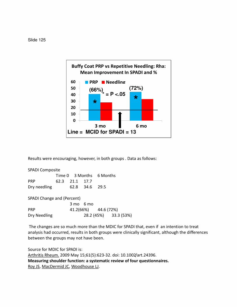
Slide 125
Buffy Coat PRP vs Repetitive Needling: Rha:
Mean Improvement In SPADI and %
0
10
20
30
40
50
60
3 mo 6 mo
PRP Needling
* = P <.05
* *
Line = MCID for SPADI = 13
(66%) (72%)
Results were encouraging, however, in both groups . Data as follows:
SPADI Composite
Time 0 3 Months 6 Months
PRP 62.3 21.1 17.7
Dry needling 62.8 34.6 29.5
SPADI Change and (Percent)
3 mo 6 mo
PRP 41.2(66%) 44.6 (72%)
Dry Needling 28.2 (45%) 33.3 (53%)
The changes are so much more than the MDIC for SPADI that, even if an intention to treat
analysis had occurred, results in both groups were clinically significant, although the differences
between the groups may not have been.
Source for MDIC for SPADI is:
Arthritis Rheum. 2009 May 15;61(5):623-32. doi: 10.1002/art.24396.
Measuring shoulder function: a systematic review of four questionnaires.
Roy JS, MacDermid JC, Woodhouse LJ.

Two patients with partial-thickness tears (1 articular tear and 1 bursal surface tear) of the
supraspinatus improved to tendinosis without tear in the platelet-rich plasma group. None did
so in the

Slide 126
Leukocyte Filtered PRP 1-2 Rx vs
Saline Injection in Knee OA
• Patel S, Dhillon MS, Aggarwal S,
Marwaha N, Jain A Treatment with
platelet-rich plasma is more effective
than placebo for knee osteoarthritis: a
prospective, double-blind, randomized
trial. Am J Sports Med. 2013
Feb;41(2):356-64. doi:
10.1177/0363546512471299. Epub 2013
Jan 8.
PDF is available via medial library. Here is the abstract.
Background: Specific growth factors have been proposed as therapeutic proteins for cartilage
repair.
Hypothesis: Platelet-rich plasma (PRP) provides symptomatic relief in early osteoarthritis (OA) of
the knee.
Study Design: Randomized controlled trial; Level of evidence, 1.
Methods: A total of 78 patients (156 knees) with bilateral OA were divided randomly into 3
groups. Group A (52 knees) received a single injection of PRP, group B (50 knees) received 2
injections of PRP 3 weeks apart, and group C (46 knees) received a single injection of normal
saline. White blood cell (WBC)–filtered PRP with a platelet count 3 times that of baseline (PRP
type 4B) was administered in all. All the groups were homogeneous and comparable in baseline
characteristics. Clinical outcome was evaluated using the Western Ontario and McMaster
Universities Arthritis Index (WOMAC) questionnaire before treatment and at 6 weeks, 3 months,
and 6 months after treatment. They were also evaluated for pain by a visual analog scale, and
overall satisfaction with the procedure and complications were noted.
Results: Statistically significant improvement in all WOMAC parameters was noted in groups A
and B within 2 to 3 weeks and lasting until the final follow-up at 6 months, with slight worsening
at the 6-month follow-up. The mean WOMAC scores (pain, stiffness, physical function, and total
score) for group A at baseline were 10.18, 3.12, 36.56, and 49.86, respectively, and at final
follow-up were 5.00, 2.10, 20.08, and 27.18, respectively, showing significant improvement.

Similar improvement was noted in group B (mean WOMAC scores at baseline: 10.62, 3.50,
39.10, and 53.20, respectively; mean WOMAC scores at final followup:
6.18, 1.88, 22.40, and 30.48, respectively). In group C, the mean WOMAC scores deteriorated
from baseline (9.04, 2.70, 33.80, and 45.54, respectively) to final follow-up (10.87, 2.76, 39.46,
and 53.09, respectively). The 3 groups were compared with each other, and no improvement
was noted in group C as compared with groups A and B (P\.001). There was no difference
between groups A and B, and there was no influence of age, sex, weight, or body mass index on
the outcome. Knees with Ahlback grade 1 fared better than those with grade 2. Mild
complications such as nausea and dizziness, which were of short duration, were observed in 6
patients (22.2%) in group A and 11 patients (44%) in group B.
Conclusion: A single dose of WBC-filtered PRP in concentrations of 10 times the normal amount
is as effective as 2 injections to alleviate symptoms in early knee OA. The results, however,
deteriorate after 6 months. Both groups treated with PRP had better results than did the group
injected with saline only.
Keywords: platelet-rich plasma; osteoarthritis

Slide 127
78 patients (156 knees) with Bilat OA (Very few Ahlback 3 (1 and 2) grade)
1 PRP Inj
6 week, 3 and 6 month WOMAC and VAS Pain
54 knees
46 knees50 knees
2 PRP Inj 0 and 3 wks 1 Saline Inj
WBC Filtered PRP
6 Months: 1 PRP ���� WOMAC improvement by 22
2 PRP ���� WOMAC improvement by 22.5
Saline ���� WOMAC decline by -7.5
1 PRP vs 2 PRP vs 1 Saline Injection in Knee OA
52 knees
46 knees50 knees Double Blind Treatment
This is the only study reviewed recently that excluded both Ahlback 3 and 4 stages.
1 patient in the single PRP group had 2 knee replacements.
Statistical correction for two knees not mentioned, and that is critical.
100 ml blood drawn. Centrifuged 15 min at 1500 RPM. The PRP was then extracted through
a pipette and transferred to a test tube, and a leucocyte filter (Imugard III-PL, Terumo Penpol
Ltd, Thiruvananthapuram, India) was then used to filter off the leucocytes. The final PRP was
assessed for platelet count and was supplied for injection in a 10-mL syringe (approximately 8
mL per knee). Mean platelet count 310,000. 1 ml CaCl was injected with each 4 ml of PRP. Ca
may have therapeutic effect.
Global WOMAC was obtained.
22% in the single PRP injection and 44% in the double PRP injection had reactions to injection
consisting of various things: Syncope, dizziness, headache, nausea, gastritis, sweating, and
tachycardia. Note pain was not mentioned as a large issue. (low WBC product) Nevertheless
saline group had none


Slide 128
Leukocyte Filtered PRP vs Saline: Patel
Mean Improvement In WOMAC Scores
-10
0
10
20
30
3 mo 6 mo
PRP 1 PRP 2 Saline
Line = MCID for WOMAC for Knee = 16
This illustrates improvement in the composite WOMAC score at 3 and 6 months. The
differences between PRP and Saline were substantial but no differences were seen between the
PRP groups. The improvement in each group decreased by 6 months but still exceeded the
MCID. The percentage improvemeat at 6 months was 45% for 1 PRP injecion and 43% in 2 PRP
injections.
The percent improvement in WOMAC at 3 months was 1 - 39.1/79.6 X 100 = 1-.49 X 100 = 51%
in the PRP group.
The percent improvement at 6 month was 1- 36.5/79.6 x 100 = 54%
.
These both imply that there is a substantial pain source not addressed by the treatment
method.

Slide 129
PRP vs HA In Knee OA: ACP
Cerza F; Carni S; Carcangiu A; Di
Vavo I; Schiavilla V; Pecora A; De
Biasi G; Ciuffreda M Comparison
Between Hyaluronic Acid and Platelet-
Rich Plasma, Intra-articular Infiltration
in the Treatment of Gonarthrosis Am J
Sports Med 2012; 40(12) p2822-7.
PDF Not available on Line:
Here is the abstract. Full article review was conducted for this summary.
AUTHOR(S): AUTHOR'S ADDRESS: Alessandro Carcangiu, Orthopaedics and Traumatology Unit
of P. Colombo Hospital of Velletri, Vicolo dell'Annunziatella, 50 Rome, Italy, 00142.
ABSTRACT: BACKGROUND: Arthrosis is particularly prevalent in the knee. Infiltration treatment
for gonarthrosis is among the most widely used techniques in orthopaedic practice. PURPOSE:
To compare the clinical response of hyaluronic acid (HA) and platelet-rich plasma (PRP)
treatment in 2 groups of patients affected by gonarthrosis. STUDY DESIGN: Randomized
controlled trial; Level of evidence, 1. METHODS: A total of 120 patients affected by clinically and
radiographically documented gonarthrosis were included in this study. The gonarthrosis was
graded using the Kellgren-Lawrence radiographic classification scale. The 120 patients were
randomized into 2 study groups in a 1:1 ratio: 60 patients received 4 intra-articular injections of
PRP (specifically, autologous conditioned plasma [ACP], 5.5 mL), and 60 patients received 4
intra-articular injections of HA (20 mg/2 mL). An unblinded physician performed infiltration once
a week for 4 weeks into the knee affected by clinically relevant gonarthrosis (in both groups). All
patients were evaluated with the Western Ontario and McMaster (WOMAC) score before the
infiltration and at 4, 12, and 24 weeks after the first injection. RESULTS: Treatment with a local
injection of ACP had a significant effect shortly after the final infiltration and a continuously
improving sustained effect up to 24 weeks (WOMAC score, 65.1 and 36.5 in the HA and ACP

groups, respectively; P < .001), where the clinical outcomes were better compared with the
results with HA. In the HA group, the worst results were obtained for grade III gonarthrosis,
whereas the clinical results obtained in the ACP group did not show any statistically significant
difference in terms of the grade of gonarthrosis. The mean WOMAC scores for grade III
gonarthrosis were 74.85 in the HA group and 41.20 in the ACP group (P < .001). CONCLUSION:
Treatment with ACP showed a significantly better clinical outcome than did treatment with HA,
with sustained lower WOMAC scores. Treatment with HA did not seem to be effective in the
patients with grade III gonarthrosis.

Slide 130
120 with OA knee . Grade IV excluded. Reason?
4 IA injection of 5.5 ml ACP, weekly
60 60
4 IA injection of 20 mg/2ml Hyalgan,
weekly.
WOMAC at 4, 12 and 24 weeks.
Single Blind
60 60#
PRP vs HA In Knee OA: ACP: Method
All subjects had received physical or pharmacologic therapy. The AP X-Ray was in full extension.
With bilateral knees only the worst side was considered in evaluation. If platelet count was less
than 150.000, patients were excluded. I did not see exclusions listed.
Mean blood draw for ACP preparation was only 12 ml. Volume if ICP injected was not stated.
Superolateral approach for injection (93% success rate noted) No guidance.
Knees were grade II most commonly then grade I and then grade III the least.
Note activation not mentioned despite very low leukocyte levels.

Slide 131
ACP vs HA in Knee OA : Cerza: Mean
Improvement In WOMAC Scores
0
10
20
30
40
50
4 12 24
PRP
HA
* P < .001
** *
Line = MCID for WOMAC for Knee = 16
This illustrates improvement in the composite WOMAC score at 4, 12 and 24 weeks. At every
point the intergroup difference of significant, and deterioration in improvement from HA was
apparent by 3 months, with less than and MCID difference at 12 and 24 weeks.
The percent improvement in WOMAC at 3 months was 1 - 39.1/79.6 X 100 = 1-.49 X 100 = 51%
in the PRP group.
The percent improvement at 6 month was 1- 36.5/79.6 x 100 = 54%
.
These both imply that there is a substantial pain source not addressed by the treatment
method.

Slide 132
PRP vs HA in Knee OA: Buffy Coat
PRP
• Spaková T, Rosocha J, Lacko M, Harvanová D,
Gharaibeh A. Treatment of knee joint
osteoarthritis with autologous platelet-rich
plasma in comparison with hyaluronic acid.
Am J Phys Med Rehabil. 2012 May;91(5):411-
7. doi: 10.1097
• 3 PRP sessions vs 3 HA sessions
• 6 month WOMAC and NRS pain signif better.
PDF requested
PDF available through medical library
Here is the Abstract:
m J Phys Med Rehabil. 2012 May;91(5):411-7. doi: 10.1097/PHM.0b013e3182aab72.
Treatment of knee joint osteoarthritis with autologous platelet-rich
plasma in comparison with hyaluronic acid.
Spaková T, Rosocha J, Lacko M, Harvanová D, Gharaibeh A.
Source
Associated Tissue Bank of Faculty of Medicine UPJS, Kosice, Slovakia.
Abstract
OBJECTIVE:
This study aimed to find a simple, cost-effective, and time-efficient method for the preparation
of platelet-rich plasma (PRP), so the acquired benefits will be readily available for multiple
procedures in smaller outpatient clinics and to explore the safety and efficacy of the application
of PRP in the treatment of degenerative lesions of articular cartilage of the knee.
DESIGN:
The study was designed as a prospective, cohort study with a control group. A total of 120
patients with Grade 1, 2, or 3 osteoarthritisaccording to the Kellgren and Lawrence grading scale
were enrolled in the study. One group of patients was treated using three intra-articular
applications of PRP, and the second group of patients was given three injections of hyaluronic
acid. Outcome measures included the Western Ontario and McMaster
Universities Osteoarthritis Index and the 11-point pain intensity Numeric Rating Scale.

RESULTS:
On average, a 4.5-fold increase in platelet concentration was obtained in the PRP group. No
severe adverse events were observed. Statistically significantly better results in the Western
Ontario and McMaster Universities Osteoarthritis Index and Numeric Rating Scale scores were
recorded in a group of patients who received PRP injections after a 3- and 6-mo follow-up.
CONCLUSIONS:
Our preliminary findings support the application of autologous PRP as an effective and safe
method in the treatment of the initial stages of knee osteoarthritis. Further studies are needed
to confirm these results and to investigate the persistence of the beneficial effects observed.

Slide 133
120 with OA knee. Grade IV excluded. Mostly grad II knees
3 IA injection of BC PRP weekly?
60 60#
3 IA injection of Erectus 1.2% HA weekly.
WOMAC & NRS at 3 and 6 months.
Non Blinded
60 60#
PRP vs HA in Knee OA: Buffy Coat PRP: Method
Goal to find a simple, cost effective and time efficient method for prep or PRP.
120 patients with grade 1=3 OA according to Kellgren and Lawrence grading scale. Also 4
excluded.
27 ml blood drawn in 3 tubes. Centrifuge X 1 15 min at 3200 RPM. They collect plasma plus
buffy coat and then 10 min at 1500 RPM to separate leukocytes. Then plasma layer was
collected and it appearss that was spun again at 1500 to prepare the PRP and PPP without
leukocytes.
Not so strict on platelet count (< 100,000) exclusions.
60 minute platelet preparation time. Concentration of platelet mean was 4.5 times.
Mean WBC was 23.2 (conc mean 3.6) and RBC was 3.8. (conc mean 0.6)
Mostly grade II subjects.

Slide 134
Buffy Coat PRP vs HA: Spakova Mean
Improvement In WOMAC Scores
0
5
10
15
20
25
30
3 mo 6 mo
PRP
HA
* P < .01
* *
Line = MCID for WOMAC for Knee = 16
This illustrates improvement in the composite WOMAC score at 4, 12 and 24 weeks. At every
point the intergroup difference of significant, and deterioration in improvement from HA was
apparent by 3 months, with less than and MCID difference at 12 and 24 weeks.
Percent improvement at 3 months in PRP group was 73% and at 6 months was 51%

Slide 135
PRP vs HA in Knee OA: Buffy Coat
Frozen PRP
• Filardo G, Kon E, Di Martino A, Di Matteo B, Merli
ML, Cenacchi A, Fornasari PM, Marcacci M.
Platelet-rich plasma vs hyaluronic acid to treat knee
degenerative pathology: study design and
preliminary results of a randomized controlled trial.
BMC Musculoskelet Disord. 2012 Nov 23;13:229.
doi: 10.1186/1471-2474-13-229.
• 3 PRP sessions vs 3 HA sessions
PDF available Free at http://www.biomedcentral.com/1471-2474/13/229
Here is the astract
Abstract
BACKGROUND:
Platelet Rich Plasma (PRP), a blood-derived product rich in growth factors, is a promising
treatment for cartilage defects but there is still a lack of clinical evidence. The aim of this study
is to show, through a randomized double blind prospective trial, the efficacy of this procedure,
by comparing PRP to Hyaluronic Acid (HA) injections for the treatment of knee chondropathy or
osteoarthritis (OA).
METHODS:
109 patients (55 treated with HA and 54 with PRP) were treated and evaluated at 12 months of
follow-up. The patients were enrolled according to the following inclusion criteria: age> 18
years, history of chronic (at least 4 months) pain or swelling of the knee and imaging findings of
degenerative changes of the joint (Kellgren-Lawrence Score up to 3). A cycle of 3 weekly
injections was administered blindly. All patients were prospectively evaluated before and at 2, 6,
and 12 months after the treatment by: IKDC, EQ-VAS, TEGNER, and KOOS scores. Range of
motion and knee circumference changes were measured over time. Adverse events and patient
satisfaction were also recorded.
RESULTS:

Only minor adverse events were detected in some patients, such as mild pain and effusion after
the injections, in particular in the PRP group, where a significantly higher post-injective pain
reaction was observed (p=0.039). At the follow-up evaluations, both groups presented a clinical
improvement but the comparison between the two groups showed a not statistically significant
difference in all scores evaluated. A trend favorable for the PRP group was only found in patients
with low grade articular degeneration (Kellgren-Lawrence score up to 2).
CONCLUSIONS:
Results suggest that PRP injections offer a significant clinical improvement up to one year of
follow-up. However, conversely to what was shown by the current literature, for middle-aged
patients with moderate signs of OA, PRP results were not better than those obtained with HA
injections, and thus it should not be considered as first line treatment. More promising results
are shown for its use in low grade degeneration, but they still have to be confirmed.

Slide 136
109 with OA knee . Grade IV excluded.
3 IA injection of 5 ml PRP weekly
54 55
3 IA injection of 20
mg/2ml Hyalubrix > 1500 KDa, weekly.
IKDC, KOOS, at 6 and 12 months and ROM
Single* Blind
60 60#
PRP vs HA in Knee OA: Buffy Coat Frozen PRP (method)
Technically single blind but evaulator was blinded as well.
150 ml blood draw venous for every knee with 1480 RPM X 6 min to separate erythrocytes,
then 3400 rpm for 15 min to produce 20 ml with 5 analyzed and 15 frozen for 5 ml X 3 visits.
This could associate with degranulation, but freeze thawing can be a method to release
intracelluar GFs.
Leukocytes has 1.2 times the usual value, clearly in between. Other components not described
but likely low RBC since first spin said to remove those.

Slide 137
Buffy Coat Frozen PRP Filardo
IKDC Improvements No Signif Differences
0
5
10
15
20
6 mo 12 mo
PRP
HA
Line = MCID for IKDC For Knee = 6.7
More pain in the PRP patients. However intolerance of treatment only occurred in the HA group
(3 subjects)
MCID = 6.7 points from previous collections of data.
Note this improvement is significant, comparable to other studies and was sustained to 1 year,
longer than other studies were followed.

Slide 138
PRGF (Endocet) vs HA in Knee OA:
• Sánchez M, Fiz N, Azofra J, Usabiaga J, Aduriz
Recalde E, Garcia Gutierrez A, Albillos J,
Gárate R, Aguirre JJ, Padilla S, Orive G, Anitua
E. A randomized clinical trial evaluating
plasma rich in growth factors (PRGF-Endoret)
versus hyaluronic acid in the short-term
treatment of symptomatic knee
osteoarthritis. Arthroscopy. 2012
Aug;28(8):1070-8.
Available via medical library but not free on line..
Here is the abstract:
Abstract
PURPOSE:
This multicenter, double-blind clinical trial evaluated and compared the efficacy and safety of
PRGF-Endoret (BTI Biotechnology Institute, Vitoria-Gasteiz, Spain), an autologous biological
therapy for regenerative purposes, versus hyaluronic acid (HA) as a short-term treatment for
knee pain from osteoarthritis.
METHODS:
We randomly assigned 176 patients with symptomatic knee osteoarthritis to receive infiltrations
with PRGF-Endoret or with HA (3 injections on a weekly basis). The primary outcome measure
was a 50% decrease in knee pain from baseline to week 24. As secondary outcomes, we also
assessed pain, stiffness, and physical function using the Western Ontario and McMaster
Universities Osteoarthritis Index; the rate of response using the criteria of the Outcome
Measures for Rheumatology Committee and Osteoarthritis Research Society International
Standing Committee for Clinical Trials Response Criteria Initiative (OMERACT-OARSI); and safety.
RESULTS:
The mean age of the patients was 59.8 years, and 52% were women. Compared with the rate of
response to HA, the rate of response to PRGF-Endoret was 14.1 percentage points higher (95%
confidence interval, 0.5 to 27.6; P = .044). Regarding the secondary outcome measures, the rate

of response to PRGF-Endoret was higher in all cases, although no significant differences were
reached. Adverse events were mild and evenly distributed between the groups.
CONCLUSIONS:
Plasma rich in growth factors showed superior short-term results when compared with HA in a
randomized controlled trial, with a comparable safety profile, in alleviating symptoms of mild to
moderate osteoarthritis of the knee.
LEVEL OF EVIDENCE:
Level I, randomized controlled multicenter trial.

Slide 139
176 with OA knee . Grade IV excluded. (Ahlback grading versus Kelgren)
3 IA injection of 8 ml PRGF* weekly
89 87
3 IA (Euflexxa) injection of weekly.
at 6 months
Random Assignment;
No blinding
79* 74*
No NSAIDs at anytime
PRGF (Endocet) vs HA in Knee OA: Sanchez
Random assignment without blinding.
* Activation required because no WBCs
Philosophy of PRGF (Plasma Rich in Growth Factors) use is as follows by authors “Recent data
support the application of platelet-rich plasma products as an effective and safe method in the
treatment of the initial stages of knee OA. Some growth factors present in platelet-rich plasma
products, including transforming growth factor β, platelet-derived growth factor, and insulin-like
growth factor 1, contribute to the maintenance of a homeostatic balanced status between
anabolism and catabolism on the articular cartilage, and others such as vascular endothelial
growth factor and basic fibroblast growth factor show chondroinductive roles.”
36 ml of blood drawn, spun at 580 g for 8 min and then pipetted and picked up 2 ml just above
the buffy coat. Activation was done. 2 ml from each of 4 tubes for an 8 ml volume.
•Exclusions from the study were considerable.
7 were excluded due to taking NSAIDs.
9 had steroid injections or surgery against the protocol
7 no improvement or lost to followup.
Total of 23/176 = 13% or more than 1/8.

Only 6 months follow-up, like most of the other knee PRP vs HA studies (only Filardo et al was to
1 year)

Slide 140
PRGF vs HA: Sanchez
WOMAC Improvement
Line = MCID for WOMAC For Knee = 16
0
5
10
15
20
25
6 Mo
PRP
HA
The values were calculated from a table in the study. The authors emphasized a barely
significant difference in the WOMAC pain which is incorrect to even mention in the presence of
no change in the WOMAC summed score. (Not even approaching significance.)
The results are better than a single minimal clinically important difference, but not approaching
double and are not different.

Slide 141

Slide 142
PRP Vs HA Study Summarization• Cerza 2012 APC (Low WBC) 6 month 54% Impr WOMAC.
WOMAC 36.5 @ 6 mo (Worse patients but no grade IV)
• Spakova 2012 Buffy Coat High WBC 6 month 51% Impr WOMAC
18.85 @ 6 mo (more grade II)
• Filardo/Kon 2012 High WBC Frozen PRP Twice MCID in both
groups (For IKDC) but no differences, sustainable to 1 year. Older
but still no grade IV.
• Sanchez 2012 Plasma Rich in Growth Factor 6 Month. Only
sightly more than MCID for WOMAC but no differences
(Incorrectly stated a subscale was different) @ 6 mo. No grade IV
• All wimply (no grade IV )
• Note in Patel 2013 Leukocyte filtered PRP vs Saline, 44% mean
WOMAC improvement (about 22.5) at 6 months in PRP group.
Rare grade 3 and no grade 4. (Less severe of all studies)
Spakova Notable is that the best the patients mean WOMAC achieved at 3 months was 14.35
and 18.85 at 6 months compared to best of 39.1 and 36.5 in Cerza with worse patients.
Some authors define PRP as only platelets and attribute better results to leucocyte depletion,
because of the deleterious effects of proteases and reactive oxygen released from white cells;
others consider them as a source of important cytokines and enzymes, that may be important
also for the prevention of infections, and report that PRP significantly inhibits the growth. (
Comment from Filardo article.
Another important comment from Filardo et al is the following: “There was a tendency
towards better improvement (or PRP over HA) only in patients affected by earlier degrees of
knee degeneration was observed, thus suggesting that the clinical application of PRP should be
mainly restricted to this patient subgroup, whereas the indication of this treatment for high
grade degeneration is lower. Due to the not significantly better results with respect to HA, PRP
cannot be considered as the first line of treatment for knee OA and should be therefore
restricted to patients who do not benefit from other conservative or injective treatments such
as HA or, if used as first line treatment, it should be mainly targeted to patients affected only by
early degrees of knee cartilage degeneration. “ (Others have found better results but only in up
to and including grade II also.


Slide 143
Systemic Effects on GFs with Focal
PRP Injection
• Wasterlain AS, Braun HJ, Harris
AH, Kim HJ, Dragoo JL. The
systemic effects of platelet-rich
plasma injection. Am J Sports
Med. 2013 Jan;41(1):186-93. doi:
10.1177/0363546512466383.
Epub 2012 Dec 4.
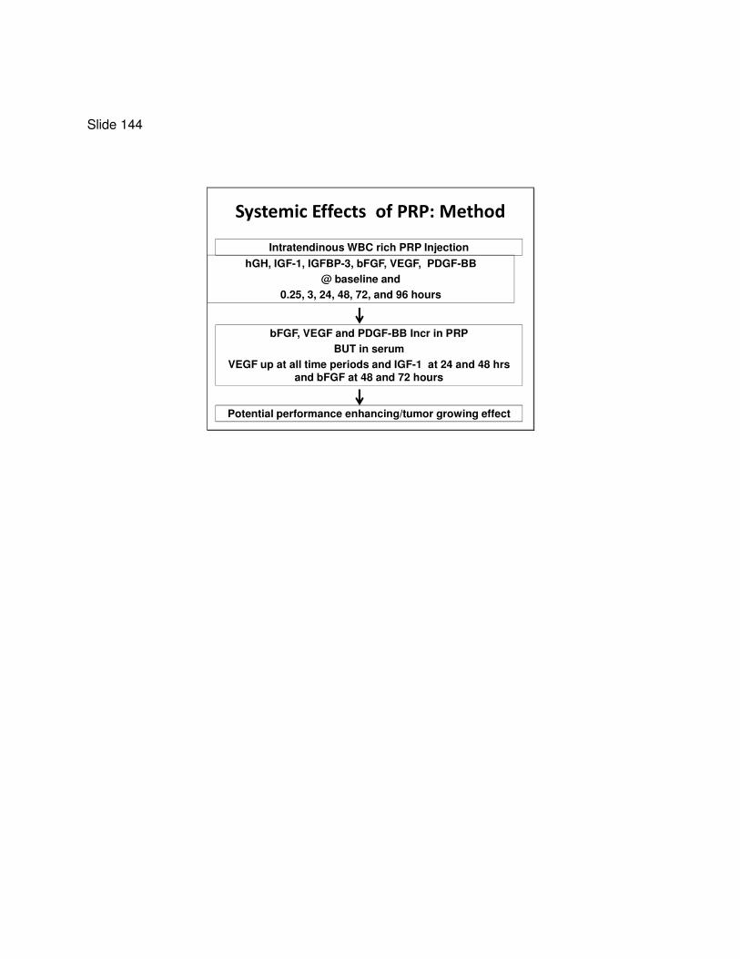
Slide 144
Systemic Effects of PRP: Method
hGH, IGF-1, IGFBP-3, bFGF, VEGF, PDGF-BB
@ baseline and
0.25, 3, 24, 48, 72, and 96 hours
Intratendinous WBC rich PRP Injection
bFGF, VEGF and PDGF-BB Incr in PRP
BUT in serum
VEGF up at all time periods and IGF-1 at 24 and 48 hrs and bFGF at 48 and 72 hours
Potential performance enhancing/tumor growing effect

Slide 145
BIOLOGIC REPAIR INJECTION:
STEM CELLS
Now we will consider some evidence of the benefit stem cells. Controlled studies are lacking.
Consecutive patient studies are worth mentioning in some cases due to durability of benefit or
objective follow-up measures.

Slide 146
Adipose Synovium Stem Cells in
Knee OA
• Koh YG, Jo SB, Kwon OR, Suh DS, Lee SW, Park SH, Choi YJ. Mesenchymal Stem Cell Injections Improve Symptoms of Knee Osteoarthritis. Arthroscopy. 2013
Jan 29. pii: S0749-8063(12)01884-1. doi: 10.1016/j.arthro.2012.11.017. [Epub ahead of print]

Slide 147
18subjects (consecutive?) 6 men and 12 women
Adipose synovium harvested at arthroscopy
Culture to 1.2 X 10⁶ with 3 ml PRP.
WOMAC Decrease by 16.6 at 24 months.
Lyshom improved by 30.3 points at 24 months.
Whole Organ MRI score improve by 11.7 points ( p .001)
The improvements at 24 months are still significant after stem cell placement by injection in the
knee.
However, harvesting was required via arthroscopy