Summary of assisting with chest tube insertion in adults ... · There should be easy access to...
6
THE THORACIC SOCIETY OF AUSTRALIA AND NEW ZEALAND 1 © Queensland Health, 2010 No part of this document may be reproduced without the express permission of Queensland Health. Summary of assisting with chest tube insertion in adults The technique for assisting with chest tube insertion is broken into 12 steps. These steps have been aligned with the doctor’s steps for inserting a chest tube to assist the nurse in timing the procedure. See the summary flowchart of the sequencing below. Nurses Doctors Step 1 – Ensure patient consent is obtained and assess risk Pre-insertion consent and risk assessment Step 1 - Select and mark insertion site Step 2 – Gather equipment and prepare UWSD Step 2 - Choose chest tube and check equipment Step 3 – Set up sterile equipment Step 4 - Inserting the chest tube – the nurse / assistant role Step 5 – Prepare the patient for insertion of chest tube Step 3 - Prepare patient Step 6 - Assist MO throughout Procedure and monitor patient Step 4 - Infiltrate anaesthetic Step 5 -Incise skin and dissect to pleura (for large bore chest tube) Step 6 - Insert chest tube Step 7- Assist in anchoring and securing the chest tube Step 7 - Anchor tube and suture Step 8 - Connect chest tube to UWSD Step 8 - Connect chest tube to UWSD Step 9 - Dress the site Step 9 - Dress the site Step 10 – Ensure confirmation of tube placement Step 10 - Confirm placement Step 11 - Document the procedure Step 11 – Document the procedure Step 12 – Monitor and record patient and chest drain observations Step 12 - Monitor patient and chest drain
-
Upload
truongtuyen -
Category
Documents
-
view
215 -
download
1
Transcript of Summary of assisting with chest tube insertion in adults ... · There should be easy access to...

THE THORACIC SOCIETYOF AUSTRALIA AND NEW ZEALAND
1
© Queensland Health, 2010 No part of this document may be reproduced without the express permission of Queensland Health.
Summary of assisting with chest tube insertion in adults The technique for assisting with chest tube insertion is broken into 12 steps. These steps have been aligned with the doctor’s steps for inserting a chest tube to assist the nurse in timing the procedure. See the summary flowchart of the sequencing below.
Nurses Doctors Step 1 – Ensure patient consent is obtained and assess risk
Pre-insertion consent and risk assessment
Step 1 - Select and mark insertion site
Step 2 – Gather equipment and prepare UWSD Step 2 - Choose chest tube and check equipment
Step 3 – Set up sterile equipment
Step 4 - Inserting the chest tube – the nurse / assistant role
Step 5 – Prepare the patient for insertion of chest tube Step 3 - Prepare patient
Step 6 - Assist MO throughout Procedure and monitor patient
Step 4 - Infiltrate anaesthetic
Step 5 -Incise skin and dissect to pleura (for large bore chest tube) Step 6 - Insert chest tube
Step 7- Assist in anchoring and securing the chest tube Step 7 - Anchor tube and suture
Step 8 - Connect chest tube to UWSD Step 8 - Connect chest tube to UWSD
Step 9 - Dress the site Step 9 - Dress the site
Step 10 – Ensure confirmation of tube placement Step 10 - Confirm placement
Step 11 - Document the procedure Step 11 – Document the procedure
Step 12 – Monitor and record patient and chest drain observations
Step 12 - Monitor patient and chest drain
Edwin
Typewritten Text
Confirm site and side for insertion with MO
Edwin
Typewritten Text
Edwin
Typewritten Text

THE THORACIC SOCIETYOF AUSTRALIA AND NEW ZEALAND
2
© Queensland Health, 2010 No part of this document may be reproduced without the express permission of Queensland Health.
Step 1: Ensure patient consent obtained and assess risk
Before starting the procedure: • Check identity of patient. • Ensure patient consent is obtained. • In collaboration with MO:
perform a risk assessment confirm side and site of insertion discuss patient position.
• Provide privacy (use of a procedure room is optimal). • Check bed positioning. • Check a chest x-ray (or digital print) is available. • Clip patient’s hair around the insertion site.
Step 2: Gather equipment and prepare UWSD
Confer with the MO to determine: • Sterile glove size • The type and volume of local anaesthetic • Suture material required • Size and type of chest tube:
Set up and prime the sterile underwater seal drainage system (UWSD) in accordance with manufacturer’s guidelines.
Safety Tip! If you are not experienced in assisting with this procedure, seek supervision from somebody who is.
Safety Tip! Should the nurse become concerned that a patient lacks a sufficient understanding of the procedure to have made an informed decision to undergo the procedure, he/she should take reasonable steps to ensure that the patient receives the necessary additional information from the Medical Officer.
Safety Tip! Check the equipment and ensure it is sterile and appropriate for your specific patient
requirements before proceeding any further. E.g. if you have an obese patient you may require longer needles for anaesthetic delivery and artery forceps.
Before the doctor commences insertion, ensure that the chest tube fits the chest drain (adaptors may be required).
Some chest drains are packaged with a cover/cap over the air vent. Ensure that the cover/cap is removed.
Safety Tip! There should be easy access to resuscitation equipment and oxygen at the site of the
procedure. Reliable venous access is recommended throughout the procedure. Ideally, monitor the the oxygen saturation continuously and vital signs intermittently
during insertion of the chest tube.
THE THORACIC SOCIETYOF AUSTRALIA AND NEW ZEALAND
3
© Queensland Health, 2010 No part of this document may be reproduced without the express permission of Queensland Health.
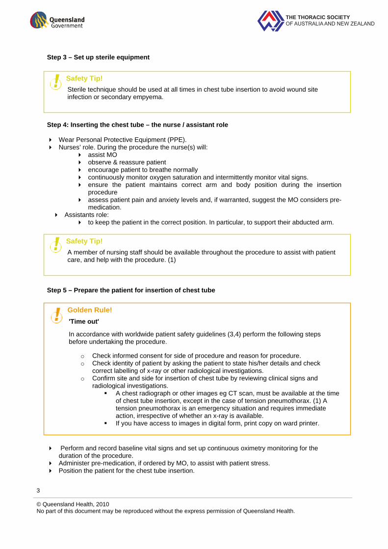
Step 3 – Set up sterile equipment Step 4: Inserting the chest tube – the nurse / assistant role
Wear Personal Protective Equipment (PPE). Nurses’ role. During the procedure the nurse(s) will:
assist MO observe & reassure patient encourage patient to breathe normally continuously monitor oxygen saturation and intermittently monitor vital signs. ensure the patient maintains correct arm and body position during the insertion
procedure assess patient pain and anxiety levels and, if warranted, suggest the MO considers pre-
medication. Assistants role:
to keep the patient in the correct position. In particular, to support their abducted arm.
Step 5 – Prepare the patient for insertion of chest tube
Perform and record baseline vital signs and set up continuous oximetry monitoring for the duration of the procedure.
Administer pre-medication, if ordered by MO, to assist with patient stress. Position the patient for the chest tube insertion.
Safety Tip! Sterile technique should be used at all times in chest tube insertion to avoid wound site infection or secondary empyema.
Safety Tip! A member of nursing staff should be available throughout the procedure to assist with patient care, and help with the procedure. (1)
Golden Rule! 'Time out'
In accordance with worldwide patient safety guidelines (3,4) perform the following steps before undertaking the procedure.
o Check informed consent for side of procedure and reason for procedure. o Check identity of patient by asking the patient to state his/her details and check
correct labelling of x-ray or other radiological investigations. o Confirm site and side for insertion of chest tube by reviewing clinical signs and
radiological investigations. A chest radiograph or other images eg CT scan, must be available at the time
of chest tube insertion, except in the case of tension pneumothorax. (1) A tension pneumothorax is an emergency situation and requires immediate action, irrespective of whether an x-ray is available.
If you have access to images in digital form, print copy on ward printer.

THE THORACIC SOCIETYOF AUSTRALIA AND NEW ZEALAND
4
© Queensland Health, 2010 No part of this document may be reproduced without the express permission of Queensland Health.
Step 6 - Assist MO throughout Procedure Assist the MO to: • draw up the appropriate local anaesthetic.
During the procedure: • pass equipment as requested • observe & reassure the patient • encourage the patient to breathe normally • monitor vital signs:
continuous oxygen saturation respiratory rate, pulse and blood pressure.
Step 7- Assist in anchoring and securing the chest tube
Anchoring suture: • Provide the MO suture material. • Support the tube to prevent it falling out while the anchoring suture is completed. • Secure the tubing to the skin using a mesenteric tag of tape applied to the chest tube.
Anchoring device: • may be used for a small bore catheter • this does not replace the need to stitch the catheter firmly in place.
Step 8 - Connect chest tube to UWSD Once the chest tube is in position and secured, immediately connect it to the chest drain. For small bore catheters, turn the 3 way tap 'off to bung' to allow free drainage from the pleural
cavity into the drain. If the chest tube has been clamped, remove clamps immediately after connection to the
drain. Tape the junction of the chest tube and drainage tube to prevent separation. Ensure the drain system is positioned below (at least 80 cm) the patient’s chest level at all times.
Safety check! Warning signs that something has gone wrong with the insertion procedure include:
the patient experiences sudden pain or distress the patient becomes hypotensive, hypoxaemic or observations deteriorate.
After the chest tube is inserted the correct placement of the chest tube can be confirmed by:
“fogging” of a large bore chest tube with expiration movement of air/fluid through the tube (1) ‘swinging’, ‘tidalling’ or ‘oscillating’ of the fluid level in the water seal chamber.
Safety Tip! After prepping and draping, and BEFORE the insertion procedure begins, ensure you return the patient back to the position they were in when the doctor marked the insertion site (especially the position of the abducted arm). This is important as the skin moves several centimetres in relation to the ribs when the arm is moved. If the Medical officer is using a skin mark made during imaging, make sure the patient is in the same position for the chest tube insertion.

THE THORACIC SOCIETYOF AUSTRALIA AND NEW ZEALAND
5
© Queensland Health, 2010 No part of this document may be reproduced without the express permission of Queensland Health.
Ask the patient to take a deep breath and slowly exhale to assist drainage of the pleural space and lung re-expansion.
Assess the patient and UWSD system. Step 9 - Dress the site
Assist the MO to dress the chest tube site. Clean and dispose of equipment in the appropriate manner. Supply the patient and or family with the post-chest tube insertion education material. Apply suction if prescribed by MO.
Step 10 – Ensure confirmation of tube placement
Ensure that a chest X-ray has been organised (as per medical order). Step 11 - Document the procedure
Ensure the MO to documents the procedure in the patient’s medical record. Document the nursing interventions, observations and that the patient education has been
provided post procedure. Step 12 – Monitor and record patient and chest drain observations
Patients with chest drains should be managed on wards where staff are familiar with chest drains and their management. (2)
Assess the patient’s clinical status hourly for 4 hours and then every 4th hour (or more frequently if patient unstable).
Safety Tip! The chest tube may not be in the correct position, or the diagnosis may be wrong, if there is:
no air bubbling in the underwater seal chamber of the drainage system after tube placement for a pneumothorax
no fluid draining after tube placement for pleural fluid no ‘swinging’, ‘tidalling’ or ‘oscillating’ of the fluid level in the water seal chamber.
Safety Tip! Ensure that the MO reviews the chest X-ray within 4 hours of insertion.
Safety Tip! If the patient has a large pleural effusion, rapid drainage may cause re-expansion pulmonary oedema. Monitor drainage closely and avoid evacuation of more than 1.5 litres in the first hour and a maximum of about 500 ml/hr thereafter. Discontinue fluid removal if the patient develops chest discomfort, persistent cough or vasovagal symptoms and notify medical officer.
THE THORACIC SOCIETYOF AUSTRALIA AND NEW ZEALAND
6
© Queensland Health, 2010 No part of this document may be reproduced without the express permission of Queensland Health.
References: 1. American College of Surgeons. Advanced Trauma Life Support ® for Doctors. Chicago: American College of Surgeons, 1997. 2. Havelock T, Teoh R, Laws D, Gleeson F, B. T. S. Pleural Disease Guideline Group. Pleural procedures and thoracic ultrasound: British Thoracic Society Pleural Disease Guideline 2010. Thorax. [serial on the internet] 2010; 65 Suppl 2:ii61-76. 3. Queensland Health. Queensland Health. Ensuring correct patient, correct site and side, correct procedure (3Cs) policy [Online]. 2009 June [cited 2010 Jan 19]. Available from: http://www.health.qld.gov.au/patientsafety/eis/documents/26961.pdf 4. World Health Organization. WHO surgical safety checklist and implementation manual. [Online]. 2009 [cited 2009 Apr 23]; Available from: http://www.who.int/patientsafety/safesurgery/ss_checklist/en/index.html