An overview on fluid resuscitation and resuscitation ...
12
s15 REVIEWS Anaesthesiology Intensive Therapy 2015, vol. 47, s15–s26 ISSN 0209–1712 10.5603/AIT.a2015.0064 www.ait.viamedica.pl An overview on fluid resuscitation and resuscitation endpoints in burns: Past, present and future. Part 2 — avoiding complications by using the right endpoints with a new personalized protocolized approach Yannick Peeters 1 , Marnix Lebeer 1 , Robert Wise 2 , Manu L.N.G. Malbrain 1 1 ICU and High Care Burn Unit, ZiekenhuisNetwerk Antwerpen, ZNA Stuivenberg, Antwerpen, Belgium 2 Head Clinical Unit Critical Care, Edendale Hospital, Pietermaritzburg, South Africa, Department of Anaesthetics, Nelson R. Mandela School of Medicine, University of KwaZulu-Natal, South Africa Abstract While organ hypoperfusion caused by inadequate resuscitation has become rare in clinical practice due to the bet- ter understanding of burn shock pathophysiology, there is growing concern that increased morbidity and mortality related to over-resuscitation induced by late 20 th century resuscitation strategies based on urine output, is occur- ring more frequently in burn care. In order to reduce complications related to this concept of “fluid creep”, such as respiratory failure and compartment syndromes, efforts should be made to resuscitate with the least amount of fluid to provide adequate organ perfusion. In this second part of a concise review, the different targets and endpoints used to guide fluid resuscitation are discussed. Special reference is made to the role of intra-abdominal hypertension in burn care and adjunctive treatments modulating the inflammatory response. Finally, as urine output has been recognized as a poor resuscitation target, a new personalized stepwise resuscitation protocol is suggested which includes targets and endpoints that can be obtained with modern, less invasive hemodynamic monitoring devices like transpulmonary thermodilution. Key words: burns; fluid resuscitation; monitoring; treatment; resuscitation endpoint/target; de-resuscitation; abdominal pressure; abdominal hypertension; abdominal compartment syndrome; personalized care; protocol; algorith Anaesthesiology Intensive Therapy 2015, vol. 47, s15–s26 As discussed in the first part of this review, following a severe burn injury, an overwhelming systemic inflamma- tory response, with an associated capillary leak syndrome, occurs. Due to fluid shifts that reach a maximum at 12 to 24 hours post injury, the severely burned patient experiences profound intravascular hypovolemia. During this initial “ebb” phase with profound intravascular underfilling, fluid resuscitation is of paramount importance. Moreover, the fluid needs can be enormous due to plasma and proteins leaking into the extravascular compartment. This results in a positive (daily and cumulative) fluid balance associated with well-known complications related to fluid-creep like renal and respiratory failure, gastro-intestinal dysfunction, abdominal hypertension and compartment syndromes [1]. As the systemic inflammatory response diminishes, a polyu- ric or “flow” phase is entered, where a negative fluid balance is seen, reflecting the loss of the initial resuscitation fluids [1]. Despite the fact that numerous articles regarding burn resuscitation have been published over recent decades, there is still no universal consensus on the optimal resus- citation fluid and how to achieve adequate resuscitation whilst avoiding the adverse effects of fluid overload. Thus, it is necessary to develop a dynamic fluid strategy, including an active de-resuscitation therapeutic protocol based on newly available physiologic parameters via transpulmonary thermodilution such as extravascular lung water (EVLW), pulmonary vascular permeability index (PVPI), in combina- tion with capillary leak index (CLI) and intra-abdominal pres-
Transcript of An overview on fluid resuscitation and resuscitation ...

s15
REVIEWS
Anaesthesiology Intensive Therapy2015, vol. 47, s15–s26
ISSN 0209–1712 10.5603/AIT.a2015.0064
www.ait.viamedica.pl
An overview on fluid resuscitation and resuscitation endpoints in burns: Past, present and future.
Part 2 — avoiding complications by using the right endpoints with a new personalized protocolized approach
Yannick Peeters1, Marnix Lebeer1, Robert Wise2, Manu L.N.G. Malbrain1
1ICU and High Care Burn Unit, ZiekenhuisNetwerk Antwerpen, ZNA Stuivenberg, Antwerpen, Belgium 2Head Clinical Unit Critical Care, Edendale Hospital, Pietermaritzburg, South Africa,
Department of Anaesthetics, Nelson R. Mandela School of Medicine, University of KwaZulu-Natal, South Africa
AbstractWhile organ hypoperfusion caused by inadequate resuscitation has become rare in clinical practice due to the bet-ter understanding of burn shock pathophysiology, there is growing concern that increased morbidity and mortality related to over-resuscitation induced by late 20th century resuscitation strategies based on urine output, is occur-ring more frequently in burn care. In order to reduce complications related to this concept of “fluid creep”, such as respiratory failure and compartment syndromes, efforts should be made to resuscitate with the least amount of fluid to provide adequate organ perfusion. In this second part of a concise review, the different targets and endpoints used to guide fluid resuscitation are discussed. Special reference is made to the role of intra-abdominal hypertension in burn care and adjunctive treatments modulating the inflammatory response. Finally, as urine output has been recognized as a poor resuscitation target, a new personalized stepwise resuscitation protocol is suggested which includes targets and endpoints that can be obtained with modern, less invasive hemodynamic monitoring devices like transpulmonary thermodilution.
Key words: burns; fluid resuscitation; monitoring; treatment; resuscitation endpoint/target; de-resuscitation; abdominal pressure; abdominal hypertension; abdominal compartment syndrome; personalized care; protocol; algorith
Anaesthesiology Intensive Therapy 2015, vol. 47, s15–s26
As discussed in the first part of this review, following a severe burn injury, an overwhelming systemic inflamma-tory response, with an associated capillary leak syndrome, occurs. Due to fluid shifts that reach a maximum at 12 to 24 hours post injury, the severely burned patient experiences profound intravascular hypovolemia. During this initial “ebb” phase with profound intravascular underfilling, fluid resuscitation is of paramount importance. Moreover, the fluid needs can be enormous due to plasma and proteins leaking into the extravascular compartment. This results in a positive (daily and cumulative) fluid balance associated with well-known complications related to fluid-creep like renal and respiratory failure, gastro-intestinal dysfunction, abdominal hypertension and compartment syndromes [1].
As the systemic inflammatory response diminishes, a polyu-ric or “flow” phase is entered, where a negative fluid balance is seen, reflecting the loss of the initial resuscitation fluids [1].
Despite the fact that numerous articles regarding burn resuscitation have been published over recent decades, there is still no universal consensus on the optimal resus-citation fluid and how to achieve adequate resuscitation whilst avoiding the adverse effects of fluid overload. Thus, it is necessary to develop a dynamic fluid strategy, including an active de-resuscitation therapeutic protocol based on newly available physiologic parameters via transpulmonary thermodilution such as extravascular lung water (EVLW), pulmonary vascular permeability index (PVPI), in combina-tion with capillary leak index (CLI) and intra-abdominal pres-
s16
Anaesthesiol Intensive Ther 2015, vol. 47, s15–s26
sure (IAP) [2−5]. The objective of this paper is to address the complications of fluid overload (especially intra-abdominal hypertension (IAH) and compartment syndromes) and to review the past and present literature regarding targets and endpoints for fluid resuscitation in burn care and to suggest a new algorithm for future clinical use. These recommenda-tions are listed in Table 1.
METHODSA MEDLINE and Pubmed search was performed using
the search terms “resuscitation”, “burn(s)”, “burn manage-ment”, “resuscitation endpoint/target”, “preload”, “resuscita-tion fluids”, “fluid creep”, “cardiac output”, “deresuscitation”, “extravascular lung water”, “abdominal pressure”, “abdomi-nal hypertension”, “abdominal compartment syndrome”. Selected articles and their bibliographies were used to supplement the authors’ knowledge and to identify other relevant citations.
ROLE OF ABDOMINAL HYPERTENSIONIntra-abdominal hypertension (IAH) and abdominal
compartment syndrome (ACS) are major complications in burn patients and contribute to multi-organ dysfunction and death. IAH/ACS requires specific strategies to prevent, monitor, diagnose and manage such conditions [6]. IAH is defined as a sustained or repeated pathological elevation in IAP ≥ 12 mm Hg, and ACS as a sustained IAP > 20 mm Hg that is associated with new organ dysfunction [5].
Although the adverse consequences of increased intra-abdominal pressure (IAP) were documented by authors in the 19th century, it appears to have been neglected in clinical practice until the beginning of the 1980s. Several publications have underlined the consequences of elevated IAP [7−10], which triggered renewed interest and research. In 2004, the World Society of the Abdominal Compartment Syndrome (WSACS) was founded in an effort to promote research, provide education and improve the survival of patients suffering from IAH and ACS [11, 12]. The definitions and guidelines regarding IAH and ACS have been developed and recently updated in order to standardize terminology, clinical applications and research [5].
The incidence of IAH in severe burns is higher than other patient populations and is around 50 to 70% with an inci-dence of ACS around 20 to 30%. The cause of IAH and ACS in burn patients is multifactorial. The fact in 2015 is that big burns get big fluid volumes, well above the classic formula. Major burns (with large resuscitation fluid) are therefore at risk of IAH. As clinical signs to detect IAH are unreliable, early IAP monitoring is warranted to detect high-risk patients. As ACS is a late development (with high morbidity and mortal-ity), there is a need to anticipate and prevent IAH. The initial idea was that if some fluid is good, more may be better,
whilst dealing with therapeutic dilemmas and choosing between renal protection vs. the risk for pulmonary edema. However, the burn specialist needs to be aware that every ml given in the first 16 hours is lost from circulation within minutes.
The major contributors of elevated IAP are indirect ef-fects of systemic inflammation and capillary leak, causing bowel edema and distention, edema of the abdominal wall and fluid accumulation in the peritoneal cavity [6, 13−15]. Once IAP is elevated, venous hypertension may follow, further aggravating fluid translocation [16]. Furthermore, direct effects of the burn insult, such as eschars, may lead to a decreased abdominal wall and thoracic compliance [17, 18]. Table 2 lists the different risk factors related to IAH and ACS in burns. Because there is no inciting intraperitoneal injury, the elevated IAP in burn patients is an example of secondary IAH/ACS. This usually develops within 48 hours after the burn injury during the acute resuscitation phase, occurring again later on as fluid accumulates in the inter-stitium and peritoneal cavities, made worse by an ileus [19]. ACS is a life-threatening complication with mortality rate of 50−80%, even in treated patients [6, 20, 21]. Patients who reach the ”flow phase” have a lower risk of IAH/ACS.
Several predisposing factors for IAH and ACS in burn patients have been identified. In 1994, it was reported that the incidence of ACS was linked with the extent of the burn injury. This relationship between TBSA and the development of ACS has been confirmed in other studies [13, 14, 22, 23]. Although not limited to this group of burn patients, ACS typically occurs when the TBSA is greater than 55−60% [22]. There is concern whether the development of IAH and ACS is iatrogenic, or if it can be avoided through different fluid strategies [14, 24, 25]. Excessive fluid resuscitation is without doubt a major predisposing factor and this has been confirmed in numerous studies [22, 23, 26−28]. In 2000, Ivy stated that a volume administration of > 250 mL kg-1 in the first 24 hours is a risk factor for ACS. This fluid quantity became known as the Ivy Index [23]. An inhalation injury is another important predisposing factor, presumably caused by aggravating the systemic inflammation and resulting in the need for a larger volume of fluid resuscitation [22, 23, 25]. Although the occurrence of IAH/ACS is usually in the burn shock resuscitation phase, each subsequent event requiring aggressive fluid resuscitation, such as sepsis or surgery, is a predisposing factor for IAH/ACS. As a result, IAH/ACS can also develop during the later course of the disease.
Preventing IAH and ACS is of paramount importance. By optimizing our resuscitation protocols we can influence one of the controllable predisposing factors. A prospective randomized trial showed it possible to significantly lower IAP by using human colloids (plasma) in comparison to crys-talloids [29]. Similar results were found by Oda when using

s17
Yannick Peeters et al., Resuscitation endpoints and algorithms in severe burns
Table 1. Recommendations regarding fluid resuscitation and resuscitation endpoints in severe burns patients
Fluids
1. Normal saline Given the fact that fluid resuscitation in burn management requires large volumes, the use of saline cannot be recommended in a burn resuscitation protocol
2. Balanced crystalloid Based on the available evidence, balanced crystalloid solutions are a pragmatic initial resuscitation fluid in the majority of acutely ill (and burn) patients
3. Semi-synthetic colloids Given the recent data concerning the use of semi-synthetic colloids (and starches in particular), their use in critically ill patients, including burn patients cannot be recommended
4. Albumin Based on the available evidence, the use of albumin 20% can be recommended in severe burns, especially in the de-resuscitation phase guided by indices of capillary leak, body weight, (cumulative) fluid balance, fluid overload, extravascular lung water, and intra-abdominal pressure
5. Hypertonic solutions To this day, there is insufficient evidence to reach consensus regarding the safety of hypertonic saline in burn resuscitation. Whenever using hypertonic saline in clinical practice, however, close monitoring of sodium levels is highly advised
Adjunctive therapy
6. Vitamin C Given the available evidence, the benefit of adjunctive high dose ascorbic acid treatment may be strongly suspected to be the limiting of fluid intake and prevention of secondary abdominal hypertension; while, equally important, no adverse effects have been reported
7. Plasmapheresis The benefit of plasmapheresis on outcomes in burn patients still needs to be validated in large prospective, randomized trials. As such its use cannot be recommended
8. Intravenous immunoglobulins (IVIG) The use of IVIG should be limited to cases of toxic epidermal necrolysis
Abdominal hypertension
9. Intra-abdominal pressure (IAP) During the resuscitation phase as well as the recovery phase intra-abdominal pressure (IAP) needs to be measured in burn patients at least 4 to 6 times per day
10. Medical treatment Medical management (improvement of abdominal compliance, evacuation of intra-abdominal contents, evacuation of intra-luminal contents, limitation of fluid intake, optimization of organ perfusion) comes first and should be initiated whenever IAP increases above 12 mm Hg
11. Surgical treatment Escharotomies should be performed in cases of circular thoracic or abdominal eschars. While surgical decompressive laparotomy is only a last resort in case medical management fails
Resuscitation endpoints
12. Monitoring Every severely burned patients (> 20% TBSA in adults or > 15% TBSA in children) should be adequately monitored with regard to fluid status, fluid responsiveness and organ perfusion
13. Urine output Diuresis is a poor endpoint that may lead to over- or under estimation of fluid resuscitation and, as such, can no longer be recommended; however in situations with limited monitoring techniques, it can still be used to guide fluid resuscitation (see further under urine output algorithm)
14. Barometric preload Barometric preload indicators, such as central venous pressure (CVP) or pulmonary artery occlusion pressure (PAOP), should not be used to guide fluid resuscitation in burn patients
15. Volumetric preload Volumetric preload indicators (such as right ventricular or global end diastolic volume) are superior compared to barometric ones and are recommended to guide fluid resuscitation, especially in burn patients with increased IAP (see further under GEDVI algorithm)
16. Lung water The use of extravascular lung water is recommended to guide de-resuscitation in burn patients not transgressing spontaneously from Ebb to Flow phase
17. Fluid responsiveness Fluid resuscitation in burn patients should be guided by physiological parameters or tests that are able to predict fluid responsiveness (see further under PPV algorithm)
18. Perfusion Fluid resuscitation should only be given/increased in case of evidence of tissue hypoperfusion (base deficit, lactate, etc.).
Stepwise approach
19. PPV Algorithm If a patient is sedated and mechanically ventilated, an algorithm based on pulse pressure variation (PPV) can be used in severe burns, on condition that PPV measurements are reliable (Fig. 3)
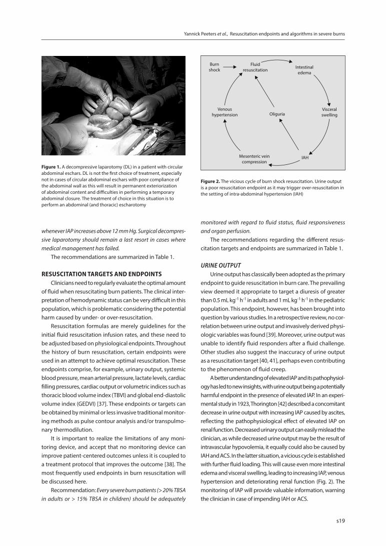
20. GEDVI algorithm If PPV is unreliable, volumetric parameters obtained with transpulmonary thermodilution may be used to guide fluid resuscitation in severe burns. Here, the GEDVI is interpreted as a measure of preload and EVLWI as a safety parameter warning for pending pulmonary edema (Fig. 4). If the GEDVI is high, the measurement needs to be corrected with the global ejection fraction as this leads to a more accurate estimation of preload
21. Urine output algorithm If PPV or volumetric parameters are unreliable, or when monitoring possibilities are limited, urine output may be used to guide fluid resuscitation in severe burns (Fig. 5)
CVP: central venous pressure; EVLWI: extravascular lung water index; GEDVI: global end diastolic volume index; IAP: intra-abdominal pressure; IVIG: intravenous immunoglobulins; PAOP: pulmonary artery occlusion pressure; PPV: pulse pressure variation; TBSA: total burned surface area

s18
Anaesthesiol Intensive Ther 2015, vol. 47, s15–s26
a hypertonic lactated saline solution [27]. However, a 2014 systematic review noted that despite the effect of colloids on decreasing resuscitation volume needs, no benefits in pre-venting IAH were observed [13]. The use of plasma or hyper-tonic saline may reduce the risk for the development of ACS and lower the volume of fluid administered, however close monitoring of sodium levels in the latter is highly advised [13, 27, 29]. Functional hemodynamics in combination with volumetric indices acquired by transpulmonary thermodilu-tion may also be useful as a resuscitation endpoint [30−32].
It is recommended that the clinician should routinely monitor IAP every 4 to 6 hours throughout the resuscitation of patients with a TBSA of more than 20% [6]. The clinical diagnosis of ACS is a very late finding and the presence of
IAH should be detected before the development of this potentially lethal complication.
Recommendation: During the resuscitation phase, as well as the recovery phase, intra-abdominal pressure (IAP) needs to be measured in burn patients at least every 4 to 6 hours
Once an elevated IAH has been diagnosed, medical treatment should be immediately initiated in order to avoid progression to ACS. This consists of nasogastric decompres-sion, the use of neuro-muscular blocking agents, percutane-ous ascites drainage, diuretics or veno-venous hemofiltra-tion [12]. Table 3 summarizes medical management options for IAH in burn patients. The clinician should also be aware of the interactions of pressures between different compart-ments referred to as polycompartment syndrome [33, 34].
If medical treatment fails or ACS is imminent, surgical treatment should be considered but never as a first choice (Fig. 1). Although a midline laparotomy is very effective in reducing IAP, its morbidity and mortality in burn patients is tremendous considering its wound healing challenges and high incidence of infectious complications [21]. One should be aware that in burn patients, decompressive laparotomy should not always be the preferred surgical salvage procedure. In case of circular truncal eschars, es-charotomy may significantly improve the compliance of the abdominal and thoracic walls. This type of procedure may also be effective at decreasing IAP and improving ventilation [35, 36].
Recommendation: Medical management (improvement of abdominal compliance, evacuation of intra-abdominal con-tents, evacuation of intra-luminal contents, limitation of fluid intake, optimization of organ perfusion) should be initiated
Table 2. Specific risk factors for abdominal hypertension in burns
A. Related to diminished abdominal wall complianceCircular abdominal eschars (diminished abdominal wall compliance)Circular thoracic eschars (diminished chest and abdominal wall compliance)Edema of the abdominal wall (fluid creep)Mechanical ventilation (especially desynchronization with the ventilator and the use of accessory muscles)Use of excessive positive end expiratory pressure (PEEP) or the presence of auto-PEEP Basal pneumonia, acute respiratory distress syndrome (primary ARDS related to inhalation injury and secondary ARDS)Prone and other body positioning High body mass index
B. Related to increased intra-abdominal contentsGastroparesis Gastric distention Ileus (bowel edema)Colonic pseudo-obstruction Enteral feedingConstipation (opioid creep)
C. Related to abdominal collections of fluid, air or bloodLiver dysfunction (forward and backward failure)Third space fluid accumulation (ascites, pleural effusions)Hematoma formation (coagulopathy)
D. Related to capillary leak and fluid resuscitationAcidosis* (pH below 7.2) Hypothermia* (core temperature below 33°C)Coagulopathy* (platelet count below 50 G L-1 or an activated partial thromboplastin time (APTT) more than twice normal or a prothrombin time (PTT) below 50% or an international standardized ratio (INR) more than 1.5) Sepsis (as defined by the American-European Consensus Conference definitions) Severe sepsis or bacteremiaSeptic shockMassive fluid resuscitation (> 7−10 L of crystalloid/24 hours with capillary leak and positive fluid balance), risk of ACS increased when fluid intake > 250 mL kg-1 in first 24 hours (Ivy index)Major burns (TBSA > 20% in adults or > 15% in children), risk of ACS increased when TBSA > 55−60%Systemic inflammatory reaction after each surgical intervention with debridement and skin graftingLoss of skin barrier and immune-deprivation (prone to infection)
*The combination of acidosis, hypothermia and coagulopathy has been labeled in the literature as the deadly triad [60, 61]
Table 3. Medical interventions and strategies in burn patients suggested by the World Society on Abdominal Compartment Syndrome (WSACS) in the management of intra-abdominal hypertension [5]
1. Improvement of abdominal wall compliance Sedation and paralysis EscharotomiesAvoiding positive fluid balance
2. Evacuation of intra-abdominal contents Ultrasound guided percutaneous ascites drainage
3. Evacuation of intraluminal contents Gastro- and colonoprokineticsStool softenersEnemasAdaptation of enteral nutrition speed
4. Correction of capillary leak and fluid balance Hypertonic solutionsAlbumin 20%Colloids (not HES, hydroxyl-ethyl starch)DiureticsRenal replacement therapy with net ultrafiltration
5. Optimisation of organ perfusionDobutamine and/or norepinephrine
s19
Yannick Peeters et al., Resuscitation endpoints and algorithms in severe burns
whenever IAP increases above 12 mm Hg. Surgical decompres-sive laparotomy should remain a last resort in cases where medical management has failed.
The recommendations are summarized in Table 1.
RESUSCITATION TARGETS AND ENDPOINTSClinicians need to regularly evaluate the optimal amount
of fluid when resuscitating burn patients. The clinical inter-pretation of hemodynamic status can be very difficult in this population, which is problematic considering the potential harm caused by under- or over-resuscitation.
Resuscitation formulas are merely guidelines for the initial fluid resuscitation infusion rates, and these need to be adjusted based on physiological endpoints. Throughout the history of burn resuscitation, certain endpoints were used in an attempt to achieve optimal resuscitation. These endpoints comprise, for example, urinary output, systemic blood pressure, mean arterial pressure, lactate levels, cardiac filling pressures, cardiac output or volumetric indices such as thoracic blood volume index (TBVI) and global end-diastolic volume index (GEDVI) [37]. These endpoints or targets can be obtained by minimal or less invasive traditional monitor-ing methods as pulse contour analysis and/or transpulmo-nary thermodilution.
It is important to realize the limitations of any moni-toring device, and accept that no monitoring device can improve patient-centered outcomes unless it is coupled to a treatment protocol that improves the outcome [38]. The most frequently used endpoints in burn resuscitation will be discussed here.
Recommendation: Every severe burn patients (> 20% TBSA in adults or > 15% TBSA in children) should be adequately
monitored with regard to fluid status, fluid responsiveness and organ perfusion.
The recommendations regarding the different resus-citation targets and endpoints are summarized in Table 1.
URINE OUTPUTUrine output has classically been adopted as the primary
endpoint to guide resuscitation in burn care. The prevailing view deemed it appropriate to target a diuresis of greater than 0.5 mL kg-1 h-1 in adults and 1 mL kg-1 h-1 in the pediatric population. This endpoint, however, has been brought into question by various studies. In a retrospective review, no cor-relation between urine output and invasively derived physi-ologic variables was found [39]. Moreover, urine output was unable to identify fluid responders after a fluid challenge. Other studies also suggest the inaccuracy of urine output as a resuscitation target [40, 41], perhaps even contributing to the phenomenon of fluid creep.
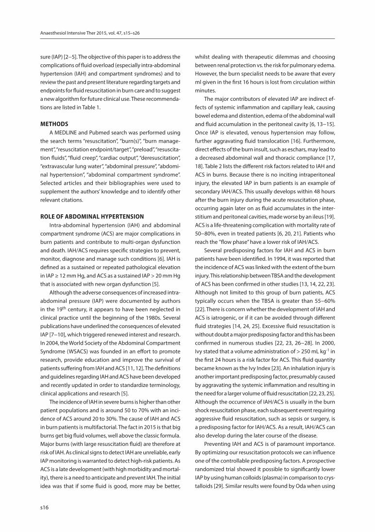
A better understanding of elevated IAP and its pathophysiol-ogy has led to new insights, with urine output being a potentially harmful endpoint in the presence of elevated IAP. In an experi-mental study in 1923, Thorington [42] described a concomitant decrease in urine output with increasing IAP caused by ascites, reflecting the pathophysiological effect of elevated IAP on renal function. Decreased urinary output can easily mislead the clinician, as while decreased urine output may be the result of intravascular hypovolemia, it equally could also be caused by IAH and ACS. In the latter situation, a vicious cycle is established with further fluid loading. This will cause even more intestinal edema and visceral swelling, leading to increasing IAP, venous hypertension and deteriorating renal function (Fig. 2). The monitoring of IAP will provide valuable information, warning the clinician in case of impending IAH or ACS.
Figure 1. A decompressive laparotomy (DL) in a patient with circular abdominal eschars. DL is not the first choice of treatment, especially not in cases of circular abdominal eschars with poor compliance of the abdominal wall as this will result in permanent exteriorization of abdominal content and difficulties in performing a temporary abdominal closure. The treatment of choice in this situation is to perform an abdominal (and thoracic) escharotomy
Figure 2. The vicious cycle of burn shock resuscitation. Urine output is a poor resuscitation endpoint as it may trigger over-resuscitation in the setting of intra-abdominal hypertension (IAH)

s20
Anaesthesiol Intensive Ther 2015, vol. 47, s15–s26
Recommendation: Urine output is a poor endpoint that may lead to over- or under estimation of fluid resuscitation and, as such, can no longer be recommended. However, in situations with limited monitoring techniques, it can still be used to guide fluid resuscitation.
BAROMETRIC PRELOADPulmonary artery occlusion pressure (PAOP) and central
venous pressure (CVP), also known as the cardiac filling pressures (or barometric preload indicators), were used as measures of preload as they were considered to be a reason-able reflection of the end-diastolic volumes of the left and right ventricles [43].
The CVP, obtained via a central venous catheter, used to be measured in almost all ICUs around the world, while clinical decisions, such as fluid or diuretic administration, were frequently based on the interpretation of this param-eter. This was encouraged by clinical guidelines such as the Surviving Sepsis Guidelines, which recommended using CVP as an endpoint of fluid resuscitation in sepsis [44]. PAOP is obtained by a pulmonary artery catheter, which not only measures pulmonary artery pressure and PAOP, but also car-diac output through thermodilution techniques, together with mixed venous oxygen saturation. This provides exten-sive information regarding hemodynamics and the oxygen supply/demand balance.
Recent studies have questioned the efficacy of CVP and PAOP as endpoints for resuscitation as these parameters do not correlate with ventricular filling pressures and ven-tricular end-diastolic volumes [45, 46]. This holds also true in burn patients with increased IAP and artificially increased CVP or PAOP [6, 33, 43].
In a systematic review by Marik [47], a very poor relation-ship was found between CVP and blood volume. Additional-ly, it was unreliable in its ability to predict the hemodynamic response to a fluid challenge. In a further study looking at the filling pressures in healthy subjects [48], there was no correlation between initial CVP/PAOP and end-diastolic ventricular volume index (EDVI), as well as stroke volume index. This lack of correlation was persistent following fluid loading. In contrast, initial EDVI and changes in EDVI follow-ing fluid loading correlate strongly with pre- and post-fluid loading changes in stroke volume index, suggesting the possible benefit of this volumetric index.
Mounting evidence demonstrates cardiac filling pres-sures are poor predictors of fluid responsiveness [47, 49, 50] while the use of a pulmonary artery catheter fails to influence outcomes in randomized controlled trials [51], thus rendering CVP and PAOP obsolete as standardized endpoints for fluid resuscitation. Therefore, their use should be reserved for specific indications (such as pericardial tam-ponade).
Recommendation: Barometric preload indicators should not be used to guide fluid resuscitation in burn patients.
VOLUMETRIC PRELOADAdvances in technology (such as transpulmonary ther-
modilution) allow the monitoring of preload in static volu-metric indices such as global end-diastolic volume (GEDV) and intrathoracic blood volume (ITBV). These parameters can be measured with the use of a conventional central venous catheter and a dedicated femoral artery catheter (PiCCO, Pulsion Medical Systems or EV1000, Edwards Lifes-ciences). During the taking of measurements, a known vol-ume of cold normal saline is injected through the central venous line. The arterial catheter detects temperature dif-ferences and generates a thermodilution curve to which the Stewart Hamilton equation is applied to calculate cardiac output (CO) and volumetric preload indices, indexed accord-ing to body surface area [52]. Some pathologies may create inaccurate measurements such as intracardiac shunts, aortic aneurysms, aortic valvular stenosis, severe mitral or tricuspid regurgitation, pneumonectomy, pulmonary embolism, the presence of a balloon pump and unstable arrhythmias. Catheters may have to be placed in other locations due to the localization of burn wounds. If both a central venous catheter and an arterial PiCCO catheter are placed in an ipsilateral femoral site, a crosstalk phenomenon can occur as the cold bolus injected through the central venous catheter passes the thermistor of the arterial catheter, leading to er-rors in measurement. This may be avoided by withdrawing the arterial PiCCO catheter a few centimetres [52].
The GEDV consists of the volumes of all four cardiac chambers, while the ITBV is the total combined volume of the heart (GEDV) and pulmonary blood volumes, both measured at the end of diastole (with ITBV = 1.25 × GEDV). Numerous studies have shown that these volumetric indices represent preload more precisely when compared to urine output [30, 31] or cardiac filling pressures [50, 53], which are prone to missing hypervolemia as it is poorly represented by the blood pressure, filling pressure and/or urine output in the early resuscitative phase [31]. By measuring the ejection fraction to correct these volumetric preload parameters, the ability of these parameters to assess changes in preload over time can be further improved [53].
Recommendation: Volumetric preload indicators are superior when compared to barometric ones and are recom-mended to guide fluid resuscitation, especially in burn patients with increased IAP.
Another parameter that can be derived from transpul-monary thermodilution is the extravascular lung water (EVLW), indexed to predicted body weight. This consists of the interstitial, intracellular and intra-alveolar water of lung tissue. This parameter, together with the pulmonary

s21
Yannick Peeters et al., Resuscitation endpoints and algorithms in severe burns
vascular permeability index (PVPI), can be used to determine the presence of lung edema which can be very useful as a safety parameter during resuscitation [31]. This is par-ticularly applicable in patients with inhalation injuries or in guiding fluid de-resuscitation if a patient fails to proceed to the “flow” phase [1−3]. Of course, as explained above, the cardiac index can also be derived from transpulmonary thermodilution, which can be used to determine the need for additional inotropic therapy.
Recommendation: The use of extravascular lung water is recommended to guide de-resuscitation in burn patients not progressing spontaneously from an “ebb” to a “flow” phase.
FLUID RESPONSIVENESSFollowing a fluid challenge, patients may be classified
as either a responder or a non-responder. Responders are patients who are on the ascending limb of the Frank Star-ling curve, during which the fluid challenge will result in an increase in stroke volume and cardiac index due to an in-creased preload. A fluid responder is defined by an increase in stroke volume of 10-15% following a fluid challenge [37].
To determine the benefit of fluid administration, several clinical and hemodynamic tests can be used. As mentioned above, although CVP does not predict fluid responsiveness [46, 47], studies have shown that dynamic parameters, ob-tained by invasive arterial monitoring and pulse contour analysis (such as pulse pressure variation [PPV] and stroke volume variation [SVV]) are highly predictive of fluid re-sponsiveness in mechanically ventilated patients [54]. Most publications report that a PPV or SVV greater than 12% is highly predictive of fluid responsiveness. Classically, fluid responsiveness is defined as an increase in cardiac index of 15% or more after a fluid bolus. It should be emphasized that PPV and SVV are unreliable in patients with spontaneous breathing activity, cardiac arrhythmias, pericarditis, cardiac tamponade, right ventricular failure (high CVP), high PEEP or high IAP and low tidal volumes (< 6 mL kg-1) [52].
An experimental study has shown that PPV and inferior vena cava flow fluctuations were dependent on IAP (the higher the IAP, the higher the SVV or PPV) [55]. Although this effect is probably related to concomitant changes in pleural pressure, further analysis showed that this was only related to a Δup phenomenon, which suggests that dynamic indices are not exclusively related to intravascular volume status in the presence of increased IAP [56]. This was confirmed in a review of all experimental studies regarding increased IAP and functional hemodynamics [56]. The conclusion was that an increase in IAP with 18 mm Hg doubles the SVV and PPV values. Subsequently, clinicians need to be cautious not to interpret increased PPV and SVV caused by IAH or ACS as hypovolemia with fluid responsiveness, and thus further over-resuscitating the patient.
Another method to determine fluid responsiveness is the passive leg raise (PLR) test, where a reversible au-totransfusion is caused by simply raising the patient’s legs to mimic the effects of a fluid challenge. Besides the ease and economic advantage, this test is completely reversible, whereas a fluid challenge could worsen resuscitation related morbidity. An increase in the cardiac index of 10% with a PLR test is an indicator of fluid responsiveness [57]. In contrast to predicting fluid responsiveness with PPV and SVV, the PLR test is reliable in patients with arrhythmias and spontaneous breathing activity. However, one must be cautious because the PLR can provide false negative results in patients with elevated IAP (and thus also burn patients), where venous return is impaired [58]. This can be avoided by performing the test in the Trendelenburg position, which gives the best endogenous transfusion in the presence of IAH. This technique is contraindicated in patients with intracranial hy-pertension, while a reflux of gastric content may also occur.
The end-expiratory occlusion test is another non-in-vasive, quick technique to predict fluid responsiveness in intubated patients. It increases cardiac preload and allows for the detection of preload dependence. Fluid responsive-ness is predicted with a high sensitivity and specificity by an increase in pulse pressure > 5% during the end-expiratory occlusion and by an increase in cardiac index > 5% [59]. Just as the passive leg raise test, the end-expiratory occlusion test can be reliably interpreted in cardiac arrhythmias or spontaneously breathing patients.
Recommendation: Fluid resuscitation in burn patients should be guided by physiological parameters or tests that are able to predict fluid responsiveness.
HOLISTIC APPROACH: INTRODUCTION OF A NEW PROTOCOL
In an attempt to guide fluid resuscitation effectively in burn patients in the future, while avoiding deleterious effects of over-resuscitation, a multimodal protocol using a modified formula and multiple endpoints is suggested. Each burn patient deserves personalized (protocolized) care using a stepwise approach.
Fluid resuscitation should be initiated in adults with > 20% TBSA and children with > 15% TBSA. A modified Park-land formula is suggested, using a balanced crystalloid (for example, Plasma-Lyte®), to be given at 2 mL kg-1 %TBSA-1
in the first 24 hours in combination with albumin 20% at 0.2 mL kg-1 %TBSA-1. Half of the total dose of crystalloids and colloids should be given in the first eight hours, and the other half between 8−24 hours. Over the next 24 hours Plasma-Lyte® is given at 0.75 mL kg-1 %TBSA-1 in combina-tion with albumin 20% at 0.075 mL kg-1 %TBSA-1. These re-suscitation rates are fixed and fluids are gradually decreased over the next 24−48 hours of burn shock resuscitation. Basic

s22
Anaesthesiol Intensive Ther 2015, vol. 47, s15–s26
fluids need to be supplemented in the form of a glucose containing solution such as Glucion® 5% at a constant rate of 30 mL kg-1 24h-1 at all times. Enteral nutrition, if given, should be included in the basic fluid administration. Fluids are changed or adapted throughout resuscitation according to biochemical analysis and concomitant medical condi-tions (see algorithms as discussed further). During the first 24 hours, the resuscitation fluids, as calculated above, are kept at a constant rate, and when needed, fluid boluses can be given at 3, 6 and 12 mL kg-1 over 30 minutes, de-pending on the different thresholds as discussed further. De-resuscitation (with a gradual decrease in resuscitation fluids) is only started after the first 24 hours. Extra albumin 20% can be administered based on serum levels of albumin (target 30 g L-1) or colloid oncotic pressure (COP, target at least 16−18 mm Hg).
PPV ALGORITHMAs discussed above, different endpoints in combination
with lactate and base excess (BE) can be used in order to guide fluid resuscitation. If a patient is sedated and mechani-cally ventilated, pulse pressure variation (PPV) is used, when-ever reliable. The subsequent PPV algorithm is presented in Figure 3. The clinician, however, needs to check whether the patient has conditions leading to incorrect interpretation of PPV, as discussed above, while before each fluid bolus, fluid responsiveness is tested [52]. In cases where PPV is unreli-able, the GEDVI algorithm needs to be used.
GEDVI ALGORITHMIf PPV is unreliable, volumetric parameters are measured
with the use of transpulmonary thermodilution using PiCCO technology. Here, the GEDVI is interpreted as a measure of preload and EVLWI as a safety parameter warning for impending pulmonary edema. The subsequent GEDVI al-gorithm is presented in Figure 4. The correct interpretation of volumetric parameters needs to be made in relation to the presence, or not, of the conditions mentioned above (valvulopathy, catheter position, extracorporeal circuit etc.) [52]. If the GEDVI is high, the measurement needs to be corrected with the global ejection fraction, as this leads to a more accurate estimation of preload [53] (Table 4).
URINE OUTPUT ALGORITHMIf PPV or volumetric parameters are unreliable, or when
monitoring possibilities are limited, urine output (UO) can be used to guide fluid resuscitation. The subsequent UO al-gorithm is presented in Figure 5. The importance of measur-ing IAP needs to be underlined when using urine output as a resuscitation target as IAH and ACS decrease urine output.
In children with TBSA > 10−15%, with a bodyweight be-low 30 kg, the Parkland formula is used (4 mL kg-1 %TBSA-1) in the first 24 hours. As with adults, half is given in the first eight hours. Basic fluid needs should always be given in glucose-containing fluids. The fluid rate in children is guided by urine output as presented in the UO algorithm in Figure 5,
Figure 3. The Pulse Pressure Variation algorithm to guide resuscitation in severely burned patients. If the patient is mechanically ventilated and PPV is reliable, fluid resuscitation is guided by the PPV algorithm
s23
Yannick Peeters et al., Resuscitation endpoints and algorithms in severe burns
however the target diuresis is 2 mL kg-1 h-1 in children weigh-ing < 10 kg and 1 mL kg-1 h-1 in children between 11−30 kg.
CONCLUSIONSDuring the last decades, burn resuscitation keeps evolv-
ing and new trends appear. Over the last fifteen years, much attention has been given to avoid over-resuscitation and subsequent morbidity and mortality. Fluid creep is recog-nized by nearly all physicians involved in burn care.
Efforts should be made to avoid excess crystalloid ad-ministration by revising resuscitation protocols. Physicians need to be aware of the harm caused by fluid overload during resuscitation. They should actively aim to avoid fluid accumulation, as this can be at least as harmful, if not more so, than under-resuscitation. Evidence suggests that the addition of a colloid, such as albumin 20%, may decrease fluid requirements and may potentially reduce resuscitation-related morbidity (especially renal, respiratory complica-tions and compartment syndromes). However, the use of
colloids in burn resuscitation continues to be a great source of controversy and discussion and, as explained in part 1 of this concise review, HES solutions should not be used. Ascorbic acid as an adjunctive therapy shows promising results, without presenting adverse effects. Its use should be considered in patients at risk of fluid overload or secondary IAH and ACS.
Moreover, the endpoints of burn resuscitation should be redefined. Although the traditional urine output tar-get does not represent preload accurately as shown in nu-merous studies, barometric preload parameters also seem to lack this ability. The evidence suggests that advanced hemodynamic monitoring with pulse contour analysis and transpulmonary thermodilution may provide superior end-points, such as pulse pressure variation (PPV) and global end diastolic volume index (GEDVI), in order to prevent under-resuscitation, while extravascular lung water index (EVLWI) can be used as a safety parameter to avoid over-resuscitation and to guide the de-resuscitation process.
Table 4. Global ejection fraction corrected volumetric target values
Ejection fraction 5% 10% 15% 20% 25% 30% 35% 40% 45% 50% 55%
GEDVI-target (normal) 1175 1050 950 850 775 700 625 575 525 475 435
GEDVI-target (critically ill burns) 1450 1300 1150 1025 925 825 750 675 600 550 500
Critically ill: refers to an unstable patient with a clinical diminished preload; GEDVI: global end-diastolic volume index; normal: refers to a stable patient
Figure 4. Global end-diastolic volume index algorithm to guide resuscitation in severely burned patients. If PPV is unreliable, the patient has a PiCCO catheter and GEDVI is reliable, fluid resuscitation is guided by the GEDVI algorithm

s24
Anaesthesiol Intensive Ther 2015, vol. 47, s15–s26
The role of intra-abdominal hypertension and its patho-physiology has been extensively investigated over recent years, while efforts should be made to prevent and treat intra-abdominal hypertension and its most lethal compli-cation — abdominal compartment syndrome. It should be emphasized that regular and routine monitoring of intra-abdominal pressure (IAP) is of paramount importance in se-verely burned patients. More studies are needed to establish the place of the proposed algorithms and personalized care in severely burned patients.
ACKNOWLEDGEMENTS1. The authors declare no financial disclosure.2. Manu Malbrain is member of the medical advisory board
of Pulsion Medical Systems (Maquet Getinge Group). The other authors have no possible conflicts of interest related to the content of this paper.
References: 1. Malbrain ML, Marik PE, Witters I et al.: Fluid overload, de−resuscitation,
and outcomes in critically ill or injured patients: a systematic review with suggestions for clinical practice. Anaesthesiol Intensive Ther 2014; 46: 361−380. doi: 10.5603/AIT.2014.0060.
2. Cordemans C, De laet I, Van Regenmortel N et al.: Fluid management in critically ill patients: The role of extravascular lung water, abdominal hypertension, capillary leak and fluid balance. Ann Intensive Care 2012; 2 (Suppl 1): S1. doi: 10.1186/2110-5820-2-S1-S1.
3. Cordemans C, De Laet I, Van Regenmortel N et al.: Aiming for a negative fluid balance in patients with acute lung injury and increased intra-
Figure 5. Urine output algorithm to guide resuscitation in severely burned patients. If the patient has no PiCCO catheter (or GEDVI is not reliable) and PPV is not reliable, fluid resuscitation is guided by the UO algorithm
-abdominal pressure: a pilot study looking at the effects of PAL-treat-ment. Ann Intensive Care 2012; 2 (Suppl 1): S15. doi: 10.1186/2110-5820-2-S1-S15.
4. Kushimoto S, Taira Y, Kitazawa Y et al.: The clinical usefulness of extra-vascular lung water and pulmonary vascular permeability index to diagnose and characterize pulmonary edema: a prospective multi-center study on the quantitative differential diagnostic definition for acute lung injury/acute respiratory distress syndrome. Crit Care 2012; 16: R232. doi: 10.1186/cc11898.
5. Kirkpatrick AW, Roberts DJ, De Waele J et al.: Intra-abdominal hyperten-sion and the abdominal compartment syndrome: updated consensus definitions and clinical practice guidelines from the World Society of the Abdominal Compartment Syndrome. Intensive Care Medicine. 2013; 39: 1190−1206. doi: 10.1007/s00134-013-2906-z.
6. Malbrain ML, De Keulenaer BL, Oda J et al.: Intra-abdominal hypertension and abdominal compartment syndrome in burns, obesity, pregnancy, and general medicine. Anaesthesiol Intensive Ther 2015; 47: 228−240. doi: 10.5603/AIT.a2015.0021.
7. Kron IL, Harman PK, Nolan SP: The measurement of intra-abdominal pressure as a criterion for abdominal re-exploration. Ann Surg 1984; 199: 28−30.
8. Fietsam R, Jr., Villalba M, Glover JL, Clark K. Intra-abdominal compartment syndrome as a complication of ruptured abdominal aortic aneurysm repair. Am Surg 1989; 55: 396−402.
9. Richards WO, Scovill W, Shin B, Reed W: Acute renal failure associated with increased intra-abdominal pressure. Ann Surg 1983; 197: 183−187.
10. Le Roith D, Bark H, Nyska M, Glick SM: The effect of abdominal pressure on plasma antidiuretic hormone levels in the dog. J Surg Res 1982; 32: 65−69.
11. Malbrain ML, Cheatham ML, Kirkpatrick A et al.: Results from the inter-national conference of experts on intra-abdominal hypertension and abdominal compartment syndrome. I. Definitions. Intensive Care Med 2006; 32: 1722−1732.
12. Cheatham ML, Malbrain ML, Kirkpatrick A et al.: Results from the inter-national conference of experts on intra-abdominal hypertension and abdominal compartment syndrome. II. Recommendations. Intensive Care Med 2007; 33: 951−962.
s25
Yannick Peeters et al., Resuscitation endpoints and algorithms in severe burns
13. Strang SG, Van Lieshout EM, Breederveld RS, Van Waes OJ: A systematic review on intra-abdominal pressure in severely burned patients. Burns 2014; 40: 9−16. doi: 10.1016/j.burns.2013.07.001.
14. Kirkpatrick AW, Ball CG, Nickerson D, D’Amours SK: Intraabdominal hypertension and the abdominal compartment syndrome in burn patients. World J Surg 2009; 33: 1142−1149. doi: 10.1007/s00268-009-9995-4.
15. Holodinsky JK, Roberts DJ, Ball CG et al.: Risk factors for intra-abdominal hypertension and abdominal compartment syndrome among adult intensive care unit patients: a systematic review and meta-analysis. Crit Care 2013; 17: R249. doi: 10.1186/cc13075.
16. Ball CG, Kirkpatrick AW, Karmali S et al.: Tertiary abdominal compartment syndrome in the burn injured patient. J Trauma 2006; 61: 1271−1273.
17. Malbrain MLNG, De laet I, De Waele J et al.: The role of abdominal com-pliance, the neglected parameter in critically ill patients — a consensus review of 16. Part 2: Measurement techniques and management recommendations. Anaesthesiol Intensive Ther 2014; 46: 406−432. doi: 10.5603/AIT.2014.0063.
18. Malbrain MLNG, Roberts DJ, De laet I et al.: The role of abdominal com-pliance, the neglected parameter in critically ill patients — a consensus review of 16. Part 1: Definitions and pathophysiology. Anaesthesiol Intensive Ther. 2014; 46: 392−405. doi: 10.5603/AIT.2014.0062.
19. Azzopardi EA, McWilliams B, Iyer S, Whitaker IS: Fluid resuscitation in adults with severe burns at risk of secondary abdominal compartment syndrome — an evidence based systematic review. Burns 2009; 35: 911−920.
20. Malbrain ML, Chiumello D, Cesana BM et al.: A systematic review and in-dividual patient data meta-analysis on intra-abdominal hypertension in critically ill patients: the wake-up project. World initiative on Abdominal Hypertension Epidemiology, a Unifying Project (WAKE−Up!). Minerva Anestesiol 2014; 80: 293−306.
21. De Waele JJ, Hoste EA, Malbrain ML: Decompressive laparotomy for abdominal compartment syndrome — a critical analysis. Crit Care 2006; 10: R51.
22. Oda J, Yamashita K, Inoue T et al.: Resuscitation fluid volume and ab-dominal compartment syndrome in patients with major burns. Burns 2006; 32: 151−154.
23. Ivy ME, Atweh NA, Palmer J, Possenti PP, Pineau M, D’Aiuto M: Intra-abdo-minal hypertension and abdominal compartment syndrome in burn patients. J Trauma 2000; 49: 387−391.
24. Kirkpatrick AW, Balogh Z, Ball CG et al.: The secondary abdominal com-partment syndrome: iatrogenic or unavoidable? J Am Coll Surg 2006; 202: 668−679.
25. McBeth PB, Sass K, Nickerson D, Ball CG, Kirkpatrick AW: A necessary evil? Intra-abdominal hypertension complicating burn patient resuscitation. J Trauma Manag Outcomes 2014; 8: 12. doi: 10.1186/1752-2897-8-12.
26. Hobson KG, Young KM, Ciraulo A, Palmieri TL, Greenhalgh DG: Release of abdominal compartment syndrome improves survival in patients with burn injury. J Trauma 2002; 53: 1129−1133.
27. Oda J, Ueyama M, Yamashita K et al.: Hypertonic lactated saline resu-scitation reduces the risk of abdominal compartment syndrome in severely burned patients. J Trauma 2006; 60: 64−71.
28. Oda J, Yamashita K, Inoue T et al.: Acute lung injury and multiple or-gan dysfunction syndrome secondary to intra-abdominal hypertension and abdominal decompression in extensively burned patients. J Trauma 2007; 62: 1365−1369.
29. O’Mara MS, Slater H, Goldfarb IW, Caushaj PF: A prospective, randomized evaluation of intra-abdominal pressures with crystalloid and colloid resuscitation in burn patients. J Trauma 2005; 58: 1011−1018.
30. Csontos C, Foldi V, Fischer T, Bogar L: Arterial thermodilution in burn patients suggests a more rapid fluid administration during early resu-scitation. Acta Anaesthesiologica Scandinavica 2008; 52: 742−749.
31. Sanchez M, Garcia-de-Lorenzo A, Herrero E et al.: A protocol for resuscita-tion of severe burn patients guided by transpulmonary thermodilution and lactate levels: a 3-year prospective cohort study. Crit Care 2013; 17: R176. doi: 10.1186/cc12855.
32. Holm C, Melcer B, Horbrand F, Worl H, von Donnersmarck GH, Muhlbauer W: Intrathoracic blood volume as an end point in resuscitation of the severely burned: an observational study of 24 patients. J Trauma 2000; 48: 728−734.
33. Malbrain ML, Wilmer A: The polycompartment syndrome: towards an understanding of the interactions between different compartments! Intensive Care Med 2007; 33: 1869−1872.
34. Malbrain MLNG, Roberts DJ, Sugrue M et al.: The polycompartment syndrome: a concise state-of-the-art review. Anaesthesiol Intensive Ther 2014; 46: 433−450. doi: 10.5603/AIT.2014.0064.
35. Oda J, Ueyama M, Yamashita K et al.: Effects of escharotomy as abdominal decompression on cardiopulmonary function and visceral perfusion in abdominal compartment syndrome with burn patients. J Trauma 2005; 59: 369−374.
36. Tsoutsos D, Rodopoulou S, Keramidas E, Lagios M, Stamatopoulos K, Io-annovich J: Early escharotomy as a measure to reduce intraabdominal hypertension in full-thickness burns of the thoracic and abdominal area. World J Surg 2003; 27: 1323−1328.
37. Marik PE, Monnet X, Teboul JL: Hemodynamic parameters to guide fluid therapy. Ann Intensive Care 2011; 1: 1. doi: 10.1186/2110-5820-1-1.
38. Ospina-Tascon GA, Cordioli RL, Vincent JL: What type of monitoring has been shown to improve outcomes in acutely ill patients? Intensive Care Med 2008; 34: 800−820. doi: 10.1007/s00134-007-0967-6.
39. Dries DJ, Waxman K: Adequate resuscitation of burn patients may not be measured by urine output and vital signs. Crit Care Med 1991; 19: 327−329.
40. Pruitt BA, Jr.: Protection from excessive resuscitation: “pushing the pendulum back”. J Trauma 2000; 49: 567−568.
41. Shah A, Kramer GC, Grady JJ, Herndon DN: Meta-analysis of fluid require-ments for burn injury 1980−2002. J Burn Care Rehabil 2003; 152: S118.
42. Thorington JM, Schmidt CF: A study of urinary output and blood-pressure changes resulting in experimental ascites. Am J Med Sci 1923; 165: 880−890.
43. Malbrain ML, De Waele JJ, De Keulenaer BL: What every ICU clinician ne-eds to know about the cardiovascular effects caused by abdominal hypertension. Anaesthesiol Intensive Ther 2015; 47: 388−399. doi: 10.5603/AIT.a2015.0028.
44. Dellinger RP, Levy MM, Rhodes A et al.: Surviving sepsis campaign: inter-national guidelines for management of severe sepsis and septic shock, 2012. Intensive Care Med 2013; 39: 165−228. doi: 10.1007/s00134-012-2769-8.
45. Marik PE: Surviving sepsis: going beyond the guidelines. Ann Intensive Care 2011; 1: 17. doi: 10.1186/2110-5820-1-17.
46. Michard F, Teboul JL: Predicting fluid responsiveness in ICU patients: a critical analysis of the evidence. Chest 2002; 121: 2000−2008.
47. Marik PE, Baram M, Vahid B: Does central venous pressure predict fluid responsiveness?: a systematic review of the literature and the tale of seven mares. Chest 2008; 134: 172−178. doi: 10.1378/chest.07-2331.
48. Kumar A, Anel R, Bunnell E et al.: Pulmonary artery occlusion pressure and central venous pressure fail to predict ventricular filling volume, cardiac performance, or the response to volume infusion in normal subjects. Crit Care Med 2004; 32: 691−699.
49. Osman D, Ridel C, Ray P et al.: Cardiac filling pressures are not appropriate to predict hemodynamic response to volume challenge. Crit Care Med 2007; 35: 64−69.
50. Lichtwarck-Aschoff M, Beale R, Pfeiffer UJ: Central venous pressure, pulmonary artery occlusion pressure, intrathoracic blood volume, and right ventricular end-diastolic volume as indicators of cardiac preload. J Crit Care 1996; 11: 180−188.
51. Shah MR, Hasselblad V, Stevenson LW et al.: Impact of the pulmonary artery catheter in critically ill patients: meta-analysis of randomized clinical trials. JAMA 2005; 294: 1664−1670.
52. Hofkens PJ, Verrijcken A, Merveille K et al.: Common pitfalls and tips and tricks to get the most out of your transpulmonary thermodilution device: results of a survey and state-of-the-art review. Anaesthesiol Intensive Ther 2015; 47: 89−116. doi: 10.5603/AIT.a2014.0068.
53. Malbrain ML, De Potter TJ, Dits H, Reuter DA: Global and right ventricular end-diastolic volumes correlate better with preload after correction for ejection fraction. Acta Anaesthesiologica Scandinavica 2010; 54: 622−631. doi: 10.1111/j.1399-6576.2009.02202.x.
54. Marik PE, Cavallazzi R, Vasu T, Hirani A: Dynamic changes in arterial waveform derived variables and fluid responsiveness in mechanically ventilated patients: a systematic review of the literature. Crit Care Med 2009; 37: 2642−2647. doi: 10.1097/CCM.0b013e3181a590da.
55. Duperret S, Lhuillier F, Piriou V et al.: Increased intra-abdominal pressure affects respiratory variations in arterial pressure in normovolaemic and hypovolaemic mechanically ventilated healthy pigs. Intensive Care Med 2007; 33: 163−171.
56. Malbrain ML, de Laet I: Functional hemodynamics and increased intra--abdominal pressure: same thresholds for different conditions ...? Crit Care Med 2009; 37: 781−783. doi: 10.1097/CCM.0b013e318194c397.

s26
Anaesthesiol Intensive Ther 2015, vol. 47, s15–s26
57. Monnet X, Rienzo M, Osman D et al.: Passive leg raising predicts fluid responsiveness in the critically ill. Crit Care Med 2006; 34]: 1402−1407.
58. Malbrain ML, Reuter DA: Assessing fluid responsiveness with the pas-sive leg raising maneuver in patients with increased intra-abdominal pressure: Be aware that not all blood returns! Crit Care Med 2010; 38: 1912−1925. doi: 10.1097/CCM.0b013e3181f1b6a2.
59. Monnet X, Osman D, Ridel C, Lamia B, Richard C, Teboul JL: Predicting volume responsiveness by using the end-expiratory occlusion in me-chanically ventilated intensive care unit patients. Crit Care Med 2009; 37: 951−956. doi: 10.1097/CCM.0b013e3181968fe1.
60. Burch JM, Moore EE, Moore FA, Franciose R: The abdominal compartment syndrome. Surg Clin North Am 1996; 76: 833−8342.
61. Ivatury RR, Sugerman HJ, Peitzman AB: Abdominal compartment syn-drome: recognition and management. Adv Surg 2001; 35: 251−269.
Corresponding author: Manu L.N.G. Malbrain, MD, PhDICU and High Care Burn Unit DirectorZNA StuivenbergLange Beeldekensstraat 267B-2060 Antwerpen 6, Belgiume-mail: [email protected]
Received: 1.09.2015 Accepted: 10.10.2015