Subtypes of breast cancer show different spatial distributions of … · 2019-09-04 · sis, hot...
10
RESEARCH ARTICLE Subtypes of breast cancer show different spatial distributions of brain metastases Sunghyon Kyeong 1 , Yoon Jin Cha 2 , Sung Gwe Ahn 3 , Sang Hyun Suh 4 , Eun Ju Son 4 , Sung Jun Ahn 4 * 1 Severance Biomedical Science Institute, Yonsei University College of Medicine, Seoul, Korea, 2 Department of Pathology, Gangnam Severance Hospital, Yonsei University, College of Medicine, Seoul, Korea, 3 Department of Surgery, Gangnam Severance Hospital, Yonsei University, College of Medicine, Seoul, Korea, 4 Department of Radiology, Gangnam Severance Hospital, Yonsei University, College of Medicine, Seoul, Korea * [email protected] Abstract The aim of our study was to test the hypothesis that the spatial distribution of breast cancer brain metastases (BM) differ according to their biological subtypes. MR images of 100 patients with BM from primary breast cancer were retrospectively reviewed. Patients were divided according to the biological subtype of the primary tumor, (triple-negative: 24, HER2 positive: 48, luminal: 28). All images marked with BMs were standardized to the human brain MRI atlas provided by the Montreal Neurological Institute 152 database. Distribution pattern of BM was evaluated with intra-group and intergroup analysis. In intra-group analy- sis, hot spots of metastases from triple-negative are evenly distributed in the brain, mean- while BMs from HER2 positive and luminal type occur dominantly in occipital lobe and cerebellum. In intergroup analysis, BMs from triple-negative type occurred more often in frontal lobe, limbic region, and parietal lobe, compared with other types (P < .05). Breast cancer subtypes tend to demonstrate different spatial distributions of their BMs. These find- ings may have direct implications for dose modulation in prophylactic irradiation as well as for differential diagnoses. Thus, this result should be validated in future study with a larger population. Introduction Brain metastases (BMs) are the most commonly encountered malignant tumors occurring in the CNS, outnumbering primary CNS tumors by more than 10-fold[1]. Breast cancer is the second most frequent cause of BM after lung cancer, with metastases occurring in 10–16% of patients[2]. Median survival ranges from 3 to 15 months following metastatic spread to the brain, making BMs one of the major causes of systemic cancer-related mortality[3]. Recently, the incidence of BMs has increased because of improvements in treatment for primary cancers and more advanced imaging techniques[4,5]. Breast cancer can be divided into several biologic subtypes on the basis of their clinical, his- topathological, and molecular features. Further, breast cancer can be classified on the basis of PLOS ONE | https://doi.org/10.1371/journal.pone.0188542 November 20, 2017 1 / 10 a1111111111 a1111111111 a1111111111 a1111111111 a1111111111 OPEN ACCESS Citation: Kyeong S, Cha YJ, Ahn SG, Suh SH, Son EJ, Ahn SJ (2017) Subtypes of breast cancer show different spatial distributions of brain metastases. PLoS ONE 12(11): e0188542. https://doi.org/ 10.1371/journal.pone.0188542 Editor: William B. Coleman, University of North Carolina at Chapel Hill School of Medicine, UNITED STATES Received: August 31, 2017 Accepted: November 8, 2017 Published: November 20, 2017 Copyright: © 2017 Kyeong et al. This is an open access article distributed under the terms of the Creative Commons Attribution License, which permits unrestricted use, distribution, and reproduction in any medium, provided the original author and source are credited. Data Availability Statement: All relevant data are within the paper and its Supporting Information files. Funding: This work was supported by the National Research Foundation of Korea(NRF) grant funded by the Korea government(MSIP) (No.2017R1C1B5014927). The funder had no role in study design, data collection and analysis, decision to publish, or preparation of the manuscript.
Transcript of Subtypes of breast cancer show different spatial distributions of … · 2019-09-04 · sis, hot...

RESEARCH ARTICLE
Subtypes of breast cancer show different
spatial distributions of brain metastases
Sunghyon Kyeong1, Yoon Jin Cha2, Sung Gwe Ahn3, Sang Hyun Suh4, Eun Ju Son4, Sung
Jun Ahn4*
1 Severance Biomedical Science Institute, Yonsei University College of Medicine, Seoul, Korea,
2 Department of Pathology, Gangnam Severance Hospital, Yonsei University, College of Medicine, Seoul,
Korea, 3 Department of Surgery, Gangnam Severance Hospital, Yonsei University, College of Medicine,
Seoul, Korea, 4 Department of Radiology, Gangnam Severance Hospital, Yonsei University, College of
Medicine, Seoul, Korea
Abstract
The aim of our study was to test the hypothesis that the spatial distribution of breast cancer
brain metastases (BM) differ according to their biological subtypes. MR images of 100
patients with BM from primary breast cancer were retrospectively reviewed. Patients were
divided according to the biological subtype of the primary tumor, (triple-negative: 24, HER2
positive: 48, luminal: 28). All images marked with BMs were standardized to the human
brain MRI atlas provided by the Montreal Neurological Institute 152 database. Distribution
pattern of BM was evaluated with intra-group and intergroup analysis. In intra-group analy-
sis, hot spots of metastases from triple-negative are evenly distributed in the brain, mean-
while BMs from HER2 positive and luminal type occur dominantly in occipital lobe and
cerebellum. In intergroup analysis, BMs from triple-negative type occurred more often in
frontal lobe, limbic region, and parietal lobe, compared with other types (P < .05). Breast
cancer subtypes tend to demonstrate different spatial distributions of their BMs. These find-
ings may have direct implications for dose modulation in prophylactic irradiation as well as
for differential diagnoses. Thus, this result should be validated in future study with a larger
population.
Introduction
Brain metastases (BMs) are the most commonly encountered malignant tumors occurring in
the CNS, outnumbering primary CNS tumors by more than 10-fold[1]. Breast cancer is the
second most frequent cause of BM after lung cancer, with metastases occurring in 10–16% of
patients[2]. Median survival ranges from 3 to 15 months following metastatic spread to the
brain, making BMs one of the major causes of systemic cancer-related mortality[3]. Recently,
the incidence of BMs has increased because of improvements in treatment for primary cancers
and more advanced imaging techniques[4,5].
Breast cancer can be divided into several biologic subtypes on the basis of their clinical, his-
topathological, and molecular features. Further, breast cancer can be classified on the basis of
PLOS ONE | https://doi.org/10.1371/journal.pone.0188542 November 20, 2017 1 / 10
a1111111111
a1111111111
a1111111111
a1111111111
a1111111111
OPENACCESS
Citation: Kyeong S, Cha YJ, Ahn SG, Suh SH, Son
EJ, Ahn SJ (2017) Subtypes of breast cancer show
different spatial distributions of brain metastases.
PLoS ONE 12(11): e0188542. https://doi.org/
10.1371/journal.pone.0188542
Editor: William B. Coleman, University of North
Carolina at Chapel Hill School of Medicine, UNITED
STATES
Received: August 31, 2017
Accepted: November 8, 2017
Published: November 20, 2017
Copyright: © 2017 Kyeong et al. This is an open
access article distributed under the terms of the
Creative Commons Attribution License, which
permits unrestricted use, distribution, and
reproduction in any medium, provided the original
author and source are credited.
Data Availability Statement: All relevant data are
within the paper and its Supporting Information
files.
Funding: This work was supported by the National
Research Foundation of Korea(NRF) grant funded
by the Korea government(MSIP)
(No.2017R1C1B5014927). The funder had no role
in study design, data collection and analysis,
decision to publish, or preparation of the
manuscript.
their gene expression profiles into luminal, basal, and HER2-positive, with each subtype show-
ing a clearly different prognostic significance[6,7]. The subgroups of patients with triple-nega-
tive and human epidermal growth factor receptor 2 (HER2)-positive breast cancer are at a
higher risk for development of BMs[8–10]. The onset of BMs in triple receptor-negative breast
cancer is earlier than that observed in other subtypes, and the overall survival rate is particu-
larly poor, when compared to other subtypes[11].
Treatment options for patients with breast cancer BMs are limited and include surgical
resection, whole-brain radiation therapy, stereotactic radiosurgery, chemotherapy, and tar-
geted therapy[12–14]. Prophylactic cranial irradiation improves the survival rate of patients
with lung cancer BMs[15,16], and may represent a novel approach for select patients with
breast cancer BMs[17]. Thus, understanding the spatial distributions of BMs by breast cancer
subtype may allow for more precise prophylactic irradiation adjustments and could lead to the
development of novel targeted therapy.
Biological characteristics of tumors could affect the spatial distribution of their BMs. For
example, the probability of cerebellar metastases is higher in lung and breast cancer[18]. BMs
are typically located in watershed areas such as the gray-white matter junction[19]. Recently,
Takano et al. reported that lung cancer BMs with an epidermal growth factor receptor (EGFR)
L858R mutation occurred more often in the caudate nucleus, cerebellum, and temporal lobe
than those with an EGFR exon 19 deletion[20]. We hypothesized that breast cancer BMs have
different spatial distributions according to their biological subtypes.
Materials and methods
Participants
We retrospectively reviewed data for breast cancer patients with BM who underwent gado-
linium-enhanced brain MRI from 2009 to 2016. A total of 128 patients were identified. Of
theses 27 patients were excluded for the following reasons (Fig 1): (1) previous neurosurgery
or brain radiation therapy (n = 10); (2) presence of other malignant disease (n = 5); and
(3) absence of the immunohistochemistry profile of breast cancer (n = 12). A total of 101
patients was remained after selection criteria. However, an unknown error occurred during
exporting from the hospital database to the local computer in one of 101 brain MRIs, and
this case was removed in the statistical mapping. Finally, gadolinium-enhanced 3D T1WIs
of 100 breast cancer patients in whom BMs were initially diagnosed were included in this
analysis (slice thickness <1.5 mm on 3.0T MRI). Patients were divided into three groups
according to biological subtypes on the basis of the expression of estrogen (ER), progester-
one (PgR), and HER2. Immunohistochemistry was carried out for the evaluation of the
level of ER, PgR, and HER2 expression of primary breast cancer. Florescence in-situ hybrid-
ization analysis of HER2 amplification was carried out in immunohistochemistry 2+ cases.
The three subtypes were triple negative (ER-, PgR-, HER2-), HER2 positive (HER2(+), any
ER/PgR), and luminal (ER/PgR(+), HER2(-)). The current study design and use of clinical
data was approved by the institutional review board of Gangnam Severance hospital The
requirement to obtain informed consent was waived, and all data were fully anonymized.
Image registration and frequency map reconstruction
DICOM gadolinium-enhanced 3D T1WIs were reviewed by Two radiologist (A.S.J, S.S.H) to
identify the focus of the brain metastases. They independently marked brain metastases and
recorded coordinates (x,y,z) of brain metastases. Interobserver agreement was assessed using
the Concordance Correlation Coefficient (CCC). The coordinates of reader 1 was used for fur-
ther analysis. Fig 1 presents the flowchart for mapping brain metastases (BMs) in the
Different spatial distribution of brain metastasis in different subtypes of breast cancer
PLOS ONE | https://doi.org/10.1371/journal.pone.0188542 November 20, 2017 2 / 10
Competing interests: The authors have declared
that no competing interests exist.

standardized automated anatomical labeling (AAL) template space[21]. The primary sites of
occurrence were then individually mapped in Neuroimaging Informatics Technology Initia-
tive format by assigning ones for the manually identified lesions and zeros otherwise (Fig 1A).
After converting DICOM images to the Neuroimaging Informatics Technology Initiative for-
mat using the dcm2nii software (http://cabiatl.com/mricro/mricron/dcm2nii.html), the
images were normalized to the standard space using the Statistical Parametric Mapping
(SPM12; http://www.fil.ion.ucl.ac.uk/spm/software/spm12) software. Individually coregistered
images of the BMs and a manually marked lesion map were standardized to the human brain
MRI atlas provided by the Montreal Neurological Institute (MNI) 152 database with a 1×1×1
mm voxel size. For frequency map reconstruction, normalized lesion maps were extended to a
site-centered spherical shape with a radius of 15 mm (Fig 1B). Subsequently, these spherical
BMs were matched with the AAL template. Initially, we assigned zeros for 116 cortical, subcor-
tical, and cerebellar regions in AAL template space, we then assigned ones if there were over-
laps between individual spherical BMs and AAL regions. Finally, all BM heat maps were
reconstructed and superimposed in the AAL space to determine the frequency of metastasis
occurrence (Fig 1C).
Analysis of spatial distribution of brain metastasis
For intra-group analysis, heat map is generated based on the percentage of brain metastasis
involving specific AAL region, which was defined as number of patients whose brain metasta-
sis involving specific AAL region per total number of patients with a specific breast cancer sub-
type. Top10% of ALL regions in frequency of metastases could be listed using heat map. For
intergroup analysis, the frequency of occurrence of metastasis was compared between biologi-
cal subtypes of the breast cancer: (1) triple-negative or non-triple-negative; (2) HER2 or non-
HER2; and (3) luminal or non-luminal. Single-subject BM maps were entered into a second-
level analysis using a χ2 test crosstab analysis to assess group level significance. The signifi-
cance threshold was set at P< .05
Fig 1. Flowchart for mapping brain metastases (BMs) in the standardized automated anatomical labeling (AAL) template space. The center
coordinates of BMs in the native space were identified by a trained radiologist (A). The individual center coordinates of BMs were normalized in the Montreal
Neurological Institute space and normalized lesion maps were extended to a site-centered spherical shape with a radius of 15 mm (B). The AAL template
was matched with the spherical BMs in the MNI space. We assigned 1 if there were overlaps between BM and AAL nodes (C). For examples, 1) if the center
coordinate of BM is located around the temporal regions and its spherical ROI with 15 mm radius were overlapped with the superior temporal gyrus and
middle temporal gyrus, then those two temporal gyri were identified as neighbor nodes (top image of Fig 1C). 2) if the center coordinate of BM is located
around the paracentral lobule and the middle cingulate gyrus and its spherical ROI with 15 mm radius were overlapped with the middle cingulate gyrus,
paracentral lobule, supplementary motor area, and precuneus, then these four medial regions were identified as neighbor nodes (bottom image of Fig 1C).
https://doi.org/10.1371/journal.pone.0188542.g001
Different spatial distribution of brain metastasis in different subtypes of breast cancer
PLOS ONE | https://doi.org/10.1371/journal.pone.0188542 November 20, 2017 3 / 10
Results
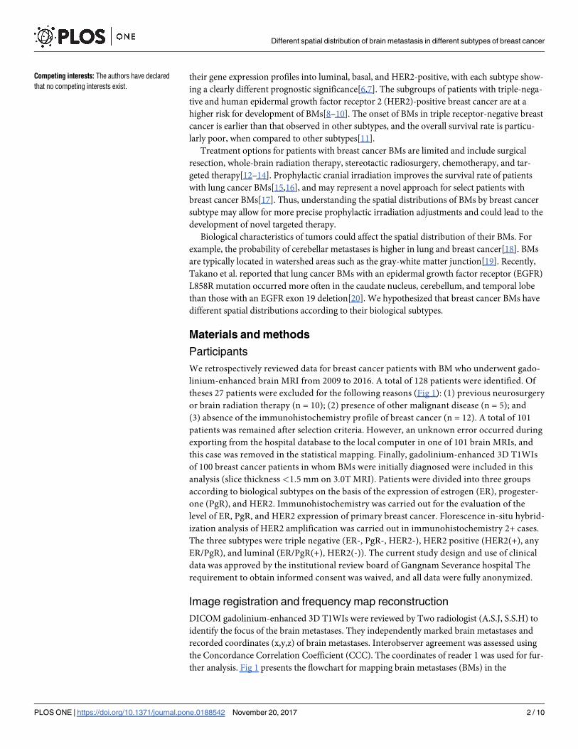
In total, 100 patients were analyzed: 24 patients had triple-negative breast cancer, 48 patients
had HER2-positive breast cancer, and 28 patients had luminal breast cancer. All patients were
female. Age at initial diagnosis of breast cancer was not significantly different among breast
cancer subtypes (46.45 ± 10.88 for triple-negative, 49.5 ± 11.45 for HER2, 46.75 ± 9.73 for
luminal; P = 0.26). Number of brain metastases per patient did not significantly differ by sub-
type (5.33 ± 5.78 for triple-negative, 4.71 ± 6.58 for HER2, 5.35 ± 6.69 for luminal; P = 0.88).
However, triple-negative and HER2-positive breast cancer showed a shorter time interval
until the onset of BMs than luminal breast cancer (23.5 ± 23.36 months for triple-negative,
19 ± 29.54 months for the HER2 subtype, and 42 ± 45.51 months for luminal subtype; P< .01,
Table 1)
Interobserver agreement
The coordinates of brain metastases had good reproducibility between two radiologists. Their
Concordance Correlation Coefficient was 0.996 (0.994–0.997)
Frequency map of brain metastases in each subtype (intra-group
analysis)
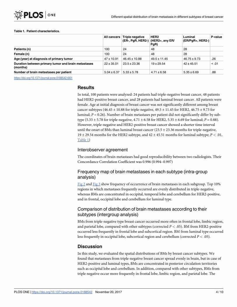
Fig 2 and Fig 3 show frequency of occurrence of brain metastases in each subgroup. Top 10%
regions in which metastases frequently occurred are evenly distributed in triple-negative,
whereas BMs are concentrated in occipital, temporal lobe and cerebellum for HER2-positive,
and in frontal, occipital lobe and cerebellum for luminal type.
Comparison of distribution of brain metastases according to their
subtypes (intergroup analysis)
BMs from triple-negative type breast cancer occurred more often in frontal lobe, limbic region,
and parietal lobe, compared with other subtypes (corrected P< .05). BM from HER2-positive
occurred less frequently in frontal lobe and subcortical region. BM from luminal type occurred
less frequently in occipital lobe, subcortical region and cerebellum (corrected P< .05).
Discussion
In this study, we evaluated the spatial distributions of BMs by breast cancer subtypes. We
found that metastases from triple-negative breast cancer spread evenly in brain, but in case of
HER2-positive and luminal types, BMs are concentrated in posterior circulation territories
such as occipital lobe and cerebellum. In addition, compared with other subtypes, BMs from
triple negative occur more frequently in frontal lobe, limbic region, and parietal lobe. The
Table 1. Patient characteristics.
All cancers Triple negative
(ER-, PgR, HER2-)
HER2
(HER2+, any ER/
PgR)
Luminal
(ER/PgR+, HER2-)
P-value
Patients (n) 100 24 48 28
Female (n) 100 24 48 28
Age (year) at diagnosis of primary tumor 47 ± 10.91 46.45 ± 10.88 49.5 ± 11.45 46.75 ± 9.73 .26
Duration between primary tumor and brain metastases
(months)
22 ± 35.01 23.5 ± 23.36 19 ± 29.54 42 ± 45.51 < .01
Number of brain metastases per patient 5.04 ± 6.37 5.33 ± 5.78 4.71 ± 6.58 5.35 ± 6.69 .88
https://doi.org/10.1371/journal.pone.0188542.t001
Different spatial distribution of brain metastasis in different subtypes of breast cancer
PLOS ONE | https://doi.org/10.1371/journal.pone.0188542 November 20, 2017 4 / 10
clinical implication of this observation is important because prophylactic irradiation with dose
modulation to the preferential site is a viable approach for breast cancer to enhance its preven-
tive effect and reduce any side effects.
Several studies have found that the cerebellum was the predominant site of metastases in
breast cancer patients.[22–24] Our data also confirm that the cerebellum is the preferential site
of breast cancer BMs. Conventionally, the “seed and soil theory” has been used to explain the
preferential involvement of specific areas within the brain: the site of metastasis depends on the
affinity of the tumor (the “seed”) to the microenvironment (the “soil”)[25]. Other potential
explanations for preferential involvement of the cerebellum are as follows: (1) high gyral density
Fig 2. Heat maps of brain metastases (BMs). Overlapping of BMs across all patients with breast cancer (A), patients with triple negative type (B),
patients with HER2+ type (C), and patients with Luminal type (D). Color-bars indicate the percentage of BMs. Abbreviations: CBL, cerebellum; IFG,
inferior frontal gyrus; MFG, middle frontal gyrus; L, left; R, right; MO, middle occipital; PMC, premotor cortex.
https://doi.org/10.1371/journal.pone.0188542.g002
Different spatial distribution of brain metastasis in different subtypes of breast cancer
PLOS ONE | https://doi.org/10.1371/journal.pone.0188542 November 20, 2017 5 / 10

Fig 3. The percentages of BMs were plotted across all patients (A), patients with triple negative gene
type (B), patients with HER2+ gene type (C), and patients with Luminal type (D). Brain regions were sorted
Different spatial distribution of brain metastasis in different subtypes of breast cancer
PLOS ONE | https://doi.org/10.1371/journal.pone.0188542 November 20, 2017 6 / 10

of the cerebellar cortex compared to that of the cerebral hemispheres; (2) higher blood volumes
and longer perfusion times of the tissue per minute in posterior circulation territories[26,27];
(3) different regional vasomotor response in the cerebellar circulation oriented toward a greater
vessel dilatation[28,29].
However, recent studies have revealed the molecular mechanism of the spatial distribution
of BMs. In breast cancer, chemokine receptors such as CXCR4 and CCR7 play a critical role in
determining the metastatic destination of tumor cells[30]. COX2, EGFR ligand, and ST6GAL-
NAC5 cross over the blood-brain barrier and enhance breast cancer metastasis to the brain.
Evidence of specific subtypes showing a preference for brain metastasis is overwhelming. Tri-
ple-negative and HER2-positive breast cancer predispose to a higher risk of BM than that
observed in luminal breast cancer, with an incidence of 30–40%[31–33]. A previous study
reported that triple-negative type and HER2-positive breast cancer show an earlier onset of
BMs than luminal breast cancer, which is consistent with our study[11,34]. In a recent study,
WNT signaling was up-regulated in the triple-negative subtype and the BMs, but down-regu-
lated in the luminal subtype and bone metastases[35]. Thus, we can assume that the spatial dis-
tribution of BMs differ according to the genetic composition of the primary breast cancer.
Interestingly, our study showed that BMs of tripe-negative spread evenly in the whole brain,
however HER-2 positive and luminal types have preferential involvements in the posterior cir-
culation territories such as occipital lobe and cerebellum.
Despite neurosurgery and radiosurgery, triple-negative type breast cancer patients have the
worst prognosis, with an overall survival duration of only 4.9 months.[36] Prophylactic cranial
irradiation is a suitable treatment option for high-risk breast cancer. A randomized controlled
trial showed that among 62 high-risk breast cancer patients receiving prophylactic cranial irra-
diation with 24 Gy in 10 fractions over 2 weeks, none developed BMs, but 6.4% of patients in
the no prophylactic cranial irradiation arm developed BMs.[37] Thus, our results suggest dif-
ferent strategies of dose modulation of radiotherapy (whole brain coverage for triple-negative
vs posterior circulation territories for HER2+ and luminal type).
Our results also may increase the diagnostic yield of brain metastases. Clinical information
of triple-negative type of primary breast cancer could make radiologist to review, with a metic-
ulous effort, occipital lobe and cerebellum as well as frontal, limbic and parietal lobe.
This study has a limitation. The number of cases was not enough to draw a solid conclusion.
However, our results may serve as a cornerstone for future studies with a larger population to
validate and extend these results.
In conclusion, different breast cancer subtypes may show different spatial distributions of
BMs. Triple-negative breast cancer BMs has a tendency to spread evenly in the whole brain,
meanwhile HER2+ and luminal types have a preponderance for posterior circulation territo-
ries. These results suggest different strategies for prophylactic irradiation according to sub-
types (triple-negative vs other types).
Supporting information
S1 File. Statistics of brain metastases in ALL regions.
(XLSX)
in the order of the frontal, limbic, occipital, parietal, subcortical (SubCor), temporal, and cerebellum. Brain regions
above dotted-line indicate the top 10% frequently occurring BMs. Reddish points indicate brain regions showing
significant group differences from χ2 test.
https://doi.org/10.1371/journal.pone.0188542.g003
Different spatial distribution of brain metastasis in different subtypes of breast cancer
PLOS ONE | https://doi.org/10.1371/journal.pone.0188542 November 20, 2017 7 / 10

Author Contributions
Conceptualization: Sung Gwe Ahn, Sang Hyun Suh, Sung Jun Ahn.
Investigation: Sang Hyun Suh, Sung Jun Ahn.
Methodology: Sunghyon Kyeong, Yoon Jin Cha.
Resources: Yoon Jin Cha, Eun Ju Son.
Software: Sunghyon Kyeong.
Writing – original draft: Sunghyon Kyeong.
Writing – review & editing: Sung Jun Ahn.
References1. Maher EA, Mietz J, Arteaga CL, DePinho RA, Mohla S (2009) Brain metastasis: opportunities in basic
and translational research. Cancer Res 69: 6015–6020. https://doi.org/10.1158/0008-5472.CAN-08-
4347 PMID: 19638593
2. Lin NU, Bellon JR, Winer EP (2004) CNS metastases in breast cancer. J Clin Oncol 22: 3608–3617.
https://doi.org/10.1200/JCO.2004.01.175 PMID: 15337811
3. Niwinska A, Murawska M, Pogoda K (2010) Breast cancer brain metastases: differences in survival
depending on biological subtype, RPA RTOG prognostic class and systemic treatment after whole-
brain radiotherapy (WBRT). Ann Oncol 21: 942–948. https://doi.org/10.1093/annonc/mdp407 PMID:
19840953
4. Weil RJ, Palmieri DC, Bronder JL, Stark AM, Steeg PS (2005) Breast cancer metastasis to the central
nervous system. Am J Pathol 167: 913–920. https://doi.org/10.1016/S0002-9440(10)61180-7 PMID:
16192626
5. Nagao E, Yoshiura T, Hiwatashi A, Obara M, Yamashita K, Kamano H (2011) 3D turbo spin-echo
sequence with motion-sensitized driven-equilibrium preparation for detection of brain metastases on 3T
MR imaging. AJNR Am J Neuroradiol 32: 664–670. https://doi.org/10.3174/ajnr.A2343 PMID:
21292797
6. Weigelt B, Baehner FL, Reis-Filho JS (2010) The contribution of gene expression profiling to breast
cancer classification, prognostication and prediction: a retrospective of the last decade. J Pathol 220:
263–280. https://doi.org/10.1002/path.2648 PMID: 19927298
7. Blows FM, Driver KE, Schmidt MK, Broeks A, van Leeuwen FE, Wesseling J. (2010) Subtyping of
breast cancer by immunohistochemistry to investigate a relationship between subtype and short and
long term survival: a collaborative analysis of data for 10,159 cases from 12 studies. PLoS Med 7:
e1000279. https://doi.org/10.1371/journal.pmed.1000279 PMID: 20520800
8. Gabos Z, Sinha R, Hanson J, Chauhan N, Hugh J, Mackey JR (2006) Prognostic significance of human
epidermal growth factor receptor positivity for the development of brain metastasis after newly diag-
nosed breast cancer. J Clin Oncol 24: 5658–5663. https://doi.org/10.1200/JCO.2006.07.0250 PMID:
17102066
9. Tham YL, Sexton K, Kramer R, Hilsenbeck S, Elledge R (2006) Primary breast cancer phenotypes
associated with propensity for central nervous system metastases. Cancer 107: 696–704. https://doi.
org/10.1002/cncr.22041 PMID: 16826579
10. Nam BH, Kim SY, Han HS, Kwon Y, Lee KS, Kim TH (2008) Breast cancer subtypes and survival in
patients with brain metastases. Breast Cancer Res 10: R20. https://doi.org/10.1186/bcr1870 PMID:
18307763
11. Dawood S, Broglio K, Esteva FJ, Yang W, Kau SW, Islam R. (2009) Survival among women with triple
receptor-negative breast cancer and brain metastases. Ann Oncol 20: 621–627. https://doi.org/10.
1093/annonc/mdn682 PMID: 19150943
12. Lee SS, Ahn JH, Kim MK, Sym SJ, Gong G, Ahn SD. (2008) Brain metastases in breast cancer: prog-
nostic factors and management. Breast Cancer Res Treat 111: 523–530. https://doi.org/10.1007/
s10549-007-9806-2 PMID: 17990100
13. Gil-Gil MJ, Martinez-Garcia M, Sierra A, Conesa G, Del Barco S, Gonzales-Jimenez S. (2014) Breast
cancer brain metastases: a review of the literature and a current multidisciplinary management guide-
line. Clin Transl Oncol 16: 436–446. https://doi.org/10.1007/s12094-013-1110-5 PMID: 24277572
Different spatial distribution of brain metastasis in different subtypes of breast cancer
PLOS ONE | https://doi.org/10.1371/journal.pone.0188542 November 20, 2017 8 / 10
14. Niwinska A, Pogoda K, Murawska M, Niwinski P (2011) Factors influencing survival in patients with
breast cancer and single or solitary brain metastasis. Eur J Surg Oncol 37: 635–642. https://doi.org/10.
1016/j.ejso.2011.05.002 PMID: 21664097
15. Slotman B, Faivre-Finn C, Kramer G, Rankin E, Snee M, Hatton M. (2007) Prophylactic cranial irradia-
tion in extensive small-cell lung cancer. N Engl J Med 357: 664–672. https://doi.org/10.1056/
NEJMoa071780 PMID: 17699816
16. Li N, Zeng ZF, Wang SY, Ou W, Ye X, Li J. (2015) Randomized phase III trial of prophylactic cranial irra-
diation versus observation in patients with fully resected stage IIIA-N2 nonsmall-cell lung cancer and
high risk of cerebral metastases after adjuvant chemotherapy. Ann Oncol 26: 504–509. https://doi.org/
10.1093/annonc/mdu567 PMID: 25515658
17. Gandhi AK, Sharma DN, Rath GK (2015) Prophylactic cranial irradiation in breast cancer: A new way
forward. Indian J Med Paediatr Oncol 36: 77–78. https://doi.org/10.4103/0971-5851.158822 PMID:
26157281
18. Bender ET, Tome WA (2011) Distribution of brain metastases: implications for non-uniform dose pre-
scriptions. Br J Radiol 84: 649–658. https://doi.org/10.1259/bjr/30173406 PMID: 21697413
19. Delattre JY, Krol G, Thaler HT, Posner JB (1988) Distribution of brain metastases. Arch Neurol 45:
741–744. PMID: 3390029
20. Takano K, Kinoshita M, Takagaki M, Sakai M, Tateishi S, Achiha T (2016) Different spatial distributions
of brain metastases from lung cancer by histological subtype and mutation status of epidermal growth
factor receptor. Neuro Oncol 18: 716–724. https://doi.org/10.1093/neuonc/nov266 PMID: 26519739
21. Tzourio-Mazoyer N, Landeau B, Papathanassiou D, Crivello F, Etard O, Delcroix N. (2002) Automated
anatomical labeling of activations in SPM using a macroscopic anatomical parcellation of the MNI MRI
single-subject brain. Neuroimage 15: 273–289. https://doi.org/10.1006/nimg.2001.0978 PMID:
11771995
22. Quattrocchi CC, Errante Y, Gaudino C, Mallio CA, Giona A, Santini D. (2012) Spatial brain distribution
of intra-axial metastatic lesions in breast and lung cancer patients. J Neurooncol 110: 79–87. https://
doi.org/10.1007/s11060-012-0937-x PMID: 22802020
23. Hengel K, Sidhu G, Choi J, Weedon J, Nwokedi E, Axiotis C (2013) Attributes of brain metastases from
breast and lung cancer. Int J Clin Oncol 18: 396–401. https://doi.org/10.1007/s10147-012-0392-x
PMID: 22383025
24. Tsukada Y, Fouad A, Pickren JW, Lane WW (1983) Central nervous system metastasis from breast
carcinoma. Autopsy study. Cancer 52: 2349–2354. PMID: 6640506
25. Ribatti D, Mangialardi G, Vacca A (2006) Stephen Paget and the ’seed and soil’ theory of metastatic dis-
semination. Clin Exp Med 6: 145–149. https://doi.org/10.1007/s10238-006-0117-4 PMID: 17191105
26. Hendrikse J, van der Grond J, Lu H, van Zijl PC, Golay X (2004) Flow territory mapping of the cerebral
arteries with regional perfusion MRI. Stroke 35: 882–887. https://doi.org/10.1161/01.STR.
0000120312.26163.EC PMID: 14988567
27. van Laar PJ, Hendrikse J, Golay X, Lu H, van Osch MJ, van der Grond J (2006) In vivo flow territory
mapping of major brain feeding arteries. Neuroimage 29: 136–144. https://doi.org/10.1016/j.
neuroimage.2005.07.011 PMID: 16095923
28. Cavaglia M, Dombrowski SM, Drazba J, Vasanji A, Bokesch PM, Janigro D (2001) Regional variation in
brain capillary density and vascular response to ischemia. Brain Res 910: 81–93. PMID: 11489257
29. Ito H, Yokoyama I, Iida H, Kinoshita T, Hatazawa J, et al. (2000) Regional differences in cerebral vascu-
lar response to PaCO2 changes in humans measured by positron emission tomography. J Cereb Blood
Flow Metab 20: 1264–1270. https://doi.org/10.1097/00004647-200008000-00011 PMID: 10950385
30. Muller A, Homey B, Soto H, Ge N, Catron D, Buchanan M. (2001) Involvement of chemokine receptors
in breast cancer metastasis. Nature 410: 50–56. https://doi.org/10.1038/35065016 PMID: 11242036
31. Foulkes WD, Smith IE, Reis-Filho JS (2010) Triple-negative breast cancer. N Engl J Med 363: 1938–
1948. https://doi.org/10.1056/NEJMra1001389 PMID: 21067385
32. Bendell JC, Domchek SM, Burstein HJ, Harris L, Younger J, Kuter I. (2003) Central nervous system
metastases in women who receive trastuzumab-based therapy for metastatic breast carcinoma. Cancer
97: 2972–2977. https://doi.org/10.1002/cncr.11436 PMID: 12784331
33. Gori S, Rimondini S, De Angelis V, Colozza M, Bisagni G, Moretti G (2007) Central nervous system
metastases in HER-2 positive metastatic breast cancer patients treated with trastuzumab: incidence,
survival, and risk factors. Oncologist 12: 766–773. https://doi.org/10.1634/theoncologist.12-7-766
PMID: 17673608
34. Sperduto PW, Kased N, Roberge D, Chao ST, Shanley R, Luo X (2013) The effect of tumor subtype on
the time from primary diagnosis to development of brain metastases and survival in patients with breast
cancer. J Neurooncol 112: 467–472. https://doi.org/10.1007/s11060-013-1083-9 PMID: 23462853
Different spatial distribution of brain metastasis in different subtypes of breast cancer
PLOS ONE | https://doi.org/10.1371/journal.pone.0188542 November 20, 2017 9 / 10

35. Bos PD, Zhang XH, Nadal C, Shu W, Gomis RR, Nguyen DX (2009) Genes that mediate breast cancer
metastasis to the brain. Nature 459: 1005–1009. https://doi.org/10.1038/nature08021 PMID: 19421193
36. Niikura N, Hayashi N, Masuda N, Takashima S, Nakamura R, Watanabe K. (2014) Treatment outcomes
and prognostic factors for patients with brain metastases from breast cancer of each subtype: a multi-
center retrospective analysis. Breast Cancer Res Treat 147: 103–112. https://doi.org/10.1007/s10549-
014-3090-8 PMID: 25106661
37. Hashem T, Eldin KK, Metwaly H, Elkholy E (2008) Prophylactic cranial irradiation (PCI) in high-risk
breast cancer patients: Preliminary data. Journal of Clinical Oncology 26.
Different spatial distribution of brain metastasis in different subtypes of breast cancer
PLOS ONE | https://doi.org/10.1371/journal.pone.0188542 November 20, 2017 10 / 10