Study to compute the current by applying nested grid technique
STUDY PROTOCOL Open Access
A nested mechanistic sub-study into theeffect of tranexamic acid versus placebo onintracranial haemorrhage and cerebralischaemia in isolated traumatic brain injury:study protocol for a randomised controlledtrial (CRASH-3 Trial Intracranial BleedingMechanistic Sub-Study [CRASH-3 IBMS])Abda Mahmood* , Ian Roberts and Haleema Shakur
Abstract
Background: Tranexamic acid prevents blood clots from breaking down and reduces bleeding. However, it isuncertain whether tranexamic acid is effective in traumatic brain injury. The CRASH-3 trial is a randomisedcontrolled trial that will examine the effect of tranexamic acid (versus placebo) on death and disability in 13,000patients with traumatic brain injury. The CRASH-3 trial hypothesizes that tranexamic acid will reduce intracranialhaemorrhage, which will reduce the risk of death. Although it is possible that tranexamic acid will reduceintracranial bleeding, there is also a potential for harm. In particular, tranexamic acid may increase the risk ofcerebral thrombosis and ischaemia. The protocol detailed here is for a mechanistic sub-study nested within theCRASH-3 trial. This mechanistic sub-study aims to examine the effect of tranexamic acid (versus placebo) onintracranial bleeding and cerebral ischaemia.
Methods: The CRASH-3 Intracranial Bleeding Mechanistic Sub-Study (CRASH-3 IBMS) is nested within a prospective,double-blind, multi-centre, parallel-arm randomised trial called the CRASH-3 trial. The CRASH-3 IBMS will beconducted in a cohort of approximately 1000 isolated traumatic brain injury patients enrolled in the CRASH-3 trial.In the CRASH-3 IBMS, brain scans acquired before and after randomisation are examined, using validated methods,for evidence of intracranial bleeding and cerebral ischaemia. The primary outcome is the total volume of intracranialbleeding measured on computed tomography after randomisation, adjusting for baseline bleeding volume. Secondaryoutcomes include progression of intracranial haemorrhage (from pre- to post-randomisation scans), new intracranialhaemorrhage (seen on post- but not pre-randomisation scans), intracranial haemorrhage following neurosurgery, andnew focal ischaemic lesions (seen on post-but not pre-randomisation scans). A linear regression model will examinewhether receipt of the trial treatment can predict haemorrhage volume. Bleeding volumes and new ischaemic lesionswill be compared across treatment groups using relative risks and 95% confidence intervals.(Continued on next page)
* Correspondence: [email protected] Trials Unit, London School of Hygiene and Tropical Medicine,University of London, Keppel Street, London WC1E 7HT, UK
© The Author(s). 2017 Open Access This article is distributed under the terms of the Creative Commons Attribution 4.0International License (http://creativecommons.org/licenses/by/4.0/), which permits unrestricted use, distribution, andreproduction in any medium, provided you give appropriate credit to the original author(s) and the source, provide a link tothe Creative Commons license, and indicate if changes were made. The Creative Commons Public Domain Dedication waiver(http://creativecommons.org/publicdomain/zero/1.0/) applies to the data made available in this article, unless otherwise stated.
Mahmood et al. Trials (2017) 18:330 DOI 10.1186/s13063-017-2073-6
(Continued from previous page)
Discussion: The CRASH-3 IBMS will provide an insight into the mechanism of action of tranexamic acid in traumaticbrain injury, as well as information about the risks and benefits. Evidence from this trial could inform the managementof patients with traumatic brain injury.
Trial registration: The CRASH-3 trial was prospectively registered and the CRASH-3 IBMS is an addition to the originalprotocol registered at the International Standard Randomised Controlled Trials registry (ISRCTN15088122) 19 July 2011,and ClinicalTrials.gov on 25 July 2011 (NCT01402882).
Keywords: Tranexamic acid, Intracranial haemorrhage, Cerebral ischaemia, Traumatic brain injury
BackgroundTraumatic brain injury (TBI) occurrenceTBI is a leading cause of death and disability worldwide.According to the World Health Organization, TBI willcontinue to be a major cause of death and disability by2020 [1]. At least 200 per 100,000 people are killed orhospitalised each year after TBI [2], resulting in over 10million deaths or hospitalisations each year [3]. TBI isthe leading cause of death and disability in people belowthe age of 45 [4].TBI patients can experience a loss in physical, behav-
ioural or emotional functioning after the injury [5]. SevereTBI often results in motor impairment that persists for atleast 3 years after the injury [6] and cognitive impairmentsare present for at least 6 months after injury [7]. Problemswith memory following TBI significantly affect an individ-ual’s quality of life [8]. Even with rehabilitation treatments,only 40–50% of TBI patients completely recover [9].The increasing incidence of TBI can be explained by the
rising frequency of traffic accidents in developing countriesand rapidly motorising middle-income countries [10]. Pro-jections of global mortality and burden of disease suggestthat road traffic accidents will be the third major cause ofdeath and disability by 2030, assuming a faster rate ofsocio-economic development [11]. Falls in older adults arethe leading cause of TBI in high-income countries [12].Given the global scope of this life threatening and poten-tially disabling condition, it is important to identify themost effective clinical care in this patient group.
Intracranial haemorrhage occurrenceIntracranial bleeding is common after TBI and the largerthe bleed the greater the risk of death and disability[13, 14]. In patients with mild TBI (Glasgow Coma Scalescore ≥ 13), although bleeding can continue for up to24 hours after injury, most bleeds stop progressing withina few hours of hospital admission [15]. Intracranial haem-orrhage progression has been observed in half of moderateto severe head injury patients who had a median GlasgowComa Scale score of 8 on admission and repeat computedtomography (CT) scans performed within 24 hours ofinjury [16, 17]. Patients who were scanned earlier afterinjury (≤3.5 hours vs. > 3.5 hours) were more likely to have
expanding haematomas on CT performed 24 hours afterinjury (57% vs. 28%) [17]. If the initial CT scan was con-ducted more than 3.5 hours after injury, the percentage ofpatients with measurable changes in haematoma volume24 hours after injury was reduced. In a subset of patientswho had an intermediate scan (most of which were be-tween 6 and 9 hours of injury), the mean volume changebetween the baseline and intermediate scan was 5.7 mL,whereas the difference in mean volume between the inter-mediate scan and the 24 hour scan was 0.03 mL [17].Thus, the maximal change in intracranial haemorrhagevolume occurs soon after injury.A meta-analysis of 34 studies that reported the fre-
quency of coagulopathy after TBI found that one thirdof patients with TBI have laboratory evidence of abnor-mal coagulation based on parameters such as fibrinogen,fibrin degradation products and antithrombin levels [18].The risk of mortality in patients with coagulopathy
after TBI is nine times higher than in TBI patients withoutcoagulopathy (odds ratio (OR) 9.0, 95% confidence inter-val (CI) 7.3–11.6). The risk of unfavourable outcome asmeasured by the Glasgow Outcome Scale (score of 1–3) ismore than 30 times higher in TBI patients with coagulop-athy (OR 36.3, 95% CI 18.7–70.5) [18]. Decreased plateletcounts, prolonged prothrombin time and partial thrombo-plastin time, and high levels of fibrinogen and D-dimerlevels are observed in patients within the first 3 hours ofTBI [19]. The highest D-dimer concentrations were foundin the most severely injured patients [20], who have ahigher risk of intracranial haemorrhage and mortality.
Effectiveness of tranexamic acid in reducing haemorrhageTranexamic acid reduces bleeding by inhibiting the en-zymatic breakdown of fibrin blood clots. Plasmin bindsto fibrin via lysine-binding sites and then splits fibrininto fibrin degradation products. Tranexamic acid is amolecular analogue of lysine that inhibits fibrinolysis byreducing the binding of plasmin to fibrin.A systematic review of 104 randomised trials of tran-
examic acid in surgical patients found that it reducedthe number of patients receiving a blood transfusion byone-third and halved the need for further surgery tocontrol bleeding [21].
Mahmood et al. Trials (2017) 18:330 Page 2 of 13
A large randomised trial of tranexamic acid treatmentwithin an hour of acute traumatic injury found thatit reduced the risk of death due to bleeding by aboutone-third (relative risk (RR) 0.68, 95% CI 0.57–0.82;P < 0.0001) [22, 23]. Treatment between 1 and 3 hours re-duced the risk by about one-fifth (RR 0.79, 0.64–0.97;P = 0.03). There was no apparent increase in the riskof vascular occlusive events with tranexamic acid follow-ing acute trauma (RR 0.69, 95% CI 0.44–1.07; P = 0.096).
Tranexamic acid as a potential treatment in TBITranexamic acid is able to penetrate the blood–brainbarrier and should be able to affect intracranial haemor-rhage [24]. If tranexamic acid is effective following TBI,it should also be most effective when given soon after in-jury when intracranial bleeding is on-going [15]. Further-more, if early increased fibrinolysis exacerbates bleedingand increases the risk of death [20], we would expect tran-examic acid to be most effective during this period.However, there is also the potential for harm. In par-
ticular, tranexamic acid may increase the risk of cerebralthrombosis and ischaemia [25]. Cerebral ischaemia is animportant secondary injury mechanism after TBI thatworsens neurologic outcome and increases mortality[26, 27]. It can be precipitated by raised intracranialpressure, which can lead to cerebral hypo-perfusion[28–31]. In addition, thrombotic disseminated intravascu-lar coagulation may increase the risk of cerebral micro-thrombi, which are often seen in the brains of TBI patientswho die within 24 hours of injury [32]. By inhibiting fibrin-olysis, tranexamic acid might increase the risk of cerebralischaemia and thrombosis in TBI patients.A systematic review identified two completed rando-
mised trials of tranexamic acid in TBI patients [33]. Thefirst randomised trial (n = 249) examined the effect oftranexamic acid in patients with extra-cranial bleedingbut who also had TBI [34]. The second randomised trial(n = 229) examined the effect of tranexamic acid in pa-tients with polytrauma and TBI or isolated TBI [35]. Bothtrials used information from pre- and post-randomisationCT scans to estimate the extent of bleeding and ischaemia.Both trials recruited patients who were within 8 hours ofinjury, yet they were not large enough to determine thebalance of risks and benefits from tranexamic acid andwhether this varies by time to treatment.When the two randomised trials were combined in a
meta-analysis, there was a statistically significant reduc-tion in intracranial haemorrhage (RR 0.75, 95% CI 0.58–0.98; P = 0.03) and mortality (RR 0.63, 95% CI 0.40–0.99;P = 0.05) with tranexamic acid. In one trial, focal ischae-mic lesions occurred in 5% of tranexamic acid-treatedpatients and 9% of placebo-treated patients (RR 0.51,95% CI 0.20–1.32; P = 1.17) [34]. In the second trial,there were three strokes in the placebo group compared
with none in the tranexamic acid group [35]. However,because the CIs for intracranial haemorrhage, death andischaemic lesion outcomes are so wide, the quality ofthis evidence is low. Furthermore, the patients in the tri-als had extra-cranial bleeding in addition to intra-cranialbleeding. Because tranexamic acid reduces mortality inextra-cranial bleeding (CRASH-2), the mortality reduc-tion seen in this trial could be from the extra-cranialinjury rather than any effect on the brain injury. Theeffect of tranexamic acid on intracranial haemorrhageand thrombotic adverse effects, including stroke, remainsuncertain.There are three ongoing randomised trials of tranex-
amic acid versus placebo in patients with isolated TBI(NCT02645552, NCT01990768, NCT01402882). Thesewill evaluate the effect of tranexamic acid on death, dis-ability, vascular occlusive events and other adverse eventsin TBI. The ongoing trials will inform whether tranexamicacid can be given to those with TBI. To date, the CRASH-3 trial, with a planned sample size of 13,000 patients, willbe the largest randomised trial into the effect of tranex-amic acid in TBI [36]. The results from the three ongoingtrials should provide clinicians with information aboutwhether tranexamic acid is effective in reducing death anddisability without increasing thrombotic events. The trialswill also provide information about whether its effect var-ies by time to treatment.However, these trials will not provide information
about the mechanism by which tranexamic acid mightexert its effects in TBI. If tranexamic acid reduces mor-tality by reducing intracranial haemorrhage, we wouldexpect there to be less blood on head CT scans of tran-examic acid-treated patients, particularly those treatedsoon after injury [25]. If tranexamic acid increases therisk of cerebral ischaemia, we would expect to see moreischaemic lesions in tranexamic acid-treated patients,particularly in those treated after a more prolonged periodfollowing injury [37]. The CRASH-3 Intracranial BleedingMechanistic Sub-Study (CRASH-3 IBMS) will examinethe effect of tranexamic acid on intracranial haemorrhageand cerebral ischaemia in a cohort of patients enrolled inthe CRASH-3 trial. This paper outlines the protocol forthe CRASH-3 IBMS and is in line with the StandardProtocol Items: Recommendations for InterventionalTrials (SPIRIT) guidelines. The SPIRIT checklist andfigure have been included as Additional file 1 and Fig. 1,respectively.
AimThe CRASH-3 IBMS aims to examine the mechanismby which tranexamic acid exerts its effects in patientswith isolated TBI. Specifically, we will assess the effect oftranexamic acid on intracranial bleeding and cerebralischaemia.
Mahmood et al. Trials (2017) 18:330 Page 3 of 13
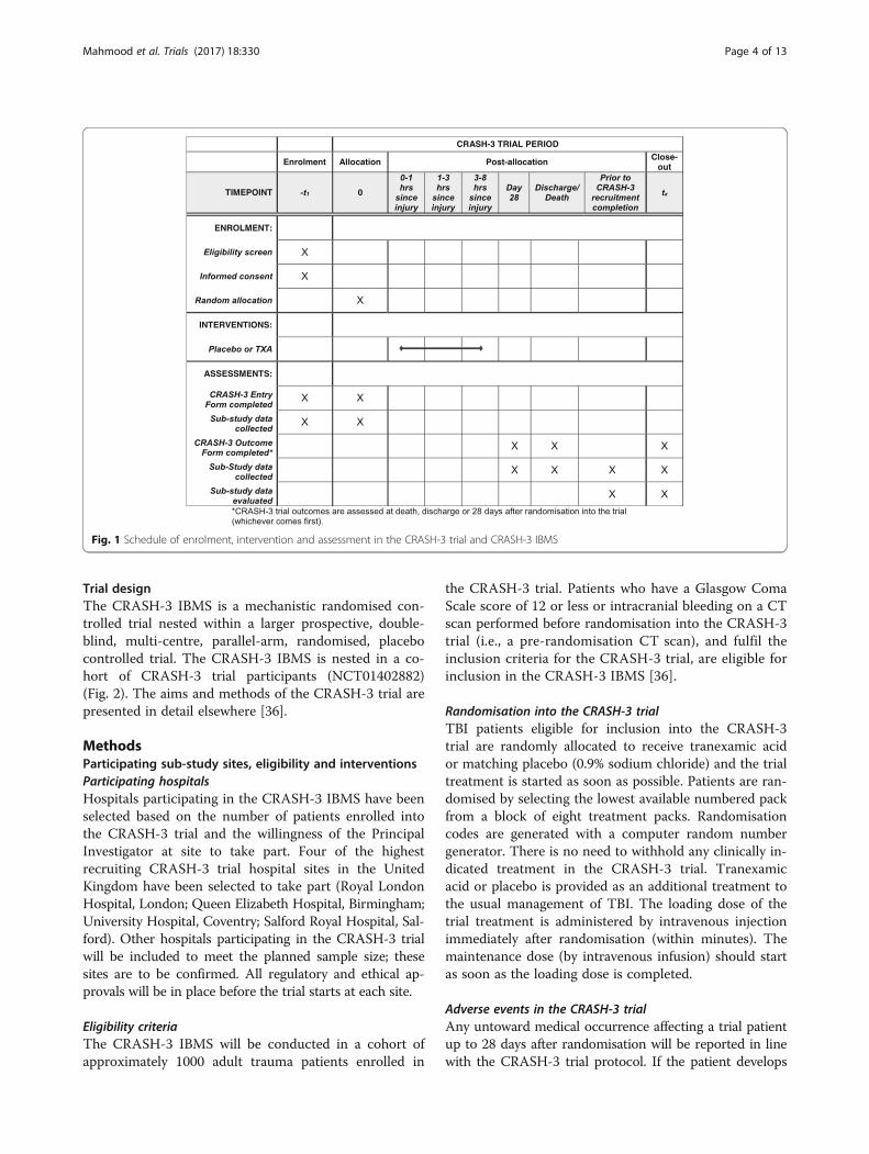
Trial designThe CRASH-3 IBMS is a mechanistic randomised con-trolled trial nested within a larger prospective, double-blind, multi-centre, parallel-arm, randomised, placebocontrolled trial. The CRASH-3 IBMS is nested in a co-hort of CRASH-3 trial participants (NCT01402882)(Fig. 2). The aims and methods of the CRASH-3 trial arepresented in detail elsewhere [36].
MethodsParticipating sub-study sites, eligibility and interventionsParticipating hospitalsHospitals participating in the CRASH-3 IBMS have beenselected based on the number of patients enrolled intothe CRASH-3 trial and the willingness of the PrincipalInvestigator at site to take part. Four of the highestrecruiting CRASH-3 trial hospital sites in the UnitedKingdom have been selected to take part (Royal LondonHospital, London; Queen Elizabeth Hospital, Birmingham;University Hospital, Coventry; Salford Royal Hospital, Sal-ford). Other hospitals participating in the CRASH-3 trialwill be included to meet the planned sample size; thesesites are to be confirmed. All regulatory and ethical ap-provals will be in place before the trial starts at each site.
Eligibility criteriaThe CRASH-3 IBMS will be conducted in a cohort ofapproximately 1000 adult trauma patients enrolled in
the CRASH-3 trial. Patients who have a Glasgow ComaScale score of 12 or less or intracranial bleeding on a CTscan performed before randomisation into the CRASH-3trial (i.e., a pre-randomisation CT scan), and fulfil theinclusion criteria for the CRASH-3 trial, are eligible forinclusion in the CRASH-3 IBMS [36].
Randomisation into the CRASH-3 trialTBI patients eligible for inclusion into the CRASH-3trial are randomly allocated to receive tranexamic acidor matching placebo (0.9% sodium chloride) and the trialtreatment is started as soon as possible. Patients are ran-domised by selecting the lowest available numbered packfrom a block of eight treatment packs. Randomisationcodes are generated with a computer random numbergenerator. There is no need to withhold any clinically in-dicated treatment in the CRASH-3 trial. Tranexamicacid or placebo is provided as an additional treatment tothe usual management of TBI. The loading dose of thetrial treatment is administered by intravenous injectionimmediately after randomisation (within minutes). Themaintenance dose (by intravenous infusion) should startas soon as the loading dose is completed.
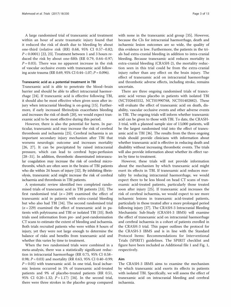
Adverse events in the CRASH-3 trialAny untoward medical occurrence affecting a trial patientup to 28 days after randomisation will be reported in linewith the CRASH-3 trial protocol. If the patient develops
Fig. 1 Schedule of enrolment, intervention and assessment in the CRASH-3 trial and CRASH-3 IBMS
Mahmood et al. Trials (2017) 18:330 Page 4 of 13
an adverse event during the treatment phase, the trial drugshould be stopped. In this situation, the patient should betreated in line with local procedures and then followed up.The independent Data Monitoring Committee may rec-ommend for the early termination of the trial, and thefinal decision lies with the Trial Steering Committee.
Unblinding before the end of the CRASH-3 trialIf there are contraindications to tranexamic acid followingrandomisation, the trial treatment should be stopped andall standard clinical care provided. Unblinding is only ne-cessary if the clinician believes that clinical managementdepends importantly upon knowledge of whether the pa-tient received tranexamic acid or placebo. In this case, a24 hour telephone service is available to confirm whetherthe patient received tranexamic acid or placebo.
Outcomes and outcome measurementPrimary outcomeThe total volume of intracranial bleeding after randomisa-tion, adjusting for total volume of intracranial bleeding atbaseline if baseline volume is available.
Secondary outcomes
(1)Frequency of progressive haemorrhage – number ofpatients with a post-randomisation CT scan withtotal haemorrhage volume of more than 25% of thevolume on the pre-randomisation scan;
(2)Frequency of new haemorrhage – number of patientswith haemorrhage on the post-randomisation CT scanwhen there was not one on the pre-randomisation scan;
(3)New focal ischaemic lesions – ischaemic lesionswhich appear on a post-randomisation scan but noton the pre-randomisation scan;
(4)Total volume of intracranial bleeding afterrandomisation in patients who undergo surgicalevacuation of haemorrhage, adjusting for volume ofbaseline bleeding.
All outcomes will be compared across treatment groups.
Outcome measurement: estimating haemorrhage volumePatients often undergo one brain CT scan as part of routinemedical care prior to randomisation into the CRASH-3
Fig. 2 CRASH-3 trial inclusion criteria (blue boxes show additional procedure for the CRASH-3 Intracranial Bleeding Mechanistic Sub-Study)
Mahmood et al. Trials (2017) 18:330 Page 5 of 13
trial. The majority of patients are scanned again afterrandomisation into the CRASH-3 trial. In the CRASH-3 IBMS, we will measure the volume of intracranialhaemorrhage on pre- and post-randomisation CT scans.A simple validated scale for measuring intracranialhaemorrhage volume shows good agreement with thegold standard of computer-assisted volumetric analysis,which requires demarcation of the haemorrhage bor-ders [38–49].The ABC/2 method is a quick and easy technique used
to estimate the volume of intracranial haemorrhage [50].This method assumes haematoma volume is approximatelyequal to an ellipsoid shape (i.e., three dimensional ovalshape). For ease of assessment, the formula for calculatingthe volume of an ellipsoid (4/3 × π × (A/2) × (B/2) × (C/2))can be simplified to ABC/2 if we assume π is equal to 3.This method selects a representative slice near the centreof the haematoma on which the bleed is most visible. Onthis slice, two measurements are taken: (A) the maximaldiameter; (B) width perpendicular to A. For the measure-ment of depth, the maximal number of slices on which thehaematoma is visible is multiplied by slice thickness (C).These three measurements are multiplied and the sum di-vided by two (ABC/2) to provide the volume measurementin cm3.Whilst some researchers have found that the ABC/2
method overestimates lesion volume compared tocomputer-assisted methods [39, 44, 45, 47, 51–55],others claim the opposite [41, 56]. Haemorrhagic lesionsthat have a regular shape are more accurately estimatedusing the ABC/2 method compared to lesions withirregular or multi-lobular shapes [43, 45–56]. Further-more, a number of variations of the ABC/2 methodadjust for the depth of a lesion. Whilst some havefound that adjusting for depth significantly underesti-mates volumes because smaller slice volumes are eli-minated [57], others found that adjusting for depth isfavourable [48].Although the ABC/2 method is a less specific measure
of haemorrhage volume and overestimation due to falsepositives would dilute the effect of the treatment to-wards the null, its low sensitivity and underestimationdue to false negatives would not impact the effect of thetreatment on haemorrhage. Furthermore, the more ac-curate method of estimating haemorrhage would havebeen more expensive and therefore administered in asmaller number of patients given the limited budget of aclinical trial. Although a more accurate method in asmall trial would result in less measurement error, a lessaccurate method in a larger trial would result in less ran-dom error. We believe that the ABC/2 method is suffi-ciently accurate and therefore chose to use this methodin a larger trial. Furthermore, the assessor rating thescans will be blind to treatment allocation and thus the
bias from measurement error should be balanced be-tween treatment groups.
Total haemorrhage volumeThe total haemorrhage volume on each scan will be cal-culated by totalling the volumes of intra-parenchymal,intra-ventricular, epidural and subdural haemorrhage.
Estimating intra-parenchymal, intra-ventricular and epiduralhaemorrhage volume using ABC/2Volume estimation of intracranial haemorrhage is aidedby the characterisation of haematomas. The final shapeof a haematoma is influenced by its location. Intra-axial(or intra-cerebral) haematomas include intra-parenchymalhaematomas, which occur in the brain tissue, and intra-ventricular haematomas, which occur in the ventricles ofthe brain. These haematomas tend to have regular shapesthat are clearly definable in every dimension (i.e., theirlength, width and depth can be measured on a CT scan).Extra-axial haematomas occur between the three mem-branes that surround the brain (dura mater, arachnoidmater and pia mater). Epidural haematomas are a type ofextra-axial haematoma and occur between the skull andouter membrane of the central nervous system (duramater). They have a clear shape that can be measured inevery dimension. The ABC/2 method assumes the haem-orrhage has an ellipsoid shape and has been validated inintra-parenchymal [38], intra-ventricular [46] and epiduralhaematomas [47, 48]. We will estimate the volume ofintra-parenchymal, intra-ventricular and epidural/extra-dural haemorrhage using the ABC/2 method.
Estimating subdural haemorrhage volume usingmaximum widthSubdural haematomas are another type of extra-axialhaemorrhage and occur between the dura mater and themiddle membrane of the central nervous system (arach-noid mater). Subdural haematomas are crescent shapedas they follow the pattern of the brain’s convexity. Theexact limits of a subdural haematoma are not clearly de-finable in any dimension. This type of haemorrhage cantheoretically occupy the entire subdural space. Giventhat the ABC/2 method assumes the haemorrhage hasan ellipsoid shape, it would not provide an accurate vol-ume estimation of subdural haemorrhage. Indeed, therehave been reports of underestimation in subdural haem-orrhage volume when using an adapted version of theABC/2 method compared with computer-assisted volu-metric analysis [41, 56].Some researchers and clinicians propose that it is
more appropriate to estimate subdural haemorrhage vol-ume using a formula which takes the difference betweentwo spheres (representing the entire subdural space),divides by two (as subdural haemorrhage is usually
Mahmood et al. Trials (2017) 18:330 Page 6 of 13
unilateral) and divide by two again (as subdural haem-orrhage tends to be thicker at the centre and thinner atthe sides). This method has been tested at the Neuro-surgical Trauma Unit at the Queen Elizabeth Hospitalin Birmingham (UK) and has been shown to provide moreclinically relevant estimates of haemorrhage volume thanthe ABC/2 method [58]. Although this method overesti-mates subdural volume, it is less than the error providedby the ABC/2 method. The key measurement in determin-ing the clinical significance of a subdural haemorrhage isits thickness (i.e., the B measurement when using theABC/2 method) [59]. In the CRASH-3 IBMS, we willmeasure the maximum width of a subdural bleed, andcompute its volume using the aforementioned formula.
Measurement of subarachnoid haemorrhageSubarachnoid bleeds are another type of extra-axialhaemorrhage and occur in the area between the arachnoidmembrane and the innermost membrane surrounding thebrain (pia mater). The shape of the subarachnoid space re-sembles a spider’s web and therefore haemorrhage in thesubarachnoid space cannot be clearly measured in any di-mension. Although there are a number of CT gradingscales that include the characterisation of subarachnoidhaemorrhage [60, 61], they are criticised for being subject-ive and not comprehensive enough to serve as a primarygrading scale for this type of haemorrhage [62]. For ex-ample, the Fisher scale and its modified version do notconsider subarachnoid haemorrhage in isolation but incombination with intraventricular haemorrhage [63].In the CRASH-3 IBMS, the size of a subarachnoid
haemorrhage will be characterised as small, medium orlarge. Each bleed will then be described as focal (local-ised to a specific location), multiple (not localised butnot widespread) or diffuse (widespread). This method isalso subjective and may have low sensitivity and specifi-city, therefore misclassification would bias the effect ofthe treatment towards the null value. We hope that, byusing this method in a large trial, the bias from measure-ment error would be offset by a reduction in randomerror.
Petechial haemorrhagePetechial haemorrhage manifests as a very small dot ona CT scan. CT scans and accompanying radiology re-ports will be examined to indicate whether petechialhaemorrhage is present.
Outcome measurement: focal ischaemic lesionsIschaemic stroke is due to the compromise of bloodand oxygen flow through either large or small arteriessupplying the brain parenchyma. Thrombotic occlusionof intracranial vessels produce wedge-shaped corticalinfarctions.
Cerebral ischaemia would reliably manifest on a CTscan performed at least 48 hours after randomisation[62]. However, given that clinical scans are performedfor diagnostic purposes, it is not possible to carry outscans at set time points post-randomisation. Brain im-aging techniques, including MRI diffusion weighted im-aging, have higher sensitivity and specificity comparedto CT in the early diagnosis of ischaemic infarction,and are often clinically warranted when there is a sus-pected stroke. Therefore, the assessor will examine allavailable brain scans performed within 28 days of ran-domisation and the accompanying radiology reports forevidence of focal ischaemic lesions and record the timefrom randomisation to detection.Furthermore, given that CT imaging is the first and
most common neuroimaging examination performed foremergency assessment of suspected acute haemorrhageand stroke around the world [64, 65], the majority ofscans included in the CRASH-3 IBMS will be CT scans.Therefore, it is important to clarify how we will capturethis endpoint when only CT scans are available. Cerebralinfarction manifests as wedge-shaped low attenuation ona CT scan. Given that oedema also manifests as low at-tenuation on CT, the radiology reports that accompanyCT scans should indicate whether the low attenuation isrepresentative of oedema or infarction. Brain imaging re-ports often refer to cerebral infarction by the affectedvascular territory (e.g., anterior cerebral artery, middlecerebral artery, posterior cerebral artery, lacunar, cere-bellar, brainstem). The assessor will examine all availablebrain imaging to assess whether oedema or infarctioncan be excluded given the appearance of earlier scans. Forexample, some patients have oedematous haemorrhagiclesions, which on CT manifests as high density haemor-rhage surrounded by low density oedema. In later scansthe haemorrhage may resolve but the oedema may remain.If only considered alone, the later CT scan may havethe appearance of infarction but could be representa-tive of residual oedema. We will attempt to minimisesuch errors by comparing the appearance of cerebralinfarction/oedema between consecutive scans, and con-sider the accompanying scan reports for radiologicalopinion. If the available scans and accompanying reportsare unable to confirm the presence of an ischaemic lesion,we would seek further radiological and clinical opinion.
Outcome measurement: mass effect and other CTendpointsSpace-occupying intracranial lesions can displace braintissue. The shift of midline structures past the centre lineof the brain will be measured in millimetres. We will alsorecord whether mass effect has caused ventricular andsulcal effacement.
Mahmood et al. Trials (2017) 18:330 Page 7 of 13
All scans will be rated according to the Marshall clas-sification – the most extensively used CT classificationscale in TBI [66]. Three main characteristics define theMarshall classification, namely presence of mass lesion,degree of compression of perimesencephalic cisterns anddegree of midline shift.
Sample sizeAssuming the average baseline intracranial bleeding volumeis 20 mL and assuming the same average increase (8 mL),standard deviation (28 mL) and correlation (rho = 0.6) be-tween baseline and follow-up bleeding volumes as in thecontrol group of the CRASH-2 Intracranial Bleeding Sub-study [34], a study with at least 1000 participants will have80% power (at alpha = 0.05) to detect a 15% lower bleedingvolume in the tranexamic acid group at follow-up (i.e.,24 mL tranexamic acid vs. 28 mL placebo). In the mainCRASH-3 trial, we hypothesise that tranexamic acidwill reduce intracranial bleeding by approximately 15%.The sample size estimates have been reviewed and ap-proved by statisticians at the London School of Hygieneand Tropical Medicine.
Data collection, management and analysisProcedures for data collectionThe CRASH-3 trial database will be used to prepare alist of all patients with a Glasgow Coma Scale score of12 or less or with a pre-randomisation CT scan at par-ticipating sub-study hospitals. The list will includeunique randomisation (box and pack) numbers, date andtime of randomisation, and time between injury and ran-domisation into the CRASH-3 trial. The randomisationnumbers will be used at the participating site to identifythe patient using their hospital number. The latter willbe used at the participating hospital to identify the pa-tient. The outcome assessor (research fellow with train-ing in brain imaging assessment) will hold a letter ofaccess at the participating hospital and use the patienthospital number to retrieve pre- and post-randomisationscans from the hospital imaging system. The outcomeassessor will complete the outcome forms at each siteusing the relevant scans and accompanying radiologyreports. All the data are collected by the same outcomeassessor who is blind to treatment allocation.If the patient does not have a pre-randomisation scan,
only the post-randomisation scan form is completed. Ifthe patient does not have a post-randomisation scan,only the pre-randomisation scan form is completed. Werecord whether pre- and/or post-randomisation scansare available such that we can examine missing data bytrial arm.In most cases, the post-randomisation scan is the first
scan performed after randomisation, which is normallywithin 72 hours of randomisation. Furthermore, due to
ongoing clinical management, some patients are scannedwithin minutes of randomisation. Tranexamic acid wouldnot have had sufficient opportunity to effect haemorrhageor infarction in such a way that would manifest on a scanthis soon after randomisation. Therefore, for patientsscanned within minutes of randomisation, we also meas-ure all the outcomes of interest on the next available post-randomisation scan, which is normally closer to 72 hoursof randomisation. All available brain imaging is examinedfor evidence of focal ischaemic lesions.The time stamped on the scans will be used to calculate
the following time intervals: (1) the time between injuryand the pre-randomisation CT scan and (2) the timebetween randomisation into the trial and the post-randomisation scan. If a patient has undergone neuro-surgery following their injury, information on the dateand time of neurosurgery will be collected using pro-spective reports including patient anaesthetic charts.The outcome data is collected for all patients includedin the CRASH-3 IBMS (unless consent was withdrawn)irrespective of whether the trial treatment was received(i.e., on an intention-to-treat basis). The outcome datais directly uploaded into an electronic database accessedat each sub-study site.
Data management planA data management plan will be prepared in advanceof data collection (Additional file 2). This will detail allaspects of data collection and recording to ensure compli-ance with International Conference on HarmonisationGood Clinical Practice guidelines (ICH-GCP) [67], UnitedKingdom Clinical Trials Regulations and the Data Pro-tection Act [68]. Data will be recorded in a database de-veloped in line with relevant regulatory requirements,including ICH-GCP guidelines.
Statistical analysis
Primary outcome A linear regression model will exam-ine the primary outcome; whether receipt of the trialtreatment can predict total haemorrhage volume follow-ing randomisation. Mean haemorrhage volume will becompared between trial arms, adjusting for baselinehaemorrhage volume. Adjusting for baseline haemorrhagevolume is important as it is a strong predictor of haema-toma increase [17, 69, 70], meaning that the baseline ad-justment can increase the power of the comparison byreducing the impact of between-patient variability. Wewill conduct subgroup analysis to examine whether the ef-fect of tranexamic acid on intracranial haemorrhage ismodified by time to treatment. A subgroup analysis bytime is important as previous evidence suggests that theeffect of tranexamic acid is strongly dependent on howquickly after injury it is received (CRASH-2).
Mahmood et al. Trials (2017) 18:330 Page 8 of 13
Secondary outcomes We will express the effect oftranexamic acid on the occurrence of dichotomous CTendpoints, including progressive haemorrhage or newhaemorrhage, using relative risks and 95% CIs esti-mated using generalised linear models.We will express the effect of tranexamic acid on newfocal cerebral ischaemic lesions measured at severalpost-randomisation time-points using relative risks and95% CIs estimated using generalised linear mixed modelsto account for the fact that this outcome could be mea-sured at several time-points following randomisation.
Missing data In line with the Consolidated Standards ofReporting Trials [71], we will report the number of pa-tients without pre- and post-randomisation scans bytreatment arm. If the outcome of interest (haemorrhageexpansion) is associated with the reason the data aremissing (patients with haemorrhage expansion may bemore likely to die before the second scan), imbalance inmissing data by treatment group can cause bias. If wesuspect that data are missing not at random [72], we willconduct sensitivity analysis to explore the implications.
Between-centre effects There is no evidence for the hy-pothesis that between-centre differences in unfavourableoutcome affect the chance of demonstrating a treatmenteffect in randomised trials of TBI [73]. This study esti-mated the between-centre differences beyond the randomvariation that may result from some centres that only treata small number of patients. Given this evidence and thatwe have no biological or mechanistic explanation to ex-pect any variation in a treatment effect between centres,we do not anticipate to find centre effects in the CRASH-3 IBMS. Furthermore, the majority of hospitals includedin the CRASH-3 IBMS are in western countries. Thehomogeneity in patient characteristics and care facilities isfurther reason not to expect a between-centre differencein treatment effect. However, for the purpose of transpar-ency we will report the interaction between centre andtreatment effect using a logistic regression model withinteraction between centre and treatment.
Inter-rater reliability The inter-rater reliability ofhaemorrhage occurrence will be assessed using relevantEntry Form data from the CRASH-3 trial to examineconsistency among ratings provided by the research fel-low and clinical staff.
Interim and final analyses There are no interim ana-lyses planned for the CRASH-3 IBMS. The final analysisfor the CRASH-3 IBMS will be undertaken followingcompletion of the main CRASH-3 trial. A complete stat-istical analysis plan will be published separately prior tocompletion of the CRASH-3 trial.
MonitoringAll data for the CRASH-3 trial will be subject to statis-tical monitoring and approximately 10% of data will besubject to on-site monitoring. Consent forms will bemonitored centrally by the Trial Coordinating Centre(where permission is given to do so). Investigators/insti-tutions are required to provide direct access to sourcedata/documents for trial-related monitoring, audits, ethicscommittee review, and regulatory inspection. All trial-related and source documents must be kept for at least5 years after the end of the trial. As all the CRASH-3IBMS data will be collected directly from source data,additional monitoring will not be carried out for this data.
Potential risksThe effective radiation dose from a CT scan is about2 mSv, which is approximately the amount received frombackground radiation in 8 months. Because CRASH-3IBMS will mainly use data from CT scans undertaken aspart of routine patient care, patients will not be exposedto extra radiation. There is no additional burden or riskto the patient as a result of CRASH-3 IBMS. It is stand-ard care for all patients with TBI and associated clinicalsigns to have a CT scan. Follow-up CT scans are oftenconducted for diagnostic purposes around 24 to 72 hoursafter the initial scan. Steps taken to minimise the risksassociated with handling personal data will be detailedin the Confidentiality section.
Confidentiality and disseminationConfidentialityOnly staff with authorised access to the scans, either asclinicians or research contract holders, will be able to re-trieve and review them. Completed scan data forms willbe uploaded onto a secure database. The scan data formswill contain no patient identifiable data (Additional file 3).Scans include the date and time of the scan and this infor-mation could potentially be used by anyone with access tothe hospital radiology system to identify the patient. Forthis reason, scan data forms will only include the random-isation number, the time interval between the injury andthe scan (pre-randomisation scan form), and the time inter-val between randomisation and the scan (post-randomisa-tion scan form). As no personal data will be collected, theanonymity of each patient will be protected.
PublicationThe results from this trial will be published in peer-reviewed medical journals. Dissemination of results topatients will take place via the media, trial website(crash3.lshtm.ac.uk) and relevant patient organisations.All participating sites will be credited in keypublications.
Mahmood et al. Trials (2017) 18:330 Page 9 of 13
DiscussionPotential benefit of CRASH-3 IBMS: furthers knowledgeabout mechanism of action of tranexamic acid in TBIThe CRASH-3 IBMS is a nested randomised trial that willreliably examine the effect of tranexamic acid on intracra-nial haemorrhage and cerebral ischaemia. We hope thatthis trial will provide information about the mechanism ofaction of tranexamic acid in isolated TBI. An understand-ing of the mechanism of action of tranexamic acid andinsight into factors that might affect this mechanism, iscritical in the appropriate generalisation of trial results[74]. If patients who receive tranexamic acid have lessintracranial bleeding on their CT scans compared to thosewho receive placebo, this information, along with the re-sults of the main CRASH-3 trial, could inform the admin-istration of tranexamic acid in TBI. If TBI patients whoreceive tranexamic acid soon after injury have less haem-orrhage expansion compared to those who receive tranex-amic later, then time between injury and treatment is afactor relevant to the mechanism of action which, with theresults of the main CRASH-3 trial, should be consideredwhen making treatment decisions. Furthermore, if we findevidence of cerebral ischaemia in patients who receivetranexamic acid and the effect varies by time to treatment,this information can be used to prevent adverse outcomesand ensure those receiving tranexamic acid are those mostlikely to benefit from it. Therefore, the knowledge gainedfrom the nested CRASH-3 IBMS will add to the evidencebase and could benefit the clinical management of patientswith head injuries.Furthermore, the patients included in the CRASH-3
IBMS are likely to have more severe head injuries com-pared to patients in the CRASH-3 trial but not included inthe CRASH-3 IBMS. The patients in the sub-study are nota random sample of patients in the CRASH-3 trial, norwill they be comparable. It is not necessary for the sub-study population to be representative of the CRASH-3trial population because knowledge about a causal mech-anism facilitates generalisation and not representativenessof the trial patients [75]. If the sub-study used a randomsample of patients from the CRASH-3 trial, the resultswould not necessarily apply to either more or less severepatients, but only to a hypothetical patient of average in-jury severity. Representativeness of trial patients does nothelp us to generalise our findings to other TBI patients.Knowledge about whether tranexamic acid reduces intra-cranial bleeding or increases cerebral ischaemia will in-form the administration of tranexamic acid in TBI andallow us to appropriately generalise the trial results.
Potential dangers of CRASH-3 IBMS: power and alternativemechanisms leading to death in TBIThe CRASH-3 trial and CRASH-3 IBMS are based on thepremise that intracranial haemorrhage is the mechanism
that leads to death in patients with TBI. We hypothesisethat tranexamic acid will reduce intracranial haemorrhage,which will in turn reduce the risk of death and disability.We assume that, by inhibiting fibrinolysis, tranexamic acidincreases blood viscosity, reduces blood flow and slowsthe rate of haemorrhage (Poiseuille’s Law [76]). However,it is possible that tranexamic acid does reduce intracranialhaemorrhage but the CRASH-3 IBMS might not have suf-ficient power to detect such an effect. Our sample size cal-culation is based on a specific difference in haemorrhagevolume between treatment groups. If receiving tranexamicacid results in a smaller reduction in haemorrhage volumethan we have assumed, the CRASH-3 IBMS might notdetect it and we may falsely conclude that tranexamicacid does not reduce intracranial haemorrhage. This isa limitation of conducting this nested sub-study in asmaller population of the main trial population. There is atrade-off between a larger sample, which would allow usto detect a smaller treatment effect and time, and re-sources; therefore, we have estimated a realistic samplesize based on the best available evidence in this area.Furthermore, if tranexamic acid reduces intracranial
haemorrhage in TBI patients and this is detected by theCRASH-3 IBMS, it is still possible that clinical outcomesmay not improve. This could be because intracranialhaemorrhage is not the mechanism that leads to deathin TBI patients. It is also possible that the potentialbenefit of tranexamic acid in reducing intracranialhaemorrhage may be offset by the increased risk of cere-bral ischaemia [29, 30], particularly when administeredseveral hours after injury when there is an increased riskof thrombotic disseminated intravascular coagulation[25]. The CRASH-3 IBMS will provide information onboth endpoints and could aid the interpretation of re-sults from the CRASH-3 trial.
Trial statusThe first patient was enrolled in the CRASH-3 trial on20 July 2012. Recruitment is currently ongoing. It is an-ticipated that recruitment for the CRASH-3 trial will becomplete by 31 December 2017. Data collection for theCRASH-3 IBMS started in February 2016. All data forthe CRASH-3 IBMS will be collected prior to completionof the CRASH-3 trial.
Additional files
Additional file 1: SPIRIT 2013 Checklist: Recommended items to addressin a clinical trial protocol and related documents. (DOC 122 kb)
Additional file 2: Data management plan. (DOCX 726 kb)
Additional file 3: CT scan outcome forms. (DOCX 52 kb)
Additional file 4: Confirmation of funding for the CRASH-3 trial fromThe Moulton Charitable Foundation. (PDF 527 kb)
Mahmood et al. Trials (2017) 18:330 Page 10 of 13
Additional file 5: Confirmation of funding for the CRASH-3 trial fromthe Joint Global Health Trials Scheme. (PDF 137 kb)
Additional file 6: Confirmation of funding for the CRASH-3 trial fromthe National Institute for Health Research. (PDF 82 kb)
Additional file 7: Confirmation of funding for the CRASH-3 trial fromthe London School of Hygiene and Tropical Medicine. (PDF 264 kb)
Additional file 8: Letter of favourable ethical opinion from the MedicalResearch and Ethics Committee and Health Research Authority. (PDF 106 kb)
Additional file 9: Letter of favourable ethical opinion from theObservational/Interventions Research Ethics Committee at the LondonSchool of Hygiene and Tropical Medicine. (PDF 267 kb)
AbbreviationsCI: confidence interval; CRASH: Clinical Randomisation of an Antifibrinolytic inSignificant Haemorrhage; CT: computed tomography; OR: odds ratio;RR: relative risk; SPIRIT: Standard Protocol Items: Recommendations forInterventional Trials; TBI: traumatic brain injury
AcknowledgementsThe authors are grateful to all clinical, research and administrative staffworking on the CRASH-3 trial. The authors thank Danielle Beaumont andLauren Frimley for managing the CRASH-3 trial, and Hakim Miah and SergeyKostrov for their help with developing the trial database. The authors aregrateful to Phil Edwards, Dan Altmann, Chris Frost, Amy Mullick and JamesCarpenter for statistical advice for this trial.
FundingThe JP Moulton Charitable Trust (United Kingdom) is funding the run-in costsfor the CRASH-3 trial and the recruitment of up to 500 patients (Additionalfile 4). Full funding for the main trial is provided through joint funding bythe United Kingdom Department for International Development/MedicalResearch Council/Wellcome Trust through the Joint Global Health TrialsScheme in low-middle income countries (Grant number MR/M009211/1;Additional file 5) and by the National Institute for Health Research, HealthTechnology Assessment programme for the United Kingdom (Grant number14/190/01; Additional file 6).Funding for recruitment in the European Union and North America isprovided by the London School of Hygiene and Tropical Medicine (Grantreference EPNPBH61; Additional file 7). The CRASH-3 IBMS is fully funded bythe London School of Hygiene and Tropical Medicine (Grant referenceEPAA6020; Additional file 7). The design, management and interpretation ofthe CRASH-3 trial and CRASH-3 IBMS are entirely independent of themanufacturers of tranexamic acid or the funders.
Availability of data and materialsNot applicable.
Access to dataAll authors will have access to the final trial dataset.
IndemnityThe London School of Hygiene and Tropical Medicine accepts responsibilityattached to its sponsorship of the CRASH-3 trial and the CRASH-3 IBMS and,as such, would be responsible for claims for any non-negligent harm sufferedby anyone as a result of participating in the CRASH-3 trial. The indemnity isrenewed on an annual basis and the London School of Hygiene and TropicalMedicine assures that it will continue renewal of the indemnity for the durationof this trial.
Sponsorship and trial managementThe CRASH-3 trial and the CRASH-3 IBMS are sponsored by the LondonSchool of Hygiene and Tropical Medicine and its responsibilities coordinatedby the Clinical Trials Unit. The responsibilities of the Clinical Trials Unit areoverseen by the Trial Management Group. The composition, roles andresponsibilities of the Trial Management Group, Protocol Committee, In-dependent Data Monitoring Committee, Trial Steering Committee andother responsible committees are detailed elsewhere [36].
Authors’ contributionsAll authors are responsible for the conception and design of the study. AMdrafted and revised this manuscript. IR and HS critically read and revised themanuscript. All authors have read and approved the final manuscript. IR andHS will provide scientific leadership for the CRASH-3 IBMS.
Authors’ informationAll authors are from the Clinical Trials Unit, Faculty of Epidemiology andPopulation Health at the London School of Hygiene and Tropical Medicine(University of London).
Ethics approval and consent to participateThe Medical Research and Ethics Committee and Health Research Authorityreviewed the protocol and supporting documents for the CRASH-3 IBMS andprovided a favourable ethical opinion on 8 June 2016 (Research EthicsCommittee Reference 12/EE/0274; Additional file 8). These committeesapproved the CRASH-3 IBMS to be conducted at the Royal London Hospital(London), Queen Elizabeth Hospital (Birmingham), University Hospital (Coventry)and Salford Royal Hospital (Salford), and in additional sites yet to be confirmed.The Royal London Hospital (London), Queen Elizabeth Hospital (Birmingham),University Hospital (Coventry) and Salford Royal Hospital (Salford) have providedlocal approvals and letters of access for the CRASH-3 IBMS to be conducted attheir respective sites. All relevant local ethical approvals will be gained fromadditional sites.Favourable ethical opinion was received from the Observational/InterventionsResearch Ethics Committee at the London School of Hygiene and TropicalMedicine on 24 May 2016 (Reference 11535; Additional file 9). Importantprotocol modifications will be submitted to and reviewed by the MedicalResearch and Ethics Committee and Health Research Authority, and registriesupdated as appropriate.TBI patients are physically and mentally incapable of providing informedconsent to participate in a clinical trial. As acknowledged in the Declarationof Helsinki, patients who are incapable of giving consent are an exception tothe general rule of informed consent in clinical trials [77]. In the CRASH-3trial, patients are unable to provide consent and so consent is sought fromthe patient’s relative, legal representative or the responsible clinician. If andwhen the patient regains capacity to provide informed consent, they areinformed about the trial and written consent sought to continue theirparticipation in the trial. If a patient or patient representative declinesconsent, they are withdrawn from the trial. For patients who were included inthe trial but did not regain capacity, written informed consent is sought from arelative or legal representative. The requirements of relevant local and nationalethics committees are adhered to at all times.The CRASH-3 trial includes consent to extract data from patient medicalrecords. Collecting CT scan data for the CRASH-3 IBMS is consistent with theconsent procedure used in the CRASH-3 trial. It would be impractical tore-consent patients or relatives/legal representatives to access CT scans,particularly for patients who have deceased or are disabled as a result oftheir injuries where re-consent would be distressing and unwelcome. TheLondon School of Hygiene and Tropical Medicine and national EthicsCommittees extended their approvals to extract CT data from the CRASH-3trial without further patient consent. Patients who withdrew from the mainCRASH-3 trial would not be included in the CRASH-3 IBMS.
Consent for publicationNot applicable.
Competing interestsThe authors declare that they have no competing interests.
Publisher’s NoteSpringer Nature remains neutral with regard to jurisdictional claims inpublished maps and institutional affiliations.
Received: 20 December 2016 Accepted: 28 June 2017
References1. Hyder AA, Wunderlich CA, Puvanachandra P, Gururaj G, Kobusingye OC. The
impact of traumatic brain injuries: a global perspective. NeuroRehabilitation.2007;22(5):341–53.
Mahmood et al. Trials (2017) 18:330 Page 11 of 13
2. Hancock CBHJ. The global burden of traumatic brain injury: preliminaryresults from the Global Burden of Disease Project. Inj Prev. 2010;16:A17.
3. Bruns Jr J, Hauser WA. The epidemiology of traumatic brain injury: a review.Epilepsia. 2003;44 Suppl 10:2–10.
4. Berkner J, Mannix R, Qiu J. Clinical traumatic brain injury in the preclinicalsetting. Methods Mol Biol. 2016;1462:11–28.
5. Bunch J, Jennifer H. Information and Resources Column. TBI challenge 4,No. 2. Alexandria: BIA; 2000.
6. Boudokhane S, Ben BH, Haj SA, Migaou HJA, Ben SFZ. Predictors offunctional and professional outcomes in patients with severe traumaticbrain injury. Ann Phys Rehabil Med. 2016;59S:e134.
7. Dikmen SS, Corrigan JD, Levin HS, Machamer J, Stiers W, Weisskopf MG.Cognitive outcome following traumatic brain injury. J Head Trauma Rehabil.2009;24(6):430–8.
8. Ghroubi S, Alila S, Feki I, Elleuch MH. Quality of life after traumatic braininjury. Ann Phys Rehabil Med. 2016;59S:e135.
9. Kirsch T, Lipinski AC. Emergency medicine: A comprehensive study guide.6th ed. New York: McGraw-Hill; 2004.
10. World Health Organization. Global Status Report on Road Safety: Supportinga Decade of Action. Geneva: WHO; 2013.
11. Mathers CD, Loncar D. Projections of global mortality and burden of diseasefrom 2002 to 2030. PLoS Med. 2006;3(11):e442.
12. Roozenbeek B, Maas AIR, Menon DK. Changing patterns in theepidemiology of traumatic brain injury. Nat Rev Neurol. 2013;9(4):231–6.
13. Edwards P, Arango M, Balica L, Cottingham R, El-Sayed H, Farrell B, et al.Final results of MRC CRASH, a randomised placebo-controlled trial ofintravenous corticosteroid in adults with head injury-outcomes at 6 months.Lancet. 2005;365(9475):1957–9.
14. Perel P, Roberts I, Bouamra O, Woodford M, Mooney J, Lecky F. Intracranialbleeding in patients with traumatic brain injury: a prognostic study. BMCEmerg Med. 2009;9:15.
15. Homnick A, Sifri Z, Yonclas P, Mohr A, Livingston D. The temporal course ofintracranial haemorrhage progression: how long is observation necessary?Injury. 2012;43(12):2122–5.
16. Oertel M, Kelly DF, McArthur D, Boscardin WJ, Glenn TC, Lee JH, et al.Progressive hemorrhage after head trauma: predictors and consequences ofthe evolving injury. J Neurosurg. 2002;96(1):109–16.
17. Narayan RK, Maas AI, Servadei F, Skolnick BE, Tillinger MN, Marshall LF, et al.Progression of traumatic intracerebral hemorrhage: a prospectiveobservational study. J Neurotrauma. 2008;25(6):629–39.
18. Harhangi BS, Kompanje EJ, Leebeek FW, Maas AI. Coagulation disorders aftertraumatic brain injury. Acta Neurochir (Wien). 2008;150(2):165–75. discussion 75.
19. Bayir A, Kalkan E, Kocak S, Ak A, Cander B, Bodur S. Fibrinolytic markers andneurologic outcome in traumatic brain injury. Neurol India. 2006;54(4):363–5.
20. Brohi K, Cohen MJ, Ganter MT, Schultz MJ, Levi M, Mackersie RC, et al. Acutecoagulopathy of trauma: hypoperfusion induces systemic anticoagulationand hyperfibrinolysis. J Trauma. 2008;64(5):1211–7. discussion 7.
21. Ker K, Prieto-Merino D, Roberts I. Systematic review, meta-analysis andmeta-regression of the effect of tranexamic acid on surgical blood loss. Br JSurg. 2013;100(10):1271–9.
22. CRASH-2 collaborators, Shakur H, Roberts I, Bautista R, Caballero J, Coats T,et al. Effects of tranexamic acid on death, vascular occlusive events, andblood transfusion in trauma patients with significant haemorrhage (CRASH-2):a randomised, placebo-controlled trial. Lancet. 2010;376(9734):23–32.
23. CRASH-2 collaborators, Roberts I, Shakur H, Afolabi A, Brohi K, Coats T, et al.The importance of early treatment with tranexamic acid in bleeding traumapatients: an exploratory analysis of the CRASH-2 randomised controlled trial.Lancet. 2011;377(9771):1096–101.
24. Schwinn DA, Mackensen GB, Brown EN. Understanding the TXA seizureconnection. J Clin Invest. 2012;122(12):4339–41.
25. Gando S, Sawamura A, Hayakawa M. Trauma, shock, and disseminatedintravascular coagulation lessons from the classical literature. Ann Surg.2011;254(1):10–9.
26. Mazzeo AT, Kunene NK, Choi S, Gilman C, Bullock RM. Quantitation ofischemic events after severe traumatic brain injury in humans: a simplescoring system. J Neurosurg Anesthesiol. 2006;18(3):170–8.
27. Hulka F, Mullins RJ, Frank EH. Blunt brain injury activates the coagulationprocess. Arch Surg. 1996;131(9):923–7. discussion 7–8.
28. Burke JF, Stulc JL, Skolarus LE, Sears ED, Zahuranec DB, Morgenstern LB.Traumatic brain injury may be an independent risk factor for stroke.Neurology. 2013;81(1):33–9.
29. Baharoglu MI, Germans MR, Rinkel GJ, Algra A, Vermeulen M, van Gijn J, etal. Antifibrinolytic therapy for aneurysmal subarachnoid haemorrhage.Cochrane Database Syst Rev. 2013;8:CD001245.
30. Roos YB, Rinkel GJ, Vermeulen M, Algra A, van Gijn J. Antifibrinolytic therapyfor aneurysmal subarachnoid haemorrhage. Cochrane Database Syst Rev.2003;2:CD001245.
31. Werner C, Engelhard K. Pathophysiology of traumatic brain injury. Br JAnaesth. 2007;99(1):4–9.
32. Kaufman HH, Hui KS, Mattson JC, Borit A, Childs TL, Hoots WK, et al.Clinicopathological correlations of disseminated intravascular coagulation inpatients with head injury. Neurosurgery. 1984;15(1):34–42.
33. Ker K, Roberts I, Shakur H, Coats TJ. Antifibrinolytic drugs for acute traumaticinjury. Cochrane Database Syst Rev. 2015;5:CD004896.
34. Perel P, Al-Shahi Salman R, Kawahara T, Morris Z, Prieto-Merino D, Roberts I,et al. CRASH-2 (Clinical Randomisation of an Antifibrinolytic in SignificantHaemorrhage) intracranial bleeding study: the effect of tranexamic acid intraumatic brain injury-a nested randomised, placebo-controlled trial. HealthTechnol Assess. 2012;16(13):iii–xii. 1–54.
35. Yutthakasemsunt S, Kittiwatanagul W, Piyavechvirat P, Thinkamrop B,Phuenpathom N, Lumbiganon P. Tranexamic acid for patients withtraumatic brain injury: a randomized, double-blinded, placebo-controlledtrial. BMC Emerg Med. 2013;13:20.
36. Dewan Y, Komolafe EO, Mejia-Mantilla JH, Perel P, Roberts I, Shakur H, et al.CRASH-3 - tranexamic acid for the treatment of significant traumatic braininjury: study protocol for an international randomized, double-blind,placebo-controlled trial. Trials. 2012;13:87.
37. Roberts I, Prieto-Merino D, Manno D. Mechanism of action of tranexamicacid in bleeding trauma patients: an exploratory analysis of data from theCRASH-2 trial. Crit Care. 2014;18(6):685.
38. Gebel JM, Sila CA, Sloan MA, Granger CB, Weisenberger JP, Green CL, et al.Comparison of the ABC/2 estimation technique to computer-assistedvolumetric analysis of intraparenchymal and subdural hematomascomplicating the GUSTO-1 trial. Stroke. 1998;29(9):1799–801.
39. Wang CW, Juan CJ, Liu YJ, Hsu HH, Liu HS, Chen CY, et al. Volume-dependent overestimation of spontaneous intracerebral hematoma volumeby the ABC/2 formula. Acta Radiol. 2009;50(3):306–11.
40. Kleinman JT, Hillis AE, Jordan LC. ABC/2: estimating intracerebralhaemorrhage volume and total brain volume, and predicting outcome inchildren. Dev Med Child Neurol. 2011;53(3):281–4.
41. Maeda AK, Aguiar LR, Martins C, Bichinho GL, Gariba MA. Hematomavolumes of spontaneous intracerebral hemorrhage: the ellipse (ABC/2)method yielded volumes smaller than those measured using theplanimetric method. Arq Neuropsiquiatr. 2013;71(8):540–4.
42. Yang W, Feng Y, Zhang Y, Yan J, Fu Y, Chen S. Volume quantification ofacute infratentorial hemorrhage with computed tomography: validation ofthe formula 1/2ABC and 2/3SH. PLoS One. 2013;8(4):e62286.
43. Yan JZK, Sun J, Yang W, Qiu Y, Kleinig T, et al. Comparison between theformula 1/2ABC and 2/3Sh in intracerebral parenchyma hemorrhage. NeurolRes. 2013;35(4):382–8.
44. Xu X, Chen X, Zhang J, Zheng Y, Sun G, Yu X, et al. Comparison of the Tadaformula with software slicer: precise and low-cost method for volumeassessment of intracerebral hematoma. Stroke. 2014;45(11):3433–5.
45. Wang CW, Liu YJ, Lee YH, Hueng DY, Fan HC, Yang FC, et al. Hematomashape, hematoma size, Glasgow coma scale score and ICH score: whichpredicts the 30-day mortality better for intracerebral hematoma? PLoS One.2014;9(7):e102326.
46. Webb AJ, Ullman NL, Morgan TC, Muschelli J, Kornbluth J, Awad IA, et al.Accuracy of the ABC/2 Score for Intracerebral Hemorrhage: Systematic Reviewand Analysis of MISTIE, CLEAR-IVH, and CLEAR III. Stroke. 2015;46(9):2470–6.
47. Hu TT, Yan L, Yan PF, Wang X, Yue GF. Assessment of the ABC/2 method ofepidural hematoma volume measurement as compared to computer-assistedplanimetric analysis. Biol Res Nurs. 2016;18(1):5–11.
48. Yan P, Yan L, Hu T, Zhang Z, Feng J, Zhao H. Assessment of the accuracy ofABC/2 variations in traumatic epidural hematoma volume estimation: aretrospective study. PeerJ. 2016;4:e1921.
49. Khan M, Baird GL, Elias R, Rodriguez-Srednicki J, Yaghi S, Yan S, et al.Comparison of intracerebral hemorrhage volume calculation methods andtheir impact on scoring tools. J Neuroimaging. 2017;27(1):144–8.
50. Kothari RU, Brott T, Broderick JP, Barsan WG, Sauerbeck LR, Zuccarello M, et al.The ABCs of measuring intracerebral hemorrhage volumes. Stroke. 1996;27(8):1304–5.
Mahmood et al. Trials (2017) 18:330 Page 12 of 13
51. Huttner HB, Steiner T, Hartmann M, Kohrmann M, Juettler E, Mueller S, et al.Comparison of ABC/2 estimation technique to computer-assistedplanimetric analysis in warfarin-related intracerebral parenchymalhemorrhage. Stroke. 2006;37(2):404–8.
52. Tsair TAA. Reproducibility and accuracy of ABC/2 and 3D techniques in ICHvolume measurement. Radiography. 2010;16:209–16.
53. Pedraza S, Puig J, Blasco G, Daunis IEJ, Boada I, Bardera A, et al. Reliability ofthe ABC/2 method in determining acute infarct volume. J Neuroimaging.2012;22(2):155–9.
54. Tao H, Ramadas G, Carrozzella J, Khatri P, Broderick J, Spilker J, et al.Parenchymal hematoma and total lesion volume in combined IV/IArevascularization stroke therapy. J Neurointerv Surg. 2012;4(4):256–60.
55. Mirsky DM SN, Bernard TJ, Armstrong-Wells J. Reproducibility and reliabilityof ABC/2 for calculating infarct volume in perinatal arterial ischemic stroke.42nd Annual Meeting of the Child-Neurology-Society. Annals Neurol.2013;74 suppl 17:Abstract 134.
56. Freeman WD, Barrett KM, Bestic JM, Meschia JF, Broderick DF, Brott TG.Computer-assisted volumetric analysis compared with ABC/2 method forassessing warfarin-related intracranial hemorrhage volumes. Neurocrit Care.2008;9(3):307–12.
57. Krishnan K, Mukhtar SF, Lingard J, Houlton A, Walker E, Jones T, et al.Performance characteristics of methods for quantifying spontaneousintracerebral haemorrhage: data from the Efficacy of Nitric Oxide in Stroke(ENOS) trial. J Neurol Neurosurg Psychiatry. 2015;86(11):1258–66.
58. Su Z, Wang M, Davies D, Belli A, et al. Preliminary results of the GoldenHour study – Part II: Multi-dimensional outcomes of traumatic brain injury incomparison with orthopaedic controls [abstract]. Madrid: EuropeanAssociation of Neurosurgical Societies; 2006.
59. Bullock MR, Chesnut R, Ghajar J, Gordon D, Hartl R, Newell DW, et al.Surgical management of acute subdural hematomas. Neurosurgery.2006;58(3 Suppl):S16–24. discussion Si-iv.
60. Fisher CM, Kistler JP, Davis JM. Relation of cerebral vasospasm tosubarachnoid hemorrhage visualized by computerized tomographicscanning. Neurosurgery. 1980;6(1):1–9.
61. Frontera JA, Claassen J, Schmidt JM, Wartenberg KE, Temes R, Connolly JrES, et al. Prediction of symptomatic vasospasm after subarachnoidhemorrhage: the modified fisher scale. Neurosurgery. 2006;59(1):21–7.discussion −7.
62. Smajlovic D, Sinanovic O. Sensitivity of the neuroimaging techniques inischemic stroke. Med Arh. 2004;58(5):282–4.
63. Rosen DS, Macdonald RL. Subarachnoid hemorrhage grading scales: Asystematic review. Neurocrit Care. 2005;2(2):110–8.
64. Sa de Camargo EC, Koroshetz WJ. Neuroimaging of ischemia and infarction.NeuroRx. 2005;2(2):265–76.
65. Barber PA, Demchuk AM, Zhang J, Buchan AM. Validity and reliability of aquantitative computed tomography score in predicting outcome ofhyperacute stroke before thrombolytic therapy. ASPECTS Study Group.Alberta Stroke Programme Early CT Score. Lancet. 2000;355(9216):1670–4.
66. Marshall LFMS, Klauber MR, Clark MV, Eisenberg HM, Jane JA, et al. A newclassification of head-injury based on computerized-tomography.Neurosurgery. 1991;75:S14–20.
67. International Council for Harmonisation of Technical Requirements forRegistration of Pharmaceuticals for Human Use (ICH). ICH HarmonisedGuideline. Integrated Addendum to ICH E6(R1): Guideline for Good ClinicalPractice E6(R2). 2016. http://www.ich.org/fileadmin/Public_Web_Site/ICH_Products/Guidelines/Efficacy/E6/E6_R2__Step_4.pdf. Accessed 7 July 2017.
68. Britain G. Data Protection Act. London: Stationery Office; 1998.69. Chieregato A, Fainardi E, Morselli-Labate AM, Antonelli V, Compagnone C,
Targa L, et al. Factors associated with neurological outcome and lesionprogression in traumatic subarachnoid hemorrhage patients. Neurosurgery.2005;56(4):671–80. discussion 80.
70. Chang EF, Meeker M, Holland MC. Acute traumatic intraparenchymalhemorrhage: risk factors for progression in the early post-injury period.Neurosurgery. 2006;58(4):647–56. discussion 56.
71. Moher D, Schulz KF, Altman DG. The CONSORT statement: revisedrecommendations for improving the quality of reports of parallel-grouprandomised trials. Lancet. 2001;357:1191–4.
72. Rubin DB. Inference with missing data. Biometrika. 1976;63:581–92.73. Lingsma HF, Roozenbeek B, Perel P, Roberts I, Maas AIR, Steyerberg EW.
Between-centre differences and treatment effects in randomized controlledtrials: A case study in traumatic brain injury. Trials. 2011;12:201.
74. Roberts I, Prieto-Merino D. Applying results from clinical trials: tranexamicacid in trauma patients. J Intensive Care. 2014;2(1):56.
75. Rothman KJ, Gallacher JEJ, Hatch EE. Why representativeness should beavoided. Int J Epidemiol. 2013;42:1012–4.
76. Bingham EC. Experimental investigations upon the flow of liquids in tubesof very small diameter, by Dr Jean Leonard Marie Poiseuille (1846)translated by W.H. Herschel. In: Rheological Memoirs, vol. 1. 1st ed.Lancaster: Lancaster Press Inc; 1940.
77. World Medical Association. Declaration of Helsinki: Ethical Principles forMedical Research involving Human Subjects. JAMA. 2013;310(20):2191–4.
• We accept pre-submission inquiries
• Our selector tool helps you to find the most relevant journal
• We provide round the clock customer support
• Convenient online submission
• Thorough peer review
• Inclusion in PubMed and all major indexing services
• Maximum visibility for your research
Submit your manuscript atwww.biomedcentral.com/submit
Submit your next manuscript to BioMed Central and we will help you at every step:
Mahmood et al. Trials (2017) 18:330 Page 13 of 13