STUDIES ON URICASE - Journal of Biological Chemistry · STUDIES ON URICASE I ... due to the...
18
STUDIES ON URICASE I. PREPARATION, PURIFICATION, AND PROPERTIES OF A CUPROPROTEIN BY H. R. MAHLER, GEORG HuBSCHER,* AND HAROLD BAUM* WITH THE TECHNICAL ASSISTAMZE OF GERMILLE COLMANO (From the Institute for Enzyme Research, University of Wisconsin, Madison, Wisconsin) (Received for publication, February 9,1956) Uricase was first isolated in a purified form by Batelli and Stern in 1909 (1). Since t,hen numerous investigators, among them Keilin and Har tree (2), Davidson (3, 4), Holmberg (5), and more recently Altman et aE. (6) and Leone (7), have published improved purificat,ion procedures. In spite of the extended and intensive investigation of the problem of purification and in spit.e of t,he fact that the best procedures available lead t,o a puri- fication of the enzyme from hog liver acetone powder of some 1200-fold, these enzyme preparations are not pure by any criteria that might be employed. Further, they cannot be maintained in solution even at a very high pH, and the over-all recoveries of purified enzyme are so low that they make the isolation of suflicient amount,s of enzyme for analyt.ical and mechanism studies a prohibitive task. The inadequacies in purification techniques are reflected in uncertainties concerning the mode of action of t.he enzyme. It has long been believed that uricase is a t,rue oxidase and does not function in conjunction with any of the known oxidation-reduction coenzymes (2, 4, 5). Thus a metal- catalyzed oxidation appeared implicated. Indeed, both iron and zinc have been suggested as possible electron carriers. Davidson demonstrated the presence of iron in his preparations of Qo, = 600 (3), but Holmberg, us- ing an improved purification procedure leading to an enzyme preparation about 10 times more active (Qos = 6000), found only one-tenth as much iron (0.02 per cent), He showed the presence of zinc in one of his prep- arations, an observation later confirmed by Davidson. The zinc content increased during purification but was not proportional to enzyme activity. The purest preparation had a zinc content of 0.09 per cent. Praetorius (8) also confirmed the presence of this metal in preparation at t.his stage of purity, but was able to remove t,he metal quantitatively with BALI without any decline in activity. * Postdoctoral Trainee of the National Heart Institute. 1 The following abbreviations will be used in this paper: BAL, British antilewisite, 2,3-dimercaptopropanol; EDTA, Versene, ethylenediaminetetraacetic acid; Tris, tris(hydroxymethyl)aminomethane; E, log Is/I; CoA, coenzyme A. 625 by guest on June 1, 2018 http://www.jbc.org/ Downloaded from
Transcript of STUDIES ON URICASE - Journal of Biological Chemistry · STUDIES ON URICASE I ... due to the...

STUDIES ON URICASE
I. PREPARATION, PURIFICATION, AND PROPERTIES OF A CUPROPROTEIN
BY H. R. MAHLER, GEORG HuBSCHER,* AND HAROLD BAUM*
WITH THE TECHNICAL ASSISTAMZE OF GERMILLE COLMANO
(From the Institute for Enzyme Research, University of Wisconsin, Madison, Wisconsin)
(Received for publication, February 9,1956)
Uricase was first isolated in a purified form by Batelli and Stern in 1909 (1). Since t,hen numerous investigators, among them Keilin and Har tree (2), Davidson (3, 4), Holmberg (5), and more recently Altman et aE. (6) and Leone (7), have published improved purificat,ion procedures. In spite of the extended and intensive investigation of the problem of purification and in spit.e of t,he fact that the best procedures available lead t,o a puri- fication of the enzyme from hog liver acetone powder of some 1200-fold, these enzyme preparations are not pure by any criteria that might be employed. Further, they cannot be maintained in solution even at a very high pH, and the over-all recoveries of purified enzyme are so low that they make the isolation of suflicient amount,s of enzyme for analyt.ical and mechanism studies a prohibitive task.
The inadequacies in purification techniques are reflected in uncertainties concerning the mode of action of t.he enzyme. It has long been believed that uricase is a t,rue oxidase and does not function in conjunction with any of the known oxidation-reduction coenzymes (2, 4, 5). Thus a metal- catalyzed oxidation appeared implicated. Indeed, both iron and zinc have been suggested as possible electron carriers. Davidson demonstrated the presence of iron in his preparations of Qo, = 600 (3), but Holmberg, us- ing an improved purification procedure leading to an enzyme preparation about 10 times more active (Qos = 6000), found only one-tenth as much iron (0.02 per cent), He showed the presence of zinc in one of his prep- arations, an observation later confirmed by Davidson. The zinc content increased during purification but was not proportional to enzyme activity. The purest preparation had a zinc content of 0.09 per cent. Praetorius (8) also confirmed the presence of this metal in preparation at t.his stage of purity, but was able to remove t,he metal quantitatively with BALI without any decline in activity.
* Postdoctoral Trainee of the National Heart Institute. 1 The following abbreviations will be used in this paper: BAL, British antilewisite,
2,3-dimercaptopropanol; EDTA, Versene, ethylenediaminetetraacetic acid; Tris, tris(hydroxymethyl)aminomethane; E, log Is/I; CoA, coenzyme A.
625
by guest on June 1, 2018http://w
ww
.jbc.org/D
ownloaded from

626 CUPROPROTEIN
Similar uncertainty surrounds the mode of action of uricase on the sub- strate. The apparently simpIe oxidative and. decarboxylative conversion of uric acid to allantoin was shown to be a complex series of reactions lead- ing to a variety of products by way of hypothetical unstable intermediates (Felix e2 al. (9)) Schuler (lo), and Klemperer (11)). Praetorius (8) was the first to bring to bear on this problem the powerful tools of ultraviolet spectrophotometry, while Bentley and Neuberger (12) used C14- and O’*- labeled uric acids to bring 6he problem into clearer focus. The recent investigations of Canellakis and Cohen (13) have aided greatly in eluci- dating the oxidative pathways followed by uric acid under the influence of uricase by the use of 2-CY4- and 8-C”-labeled uric acids.
It has been the aim of this invest,igation to make a systematic study of some of the outstanding problems in connection with uricase action. This, the first paper in the series, will deal with a saWactory method of prep- arat,ion of apparently pure enzyme in good yield from hog liver mito- chondria, and \\<ith the demonstration of enzyme-bound copper as a com- ponent essential to uricase action.2
Materials-The uric acid used for routine assays was a commercial product (Amend Drug and Chemical Company, c.p.). For experiments in which metal effects were important, a sample of uric acid freed of heavy metals was obtained as follows: Tris urate was prepared by adding solid Tris base to a suspension of uric acid in dist,illed water until all the uric acid had gone int,o solution (final concentration of uric acid about 0.01 M, final pH about 8.5). The solution was freed of a small amount of impuri- ties by filtration and passed through a column containing Dowex 50 cation resin in the Tris form (obtained by treating Dowex 50 = H+ form with an excess of Tris base). The solution was then acidified with diluOe HCl and the precipitated uric acid collected on a Biichner funnel and washed with cold alcohol and ether. The acid was redissolved in a 0.01 M solu- tion of Tris Versenate (obt,ained by the neutralizat,ion of EDTA by Tris base), allowed to stand at room t.emperat,ure for 1 hour, and at 0” overnight, and was then reprecipitated by means of hydrochloric acid. The precipitate containing uric acid and some EDTA was then collected as before and recrystallized from boiling wat,er.
The various other reagent,s and inhibitors were commercial samples of the highest purity available. The iron-binding globulin of human plasma was a gift of Dr. Surgenor.
Z The second paper will cover some experiments bearing on the mechanism of the conversion of uric acid to an unstable intermediate by the enzyme, while subsequent phases of the investigation will be concerned with the possible intermediates and products under a wide variety of experimental conditions, with the nature of the active Cu-binding site on the enzyme, etc.
by guest on June 1, 2018http://w
ww
.jbc.org/D
ownloaded from

Il. R. MAHLER, G. HijBSCHER, AXD H. BAUM 627
Methods
Assay-The assay used is a modification of Kalckar’s spectrophotomet- ric method (14) studied extensively by Praetorius. A suitable amount of a freshly prepared solution of potassium urate (approximately 2.0 mg. per ml., neutralized to pH 8.0 with KOH) is added to a micro cuvette (1.0 cm. light path) containing 0.02 M borate buffer, pH 8.0, to give an ,?& of about 0.500 in a final volume of 0.98 ml. when read against a blank consisting of the same volume of borate buffer alone. Enzyme is then added in equal volumes (0.02 ml.) to the experimental and blank cuvettes, and the decrease in E293 (AEza3) determined at suitable intervals (15 or 30 seconds). If the AEzes per minute is less than 0.100, the rate is zero order for about 3 minutes or more after the first 30 seconds, and, since it is also strictly proportional to enzyme concentration in dilute solutions, this zero order rate provides a satisfactory assay for the enzyme from the first crude extracts to the stage of final purity.
Units and Speci$ic Activity-l enzyme unit is defined as that amount which will give rise to a AEz93 equal to 1.00 per minute under the condi- tions just described. Since the molar extinction coefficient of uric acid equals 12.3 X lo3 (15) under the same conditions of pH, buffer composi- tion, and ionic strength, 1 unit corresponds to the oxidation of 8.13 X 1W2 rmole of uric acid per minute. Specific activity is defined as the number of enzyme units per mg. of protein, as measured by the use of the biuret reaction (16) with crystalline bovine plasma albumin (Armour) as a pri- mary protein standard. The concentration of protein determined in this manner for an enzyme of highest purity attainable is in satisfactory agree- ment (5 10 per cent variance) with the value determined from the absorb- ancy at the peak in the ultraviolet (17).
Metal Analyses; Treatment of Samples--For metal analyses of samples, all chemical operations, dialyses, etc., were carried out with glass-distilled water and glassware which had been rinsed in order with deionized water, glass-distilled water, 0.001 M Tris Versenate, and finally with a solution of dithizone in carbon tetrachloride. For spectrographic analyses the samples were dialyzed first against 0.01 M carbonate or phosphate, pH 10.5, containing 0.001 M disodium Versenate for 12 hours. This was fol- lowed by dialysis against 0.01 M Tris Versenate, pH 8.1, for 24 to 48 hours. The enzyme precipitated under these conditions. The preparation was transferred to thick walled glass centrifuge tubes and spun at top speed in the high speed head of the International centrifuge for 10 minutes. The supernatant buffer solution was then poured off and the bulk of the remaining fluid removed by inverting the tubes over filter paper and al- lowing them to drain. Final drying was achieved in vacua over PzOs.
For quantitative chemical analyses and determination of activity, the
by guest on June 1, 2018http://w
ww
.jbc.org/D
ownloaded from

628 CUPROPROTEIK
dialysis procedure was reversed. The enzyme was first precipitated by dialysis against Tris Versenate, pH 8.1, followed by a change in the dialy- sis medium to the phosphate-Versenate or the carbonate-Versenate buffer; the enzyme redissolves under these conditions.
Qualitative Metal Analyses-Qualitative analyses were performed by arc spectroscopy in a manner described previously (18). Duplicate samples were submitted to the Department of Chemistry, University of Wisconsin,3 and to a commercial organization.4 The blank in each case consisted of an equal volume of dialysis buffer treated in a manner entirely analogous to the sample.
Quantitative Copper Analyses-The method used was a micromodifica- tion of the dithizone procedure (19), involving a wet digestion of the sam- ple with concentrated H2‘304, followed by a digestion with H&04 and HzOz. For greatest precision we have carried a known amount of a stand- ard solution of copper through the digestion procedure, both in the pres- ence and in the absence of the sample. In this manner we have obtained adequate checks on the amount and reproducibility of the color yield due to a known amount of standard under the exact conditions of each individual determination. This procedure eliminates non-specific effects due to the presence of impurities and variation in the base-line absorption of.reagents. The value for each experimental determination was corrected for a blank obtained by determination of the copper content in a vol- ume of dialysis fluid equal to that in which the sample was contained.
Isolation and Purifiation of Enzyme
The method of isolation takes advantage of the fact that uricase is associated with the mitochondrial fraction of liver cells (20). Pork liver mitochondria are prepared as previously described (21) and are then sub- jected to acetone drying. Essentially the same number of total units is obtained if an alkaline extract of an acetone powder of whole liver is com- pared with a similar extract of acetone-dried particles, although the total protein concentration is considerably lower in the latter case.
The further purification stages, given in Table I, make use of the fact that uricase is very stable and particularly susceptible to isoelectric pre- cipitation under a variety of experimental conditions. All operations are carried out at O-2”, unless otherwise indicated. Some of the steps are similar to those used by Oppenheimer and Kunkel (22) and by Leone (7) in their purification procedures.
Extraction of Acetone Powder-100 gm. of fresh acetone powder of pig mitochondria are suspended in 1 liter of 0.1 M phosphate, pH 7.8, and
3 We are indebted to Professor V. W. Meloche for performing these analyses. 4 American Spectrographic Laboratories, San Francisco, California.
by guest on June 1, 2018http://w
ww
.jbc.org/D
ownloaded from

II. R. MAHLER, G. HijBSCHER, AND H. BAUM 629
stirred vigorously for 30 minutes at 0”. At the end of this period the extract of soluble protein is separated from the residue by centrifugation at the highest g value practicable, and discarded. This supernatant fluid contains essentially no uricase, but between 73 and 80 per cent of the total protein of the powder. The residue, freed as much as possible from adhering buffer, is resuspended in 1000 ml. of 0.15 per cent NazCOa and st.irred vigorously for 30 minutes. The suspension is freed of particles by high speed centrifugation; the supernatant fluid contains essentially all the uricase activity. (F rat ion A, specific activity, 0.075 to 0.2 unit t per mg.; total activity, 600 to 1200 units.)
Precipitation with Ammonium Sulfate and Heat Treatment-To Fraction A (600 ml.), 95 ml. of an alkaline (pH 8.5)~saturated ammonium sulfate solution are added slowly with stirring. The resulting suspension is cen-
TABLE I
Purification of Typical Uricase Preparation
FWC- tion Description
A Carbonate extract B Heat-treated supernatant c Ammonium sulfate ppt . E ted-Butanol extract F Tris Versenate ppt. G Alkaline ammonium sulfate
Total Total VOlllXll‘2 protein --
ml. mg.
600 5550 200 760
17 127 3.5 21 2.0 6.6 2.0 2.0
Total units
Specific activity
1170 730 700 500 400 250
rttits gcr m.
0.21 0.96 5.5
23.8 60
125
-
-
pn cent
100 63 60 43 34 21
trifuged for 20 minutes at full speed in the No. 845 head of the Interna- tional centrifuge, -and the residue discarded. To the supernatant fluid are added 450 ml. of the ammonium sulfate solution, and the mixture is again centrifuged for 30 minutes. The residue is freed carefully of the adhering salt solution and is finely suspended in 180 ml. of distilled water by means of a Potter-Elvehjem homogenizer. The temperature of the mixture is raised quickly to 55-6OO” by means of a water bath and is main- tained in this range for exactly 5 minutes. The suspension is rapidly chilled to 0” and then centrifuged for 10 t.o 15 minutes. The residue is discarded; the clean yellow supernatant fluid constitutes Fraction B (500 to 1000 units, specific activity, 0.40 to 1.0).
Treatment with Calcium Phosphate Gel and Precipitation un’th Acid Am- monium Sulfate-To 220 ml. of Fraction B are added dropwise 44 ml. of tricalcium phosphate gel (well aged, about 20 mg. of dry matter per ml.). The gel is removed by centrifugation, and to the supernatant fluid (256 ml.) are added, in small increments, with constant stirring, 40 gm. of
by guest on June 1, 2018http://w
ww
.jbc.org/D
ownloaded from

630 CUPROPROTEIN
solid ammonium sulfate. The mixture is then allowed to stand, again with stirring, for an additional 20 minutes, centrifuged for 20 minutes at full speed, and the residue dissolved in the minimal volume of 0.1 M XazCOa (10 to 20 ml. will be necessary). This solution constitutes Fraction C (400 to 800 units, specific activity, 2.0 to 8.0).
Dialysis against Bicarbonate-Fraction C is dialyzed against 50 volumes of 0.02 M KHCO, for 12 hours, followed by dialysis against a second batch of the same amount of the same buffer. The enzyme precipitates under these conditions. It is collected by high speed centrifugation, and sus- pended by homogenization in 2.0 ml. of 1 per cent Na&O,. (Fraction D, specific activity 10 to 20 units per mg.; total activity 400 to 750 units.)
Solutility with ted-Butyl Alcohol-To Fraction D is added 0.25 ml. of a mixture of tert-butyl alcohol (60 per cent)-water (40 per cent), and the preparation is allowed to stand at 0” with occasional stirring for 4 to 6 hours. It is then frozen, stored at -12’ overnight, thawed, and centri- fuged at high speed. The enzyme is recovered almost quantitatively in the supernatant fluid (Fraction E, specific activity, 20 to 40 units per mg.; total activity, about 400 to 500 units). Until this point, spectra of the enzyme show a higher extinction at 260 than at 280 rnp, but after this step the &a0 drops abruptly and subsequent purification steps increase the value of &s~:Ezs~ even further. This has been interpreted to mean that crude uricase preparations are associated with nucleic acid or nucleic acid breakdown products, and many of the apparent anomalies of uricase preparations described in the literature may be explained in this manner.
Precipitation with Tris Verse&e--Fraction E is dialyzed for 18 to 24 hours against 0.01 M Tris Versenate, pH 8.0 to 8.1. The enzyme precip- itated in this manner is collected by high speed centrifugation and is then redissolved in 1 per cent NatCOo or in 0.02 to 0.1 M phosphate buffer, pH 10.5. About 50 to 60 per cent of the activity originally present in Frac- tion E is found in this solution (Fraction F, specific activity, 40 to 70 units per mg.; total activity, 250 to 450 units).
Fractknatbn with Alhxdine Ammonium Sulfate-A solution of saturated alkaline (ammoniacal) ammonium sulfate is added dropwise with stirring to Fraction F. Four fractions precipitating with increasing salt concen- trations are collected serially. After centrifugation, the fractions are dissolved in alkaline phosphate or carbonate, and assayed for enzymatic activity. The highest specific activity is generally found in one or both of the two center fractions, precipitating usually at an ammonium sulfate concentration around 30 to 35 per cent of saturation. (Fraction G, spe- cific activity, 100 to 125 units per mg.; total activity, 150 to 275 units.)
Preparations of specific activity 120 to 125 cannot be purified further by repetition of any of the above purification steps either singly or in combination, by fractionation with salts either at alkaline or neutral pH.
by guest on June 1, 2018http://w
ww
.jbc.org/D
ownloaded from
H. R. MAHLER, G. HijBSCHER, AND H. BAUM 631
by further isoelectric precipitation, by fractionation with alcohol in the presence or absence of Zn++, or by adsorption on and elution from calcium phosphate, alumina Cy, or zinc hydroxide gels.
Purijication and RecouerQ-The method described leads to an over-all purification of some lOOO-fold over the first ‘alkaline mitochondrial ex- tract, or some 4000- to 5000-fold over the total extractable proteins from acetone-dried mitochondria. The over-all recoveries observed ranged from 30 to 80 per cent to the stage of Fraction E, and from 10 to 30 per cent to the stage of Fraction G.
Properties of Purified Enzyme
Examination in Ultracenttijuge6-A preparation with a specific activity of 120 was examined in the analytical ultracentrifuge with a rotor speed of 53,000 r.p.m. Under the conditions of the experiment, i.e. a 0.2 per cent protein solution in 0.1 M phosphate buffer, pH 10.5, at 22.5’, only one sharp, symmetridal boundary was observed. The sedimentation constant, corrected to water and 20” (.Y~~,~), was calculated to equal 5.5 X KPs sec. Assuming a partial specific volume of 0.74 f 0.01 and a frictional ratio of 1.30 f 0.15, this would correspond to a molecular weight of ap- proximately 1 X 10⁵.
Examination on Electrophoresi&--After preliminary dialysis against 0.1 M phosphate buffer, pH 10.5, a sample (specific activity, 120) was examined in a 4 ml. electrophoresis cell in the same buffer, with a 3 ma. current. After a total migration time of 120 minutes, only one sharp, symmetrical boundary could be detected. The mobility was calculated to equal 3.8 X 1O-5 cm.2 volt? sec.-l.
Appearance and Stability of Enzyme-Solutions of the enzyme, even those of fairly high concentration, are completely transparent and show no trace of color. At pH values greater than 9.8 to 10, the enzyme is soluble in buffers of ionic strength 0.05 to 0.20 up to a protein concentra- tion of 1.5 per cent, the highest value tested. In a 0.1 M phosphate buffer of pH 10.5 or a 1 per cent NazCOa solution, the enzyme remained per- fectly soluble up to 100 hours at 2”, the longest time tested, and full ac- tivity was maintained throughout this period. No impairment in enzy- matic activity was observed by exposure of the enzyme to hydrogen ion concentrations corresponding to pH values ranging from 1.0 to 12.5 for 10 minutes at 0”. Similarly, full catalytic efficiency is maintained even in dilute aqueous, or aqueous, buffer solutions down to a protein concen-
6 We are indebted to Professor R. M. Bock for carrying out these determinations. In the electrophoresis experiment the boundary appeared to migrate aa a steady state spike. This criterion for homogeneity has been described and discussed by Hoch (23) and Anderson (24).
by guest on June 1, 2018http://w
ww
.jbc.org/D
ownloaded from

632 CUPROPROTEIN
tration of 0.01 per cent on storage at 2’ overnight or at - 12” in the frozen state for several days. Repeated freezing and thawing also do not seem to impair the activity of the preparation. If the enzyme is kept frozen for periods greater than 2 weeks, a gradual decrease in activity can be observed.
Activity and Turnover Number-Assuming a molecular weight of 100,000, a specific activity of 125 corresponds to a turnover number of 1000 moles of uric acid oxidized per mole of enzyme per minute at 22”, at pH 8.0, in water in equilibrium with air and at a uric acid concentration of 4 X 1O-6 M. By using the factors to be reported subsequently to correct for a tem- perature of 38”, an oxygen atmosphere, and limiting uric acid concen- tration, we find a turnover number of 1000 X 2.6 X 1.8 X 1.7 = 8000.6 If the reaction were to be run at the optimal pH of 9.0, this value would be raised to 12,000.
A turnover number of 8000 corresponds to a Qo, (protein) of 117,500. Experimentally, 4.0 y of enzyme were observed to catalyze the uptake of 36 c.mm. of O2 in 10 minutes at 38” (gas phase, 02, uric acid concentra- tion 1 X 1O-4 ~3, 0.02 M borate buffer, pH S.O), corresponding t.o a Qol of 120,000.
Identijication of Cu As l%osthetic Metal of Enzyme
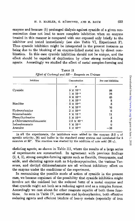
Experiments with Complex-Forming Agents-In accord with previous investigators, we believed that the rapid, reversible, and efficient inhibi- tion of uricase action by cyanide ions (2) provided a clue as to the nature of the prosthetic group of the enzyme. Cyanide can function as a re- agent for binding either a prosthetic metal or a carbonyl group. To investigate the second possibility, a series of carbonyl reagents was tested under conditions of pH and incubation time corresponding to those ob- taining for cyanide inhibition, but at concentrations exceeding t,hat of cyanide by at least a factor of 10. These data are summarized in Table II. It can be seen that, even under these favorable conditions, carbonyl reagents are incapable of blocking uricase action effectively.
The metal-binding capacities of cyanide may manifest themselves in two ways, either by actual removal of the metal from the enzyme and binding of the cation as an inorganic cyano complex or by forming a cyan0 complex with the enzyme-bound metal. The former mechanism, exemplified by Kubowitz’ experiments on phenoloxidase (25) and our own on butyryl CoA dehydrogenase (18), can be ruled out in the present case because (a) cyanide inhibition can be overcome by dialysis of the cyanide-treated
6 The rate at 38” is approximately 2.6 times that at 22“; at 38” the rate in pure 02 is 1.8 times that in air; the extrapolated rate at infinite uric acid concentration (v mar.) is 1.7 times that observed under standard assay conditions.
by guest on June 1, 2018http://w
ww
.jbc.org/D
ownloaded from
II. R. MAHLER, G. I@BSCHER, AND H. BAUM 633
enzyme and because (b) prolonged dialysis against cyanide of a given con- centration does not lead to more’ complete inhibition when an enzyme treated in this manner is compared with one exposed only briefly to the inhibitor and tested immediately (see also Table VI, Experiment F). Thus cyanide inhibition might be interpreted in the present instance as being due to the blocking of an enzyme-linked metal ion by direct com- bination. In this case cyanide inhibition should not be unique, and the effect should be capable of duplication by other strong metal-binding agents. Accordingly we studied the effect of metal complex-forming and
E$ect of Carboy I and SH- Reagents on Uricase
Inhibitor Concentration Per cent inhibition
Cyanide
Bisulfite
Hydroxylamine Semicarbaaide Phenylhydrazine p-Chloromercuribenzoate Iodosobenzoate Arsenite
M
5 x 16-6 1 x 16-6 5 x 10⁻⁶ 1 x lo-” 5 x 10⁻¹ 1 x lo-8 2 x 10-z 1 x 16-3 1 x 10-z 2.5 x 16-4 1 x 10-s 1 x 10-z I
98 76 45
8 5
10 0
11 5 5 0
-10
TABLE II
In all the experiments, the inhibitors were added to the enzyme (3.5 y of specific activity, 38) and buffer in the standard assay system and incubated for 5 minutes at 22”. The reaction was started by the addition of uric acid (26 7).
chelating agents, as shown in Table III, where the results of a 1,arge series of experiments; are summarized. In agreement with previous findings (2, 4, 5), strong complex-forming agents such as fluoride, thiocyanate, and azide, and chelating agents such as S-hydroxyquinoline, the various Ver- senes, and diethyl dithiocarbamate are all without inhibitory effect on the enzyme under the conditions of the experiment.
In reexamining the possible mode of action of cyanide in the present case, we became cognizant of the possibility that cyanide inhibition might involve not the oxidized but the reduced fo&n of a metal enzyme and that cyanide might act both as a reducing agent and as a complex former. Accordingly we cast about for other reagents capable of both these func- tions. As seen in Table IV, ascorbate and hydroxylamine, both effective reducing agents and efhcient binders of heavy metals (especially of iron
by guest on June 1, 2018http://w
ww
.jbc.org/D
ownloaded from

634 CUPROPROTEIN
and copper), are effective inhibitors of uricase under appropriate condi- tions; i.e., a period of preincubation. The experiments involving fluoride
TABLE III
Eflect of Metal-Binding Agents on Uricase
Reagent Per cent inhibition*
Thenoyl trifluoroacetone. . 0 Diethyl dithiocarbamate. -10 EDTA. 0 N-Hydroxyethylethylenediaminetriacetic acid.. 0 S-Hydroxyquinoline. 2 Tiron............................................. 0 Fluoride.......................................... 0 ~hiocyanate....................................... 15 Aeide 0
All inhibitors at 10-* M preincubated with the enzyme (2 y of specific activity of 100 in 0.01 M borate buffer, pH 8.0) for 30 minutes at 30”; reaction wae started by the addition of uric acid.
* For a highly purified enzyme (specific activity >60), with enzymes of lower spe- cific activity, metal-binding agents frequently appear to stimulate the activity.
TABLE IV
Effect of Hydroxylamine and Aacorbate on Uricase
Reagent
Aacorbate . . . . . Hydroxylamine .
“ + fluoride. . . ‘I +‘KCNS..
Sulfide. . ..___......_ Semicarbazide . . CN- (2.5 X 10⁻⁶ M).
None. . . . . ‘I
Ascorbate . CN- (2.5 x lo-‘ M).
“ (2.5 x 10⁻⁶ “), Hydroxylamine f fluoride. . .
-
-- Previous treatment of enzyme
None ‘I I‘ “ “ “ I‘
S-Hydroxyquinoline Diethyl dithiocarbamate %Hydroxyquinoline
“ ‘I
Diethyl dithiocarbamate
-
_- Per cent inhibition
75 48 95 73 5 0
75
0 0 7 5
50 0
All the conditions are similar to those in Table III. The S-hydroxyquinoline- and diithyl dithiocarbamate-treated enzymes were prepared by dialysis of the prep- aration against these agents and are described in the text.
are of interest; fluoride alone is completely ineffective as an inhibitor (Table III). Hydroxylamine at the concentration shown leads to approxi-
by guest on June 1, 2018http://w
ww
.jbc.org/D
ownloaded from

H. R. MAHLER, G. HtiBSCHER, AliD H. BAUM 635
mately 50 per cent inhibition. This effect is probably due to simultaneous (or closely consecutive) reduction by one hydroxylamine molecule and binding of a second one. Hydroxylamine in the present case is a more effective reducing agent than a metal complex-forming agent. Thus an enzyme prereduced by hydroxylamine is now capable of binding and being inhibited by fluoride. The fact that sulfide, which should be able to per- form these two functions, is not a good inhibitor might be explained by steric considerations.7
Binding of Chelating Agents by Enzyme-Even though most ordinary chelating agents do not function as inhibitors, they can be shown to be bound by the enzyme. Thus, dialysis of the enzyme against 0.01 M Tris Versenate at pH 8.0, followed by’ a second period of dialysis against the same buffer containing 0.001 M sodium diethyl dithiocarbamate, leads to an accumulation of a yellow-colored material inside the dialysis bag. Examination of the contents indicates that it is the enzyme which can be shown to have acquired a brilliant yellow color with an absorption spec- trum (Fig. l), which appears to be typical of dilute solutions of Cu diethyl dithiocarbamate complex, either in the presence or in the absence of Cu- binding proteins. No other metal complexes portray a spectrum re- sembling the one observed, nor even any appreciable absorption in the characteristic region around 450 rnp (Fig. 1). Similar experiments can also be performed with 8-hydroxyquinoline, leading to a green complex. The spectrum of this complex is not as specific, however, and might be due to the presence of any of several different metals.
When the enzymatic activity of these presumptive complexes of a pro- teinimetal-chelating agent is compared with that of the untreated enzyme, the results in the lower half of Table IV are obtained. The complex- forming enzyme is as effective as the untreated variety. This suggests that the substrate might be bound to the enzyme in a manner analogous to that of the chelating agents, and is capable of competing successfully with these reagents for the metallic site on the enzyme. The chelated enzyme preparations do portray a significant difference in their behavior toward the metal-binding inhibitors (cyanide, etc.) discussed in the previ- ous section. Thus, as the data of Table IV indicate, these transformed enzymes are completely protected against ascorbate and hydroxylamine inhibition, and partially protected against cyanide inhibition as well.
Substrate, if bound in the same manner at the same metal site, should then produce similar protective effects. This is borne out by the data in Table V. Preincubation with substrate leads to a decrease in the in-
7 The addition of sodium sulfide in solution does not provide the ions S- or HS- but polymeric forms also involving neutral sulfur, which might well be too bulky to be able to penetrate the active site on the enzyme.
by guest on June 1, 2018http://w
ww
.jbc.org/D
ownloaded from

636 CUPROPROTEIN
WAVELENGTH IN b.4~
FIQ. 1. Spectra of Cu diethyl dithiocarbamate chelates. Curve A, uricase (Cu content, 2 y per ml., specific activity, 40) dialyzed for 12 hours against 0.01 M Tris Versenate, pH 8.0, followed by dialysis against the same buffer containing 0.001 M sodium diethyl dithiocarbamate for 24 hours. Curve B, 2 y of Cu++ per ml. (as the sulfate) treated for 60 minutes with 0.001 M diethyl dithiocarbamate in 0.01 M Tris Versenate, pH 8.0, at 38”. Curve C, conditions as in Curve B, but 0.93 mg. per ml. of recrystallized bovine albumin or 1.0 mg. per ml. of metal-binding human globulin were added. The solid circles indicate Mn++, Co++, etc., obtained under conditions similar to those used for the data of Curve B, but with the appropriate ion at the same concentration substituted for Cu*.
TABLE V E#ect of Preincubation with Uric Acid on Uricase Inhibited by Metal
Complex-Forming Agents
Reagent Per cent inhibition ,
Hydroxylamine. ................................. I‘ + fluoride. .......................
Ascorbate ......................................... Hydroxylamine + uric acid, ......................
“ + ‘I “ + fluoride. .........
Ascorbate + uric acid. .......................... Cysteine + “ “ ........................... Diethyl dithiooarbamate + uric acid, ............ EDTA + uric acid ................................ N-Hydroxyethyliminodiacetic acid + uric acid .... S-Hydroxyquinoline + uric acid ................... Furoyl trifluoroacetone + uric acid ................
54 95 98
0 5 5 0 0
10 15 12
4 O 20 pmoles of diol buffer, pH 35,300 y of uric acid (where indicated), 20 y of enzyme
(specific activity, 60), and 0.1 amole of inhibitor were incubated for 2 hours at 22” in a total volume of 0.10 ml. At the end of this period 0.01 ml. aliquots were with- drawn and tested in the standard assay system.
by guest on June 1, 2018http://w
ww
.jbc.org/D
ownloaded from

H. R. MAHLER, C+. HijBSCHER, AND H. BAUM 637
hibitory efEiciency of hydroxylamine and ascorbate. No great enhance- ment of the inhibitory power of the metal-binding agents of Table III, and of numerous others such as cysteine, o-phenanthroline, or -SH inhibitors, is observed under these reducing conditions. Is substrate then bound at the same site as the chelating agents? From the following experiment, this would appear to be the case: A diethyl dithiocarbamate-linked en- zyme, well dialyzed against buffer (0.1 M borate, pH 9.0) wits made 10-4 M with respect to sodium urate, while another aliquot of the same en- zyme was made 1O-4 M with respect to sodium chloride. Dialysis at 0” was then resumed against the same buffer. After 1 hour, the two bags were opened and the Ed60 determined. Since the value of the chloride-
TABLE VI Copper Content of Purified Uricase Preparations
Preparation
A B C D E F* G H I K
- I
-
Protein cu content
mg. per ml. Y w %-. 3.0 0.45 3.0 0.67 2.4 0.22 4.2 0.071 2.0 0.18 1.05 0.17 4.0 0.15 1.4 0.12 3.1 0.12 1.5 0.15
Specific activity
urils per mg. 120 125 40 15 40 40 40 16 35 37
7-
Activity
rnits per y cu 270 180 185 211 220 240 260 133 290 245
Mol. wt., 1 atom Cu per mole
145,006 97,000 87,000 73,500
108,600 115,000 130,500 43,000
187,000 138,000
* Preparation E was dialyzed versus 0.01 M cyanide for 48 hours and retested.
containing enzyme was 0.150 and that of the urate-containing enzyme 0.050, a significant dissociation of the chelating agent from the enzyme under the influence of the substrate could be inferred.
Qualitative Metal Analysis--The experiments just described made it appear profitable to consider again a possible r61e of metals in uricase action, with special emphasis on copper, highly purified preparations of the enzyme being used. Therefore two samples of uricase of specific activity 40 and 120, respectively, were subjected to qualitative spcctro- scopic analysis. The results obtained in two different laboratories were in essential agreement. Copper was present in all the experimental samples. The copper content of the samples corrected for blank values appeared to increase with increasing specific activity when compared with the experi- mental sample of lower specific activity. Other metals, except iron (but, including Mg, Mn, Zn, Co, Ni, Ag, Hg), appeared to be either absent, or
by guest on June 1, 2018http://w
ww
.jbc.org/D
ownloaded from

638 CUPROPROTIGIX
present at the same level in the experimental sample as in the blank. Iron was present only in the sample of low specific activity, absent in that of high activity.
Quantitative Copper Analysis-In order to investigate the possible r61e of copper aa ,an electron carrier in the enzyme, a systematic study of the copper content of numerous enzyme preparations at different purity levels was undertaken. The results of this study are given in Table VI. There appears to be donsiderable scatter among the results, the cause for which
I ; I I ; I I I I I I I I I I I
I I
3 3 1.80- 1 l.80- 1
I I
s: s: 1.80- \ l.80- \
I I
;: 0.200- u I= I= I I I I I I I I I I I I I I
oa oa 230 230 270 270 310 310 350 350
WAVELENGTH IN t$.J WAVELENGTH IN t$.J
FIQ. 2. Spectra of highly purified uricase. Curve A, enzyme of specific activity 120 in 1 per cent carbonate. Curve B, enzyme of specific activity 60.
experimental variation is not apparent at the present time. In spite of these variations, the following facts emerge from Table VI: (a) There is an increase in the copper content paralleling an increase in specific activity; (b) the characteristic ratio of Cu content per enzyme unit (units per micro- gram of Cu), which is proportional to the ratio of copper atoms per enzyme molecule, gives a sufficient constancy throughout the purificatioti range (i.e., for enzymes of approximately 10 per cent purity or better) to suggest a definite catalytic r61e for the metal; (c) the molecular weight of the enzyme calculated from copper content, assuming 1 copper atom per en- zyme molecule, equals 120,000 f 40,000 in satisfactory agreement with the value calculated from the sedimentation constant.
Absorption Spechm- Contributory evidence for the hypothesis that
by guest on June 1, 2018http://w
ww
.jbc.org/D
ownloaded from

II. R. MAELER, (f. HOBSCHER, AND H. BAUM 639
uricase is a copper protein comes from a study of appropriate absorption spectra in the ultraviolet region. The absorption spectra of two highly purified uricme preparations are shown in Fig. 2. Although apparently quite similar to t,hat of a pure protein, not in a complex with any prosthetic group, certain significant differences emerge if the spectrum of the enzyme is compared with that of a pure “typical” protein; e.g., crystalline bovine serum albumin or t.he met,al-binding globulin of human plasma (26). When the latter two proteins are incubated with a Cu++ solution, and their spectra redetermined wit,h the meta,l solution as t.he blank in this determination at least a qualitative similarity between these copper pro- tein spectra and that of uricase may be observed. This similarity becomes
TABLE VII Optical Properties of Various Copper-Containing Proteins
Protein
Uricase (specific activity 55) ‘I ( I‘ “ 120)
Bovine plasma albumin, ‘f “ “ + cu.
Metal-binding globulin. “ “
+ cu. “ “ + Fe.
bu. I
h&l. Etu:EIM
277 261 9.5 276 261 5.55 280 265 17.6 282 275 3.8 282 265 27.5 278 270 9.3 281 265 13.5
All spectra in 1 per cent NazCOa, with proteins at a concentration of 1.0 mg. per ml., against blanks containing all components except the protein. Spectra in the presence of metal were obtained by incubating the protein in question with 5 X 10-b M metal ion at room temperature for 45 minutes.
even more apparent when the optical properties of these various proteins are compared as in Table VII. There the most characteristic feature appears to be the ratio of the optical densities at 280 and at 330 rnp. This ratio is high for all “ordinary” proteins and for the iron-globulin complex, but low for all the copper proteins and for uricase.*
DISCUSSION
The implication of copper as an integral part of uricase appears some- what surprising in view of two well substantiated observations reported in the literature (2-5) and fully confirmed in the course of the present in- vestigations. These are that (a) diethyl dithiocarbamate, a complex-
8 The composition and the properties of metal complexes of the metal-binding globulin and of albumin have been studied extensively. These studies have recently been reviewed by Klotz (27). 2 gm. atoms of metal are bound per approximately 100,000 gm. of either protein.
by guest on June 1, 2018http://w
ww
.jbc.org/D
ownloaded from
640 CUPROPROTEIN
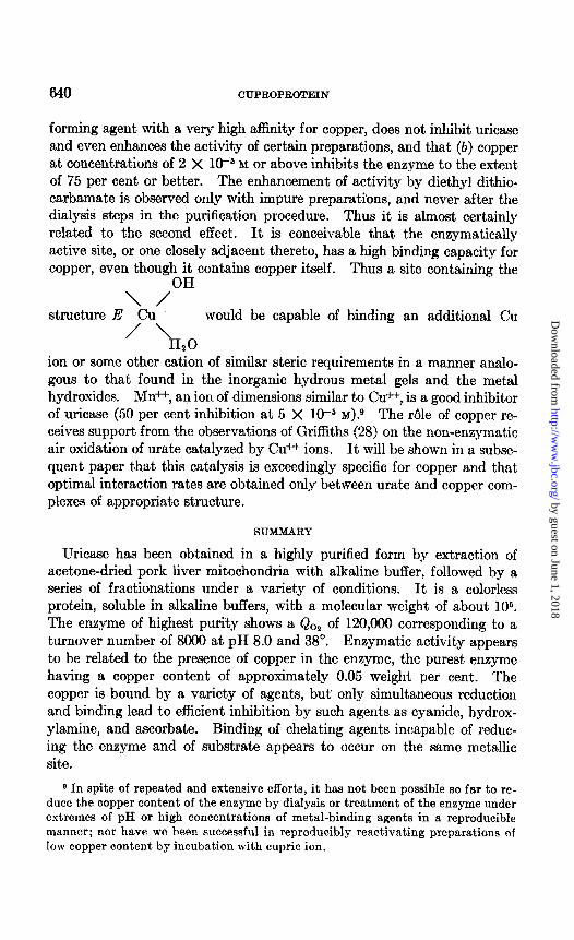
forming agent with a very high affinity for copper, does not inhibit uricase and even enhances the activity of certain preparations, and that (b) copper at concentrations of 2 X 1O-6 M or above inhibits the enzyme to the extent of 75 per cent or better. The enhancement of activity by diethyl dithio- carbamate is observed only with impure preparations, and never after the dialysis steps in the purification procedure. Thus it is almost certainly related to the second effect. It is conceivable that the enzymatically active site, or one closely adjacent thereto, has a high binding capacity for copper, even though it contains copper itself.
OH
structure Au’
Thus a site containing the
’ ‘HO
would be capable of binding an additional Cu
2 ion or some other cation of similar steric requirements in a manner analo- gous to that found in the inorganic hydrous metal gels and the metal hydroxides. Mn++, an ion of dimensions similar to Cu++, is a good inhibitor of uricase (50 per cent inhibition at 5 X lO+ M)? The Ale of copper re- ceives support from the observations of Griffiths (28) on the non-enzymat,ic air oxidation of urate catalyzed by CL++ ions. It will be shown in a subse- quent paper that this catalysis is exceedingly specific for copper and that optimal interaction rates are obtained only between urate and copper com- plexes of appropriate structure.
SUMMARY
Uricase has been obtained in a highly purified form by extraction of acetone-dried pork liver mitochondria with alkaline buffer, followed by a series of fractionations under a variety of conditions. It is a colorless protein, soluble in alkaline buffers, with a molecular weight of about 106. The enzyme of highest purity shows a QO, of 120,000 corresponding to a turnover number of 8000 at pH 8.0 and 38”. Enzymat,ic activity appears to be related to the presence of copper in the enzyme, the purest enzyme having a copper content of approximately 0.05 weight per cent. The copper is bound by a variety of agents, but only simultaneous reduction and binding lead to efficient inhibition by such agents as cyanide, hydrox- ylamine, and ascorbate. Binding of chelating agents incapable of reduc- ing the enzyme and of substrate appears to occur on the same metallic site.
9 In spite of repeated and extensive efforts, it has not been possible so far to re- duce the copper content of the enzyme by dialysis or treatment of the enzyme under extremes of pH or high concentrations of metal-binding agents in a reproducible manner; nor have ne been successful in reproducibly reactivating preparations of low copper content by incubation with cupric ion.
by guest on June 1, 2018http://w
ww
.jbc.org/D
ownloaded from

H. R. MAHLER, G. HCBSCHER, AND H. BAUM 641
We wish to thank Dr. David E. Green for many stimulating discussions. Mrs. Dorothee Elowe provided capable technical ‘assistance. These in- vestigations were supported by a grant-in-aid of, the Nutrition Founda- tion, Inc., to’ Dr. David E. Green, and by a research grant, No. G-4128, from the National Heart Institute of the National Institutes of Health, Public Health Service, to one of us (H. R. M.).
BIBLIOGRAPHY
1. Batelli, F., and Stern, L., Biochem. Z., 19, 219 (1909). 2. Keilin, D., and Hartree, E. F., Proc. Roy. Sot. London, Series B, 119, 114 (1936);
121, 173 (1936). 3. Davidson, J. N., Biochem. J., 32, 1386 (1938). 4. Davidson, J. X., Biochem. J., 36, 252 (1942). 5. Holmberg, C. G., Biochem. J., 33, 1901 (1939). 6. Altman, K. I., Smull, K., and Barron, E. S. G., Arch. Biochem., 21, 153 (1949). 7. Leone, E., Biochem. J., 64, 393 (1953). 8. Praetorius, E., Biochim. et biophys. acta, 2, 602 (1948). 9. Felix, F., Schecl, F., and Schuler, W., 2. physiol. Chem., 186, 90 (1929).
10. Schuler, W., 2. physiol. Chem., 208, 237 (1932). 11. Klemperer, F. W., J. Biol. Chem., 160, 111 (1945). 12. Bentley, R., and Neuberger, A., Biochem. J., 62,694 (1952). 13. Canellakis, E. S., and Cohen, P. P., J. Biol. Chem., 213, 335 (1956). 14. Kalckar, H. M., J. Biol. Chem., 167, 429 (1947). 15. Stimson, M. M., and Reuter, LM. A., J. Am. Chem. Sot., 66, 153 (1943). 16. Gornall, A. G., Bardawill, C. J., and David, M. M., J. Biol. Chem., 177,751 (1949). 17. Warburg, O., and Christian, W., Biochem. Z., 310, 384 (1941). 18. Mahler, H. R., J. Biol. Chem., 206, 13 (1954). 19. Sandell, E. B., in Calorimetric determination of traces of metals, New York,
226 (1944). 26. Schein, A. H., Podber, E., and Novikoff, A. B., J. BioZ. Chem., 190, 331 (1951). 21. Mahler, H. R., Wakil, S. J., and Bock, R. M., J. BioZ. Chem., 204, 453 (1953). 22. Oppenheimer, E. H., and Kunkel, H. G., Bull. Johns Hopkins Hosp., 73,40 (1943);
quoted by McShan, W. H., in Respiratory enzymes, Minneapolis, 111 (1949). 23. Hoch, H., Riochem. J., 46, 199 (1950). 24. Anderson, E. A., and Alberty, R. A., J. Phys. and CoZZoid Chem., 62, 1345 (1948). 25. Kubowita, F., Biochem. Z., 292, 221 (1937); 299, 32 (1938). 26. Surgenor, D. M., Koechlin, B. A., and Strong, L. E., J. CZin. Invest., 28,73 (1949). 27. Klotz, I. M., in McElroy, W. D., and Glass, B., The mechanism of enzyme action,
Baltimore (1954). 23. Griffiths, M., J. BioZ. Chem., 197, 399 (1952).
by guest on June 1, 2018http://w
ww
.jbc.org/D
ownloaded from

Colmanoand With the technical assistance of Germille H. R. Mahler, Georg Hübscher, Harold BaumPROPERTIES OF A CUPROPROTEIN
PREPARATION, PURIFICATION, AND STUDIES ON URICASE: I.
1955, 216:625-642.J. Biol. Chem.
http://www.jbc.org/content/216/2/625.citation
Access the most updated version of this article at
Alerts:
When a correction for this article is posted•
When this article is cited•
alerts to choose from all of JBC's e-mailClick here
tml#ref-list-1
http://www.jbc.org/content/216/2/625.citation.full.haccessed free atThis article cites 0 references, 0 of which can be
by guest on June 1, 2018http://w
ww
.jbc.org/D
ownloaded from